Article

Fullerene-Functionalized Cellulosic Hydrogel Biosensor with Bacterial Turn-on Fluorescence Response Derived from Carboxymethyl Cellulose for Intelligent Food Packaging with DFT Calculations and Molecular Docking

by
Hebat-Allah S. Tohamy
Cellulose and Paper Department, National Research Centre, 33 El Bohouth Str., Dokki Giza P.O. 12622, Egypt
Gels 2025, 11(5), 329; https://doi.org/10.3390/gels11050329
Submission received: 29 January 2025 / Revised: 17 February 2025 / Accepted: 21 February 2025 / Published: 28 April 2025
(This article belongs to the Special Issue Recent Progress of Hydrogel Sensors and Biosensors)

Abstract

:
This study reports the synthesis and characterization of a novel carboxymethyl cellulose–N-fullerene–g-poly(co-acrylamido-2-methyl-1-propane sulfonic acid) (CMC–N-fullerene–AMPS) hydrogel for potential application in biosensing within food packaging. The hydrogel was synthesized via free radical polymerization and characterized using FTIR, SEM, and fluorescence microscopy. FTIR analysis confirmed the successful grafting of AMPS and incorporation of N-fullerenes, indicated by characteristic peaks and a shift in the N–H/O–H stretching frequency. Density Functional Theory (DFT) calculations revealed that the CMC–N-fullerene–AMPS hydrogel exhibited higher stability and a lower band gap energy (0.0871 eV) compared to the CMC–AMPS hydrogel, which means a high reactivity of CMC–N-fullerene–AMPS. The incorporation of N-fullerenes significantly enhanced the hydrogel’s antibacterial activity, demonstrating a 22 mm inhibition zone against E. coli and a 24 mm zone against S. aureus, suggesting potential for active food packaging applications. Critically, the hydrogel displayed a unique “turn-on” fluorescence response in the presence of bacteria, with distinct color changes observed upon interaction with E. coli (orange-red) and S. aureus (bright green). This fluorescence enhancement, coupled with the porous morphology observed via SEM (pore size 377–931 µm), suggests the potential of this hydrogel as a sensing platform for bacterial contamination within food packaging. These combined properties of enhanced antibacterial activity and a distinct, bacteria-induced fluorescence signal make the CMC–N-fullerene–AMPS hydrogel a promising candidate for developing intelligent food packaging materials capable of detecting bacterial spoilage.

Graphical Abstract

1. Introduction

The sheer scale of food waste globally presents a critical challenge with wide-ranging repercussions. A substantial portion of food intended for human consumption, roughly a third, or about 1.3 billion tons each year, is either lost or wasted. This loss and waste occur at every stage, from farm production and processing to retail sales and household consumption. In developing nations, much of the loss occurs during production, often due to inefficient harvesting methods, inadequate storage, and limited market access. Conversely, in wealthier nations, waste is more common at the retail and consumer levels, driven by factors like cosmetic imperfections in produce, purchasing more than needed, and improper food storage leading to spoilage [1,2,3]. The consequences of this waste are considerable. Food waste is responsible for a significant portion of global greenhouse gas emissions, estimated at 8–10%, making it a key contributor to climate change [4,5,6]. Furthermore, discarded food frequently ends up in landfills, where it decomposes and generates methane, a powerful greenhouse gas [7,8]. The economic implications are also significant. Food waste represents a substantial loss of resources and income for farmers, businesses, and individuals, affecting both rich and poor countries [9,10]. While food packaging plays a vital role in protecting and preserving food, its current forms often present challenges that worsen the issue of food waste. A key concern is the lack of biodegradability in many commonly used materials, which significantly contributes to environmental pollution. For instance, widely adopted plastics like polyethylene (PE) and polypropylene (PP), favored for their affordability and adaptability, can persist for centuries in the environment [11,12]. This slow decomposition results in the build-up of waste in landfills and oceans, negatively impacting ecosystems and potentially introducing microplastics into the food chain. Moreover, the recycling of these plastics, even when feasible, can require substantial energy input and may yield materials with diminished quality, restricting their ability to be reused effectively [13,14]. Smart packaging marks a major leap forward in food technology, providing innovative solutions to the intertwined challenges of food safety and waste. Moving beyond the passive role of traditional packaging, smart packaging actively engages with food and its surroundings, offering insights into quality, freshness, and safety. This is accomplished by incorporating technologies like sensors, indicators, and antimicrobial agents. These advancements offer significant promise for minimizing food waste by prolonging shelf life, delivering real-time data on food condition, and inhibiting spoilage [15,16]. The unique characteristics of fullerenes, particularly C60, including their spherical shape, extensive surface area, and ability to accept electrons, position them as promising candidates for revolutionizing food packaging [17,18,19,20]. These properties endow them with potent antimicrobial capabilities [21,22]. When integrated into food packaging materials, fullerenes can effectively extend the shelf life of perishable foods by inhibiting microbial growth and safeguarding against oxidative spoilage [23,24,25,26]. This translates to a reduction in food waste and a significant enhancement in food safety [25,27]. Conventional fullerene synthesis methods, such as the energy-intensive and environmentally detrimental arc-discharge technique, present significant drawbacks [27,28,29,30]. Conversely, utilizing sugarcane bagasse (SB), a readily available and renewable byproduct of the sugar industry, offers a sustainable alternative for fullerene production [31,32,33]. Transforming this agricultural waste into valuable materials like fullerenes not only generates economic advantages but also mitigates environmental pollution [34,35,36,37]. The production of fullerenes from SB typically entails a multi-step process encompassing carbonization, purification, and subsequent processing to isolate C60. Its environmental and economic benefits make it a highly promising avenue for sustainable fullerene production [38]. Fullerenes exhibit inherent fluorescence properties that make them valuable for developing innovative food packaging sensors [39]. One key characteristic is their ability to undergo thermally activated delayed fluorescence (TADF). In TADF, upon absorbing light, the fullerene molecule enters an excited state [40,41,42,43]. Instead of immediately emitting fluorescence, it undergoes a thermally activated process, transitioning to a lower energy state before emitting light. This delayed fluorescence emission provides a unique mechanism for sensing applications [44,45]. Furthermore, fullerenes can act as efficient fluorescence quenchers [40,46]. When a fluorescent molecule is in close proximity to a fullerene, the energy of the excited state of the fluorescent molecule can be transferred to the fullerene, leading to a decrease in the fluorescence intensity of the molecule [46,47]. This fluorescence-quenching phenomenon can be effectively utilized to create sensors that detect the presence of specific analytes in food, such as pathogens or toxins [48,49,50]. By strategically incorporating fullerenes into food packaging materials and designing appropriate sensing mechanisms, it becomes possible to develop systems that can detect the presence of contaminants or spoilage in food products. These sensors have the potential to provide real-time information about the safety and quality of food, ultimately contributing to a reduction in food waste and an improvement in consumer health.
Hydrogels, with their high water content, responsiveness, and safety, are in high demand for biological applications [51,52,53]. pH-sensitive hydrogels offer enhanced controllability [15]. Incorporating 2-Acrylamido-2-methyl-1-propane-sulfonic acid (AMPS) into carboxymethyl cellulose (CMC) enables the creation of hydrogels with enhanced properties for food packaging [54,55]. The sulfonic acid groups of AMPS impart hydrophilicity and an ionic character, while the acrylamide moiety facilitates crosslinking and the formation of a robust three-dimensional network [56,57,58,59]. These combined features position AMPS-modified CMC hydrogels as promising candidates for developing innovative and sustainable food packaging solutions. The microwave method for preparing CMC offers significant advantages over conventional methods. It is significantly faster, reducing reaction times considerably compared to traditional heating techniques. This expedited process translates to increased efficiency and reduced energy consumption, making it a more environmentally friendly approach. Moreover, microwave irradiation often results in improved product quality and reduced byproduct formation, further enhancing the sustainability and overall appeal of this method for CMC production [16,60]. This article presents a green and sustainable method for synthesizing antimicrobial/sensing carboxymethyl cellulose–fullerene–g-poly(co-acrylamido-2-methyl-1-propane sulfonic acid) hydrogel. The prepared carboxymethyl cellulose–fullerene–g-poly(co-acrylamido-2-methyl-1-propane sulfonic acid) was characterized using FTIR, SEM, DFT, molecular docking, and antimicrobial activity assays.

2. Results and Discussion

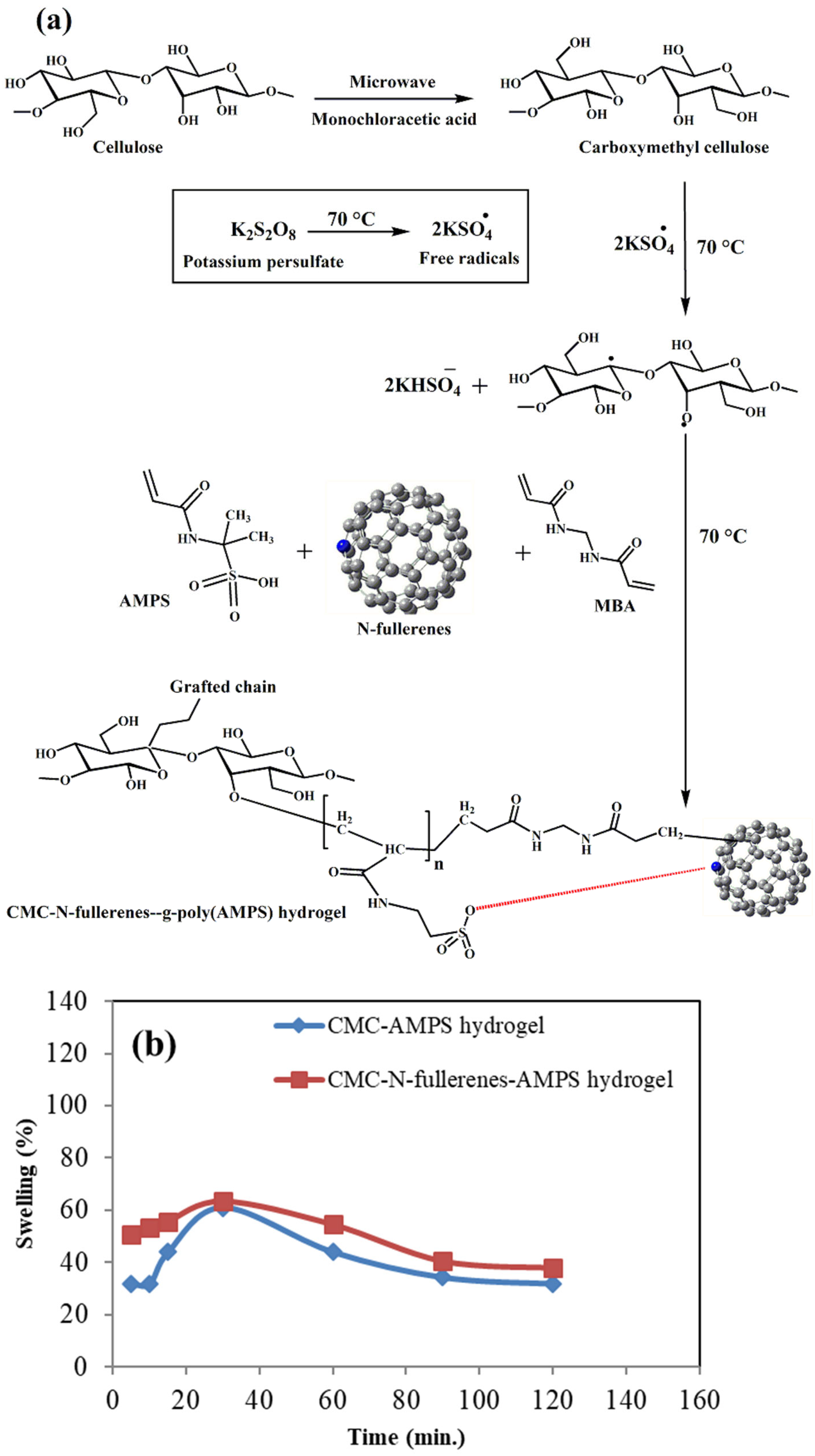
2.1. Proposed Mechanism of Carboxymethyl Cellulose–N-fullerene–g-poly(co-acrylamido-2-methyl-1-propane Sulfonic Acid) Hydrogel Formation and the Swelling Study

When heated, the KPS initiator breaks down into sulfate anion radicals (SO4) [61,62,63]. The grafting and crosslinking mechanism is depicted in Figure 1. Initiation begins with the thermal decomposition of KPS, producing sulfate anion radicals (SO4). These radicals abstract hydrogen atoms from CMC’s hydroxyl groups, generating alkoxy radicals along the CMC backbone. These alkoxy radicals then react with adjacent AMPS monomers, initiating polymerization. The growing polymer chains subsequently transfer free radicals to N-fullerene molecules, promoting further chain propagation and grafting. Simultaneously, the crosslinking agent, MBA, reacts with the propagating polymer chains via its vinyl groups, leading to the formation of a crosslinked copolymer network [56,64].
The swelling behavior of both hydrogels exhibited an initial increase within the first 30 min, with the CMC–AMPS hydrogel swelling from 31.70% to 60.97% and the CMC–N-fullerene–AMPS hydrogel swelling from 50.63% to 63.29%. This initial increase is likely due to the hydrophilic nature of the AMPS groups and water molecules penetrating the hydrogel matrix. However, after 60 min, a decrease in swelling was observed for both hydrogels. The subsequent decrease in swelling after 60 min could indicate possible changes in the hydrogel structure; e.g., further crosslinking occurring within the hydrogel network, or perhaps some breakdown of the network. This change in crosslinking density over time may suggest that the hydrogel network is starting to degrade.

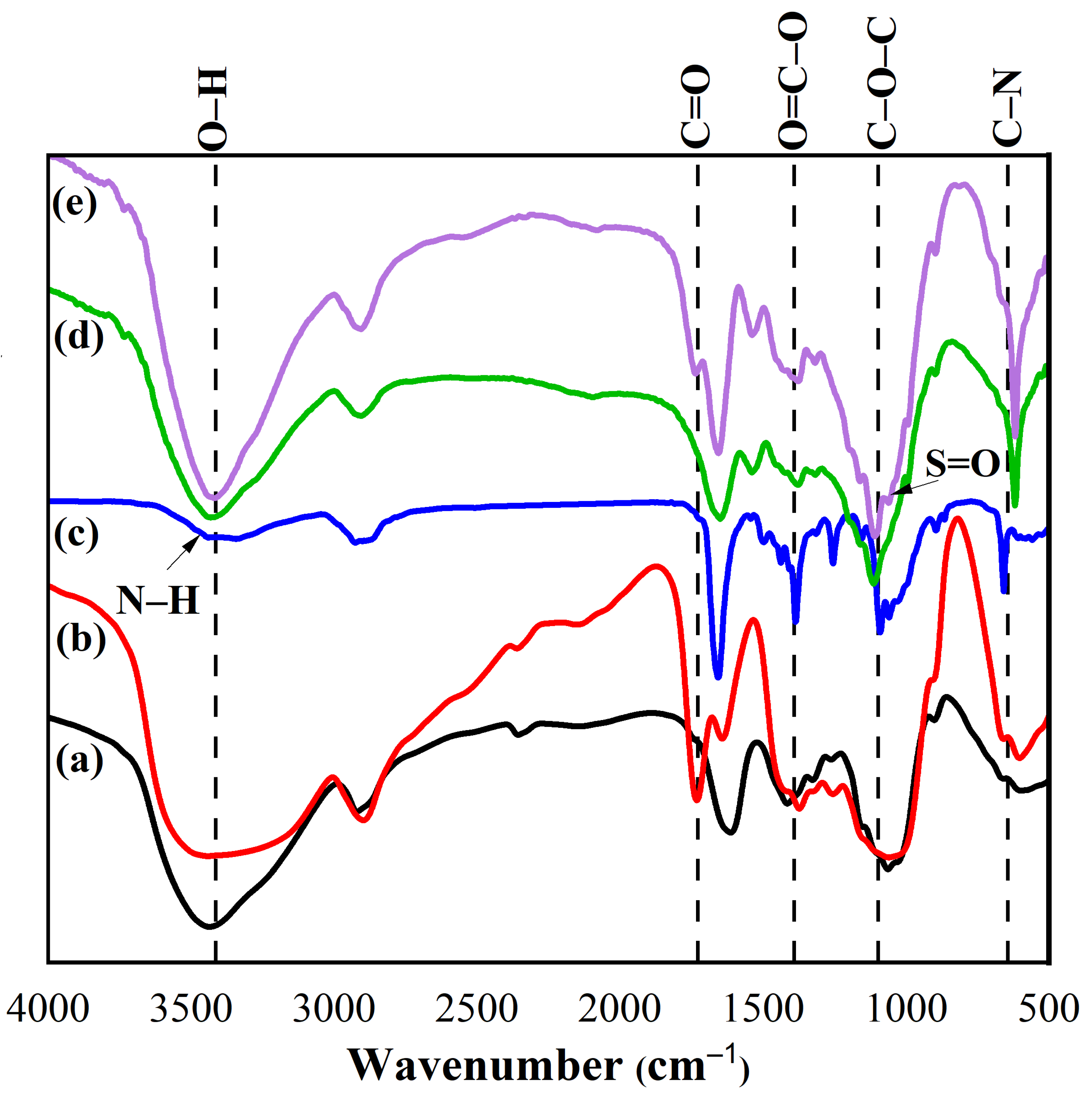
2.2. Fourier Transform Infrared Spectroscopy (FTIR) Spectra

The prepared cellulose and CMC displayed peaks between 3221–3344 cm−1, 1450–1455 cm−1, 1315–1386 cm−1, 1029–1037 cm−1, and 882–896 cm−1, which are related to O–H, C–O bending, C–H bending, the C–O–C of pyranose ring vibration, and β-glycosidic linkage, respectively [65,66,67,68,69]. The additional peak at 1745 cm−1 in CMC is related to the C=O peak, while the N–fullerenes showed peaks at 3461 cm−1 (N–H), 3207 cm−1 (O–H), 1662 cm−1 (C=O), 1386 cm−1 (O=C–O), 1087 cm−1 (C–O–C), and 657 cm−1 (C–N) [70,71].
The prepared CMC–AMPS and CMC–N-fullerene–AMPS hydrogels showed the same peaks of CMC with additional peaks between 986 and 1076 cm−1 (S=O), which proves the AMPS grafting (Figure 2) [56]. From FTIR observations, we found that the broad peak of N–H/O–H groups was shifted from a high value (3431 cm−1) for the CMC–AMPS hydrogel to a lower value (i.e., 3406 cm−1) for the CMC–N-fullerene–AMPS hydrogel, indicating strong H bonding between the CMC–N-fullerene–AMPS hydrogel compared to the CMC–AMPS hydrogel due to the incorporation of N–fullerenes [38,72].

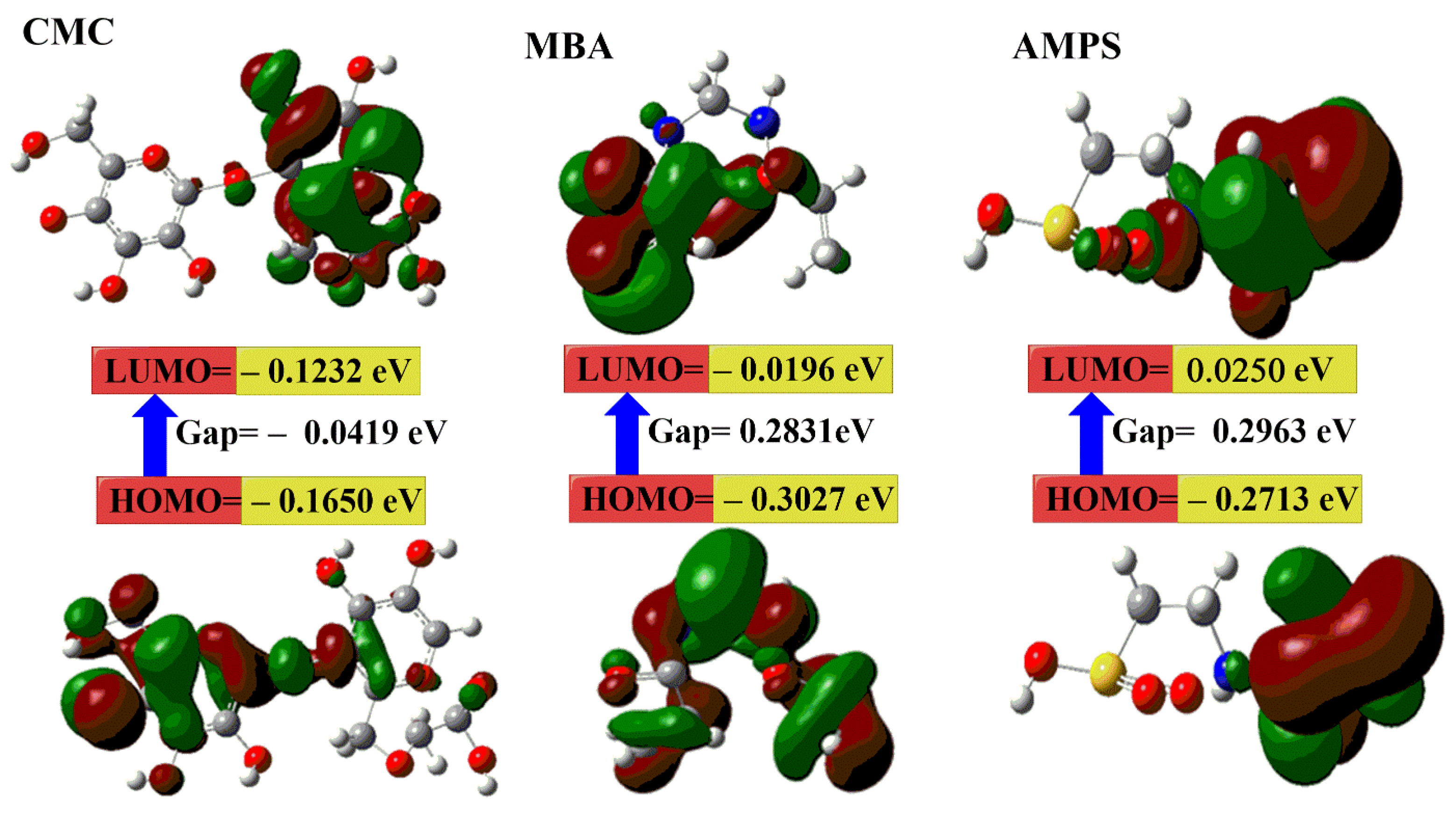
2.3. DFT Calculations

To gain deeper insights into the structural and electronic properties of the synthesized hydrogels and to understand the impact of N-fullerene incorporation on their stability and reactivity, Density Functional Theory (DFT) calculations were performed. From Figure 3 and Table 1, the results show the following:
a.
The μ and ω for the CMC–N-fullerene–AMPS hydrogel (i.e., 41.321 Debye and 0.2543 eV) is higher than the CMC–AMPS hydrogel (i.e., 24.309 Debye and 0.1172 eV) because of the presence of N-fullerenes [16,73,74].
b.
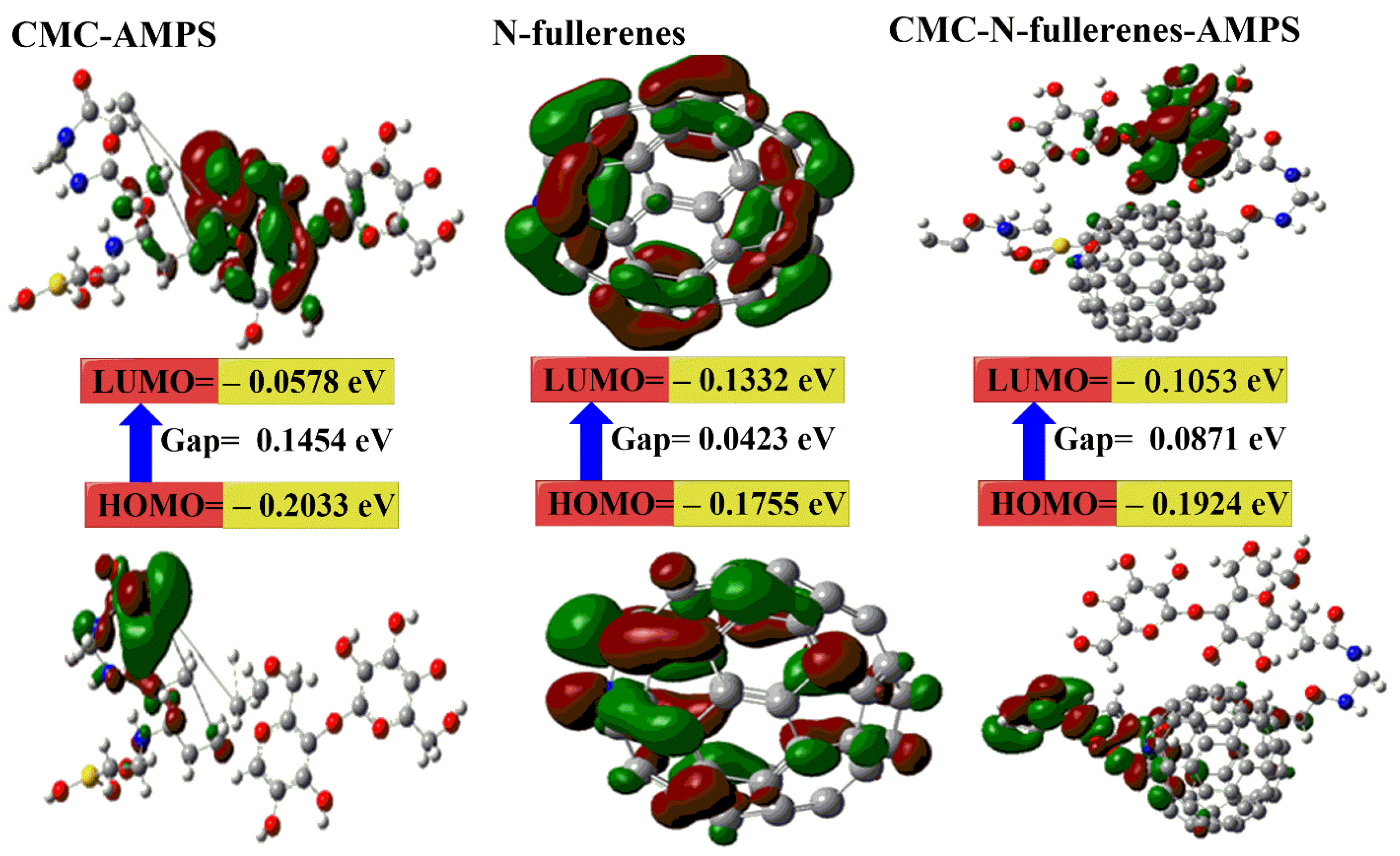
The calculated Eg for the CMC–N-fullerene–AMPS hydrogel is the lowest (i.e., 0.0871 eV) compared to the CMC–AMPS hydrogel (i.e., 0.1454 eV), which in turn proves the strong chemical reaction between CMC, MBA, AMPS, and N-fullerenes in the CMC–N-fullerene–AMPS hydrogel compared to the CMC–AMPS hydrogel due to the presence of N-fullerenes [75,76].
c.
The ET of the CMC–N-fullerene–AMPS hydrogel (−5158.364 au) is lower than the CMC–AMPS hydrogel (−2741.402 au), which means that the formation of the CMC–N-fullerene–AMPS hydrogel releases energy. This released energy makes it harder to break the bonds in the CMC–N-fullerene–AMPS hydrogel [15,72].
d.
The CMC–N-fullerene–AMPS hydrogel is much softer than the CMC–AMPS hydrogel, which is a good approximation of the strong energy changes between the donor (HOMO) and acceptor (LUMO) in the CMC–N-fullerene–AMPS hydrogel [75].
These DFT findings provide theoretical support for the enhanced properties observed experimentally. For instance, the lower band gap energy may contribute to the enhanced fluorescence or antibacterial activity (this will be discussed later). This suggests that N-fullerenes play a crucial role in modulating the electronic and structural characteristics of the hydrogel, leading to the observed improvements in performance.

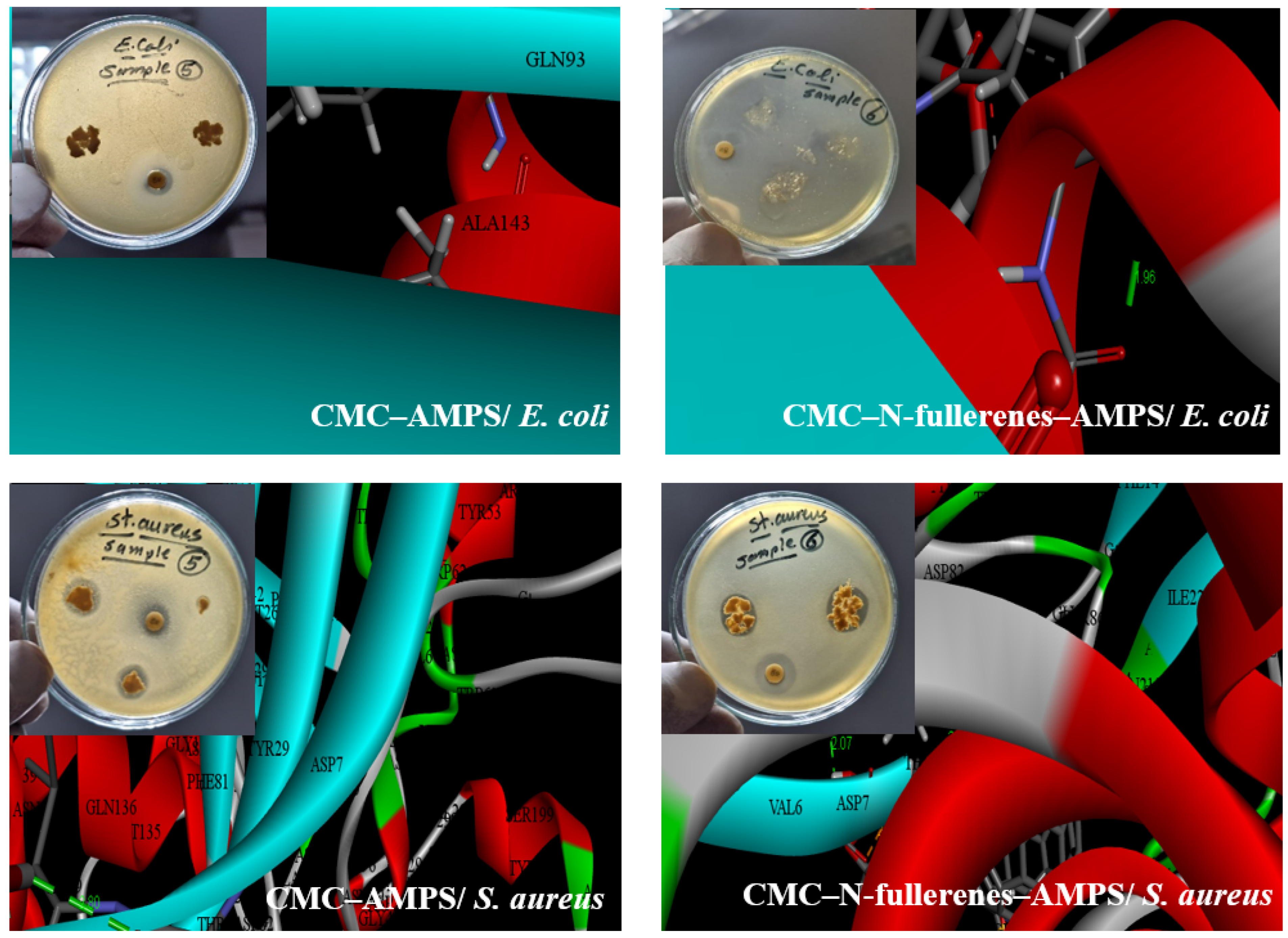
2.4. Antibacterial Activity and Molecular Docking Study

Our investigation demonstrated that the incorporation of N-fullerenes into the CMC–AMPS hydrogel significantly enhanced its antibacterial properties against both Gram-negative (Escherichia coli) and Gram-positive (Staphylococcus aureus) bacteria. While CMC–AMPS hydrogel exhibited no inhibition against E. coli, the CMC–N-fullerene–AMPS hydrogel displayed a substantial inhibition zone of 22 mm. Similarly, for S. aureus, the CMC–N-fullerene–AMPS hydrogel demonstrated a larger inhibition zone (24 mm) compared to CMC–AMPS hydrogel (15 mm). This enhanced antibacterial activity against Gram-positive bacteria is likely attributed to the presence of oxygen (O), nitrogen (N), and sulfur (S) functionalities within the CMC–N-fullerene–AMPS hydrogel (Figure 4) [77,78,79,80,81]. These functional groups are capable of interacting with key bacterial cell components, such as lipids, proteins, and DNA/RNA, through various mechanisms, including hydrogen bonding, π–π interactions, and electrostatic interactions [82,83]. Furthermore, the presence of N-fullerenes within the hydrogel may contribute to the observed antibacterial effect by regulating the generation of reactive oxygen species, potentially derived from bacterial surface proteins [84,85,86]. These findings were aligned with the docking results where the N–CDs showed binding with proteins with bond lengths of 0, 1.96, 2.80, and 2.07 A° for CMC–AMPS hydrogel/Escherichia coli, CMC–N-fullerene–AMPS hydrogel/Escherichia coli, CMC–AMPS hydrogel/Staphylococcus aureus, and CMC–N-fullerene–AMPS hydrogel/Staphylococcus aureus, respectively. Shorter bond lengths between the ligand and the protein result in a more reactive ligand. This increased reactivity enables the ligand to form a stronger bond (chelate) with the protein, potentially impairing its function and killing the microbe [15,16].

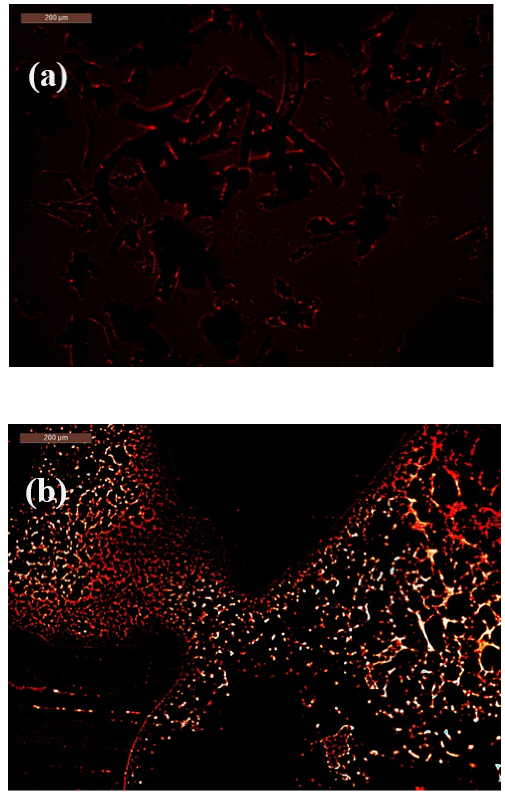
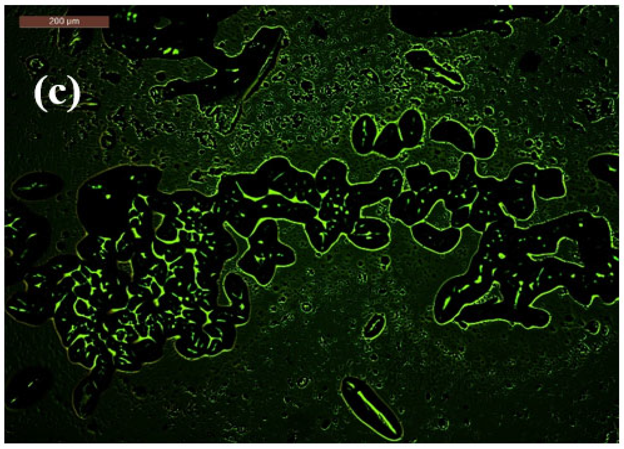
2.5. Mechanism of Bacterial-Induced Fluorescence Enhancement After Bacterial Contact

Initially, the CMC–N-fullerene–AMPS hydrogel appeared dark red. However, upon exposure to E. coli, a noticeable shift towards a brighter orange-red color was observed, accompanied by a broader color distribution. In contrast, interaction with S. aureus led to a more pronounced and uniform bright green coloration, demonstrating the “turn-on” fluorescence response of the hydrogel in the presence of both Gram-negative and Gram-positive bacteria (Figure 5).
The “turn-on” fluorescence observed in the CMC–N-fullerene–AMPS hydrogel upon bacterial contact suggests an intriguing interaction. Several mechanisms could be responsible for this phenomenon. One possibility is Fluorescence Resonance Energy Transfer (FRET). FRET is a distance-dependent energy transfer process where an excited donor fluorophore transfers energy to an acceptor chromophore. In this scenario, bacterial components, such as proteins or DNA, could act as energy acceptors. Upon bacterial interaction, energy transfer might occur from N-fullerenes within the hydrogel to these bacterial components, leading to an increase in fluorescence emission [87,88,89,90,91]. Another possible mechanism involves environmental changes induced by a bacterial metabolism [92,93]. Bacterial activity could alter the local environment of the hydrogel, such as pH or ionic strength. These changes could subsequently influence the electronic structure of the N-fullerenes, leading to enhanced fluorescence. Furthermore, direct N-fullerene–bacteria interactions cannot be ruled out. The N-fullerenes within the hydrogel might interact directly with bacterial components, potentially causing conformational changes in the N-fullerenes and consequently increasing their fluorescence quantum yield.
While Tohamy et al. observed a red-to-blue fluorescence shift with E. coli and a red emission with S. aureus using CMC@Ca-N-CDs, our CMC–N-fullerene–AMPS hydrogel exhibited a distinct “turn-on” fluorescence response, shifting from dark red to brighter orange-red with E. coli and to bright green with S. aureus [16]. A key advantage of our CMC–N-fullerene–AMPS hydrogel is the significant enhancement of fluorescence upon bacterial interaction. Unlike the subtle color changes reported by Tohamy et al., our system exhibited a dramatic increase in fluorescence intensity, transitioning from a dark red to bright orange-red with E. coli and bright green with S. aureus, suggesting a more sensitive detection mechanism.

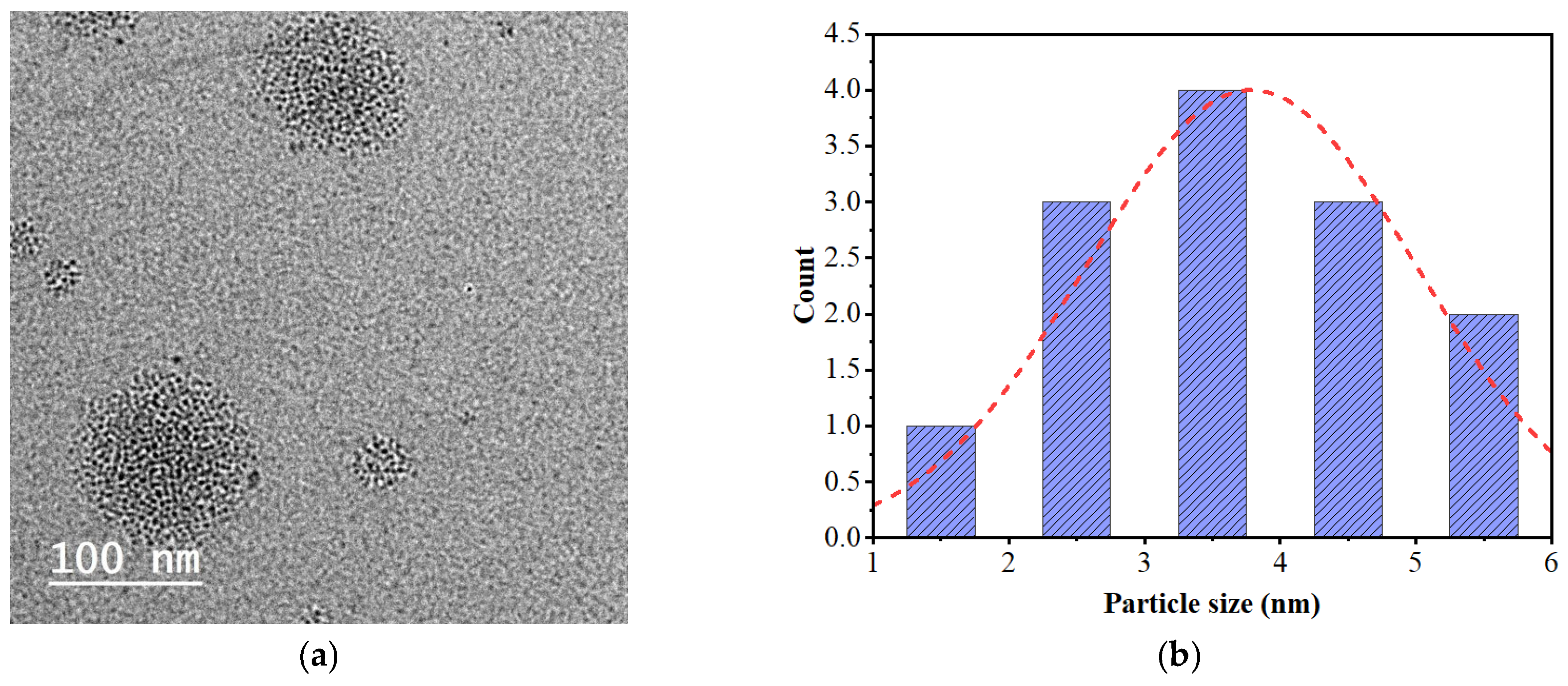
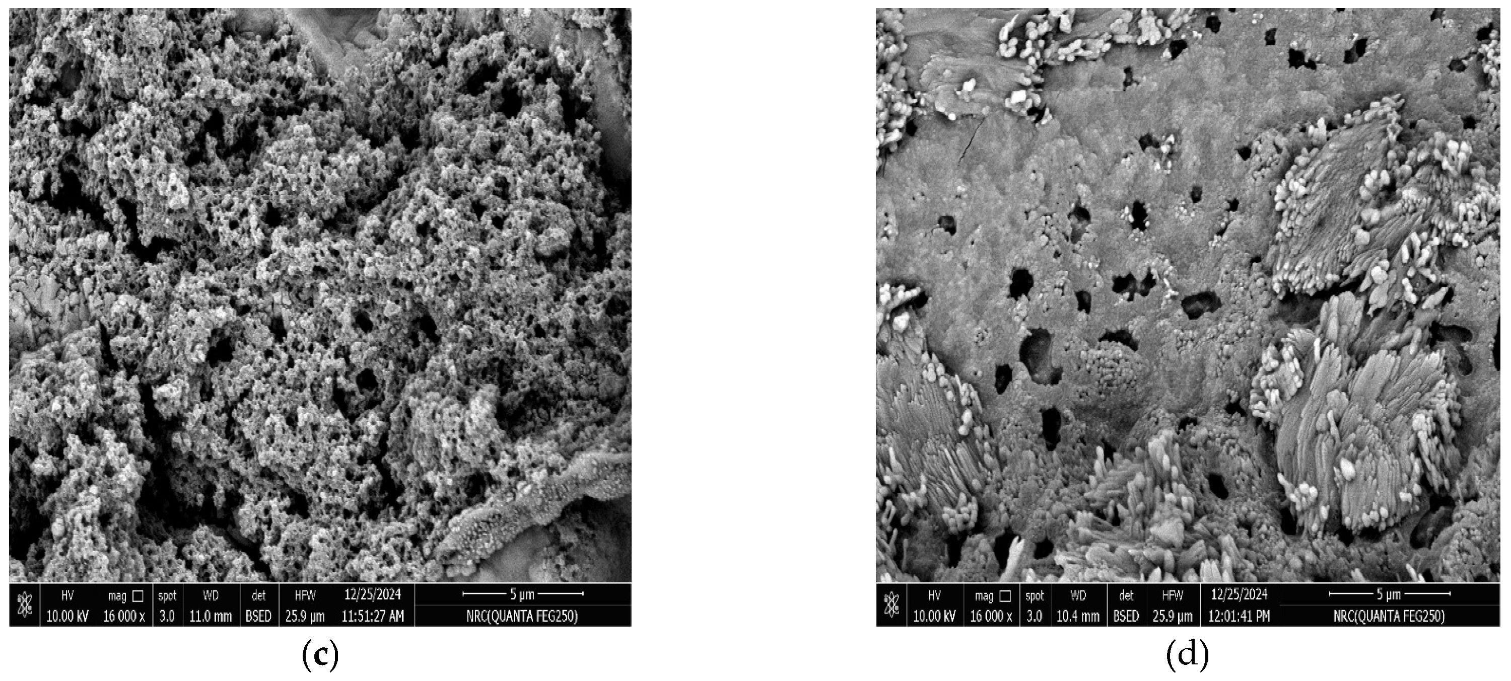
2.6. Morphological Observations

TEM analysis of N-fullerenes illustrates big bucky ball-shaped carbon dots composed of individual carbon nanodots with a diameter ranging between ~1.85 and 4.93 nm (Figure 6a).
Figure 6b,c illustrates the surface morphology of the CMC–AMPS hydrogel and the CMC–N-fullerene–AMPS hydrogel. The CMC–AMPS hydrogel showed a porous-shaped film with a pore size ranging between 309–930 µm, while the CMC–N-fullerene–AMPS hydrogel showed a porous shape with flowery structures due to the presence of N-fullerenes. The porosity for the CMC–N-fullerene–AMPS hydrogel is equal to 377–931 µm. The porous structure of the CMC–N-fullerene–AMPS hydrogel enhances the interaction between the MCD sensor and its surroundings. This increased accessibility results in improved sensitivity and faster response times.

3. Conclusions

In conclusion, this study successfully synthesized a novel carboxymethyl cellulose–N-fullerene–g-poly(co-acrylamido-2-methyl-1-propane sulfonic acid) (CMC–N-fullerene–AMPS) hydrogel via a free radical polymerization method. FTIR analysis confirmed the successful grafting of AMPS and incorporation of N-fullerenes onto the CMC backbone, evidenced by characteristic S=O peaks and a shift in the N–H/O–H peak from 3431 cm−1 in CMC–AMPS to 3406 cm−1 in CMC–N-fullerene–AMPS, indicating strong hydrogen bonding. DFT calculations further corroborated the successful synthesis, demonstrating that the CMC–N-fullerene–AMPS hydrogel exhibited higher stability (μ = 41.321 Debye), a lower band gap energy (Eg = 0.0871 eV), and greater energy release upon formation (ET = −5158.364 au) compared to the CMC–AMPS hydrogel, attributed to the presence of N-fullerenes. The incorporation of N-fullerenes significantly enhanced the hydrogel’s antibacterial activity, increasing the inhibition zone against E. coli from 0 mm to 22 mm and against S. aureus from 15 mm to 24 mm. Molecular docking studies supported these findings, revealing shorter bond lengths between the hydrogel and bacterial proteins in the presence of N-fullerenes. Furthermore, the hydrogel displayed a unique “turn-on” fluorescence response upon interaction with bacteria, shifting from dark red to bright orange-red with E. coli and bright green with S. aureus, suggesting potential applications in bacterial detection and sensing. SEM analysis revealed a porous structure in both hydrogels, with the CMC–N-fullerene–AMPS hydrogel exhibiting a flowery morphology and pore sizes ranging from 377 to 931 µm due to the N-fullerenes, which is expected to contribute to enhanced interaction with the surrounding environment. These combined properties of enhanced antibacterial activity, fluorescence response, and porous structure suggest that the synthesized CMC–N-fullerene–AMPS hydrogel holds significant promise for applications in various fields, including biosensing in food packaging.

4. Materials and Methods

4.1. Materials

Sugarcane bagasse (SB) was obtained from the Quena Company for Paper Industry (Quena, Egypt). 2-Acrylamido-2-methyl-1-propanesulfonic acid (AMPS) was purchased from Alfa Aesar. N,N′-Methylenebisacrylamide (MBA) and potassium persulfate (KPS) were acquired from Sigma-Aldrich (Burlington, MA, USA). All chemicals and reagents were of analytical grade and used without further purification.

4.2. Preparation of Cellulose

Sugarcane bagasse (SB) was hydrolyzed by using 1.5% HCl at 120 °C for 2 h. The acid-treated SB was washed and then alkali-treated with 20 g NaOH in 300 mL water at 170 °C for 2 h to obtain pulp. Lignin was removed from 80 g of pulp by bleaching with 3% HClO2 and acetic acid at 80 °C for 2 h [65].

4.3. Microwave-Assisted Synthesis of Carboxymethyl Cellulose

An amount of 15 g of prepared cellulose was mixed with 30% NaOH solution and 18 g of monochloroacetic acid. This mixture was then subjected to microwave heating until complete dissolution. The synthesized CMC was then precipitated by adding 70% methanol, followed by filtration and oven drying [16,76].

4.4. Preparation of Nitrogen-Doped Fullerenes (N-fullerenes)

An amount of 1 g of CMC and 0.1 g of citric acid were dissolved in 20 mL of dimethylformamide. The mixture was microwave-irradiated at 100 °C and 750 W for 135 min using a Milestone Italy StartSynth microwave reactor (Pack2B Basic Single Vessel Kit, Milestone Inc., Bergamo, Italy) . The final solution was cooled to room temperature, centrifuged, filtered, and dried [38].

4.5. Preparation of Carboxymethyl cellulose–N-fullerene–g-poly(co-acrylamido-2-methyl-1-propane Sulfonic Acid)

An amount of 2 g of CMC was dissolved in 50 mL of distilled water at 60 °C with stirring. Then, 0.24 g of KPS diluted in 5 mL of distilled water was added. An aqueous suspension containing 2 g of fullerenes, 2 g of AMPS neutralized to pH 5.5 with NaOH, and 0.24 g of MBA was then added to the solution. The temperature was maintained at 65 °C for 10 min to initiate free radical polymerization on the CMC chains. Subsequently, the pH was adjusted to 5.5 and the temperature was set to 70 °C for 3 h. The resulting CMC–N-fullerene–AMPS hydrogel was washed with distilled water to remove any unreacted monomers, homopolymers, and crosslinkers. The hydrogel was then frozen at −80 °C for 3 h and freeze-dried. Another blank hydrogel was prepared without fullerenes and denoted as CMC–AMPS hydrogel.

4.6. Characterization

4.6.1. Morphological Observations

Transmission electron microscopy (TEM) was performed using a JEOL JEM-2100 microscope (TEM, JEM-1230, Gunma, Japan) operating at 120 kV. Scanning electron microscopy (SEM) and energy-dispersive X-ray spectroscopy (EDX) were conducted with a Quanta/250-FEG scanning electron microscope (Thermo Fisher Scientific, Waltham, MA, USA) equipped with an EDX analyzer, using an acceleration voltage of 30 kV.

4.6.2. Fluorescence Microscope

Fluorescence microscopy was carried out using a Jasco FP-6500 spectrofluorometer (Tokyo, Japan) equipped with a 150-watt xenon arc lamp.

4.6.3. Fourier-Transform Infrared (FTIR) Spectra

Fourier-transform infrared (FTIR) spectra were acquired using a Mattson 5000 spectrometer (Unicam, UK) via the potassium bromide (KBr) disk method. The crystallinity index (LOI) and mean hydrogen bond strength (MHBS) were determined using Equations (1) and (2).
LOI = A 1425 A 900
MHBS = A O H A C H
where A1425 and A900 refer to the FTIR absorbance at 1425 and 900 cm−1, respectively. In addition, AOH and ACH refer to the FTIR absorbance of the OH and CH peaks, respectively [66,94].

4.6.4. DFT Calculations

Density Functional Theory (DFT) calculations were performed using Gaussian 09W at the B3LYP/6-31G(d) level of theory with Berny optimization. The investigated parameters included optimized geometries, ground state energies (ET), HOMO-LUMO energies (EHOMO, ELUMO, and Eg), dipole moment (μ), absolute hardness (η), softness (σ, S), and additional electronic charge (ΔNmax) [72,75].
E g a p = ( E L U M O E H O M O )
η = ( E L U M O + E H O M O ) 2
σ = 1 η
S = 1 2 η
Δ N m a x = P i η

4.6.5. Swelling Behavior

The swelling behavior of the hydrogels was studied over time. A pre-weighed dry hydrogel sample was immersed in distilled water at room temperature. At designated intervals, the swollen hydrogel was removed and blotted dry with filter paper for 24 h to eliminate excess surface water. The swelling percentage was subsequently calculated using Equation (8).
S w e l l i n g % = W t W 0 W 0 × 100
where W0 and Wt stand for the hydrogel’s dry and swelling weights, respectively. For every hydrogel, the experiment was conducted three times [56].

Funding

The author appreciates the National Research Centre and the PRIMA International Project, between the Academy of Scientific Research and Technology (ASRT) and PRIMA, through the Joint Agreement project “Sustainable Antimicrobial Packaging based on a Healthy Intelligent Renewable Approach” with the acronym “SAPHIRA”.

Institutional Review Board Statement

Not applicable.

Informed Consent Statement

Not applicable.

Data Availability Statement

The original contributions presented in the study are included in the article material; further inquiries can be directed to the author.

Acknowledgments

The author wants to acknowledge the National Research Centre, Cellulose and Paper Department, Egypt, for supporting us with the materials needed for this work.

Conflicts of Interest

The author declares no conflict of interest.

References

  1. Squires, V.R.; Gaur, M.K. Food Security and Land Use Change Under Conditions of Climatic Variability: A Multidimensional Perspective; Springer: Berlin/Heidelberg, Germany, 2020. [Google Scholar]
  2. Martindale, W. Global Food Security and Supply; John Wiley & Sons: Hoboken, NJ, USA, 2014. [Google Scholar]
  3. Aslam, J.; Parray, H.A.; Aslam, A.; Aslam, R. Food Waste Environmental Impact Assessment. In Sustainable Food Waste Management: Anti-corrosion Applications; Springer: Berlin/Heidelberg, Germany, 2024; pp. 87–105. [Google Scholar]
  4. Singh, A.; Tyagi, P.K.; Garg, A. Sustainable Disposal Methods of Food Wastes in Hospitality Operations; IGI Global: Hershey, PA, USA, 2024. [Google Scholar]
  5. Wong, M.H.; Purchase, D.; Dickinson, N. Impacts, Management, and Recycling of Food Waste: Global Emerging Issues. In Food Waste Valorisation: Food, Feed, Fertiliser, Fuel and Value-Added Products; World Scientific: Singapore, 2023; pp. 3–31. [Google Scholar]
  6. Chapagain, A.; James, K. Accounting for the impact of food waste on water resources and climate change. In Food Industry Wastes; Academic Press: Cambridge, MA, USA, 2013; pp. 217–236. [Google Scholar] [CrossRef]
  7. Chai, X.; Tonjes, D.J.; Mahajan, D. Methane emissions as energy reservoir: Context, scope, causes and mitigation strategies. Prog. Energy Combust. Sci. 2016, 56, 33–70. [Google Scholar] [CrossRef]
  8. Zuberi, M.J.S.; Ali, S.F. Greenhouse effect reduction by recovering energy from waste landfills in Pakistan. Renew. Sustain. Energy Rev. 2015, 44, 117–131. [Google Scholar] [CrossRef]
  9. Ishangulyyev, R.; Kim, S.; Lee, S.H. Understanding food loss and waste—Why are we losing and wasting food? Foods 2019, 8, 297. [Google Scholar] [CrossRef] [PubMed]
  10. Thyberg, K.L.; Tonjes, D.J. Drivers of food waste and their implications for sustainable policy development. Resour. Conserv. Recycl. 2016, 106, 110–123. [Google Scholar] [CrossRef]
  11. Mosomi, E.K.; Olanrewaju, O.A.; Adeosun, S.O. Pivotal role of polylactide in carbon emission reduction: A comprehensive review. Eng. Rep. 2024, 6, e12909. [Google Scholar] [CrossRef]
  12. Soo, X.Y.D.; Muiruri, J.K.; Wu, W.Y.; Yeo, J.C.C.; Wang, S.; Tomczak, N.; Thitsartarn, W.; Tan, B.H.; Wang, P.; Wei, F. Bio-Polyethylene and Polyethylene Biocomposites: An Alternative toward a Sustainable Future. Macromol. Rapid Commun. 2024, 45, 2400064. [Google Scholar] [CrossRef]
  13. Macheca, A.D.; Mutuma, B.; Adalima, J.L.; Midheme, E.; Lúcas, L.H.; Ochanda, V.K.; Mhlanga, S.D. Perspectives on plastic waste management: Challenges and possible solutions to ensure its sustainable use. Recycling 2024, 9, 77. [Google Scholar] [CrossRef]
  14. Jiao, H.; Ali, S.S.; Alsharbaty, M.H.M.; Elsamahy, T.; Abdelkarim, E.; Schagerl, M.; Al-Tohamy, R.; Sun, J. A critical review on plastic waste life cycle assessment and management: Challenges, research gaps, and future perspectives. Ecotoxicol. Environ. Saf. 2024, 271, 115942. [Google Scholar] [CrossRef]
  15. Tohamy, H.-A.S. Cellulosic schiff base hydrogel biosensor for bacterial detection with pH/thermo-responsitivity: DFT calculations and molecular docking. Int. J. Biol. Macromol. 2024, 283, 137389. [Google Scholar] [CrossRef]
  16. Tohamy, H.-A.S. Novel, Speedy, and Eco-Friendly Carboxymethyl Cellulose-Nitrogen Doped Carbon Dots Biosensors with DFT Calculations, Molecular Docking, and Experimental Validation. Gels 2024, 10, 686. [Google Scholar] [CrossRef] [PubMed]
  17. Afolabi, O.A.; Turup Pandurangan, M.; Kanny, K. Types of Biobased Nanomaterials. In Biobased Nanomaterials: Applications in Biomedicine, Food Industry, Agriculture, and Environmental Sustainability; Springer: Berlin/Heidelberg, Germany, 2024; pp. 17–43. [Google Scholar]
  18. Gaur, M.; Misra, C.; Yadav, A.B.; Swaroop, S.; Maolmhuaidh, F.Ó.; Bechelany, M.; Barhoum, A. Biomedical applications of carbon nanomaterials: Fullerenes, quantum dots, nanotubes, nanofibers, and graphene. Materials 2021, 14, 5978. [Google Scholar] [CrossRef]
  19. Khan, T.; Singh, B.; Manikandan, M. Synthesis, Characteristics, and Applications of Nanomaterials. In Nanomaterials: The Building Blocks of Modern Technology: Synthesis, Properties and Applications; Springer: Berlin/Heidelberg, Germany, 2023; pp. 11–26. [Google Scholar]
  20. Prasad, R.D.; Sahoo, A.; Shrivastav, O.P.; Charmode, N.; Prasad, S.R.; Kamat, R.; Kajave, N.; Chauhan, J.; Banga, S.; Tamboli, U. A review on aspects of nanotechnology in food science and animal nutrition. ES Food Agrofor. 2022, 8, 12–46. [Google Scholar]
  21. Markovic, Z.; Trajkovic, V. Biomedical potential of the reactive oxygen species generation and quenching by fullerenes (C60). Biomaterials 2008, 29, 3561–3573. [Google Scholar] [CrossRef] [PubMed]
  22. Pantarotto, D.; Tagmatarchis, N.; Bianco, A.; Prato, M. Synthesis and biological properties of fullerene-containing amino acids and peptides. Mini Rev. Med. Chem. 2004, 4, 805–814. [Google Scholar] [CrossRef]
  23. Nile, S.H.; Baskar, V.; Selvaraj, D.; Nile, A.; Xiao, J.; Kai, G. Nanotechnologies in food science: Applications, recent trends, and future perspectives. Nano-Micro Lett. 2020, 12, 1–34. [Google Scholar] [CrossRef]
  24. Papadochristopoulos, A.; Kerry, J.P.; Fegan, N.; Burgess, C.M.; Duffy, G. Natural anti-microbials for enhanced microbial safety and shelf-life of processed packaged meat. Foods 2021, 10, 1598. [Google Scholar] [CrossRef]
  25. Balasundaram, R.P.; Gunasekaran, P.; Ramaraj, A.; Jothiprakash, G.; Jaganathan, D. Role of Nanomaterials in Food Packaging Applications. In Introduction to Functional Nanomaterials; CRC Press: Boca Raton, FL, USA, 2024; pp. 290–301. [Google Scholar]
  26. Khan, A.; Ezati, P.; Tammina, S.K.; Riahi, Z. Packaging and Preservation. In Functional Nanomaterials and Nanocomposites for Biodegradable Food Packaging; Springer: Berlin/Heidelberg, Germany, 2025; p. 109. [Google Scholar]
  27. Sinha, S.; Sanfo, K.; Dallas, P.; Kumar, S.; Porfyrakis, K. Process parameter optimisation for endohedral metallofullerene synthesis via the arc-discharge method. Inorganics 2024, 12, 38. [Google Scholar] [CrossRef]
  28. Gupta, P.K. Mechanism of Nanotoxicity. In Nanotoxicology in Nanobiomedicine; Springer International Publishing: Cham, Switzerland; Springer: Berlin/Heidelberg, Germany, 2023; pp. 37–53. [Google Scholar]
  29. Gaganpreet; Pathania, Y. Carbon-Based Nanomaterials for Pollutants’ Treatment. In Carbon-Based Nanomaterials for Green Applications; John Wiley & Sons: Hoboken, NJ, USA, 2024; pp. 355–381. [Google Scholar]
  30. Bharti, R.; Kaushik, P.; Naik, S.; Thakur, A.; Verma, M.; Sharma, R. Low Temperature Synthesis of Carbon Nanostructures. In Handbook of Functionalized Carbon Nanostructures: From Synthesis Methods to Applications; Springer: Berlin/Heidelberg, Germany, 2024; pp. 1–45. [Google Scholar]
  31. Sharma, G. Biogenic carbon nanostructured materials for detection of cancer and medical applications: A mini review. Hybrid Adv. 2024, 5, 100166. [Google Scholar] [CrossRef]
  32. Danial, W.H. Synthesis of Carbon Nanostructures from Waste Materials. In Handbook of Functionalized Carbon Nanostructures: From Synthesis Methods to Applications; Springer: Berlin/Heidelberg, Germany, 2024; pp. 1–38. [Google Scholar]
  33. Tohamy, H.-A.S.; El-Sakhawy, M.; Kamel, S. A Greener Future: Carbon Nanomaterials from Lignocellulose. J. Renew. Mater. 2024, 13, 21–47. [Google Scholar]
  34. Levchenko, I.; Mandhakini, M.; Prasad, K.; Bazaka, O.; Ivanova, E.P.; Jacob, M.V.; Baranov, O.; Riccardi, C.; Roman, H.E.; Xu, S.; et al. Functional Nanomaterials from Waste and Low-Value Natural Products: A Technological Approach Level. Adv. Mater. Technol. 2022, 7, 2101471. [Google Scholar] [CrossRef]
  35. Kumar, K.; Kumar, R.; Kaushal, S.; Thakur, N.; Umar, A.; Akbar, S.; Ibrahim, A.A.; Baskoutas, S. Biomass waste-derived carbon materials for sustainable remediation of polluted environments: A comprehensive review. Chemosphere 2023, 345, 140419. [Google Scholar] [CrossRef] [PubMed]
  36. Poonia, K.; Singh, P.; Ahamad, T.; Van Le, Q.; Quang, H.H.P.; Thakur, S.; Mishra, A.K.; Selvasembian, R.; Hussain, C.M.; Nguyen, V.-H.; et al. Sustainability, performance, and production perspectives of waste-derived functional carbon nanomaterials towards a sustainable environment: A review. Chemosphere 2024, 352, 141419. [Google Scholar] [CrossRef]
  37. Salehi, B.; Wang, L. Critical review on nanomaterials for enhancing bioconversion and bioremediation of agricultural wastes and wastewater. Energies 2022, 15, 5387. [Google Scholar] [CrossRef]
  38. Tohamy, H.-A.S.; El-Sakhawy, M.; Kamel, S. Fullerenes and tree-shaped/fingerprinted carbon quantum dots for chromium adsorption via microwave-assisted synthesis. RSC Adv. 2024, 14, 25785–25792. [Google Scholar] [CrossRef]
  39. Sabui, P.; Mallick, S.; Singh, K.R.; Natarajan, A.; Verma, R.; Singh, J.; Singh, R.P. Potentialities of fluorescent carbon nanomaterials as sensor for food analysis. Luminescence 2023, 38, 1047–1063. [Google Scholar] [CrossRef]
  40. Chen, D.; Gong, J.; Grüne, J.; Matulaitis, T.; Gillett, A.J.; Zhang, X.H.; Samuel, I.D.; Turnbull, G.A.; Zysman-Colman, E. Tetra-Donor Pyrazine Based Thermally Activated Delayed Fluorescence Emitters for Electroluminescence and Amplified Spontaneous Emission. Adv. Funct. Mater. 2024, 34, 2409592. [Google Scholar] [CrossRef]
  41. Hasan, M.; Shukla, A.; Mamada, M.; Adachi, C.; Lo, S.-C.; Namdas, E.B. Correlating exciton dynamics of thermally activated delayed-fluorescence emitters to efficiency roll-off in OLEDs. Phys. Rev. Appl. 2022, 18, 054082. [Google Scholar] [CrossRef]
  42. Yuan, S.; Zhang, G.; Chen, F.; Chen, J.; Zhang, Y.; Di, Y.; Chen, Y.; Zhu, Y.; Lin, M.; Chen, H. Thermally Activated Delayed Fluorescent Ag (I) Complexes for Highly Efficient Scintillation and High-Resolution X-Ray Imaging. Adv. Funct. Mater. 2024, 34, 2400436. [Google Scholar] [CrossRef]
  43. Feng, Q.; Zhu, S.; Wang, B.; Yu, F.; Li, H.; Yu, M.; Xu, M.; Xie, L. Thermally Activated Delayed Fluorescence Macrocycles for Organic Light-Emitting Diodes. Adv. Funct. Mater. 2024, 34, 2312622. [Google Scholar] [CrossRef]
  44. Baleizão, C.; Berberan-Santos, M.N. Thermally activated delayed fluorescence in fullerenes. Ann. N. Y. Acad. Sci. 2008, 1130, 224–234. [Google Scholar] [CrossRef] [PubMed]
  45. Tao, Y.; Yuan, K.; Chen, T.; Xu, P.; Li, H.; Chen, R.; Zheng, C.; Zhang, L.; Huang, W. Thermally activated delayed fluorescence materials towards the breakthrough of organoelectronics. Adv. Mater. 2014, 26, 7931–7958. [Google Scholar] [CrossRef] [PubMed]
  46. Lichota, A.; Szabelski, M.; Krokosz, A. Quenching of protein fluorescence by fullerenol C60(OH)36 nanoparticles. Int. J. Mol. Sci. 2022, 23, 12382. [Google Scholar] [CrossRef] [PubMed]
  47. Wu, F.; Bai, Y.; Mu, Y.; Pan, B.; Xing, B.; Lin, Y. Fluorescence quenching of fulvic acids by fullerene in water. Environ. Pollut. 2013, 172, 100–107. [Google Scholar] [CrossRef]
  48. Chen, H.; Zhang, L.; Hu, Y.; Zhou, C.; Lan, W.; Fu, H.; She, Y. Nanomaterials as optical sensors for application in rapid detection of food contaminants, quality and authenticity. Sens. Actuators B Chem. 2021, 329, 129135. [Google Scholar] [CrossRef]
  49. Das, J.; Mishra, H.N. Recent advances in sensors for detecting food pathogens, contaminants, and toxins: A review. Eur. Food Res. Technol. 2022, 248, 1125–1148. [Google Scholar] [CrossRef]
  50. Bulgakov, R.; Galimov, D. Fullerene C 60 as a superefficient quencher of singlet exited states of polycyclic aromatic hydrocarbons. Russ. Chem. Bull. 2007, 56, 446–451. [Google Scholar] [CrossRef]
  51. Liu, S.; Ji, X.-X.; Zhu, J.-F. Recent progress in the synthesis and biomedical properties of natural biopolymer composites. Curr. Med. Chem. 2021, 28, 8243–8266. [Google Scholar] [CrossRef]
  52. Dobrzyńska-Mizera, M.; Dodda, J.M.; Liu, X.; Knitter, M.; Oosterbeek, R.N.; Salinas, P.; Pozo, E.; Ferreira, A.M.; Sadiku, E.R. Engineering of Bioresorbable Polymers for Tissue Engineering and Drug Delivery Applications. Adv. Healthc. Mater. 2024, 13, 2401674. [Google Scholar] [CrossRef]
  53. Dispenza, C.; Giacomazza, D.; Jonsson, M. Micro-to Nanoscale Bio-Hybrid Hydrogels Engineered by Ionizing Radiation. Biomolecules 2021, 11, 47. [Google Scholar] [CrossRef]
  54. Gurusamy, L.C. AMPs Based Rheology Modifier for Personal Care Industry. Ph.D. Thesis, Lehigh University, Bethlehem, PA, USA, 2019. [Google Scholar]
  55. Karchoubi, F.; Ghotli, R.A.; Pahlevani, H.; Salehi, M.B. New insights into nanocomposite hydrogels; A review on recent advances in characteristics and applications. Adv. Ind. Eng. Polym. Res. 2024, 7, 54–78. [Google Scholar] [CrossRef]
  56. Tohamy, H.-A.S. Fluorescence ‘Turn-on’ Probe for Chromium Reduction, Adsorption and Detection Based on Cellulosic Nitrogen-Doped Carbon Quantum Dots Hydrogels. Gels 2024, 10, 296. [Google Scholar] [CrossRef] [PubMed]
  57. Shoukat, H.; Pervaiz, F.; Rehman, S.; Noreen, S. Development of β-cyclodextrin/chitosan-co-poly (2-acrylamide-2-methylpropane sulphonic acid) cross-linked hybrid IPN-nanogels to enhance the solubility of rosuvastatin: An in vitro and in vivo attributes. J. Drug Deliv. Sci. Technol. 2022, 75, 103696. [Google Scholar] [CrossRef]
  58. Singha, N.R.; Dutta, A.; Mahapatra, M.; Roy, J.S.D.; Mitra, M.; Deb, M.; Chattopadhyay, P.K. In situ attachment of acrylamido sulfonic acid-based monomer in terpolymer hydrogel optimized by response surface methodology for individual and/or simultaneous removal (s) of M (III) and cationic dyes. ACS Omega 2019, 4, 1763–1780. [Google Scholar] [CrossRef]
  59. Mondal, S.; Das, S.; Nandi, A.K. A review on recent advances in polymer and peptide hydrogels. Soft Matter 2020, 16, 1404–1454. [Google Scholar] [CrossRef]
  60. Tohamy, H.-A.S.; Mohamed, F.E.-Z.S.; El-Sakhawy, M. Novel microwave assisted carboxymethyl-graphene oxide and its hepatoprotective activity. BMC Pharmacol. Toxicol. 2024, 25, 50. [Google Scholar] [CrossRef]
  61. Li, Z.; Sun, Y.; Liu, D.; Yi, M.; Chang, F.; Li, H.; Du, Y. A review of sulfate radical-based and singlet oxygen-based advanced oxidation technologies: Recent advances and prospects. Catalysts 2022, 12, 1092. [Google Scholar] [CrossRef]
  62. Seo, K.; Chung, I.-J. Reaction analysis of 3, 4-ethylenedioxythiophene with potassium persulfate in aqueous solution by using a calorimeter. Polymer 2000, 41, 4491–4499. [Google Scholar] [CrossRef]
  63. Hsu, S.-C.; Don, T.-M.; Chiu, W.-Y. Free radical degradation of chitosan with potassium persulfate. Polym. Degrad. Stab. 2002, 75, 73–83. [Google Scholar] [CrossRef]
  64. Ma, J.; Li, X.; Bao, Y. Advances in cellulose-based superabsorbent hydrogels. RSC Adv. 2015, 5, 59745–59757. [Google Scholar] [CrossRef]
  65. El-Sakhawy, M.; Tohamy, H.-A.S.; AbdelMohsen, M.M.; El-Missiry, M. Biodegradable carboxymethyl cellulose based material for sustainable/active food packaging application. J. Thermoplast. Compos. Mater. 2024, 37, 2035–2050. [Google Scholar] [CrossRef]
  66. Tohamy, H.-A.S.; El-Sakhawy, M.; Abdel-Halim, S.A.; El-Masry, H.M.; AbdelMohsen, M.M. Antimicrobial Plectranthus amboinicus emulsions prepared with amphiphilic cellulose stearate. In Euro-Mediterranean Journal for Environmental Integration; Springer: Berlin/Heidelberg, Germany, 2024; pp. 1–12. [Google Scholar]
  67. Cuba-Chiem, L.T.; Huynh, L.; Ralston, J.; Beattie, D.A. In situ particle film ATR FTIR spectroscopy of carboxymethyl cellulose adsorption on talc: Binding mechanism, pH effects, and adsorption kinetics. Langmuir 2008, 24, 8036–8044. [Google Scholar] [CrossRef] [PubMed]
  68. Cukrowicz, S.; Grabowski, B.; Kaczmarska, K.; Bobrowski, A.; Sitarz, M.; Tyliszczak, B. Structural studies (FTIR, XRD) of sodium carboxymethyl cellulose modified bentonite. Arch. Foundry Eng. 2020, 20, 119–125. [Google Scholar] [CrossRef]
  69. Jahit, I.; Nazmi, N.; Isa, M.; Sarbon, N. Preparation and physical properties of gelatin/CMC/chitosan composite films as affected by drying temperature. Int. Food Res. J. 2015, 23, 1068–1074. [Google Scholar]
  70. Mottola, S.; Viscusi, G.; Tohamy, H.-A.S.; El-Sakhawy, M.; Gorrasi, G.; De Marco, I. Application of electrospun N-doped carbon dots loaded cellulose acetate membranes as cationic dyes adsorbent. J. Environ. Manag. 2024, 370, 122714. [Google Scholar] [CrossRef] [PubMed]
  71. Mohkami, M.; Talaeipour, M. Investigation of the chemical structure of carboxylated and carboxymethylated fibres from waste paper via XRD and FTIR analysis. Bioresources 2011, 6, 1988–2003. [Google Scholar] [CrossRef]
  72. Tohamy, H.-A.S. Speedy synthesis of magnetite/carbon dots for efficient chromium removal and reduction: A combined experimental and DFT approach. In Emergent Materials; Springer: Berlin/Heidelberg, Germany, 2024; pp. 1–13. [Google Scholar]
  73. Garcia-Borras, M.; Osuna, S.; Luis, J.M.; Swart, M.; Sola, M. The role of aromaticity in determining the molecular structure and reactivity of (endohedral metallo) fullerenes. Chem. Soc. Rev. 2014, 43, 5089–5105. [Google Scholar] [CrossRef]
  74. Nattagh, F.; Hosseini, S.; Esrafili, M.D. Effects of B and N doping/codoping on the adsorption behavior of C60 fullerene towards aspirin: A DFT investigation. J. Mol. Liq. 2021, 342, 117459. [Google Scholar] [CrossRef]
  75. Tohamy, H.-A.S. Greener, Safer Packaging: Carbon Nanotubes/Gelatin-Enhanced Recycled Paper for Fire Retardation with DFT Calculations. J. Renew. Mater. 2024, 12, 1963–1983. [Google Scholar] [CrossRef]
  76. Tohamy, H.-A.S. Carboxymethyl hemicellulose hydrogel as a fluorescent biosensor for bacterial and fungal detection with DFT and molecular docking studies. Sci. Rep. 2025, 15, 741. [Google Scholar] [CrossRef]
  77. Varghese, M.; Balachandran, M. Antibacterial efficiency of carbon dots against Gram-positive and Gram-negative bacteria: A review. J. Environ. Chem. Eng. 2021, 9, 106821. [Google Scholar] [CrossRef]
  78. Niranjan, R.; Zafar, S.; Lochab, B.; Priyadarshini, R. Synthesis and characterization of sulfur and sulfur-selenium nanoparticles loaded on reduced graphene oxide and their antibacterial activity against gram-positive pathogens. Nanomaterials 2022, 12, 191. [Google Scholar] [CrossRef] [PubMed]
  79. Travlou, N.A.; Giannakoudakis, D.A.; Algarra, M.; Labella, A.M.; Rodríguez-Castellón, E.; Bandosz, T.J. S-and N-doped carbon quantum dots: Surface chemistry dependent antibacterial activity. Carbon 2018, 135, 104–111. [Google Scholar] [CrossRef]
  80. Ruan, Z.; Xu, Z.; Liu, T.; Chen, L.; Liu, X.; Chen, K.; Zhao, C. Multifunctional nitrogen-sulfur codoped carbon quantum dots: Determining reduced glutathione, broad-spectrum antibacterial activity, and cell imaging. Heliyon 2024, 10, e38177. [Google Scholar] [CrossRef]
  81. Saravanan, A.; Das, P.; Maruthapandi, M.; Aryal, S.; Michaeli, S.; Mastai, Y.; Luong, J.H.; Gedanken, A. Heteroatom co-doping (N, NS, NB) on carbon dots and their antibacterial and antioxidant properties. Surf. Interfaces 2024, 46, 103857. [Google Scholar] [CrossRef]
  82. Li, Y.; Han, Y.; Li, H.; Niu, X.; Zhang, D.; Wang, K. Antimicrobial hydrogels: Potential materials for medical application. Small 2024, 20, 2304047. [Google Scholar] [CrossRef]
  83. Fantini, J.; Yahi, N. Brain Lipids in Synaptic Function and Neurological Disease: Clues to Innovative Therapeutic Strategies for Brain Disorders; Academic Press: Cambridge, MA, USA, 2015. [Google Scholar]
  84. Baskar, A.V.; Benzigar, M.R.; Talapaneni, S.N.; Singh, G.; Karakoti, A.S.; Yi, J.; Al-Muhtaseb, A.a.H.; Ariga, K.; Ajayan, P.M.; Vinu, A. Self-assembled fullerene nanostructures: Synthesis and applications. Adv. Funct. Mater. 2022, 32, 2106924. [Google Scholar] [CrossRef]
  85. Lee, C.; Seo, Y.; Han, J.; Hwang, J.; Jeon, I. Perspectives on critical properties of fullerene derivatives for rechargeable battery applications. Carbon 2023, 210, 118041. [Google Scholar] [CrossRef]
  86. Omar Khudhur, Z.; HMaad, A.; AGhanimi, H.; Abdolmaleki, A. Fullerene nanoparticle as new therapeutic agent for the nervous system disorders. Nanomed. J. (NMJ) 2024, 11, 342–359. [Google Scholar]
  87. Hildebrandt, N.; Spillmann, C.M.; Algar, W.R.; Pons, T.; Stewart, M.H.; Oh, E.; Susumu, K.; Diaz, S.A.; Delehanty, J.B.; Medintz, I.L. Energy transfer with semiconductor quantum dot bioconjugates: A versatile platform for biosensing, energy harvesting, and other developing applications. Chem. Rev. 2017, 117, 536–711. [Google Scholar] [CrossRef]
  88. Szöllosi, J.; Damjanovich, S.; Mátyus, L. Application of fluorescence resonance energy transfer in the clinical laboratory: Routine and research. Cytom. J. Int. Soc. Anal. Cytol. 1998, 34, 159–179. [Google Scholar]
  89. Bhupathi, P.; Elhassan A-Elgadir, T.M.; Mohammed Ali, R.H.; Sanaan Jabbar, H.; Gulnoza, D.; Joshi, S.; Kadhem Abid, M.; Ahmed Said, E.; Alawadi, A.; Alsaalamy, A. Fluorescence Resonance Energy Transfer (FRET)-Based Sensor for Detection of Foodborne Pathogenic Bacteria: A Review. Crit. Rev. Anal. Chem. 2023, 1–18. [Google Scholar] [CrossRef] [PubMed]
  90. De, A.; Arora, R.; Jasani, A. Engineering aspects of bioluminescence resonance energy transfer systems. In Engineering in Translational Medicine; Springer: London, UK, 2013; pp. 257–300. [Google Scholar]
  91. Lone, M.S.; Bhat, P.A.; Afzal, S.; Chat, O.A.; Dar, A.A. Energy transduction through FRET in self-assembled soft nanostructures based on surfactants/polymers: Current scenario and prospects. Soft Matter 2021, 17, 425–446. [Google Scholar] [CrossRef]
  92. Gottschalk, G. Regulation of Bacterial Metabolism; Springer: Berlin/Heidelberg, Germany, 1986; pp. 178–207. [Google Scholar] [CrossRef]
  93. Somerville, G.A.; Proctor, R.A. At the crossroads of bacterial metabolism and virulence factor synthesis in Staphylococci. Microbiol. Mol. Biol. Rev. 2009, 73, 233–248. [Google Scholar] [CrossRef] [PubMed]
  94. Hassanein, H.D.; Tohamy, H.A.S.; AbdelMohsen, M.M.; El-Masry, H.M.; El-Sakhawy, M. Preparation and characterization of cellulose acetate/corn silk extract films for potential antimicrobial application. Egypt. J. Chem. 2024, 68, 311–318. [Google Scholar] [CrossRef]
Figure 1. (a) Reaction mechanism for synthesizing carboxymethyl cellulose–N-fullerene–g-poly(co-acrylamido-2-methyl-1-propane sulfonic acid) hydrogel and (b) the swelling study of CMC–AMPS hydrogel and CMC–N-fullerene–AMPS hydrogel.
Gels 11 00329 g001
Figure 2. FTIR spectra of (a) cellulose, (b) CMC, (c) N-fullerenes, (d) CMC–AMPS hydrogel, and (e) CMC–N-fullerene–AMPS hydrogel.
Gels 11 00329 g002
Figure 3. The gap energies (HOMO–LUMO) (eV) were calculated for the hydrogels using DFT B3LYP/6–31G (d), as was the molecular orbital interaction between CMC, MBA, AMPS, CMC–AMPS hydrogel, N-fullerenes, and CMC–N-fullerene–AMPS hydrogel.
Gels 11 00329 g003aGels 11 00329 g003b
Figure 4. The antimicrobial and biological activity of CMC–AMPS hydrogel (denoted as 5) and CMC–N-fullerene–AMPS hydrogel (denoted as 6) as ligands against Escherichia coli PDB (3ZH7), and Staphylococcus aureus PDB (4QLO).
Gels 11 00329 g004
Figure 5. Fluorescence microscope for (a) CMC–N-fullerene–AMPS hydrogel before bacteria, (b) CMC–N-fullerene–AMPS hydrogel/Escherichia coli, and (c) CMC–N-fullerene–AMPS hydrogel/Staphylococcus aureus.
Gels 11 00329 g005aGels 11 00329 g005b
Figure 6. (a) TEM analysis for N-fullerenes, (b) particle size distribution for N-fullerenes, (c) SEM analysis for CMC–AMPS hydrogel, and (d) SEM analysis for CMC–N-fullerene–AMPS hydrogel.
Gels 11 00329 g006aGels 11 00329 g006b
Table 1. The quantum chemical parameters of CMC, MBA, AMPS, CMC–AMPS hydrogel, N-fullerenes, and CMC–N-fullerene–AMPS hydrogel.
DFT B3LYP/6–31G (d)CMCMBAAMPSCMC–AMPSN-FullerenesCMC–N-fullerene–AMPS
ELUMO (eV)−0.1232−0.01960.0250−0.0578−0.1332−0.1053
EHOMO (eV)−0.1650−0.3027−0.2713−0.2033−0.1755−0.1924
Eg (eV)0.04190.28310.29630.14540.04230.0871
ET (au)1434.557−525.486−939.989−2741.402−2290.040−5158.364
μ (Debye)11.1413.7819.21924.3091.33441.321
ʷ (eV)0.02090.14150.14810.07270.02110.0435
σ (eV)47.7327.06466.74813.75047.28122.962
S (eV)23.8663.53233.3746.87523.64011.481
ω (eV)0.49550.09170.05110.11720.56320.2543
Disclaimer/Publisher’s Note: The statements, opinions and data contained in all publications are solely those of the individual author(s) and contributor(s) and not of MDPI and/or the editor(s). MDPI and/or the editor(s) disclaim responsibility for any injury to people or property resulting from any ideas, methods, instructions or products referred to in the content.

