Article

Dual Functionalization of Hyaluronan Dermal Fillers with Vitamin B3: Efficient Combination of Bio-Stimulation Properties with Hydrogel System Resilience Enhancement

by
Alexandre Porcello
1,*,†,
Michèle Chemali
2,†,
Cíntia Marques
1,
Corinne Scaletta
3,
Kelly Lourenço
1,
Philippe Abdel-Sayed
3,4,
Wassim Raffoul
2,3,
Nathalie Hirt-Burri
3,
Lee Ann Applegate
3,5,6 and
Alexis Laurent
3,7,8,*
1
Development Department, LOUNA REGENERATIVE SA, CH-1207 Geneva, Switzerland
2
Plastic and Reconstructive Surgery, Ensemble Hospitalier de la Côte, CH-1110 Morges, Switzerland
3
Regenerative Therapy Unit, Lausanne University Hospital, University of Lausanne, CH-1066 Epalinges, Switzerland
4
STI School of Engineering, Federal Polytechnical School of Lausanne, CH-1015 Lausanne, Switzerland
5
Center for Applied Biotechnology and Molecular Medicine, University of Zurich, CH-8057 Zurich, Switzerland
6
Oxford OSCAR Suzhou Center, Oxford University, Suzhou 215123, China
7
Manufacturing Department, LAM Biotechnologies SA, CH-1066 Epalinges, Switzerland
8
Manufacturing Department, TEC-PHARMA SA, CH-1038 Bercher, Switzerland
*
Authors to whom correspondence should be addressed.
These authors contributed equally to this work.
Gels 2024, 10(6), 361; https://doi.org/10.3390/gels10060361
Submission received: 6 May 2024 / Revised: 17 May 2024 / Accepted: 22 May 2024 / Published: 24 May 2024

Abstract

:
Hyaluronic acid (HA) hydrogels are commonly used for facial dermal filling and for alternative medical aesthetic purposes. High diversity exists in commercial formulations, notably for the optimization of finished product stability, functionality, and performance. Polyvalent ingredients such as calcium hydroxylapatite (CaHA) or vitamin B3 (niacinamide) are notably used as bio-stimulants to improve skin quality attributes at the administration site. The aim of the present study was to perform multi-parametric characterization of two novel cross-linked dermal filler formulas (HAR-1 “Instant Refine” and HAR-3 “Maxi Lift”) for elucidation of the various functional impacts of vitamin B3 incorporation. Therefore, the HAR products were firstly comparatively characterized in terms of in vitro rheology, cohesivity, injectability, and resistance to chemical or enzymatic degradation (exposition to H2O2, AAPH, hyaluronidases, or xanthine oxidase). Then, the HAR products were assessed for cytocompatibility and in vitro bio-stimulation attributes in a primary dermal fibroblast model. The results showed enhanced resilience of the cohesive HAR hydrogels as compared to JUVÉDERM® VOLBELLA® and VOLUMA® reference products in a controlled degradation assay panel. Furthermore, significant induction of total collagen synthesis in primary dermal fibroblast cultures was recorded for HAR-1 and HAR-3, denoting intrinsic bio-stimulatory effects comparable or superior to those of the Radiesse® and Sculptra reference products. Original results of high translational relevance were generated herein using robust and orthogonal experimental methodologies (hydrogel degradation, functional benchmarking) and study designs. Overall, the reported results confirmed the dual functionalization role of vitamin B3 in cross-linked HA dermal fillers, with a significant enhancement of hydrogel system stability attributes and the deployment of potent bio-stimulatory capacities.

1. Introduction

Hyaluronic acid (HA) is a natural glycosaminoglycan and an important component of the extracellular matrix (ECM) of many tissues and organs such as the skin, limb joints, and eyes [1]. The disaccharide chemical structure of HA was described in 1954 by Meyer and Weissmann [2]. Many years after its discovery, HA continues to attract a great deal of interest in various fields. This prominence is mainly due to the well-documented safety, biocompatibility, and biodegradability of HA. Of note, this polymer is used in rheumatology, dermatology, oncology, ophthalmology, tissue engineering, aesthetic surgery, and cosmetology [2,3,4]. Specifically, HA-based dermal fillers may be considered as components of aesthetic and/or regenerative medicine formulations.
Dermal fillers, also known as injectable implants, soft tissue fillers, or wrinkle fillers, are defined by the US Food and Drug Administration (FDA) as medical device (MD) implants. They are notably approved for human use in enhancing facial aesthetics by creating a smoother and/or fuller appearance in the face [5]. Therein, most commercial HA-based products are indicated for facial volume loss due to age-related bone and fat resorption. Importantly, such devices are often obtained via HA chemical cross-linking with 1,4-butanediol diglycidylether (BDDE), as described in various manufacturing technologies (e.g., VYCROSS®, CPM®, Preserved Network®, IPN-Like®, or OxiFree™) [6,7,8,9,10,11,12,13]. The numerous variants for chemical modifications of HA, exploited in order to obtain physico-chemical advantages, are well-described in the literature [14]. Cross-linked HA dermal fillers are classified as «volumetric» or «volumizing fillers», which are temporary and biodegradable, and which act mechanically by direct and indirect tissular displacement [13,15].
Stimulatory fillers or bio-stimulators are often presented as skin collagen enhancers (i.e., products stimulating the synthesis of endogenous collagens by the body) and are classified as synthetic fillers. Two major commercial bio-stimulators are calcium hydroxylapatite (CaHA; e.g., Radiesse®) and poly-L-lactic acid (PLLA; e.g., Sculptra™). Of note, injectable PLLA has been used as a cosmetic filler since 1999, while CaHA was approved by the FDA in 2006 [16,17]. Clinically, CaHA is biodegradable and may be considered as a synthetic and long-lasting/semi-permanent dermal filler [18]. Similarly, Sculptra™ (i.e., PLLA microparticles) is used as a facial volumizer which promotes the gradual physiological deposition of fibrous tissues or collagen in the skin [16,19].
In the field of HA-based product characterization, a substantial body of literature compares the rheological properties of volumizing fillers [20,21,22,23]. Additionally, numerous studies evaluated and benchmarked various finished product bio-physical attributes (e.g., extrusion forces, cohesivity, and swelling ratio). Importantly, the hydrogel characterization methodologies were largely consistent across studies, with minimal variations in approaches and results [22,23,24,25,26,27,28,29,30,31,32]. However, experimental unraveling of the degradation mechanisms in cross-linked HA-based fillers is highly complex and challenging. Specifically, methodological variations (i.e., polymer breakdown model and analytical means) were reported for characterizing such HA-based system degradation processes [22,23,24,25,26,27,28,29,30,31,32].
HA hydrogel physiological degradation is mainly mediated by specific catabolic enzymes (i.e., hyaluronidases, HYAL) or by reactive oxygen species (ROS) and secondly by mechanical stress or thermal hydrolysis [13,32,33,34,35]. Notably, the extent of polymer degradation induced by HYAL is influenced by multiple factors. These include the retained concentration of the enzyme (e.g., from 5 to 100 U/mL), the specific type of hyaluronidase (e.g., ovine, bovine, or commercial products including Hyalase®), the duration of exposure, and the methods for quantification of the degradation (e.g., time-lapse microscopy, in vivo assay, colorimetric assay, or rheology) [13,21,25,31,36,37,38].
Furthermore, when examining oxidative stress-induced HA-based hydrogel degradation, the diversity in the applied experimental methodologies is high. Therein, H2O2 alone or in combination with copper (II) or iron (II) is predominantly used for inducing ROS-mediated degradation. In these studies, rheological values are often used as the primary outcome measure to assess the extent of system degradation [13,25,39,40,41]. The selection of H2O2 as an oxidant source stems from its role as a physiological ROS within the skin. Notwithstanding, alternative oxidant sources exist, such as xanthine oxidase/xanthine, nitric oxide (NO), or 2,2’-azobis(2-amidinopropane) dihydrochloride (AAPH), which are small-molecule ROS generators [34,39,42,43,44]. The oxidative stress generated by ROS in the skin can cause premature aging and lead to skin damage (e.g., hyperpigmentation or dryness), hence the success of antioxidants in the cosmetic industry [44,45,46]. As concerns bio-stimulators, the methods of functional characterization are different and scarcer than for HA-based dermal fillers. Nevertheless, various clinical and histological reports have shown the effects of Radiesse® and Sculptra [16,47,48,49,50]. However, in vitro studies aiming to quantify the effects of Radiesse® and Sculptra on collagen synthesis by dermal fibroblasts are scarcely reported [16].
The present study was designed during the development of a novel dermal filler technology (i.e., “Hyaluronic Acid-Reticulated” [HAR] boosting fillers) based on BDDE-cross-linked HA and vitamin B3 (niacinamide). Specifically, the HAR formulas were tailored to combine the volumizing attributes of cross-linked HA and the bio-stimulatory properties of vitamin B3. Additionally, the latter is an antioxidant which may exert a protective effect against the degradation of HA polymer chains [51,52,53]. Thus, the aim of this study was to perform multi-parametric in vitro characterization of the HAR-1 “Instant Refine” and HAR-3 “Maxi Lift” dermal fillers and to provide comparative functional results (system stability, bio-stimulatory attributes) of high translational relevance. A primary hypothesis of the study was that vitamin B3 incorporation in cross-linked HA systems results in a dual enhancement of hydrogel stability and in vitro bioactivity. A secondary hypothesis of the study was that the use of orthogonal methods for oxidative degradation assessment provides an enhanced understanding of in situ hydrogel catabolism in the skin.
Generally, the originality and significance of the study resided in the combination of the described methodological approaches for commercial HA- and vitamin B3-based dermal filler characterization. In detail, the reported characterization of the HAR formulations included assays which are typically used for traditional dermal fillers as well as those used for bio-stimulator products. To the best of our knowledge, we are the first to analyze functionalized dermal filler degradation by rheology using three ROS sources, as other groups usually only use H2O2. Therefore, the study novelly reported several method combinations and complementary in vitro datasets for an enhanced understanding of the in situ fate and effects of the investigated devices. Furthermore, this study was the first to report experimental data on cross-linked dermal filler hydrogels containing vitamin B3, manufactured for the first time with the Boost and Fusion technology (i.e., HAR hydrogels). Overall, the present study contributed to advancing and standardizing the methods for polyvalent hydrogel function-oriented development, ultimately aiming to bring safe and performant products to patients.

2. Results and Discussion

2.1. HAR Hydrogel Systems: Technical Benchmarking Study and Product Formulation Analyses

Prior to in vitro laboratory testing, the HAR hydrogels were firstly technically benchmarked and analyzed from a product formulation viewpoint. Importantly, when developing a novel HA-based hydrogel technology, the design considerations always depend on the intended clinical use(s) [25]. Furthermore, in the field of aesthetic medicine, product design considerations are critically related to the choice of raw materials (i.e., HA source, cross-linking agent, additives, and anesthetics). Specifically, the starting HA polymer concentration played a major role in the first steps of HAR hydrogel development, along with the time and temperature of the BDDE cross-linking reaction (i.e., performed at basic pH). Of note, the HA starting material for both of the HAR formulations had an intrinsic viscosity of 2.5 m3/kg (i.e., determined following Ph. Eur., in a pH 7.0 phosphate buffer). As regards the clinical indications, HAR-1 was mainly designed to be injected in the dermis, while HAR-3 was designed for subcutaneous fat tissue or for supraperiostic zone administration (Table 1).
From a detailed formulation and manufacturing process standpoint, both HAR hydrogels were obtained using the Boost and Fusion technology (Table 2).
The modern landscape of HA-based dermal fillers is commercially populated by multiple injectable products [25]. Therein, the JUVÉDERM® brand (i.e., VYCROSS® technology) is often cited as a gold standard [6]. Notably, JUVÉDERM® VOLUMA® with lidocaine is intended to be used to restore volume in the face [6]. On the other hand, JUVÉDERM® VOLBELLA® with lidocaine is intended for enhancement and pouting of the lips (i.e., to correct volume loss via labial mucosal injection), to treat perioral lines and oral commissures, and to correct infraorbital skin depressions [6]. Importantly, both JUVÉDERM® products are cross-linked with BDDE (Table 1). Due to respective technical similarities between the VOLBELLA® and HAR-1 hydrogels or between the VOLUMA® and HAR-3 hydrogels, the JUVÉDERM® products were retained for analysis as reference commercial comparators (Table 1). Furthermore, the JUVÉDERM® products were retained for analysis as they do not contain vitamin B3 or any other antioxidant excipient, thereby enabling to individually/specifically assess the functional impacts of vitamin B3 incorporation in the HAR systems. Overall, this choice of commercial comparators was made to enhance the translational relevance of the study and to experimentally investigate the primary hypothesis of the study.

2.2. HAR Hydrogel Rheological Characterization

In order to firstly perform basic physico-chemical characterization of the HAR-1 and HAR-3 hydrogels, oscillatory rheology analyses were performed. The results enabled to experimentally compare the storage modulus (G′), loss modulus (G″), and tan δ values of the two HAR systems to those of the VOLBELLA® and VOLUMA® controls (Figure 1).
Among the tested hydrogel products, HAR-3 and VOLUMA® exhibited the highest storage modulus G′ values (Figure 1A, Figure S1). The values of the VOLUMA® samples were linked to a relatively high reported percentage of cross-linking (i.e., a larger quantity of incorporated BDDE) in the VOLUMA® hydrogel [29,54]. Specifically, the VYCROSS® technology is known to leverage important polymer cross-linking percentages as compared to alternative commercially available products [29,54]. On a technical note, the degree of cross-linking in JUVÉDERM® products refers to how many covalent bonds are established between two HA molecules on an average basis [54]. The rheological values of HAR-3 were linked to cross-linking specificities and the possible interactions between the HA chains and vitamin B3 (Figure S2). As G′ corresponds to the contribution of the resistance to deformation of HA polymer chains (i.e., the gel firmness in dynamic conditions), it may be expected that higher cross-linking degrees in the hydrogel would lead to higher G′ values [20,24]. Parallelly, the G″ modulus corresponds to the contribution of the liquid phase of the system and represents the inability of the hydrogel to recover its shape completely after shear deformation [55].
Regarding the experimental loss moduli G″ values, the VOLUMA®, VOLBELLA®, and HAR-1 samples were found to be in the same range of 31–37 Pa (Figure 1B). Contrastingly, HAR-3 exhibited significantly higher loss moduli G″ values. Notably, the quantitative rheological attributes which were determined for the VOLBELLA® and VOLUMA® products were found to be consistent with the literature [55].
When focusing on HAR-1, the storage modulus results showed lower values than for the other products (Figure 1). Namely, HAR-1 presented an average G′ value of 174 Pa, which was approximately 1.9 times lower than that of HAR-3 and 1.3 times lower than that of VOLBELLA® (Figure 1A). Interestingly, HAR-1 displayed significantly higher tan δ values (Figure 1C). HAR-3 and VOLBELLA® were in a similar range and VOLUMA® was the only product presenting significantly lower tan δ values (i.e., averaging at 0.096; Figure 1C). Of note, the G″/G′ ratio yields the tan δ value of the system, which inversely correlates to the elasticity of the HA filler. Importantly, tan δ is a good indicator for determining whether the filler can be injected more superficially (i.e., higher tan δ values) or deeper (i.e., lower tan δ values) in/under the skin [55].
In general, higher degrees of polymer cross-linking render the gel “harder”, augment the persistence of the filler after injection into soft tissues, and lower the hydrophilicity of the filler. Notwithstanding, manufacturers are not required to disclose the applied degree of cross-linking for a given product. Furthermore, there are various cross-linking degree measurement methods (e.g., size-exclusion chromatography with mass spectrometry [SEC-MS] or nuclear magnetic resonance [NMR]), wherein a lack of methodological homogeneity exists. Currently, there is no consensus on the optimal method to use in order to determine these parameters [12,55,56].
For a linear HA-based hydrogel at 10 mg/mL or 20 mg/mL HA, the addition of vitamin B3 at a concentration of 10 mg/mL does not alter its rheology. Similar observations were previously reported for a solution of hydroxypropyl methylcellulose at various concentrations of vitamin B3 [57]. Thus, it may be considered that vitamin B3 (i.e., as an additive at a concentration of 7.5 mg/mL) does not play a significant role in the rheology of the system. However, in the manufacturing process of the HAR-1 and HAR-3 gels, vitamin B3 is added in two stages, namely during the cross-linking phase (i.e., at basic pH) and after the cross-linking phase (i.e., at neutral pH). Therefore, it is difficult to compare this process to that of a simple excipient which is added by terminal incorporation. Of note, vitamin B3 more likely acts as a salt, with possible hydrogen bonding (i.e., the nitrogen atom on the pyridine ring and the carbonyl oxygen atom on the amide group) with the hydrophilic groups of the HA polymer (Figure S2) [57,58,59].
Of note, the rheological values of commercial products are generally highlighted by manufacturers as functional proxies. From a rheological point-of-view, the HAR-1 hydrogel can also be compared to the STYLAGE® M product from Vivacy (Paris, France). Specifically, STYLAGE® M is composed of 20 mg/mL HA and presents G′ values of 195 Pa and G″ values of 35 Pa at 1 Hz. In contrast, the HAR-3 product can also be compared to the Teosyal RHA® 4 product from Teoxane (Geneva, Switzerland) or to the STYLAGE® XL product from Vivacy [60,61]. Specifically, RHA® 4 is composed of 23 mg/mL HA, and displays mean G′ values of 346 Pa and G″ values of 62 Pa at 5 Hz, whereas STYLAGE® XL is composed of 26 mg/mL HA and presents G′ values of 290 Pa and G″ values of 46 Pa at 1 Hz [60,61]. Overall, the gathered rheological data confirmed that both HAR hydrogels were comparable in terms of physical behavior with widely applied commercial dermal fillers (Figure 1).

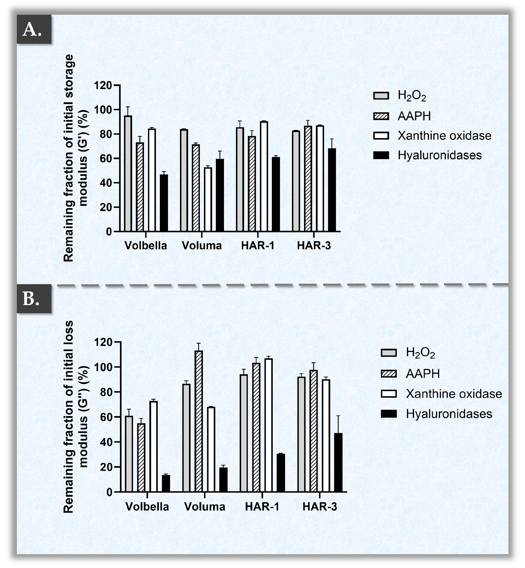
2.3. HAR Hydrogel Multi-Parametric Accelerated Degradation Assays

In vivo, HA hydrogel degradation is mainly mediated by HYAL and ROS. In order to capture a more advanced and thorough understanding of the biophysical attributes of the HAR hydrogels, accelerated product degradation assays were performed. These studies were specifically designed to include standardized orthogonal methods (i.e., oxidant- and enzyme-mediated polymeric system degradation) and to provide quantitative data of enhanced translational relevance. Specifically, while HYAL- or H2O2-based in vitro hydrogel challenge assays are of use for basic product benchmarking, such experimental designs fail to comprehensively mimic or approximate the multi-parametric catabolic drivers at play in vivo [21,26]. The HAR-1, HAR-3, and the two JUVÉDERM® control products were therefore parallelly submitted to simulated in vitro challenge using H2O2, AAPH, hyaluronidases, or xanthine oxidase sources. Interestingly, distinctive endpoint rheological behaviors were evidenced for the respective hydrogel systems, depending on the specific challenge item (Figure 2).
As previously mentioned, two major mechanisms that contribute to the degradation of HA-based gels are oxidative reactions and enzymatic catabolism (i.e., primarily via hyaluronidases). Specifically, the degradation of HA-based gels is a result of the cleavage of hyaluronic acid chains, leading to a decrease in the rheological attribute values (e.g., G′ and G″ moduli) of the system [13,35,39,40,41]. Firstly, when considering the reduction in storage moduli G′ associated with exposition to H2O2, the compared products did not behave significantly differently (Figure 2A). Contrastingly, the remaining fractions of the loss moduli G″ were found to be significantly lower for the VOLBELLA® product as compared to the other tested products (Figure 2B, Table S3). Of note, H2O2 is the most common and simplest ROS generator to be used when evaluating the in vitro oxidative degradation of HA [25].
Secondly, when comparing the reductions in storage moduli G′ induced by AAPH (i.e., a free radical generator) exposure, HAR-3 surprisingly exhibited significantly higher remaining fractions as compared to VOLUMA®, VOLBELLA®, and HAR-1 (Figure 2A, Table S2). Furthermore, VOLUMA® was recorded as the most sensitive formulation in terms of storage modulus G′ under the AAPH challenge (Figure 2A). Then, when analyzing the remaining fractions of loss moduli G″, VOLBELLA® exhibited significantly lower viscosity values as compared to HAR-1, HAR-3, and VOLUMA® products (Figure 2B, Table S3). Surprisingly, the ratio of G″ to G′ (i.e., tan δ values, elasticity proxy of the material) of the VOLUMA® product increased by 63% after exposure to AAPH. Such experimental tan δ values were the highest for the VOLUMA® samples.
Thirdly, when comparing the extent of the oxidative degradation induced by the mixture of xanthine/xanthine oxidase, HAR-1 and HAR-3 displayed significantly higher remaining fractions of G′ and G″ (Figure 2, Tables S2 and S3). These results indicated superior resilience of the HAR hydrogels against xanthine oxidase as compared to VOLUMA® (i.e., close to 1.7 times for G′ and close to 1.3 times for G″, Figure 2, Tables S2 and S3). Interestingly, VOLBELLA® displayed similar extents of degradation to those of both HAR formulations for G′ but similar degradation to that of VOLUMA® for G″ (Figure 2). Overall, the presented results confirmed oxidant-specific catabolic behaviors of the investigated cross-linked HA hydrogels (Figure 2). Therefore, the use of several oxidative challenge items is highly recommended in accelerated stability studies or in simulations of in vivo product degradation to comprehensively characterize the resilience of a given system.
In vivo, free radicals are generated as radicals of lipids, proteins, and DNA alongside various ROS, which are also categorized as free radicals (e.g., superoxide anion [O2], hydroxyl radical [•OH], nitric oxide radical [•NO], and peroxynitrite anion [ONOO]). Oxidative stress is characterized by an elevated level of ROS generation and the presence of other oxidative species, which exceed the capacity of the cellular antioxidant mechanisms to neutralize them [44]. Xanthine oxidase is an enzyme that catalyzes the production of the superoxide anion (O2•−) and which is expressed in skin cells [44,62,63]. AAPH is a water-soluble compound primarily used as a source of peroxyl radicals (ROO•). It is commonly employed to induce oxidative stress and cellular senescence in skin cells or as an oxidation model in various studies [64,65,66]. Hydrogel product rheological attributes, particularly the storage modulus G′, influence the in vivo performance of dermal fillers. Namely, rheology provides a straightforward and rapid method to assess a product’s resistance to oxidative stress by using the parameters G′ and G″ and an appropriate oxidant source.
Fourthly, a few significant differences were evidenced between the groups in residual endpoint storage modulus G′ values following HYAL exposure (Figure 2A, Table S2). Therein, HAR-3 displayed higher remaining fractions of storage modulus G′ as compared to the other tested products (Figure 2A). Of note, VOLBELLA® displayed the most extensive enzymatic degradation, as it was the only product with a remaining average fraction of storage modulus G′ below 50% (Figure 2A). Interestingly, more pronounced differences were recorded between the groups in terms of residual loss modulus G″ (Figure 2B, Table S3). Therein, HAR-3 exhibited higher resistance towards HYAL, and VOLUMA® showed lower resistance (Figure 2B). Notwithstanding the reported results on product resilience toward enzymatic degradation, it is underscored that the performance and longevity of HA-based fillers are more closely associated with the storage modulus G′ (Figure 2) [24,25].
Interestingly, HAR-1 and HAR-3 exhibited similar remaining rheological fraction values in oxidative environments (i.e., low dispersion of values between assay types for given products) despite their differences in formulation (Figure 2, Table 1 and Table 2). Namely, both HAR products displayed remaining fractions > 78% for both moduli following exposure to three different ROS sources (Figure 2). In contrast, VOLBELLA® displayed a disparity in sensitivity to the various ROS sources, with values dropping as low as approximately 52% (Figure 2). Generally, it was set forth that the HAR hydrogels presented enhanced global resilience towards oxidant-mediated degradation as compared to the JUVÉDERM® control products.
Vitamin B3 is known for its antioxidant properties [52,53,67]. However, the presence of an antioxidant molecule does not necessarily equate to the effective protection of HA-based gels from degradation under oxidative stress. For instance, vitamin C is recognized for its potent antioxidant capacity but can significantly degrade HA [68]. Of note, the precise mechanism of interaction between vitamin B3 and HA polymers under high concentrations of H2O2, AAPH, or xanthine oxidase is not yet fully understood. This mechanism may be influenced by hydrogen bonding and a limiting effect on HA chain breakdown by vitamin B3.
Overall, the gathered experimental results confirmed that cross-linked HA hydrogel formulations containing vitamin B3 benefit from a protective effect against oxidative stress but not against the action of hyaluronidases (Figure 2). Such datasets confirmed the technical rationale of incorporating vitamin B3 in the HAR formulations to benefit from its polyvalent antioxidant attributes and effects. Namely, vitamin B3 may be considered as a product stability and performance enhancer due to its ancillary internal action on the cross-linked hydrogel system. From a methodological viewpoint, the use of close commercial comparators (i.e., the JUVÉDERM® control products, technically similar to the HAR systems but without vitamin B3) enabled us to specifically experimentally study the functional impacts of vitamin B3 incorporation. However, further research is warranted, notably around the exact mechanisms of chemical interaction and incorporation of vitamin B3 in the HA polymer network (Figure S2). The unraveling of such mechanisms could potentially shed more light on the overall functional contributions of vitamin B3 in HA-based hydrogels.
Finally, this was the first study to use advanced rheological analysis to assess dermal filler degradation by multiple ROS, offering new insights into the stability and resilience of HA-based dermal fillers under complex oxidative stress. Therefore, this contribution addressed significant gaps in the understanding of dermal filler degradation and introduced innovative methodologies to the field.

2.4. HAR Hydrogel System Cohesivity and Injectability Assays

In order to further functionally characterize the hydrogel systems of interest, cohesivity quantifications were performed. This biophysical attribute characterizes how a dermal filler behaves in the form of a gel deposit once it is injected and subjected to mechanical forces and constraints in the body. Of note, when high compression is applied to a low-cohesivity gel, there is a risk of detachment/separation from the original deposit location, which can potentially result in adverse dermal filler migration. Contrastingly, when high compression is applied to a high-cohesivity gel, the deposit resists displacement more easily and retains its original shape [25,27].
From a dermal filler formulation standpoint, system cohesivity is a function of both the HA concentration and the degree of polymer cross-linking. Specifically, using the same cross-linking technology, the action of increasing either the HA concentration or the HA cross-linking degree generally leads to increases in system cohesivity. For the experimental assay, the drop-weight method was retained, as there was a good correlation with the perceived cohesion attributes of the hydrogels (Figure 3) [27].
Importantly, drop-weight cohesivity values are dependent upon hydrogel extrusion speed and the dimensions of the needle. Quantitatively, with a 30 G needle and a constant plunger speed of 12 mm·min−1, the mean drop-weight values of HAR-1 and VOLBELLA® were almost identical (Figure 3A). In the case of HAR-3, however, mean values 1.5 times higher than those of VOLBELLA® and HAR-1 were recorded (Figure 3A). Of note, no experimental values were obtained for the VOLUMA® product in this setup because it did not form drops but a continuous filament (Video S1). A possible technical explanation for this observation is the presence of a linear HA phase in the VOLUMA® hydrogel system. Specifically, adding and mixing a linear HA phase to a cross-linked product notably facilitates its injectability. However, no detailed quantitative aspects of this linear HA phase are available, as manufacturers do not generally disclose HA molecular weight ranges, degrees of cross-linking, or linear HA phase contents.
Quantitatively, with an 18 G needle and a constant plunger speed of 7.5 mm·min−1, the mean drop-weight values of HAR-1 and VOLUMA® were almost identical (Figure 3B). Therein, the cohesivity values of VOLBELLA® were significantly lower than those of HAR-1 (i.e., 16.6 mg per drop for VOLBELLA® versus 19.5 mg per drop for HAR-1, Figure 3B, Table S5). Of note, HAR-3 presented a significantly higher average drop-weight as compared to the other products, confirming that the HAR-3 product is characterized by superior cohesiveness (Figure 3A,B, Table S5). Importantly, the use of 18 G needles and an extrusion speed of 7.5 mm·min−1 are standard parameters that are frequently reported in the literature. The experimental values obtained for VOLUMA® and VOLBELLA® were almost identical to those previously published [27]. Interestingly, as compared to VOLUMA®, HAR-1 contains a lower hyaluronic acid concentration, presents lower G′ values, uses less BDDE and a lower degree of modification, but exhibits higher cohesiveness values (Figure 3, Table 1 and Table 2). These results confirmed the potential role of vitamin B3 as a potent stabilizer of the HA matrix, which acts by enhancing the internal adhesion forces that hold together the individual cross-linked HA units composing the system via hydrogen bonds. Overall, the HAR-1 and HAR-3 formulations allow for a reduction in the amount of incorporated BDDE while maintaining high cohesivity properties.
Regarding the injectability of the HAR hydrogels, both products showed high homogeneity in their injection force profiles at a relatively high speed of injection (Figure 3C,D). The injection force profile of an empty syringe (i.e., without any product) served as a control, and a representative dataset is presented in Figure S5. For optimal data comparability, the experimental setup of this test was identical to that of a previous study by the authors [69]. Of note, HAR-1 demonstrated significantly higher injection forces as compared to HAR-3 (Figure 3C,D, Table 3).
This difference was notably attributed to HAR-1 being administered using a 27 G needle, whereas HAR-3 was injected using a 30 G needle (i.e., 1.3 times narrower than 27 G needles). Nevertheless, in comparison with previously reported injection force profiles, those of HAR-1 and HAR-3 did not show any plateau heterogeneity (i.e., achievement of seamless injectability), wherein the three experimental replicates overlapped (Figure 3C,D) [69].
Interestingly, in terms of average plateau injection force, the HAR-3 product fell within the same range as the VOLBELLA® product (i.e., 20–25 N) and the HAR-1 product fell within the same range as the VOLUMA® product (i.e., 40–50 N; Figure 3C,D) [69]. Of note, the plateau injection force does not linearly correlate with the HA concentration or the needle gauge, suggesting that the product extrusion force is likely determined by a complex interplay of factors [69]. Such factors include the HA concentration, needle gauge, formulation parameters, degree of cross-linking, percentage of HA linear phase, and the specific technology used during product manufacturing. Importantly, hydrogel injectability is an important functional attribute of dermal fillers [69]. Indeed, various issues may occur in cases of non-homogeneity during the viscous regime (i.e., linear injection phase) of the injection process. Specifically, hydrogel system non-homogeneity can lead to an uneven in situ distribution of the product, potentially resulting in undesirable events (e.g., Tyndall effect) and uneven aesthetic outcomes [29].

2.5. HAR Hydrogel Bio-Stimulatory Attribute Assessment in a Cutaneous Cell Model

The first part of the study enabled us to comparatively characterize several hydrogel intrinsic attributes or the internal effects of the incorporated excipients (i.e., wide-spectrum protective antioxidant action of vitamin B3; Figure 1, Figure 2 and Figure 3). Then, the second part of the study was designed to assess the external effects or the interactions of the investigated products with relevant biological entities. Therefore, based on the clinical indications of the HAR-1 and HAR-3 products, an in vitro primary dermal fibroblast model was established. Similarly to the first part of the study, the cell-based assays were designed around the most relevant controls and commercial reference products available. Therefore, in the context of bio-stimulatory effect investigation, gold-standard commercial bio-stimulants such as Radiesse® and Sculptra™ were included for analysis (Table 4).
Specifically, the Radiesse® and Sculptra™ comparator products were retained because they have been clinically used for several years, have displayed clinical efficacy results, and were historically assessed as being safe for human use. Based on professional market reviews, Radiesse® and Sculptra™ were determined to be the best available options for bio-stimulant treatments on the market, which is why they were included as positive controls in the study. Overall, this choice of commercial comparators contributed to enhancing the translational relevance of the study and was preferred over the use of PLLA or CaHA chemical reference standards, for example.
Firstly, the cellular viability of primary dermal fibroblasts that were directly exposed to the investigated products was assessed in a WST-1 mitochondrial activity assay. Therein, all of the included product groups retained viability after 96 h of incubation, with only the PBS control showing a decrease in average cell viability (i.e., 63.5%; Figure 4A).
Interestingly, the two reference products, which are classified as bio-stimulators (i.e., Radiesse® and Sculptra™), demonstrated higher cellular viability values than those based on HA (i.e., HAR-1, HAR-3, and VOLUMA®; Figure 4A). Specifically, Radiesse® showed the highest cellular metabolic activity levels, followed by Sculptra™ (Figure 4A). Contrastingly, VOLUMA®, HAR-1, and HAR-3 were in the same range and did not show significant differences among them (Figure 4A, Table S6).
Generally, the observed results of cellular viability/metabolic activity could be explained by the differential formulation attributes of the investigated products (Figure 4A, Table 4). Specifically, the CaHA and PLLA particles, respectively, contained in Radiesse® and Sculptra™, are known to stimulate cellular activity [16,17,18,19]. Furthermore, the results related to cross-linked HA were consistent with the literature [25]. However, it is important to note that the tests were conducted on large multi-well plates due to the difficulty of viscous product removal in smaller wells. Specifically, Radiesse® was particularly difficult to eliminate, as it adhered to the bottom and sides of the assay well (Figure S6). Therefore, Radiesse® syringes were diluted five-fold to avoid potential bias during the analyses, especially for colorimetric readouts (Table 4). Additionally, to ensure uniformity of endpoint analysis, all wells were rinsed six times with 0.5 mL of PBS before further processing.
As regards total proteins and collagen quantification, HAR-1 and HAR-3 stimulated their production more effectively than VOLUMA® and PBS (Figure 4B,C). Such results could notably be attributed to the action of vitamin B3, which is well-known for stimulating collagen production, a keynote of skin repair and regeneration [52,53,67]. Furthermore, despite their differences in HA contents, no significant differences were observed in terms of total proteins and collagens between HAR-1 and HAR-3, underscoring the major role of vitamin B3 in collagen and total protein synthesis stimulation (Figure 4B,C, Table 1, Table S7 and S8). When compared to the reference bio-stimulators, HAR-1 and HAR-3 indicated a trend towards higher collagen concentrations, but no significant differences with Sculptra™ were found (Figure 4B, Table S7).
Generally, the dual functionalization of cross-linked HA-based hydrogels by vitamin B3 necessitated dual characterization, with respective standards or commercial comparators for both aspects (i.e., physical attributes and bio-stimulation effects). Therein, Radiesse® and Sculptra™ were selected as comparators for the bio-stimulation assays due to their international and widespread clinical use as injectable bio-stimulators. Interestingly, HAR-1 and HAR-3 demonstrated significantly greater collagen production induction than Radiesse® and showed a superior trend to Sculptra™ (Figure 4B, Table S7). In terms of total proteins, Radiesse® exhibited comparative values within the same range as both HAR products, while Sculptra™ showed a trend of lower values (Figure 4C, Table S8). Importantly, despite significantly higher cellular activity levels for Radiesse® as compared to the two HAR products, the collagen quantity was significantly greater for the latter (Figure 4A,B, Tables S6 and S7). Therefore, the reported results confirmed that both of the HAR products boosted fibroblasts to produce more collagen, yet they did not exert significant effects on cellular metabolic activity (i.e., a characteristic of classical bio-stimulants; Figure 4, Figure S7).
Of note, the tested primary dermal fibroblasts retained their characteristic morphology in vitro following incubation with the investigated products (Figure S8). Previously, vitamin B3/niacinamide was proven to prevent cellular senescence (i.e., modeled by a decrease in protein levels, including collagen/elastin) in fibroblast models [67,70]. The anti-senescence effects of niacinamide largely stem from its anti-inflammatory properties, which include reducing inflammatory mediators and inhibiting mast cell degranulation [67,71,72]. Additionally, as a precursor to NAD+, niacinamide helps to prevent the depletion of cellular energy and regulates the cell’s redox status, both of which are essential for collagen synthesis [53,67,73]. Overall, the gathered experimental results demonstrated that the vitamin B3 present in HAR-1 and HAR-3 conferred multifactorial benefits to the HAR hydrogel systems, notably in terms of potent oxidative resilience and pro-collagenesis (Figure 2 and Figure 4).
From a specific mechanistic viewpoint, the bio-stimulant actions of CaHA, PLLA, and vitamin B3 were well-described in the literature, among which the aforementioned “skin collagen enhancement” attributes. Notably, the resorbable PLLA of the Sculptra™ stimulatory filler was described to exert its effects by promoting neo-collagenesis upon injection [19]. The detailed mechanism of action is thought to comprise the induction of a foreign-body reaction characterized by increased macrophage, mast cell, and lymphocyte presence. In turn, this cellular response is set forth to slowly degrade the product, increase fibroblastic activity, and result in gradual neo-collagenesis [74]. By immunofluorescence staining, Stein et al. confirmed that PLLA-induced augmentation is most likely based on capsule formation orchestrated by macrophages, (myo-)fibroblasts, and collagen type I and III fibers [75].
As regards the CaHA microspheres in Radiesse®, in vitro analyses showed a significant increase in COLIII expression by dermal fibroblasts, which was 123% higher than baseline at 24 h post-incubation. Interestingly, this enhanced expression of COLIII was observed only in fibroblasts that were in direct contact with CaHA, indicating that the recorded increase in collagen production was rather due to the activation of more fibroblasts than to a change in the rate of cellular expression [76]. Furthermore, a systematic review by Amiri et al. compiled data on the skin regeneration mechanisms related to CaHA, focusing particularly on neo-collagenesis. The authors highlighted supporting evidence that demonstrated that collagen production increased in proportion to the amount of applied CaHA, further substantiating the potential of CaHA in skin regenerative applications [77].
Finally, concerning the activity of vitamin B3, we recently described that the intrinsic antioxidant activity of the molecule contributes to cellular homeostasis, thereby maintaining skin ECM integrity [67]. As mast cell degranulation leads to fibroblast senescence, vitamin B3 contributes to normal fibroblast activity maintenance by its stabilizing effects. Of note, vitamin B3 also inhibits MMPs due to its antioxidant and anti-inflammatory activities. Furthermore, it was shown that vitamin B3 inhibits elastase activity, preserving the integrity of elastin in cutaneous ECM [67]. Overall, while the reported results did not mechanistically describe the exact bio-stimulatory pathway of the considered HAR hydrogel systems, strong parallels could be made with previous reports on the beneficial effects of selected ingredients. Interestingly, the reported functional performance of the HAR systems in experimental benchmarking with PLLA- and CaHA-based products confirmed their significant intrinsic bioactivity in a relevant cutaneous cell-based model (Figure 4). Thus, the present study set forth several datasets confirming the multiple functionalization roles of vitamin B3 and supporting its use as a versatile, functional ingredient in medicalized skincare.

2.6. Study Limitations and Future Perspectives

As regards the main identified limitations of the present study, it was outlined that the reported results were limited to in vitro setups. Specifically, injecting the HAR products into ex vivo skin explants (e.g., abdominoplasty sections, porcine tissues) would further enable us to observe and compare their tissue integration properties. Additionally, semi-quantitative evaluation of collagen contents and quality via appropriate staining (e.g., Picrosirius Red, Masson’s Trichrome) would then be possible. A second technical limitation of the present study concerned the cell-based assays, which did not include orthogonal tests, notably for collagen quantification (e.g., by proteomics, ELISA). A third technical limitation concerned the low number of commercial comparators to the HAR gels, particularly cross-linked HA gels. Therein, alternative products from other well-established brands (e.g., Teoxane, Merz Aesthetics, or Vivacy) could have been used as comparators. Specifically, as the cross-linking techniques with BDDE are relatively similar, it would have been interesting to compare a hydrogel with a degree of cross-linking that is as close as possible to that of the HAR products.
Regarding future perspectives on this research, the HAR hydrogels could be injected subcutaneously in rats or intradermally in rabbits in order to monitor the in vivo mid-to-long-term performance of the products. Additionally, the HAR hydrogels could then be the subjects of a prospective clinical study, wherein cutaneous biopsies at various time-points would be performed on patients in order to quantify the collagen produced over time under real-world conditions. Finally, vitamin B3 could be formulated in alternative hydrogels in order to further study its intrinsic properties and effects from multi-parametric functional standpoints.

3. Conclusions

The results reported herein for the HAR-1 “Instant Refine” and HAR-3 “Maxi Lift” dermal fillers experimentally confirmed several functional impacts of vitamin B3 incorporation. The observed resilience of the HAR hydrogels (H2O2, AAPH, and xanthine oxidase assays) was interpreted as being highly beneficial from a post-administration system stability standpoint. Such attributes were specifically linked to the incorporation of vitamin B3 in the HA polymeric network, yielding a significant protective effect. Furthermore, the significant promotion of collagen production by dermal fibroblasts treated with HAR-1 and HAR-3 confirmed the intrinsic bio-stimulatory effects of both formulas, which differed from those of Radiesse® and Sculptra™ comparators. The retained experimental methodologies of the study enabled to obtain characterization datasets of high translational relevance. Overall, the dual functionalization role of vitamin B3 in cross-linked HA-based dermal fillers was confirmed herein, providing a polyvalent treatment option (i.e., combined volumizer and bio-stimulant) for medical aesthetic professionals.

4. Materials and Methods

4.1. Reagents and Consumables Used for the Study

The reagents and consumables used in the present study are listed hereafter along with the corresponding manufacturers. The HAR-1 “Instant Refine” and HAR-3 “Maxi Lift” commercial dermal filler products were obtained from LOUNA AESTHETICS (Poisy, France). A 1 mL syringe of HAR-1 contained 15 mg of HA, 7.5 mg of vitamin B3, and 3 mg of lidocaine. A 1 mL syringe of HAR-3 contained 20 mg of HA, 7.5 mg of vitamin B3, and 3 mg of lidocaine. The initial HA in the HAR-1 and HAR-3 formulations had an intrinsic viscosity of 2.5 m3/kg. The JUVÉDERM® VOLBELLA® and VOLUMA® products were purchased from Allergan Aesthetics (Annecy, France). The Radiesse® product was purchased from Merz Aesthetics (Plan-les-Ouates, Switzerland). The Sculptra product was purchased from Galderma (Zug, Switzerland). Hyaluronidase from bovine testes (i.e., type VI-S), Collagen Assay Kits (i.e., reference MAK332), and BCA Protein Assay Kits were purchased from Sigma-Aldrich (St. Louis, MO, USA). Pharmaceutical-grade purified water and sterile phosphate-buffered saline (PBS) solutions were purchased from Laboratorium Dr. G. Bichsel (Unterseen, Switzerland). TrypLE™, DMEM, and FBS cell culture reagents were purchased from Life Technologies (Thermo Fisher Scientific, Waltham, MA, USA). Cell culture surfaces and disposable plastics were purchased from Greiner (Frickenhausen, Germany). Penicillin-streptomycin was obtained from the CHUV pharmacy (Lausanne, Switzerland). Except for the commercial analytical kits, all the reagents and consumables that were used in the study were procured in sterile form.

4.2. Hydrogel Rheological Characterization Method

In order to obtain quantitative and comparative information about important bio-physical attributes of the investigated hydrogels, a standard rheological setup was used. The basic rheological profiles of the selected commercial products were experimentally determined in oscillatory rheology using an HR 10 rheometer (TA Instruments, Guyancourt, France) equipped with a Peltier plate-plate. The storage modulus (G′), loss modulus (G″), and complex viscosity (η*) values of the samples were determined as a function of the applied oscillatory frequency. To this end, a frequency sweep was performed from 0.1 Hz to 10 Hz at a constant temperature of 25 °C. All measurements were then performed at 1 Hz and at 25 °C on volumes of 600 μL for the hydrogel samples and for the control groups. For the analyses, undiluted samples were dispensed on the baseplate of the rheometer and were submitted to controlled compression and shear stress by the instrument sensor plate. The shear stress was set at 3 N/m2 in all experiments to respect the linear viscoelastic region (LVE). Tan delta (tan δ) values were calculated from experimental G′ and G″ results (i.e., G″/G′ ratio). A sample hood was used during the rheological measurements to minimize sample water evaporation. The experimental storage moduli (G′) and loss moduli (G″) values of the samples were determined by the instrument using three experimental replicates for all of the assays. In detail, each replicate was obtained from a new hydrogel product syringe.

4.3. Hydrogel Accelerated Degradation Assays with Rheological Readouts

In order to study the breakdown of the hydrogel systems of interest (i.e., as a proxy for product resilience), various in vitro accelerated degradation assays were performed. The assays were specifically designed to compare the breakdown behaviors of the products in various setups. Firstly, the accelerated oxidative degradation of the included dermal fillers was assessed by means of rheology. Therefore, a volume of 300 µL of 30% H2O2, a volume of 300 µL of 2% 2,2’-azobis(2-amidinopropane) dihydrochloride (AAPH), or a volume of 300 µL of xanthine/xanthine oxidase mixture was added to 300 µL of the hydrogel sample. By direct contact, the various oxidant sources reacted with the samples and started to break down the polymer networks. The reaction tubes were then incubated at 37 °C under gentle shaking for 10 min before rheological readout. Following this standardized challenge period, the storage (G′) and loss (G″) moduli of the hydrogels were measured on an HR 10 rheometer (TA Instruments, Guyancourt, France) equipped with a Peltier plate-plate on 600 µL of sample. The constant oscillatory frequency was set at 1 Hz, and the assay temperature was set at 22 °C. The shear stress was set at 3 N/m2 in all experiments to respect the linear viscoelastic region (LVE). Tan delta (tan δ) values were calculated from experimental G′ and G″ results (i.e., G″/G′ ratio). A sample hood was used during the rheological measurements to minimize sample water evaporation. Three experimental replicates were used for all of the assays.
Secondly, the accelerated enzymatic degradation of the included dermal fillers was assessed by means of rheology. Therefore, a volume of 300 µL of 100 U/mL hyaluronidase was added to 300 µL of the hydrogel sample. By direct contact, the enzyme reacted with the samples and started to break down the polymer networks. The reaction tubes were then incubated at 37 °C under gentle shaking for 10 min before rheological readout. Following this standardized challenge period, the storage (G′) and loss (G″) moduli were measured on an HR 10 rheometer (TA Instruments, Guyancourt, France) equipped with a Peltier plate-plate on 600 µL of sample. The constant oscillatory frequency was set at 1 Hz, and the assay temperature was set at 22 °C. The shear stress was set at 3 N/m2 in all experiments to respect the linear viscoelastic region (LVE). A sample hood was used during the rheological measurements to minimize sample water evaporation. Three experimental replicates were used for all of the assays.

4.4. Hydrogel Cohesivity Analysis Method

In order to comparatively study the in situ behavior of the various hydrogels from another bio-physical attribute standpoint, the cohesiveness or cohesivity of the products was quantitatively assessed. Sample cohesivity attributes were determined using a standard drop-weight method. Therefore, the weight of an average fragment/drop of undiluted HA hydrogel was experimentally determined for each of the included dermal fillers using an 18 G or a 30 G needle. In detail, an automated setup was used to extrude the hydrogels drop-by-drop, in view of weighing each drop (i.e., ponderal proxy of system cohesivity). Therefore, the product syringe was mounted on the baseplate of a Texture Analyzer TA.XT. Plus instrument (Tracomme, Schlieren, Switzerland). The syringe plunger rod was automatically actuated by the instrument at a constant downward speed of 7.5 mm·min−1 or 12 mm·min−1. During this process, the hydrogels were automatically extruded into ambient air. When a constant extrusion force was attained (i.e., plateau extrusion force), at least 6 fragments/drops were collected and weighed to determine the average weight per drop. Three experimental replicates and three repetitions were used for all of the assays.

4.5. Hydrogel Injectability Analysis Method

In order to confirm that the considered hydrogels can be administered using standard medical equipment and normal operator strength, product injectability assays were performed. Standardized dermal filler injectability measurements were performed at a constant speed on a Texture Analyzer TA.XT. Plus instrument (Tracomme, Schlieren, Switzerland). An automated setup was used to continuously measure the force required for hydrogel product injection (i.e., the force that is applied on the plunger rod hilt). Therefore, the product syringe was mounted on the baseplate of the Texture Analyzer instrument. The injection force profiles of both HAR products were determined using the original syringes, with 30 G needles for HAR-1 and 27 G needles for HAR-3. The products were automatically injected by the instrument into a synthetic skin substitute (i.e., SimSkin® from Wallcur™, San Diego, CA, USA) at a relatively fast and constant piston speed of 60 mm·min−1 at ambient temperature. Plateau injection forces were determined using force values acquired between 50% and 85% of the piston travel distance. Three experimental replicates were used for all of the assays. In detail, each replicate was obtained from a new hydrogel product syringe.

4.6. Product Biological Effect Evaluations in an In Vitro Dermal Fibroblast Model

4.6.1. In Vitro Assessment of Cellular Viability/Metabolic Activity

In order to first assess the cytocompatibility/toxicity of the products of interest, these were incubated in direct contact with primary cells. In detail, primary dermal fibroblasts were retained for the study, as they are the most relevant target cellular population for dermal fillers (i.e., cells present in dermal tissues). Human primary dermal fibroblasts were grown in vitro in culture flasks containing Dulbecco’s modified Eagle medium (DMEM), supplemented with 10% fetal bovine serum (FBS) and 1% antibiotic–antimycotic solution. The cell cultures were incubated at 37 °C in a humidified atmosphere under 5% CO2 and were monitored daily using an inverted microscope. Subcultures were performed when a confluency level of 80% was observed. Therefore, the confluent fibroblasts were transferred to a 24-well plate (i.e., 104 cells/well). After 24 h, the culture medium was replaced with a mix of hydrogel sample:medium at a 1:1 ratio. The cells were treated with the test-items for up to 5 days of incubation. Then, the adherent cells were rinsed and tested for viability/metabolic activity using the cell proliferation reagent WST-1 according to the specifications of the manufacturer. The assay was carried out four times, using four experimental replicates.

4.6.2. In Vitro Assessment of Collagen Synthesis Stimulation Activity

In order to secondly assess the potential of the investigated products to stimulate collagen synthesis in the target cell population, endpoint quantifications of total collagen levels in the co-cultures (i.e., cell monolayers in contact with the test-items) were performed. For 96 h collagen quantification, the harvested cells were seeded into 24-well culture plates at a density of 105 cells per well. After 24 h, the culture medium was replaced with 1000 µL of a mix of hydrogel sample:medium at a 1:1 ratio. The assay was carried out four times, using four experimental replicates. After 96 h of incubation, the supernatant was removed, and the wells were washed multiple times with PBS. The cells were then detached with TrypLE™ and hydrolyzed through cycles of freezing and thawing in order to measure the total collagen contents by fluorescence with the Collagen Assay Kit. Firstly, the collagen in the samples was enzymatically digested into peptides. Subsequently, the N-terminal glycine-containing peptides reacted with the dye reagent to form a fluorescent complex. The fluorescence intensity of this product, which was measured at λex = 375/λem = 465 nm on a Varioskan™ LUX (Thermo Fisher Scientific, Waltham, MA, USA), was directly proportional to the total collagen concentration in the samples.

4.6.3. In Vitro Assessment of Total Protein Synthesis Stimulation Activity

In order to thirdly assess the potential of the investigated products to stimulate protein synthesis in the target cell population, endpoint quantifications of total protein levels in the co-cultures (i.e., cell monolayers in contact with the test-items) were performed. Following the same procedure as for total collagen quantification, total protein concentrations after 96 h of incubation of the fibroblasts with the test-items were determined using a BCA assay kit. Bovine serum albumin (BSA) protein standards consisted of known concentrations of BSA and were prepared in a range of 200–1000 μg/mL. Following sample treatment with the assay reagent, the optical density at 562 nm was determined spectrophotometrically on a Varioskan™ LUX (Thermo Fisher Scientific, Waltham, MA, USA). The assay was carried out four times, using four experimental replicates.

4.7. Statistical Analyses and Data Presentation

The experimental data were reported herein as mean values and standard deviations, which were plotted as error bars in the graphs. For the statistical comparison of values from multi-group quantitative datasets, a one-way ANOVA test or a two-way ANOVA test was performed and was followed by a post hoc Tukey’s multiple comparison test. A p-value < 0.05 was retained as a general base for statistical significance determination. Detailed levels of statistical significance may be found in the Results section and in the Supplementary Tables. The statistical calculations and/or data presentation were performed using Microsoft Excel 365 (Microsoft Corporation, Redmond, WA, USA), Microsoft PowerPoint 365, and GraphPad Prism v.8.0.2 (GraphPad Software, San Diego, CA, USA).

Supplementary Materials

The following supporting information can be downloaded at: https://www.mdpi.com/article/10.3390/gels10060361/s1, Figure S1: Supporting data for the rheological analyses; Figure S2: Supporting data for chemical mechanism investigation; Figure S3: Supporting data for the rheological analyses; Figure S4: Illustration of the investigated hydrogels; Figure S5: Supporting data for the injectability analyses; Figure S6: Illustration of the in vitro cell-based functional assay; Figure S7: Mechanistic explanation of the in vitro cell-based functional assay; Figure S8: Data illustrations from the in vitro cell-based functional assay; Table S1: Statistical analysis details; Table S2: Statistical analysis details; Table S3: Statistical analysis details; Table S4: Statistical analysis details; Table S5: Statistical analysis details; Table S6: Statistical analysis details; Table S7: Statistical analysis details; Table S8: Statistical analysis details; Video S1: Illustration of the drop-weight cohesivity determination setup.

Author Contributions

Conceptualization, A.P., M.C., C.S., N.H.-B., L.A.A. and A.L.; methodology, A.P., C.M., C.S., N.H.-B., L.A.A. and A.L.; software, A.P. and A.L.; validation, A.P., M.C., C.M., C.S., K.L., P.A.-S., W.R., N.H.-B., L.A.A. and A.L.; formal analysis, A.P., M.C., K.L., N.H.-B., L.A.A. and A.L.; investigation, A.P., C.S., K.L. and A.L.; resources, A.P., L.A.A. and A.L.; data curation, A.P., M.C., C.M., K.L., N.H.-B., L.A.A. and A.L.; writing—original draft preparation, A.P. and A.L.; writing—review and editing, A.P., M.C., C.M., C.S., K.L., P.A.-S., W.R., N.H.-B., L.A.A. and A.L.; visualization, A.P., K.L., L.A.A. and A.L.; supervision A.P., L.A.A. and A.L.; project administration, A.P. and A.L.; funding acquisition, A.P. and A.L. All authors have read and agreed to the published version of the manuscript.

Funding

This study received no external funding and was not supported by specific grants or institutional programs.

Institutional Review Board Statement

The biological starting materials used for the present study were procured according to the guidelines of the Declaration of Helsinki and approved by the appropriate Cantonal Ethics Committee [78]. Obtention and use of patient cellular materials followed the regulations of the Biobank of the CHUV Department of Musculoskeletal Medicine. Materials from patients were included in the study under an Ethics Committee Protocol (Vaud State Ethics Committee, Ref. CHUV [BB_029_DAL]). Specifically, the primary human fibroblasts used for the present study (i.e., DECH-2jM-Fib cell type) were obtained by culture and expansion from an extra finger of a polydactyly patient, which was registered in the Department Biobank, as validated by the Operational Committee of Biobanks and Official Registers of the CHUV.

Informed Consent Statement

Informed consent (i.e., formalized in a general informed consent agreement) was obtained from all patients or from their legal representatives at the time of treatment for unrestricted use of the gathered and anonymized patient data or anonymized biological materials (i.e., donors of primary cell types).

Data Availability Statement

The data presented in this study are openly available within the article files.

Acknowledgments

The authors are thankful to Marco Cerrano for his continued and passionate clinical expertise and feedback. Artwork templates were partly generated with www.biorender.com, accessed on 25 March 2024.

Conflicts of Interest

Authors A.P., C.M., and K.L. were employed by LOUNA REGENERATIVE SA (Geneva, Switzerland) and were consultants for LOUNA AESTHETICS SAS (Poisy, France) during the course of this study. Author A.L. was employed by LAM Biotechnologies SA (Epalinges, Switzerland) and by TEC-PHARMA SA (Bercher, Switzerland) during the course of this study. The remaining authors declare no conflict of interest for this study.

List of Abbreviations

AAPH2,2′-azobis(2-amidinopropane) dihydrochloride
BCABicinchoninic acid
BDDE1,4-butanediol diglycidylether
BSABovine serum albumin
CaHACalcium hydroxylapatite
CHUVLausanne University Hospital
CMCCarboxymethylcellulose
DMEMDulbecco’s modified Eagle medium
ECMExtracellular matrix
FBSFetal bovine serum
FDAUS Food and Drug Administration
G′Storage modulus
G″Loss modulus
HAHyaluronic acid
HARBoosting dermal fillers
HYALHyaluronidase
H2O2Hydrogen peroxide
LVELinear viscoelastic region
MDMedical device
minMinute
NANon-applicable
NMRNuclear magnetic resonance
NONitric oxide
nsNon-significant
PaPascals
Pa·sPascal seconds
PBSPhosphate-buffered saline
PLLAPoly-l-lactic acid
ROSReactive oxygen species
sSecond
SEC-MSSize-exclusion chromatography with mass spectrometry
USAUnited States of America
UVUltraviolet

References

  1. Olczyk, P.; Komosińska-Vassev, K.; Winsz-Szczotka, K.; Kuźnik-Trocha, K.; Olczyk, K. Hyaluronan: Structure, metabolism, functions, and role in wound healing. Adv. Hyg. Exp. Med. 2008, 62, 651–659. [Google Scholar]
  2. Fallacara, A.; Baldini, E.; Manfredini, S.; Vertuani, S. Hyaluronic acid in the third millennium. Polymers 2018, 10, 701. [Google Scholar] [CrossRef] [PubMed]
  3. Saravanakumar, K.; Park, S.; Santosh, S.S.; Ganeshalingam, A.; Thiripuranathar, G.; Sathiyaseelan, A.; Vijayasarathy, S.; Swaminathan, A.; Priya, V.V.; Wang, M.H. Application of hyaluronic acid in tissue engineering, regenerative medicine, and nanomedicine: A review. Int. J. Biol. Macromol. 2022, 222, 2744–2760. [Google Scholar] [CrossRef] [PubMed]
  4. Huynh, A.; Priefer, R. Hyaluronic acid applications in ophthalmology, rheumatology, and dermatology. Carbohydr. Res. 2020, 489, 107950. [Google Scholar] [CrossRef] [PubMed]
  5. Available online: https://www.fda.gov/medical-devices/aesthetic-cosmetic-devices/dermal-fillers-soft-tissue-fillers (accessed on 9 April 2024).
  6. Allergan Aesthetics, Allergan. Instructions for Use of the Juvéderm® Gel Range (Volbella®, Volift®, Voluma®, Juvéderm® Ultra 2, 3); Allergan: Annecy, France, 2022; unpublished work; Available online: https://media.allergan.com/actavis/actavis/media/general/Juvederm-voluma-IFU.pdf (accessed on 3 April 2024).
  7. Merz Aesthetics, Anteis SA. Instructions for Use of the Belotero® Gel Range (Belotero® Soft, Balance, Intense, Volume); Anteis: Plan-les-Ouates, Switzerland, 2015; unpublished work; Available online: https://www.merz.ch/wp-content/uploads/2016/03/BELOTERO_Volume_Lidocaine.pdf (accessed on 23 January 2024).
  8. Teoxane, S.A. Instructions for Use of the RHA® Gel Range (RHA® 1, 2, 3, 4); Teoxane: Geneva, Switzerland, 2015; unpublished work; Available online: https://irp-cdn.multiscreensite.com/e193bd60/files/uploaded/Teosyal-Rha-Dr.-brochure-en-1.pdf (accessed on 25 January 2024).
  9. Sinclair Pharma. Instructions for Use of the MaiLi® Gel Range; Sinclair Pharma Ltd.: London, UK, 2023; unpublished work; Available online: https://maili.com/physicians/maili-an-expert-s-guide/ (accessed on 7 March 2024).
  10. Laboratoires VIVACY. Instructions for Use of the Stylage® Gel Range (Stylage® S, M, L, XL, XXL); Vivacy: Paris, France, 2024; unpublished work; Available online: https://vivacy.com/fr/produits/medecine-esthetique/ (accessed on 23 January 2024).
  11. Faivre, J.; Pigweh, A.I.; Iehl, J.; Maffert, P.; Goekjian, P.; Bourdon, F. Crosslinking hyaluronic acid soft-tissue fillers: Current status and perspectives from an industrial point of view. Exp. Rev. Med. Dev. 2021, 18, 1175–1187. [Google Scholar] [CrossRef] [PubMed]
  12. Pluda, S.; Salvagnini, C.; Fontana, A.; Marchetti, A.; Di Lucia, A.; Galesso, D.; Guarise, C. Investigation of crosslinking parameters and characterization of hyaluronic acid dermal fillers: From design to product performances. Gels 2023, 9, 733. [Google Scholar] [CrossRef] [PubMed]
  13. La Gatta, A.; Salzillo, R.; Catalano, C.; D’Agostino, A.; Pirozzi, A.V.A.; De Rosa, M.; Schiraldi, C. Hyaluronan-based hydrogels as dermal fillers: The biophysical properties that translate into a “volumetric” effect. PLoS ONE 2019, 14, e0218287. [Google Scholar] [CrossRef] [PubMed]
  14. Bokatyi, A.N.; Dubashynskaya, N.V.; Skorik, Y.A. Chemical modification of hyaluronic acid as a strategy for the development of advanced drug delivery systems. Carbohydr. Polym. 2024, 337, 122145. [Google Scholar] [CrossRef] [PubMed]
  15. Sánchez-Carpintero, I.; Candelas, D.; Ruiz-Rodríguez, R. Dermal fillers: Types, indications, and complications. Acta Dermo-Sifilio. 2010, 101, 381–393. [Google Scholar] [CrossRef]
  16. Ray, S.; Ta, H.T. Investigating the effect of biomaterials such as poly-(L-lactic acid) particles on collagen synthesis in vitro: Method is matter. J. Funct. Biomater. 2020, 11, 51. [Google Scholar] [CrossRef]
  17. Attenello, N.H.; Maas, C.S. Injectable fillers: Review of material and properties. Facial Plast. Surg. 2015, 31, 29–34. [Google Scholar] [CrossRef] [PubMed]
  18. Jacovella, P.F. Use of calcium hydroxylapatite (Radiesse) for facial augmentation. Clin. Interv. Aging. 2008, 3, 161–174. [Google Scholar] [CrossRef] [PubMed]
  19. Lacombe, V. Sculptra: A stimulatory filler. Facial Plast. Surg. 2009, 25, 95–99. [Google Scholar] [CrossRef] [PubMed]
  20. Molliard, S.G.; Bétemps, J.B.; Hadjab, B.; Topchian, D.; Micheels, P.; Salomon, D. Key rheological properties of hyaluronic acid fillers: From tissue integration to product degradation. Plast. Aesthet. Res. 2018, 5, 17. [Google Scholar] [CrossRef]
  21. Pierre, S.; Liew, S.; Bernardin, A. Basics of dermal filler rheology. Dermatol. Surg. 2015, 41, S120–S126. [Google Scholar] [CrossRef]
  22. Sparavigna, A.; La Gatta, A.; Bellia, G.; La Penna, L.; Giori, A.M.; Vecchi, G.; Tenconi, B.; Schiraldi, C. Evaluation of the volumizing performance of a new volumizer filler in volunteers with age-related midfacial volume defects. Clin. Cosmet. Investig. Dermatol. 2020, 16, 683–690. [Google Scholar] [CrossRef] [PubMed]
  23. Choi, M.S. Basic rheology of dermal filler. Arch. Plast. Surg. 2020, 47, 301–304. [Google Scholar] [CrossRef] [PubMed]
  24. Santoro, S.; Russo, L.; Argenzio, V.; Borzacchiello, A. Rheological properties of cross-linked hyaluronic acid dermal fillers. J. Appl. Biomater. Biomech. 2011, 9, 127–136. [Google Scholar] [CrossRef]
  25. Gou, S.; Porcello, A.; Allémann, E.; Salomon, D.; Micheels, P.; Jordan, O.; Kalia, Y.N. Injectable hyaluronan-based thermoresponsive hydrogels for dermatological applications. Pharmaceutics 2023, 15, 1708. [Google Scholar] [CrossRef] [PubMed]
  26. Sundaram, H.; Rohrich, R.J.; Liew, S.; Sattler, G.; Talarico, S.; Trévidic, P.; Gavard Molliard, S. Cohesivity of hyaluronic acid fillers: Development and clinical implications of a novel assay, pilot validation with a five-point grading scale, and evaluation of six U.S. Food and Drug Administration-approved fillers. Plast. Reconstr. Surg. 2015, 136, 678–686. [Google Scholar] [CrossRef]
  27. Edsman, K.L.M.; Öhrlund, Å. Cohesion of hyaluronic acid fillers: Correlation between cohesion and other physicochemical properties. Dermatol. Surg. 2018, 44, 557–562. [Google Scholar] [CrossRef] [PubMed]
  28. Kleine-Börger, L.; Meyer, R.; Kalies, A.; Kerscher, M. Approach to differentiate between hyaluronic acid skin quality boosters and fillers based on their physicochemical properties. J. Cosmet. Dermatol. 2022, 21, 149–157. [Google Scholar] [CrossRef]
  29. Wongprasert, P.; Dreiss, C.A.; Murray, G. Evaluating hyaluronic acid dermal fillers: A critique of current characterization methods. Dermatol. Ther. 2022, 35, e15453. [Google Scholar] [CrossRef]
  30. Guo, J.; Fang, W.; Wang, F. Injectable fillers: Current status, physicochemical properties, function mechanism, and perspectives. RSC Adv. 2023, 13, 23841. [Google Scholar] [CrossRef]
  31. Buhren, B.A.; Schrumpf, H.; Bölke, E.; Kammers, K.; Gerber, P.A. Standardized in vitro analysis of the degradability of hyaluronic acid fillers by hyaluronidase. Eur. J. Med. Res. 2018, 23, 37. [Google Scholar] [CrossRef] [PubMed]
  32. Porcello, A.; Laurent, A.; Hirt-Burri, N.; Abdel-Sayed, P.; de Buys Roessingh, A.; Raffoul, W.; Jordan, O.; Allémann, E.; Applegate, L.A. Hyaluronan-based hydrogels as functional vectors for standardized therapeutics in tissue engineering and regenerative medicine. In Nanopharmaceuticals in Regenerative Medicine; CRC Press: Boca Raton, FL, USA, 2022. [Google Scholar]
  33. Stern, R.; Kogan, G.; Jedrzejas, M.J.; Soltés, L. The many ways to cleave hyaluronan. Biotechnol. Adv. 2007, 25, 537–557. [Google Scholar] [CrossRef]
  34. Žádníková, P.; Šínová, R.; Pavlík, V.; Šimek, M.; Šafránková, B.; Hermannová, M.; Nešporová, K.; Velebný, V. The degradation of hyaluronan in the skin. Biomolecules 2022, 12, 251. [Google Scholar] [CrossRef]
  35. Ferraz, R.M.; Sandkvist, U.; Lundgren, B. Degradation of hylauronic acid fillers using hyaluronidase in an in vivo model. J. Drugs Dermatol. 2018, 17, 548–553. [Google Scholar] [PubMed]
  36. Schanté, C.; Zuber, G.; Herlin, C.; Vandamme, T.F. Synthesis of N-alanyl-hyaluronamide with high degree of substitution for enhanced resistance to hyaluronidase-mediated digestion. Carbohydr. Polym. 2011, 86, 747–752. [Google Scholar] [CrossRef]
  37. Flégeau, K.; Jing, J.; Brusini, R.; Gallet, M.; Moreno, C.; Walker, L.; Bourdon, F.; Faivre, J. Multidose hyaluronidase administration as an optimal procedure to degrade resilient hyaluronic acid soft tissue fillers. Molecules 2023, 28, 1003. [Google Scholar] [CrossRef]
  38. Paap, M.K.; Silkiss, R.Z. The interaction between hyaluronidase and hyaluronic acid gel fillers—A review of the literature and comparative analysis. Plast. Aesthet. Res. 2020, 7, 36. [Google Scholar] [CrossRef]
  39. Šoltés, L.; Mendichi, R.; Kogan, G.; Schiller, J.; Stankovska, M.; Arnhold, J. Degradative action of reactive oxygen species on hyaluronan. Biomacromolecules 2006, 7, 659–668. [Google Scholar] [CrossRef] [PubMed]
  40. Hrabárová, E.; Valachová, K.; Juránek, I.; Soltés, L. Free-radical degradation of high-molar-mass hyaluronan induced by ascorbate plus cupric ions: Evaluation of antioxidative effect of cysteine-derived compounds. Chem. Biodivers. 2012, 9, 309–317. [Google Scholar] [CrossRef] [PubMed]
  41. Darsy, G.; Patarin, J.; Conrozier, T. Large variations in resistance to degradation between hyaluronic acid viscosupplements: A comparative rheological study. Cartilage 2023, 19476035231205696. [Google Scholar] [CrossRef] [PubMed]
  42. Barygina, V.; Becatti, M.; Lotti, T.; Moretti, S.; Taddei, N.; Fiorillo, C. ROS-challenged keratinocytes as a new model for oxidative stress-mediated skin diseases. J. Cell. Biochem. 2019, 120, 28–36. [Google Scholar] [CrossRef] [PubMed]
  43. Alsharabasy, A.M.; Glynn, S.; Farràs, P.; Pandit, A. Interactions between nitric oxide and hyaluronan implicate the migration of breast cancer cells. Biomacromolecules 2022, 23, 3621–3647. [Google Scholar] [CrossRef] [PubMed]
  44. Nakai, K.; Tsuruta, D. What are reactive oxygen species, free radicals, and oxidative stress in skin diseases? Int. J. Mol. Sci. 2021, 22, 10799. [Google Scholar] [CrossRef] [PubMed]
  45. Kusumawati, I.; Indrayanto, G. Chapter 15—Natural antioxidants in cosmetics. In Atta-ur-Rahman; 2013; pp. 485–505. Available online: https://repository.unair.ac.id/117006/3/C-25%20Similarity.pdf (accessed on 2 May 2024).
  46. Ngoc, L.T.N.; Moon, J.-Y.; Lee, Y.-C. Antioxidants for improved skin appearance: Intracellular mechanism, challenges and future strategies. Int. J. Cosmet. Sci. 2023, 45, 299–314. [Google Scholar] [CrossRef]
  47. Fitzgerald, R.; Bass, L.M.; Goldberg, D.J.; Graivier, M.H.; Lorenc, Z.P. Physiochemical characteristics of poly-L-lactic acid (PLLA). Aesthet. Surg. J. 2018, 38, S13–S17. [Google Scholar] [CrossRef]
  48. Vleggaar, D. Facial volumetric correction with injectable poly-L-lactic acid. Dermatol. Surg. 2005, 31, 1511–1518. [Google Scholar] [CrossRef]
  49. Lorenc, Z.P.; Bass, L.M.; Fitzgerald, R.; Goldberg, D.J.; Graivier, M.H. Physiochemical characteristics of calcium hydroxylapatite (CaHA). Aesthet. Surg. J. 2018, 38, S8–S12. [Google Scholar] [CrossRef] [PubMed]
  50. Berlin, A.L.; Hussain, M.; Goldberg, D.J. Calcium hydroxylapatite filler for facial rejuvenation: A histologic and immunohistochemical analysis. Dermatol. Surg. 2008, 34, S64–S67. [Google Scholar] [CrossRef] [PubMed]
  51. Porcello, A.; Hadjab, F.; Ajouaou, M.; Philippe, V.; Martin, R.; Abdel-Sayed, P.; Hirt-Burri, N.; Scaletta, C.; Raffoul, W.; Applegate, L.A.; et al. Ex vivo functional benchmarking of hyaluronan-based osteoarthritis viscosupplement products: Comprehensive assessment of rheological, lubricative, adhesive, and stability attributes. Gels 2023, 9, 808. [Google Scholar] [CrossRef] [PubMed]
  52. Zhen, A.X.; Piao, M.J.; Kang, K.A.; Madushan Fernando, P.D.S.; Kang, H.K.; Koh, Y.S.; Yi, J.M.; Hyun, J.W. Niacinamide protects skin cells from oxidative stress induced by particulate matter. Biomol. Ther. 2019, 27, 562–569. [Google Scholar] [CrossRef] [PubMed]
  53. Boo, Y.C. Mechanistic basis and clinical evidence for the applications of nicotinamide (niacinamide) to control skin aging and pigmentation. Antioxidants 2021, 10, 1315. [Google Scholar] [CrossRef] [PubMed]
  54. Wu, G.T.; Kam, J.; Bloom, J.D. Hyaluronic acid basics and rheology. Facial Plast. Surg. Clin. N. Am. 2022, 30, 301–308. [Google Scholar] [CrossRef] [PubMed]
  55. Fundarò, S.P.; Salti, G.; Malgapo, D.M.H.; Innocenti, S. The rheology and physicochemical characteristics of hyaluronic acid fillers: Their clinical implications. Int. J. Mol. Sci. 2022, 23, 10518. [Google Scholar] [CrossRef] [PubMed]
  56. Chang, L.; Zhang, J.; Jiang, X. Comparative properties of hyaluronic acid hydrogel cross-linked with 1,4-butanediol diglycidyl ether assayed using a marine hyaluronidase. Mat. Sci. Eng. 2019, 493, 012007. [Google Scholar] [CrossRef]
  57. Hino, T.; Ford, J.L. Effect of nicotinamide on the properties of aqueous HPMC solutions. Int. J. Pharm. 2001, 226, 53–60. [Google Scholar] [CrossRef]
  58. Cysewski, P.; Przybyłek, M.; Kowalska, A.; Tymorek, N. Thermodynamics and intermolecular interactions of nicotinamide in neat and binary solutions: Experimental measurements and COSMO-RS concentration dependent reactions investigations. Int. J. Mol. Sci. 2021, 22, 7365. [Google Scholar] [CrossRef]
  59. Zhai, C.; Hou, B.; Peng, P.; Zhang, P.; Li, L.; Chen, X. Hydrogen bonding interaction of ascorbic acid with nicotinamide: Experimental and theoretical study. J. Mol. Liq. 2018, 249, 9–15. [Google Scholar] [CrossRef]
  60. De la Guardia, C.; Virno, A.; Musumeci, M.; Bernardin, A.; Silberberg, M.B. Rheologic and physicochemical characteristics of hyaluronic acid fillers: Overview and relationship to product performance. Facial Plast. Surg. 2022, 38, 116–123. [Google Scholar] [CrossRef] [PubMed]
  61. Available online: https://www.postersessiononline.eu/173580348_eu/congresos/WBC2020/aula/-WBC2020-LATE_4410_WBC2020.pdf (accessed on 10 February 2024).
  62. Battelli, M.G.; Bortolotti, M.; Bolognesi, A.; Polito, L. Pro-aging effects of xanthine oxidoreductase products. Antioxidants 2020, 9, 839. [Google Scholar] [CrossRef] [PubMed]
  63. Meneshian, A.; Bulkley, G.B. The physiology of endothelial xanthine oxidase: From urate catabolism to reperfusion injury to inflammatory signal transduction. Microcirculation 2002, 9, 161–175. [Google Scholar] [CrossRef] [PubMed]
  64. Rojo-Vergara, C.; López-Alarcón, C.; Alvarez-Figueroa, M.J. Transdermal penetration of diclofenac in the presence of AAPH-derived peroxyl radicals. Drug Dev. Industr. Pharm. 2009, 35, 976–980. [Google Scholar] [CrossRef] [PubMed]
  65. Ximenes, V.F.; Pessoa, A.S.; Padovan, C.Z.; Abrantes, D.C.; Gomes, F.H.; Maticoli, M.A.; de Menezes, M.L. Oxidation of melatonin by AAPH-derived peroxyl radicals: Evidence of a pro-oxidant effect of melatonin. Biochim. Biophys. Acta 2009, 1790, 787–792. [Google Scholar] [CrossRef] [PubMed]
  66. Li, Y.F.; Ouyang, S.H.; Tu, L.F.; Wang, X.; Yuan, W.L.; Wang, G.E.; Wu, Y.P.; Duan, W.J.; Yu, H.M.; Fang, Z.Z.; et al. Caffeine protects skin from oxidative stress-induced senescence through the activation of autophagy. Theranostics 2018, 8, 5713–5730. [Google Scholar] [CrossRef] [PubMed]
  67. Marques, C.; Hadjab, F.; Porcello, A.; Lourenço, K.; Scaletta, C.; Abdel-Sayed, P.; Hirt-Burri, N.; Applegate, L.A.; Laurent, A. Mechanistic insights into the multiple functions of niacinamide: Therapeutic implications and cosmeceutical applications in functional skincare products. Antioxidants 2024, 13, 425. [Google Scholar] [CrossRef] [PubMed]
  68. Fink, R.M.; Lengfelder, E. Hyaluronic acid degradation by ascorbic acid and influence of iron. Free Radic. Res. Commun. 1987, 3, 85–92. [Google Scholar] [CrossRef]
  69. Micheels, P.; Porcello, A.; Bezzola, T.; Perrenoud, D.; Quinodoz, P.; Kalia, Y.; Allémann, E.; Laurent, A.; Jordan, O. Clinical perspectives on the injectability of cross-linked hyaluronic acid dermal fillers: A standardized methodology for commercial product benchmarking with inter-injector assessments. Gels 2024, 10, 101. [Google Scholar] [CrossRef]
  70. Chang, T.-M.; Yang, T.-Y.; Huang, H.-C. Nicotinamide mononucleotide and coenzyme Q10 protects fibroblast senescence induced by particulate matter preconditioned mast cells. Int. J. Mol. Sci. 2022, 23, 7539. [Google Scholar] [CrossRef] [PubMed]
  71. Tan, C.Y.R.; Tan, C.L.; Chin, T.; Morenc, M.; Ho, C.Y.; Rovito, H.A.; Quek, L.S.; Soon, A.L.; Lim, J.S.Y.; Dreesen, O.; et al. Nicotinamide prevents UVB- and oxidative stress–induced photoaging in human primary keratinocytes. J. Investig. Dermatol. 2022, 142, 1670–1681. [Google Scholar] [CrossRef] [PubMed]
  72. Bierman, J.C.; Laughlin, T.; Tamura, M.; Hulette, B.C.; Mack, C.E.; Sherrill, J.D.; Tan, C.Y.R.; Morenc, M.; Bellanger, S.; Oblong, J.E. Niacinamide mitigates SASP-related inflammation induced by environmental stressors in human epidermal keratinocytes and skin. Int. J. Cosmet. Sci. 2020, 42, 501–511. [Google Scholar] [CrossRef] [PubMed]
  73. Alyoussef, A.; Nasr, M.; Ahmed, R.F.; Ahmed Farid, O.A.H.; Bakeer, R.; Karandikar, H.; Paradkar, A. Nicotinamide extrudates as novel anti-aging and collagen promoting platform: A comparative cosmeceutical study versus the gel form. Pharm. Dev. Technol. 2020, 25, 1139–1149. [Google Scholar] [CrossRef] [PubMed]
  74. Lowe, N.J. Optimizing poly-L-lactic acid use. J. Cosmet. Laser Ther. 2008, 10, 43–46. [Google Scholar] [CrossRef] [PubMed]
  75. Stein, P.; Vitavska, O.; Kind, P.; Hoppe, W.; Wieczorek, H.; Schürer, N.Y. The biological basis for poly-L-lactic acid-induced augmentation. J. Dermatol. Sci. 2015, 78, 26–33. [Google Scholar] [CrossRef] [PubMed]
  76. Nowag, B.; Casabona, G.; Kippenberger, S.; Zöller, N.; Hengl, T. Calcium hydroxylapatite microspheres activate fibroblasts through direct contact to stimulate neocollagenesis. J. Cosmet. Dermatol. 2023, 22, 426–432. [Google Scholar] [CrossRef] [PubMed]
  77. Amiri, M.; Meçani, R.; Niehot, C.D.; Phillips, T.; Kolb, J.; Daughtry, H.; Muka, T. Skin regeneration-related mechanisms of Calcium Hydroxylapatite (CaHA): A systematic review. Front. Med. 2023, 10, 1195934. [Google Scholar] [CrossRef]
  78. World Medical Association. World Medical Association Declaration of Helsinki: Ethical principles for medical research involving human subjects. JAMA 2013, 310, 2191–2194. [Google Scholar] [CrossRef]
Figure 1. Results of basic rheological characterization studies for the hydrogels of interest. (A) Storage modulus G′ values for the undiluted samples at 25 °C. (B) Loss modulus G″ values for the undiluted samples at 25 °C. (C) Tan delta (tan δ) values for the undiluted samples at 25 °C. Measurements were performed in triplicate and standard deviations were reported as error bars around mean values. Detailed results of the statistical analyses are presented in Table S1. Pa, Pascals.
Gels 10 00361 g001
Figure 2. Results of accelerated hydrogel degradation studies expressed as endpoint residual fractions of the rheological attributes of the samples as compared to their initial (i.e., unchallenged) values. (A) Experimental endpoint storage modulus G′ values for the challenged samples. (B) Experimental endpoint loss modulus G″ values for the challenged samples. All the samples were parallelly exposed to strong oxidant sources and to hyaluronidases. After 10 min of exposure, the samples were analyzed in oscillatory rheology at 22 °C with a frequency of 1 Hz. Measurements were performed in triplicate and standard deviations were reported as error bars around mean values. The corresponding complex viscosity η* values are presented in Figure S3. Detailed results of the statistical analyses are presented in Tables S2–S4.
Gels 10 00361 g002
Figure 3. Comparative hydrogel system biophysical characterization results. (A) System cohesivity determined by the drop-weight method (30 G needle, 12 mm·min−1 extrusion). An illustration of the investigated hydrogels is presented in Figure S4. An illustration of the experimental setup is presented in Video S1 (i.e., VOLUMA® product). (B) System cohesivity determined by the drop-weight method (18 G needle, 7.5 mm·min−1 extrusion). Detailed results of the statistical analyses are presented in Table S5. (C) Injection force profiles of HAR-1 in an automated measurement setup. (D) Injection force profiles of HAR-3 in an automated measurement setup. Injectability results were presented for three distinct product syringes, corresponding to profiles N°1, N°2, and N°3. Injectability assays were performed at ambient temperature using a constant plunger rod actuation speed of 1 mm·s−1. All measurements were performed in triplicate. G, gauge.
Gels 10 00361 g003
Figure 4. Comparative product bio-stimulatory effect assessments in an in vitro cell-based model. (A) Cellular viability of primary dermal fibroblasts incubated in contact with the samples for 96 h. (B) Absolute values of the total collagen produced by primary dermal fibroblasts incubated with the samples for 96 h. (C) Absolute values of the total proteins produced by primary dermal fibroblasts incubated with the samples for 96 h. Measurements were performed four times and standard deviations were reported as error bars around mean values. Significant differences with the PBS control groups were evidenced by asterisks (i.e., two asterisks “**” corresponded to a p-value between 0.001 and 0.01, four asterisks “****” corresponded to a p-value inferior to 0.0001). Photographic and mechanistic illustrations of the experimental setup are presented in Figures S6–S8. Detailed results of the statistical analyses are presented in Tables S6–S8. PBS, phosphate-buffered saline.
Gels 10 00361 g004
Table 1. Technical overview of the HA-based dermal filler products which were retained for experimental physico-mechanical characterization. Two JUVÉDERM® reference products were included in the study as commercial comparators, based on finished product technical specification benchmarking [6]. G, gauge; HA, hyaluronic acid.
Product Commercial NameIntended Product Uses 1Needle Gauge 2 (G)HA
Concentration 3
Cross-LinkerManufacturing Technology
HAR-1 “Instant Refine”Fine lines; Peri-oral lines; Tear through30 G15 mg/mLBDDEBoost and Fusion
HAR-3 “Maxi Lift”Volumizer27 G20 mg/mLBDDEBoost and Fusion
JUVÉDERM® VOLBELLA®Fine lines; Tear through30 G15 mg/mLBDDEVYCROSS®
JUVÉDERM® VOLUMA®Volumizer27 G20 mg/mLBDDEVYCROSS®
1 Product intended uses as specified by the manufacturers. 2 Needles as supplied with the commercial products. 3 HA contents as labeled on the products.
Table 2. Formulation details and selected hydrogel manufacturing process parameters for the HAR-1 and HAR-3 dermal fillers as reported by the manufacturer. HA, hyaluronic acid.
Product ParametersHydrogel Product
HAR-1HAR-3
HA Concentration (mg/mL)15.020.0
Vitamin B3 Concentration 1 (mg/mL)7.507.50
Lidocaine Concentration (mg/mL)3.003.00
Degree of HA Modification (%)~3.8~4.2
1 For both HAR products, the incorporation of vitamin B3 was performed in two steps.
Table 3. Injection force experimental values for the investigated HAR products at high injection speed (i.e., 1 mm·s−1). G, gauge; N, Newtons.
Product NamePlateau Injection Force (N)Maximum Injection Force (N)
HAR-1 “Instant Refine”43.78 ± 0.5144.62 ± 0.58
HAR-3 “Maxi Lift”23.33 ± 0.6523.49 ± 0.65
Empty Syringe Control 12.39 ± 0.132.48 ± 0.16
1 Empty syringes identical to those used for primary conditioning of the HAR products were used as controls.
Table 4. Technical description of the bio-stimulatory products included for biological effect characterization. CaHA, calcium hydroxylapatite; CMC, carboxymethylcellulose; HA, hyaluronic acid; PBS, phosphate-buffered saline; PLLA, poly-L-lactic acid.
Product NameIntended Clinical Uses
(Experimental Uses)
Product Conditioning 1 (Sample Preparation Method)Main Product CompositionRatio Prepared Product: Culture Medium 2
HAR-1 “Instant Refine”Fine lines; Peri-oral lines; Tear through1 mL syringeHA; Vitamin B31:1
HAR-3 “Maxi Lift”Volumizer1 mL syringeHA; Vitamin B31:1
Radiesse®Moderate to severe facial wrinkles and folds; Lipoatrophy
(Bio-stimulant control)
1.5 mL syringe (mixed with 6 mL water)CaHA; CMC; Glycerin1:1
Sculptra™Volumizer; Lipoatrophy
(Bio-stimulant control)
1 vial (lyophilizate resuspended in 5 mL water)PLLA; CMC; Mannitol1:1
JUVÉDERM® VOLUMA®Volumizer
(Volumizer control)
1 mL syringeHA1:1
PBS(Sham control)500 mL bottleMonopotassium phosphate; Disodium phosphate; Sodium chloride1:1
1 Product conditioning unit as supplied by the manufacturers. 2 Proportion of each product in the cell-based assay, following the sample preparation step and sample dilution with equal volumes of culture medium.
Disclaimer/Publisher’s Note: The statements, opinions and data contained in all publications are solely those of the individual author(s) and contributor(s) and not of MDPI and/or the editor(s). MDPI and/or the editor(s) disclaim responsibility for any injury to people or property resulting from any ideas, methods, instructions or products referred to in the content.

