Article

Process Mapping of the Sol–Gel Transition in Acid-Initiated Sodium Silicate Solutions

Department of Chemical and Materials Engineering, University of Alberta, Edmonton, AB T6G 1H9, Canada
*
Author to whom correspondence should be addressed.
Gels 2024, 10(10), 673; https://doi.org/10.3390/gels10100673
Submission received: 12 September 2024 / Revised: 16 October 2024 / Accepted: 18 October 2024 / Published: 21 October 2024

Abstract

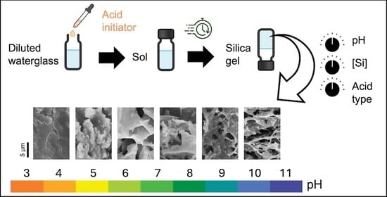
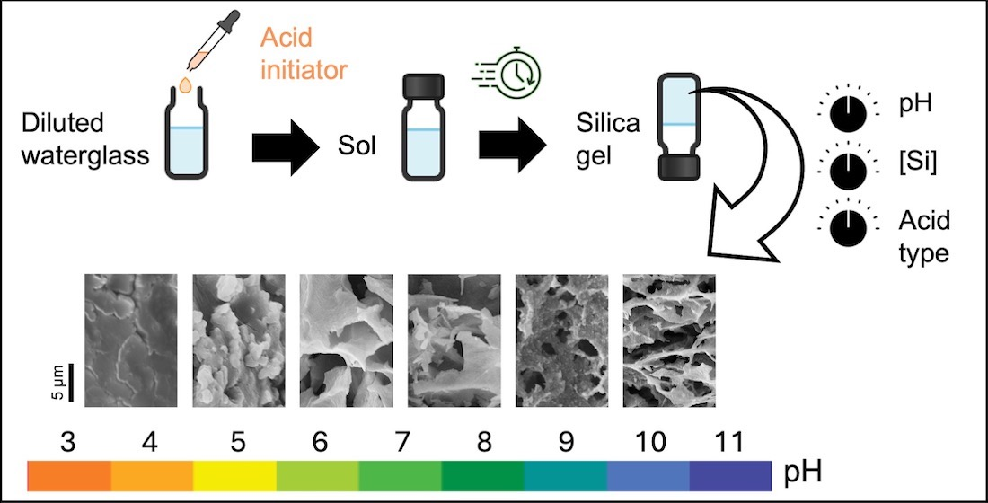
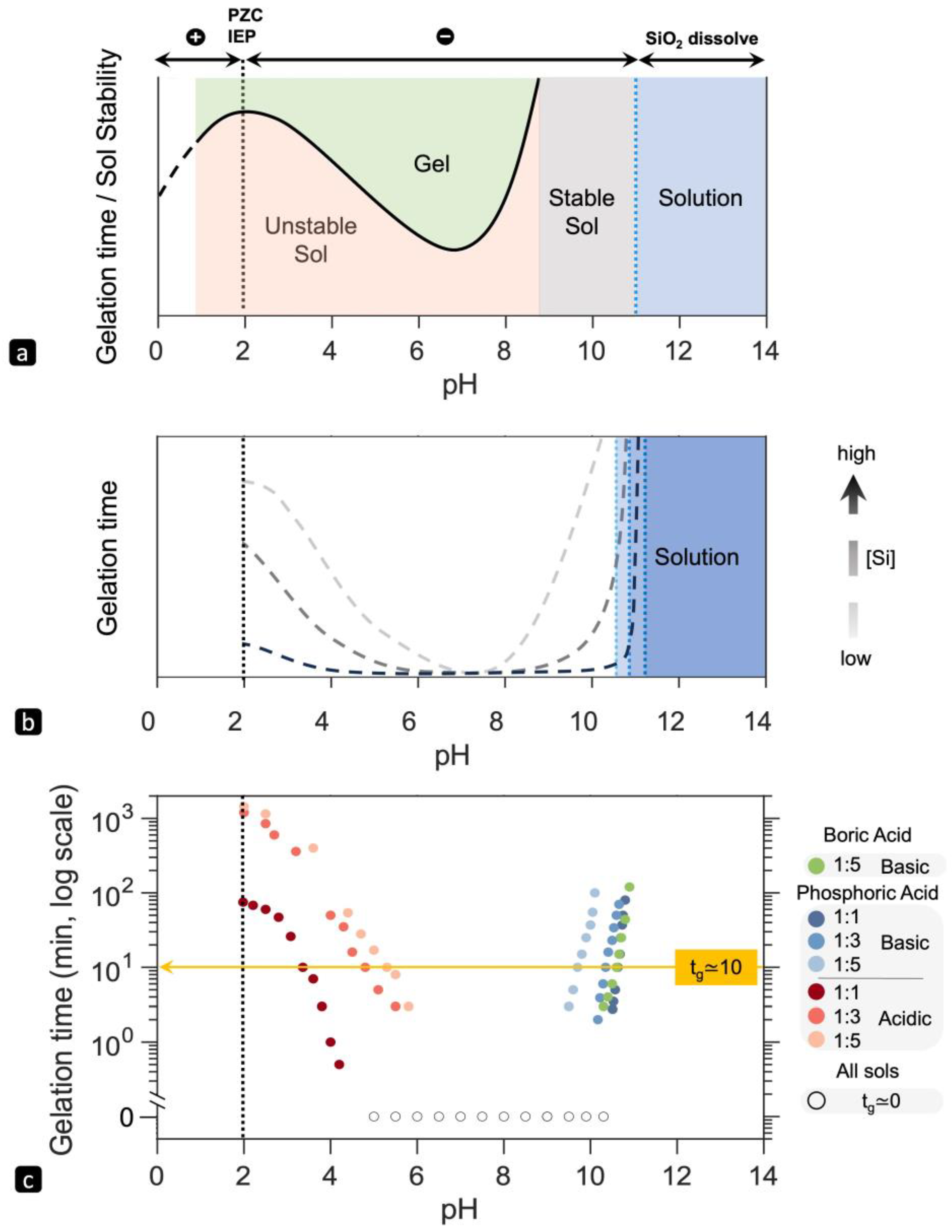
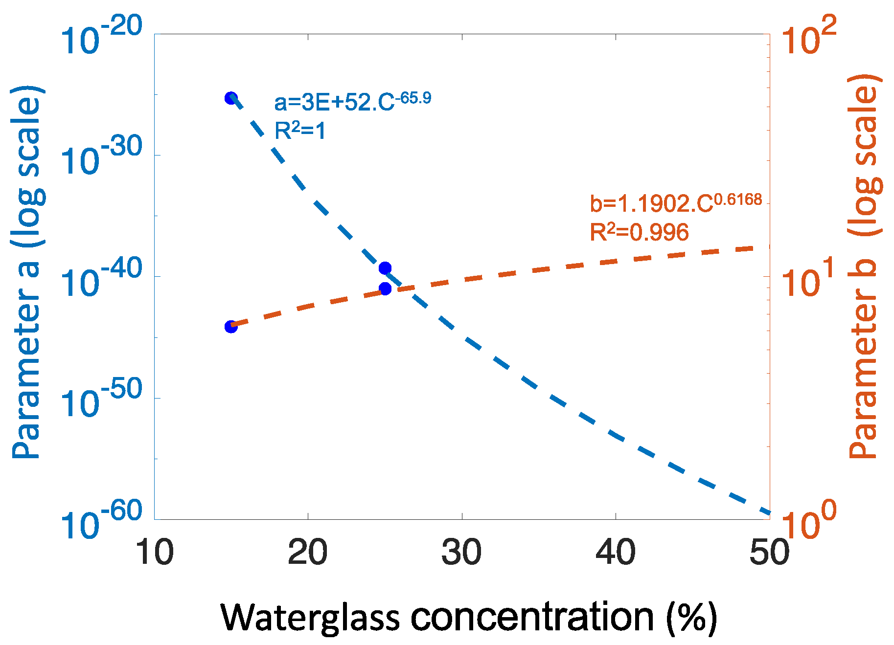
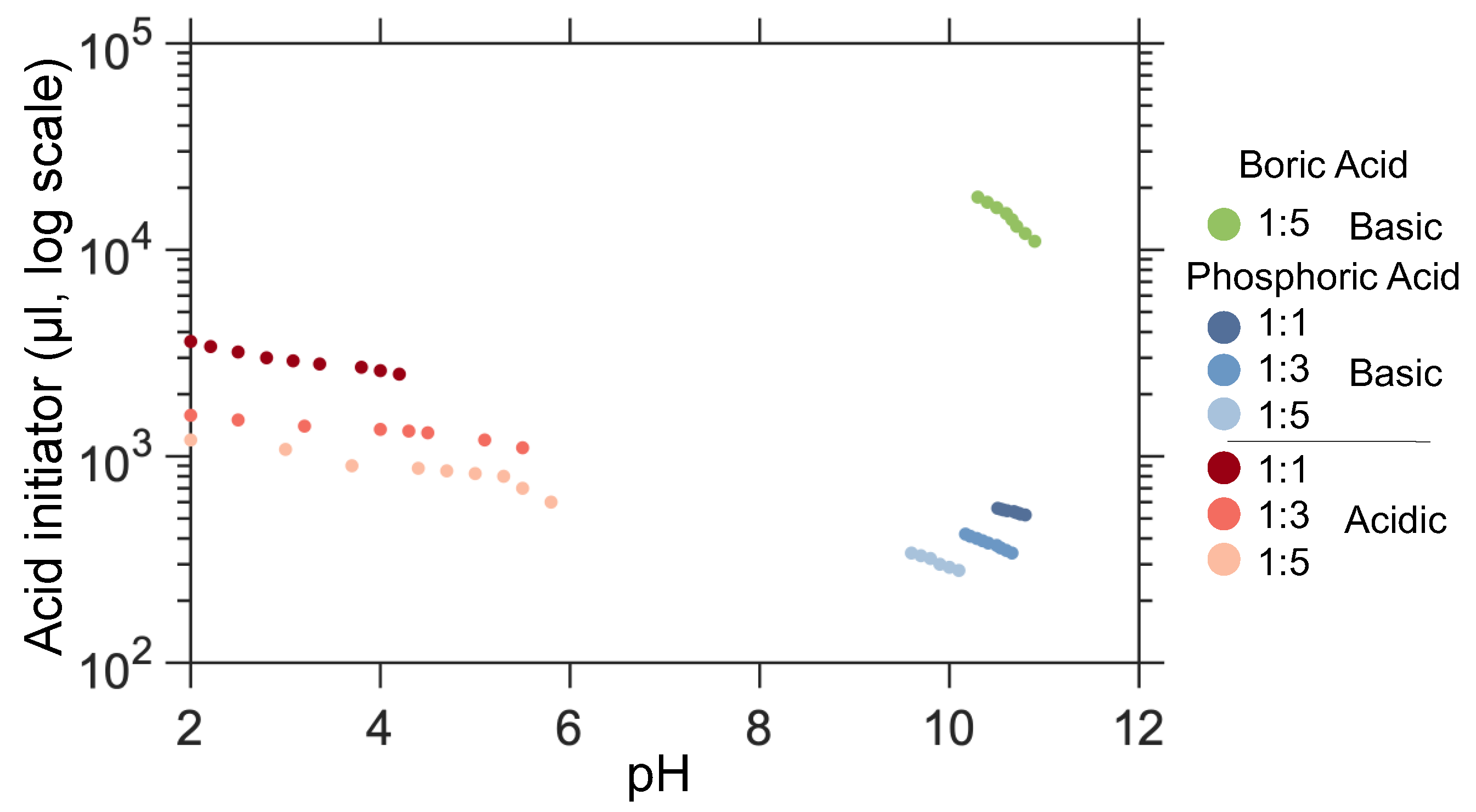
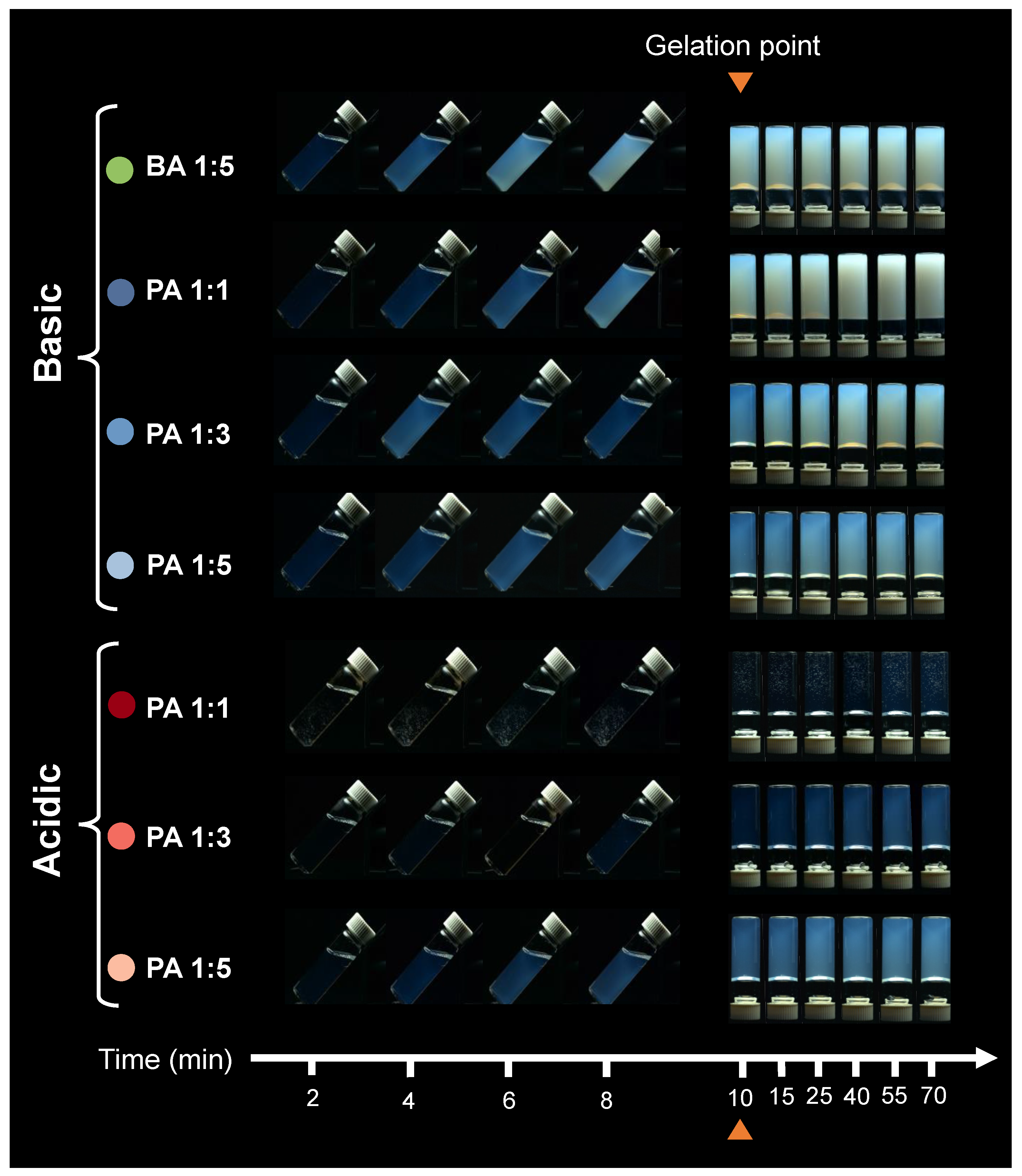
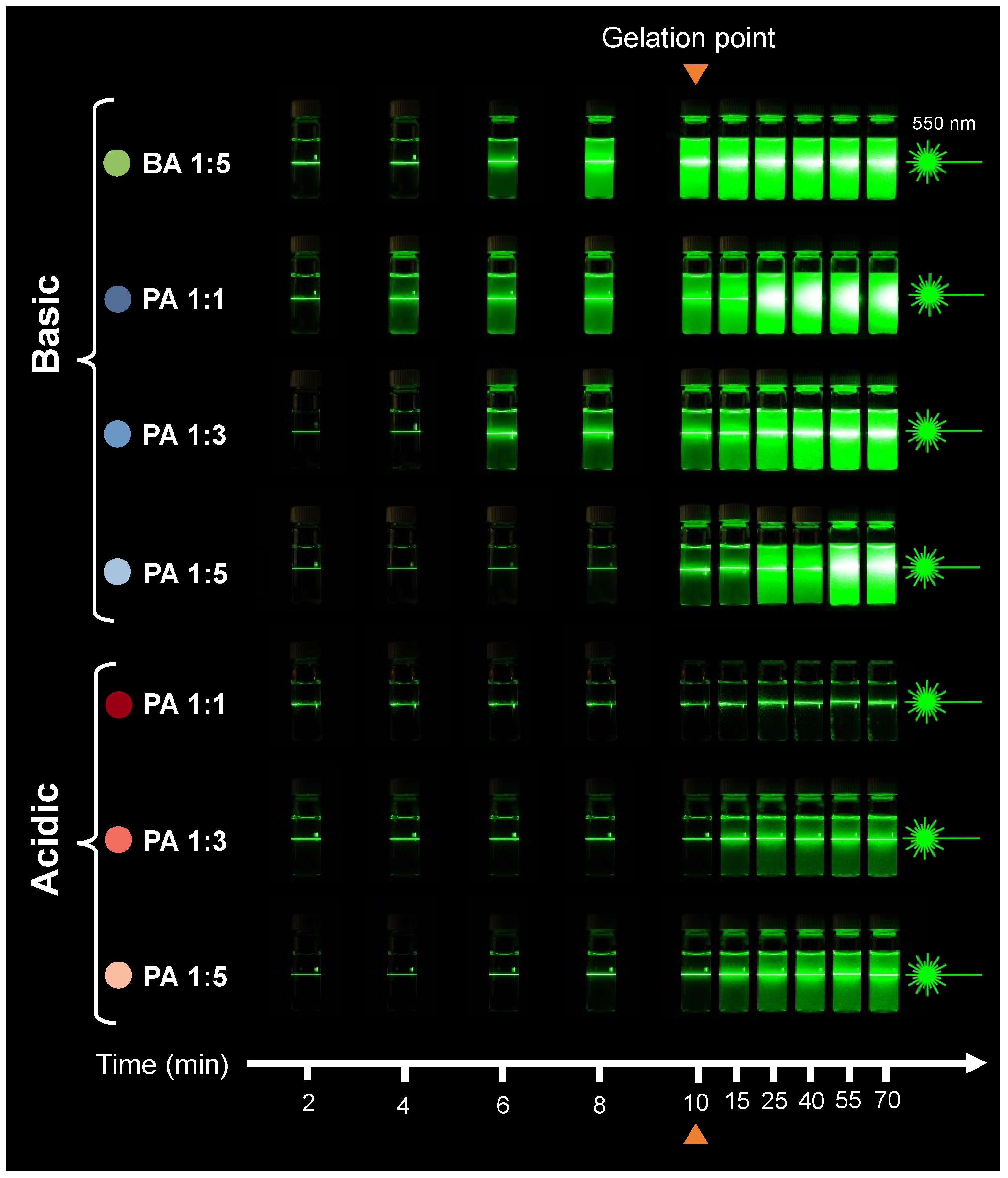
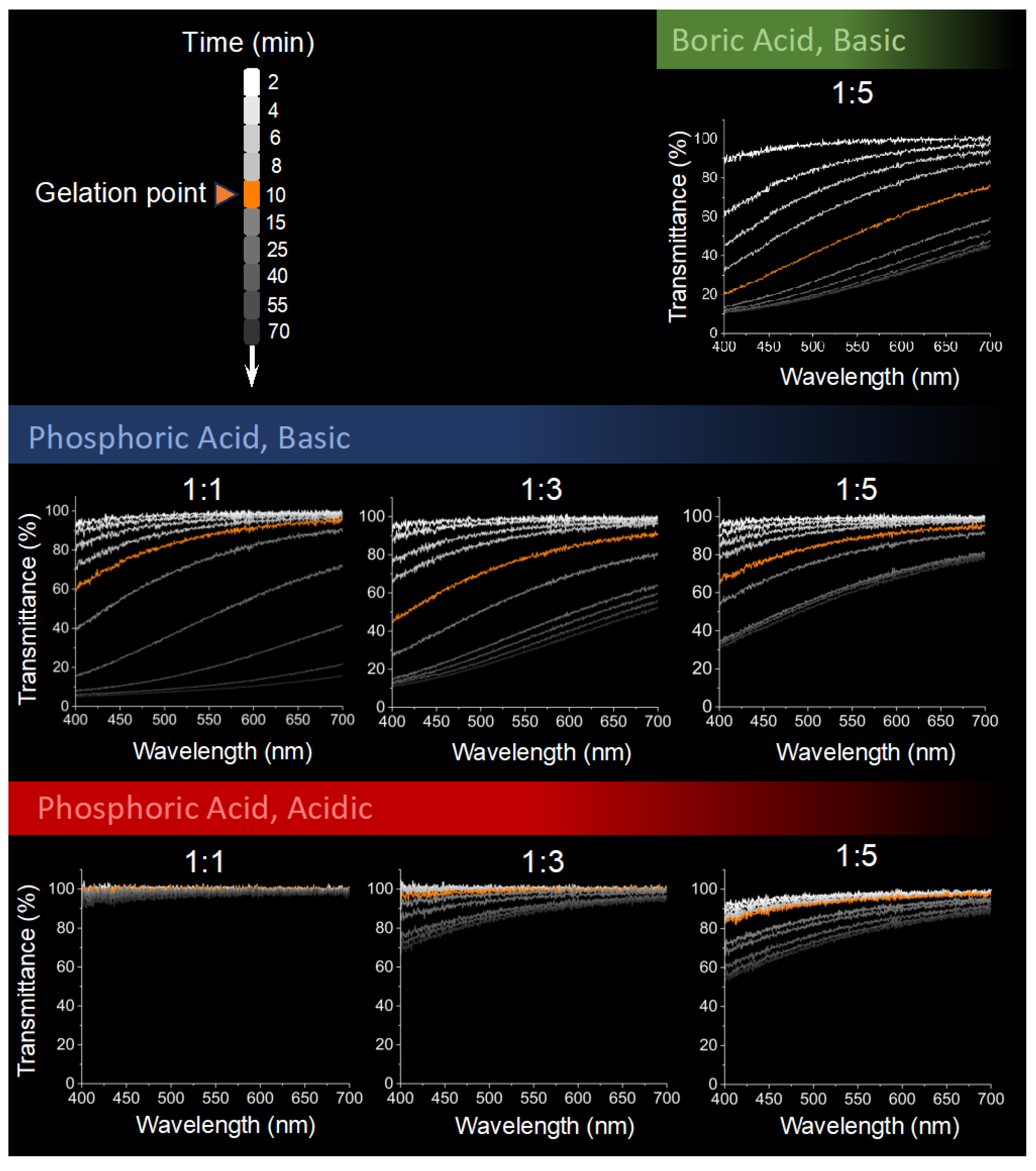
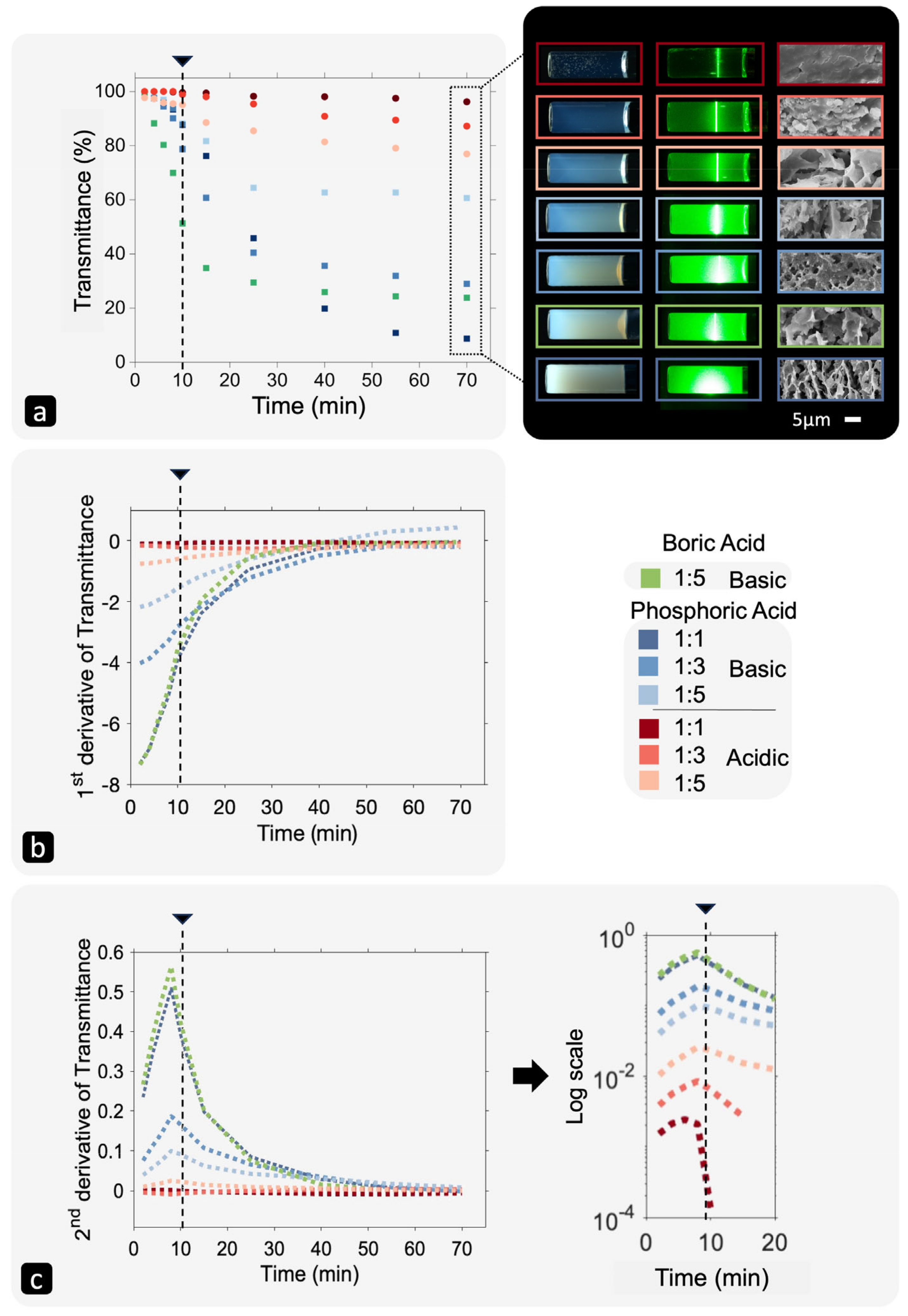
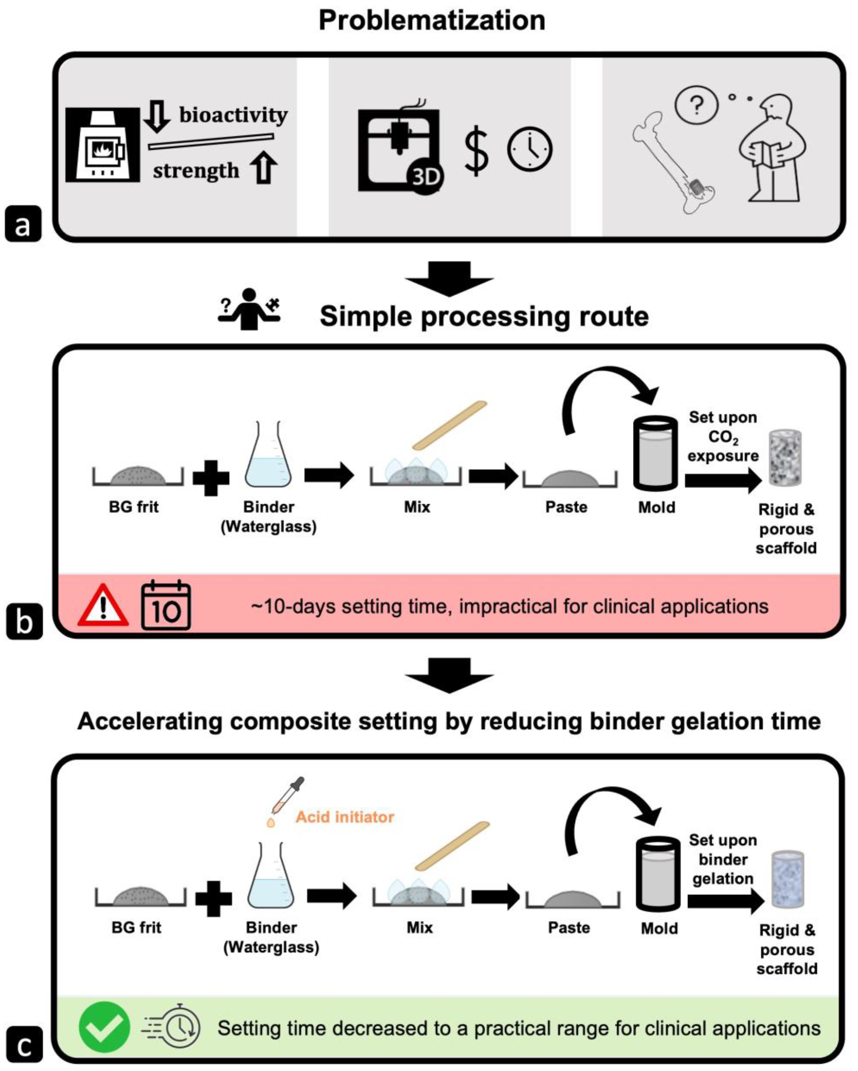
Fabricating large-scale porous bioactive glass bone scaffolds presents significant challenges. This study aims to develop formable, in situ setting scaffolds with a practical gelation time of about 10 min by mixing 45S5 bioactive glass with sodium silicate (waterglass) and an acid initiator. The effects of pH (2–11), waterglass concentration (15–50 wt.%), and acid initiator type (phosphoric or boric acid) were examined to optimize gelation kinetics and microstructure. A 10 min gelation time was achieved with boric acid and phosphoric acid at various pH levels by adjusting the waterglass concentration. Exponential and polynomial models were proposed to predict gelation times in basic and acidic environments, respectively. The optical properties of the gels were studied qualitatively and quantitatively, providing insights into gelation kinetics and structure. Acidic gels formed smaller particles in a dense network (pores < 550 nm) with higher light transmittance, while basic gels had larger aggregates (pores ~5 µm) and lower transmittance. As the waterglass concentration decreased, pore size and transmittance converged in both groups. The onset of gelation was detected around 8 min using the derivative of light transmittance. This work identifies the key factors controlling waterglass gelation and their impact on gel structure, enabling the tailored creation of formable, in situ setting bioactive glass bone scaffolds.
Keywords: silica gel; colloidal silica; sodium silicate solution; sol–gel; gelation kinetics; optical properties; bone tissue scaffold silica gel; colloidal silica; sodium silicate solution; sol–gel; gelation kinetics; optical properties; bone tissue scaffold
Graphical Abstract