Article

Isolation of Tulasnella spp. from Cultivated Paphiopedilum Orchids and Screening of Germination-Enhancing Fungi

1
State Key Laboratory of Tree Genetics and Breeding, Key Laboratory of Tree Breeding and Cultivation of the National Forestry and Grassland Administration, Research Institute of Forestry, Chinese Academy of Forestry, Beijing 100091, China
2
Beijing Laboratory of Urban and Rural Ecological Environment, Beijing Floriculture Engineering Technology Research Centre, China National Botanical Garden (North Garden), Beijing 100093, China
*
Author to whom correspondence should be addressed.
J. Fungi 2023, 9(6), 597; https://doi.org/10.3390/jof9060597
Submission received: 24 April 2023 / Revised: 18 May 2023 / Accepted: 18 May 2023 / Published: 23 May 2023

Abstract

:
Ex situ conservation, an important way to increase the survival and sustainability of endangered species, is widely used in the conservation of endangered orchids. However, long-term ex situ conservation might affect the dominant group of orchid symbiotic fungi, which are crucial for orchid growth and reintroduction. This study investigated the culturable Tulasnella spp. associated with Paphiopedilum orchids after long-term greenhouse cultivation, and identified germination-enhancing isolates. A total of 44 Tulasnella isolates were obtained from the roots of 14 Paphiopedilum spp., and 29 of them were selected for phylogenetic analysis. They clustered mainly with Tulasnella deliquescens, Tulasnella calospora, Tulasnella bifrons, and Tulasnella irregularis, but included two potential new groups. Compared with published uncultured data, most of the isolates were grouped together with the reported types, and the dominant Tulasnella associated with P. armeniacum and P. micranthum could still be isolated after ten years of cultivation, most of which were the first isolation. In vitro symbiotic germination showed that certain root isolates could promote seed germination (e.g., parm152 isolated from P. armeniacum, Php12 from P. hirsutissimum, and prhi68 from P. rhizomatosum). These data indicated that the dominant Tulasnella types colonizing the roots of cultivated Paphiopedilum are stable over time, and germination-enhancing fungi colonizing the roots would benefit for seed reproduction after population reintroduction into the wild.

1. Introduction

Paphiopedilum Pfitzer (Orchidaceae), comprising about 96–100 species around the globe, is mainly distributed in Southeast Asia [1,2] and is popular in the horticultural market and exhibitions as a pot plant because of its slipper-like flower shape and abundant color (Figure 1A–D,F). China is one of the distribution centers of Paphiopedilum. There are about 27 native species found in China, most of which are distributed in Yunnan or Guangxi province [3]. Over-collection and loss of suitable habitats have led to the wild population of Paphiopedilum being under the threat of extinction [4]. All species in Paphiopedilum are listed in the Convention on International Trade in Endangered Species of Wild Fauna and Flora (CITES) [5].
To conserve and restore the population of Paphiopedilum, studies of population geographical distribution [6], embryo development [7,8], in vitro tissue culture [9,10], ex situ seed baiting [11], and mycorrhizal fungi associated with Paphiopedilum [12] have been carried out. At the same time, in situ/ex situ conservation of Paphiopedilum orchids has also been implemented [3]. The limited distribution and endangered status [13] of Paphiopedilum means that it has only been sporadically studied compared with other commercial orchids.
Like other orchids, Paphiopedilum spp. depend on mycorrhizal fungi from seed germination to seedling growth [14], to obtain organic nutrients and carbon energy. Orchid mycorrhizal fungi (OMF) form characteristic intracellular hyphae (Figure 1G), called a peloton, within the cells of Paphiopedilum orchid roots or protocorms [15]. Most of the OMF associated with Paphiopedilum orchids are members of the genus Tulasnella [12,16,17] and Ceratobasidium [4,15].
Tulasnella J. Schröt. (Tulasnellaceae) is an important genus of OMF that occurs worldwide in the form of mycobionts associated with orchids [18,19,20], which is also a frequently detected fungal group in the roots of Paphiopedilum [21]. Diverse Tulasnella fungi colonize Paphiopedilum. In southwestern China, Yuan et al. [12] amplified 25 types of internal transcribed spacer (ITS) sequence representing Tulasnellaceae from the roots of Paphiopedilum and Cypripedium. Similarly, Tulasnella species such as T. calospora, T. violea, and T. pruinosa were isolated from five species of Paphiopedilum in Thailand [16].
To date, most species in Paphiopedilum have been successfully cultivated in the greenhouse for ex situ conservation. In the present study, we isolated Tulasnella spp. from 14 Paphiopedilum species cultivated in greenhouses in southwestern China, and used phylogenetic analysis of ITS sequences to identify and characterize the isolates. We focused on the following issues: (1) After long-term cultivation, do Tulasnella spp. exist in specific Paphiopedilum species? (2) Do Tulasnella spp. isolated from cultivated Paphiopedilum have a positive effect in promoting seed germination? These issues are very important for seedling adaptability and seed germination of Paphiopedilum population reintroduction into the wild.

2. Materials and Methods

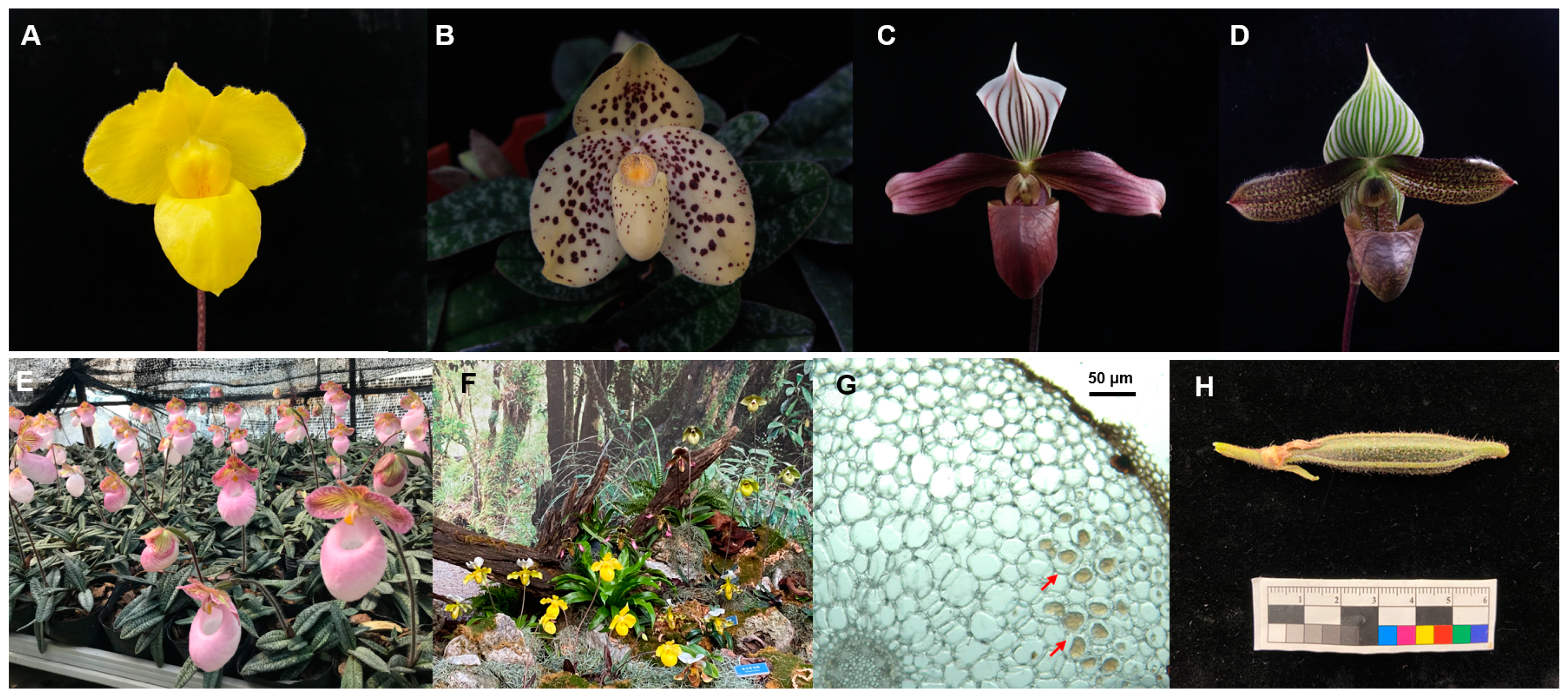
2.1. Study Materials

Paphiopedilum species, including P. armeniacum (Figure 1A), P. callosum, P. emersonii, P. gratrixianum, P. henryanum, P. hirsutissimum, P. malipoense, P. micranthum (Figure 1E), P. purpuratum (Figure 1C), P. tigrinum, P. villosum, P. wardii (Figure 1D), P. wenshanense (Figure 1B), and P. rhizomatosum, were ex situ conserved in a greenhouse in Xingyi, Guizhou province, southwestern China. The orchids were cultivated on pine bark substrate in the greenhouse for at least 3 years before root sampling. The seedlings were healthy, and some were at the flowering stage or fruiting stage after artificial pollination. The root samples were collected from September 2017 to May 2021 (Table S1). Paphiopedilum spp. at different growth stages were randomly selected for sampling, with three plants for each stage, and 2–3 healthy roots from each plant. Sampled seedlings were marked to avoid subsequent sampling. About 5–10 cm of the healthy root was cut from each seedling using a sterile scalpel, put into a sterile bag, and transported at 4 °C for 24 h until fungal isolation.
The fruit of P. hirsutissimum were obtained by artificial pollination (Figure 1H). Seeds at approximately 130–160 days after pollination were used for in vitro germination. Healthy uncracked capsules were harvested from 2019 to 2021, which were cultivated in the greenhouse of Chinese Academy of Forestry, Beijing, China.
Figure 1. Cultivated Paphiopedilum spp. (A) P. armeniacum. (B) P. wenshanense. (C) P. purpuratum. (D) P. wardii. (E) P. micranthum cultivated in Xingyi greenhouse. (F) Landscape display arranged with Paphiopedilum spp. (G) Intracellular fungal pelotons in the root of Paphiopedilum sp. The red arrows indicate the intracellular fungal pelotons. (H) Fruit of P. hirsutissimum.
Jof 09 00597 g001

2.2. Fungal Isolation and Identification

The roots were transferred to the laboratory, washed using sterile water, and surface sterilized (30 s submergence in 75% ethanol, 30 s rinsing with sterile distilled water, 5 min submergence in 1% sodium hypochlorite, and 30 s rinsing in sterile distilled water five times). The sterilized roots were sliced into 0.5–1.0 mm sections and inoculated on one-quarter strength potato dextrose agar (PDA) from BD Difco (Franklin Lakes, NJ, USA) with 11.25 g/L agar at 25 ± 2 °C. Five sections were placed in one Petri dish for fungal isolation, with 10 dishes per sample. When hyphae growing from the section exceeded 0.5 cm in length, the tips of the hyphae were cut and transferred to new one-quarter strength PDA medium for purification. After repeating this purification step four to five times, purified isolates were obtained.
The purified isolates were identified morphologically by observing and recording the color and surface shape of their culture characteristics [22,23,24] on PDA medium at 25 °C in the dark for about 1–4 weeks, depending on their growth rates. The condition of their nuclei was observed from young hyphae after staining with SYBR Green I according to a previous publication [25]. Observation, measurement, and photography of microscopic fungal structures were recorded using a ZEISS Axio Imager M2 microscope (ZEISS, Oberkochen, Germany), with a ZEISS Axiocam 503 mono camera and differential interference contrast (DIC) illumination. ZEISS Zen 3.3 was used for image processing and synthesis.
For molecular identification of the isolates, the ribosomal DNA (rDNA) region containing the two ITS regions and the 5.8S gene was amplified using primers ITS1 and ITS4 [26]. The PCR was performed as previously described [27]. The PCR products were purified and sequenced at Sangon Biotech Co., Ltd. (Beijing, China) using the same primers. The sequences were BLAST searched against the GenBank database of the National Center for Biotechnology Information (NCBI) for identification. The sequences were then deposited in the GenBank database (Table S1). The isolates were maintained at the Research Institute of Forest, Chinese Academy of Forestry.

2.3. Phylogenetic Analysis of the ITS Sequences

As previously noted, within Tulasnella, the ITS sequences could reveal congruent species delimitation and phylogenetic outcomes [28]. The sequences representing the two ITS regions and the 5.8S gene were used for the phylogenetic analysis of Tulasnella. One phylogenetic tree was constructed by combining the ITS sequence of the isolates with the published ITS sequences of Tulasnella spp. downloaded from GenBank in NCBI (Table S2) to determine the taxonomic status of the isolates [20,29]. Another phylogenetic tree was analyzed to clarify the relationship among the isolates associated with Paphiopedilum spp. The uncultured ITS sequences of Tulasnellaceae amplified from roots of Paphiopedilum spp. and Cypripedium spp. in a previous study [12] were used as reference sequences to investigate the continuous colonization of Tulasnella associated with Paphiopedilum spp.
All the sequences used for phylogenetic analyses were aligned using MAFFT v7.311 [30] and manually adjusted using MEGA 7 [31]. MEGA 7 was used to construct the maximum likelihood (ML) trees under the general time reversible model (GTR+I+G), with 1000 bootstrap replicates. MrModeltest v2.3 [32,33] was used to determine the best-fit evolution model for the combined dataset for Bayesian inference (BI). BI was calculated using MrBayes v3.2.7 [34], with a GTR+I+G model of DNA substitution and a gamma distribution rate variation across sites. Four Markov chains were run for two runs from random starting trees for 1 million generations and were sampled every 100 generations. The phylogenetic trees were visualized using FigTree v1.4.2 (http://tree.bio.ed.ac.uk/software/figtree/ (accessed on 16 November 2021)). Branches that received bootstrap support for ML and Bayesian posterior probabilities greater than or equal to 50% and 0.70, respectively, were considered to be significantly supported.

2.4. Screening of Germination-Enhancing Fungi

Fungi from different clades in the phylogenetic tree were tested for their ability to enhance the germination of P. hirsutissimum seeds. After surface sterilization of the capsules as described by Zi et al. [35], approximately 200–300 seeds were sown on oatmeal agar (OMA) medium (3.0 g of oatmeal (Solarbio, Beijing, China) was added to 800 mL of distilled water, boiled for 30 min, filtered through a 50-mesh sieve to remove large particles, added 6.0 g of agar, and then added to 1000 mL with water ) in a Petri dish. A fungal inoculum was placed in the center of each dish and co-inoculated with the seeds. Asymbiotic germination was performed as a control on OMA or MS1 (MS medium with one-quarter strength MS macronutrients; 0.4 mg/L 6-benzylaminopurine, 10.0 g/L sucrose, 6.0 g/L agar, pH 6.3). There were three replicates for each treatment. All replicates were placed in germination chambers at 25 ± 2 °C and subjected to alternating 12 h periods of light and dark. Three independent sowing experiments were performed.
The number of seeds and the germination status in each dish were assessed after 90 days of incubation, according to previously defined stages (Figure 2) [36]. The percentages of germinated seeds at each developmental stage were calculated. Seed germination data were recorded as previously described [37]: 0, 1, (2 + 3), and (4 + 5) stages were defined based on the number of ungerminated, germinated (g) seeds, protocorms (p), and seedlings (s). The total number of seeds (t) constituted the seeds with well-developed embryos. Germination rate (G), protocorm formation rate (P), and seedling formation rate (S) were calculated 90 days after inoculation. The following calculations were used:
G = 100 × (g + p + s)/t.
P = 100 × p/t.
S = 100 × s/t.
Analysis of variance was performed using SPSS 16.0 (IBM Corp., Armonk, NY, USA). The data were analyzed using one-way analysis of variance (ANOVA) after inverse sine transformation. Statistical significance was set at p < 0.05.

3. Results

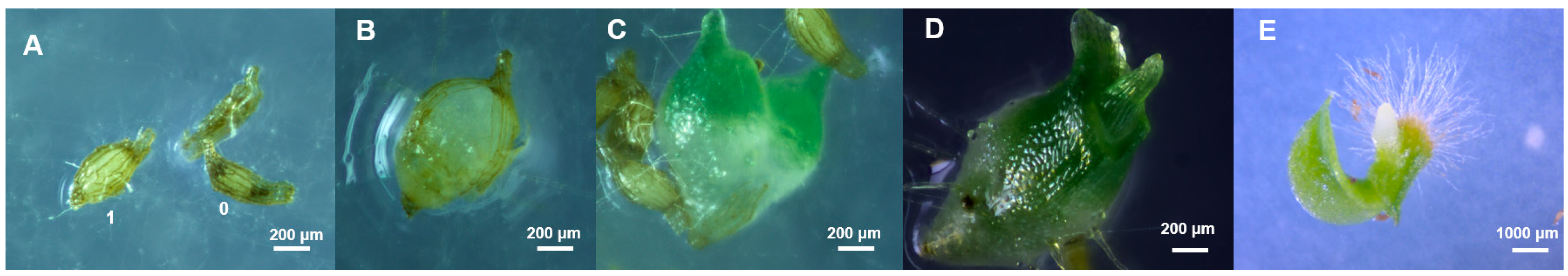
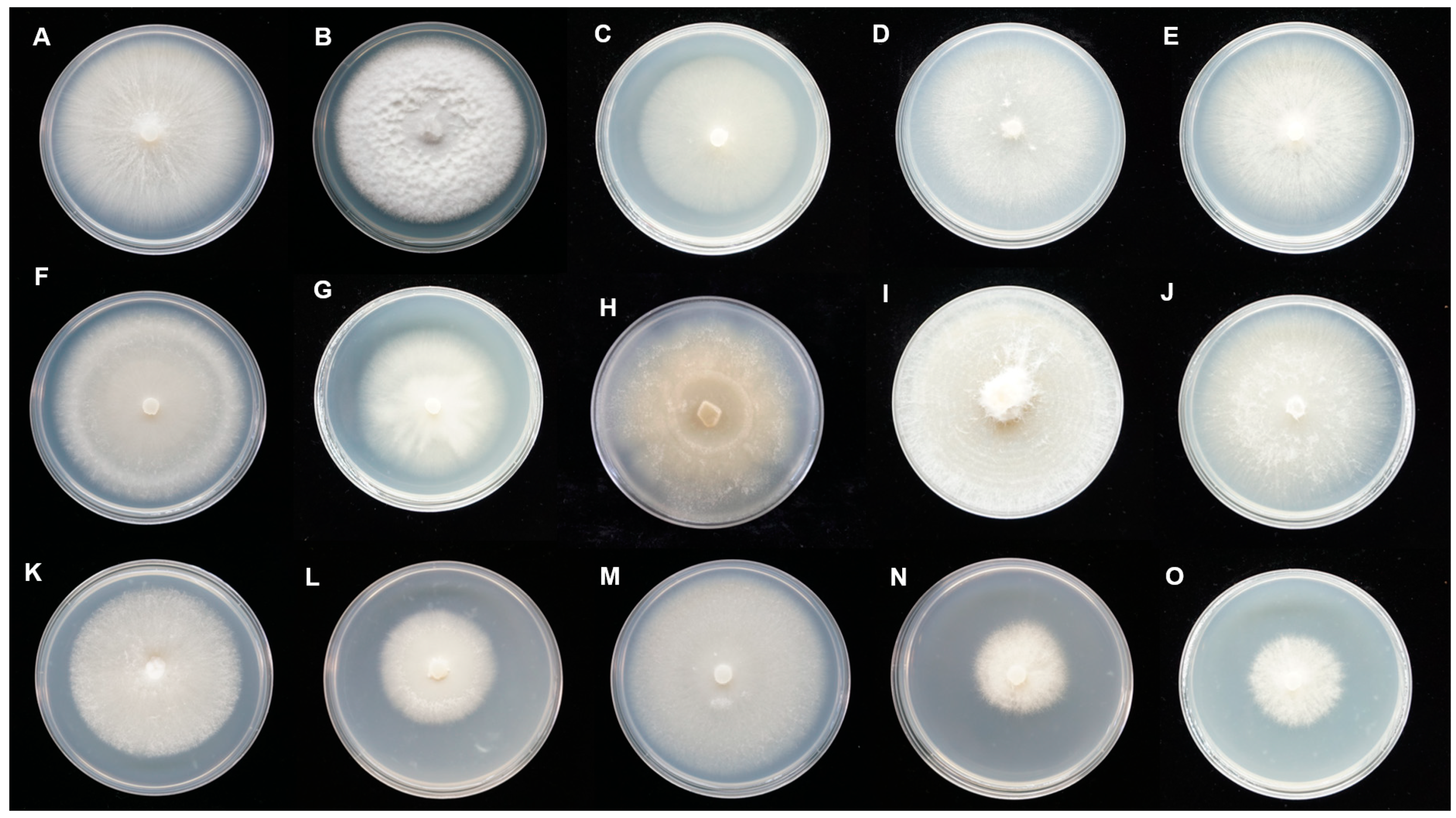
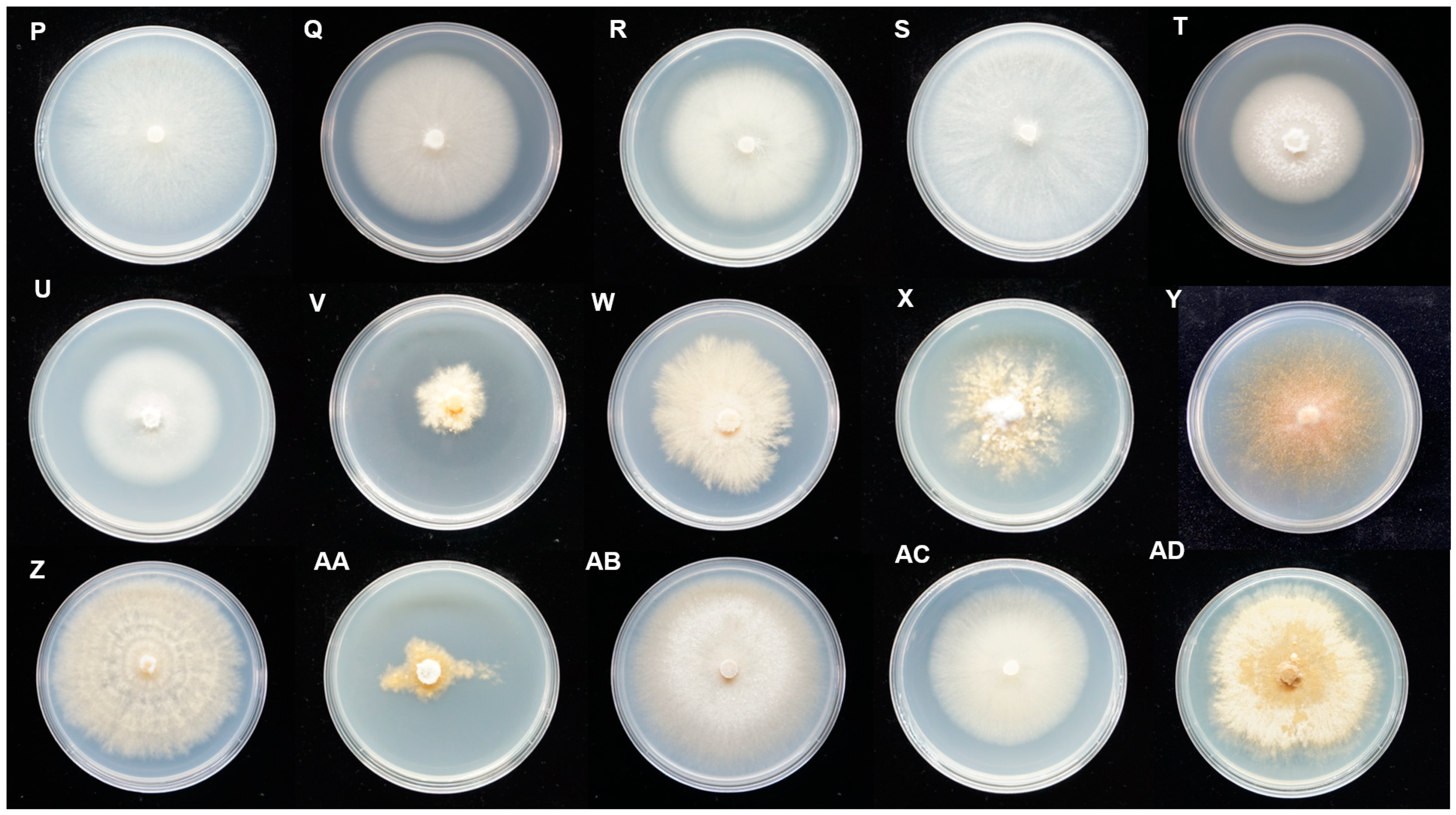
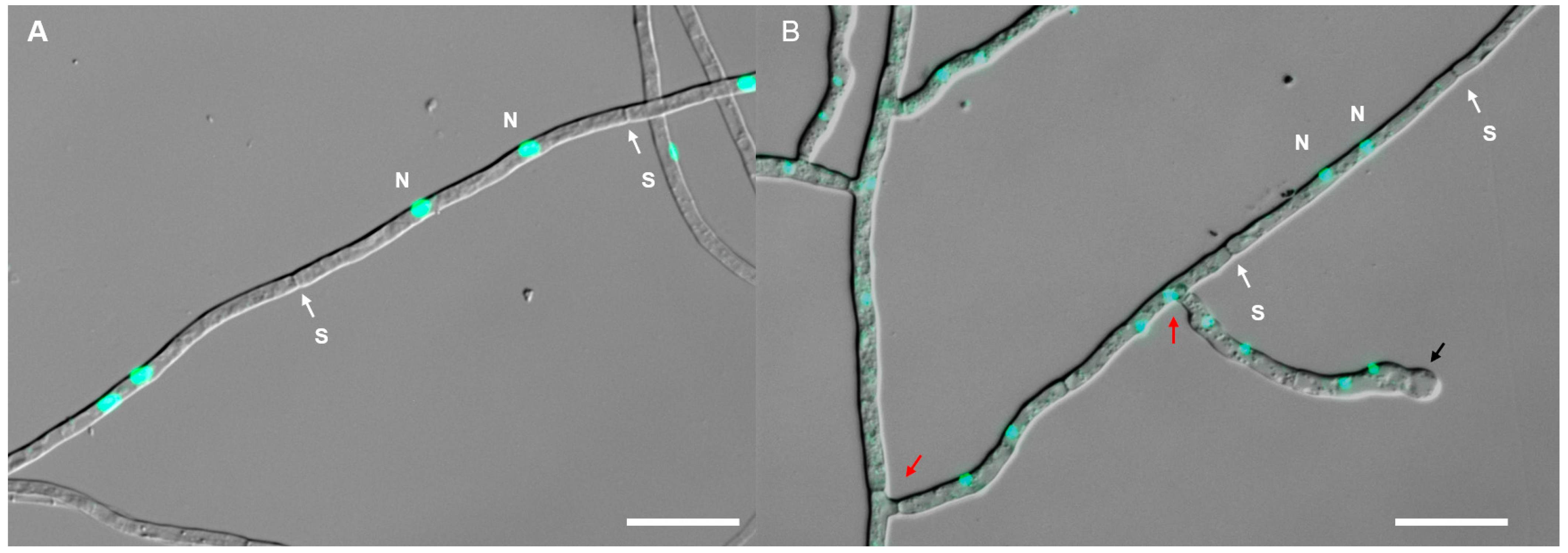
3.1. Fungal Isolation and Morphological Characterization

A total of 14 Paphiopedilum species cultivated in a Xingyi greenhouse were sampled at different growing stages, including the vegetative growth, flowering, or fruiting stage. In total, 44 fungal isolates belonging to Tulasnella were obtained, which were identified by BLAST analysis of the ITS sequences. For the closest matching ITS sequence, 25 isolates were BLAST searched against uncultured Tulasnellaceae clones. For the source of the closest matching fungi, 28 isolates were BLAST searched against Paphiopedilum spp., and 2 isolates were BLAST searched against Phragmipedium longifolium (Table S1), which is a member of the subfamily Cypripedioideae.
These isolates showed diverse colonial morphology on PDA medium (Figure 3), and could be divided into two groups. In Group A, the colonies were white or beige with aerial and submerged hyphae. In Group B, the colonies were light buff to orange with submerged hyphae. The hyphae were regularly septate with branching at right angles, hyaline, and with binucleate cells (Figure 4). Molinioid cells were hyaline, barrel to elliptical-shaped, and occurred in branched chains (Figure 4B). Sexual morphology was not observed for all the isolates. Combined with the BLAST results and colonial characteristics, 29 representative isolates (Figure 3), removing the isolates that were repeatedly acquired at each isolation, were finally selected for the subsequent phylogenetic analysis (Table 1).

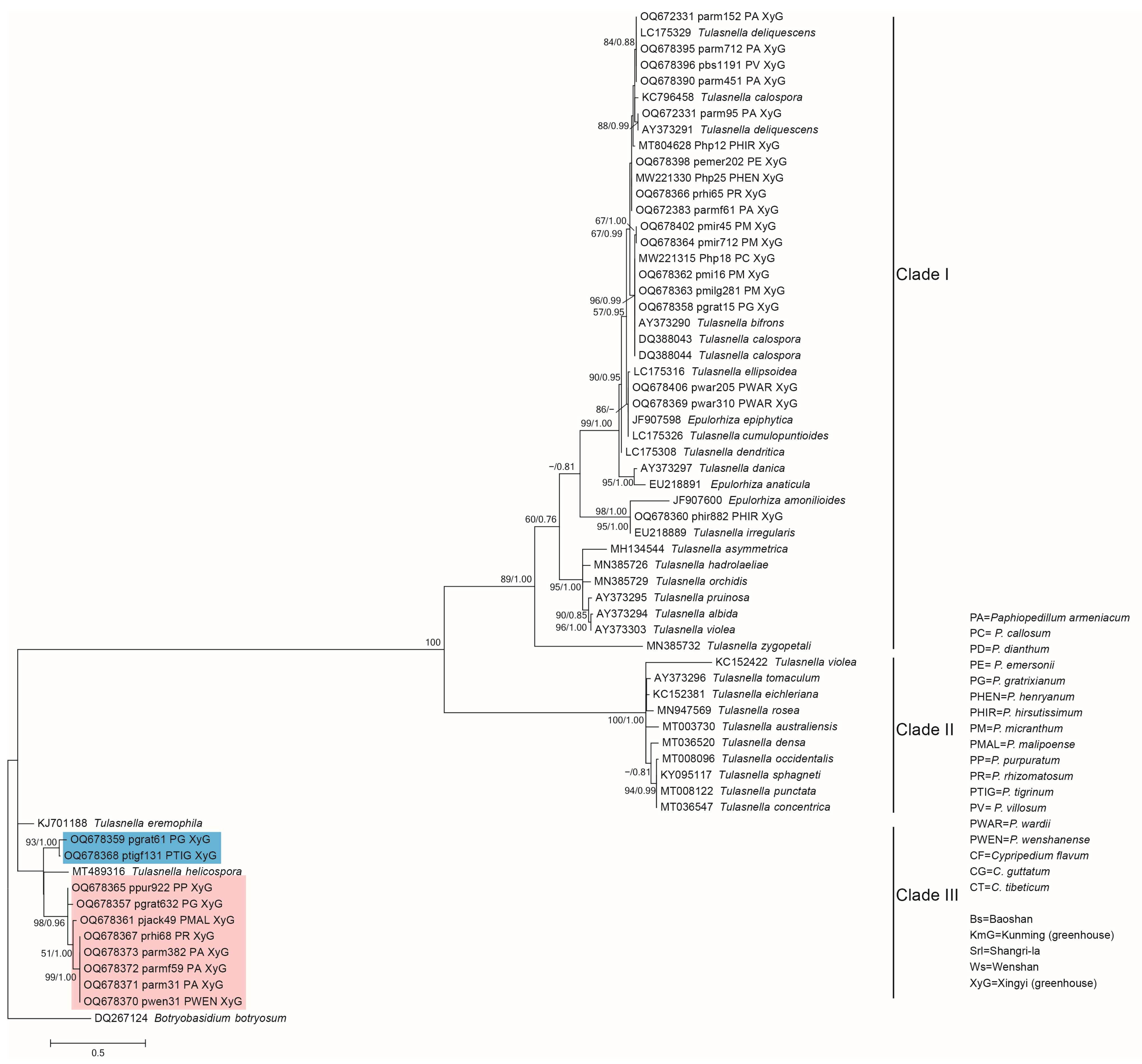
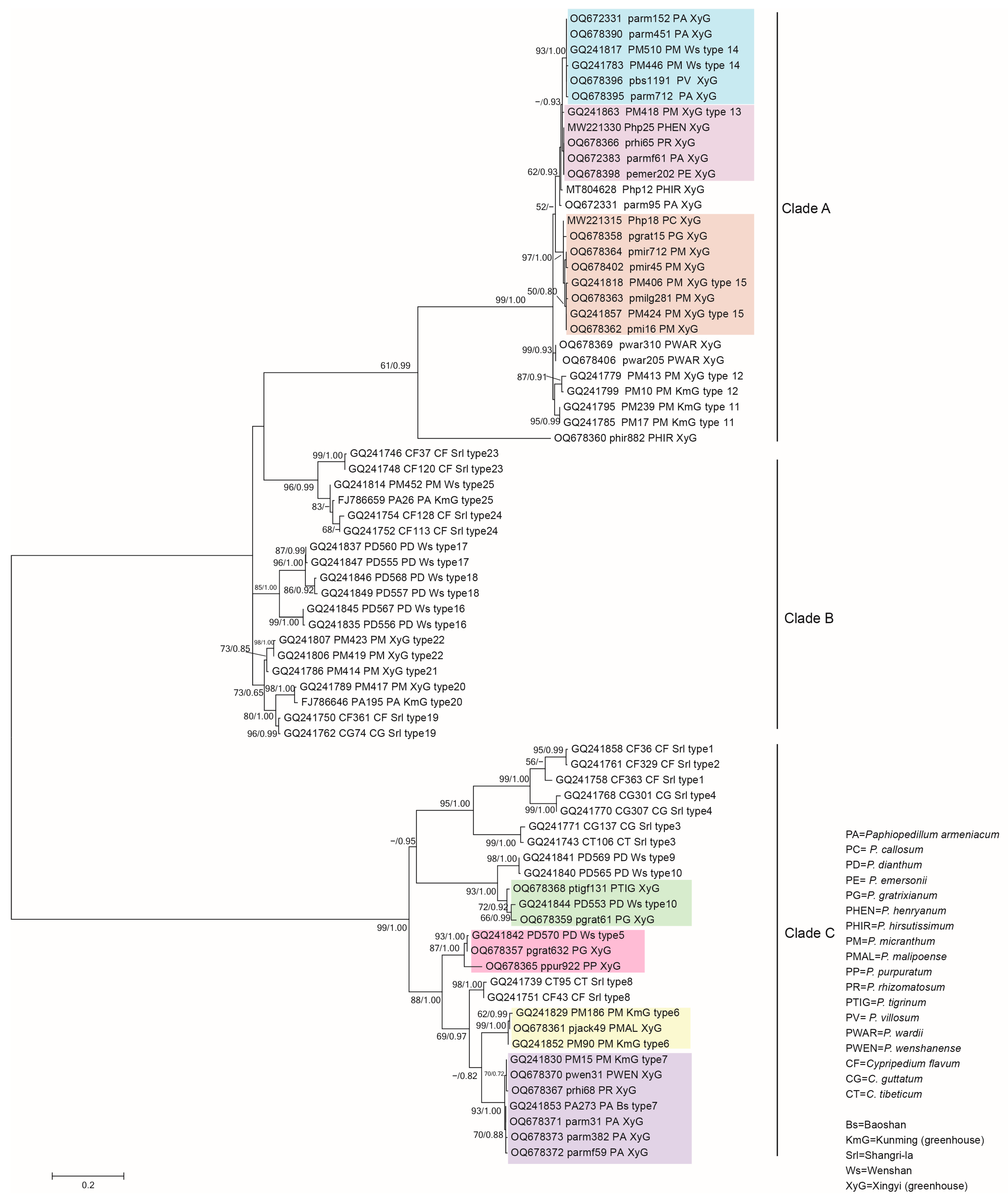
3.2. Phylogenetic Analysis of the Representative Isolates

The ITS sequences consisted of 63 isolates (including the outgroup sequence DQ267124 Botryobasidium botryosum), of which 34 were published sequences downloaded from NCBI (Table S2) and 29 were the representative isolates from this study (Table 1), which were used for phylogenetic analysis. The phylogenetic tree showed that mycorrhizal fungi isolated from Paphiopedilum orchids were in the genus Tulasnella (Figure 5), which were mainly clustered with Tulasnella deliquescens, Tulasnella calospora, Tulasnella bifrons, and Tulasnella irregularis. There were two potential new groups in Clade III (new group one: pgrat61 and ptigf131; new group two: pgrat632, ppur922, pjack49, prhi68, parmf59, parm382, parm31, and pwen31), which were not clustered with previously reported Tulasnella species. This indicated that after long-term cultivation in similar environments, the Tulasnella fungi in different Paphiopedilum orchids belonged to different species, rather than converging on a few/one species.
In the tree of Tulasnella, the 29 isolates fell into two clades (Figure 5). Nineteen isolates were clustered in Clade I, and ten isolates were clustered in Clade III. Meanwhile, the fungi in Clade II were not isolated. The 19 isolates belonging to Clade I were mainly from 10 Paphiopedilum species, and the isolates belonging to Clade III were from 7 Paphiopedilum species. The roots of P. armeniacum, P. gratrixianum, and P. rhizomatosum were colonized by two types of isolates belonging to Clade I and Clade III.
Some isolates with a similar phylogenetic status were acquired multiple times from one Paphiopedilum species at different isolations (Table 1, Figure 5), such as pmi16, pmilg281, pmir45, and pmir712 from P. micranthum and parm31, parm382, and parmf59 from P. armeniacum, which indicated that some Tulasnelloid fungi could stably colonize the roots of Paphiopedilum orchids within a few years. Most of the above isolates were isolated for the first time in this study, because the closest sequence alignments in NCBI were from uncultured clones.
Another phylogenetic tree (Figure 6) was constructed based on the ITS sequences of the 29 isolates and the 25 types of published Tulasnellaceae sequences amplified from the roots of Paphiopedilum and Cypripedium. Seventeen of the twenty-five types were from Paphiopedilum, and eight types were from Cypripedium. The phylogenetic tree showed that 24 isolates in this study were clustered with 7 of the 17 types from Paphiopedilum. The eight types from Cypripedium were not isolated from Paphiopedilum. The isolates parm95, Php12, pwar310, pwar205, and phir882 were new types. As in the Tulasnella tree (Figure 5), all the 29 isolates were mainly clustered into two clades. Clade A and Clade C corresponded to Clade I and Clade III in the tree of Tulasnella, respectively.
Fourteen isolates were clustered with type 15 and type 13 from the Xingyi greenhouse, and type 14 from Wenshan, which were in Clade A. Clade C included 10 isolates, which were clustered with type 10 and type 5 from Wenshan, type 6 from Kunming, and type 7 from different regions (Kunming and Baoshan).
It should be noted that fungi belonging to type 15 and type 7 could still be isolated after about ten years of cultivation, which indicated that some Tulasnella spp. could colonize the roots of P. micranthum and P. armeniacum for a long time. Meanwhile, some types included isolates from different Paphiopedilum species, such as the isolates (parm152, parm415, pbs1191) from P. armeniacum, and P. villosum clustered into type 14 and isolates (Php18, pgrat15, pmir712) from P. callosum, P. gratrixianum, and P. micranthum clustered into type 15.

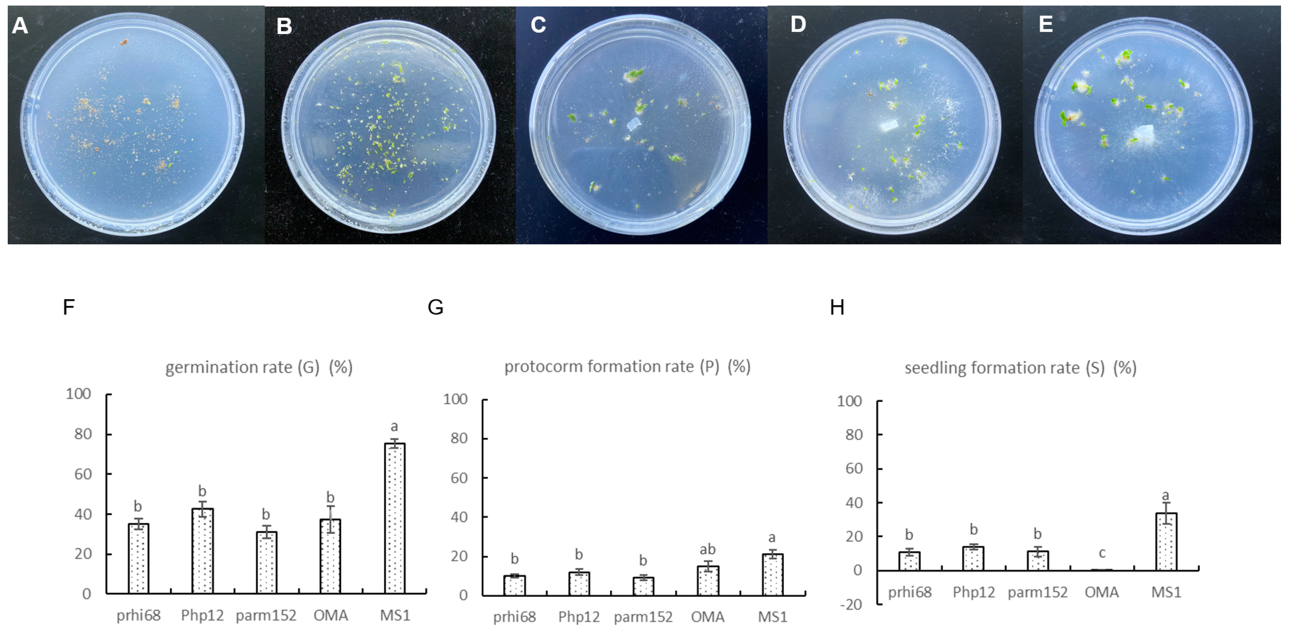
3.3. Fungal Capacity to Enhance Germination

After 90 days of in vitro symbiotic germination on OMA medium, three isolates could significantly promote the seedling formation of P. hirsutissimum (Figure 7C–E) compared with that on the asymbiotic negative control (OMA) (Figure 7A). The in vitro symbiotic germinated protocorms were checked microscopically for mycorrhizal colonization. The three isolates could colonize protocorm cells of P. hirsutissimum and formed the typical intracellular hyphae coils of OMF (Figure S1).
The asymbiotic positive control (MS1) allowed more than 75% seed germination (Figure 7F), leading to large proportions of seedlings (33.98%; Figure 7B). In the asymbiotic negative control treatment (OMA), seedling formation was difficult. With the extension of incubation time, most of the protocorms and germinated seeds on OMA at stage 1–3 gradually became brown and died instead of developing into seedlings. Meanwhile, during symbiotic germination, about 10% of healthy seeds could develop into seedlings (Figure 7H).
Three germination-enhancing fungi (prhi68, Php12, and parm152) were isolated from three different Paphiopedilum species: P. rhizomatosum, P. hirsutissimum, and P. armeniacum. Isolate prhi68 belonged to Clade III/Clade C in the phylogenetic trees, which was a potential new taxon. Isolate Php12, as a native fungus for the seeds of P. hirsutissimum, belonged to a new type in Clade A (Figure 6). Isolate parm152 belonged to type 14 reported previously. The three isolates had similar efficiencies in promoting protocorm and seedling formation for P. hirsutissimum, which were significantly higher than in the asymbiotic negative control treatment (OMA) (Figure 7G,H). The seedling morphologies after symbiotic germination with the three fungi were different. Isolate parm152 promoted more obvious root development.

4. Discussion

Tulasnella spp. are among the most common OMF found in the roots of orchids [38]. The main results in this study indicated that long-term greenhouse cultivation might not lead to a complete change of the dominant group of Tulasnella associated with Paphiopedilum spp. At the same time, we identified germination-enhancing isolates among the fungi isolated from the roots of Paphiopedilum spp., indicating the potential for reintroduction and seed reproduction of ex situ conserved Paphiopedilum spp., which could release the pressure of population extinction for orchids in China [39].

4.1. Fungal Isolates Associated with Paphiopedilum

The Paphiopedilum spp. in this study have been raised in the greenhouse for more than 3 years; therefore, we speculated that under long-term cultivation, the Tulasnella spp. in the Xingyi greenhouse would tend to lose their diversity [40]. However, by analyzing the ITS sequences of 29 representative isolates, it was found that they were mainly clustered with four Tulasnella species, and two potential new taxa were observed, which indicated that the culturable Tulasnella spp. associated with Paphiopedilum spp. in the Xingyi greenhouse were certainly diverse.
The isolates in Clade I were represented mainly by Tulasnella deliquescens and Tulasnella calospora, which are common mycorrhizal fungi found in Paphiopedilum species [21]. Tulasnella calospora has been proven to have the effect of promoting seed germination and plant growth of many orchid plants, such as Cymbidium [41], Dendrobium [35], Paphiopedilum [14], and Bletilla [42], and is also an important species in research on the symbiosis mechanism of OMF [43]. In this study, different isolates were acquired from cultivated Paphiopedilum spp., including germination-enhancing isolates, which can provide microbial resources for comparative research on the functional diversity of Tulasnella spp. associated with Paphiopedilum spp.
The isolates from P. armeniacum, P. gratrixianum, and P. rhizomatosum included both Clade I and Clade III species. Tulasnella spp. isolated from Paphiopedilum in Thailand also included two clades, which corresponded to Clade I and Clade III in this study [16]. Meanwhile, in most related research, only one clade of Tulasnella was isolated. This might have been caused by the unsuitable culture conditions for the growth of some Tulasnella fungi. In future research, a culturomics approach for Tulasnella isolation could be adopted according to the techniques in environmental microbial culturomics [44].
The isolates in Clade III contained potential new fungal taxa, which were not clustered with previously reported Tulasnella species. The best matching fungal ITS sequences close to these isolates were mainly from the orchids in the subfamily Cypripedioideae, such as P. armeniacum [12], P. micranthum [12], P. callosum [16], P. dianthum [12], and Phragmipedium longifolium (direct submission). Some of the isolates in Clade III were first reported and described in this study because the best matching fungal ITS sequences were obtained through uncultured methods. Strict identification of new fungal taxa requires obtaining more isolates with intraspecific differences [45,46]. In this study, only one new taxon included more than three different isolates. Therefore, currently, they can only be referred to as potential new taxa. In future research, we hope that we can obtain more isolates to determine their taxonomic status.

4.2. Continuous Colonization of Tulasnella in Paphiopedilum

Wild populations of Paphiopedilum in China have been strictly protected in recent years [47,48]. The Paphiopedilum spp. used in this study were collected from the germplasm conservation greenhouse in Xingyi. After long-term greenhouse cultivation, whether Tulasnella spp. that once colonized in the roots still exist, and whether these Tulasnella spp. could promote seed germination are issues of great concern to us. The diversity of Tulasnella in some Paphiopedilum species in this greenhouse had been studied using an unculturable method [12]. In this study, the phylogenetic tree indicated that most of the isolates could be grouped together with Tulasnella associated with Paphiopedilum that were published previously. Especially for P. armeniacum and P. micranthum, their dominant Tulasnella groups were basically consistent with Yuan’s report [12]. This means that some isolates can still colonize Paphiopedilum spp. for a long period of time.
We speculated that Tulasnella spp. could continue to form symbiosis with Paphiopedilum, considering that the geographical location of the Xingyi greenhouse is close to the original habitat of Paphiopedilum, and the samples collected in the greenhouse are propagated by division mother plants. The reason for this continuous colonization might be that the cultivation environment, such as the nutrient and water conditions, are close to the wild conditions [49,50,51], or that Tulasnella spp. survived in cultivation substrates could be recruited by species of Paphiopedilum, or that the propagation method facilitates the colonization of the fungi from mother plants [52], or that like other orchids, Paphiopedilum spp. prefer specific fungal groups [53,54,55]. Currently the results of this study are supported by culturable data from isolates, and it is expected that, in future research, amplification sequencing based on high throughput sequencing will be conducted for in-depth analysis [56,57].

4.3. In Vitro Symbiotic Germination of Paphiopedilum

In the wild, Paphiopedilum seeds germinate relatively slowly because of the seeds’ morphological and physiological characteristics [10,58]. In asymbiotic germination, the germination percentage of many Paphiopedilum species is extremely low and is often affected by many factors [9], such as plant hormone content in the embryo [8] or non-methylated lignin accumulation in the seeds [59]. Therefore, advances in hybrid breeding of Paphiopedilum and rapid propagation through seed germination is relatively slow.
Tulasnella spp. play an important role in enhancing the symbiotic germination of orchid seeds. However, there is a limited number of isolates that can promote the germination of Paphiopedilum [11,14,15]. At the beginning of the study, we used the seeds of P. armeniacum and P. micranthum for in vitro symbiotic germination; however, the germination rates were too low to conduct statistical analyses. After the pre-experiment, the seeds of P. hirsutissimum were used to test the capacity of fungi to promote seed germination. Finally, we screened three isolates from different Paphiopedillum spp. that could stably promote the germination of P. hirsutissimum, which suggested that the specificity of germination enhancement between species of Tulasnella and P. hirsutissimum was not very strict. Similar results were found for Cynorkis [60]. However, this was not consistent with a previous report on fungi isolated from naturally occurring protocorms [11,61], which might be because the fungi isolated from in situ/ex situ protocorms or seedlings were more specific.
Not all the Tulasnelloid fungi colonizing roots were active in seed germination or seedling formation [37]. Some orchids might use certain mycorrhizal fungi only in the early stages and they do not necessarily continue to be associated with them in the adult stage [62,63]. The studies on fungal isolation from in situ/ex situ germinated protocorms are gradually increasing, and these fungi are believed to be more specific for seed germination [64,65,66,67]. The germination efficiency of Paphiopedilum in the wild is very low; therefore, we used the traditional method to isolate fungi from the root. Many germination-enhancing fungi were acquired by this method in previous reports [68,69]. In future research, we will attempt to conduct ex situ symbiotic germination in the greenhouse to acquire more specific mycorrhizal fungi.

5. Conclusions

Based on the published ITS sequences of Tulasnella spp. and the 29 representative isolates in this study, we found that after long-term greenhouse cultivation, some Paphiopedilum spp. could still maintain colonization of the dominant Tulasnella type in their roots. Through in vitro symbiotic germination, germination-enhancing isolates were screened, which indicated that some of the Paphiopedilum orchids in the greenhouse had the potential for seed reproduction after reintroduction to the wild. These findings will provide abundant fungal resources for future studies of the mechanism of the specific interaction between Paphiopedilum and Tulasnella, and the symbiotic germination mechanism. Meanwhile, we could also provide beneficial Tulasnella fungi to construct a healthy root fungal community for tissue-cultured seedlings of Paphiopedilum in a greenhouse, which is beneficial for the artificial reproduction of Paphiopedilum. Furthermore, it will promote the commercial cultivation of Paphiopedilum and drive the development of flower planting in remote areas in southwest China.

Supplementary Materials

The following supporting information can be downloaded at: https://www.mdpi.com/article/10.3390/jof9060597/s1, Figure S1: Fungal hyphae colonizing the inner cells of protocorms; Table S1: Forty-four fungal isolates from roots of Paphiopedilum; Table S2: Thirty-three published Tulasnella spp.

Author Contributions

N.Y. performed the experiments and wrote the manuscript; N.Y. and X.C. analyzed the data; B.Z. and T.W. prepared the plant materials; X.C. and T.W. reviewed and supervised the research. All authors have read and agreed to the published version of the manuscript.

Funding

This work was supported by National Non-profit Institute Research Fund of Chinese Academy of Forestry (CAFYBB2019ZA001) and National Natural Science Foundation of China (No. 31700547).

Institutional Review Board Statement

Not applicable.

Informed Consent Statement

Not applicable.

Data Availability Statement

The data used in the study are available upon request from the corresponding author.

Acknowledgments

We are very grateful to Keyun Deng in the National Flower Germplasm Resource Nursery of Paphiopedilum in Guizhou Qianxinan Luyuan Animals and Plants Science and Technology Development Co., Ltd. in Xingyi for providing the materials used for experiments.

Conflicts of Interest

The authors declare no conflict of interest.

References

  1. Cribb, P. The Genus Paphiopedilum, 2nd ed.; Natural History Publications: Kota Kinabalu, Malaysia, 1998; pp. 254–260. [Google Scholar]
  2. Wu, Z.Y.; Raven, P.H.; Hong, D.Y. Flora of China, Volume 25 (Orchidaceae); Science Press: Beijing, China; Missouri Botanical Garden Press: St. Louis, MO, USA, 2009; pp. 33–44. [Google Scholar]
  3. Liu, Z.J.; Chen, X.Q.; Chen, L.J.; Lei, S.P. The Genus Paphiopedilum in China; Science Press: Beijing, China, 2009; pp. 1–35. [Google Scholar]
  4. Han, J.Y.; Xiao, H.F.; Gao, J.Y. Seasonal dynamics of mycorrhizal fungi in Paphiopedilum spicerianum (Rchb. f) Pfitzer—A critically endangered orchid from China. Glob. Ecol. Conserv. 2016, 6, 327–338. [Google Scholar] [CrossRef]
  5. UNEP-WCMC (Comps.). Checklist of CITES Species–CITES Identification Manual; CITES Secretariat: Geneva, Switzerland; UNEPWCMC: Cambridge, UK, 2020; Available online: https://checklist.cites.org/#/en (accessed on 9 March 2023).
  6. Ye, P.; Wu, J.; An, M.; Chen, H.; Zhao, X.; Jin, X.; Si, Q. Geographical distribution and relationship with environmental factors of Paphiopedilum Subgenus Brachypetalum Hallier (Orchidaceae) taxa in southwest China. Diversity 2021, 13, 634. [Google Scholar] [CrossRef]
  7. Zhang, Y.Y.; Wu, K.L.; Zhang, J.X.; Deng, R.F.; Duan, J.; da Silva, J.A.T.; Huang, W.C.; Zeng, S.J. Embryo development in association with asymbiotic seed germination in vitro of Paphiopedilum armeniacum S. C. Chen et F. Y. Liu. Sci. Rep. 2015, 5, 16356. [Google Scholar] [CrossRef]
  8. Fang, L.; Kong, X.P.; Wen, Y.T.; Li, J.; Yin, Y.Y.; Li, L.; Ma, G.H.; Wu, K.L.; Zeng, S.J. Characterization of embryo and protocorm development of Paphiopedilum spicerianum. Plant Physiol. Biochem. 2021, 167, 1024–1034. [Google Scholar] [CrossRef] [PubMed]
  9. Zeng, S.J.; Huang, W.C.; Wu, K.L.; Zhang, J.X.; da Silva, J.A.T.; Duan, J. In vitro propagation of Paphiopedilum orchids. Crit. Rev. Biotechnol. 2016, 36, 521–534. [Google Scholar] [PubMed]
  10. Zeng, S.J.; Wu, K.L.; da Silva, J.A.T.; Zhang, J.X.; Chen, Z.L.; Xia, N.H.; Duan, J. Asymbiotic seed germination, seedling development and reintroduction of Paphiopedilum wardii Sumerh., an endangered terrestrial orchid. Sci. Hortic. 2012, 138, 198–209. [Google Scholar] [CrossRef]
  11. Yang, W.K.; Li, T.Q.; Wu, S.M.; Finnegan, P.M.; Gao, J.Y. Ex situ seed baiting to isolate germination-enhancing fungi for assisted colonization in Paphiopedilum spicerianum, a critically endangered orchid in China. Glob. Ecol. Conserv. 2020, 23, e01147. [Google Scholar] [CrossRef]
  12. Yuan, L.; Yang, Z.L.; Li, S.Y.; Hu, H.; Huang, J.L. Mycorrhizal specificity, preference, and plasticity of six slipper orchids from South Western China. Mycorrhiza 2010, 20, 559–568. [Google Scholar] [CrossRef]
  13. Guo, Y.Y.; Luo, Y.B.; Liu, Z.J.; Wang, X.Q. Reticulate evolution and sea-level fluctuations together drove species diversification of slipper orchids (Paphiopedilum) in South-East Asia. Mol. Ecol. 2015, 24, 2838–2855. [Google Scholar] [CrossRef]
  14. Tian, F.; Liao, X.F.; Wang, L.H.; Bai, X.X.; Yang, Y.B.; Luo, Z.Q.; Yan, F.X. Isolation and identification of beneficial orchid mycorrhizal fungi in Paphiopedilum barbigerum (Orchidaceae). Plant. Signal. Behav. 2022, 31, 2005882. [Google Scholar] [CrossRef]
  15. Khamchatra, N.; Dixon, K.W.; Tantiwiwat, S.; Piapukiew, J. Symbiotic seed germination of an endangered epiphytic slipper orchid, Paphiopedilum villosum (Lindl.) Stein. from Thailand. S. Afr. J. 2016, 104, 76–81. [Google Scholar] [CrossRef]
  16. Nontachaiyapoom, S.; Sasirat, S.; Manoch, L. Isolation and identification of rhizoctonia-like fungi from roots of three orchid genera, Paphiopedilum, Dendrobium, and Cymbidium, collected in Chiang Rai and Chiang Mai provinces of Thailand. Mycorrhiza 2010, 20, 459–471. [Google Scholar] [CrossRef]
  17. Cao, X.; Wang, X.; Wang, T.; Chen, Y.; Yao, N. Dynamic shifts in the root microbiota of cultivated Paphiopedilum armeniacum during different stages of growth. Diversity 2022, 14, 321. [Google Scholar] [CrossRef]
  18. Linde, C.C.; May, T.W.; Phillips, R.D.; Ruibal, M.; Smith, L.M.; Peakall, R. New species of Tulasnella associated with terrestrial orchids in Australia. IMA Fungus 2017, 8, 27–47. [Google Scholar] [CrossRef] [PubMed]
  19. Arifin, A.R.; May, T.W.; Linde, C.C. New species of Tulasnella associated with Australian terrestrial orchids in the Cryptostylidinae and Drakaeinae. Mycologia 2020, 113, 212–230. [Google Scholar] [CrossRef] [PubMed]
  20. Freitas, E.F.S.; Silva, M.D.; Cruz, E.D.S.; Mangaravite, E.; Bocayuva, M.F.; Veloso, T.G.R.; Selosse, M.A.; Kasuya, M.C.M. Diversity of mycorrhizal Tulasnella associated with epiphytic and rupicolous orchids from the Brazilian Atlantic Forest, including four new species. Sci. Rep. 2020, 10, 7069. [Google Scholar] [CrossRef] [PubMed]
  21. Shefferson, R.P.; Bunch, W.; Cowden, C.C.; Lee, Y.I.; Kartzinel, T.R.; Yukawa, T.; Downing, J.; Jiang, H. Does evolutionary history determine specificity in broad ecological interactions? J. Ecol. 2019, 107, 1582–1593. [Google Scholar] [CrossRef]
  22. Rayner, R.W. A Mycological Color Chart; Commonwealth Mycological Institute: Kew, UK, 1970. [Google Scholar]
  23. Wei, J.C. Identification Manual for Fungi; Shanghai Scientific & Technical Publishers: Shanghai, China, 1979. [Google Scholar]
  24. Pereira, O.L.; Kasuya, M.C.M.; Borges, A.C.; Araújo, E.F. Morphological and molecular characterization of mycorrhizal fungi isolated from neotropical orchids in Brazil. Botany 2005, 83, 54–65. [Google Scholar] [CrossRef]
  25. Meinhardt, L.W.; Bellato, C.M.; Tsai, S.M. SYBR green I used to evaluate the nuclei number of fungal mycelia. Biotechniques 2001, 31, 42–46. [Google Scholar] [CrossRef]
  26. White, T.J.; Bruns, T.D.; Lee, S.; Taylor, J.W. Amplification and direct sequencing of fungal ribosomal RNA genes for phylogenetics. In PCR Protocols: A Guide to Methods and Applications; Innis, M.A., Gelfand, D.H., Sninsky, J.J., White, T.J., Eds.; Academic Press: San Diego, CA, USA, 1990; pp. 315–322. [Google Scholar]
  27. Ji, X.; Wu, D.M.; Song, C.G.; Liu, S.; Si, J.; Cui, B.K. Two new Neofomitella species (Polyporaceae, Basidiomycota) based on morphological and molecular evidence. Mycol. Prog. 2019, 18, 593–602. [Google Scholar] [CrossRef]
  28. Linde, C.C.; Phillips, R.D.; Crisp, M.D.; Peakall, R. Congruent species delineation of Tulasnella using multiple loci and methods. New Phytol. 2014, 201, 6–12. [Google Scholar] [CrossRef] [PubMed]
  29. Crous, P.W.; Wingfield, M.J.; Guarro, J.; Hernández-Restrepo, M.; Sutton, D.A.; Acharya, K.; Barber, P.A.; Boekhout, T.; Dimitrov, R.A.; Dueñas, M.; et al. Fungal planet description sheets: 320–370. Persoonia 2015, 34, 250–251. [Google Scholar] [CrossRef] [PubMed]
  30. Katoh, K.; Standley, D.M. MAFFT multiple sequence alignment software version 7: Improvements in performance and usability. Mol. Biol. Evol. 2013, 30, 772–780. [Google Scholar] [CrossRef] [PubMed]
  31. Kumar, S.; Stecher, G.; Tamura, K. MEGA 7: Molecular evolutionary genetics analysis version 7.0 for bigger datasets. Mol. Biol. Evol. 2016, 33, 1870–1874. [Google Scholar] [CrossRef] [PubMed]
  32. Posada, D.; Crandall, K.A. MODELTEST: Testing the model of DNA substitution. Bioinformatics 1998, 14, 817–818. [Google Scholar] [CrossRef]
  33. Nylander, J.A.A. MrModeltest v2. Program Distributed by the Author; Evolutionary Biology Centre, Uppsala University: Uppsala, Sweden, 2004. [Google Scholar]
  34. Ronquist, F.; Huelsenbeck, J.P. MrBayes 3: Bayesian phylogenetic inference under mixed models. Bioinformatics 2003, 19, 1572–1574. [Google Scholar] [CrossRef]
  35. Zi, X.M.; Sheng, C.L.; Goodale, U.M.; Shao, S.C.; Gao, J.Y. In situ seed baiting to isolate germination-enhancing fungi for an epiphytic orchid, Dendrobium aphyllum (Orchidaceae). Mycorrhiza 2014, 24, 487–499. [Google Scholar] [CrossRef]
  36. Arditti, J. Factors affecting the germination of orchid seeds. Bot. Rev. 1967, 33, 1–97. [Google Scholar] [CrossRef]
  37. Meng, Y.Y.; Zhang, W.L.; Selosse, M.A.; Gao, J.Y. Are fungi from adult orchid roots the best symbionts at germination? A case study. Mycorrhiza 2019, 29, 541–547. [Google Scholar] [CrossRef]
  38. Oberwinkler, F.; Cruz, D.; Suárez, J.P. Biogeography and Ecology of Tulasnellaceae. Ecol. Stud. 2017, 230, 237–271. [Google Scholar]
  39. Shao, S.C.; Luo, Y.; Jacquemyn, H. Successful reintroduction releases pressure on China’s orchid species. Trends Plant Sci. 2022, 27, 211–213. [Google Scholar] [CrossRef] [PubMed]
  40. McCormick, M.K.; Whigham, D.F.; O’Neill, J. Mycorrhizal diversity in photosynthetic terrestrial orchids. New Phytol. 2004, 163, 425–438. [Google Scholar] [CrossRef]
  41. Zhang, J. Research on diversity of endosymbiotic fungi in roots of Cymbidium. Master’s Dissertation, Chinese Academy of Forestry, Beijing, China, 2012. [Google Scholar]
  42. Yamamoto, T.; Miura, C.; Fuji, M.; Nagata, S.; Otani, Y.; Yagame, T.; Yamato, M.; Kaminaka, H. Quantitative evaluation of protocorm growth and fungal colonization in Bletilla striata (Orchidaceae) reveals less-productive symbiosis with a non-native symbiotic fungus. BMC Plant Biol. 2017, 17, 50. [Google Scholar] [CrossRef] [PubMed]
  43. Ghirardo, A.; Fochi, V.; Lange, B.; Witting, M.; Schnitzler, J.P.; Perotto, S.; Balestrini, R. Metabolomic adjustments in the orchid mycorrhizal fungus Tulasnella calospora during symbiosis with Serapias vomeracea. New Phytol. 2020, 228, 1939–1952. [Google Scholar] [CrossRef] [PubMed]
  44. Zhou, S.Y.; Zhou, X.; Zhang, Z.F.; Liang, J.M.; Cai, L. Attempt to isolate hitherto uncultured fungi. Mycosystema 2020, 39, 766–776. [Google Scholar]
  45. Fujimori, S.; Abe, J.P.; Okane, I.; Yamaoka, Y. Three new species in the genus Tulasnella isolated from orchid mycorrhiza of Spiranthes sinensis var. amoena (Orchidaceae). Mycoscience 2019, 60, 71–81. [Google Scholar] [CrossRef]
  46. Arifin, A.R.; Reiter, N.H.; May, T.W.; Linde, C.C. New species of Tulasnella associated with Australian terrestrial orchids in the subtribes Megastylidinae and Thelymitrinae. Mycologia 2022, 114, 388–412. [Google Scholar] [CrossRef]
  47. Luo, Y.B.; Jia, J.S.; Wang, C.L. Conservation strategy and potential advantages of the Chinese Paphiopedilum. Biodivers. Sci. 2003, 11, 491–498. [Google Scholar]
  48. Zhang, F.P.; Huang, J.L.; Zhang, S.B. Trait evolution in the slipper orchid Paphiopedilum (Orchidaceae) in China. Plant Signal. Behav. 2016, 11, e1149668. [Google Scholar] [CrossRef]
  49. Shefferson, R.P.; Weiß, M.; Kull, T.; Taylor, D.L. High specificity generally characterizes mycorrhizal association in rare lady’s slipper orchids, genus Cypripedium. Mol. Ecol. 2005, 14, 613–626. [Google Scholar] [CrossRef]
  50. Shefferson, R.P.; Taylor, D.L.; Weiß, M.; Garnica, S.; McCormick, M.K.; Adams, S.; Gray, H.M.; McFarland, J.W.; Kull, T.; Tali, K.; et al. The evolutionary history of mycorrhizal specificity among lady’s slipper orchids. Evolution 2007, 61, 1380–1390. [Google Scholar] [CrossRef] [PubMed]
  51. Waud, M.; Brys, R.; Landuyt, W.V.; Lievens, B.; Jacquemyn, H. Mycorrhizal specificity does not limit the distribution of an endangered orchid species. Mol. Ecol. 2017, 26, 1687–1701. [Google Scholar] [CrossRef] [PubMed]
  52. Vannier, N.; Mony, C.; Bittebiere, A.K.; Michon-Coudouel, S.; Biget, M.; Vandenkoornhuyse, P. A microorganisms’ journey between plant generations. Microbiome 2018, 6, 79. [Google Scholar] [CrossRef] [PubMed]
  53. Perotto, S.; Rodda, M.; Benetti, A.; Sillo, F.; Ercole, E.; Rodda, M.; Girlanda, M.; Murat, C.; Balestrini, R. Gene expression in mycorrhizal orchid protocorms suggests a friendly plant-fungus relationship. Planta 2014, 239, 1337–1349. [Google Scholar] [CrossRef] [PubMed]
  54. Favre-Godal, Q.; Gourguillon, L.; Lordel-Madeleine, S.; Gindro, K.; Choisy, P. Orchids and their mycorrhizal fungi: An insufficiently explored relationship. Mycorrhiza 2020, 30, 5–22. [Google Scholar] [CrossRef] [PubMed]
  55. Moreno-Camarena, M.; Ortega-Larrocea, M.P. Mesoamerican Cypripedium: Mycorrhizal Contributions to Promote Their Conservation as Critically Endangered Species. Plants 2022, 11, 1554. [Google Scholar] [CrossRef] [PubMed]
  56. Voyron, S.; Ercole, E.; Ghignone, S.; Perotto, S.; Girlanda, M. Fine-scale spatial distribution of orchid mycorrhizal fungi in the soil of host-rich grasslands. New Phytol. 2017, 213, 1428–1439. [Google Scholar] [CrossRef]
  57. Kaur, J.; Phillips, C.; Sharma, J. Host population size is linked to orchid mycorrhizal fungal communities in roots and soil, which are shaped by microenvironment. Mycorrhiza 2021, 31, 17–30. [Google Scholar] [CrossRef]
  58. Lee, Y.I.; Yeung, E.C.; Lee, N.; Chung, M.C. Embryo development in the lady’s slipper orchid, Paphiopedilum delenatii, with emphasis on the ultrastructure of the suspensor. Ann. Bot. 2006, 98, 1311–1319. [Google Scholar] [CrossRef]
  59. Fang, L.; Xu, X.; Li, J.; Zheng, F.; Li, M.Z.; Yan, J.W.; Li, Y.; Zhang, X.H.; Li, L.; Ma, G.H.; et al. Transcriptome analysis provides insights into the non-methylated lignin synthesis in Paphiopedilum armeniacum seed. BMC Genom. 2020, 29, 524. [Google Scholar] [CrossRef]
  60. Rafter, M.; Yokoya, K.; Schofield, E.J.; Zettler, L.W.; Sarasan, V. Non-specific symbiotic germination of Cynorkis purpurea (Thouars) Kraezl., a habitat-specific terrestrial orchid from the Central Highlands of Madagascar. Mycorrhiza 2016, 26, 541–552. [Google Scholar] [CrossRef] [PubMed]
  61. Huang, H.; Zi, X.M.; Lin, H.; Gao, J.Y. Host-specificity of symbiotic mycorrhizal fungi for enhancing seed germination, protocorm formation and seedling development of over-collected medicinal orchid, Dendrobium devonianum. J. Microbiol. 2018, 56, 42–48. [Google Scholar] [CrossRef] [PubMed]
  62. Bidartondo, M.I.; Read, D.J. Fungal specificity bottlenecks during orchid germination and development. Mol. Ecol. 2008, 17, 3707–3716. [Google Scholar] [CrossRef] [PubMed]
  63. Swarts, N.D.; Dixon, K.W. Terrestrial orchid conservation in the age of extinction. Ann. Bot. 2009, 104, 543–556. [Google Scholar] [CrossRef]
  64. Brundrett, M.C.; Scade, A.; Batty, A.L.; Dixon, K.W.; Sivasithamparam, K. Development of in situ and ex situ seed baiting techniques to detect mycorrhizal fungi from terrestrial orchid habitats. Mycol. Res. 2003, 107, 1210–1220. [Google Scholar] [CrossRef] [PubMed]
  65. Wu, Y.H.; Chen, D.Y.; Wang, X.J.; Li, N.Q.; Gao, J.Y. Using ex situ seedling baiting to capture seedling associated mycorrhizal fungi in medicinal orchid Dendrobium officinale. J. Fungi 2022, 8, 1036. [Google Scholar] [CrossRef] [PubMed]
  66. Chen, D.Y.; Wang, X.J.; Li, T.Q.; Li, N.Q.; Gao, J.Y. In situ seedling baiting to isolate plant growth-promoting fungi from Dendrobium officinale, an over-collected medicinal orchid in China. Glob. Ecol. Conserv. 2021, 28, e01659. [Google Scholar] [CrossRef]
  67. Shao, S.C.; Wang, Q.X.; Beng, K.C.; Zhao, D.K.; Jacquemyn, H. Fungi isolated from host protocorms accelerate symbiotic seed germination in an endangered orchid species (Dendrobium chrysotoxum) from southern China. Mycorrhiza 2020, 30, 529–539. [Google Scholar] [CrossRef]
  68. Gao, Y.Y.; Peng, S.J.; Hang, Y.; Xie, G.F.; Ji, N.; Zhang, M.S. Mycorrhizal fungus Coprinellus disseminatus influences seed germination of the terrestrial orchid Cremastra appendiculata (D. Don) Makino. Sci. Hortic-Amst. 2022, 293, 110724. [Google Scholar] [CrossRef]
  69. Herrera, H.; Valadares, R.; Contreras, D.; Bashan, Y.; Arriagada, C. Mycorrhizal compatibility and symbiotic seed germination of orchids from the Coastal Range and Andes in south central Chile. Mycorrhiza 2017, 27, 175–188. [Google Scholar] [CrossRef]
Figure 2. Seed germination stages of P. hirsutissimum. (A) 0. Seed of P. hirsutissimum, ungerminated; 1. Embryo swells and testa are propped up (germinated). (B) Embryo enlargement results in a spherule, and the seed coat is broken (protocorm formation). (C) Appearance of the protomeristem (protocorm differentiation). (D) A seedling with leaf emergence. (E) A seedling with root emergence.
Jof 09 00597 g002
Figure 3. The colonial morphological characteristics of representative Tulasnella spp. isolated from Paphiopedilum cultured on potato dextrose agar (PDA). Group A: (A) parm152; (B) parm95; (C) parmf61; (D) parm451; (E) parm712; (F) Php18; (G) pemer202; (H) pgrat15; (I) pgrat61; (J) phir882; (K) Php12; (L) Php25; (M) pmi16; (N) pmilg281; (O) pmir712; (P) pmir45; (Q) prhi65; (R) ptigf131; (S) pbs1191; (T) pwar310; (U) pwar205. Group B: (V) parm31; (W) parm382; (X) parmf59; (Y) pgrat632; (Z) pjack49; (AA) ppur922; (AB) prhi68; (AC) pwen31; (AD) parm11.
Jof 09 00597 g003aJof 09 00597 g003b
Figure 4. Hyphae of Tulasnella spp. stained with SYBR Green I showing binucleate cells. (A) Php12; (B) pjack49 (N = nuclei; S = septa; the red arrows indicate hyphae with branching at right angles; the black arrow indicates a monilioid cell chain). Bars = 20 µm.
Jof 09 00597 g004
Figure 5. Maximum likelihood phylogenetic tree for Tulasnella based on internal transcribed spacer (ITS) sequence alignment. Maximum likelihood bootstrap support (ML > 50) and Bayesian posterior probability (PP > 0.70) values are indicated next to the nodes (ML/PP). Botryobasidium botryosum was used as the outgroup. Color blocks highlight the two potential new groups.
Jof 09 00597 g005
Figure 6. Maximum likelihood phylogenetic tree for mycorrhizal fungi obtained from the subfamily Cypripedioideae based on ITS sequence alignment. Maximum likelihood bootstrap support (ML > 50) and Bayesian posterior probability (PP > 0.70) values are indicated next to the nodes (ML/PP). The sequences with types were Tulasnellaceae sequences obtained and typed by Yuan [12] from the subfamily Cypripedioideae. Color blocks highlight the seven common types.
Jof 09 00597 g006
Figure 7. In vitro symbiotic germination of P. hirsutissimum with germination-enhancing fungi. (A,B) In vitro germination of seeds on oatmeal agar (OMA) (A) and MS1 (B) media in 90 mm Petri dishes. (CE) In vitro symbiotic germination of seeds with fungal isolates prhi68 isolated from P. rhizomatosum, Php12 from P. hirsutissimum, and parm152 from P. armeniacum in 90 mm Petri dishes. (FH) Rates of seed germination, protocorm formation, and seedling development after 90 days of germination. Different lowercase letters are significantly different (p ˂ 0.05).
Jof 09 00597 g007
Table 1. The 29 representative Tulasnella spp. isolates from Paphiopedilum.
No.Fungal IsolateOrchidAccession NumberClosest Match in GenBank Percentage Identity (%)Source of Closest Matching FungiCulturable
1parm152P. armeniacumOQ672311MN918479100NAIsolate
2parm31P. armeniacumOQ678371GQ24185399.39P. armeniacumUncultured
3parm382P. armeniacumOQ678373GQ24185398.69P. armeniacumUncultured
4parm451P. armeniacumOQ678390GQ24181799.51P. micranthumUncultured
5parm712P. armeniacumOQ678395KF53764799.01Liparis japonicaIsolate
6parm95P. armeniacumOQ672331JQ247568100soilIsolate
7parmf59P. armeniacumOQ678372GQ241854100P. armeniacumUncultured
8parmf61P. armeniacumOQ672383KX38761699.6P. hirsutissimumUncultured
9pbs1191P. villosumOQ678396GU16641599.66P. charlesworthiiIsolate
10pemer202P. emersoniiOQ678398OP740400100Habenaria longicornuIsolate
11pgrat15P. gratrixianumOQ678358EF39362198.99Cymbidium floribundumIsolate
12pgrat61P. gratrixianumOQ678359FJ94090396.64Pecteilis susannaeIsolate
13pgrat632P. gratrixianumOQ678357GQ24184298.96P. dianthumUncultured
14phir882P. hirsutissimumOQ678360GU16642399.45CymbidiumIsolate
15Php12P. hirsutissimumMT804628OQ14869398.94Dendrobium flexicauleIsolate
16Php18P. callosumMW221315GU16642199.82P. villosumIsolate
17Php25P. henryanumMW221330KX38760099.81P. dianthumUncultured
18pjack49P. malipoenseOQ678361GQ24182998.76P. micranthumUncultured
19pmi16P. micranthumOQ678362GQ24181899.82P. micranthumUncultured
20pmilg281P. micranthumOQ678363GQ24185798.05P. micranthumUncultured
21pmir45P. micranthumOQ678402GQ24181897.47P. micranthumUncultured
22pmir712P. micranthumOQ678364OL63825598.15Cymbidium goeringiiIsolate
23ppur922P. purpuratumOQ678365GU16642497P. callosumIsolate
24prhi65P. rhizomatosumOQ678366KX38760099.64P. dianthumUncultured
25prhi68P. rhizomatosumOQ678367GQ24183099.64P. micranthumUncultured
26ptigf131P. tigrinumOQ678368KC47857497.34Phragmipedium longifoliumUncultured
27pwar205P. wardiiOQ678406AJ31344899.16OncidiumIsolate
28pwar310P. wardiiOQ678369MW43218999.82Dendrobium officinaleIsolate
29pwen31P. wenshanenseOQ678370GQ24183099.53P. micranthumUncultured
NA: no separation information. Source of Closest Matching Fungi: except for special annotations, the fungal sequences are from the roots of orchids.
Disclaimer/Publisher’s Note: The statements, opinions and data contained in all publications are solely those of the individual author(s) and contributor(s) and not of MDPI and/or the editor(s). MDPI and/or the editor(s) disclaim responsibility for any injury to people or property resulting from any ideas, methods, instructions or products referred to in the content.

