Review

Secondary Metabolites Produced by the Blue-Cheese Ripening Mold Penicillium roqueforti; Biosynthesis and Regulation Mechanisms

by
Renato Chávez
1,*,
Inmaculada Vaca
2,* and
Carlos García-Estrada
3,*
1
Departamento de Biología, Facultad de Química y Biología, Universidad de Santiago de Chile (USACH), Santiago 9170022, Chile
2
Departamento de Química, Facultad de Ciencias, Universidad de Chile, Santiago 7800003, Chile
3
Departamento de Ciencias Biomédicas, Facultad de Veterinaria, Campus de Vegazana, Universidad de León, 24071 León, Spain
*
Authors to whom correspondence should be addressed.
J. Fungi 2023, 9(4), 459; https://doi.org/10.3390/jof9040459
Submission received: 9 March 2023 / Revised: 29 March 2023 / Accepted: 6 April 2023 / Published: 10 April 2023
(This article belongs to the Special Issue Recent Advances in Fungal Secondary Metabolism)

Abstract

:
Filamentous fungi are an important source of natural products. The mold Penicillium roqueforti, which is well-known for being responsible for the characteristic texture, blue-green spots, and aroma of the so-called blue-veined cheeses (French Bleu, Roquefort, Gorgonzola, Stilton, Cabrales, and Valdeón, among others), is able to synthesize different secondary metabolites, including andrastins and mycophenolic acid, as well as several mycotoxins, such as Roquefortines C and D, PR-toxin and eremofortins, Isofumigaclavines A and B, festuclavine, and Annullatins D and F. This review provides a detailed description of the biosynthetic gene clusters and pathways of the main secondary metabolites produced by P. roqueforti, as well as an overview of the regulatory mechanisms controlling secondary metabolism in this filamentous fungus.

1. Introduction

Filamentous fungi are prolific producers of secondary metabolites. These metabolites are a wide group of diverse organic molecules that contribute to fundamental biological processes in fungi, including defense, communication with other microorganisms, and virulence in pathogenic interactions [1,2]. Many fungal secondary metabolites are bioactive molecules with medical and/or commercial interests [1,3]. Therefore, their study is an exciting topic of scientific and applied interest.
Penicillium is one of the most important fungal genera in the field of secondary metabolites. The most emblematic example is Penicillium chrysogenum (reclassified as Penicillium rubens), the industrial producer of penicillin [4,5]. Beyond P. chrysogenum, several studies have highlighted the potential of other members of the genus Penicillium as potential producers of a vast and diverse array of secondary metabolites of interest [6,7,8,9]. Among these fungi, Penicillium roqueforti emerges as one of the most interesting species. During the last years, much progress has been made in the secondary metabolism of this fungus. In this review, we summarize and update the main aspects of the secondary metabolism of P. roqueforti focusing on the metabolites produced by this fungus, their biosynthesis, and regulatory mechanisms.

2. Brief Overview of Taxonomic and Biotechnological Aspects of P. roqueforti

P. roqueforti is a saprophytic filamentous fungus (mold) whose colonies show a color from light to dark greenish gray and can include gray, yellowish, and olive-green shades. It also has a texture that can vary from velvety to fasciculate. Conidiophores constitute a velutinous felt with phialides, which produce spherical, smooth, and dark green conidia (3 to 4.5 μm diameter) included in terminal penicilli, which are typically terverticillate (quaterverticillate and more rarely biverticillate can also be observed) [10].
The original taxonomic description of the species P. roqueforti was performed by Thom in 1906 [11], using a strain isolated from a Roquefort cheese purchased in a market in the United States. Despite this early taxonomic description, the taxonomy of this species has been complex. In the past, the denomination P. roqueforti included a group of heterogeneous fungi (the “P. roqueforti group”) with very similar phenotypic characteristics, which are hard to distinguish by the traditional morphological and physiological methods [12]. In addition, through the years, numerous strains of P. roqueforti independently isolated by different researchers were designated with different names, making more complex the establishment of accurate taxonomic denominations for new isolates. With the advent of molecular techniques, the taxonomy of the “P. roqueforti group” began to be clarified [12]. In 2004, Frisvad and Samson accomplished the full taxonomy of this species synonymizing many of the different names given to P. roqueforti over the years and designating P. roqueforti IMI 024313 as the neotype for the species [13].
Currently, P. roqueforti is classified within the subgenus Penicillium, section Roquefortorum, and series Roquefortorum along the closely related species P. carneum, P. mediterraneum, P. paneum, and P. psychrosexuale [14]. As all the members of this series, P. roqueforti is characterized by having large globose conidia and rough-walled conidiophore stipes, the ability to grow at elevated carbon dioxide levels, and the ability to produce the mycotoxin roquefortine C, but it can be clearly distinguished from the other members of the series by phylogenetic analyses using concatenated markers, its ability to produce specific secondary metabolites, and its capability of performing heterothallic sexual reproduction [14,15].
P. roqueforti has been used for centuries as a maturation agent in blue cheese, which includes several varieties such as French Bleu and Roquefort, Italian Gorgonzola, English Stilton, Spanish Cabrales, Picón Bejes-Tresviso and Valdeón, and many others from Denmark and the United States [10,16]. Blue cheese receives this name due to the blue-veined appearance that results from the proliferation of the melanized fungal conidia (asexual spores) within aerated cavities in the cheese [17]. P. roqueforti spores spontaneously contaminating milk were the origin of blue cheese. However, since the end of the 18th century, conidia are inoculated during the production process [18,19], which is slightly different according to the variety. This process mainly comprises the inoculation of the ripening mold P. roqueforti in liquid suspensions, which are added either to milk batches containing high levels of fat obtained from sheep or cow, depending on the variety, or the curd. Fermentation of this type of cheese is carried out by mesophilic lactic acid bacteria: Streptococcus lactis, Streptococcus lactis subsp. diacetylactis and Leuconostoc spp. [16]. In addition, P. roqueforti acts as a secondary starter providing cheese with a characteristic intense and spicy flavour because of the proteolytic and lipolytic activities, which, during ripening, generate volatile and non-volatile compounds responsible for the mouldy aromas [20,21]. During the maturation process, other microorganisms, such as Brevibacterium linens, also proliferate and add specific aroma to many blue cheeses [22].
In addition to the main use of this microorganism in the production of blue cheese, P. roqueforti has also been considered for other biotechnological purposes, such as the production of different metabolites, including the immunosuppressant agent mycophenolic acid [23], lipase extracts on solid state fermentation using cocoa shells as a substrate [24], or cellulolytic enzyme extracts upon cultivation on residue of yellow mombin fruit [25].

3. Secondary Metabolites Produced by P. roqueforti

For many years, P. roqueforti has been known to produce several secondary metabolites with biological properties, which encouraged the chemical study of this fungal species. As a result, numerous secondary metabolites were purified for the first time from P. roqueforti cultures, and their structures were clarified. In addition, other compounds already known to be produced by other fungi are also produced by P. roqueforti. A brief overview of these secondary metabolites (Figure 1) is described below.

3.1. PR-Toxin and Related Compounds

During the 70s, a potent toxin from P. roqueforti, named PR-toxin (Figure 1a), was discovered by Wei et al. [26]. The compound was purified from stationary cultures of the fungus on liquid YES medium (2% yeast extract, 15% sucrose), and its toxicity to rats was demonstrated [26]. In this original description, the chemical structure of PR-toxin was only partially elucidated. The full elucidation of its structure was achieved in a subsequent work [27]. From a chemical point of view, PR-toxin belongs to the eremophilane terpenoid class, it is a bicyclic sesquiterpene with the presence of two stable epoxide rings, and it has several functional groups [28].
PR-toxin is unstable and can be readily converted into other compounds. This property led to the identification of some PR-toxin related metabolites. Chang et al. [29] found that two compounds appeared in the culture medium of P. roqueforti, while PR toxin decreased. These compounds were purified, and their structures were elucidated, revealing that they were PR-imine and PR-amide (also known as Eremofortin E) [29]. Later, the same group purified and identified a third degradation product, which was named PR-acid [30].
As was mentioned before, PR-toxin belongs to the eremophilane terpenoid class. Accordingly, eremophilane compounds related to PR-toxin have also been purified and characterized. In this way, in three consecutive papers, Moreau and co-workers reported five eremofortins (A–E) related to PR-toxin, which were isolated from filtrates of P. roqueforti [31,32,33].

3.2. Roquefortine C and Related Compounds

Roquefortines are a family of prenylated diketopiperazine indole alkaloids, whose core structure is formed by the condensation of L-tryptophan and L-histidine [34]. Roquefortines are produced by several fungi from the genus Penicillium [6]. The main member of this family is Roquefortine C (Figure 1b), which is also one of the major secondary metabolites produced by P. roqueforti [34]. Roquefortine C was originally isolated from cultures of P. roqueforti in 1975 [35], although its full structural elucidation was achieved a couple of years after [36,37].
In addition to roquefortine C, the other member of the family that has been isolated from cultures of Penicillium roqueforti is Roquefortine D [38]. At this point, it should be mentioned that there are other members of the roquefortine family as well as related compounds (meleagrin, glandicolins), but they are not produced by P. roqueforti [34]. This point will be revisited below.

3.3. Other Secondary Metabolites Produced by P. roqueforti

PR-toxin and Roquefortine C are the main toxic secondary metabolites produced by P. roqueforti, so they were treated separately before. However, this fungus can produce other minor mycotoxins as well as non-toxic compounds, which are briefly described in this section.
In early studies on purification of Roquefortine C, some ergot alkaloids were co-purified [35]. They included festuclavine and two other compounds that were originally named Roquefortine A and Roquefortine B, which correspond to the ergot alkaloids Isofumigaclavine A (Figure 1c) and Isofumigaclavine B, respectively [35]. In addition, another ergot alkaloid, agroclavine, was detected by mass spectrometry in P. roqueforti [39].
Another interesting compound produced by P. roqueforti is mycophenolic acid (Figure 1d). This compound is a meroterpenoid with a phthalide moiety that exhibits several biological activities, being widely used as immunosuppressant for the prevention of organ transplant rejection [40]. Mycophenolic acid is commonly detected in many P. roqueforti strains [39,41].
Andrastins A–D are a family of meroterpenoid compounds derived from dimethyl orsellinic acid, which are interesting candidates as anticancer drugs [34]. Andrastins, especially the main member of the family, Andrastin A (Figure 1e), are usually detected in P. roqueforti [39,41,42].
In recent years, a family of sesterterpenoid compounds were isolated from P. roqueforti. The first compounds isolated from this family were the pentacyclic sesterterpenes Peniroquesines A–C [43]. Later, Roquefornine A (Figure 1f), another sesterterpene with an unprecedented pentacyclic system and cytotoxic activity, was described by Wang et al. [44]. Finally, seven other compounds related to Peniroquesines A–C were recently described, some of them showing interesting cytotoxic and anti-inflammatory activities [45].

4. Biosynthetic Gene Clusters and Pathways Functionally Characterized in P. roqueforti

During the last decade, several research groups were able to elucidate the gene clusters and enzymatic pathways responsible for the biosynthesis of the main secondary metabolites produced by P. roqueforti, i.e., Roquefortine C, Isofumigaclavine A, PR-toxin, Andrastin A, and mycophenolic acid. In addition, a silent gene cluster producing annullatins was recently characterized. The main findings are summarized below.

4.1. Roquefortine C Biosynthetic Gene Cluster and Pathway

Roquefortine C is produced in P. roqueforti by means of a biosynthetic pathway encoded by a gene cluster comprising four genes (gmt, rpt, rdh and rds) and one pseudogene [34,46] (Figure 2a, Table 1).
Starting from the gmt gene and downstream of it, there is a DNA fragment that shows several in-frame translation stop codons. Upon translation, it would give rise to several protein fragments showing homology to RoqT, an MSF transporter present in the roquefortine C/meleagrin biosynthetic cluster of P. chrysogenum that is not essential for the secretion of these metabolites [47]. Due to the organization of this DNA fragment, these peptides seem not to constitute a functional protein. Therefore, this DNA may represent a pseudogene that has not been annotated in the genome of P. roqueforti FM164 [48]. Downstream of the pseudogene, the next genes in the cluster are rpt (initially dubbed dimethylallyltryptophan synthetase dmaW), rdh, and rds. These last two genes are organized in divergent orientation (head-to-head) and share a divergent promoter (Figure 2a).
The Roquefortine C/meleagrin biosynthetic pathway was first elucidated in P. chrysogenum and included a metabolic grid in the early steps and further branching, thus giving rise to different roquefortines, Glandicolines A and B, and meleagrin [47,49,50]. Based on these studies, the Roquefortine C biosynthetic pathway was also characterized in P. roqueforti [46]. These two pathways have the initial steps in common and differ in the late steps involved in the formation of Roquefortine L (Glandicoline A) and Glandicoline B (Figure 2b). L-histidine and L-tryptophan are the precursor amino acids of these indole alkaloids [51,52]. They are condensed by the rds-encoded dimodular nonribosomal peptide synthetase to form the cyclopeptide histidyltryptophanyldiketopiperazine (HTD), which can be converted either into dehydrohistidyltryptophanyldiketopiperazine (DHTD) by the rdh-encoded dehydrogenase, or into Roquefortine D (3,12-dihydro-roquefortine C) by the rpt-encoded reverse prenyltransferase, which introduces an isopentenyl group from dimethylallyl diphosphate at the C-3 of the indole moiety. Then, DHTD and Roquefortine D are converted into Roquefortine C by means of the rpt-encoded prenyltransferase or by the rdh-encoded dehydrogenase, respectively, thus closing the metabolic grid [34,46] (Figure 2b).
P. roqueforti cannot produce meleagrin due to the absence of the genes encoding late enzymes for the conversion of Roquefortine C to Roquefortine L (Glandicoline A) (nox), and the latter to Glandicoline B (sro) that are present in P. chrysogenum. However, it contains the gmt gene that encodes the methyltransferase catalyzing the methylation of Glandicoline B (yielding meleagrin) and hydroxylated Roquefortine C (yielding roquefortine F) in P. chrysogenum [47,49,50]. Therefore, it has been suggested that the P. roqueforti methyltransferase encoded by the gmt gene could be involved in the conversion of hydroxylated Roquefortine C into minor methylated derivatives (e.g., roquefortine F) [46,53] (Figure 2b).

4.2. Isofumigaclavine A Biosynthetic Gene Cluster and Pathway

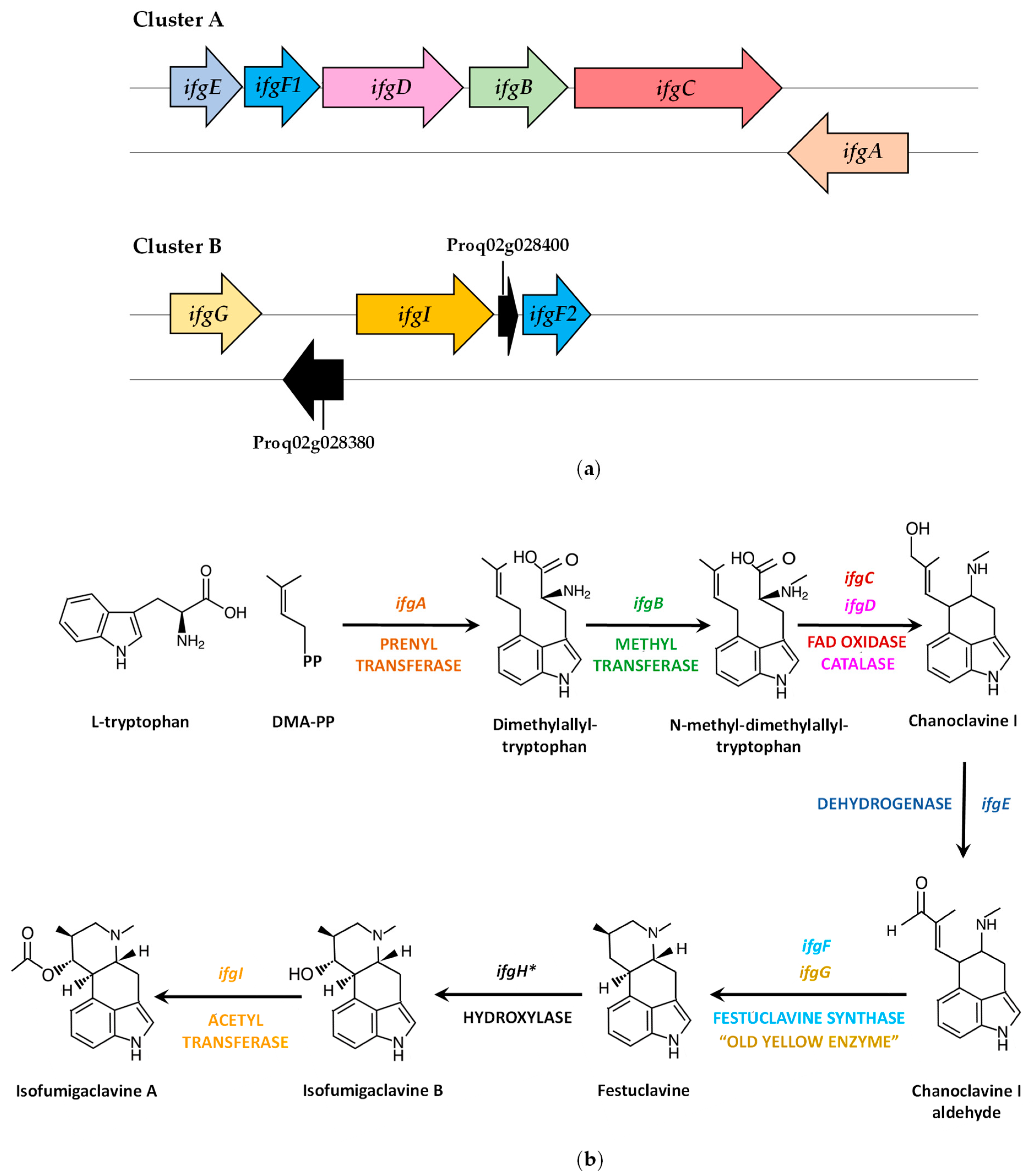
The identification of a second dimethylallyltryptophan synthase, (dmaW2, current annotation: Proq05g069310), different from the roquefortine prenyltransferase (rpt, dmaW Proq01g022770) involved in the biosynthesis of Roquefortine C, allowed the characterization of the Isofumigaclavine A biosynthetic gene cluster (Figure 3a, Table 2) and elucidation of the biosynthetic pathway in P. roqueforti [54] (Figure 3b).
Knock-down mutants in the dmaW2 gene were able to produce Roquefortine C but were blocked in the biosynthesis of Isofumigaclavine A, thus confirming the role of the protein encoded by dmaW2 gene in the Isofumigaclavine A biosynthetic pathway of P. roqueforti. Consequently, the dmaW2 gene was renamed ifgA (for isofumigaclavine A biosynthesis), and two clusters (A and B) containing the genes putatively involved in the biosynthesis of Isofumigaclavine A were identified in the P. roqueforti genome [54,55] (Figure 3a, Table 2). Cluster A includes ifgE, ifgF1, ifgD, igfB, ifgC and ifgA genes, whereas Cluster B contains three genes related to Isofumigaclavine A biosynthesis (ifgG, ifgI and ifgF2) and two extra genes encoding a phytanoyl-CoA dioxygenase (CDM30152) and an unnamed protein product (CDM30154), respectively, of uncharacterized role in this biosynthetic pathway (Figure 3a, Table 2).
The P. roqueforti Isofumigaclavine A biosynthetic pathway was proposed [54,55] by comparing the enzymes putatively encoded in Clusters A and B with orthologs of Neosartorya fumigata, whose function was characterized by different molecular techniques [56,57]. The formation of dimethylallyltryptophan, specific for clavine biosynthesis, is carried out by the prenyltransferase encoded by ifgA. In the next step, the N-methyltransferase encoded by ifgB adds a methyl group in the amino group of dimethylallyltryptophan, thereby forming N-methyl-dimethylallyltryptophan. This compound is converted into Chanoclavine I by the coordinated action of the FAD-dependent oxidoreductase (encoded by ifgC) and the catalase (encoded by ifgD). Then, the short chain dehydrogenase encoded by ifgE transforms Chanoclavine I into the aldehyde form, which is converted into festuclavine by the festuclavine synthases (encoded by ifgF1 and ifgF2, the latter located on Cluster B) and the FMN-containing “old yellow enzyme” (encoded by the ifgG, located on Cluster B). The next step is catalyzed by the festuclavine hydroxylase, which is a cytochrome P450 that carries out the hydroxylation of C-9, thus forming Isofumigaclavine B. The gene encoding this protein (ifgH) is not present in Clusters A or B. Therefore, alternative genes for P450 cytochrome monooxygenases may encode this enzyme. Finally, the acetyltransferase encoded by ifgI (located on Cluster B) has been proposed to participate in the conversion of Isofumigaclavine B to Isofumigaclavine A [54,55].
In addition to these enzymes, a second “old yellow enzyme” named FgaOx3Pr3 has been reported in P. roqueforti, and the gene encoding this protein is outside Clusters A and B. FgaOx3Pr3 was able to catalyze the formation of festuclavine in the presence of a festuclavine synthase FgaFS [58], thus suggesting that at least two different “old yellow enzymes” may contribute to isofumigaclavine biosynthesis in P. roqueforti [55].

4.3. PR-Toxin Biosynthetic Gene Cluster and Pathway

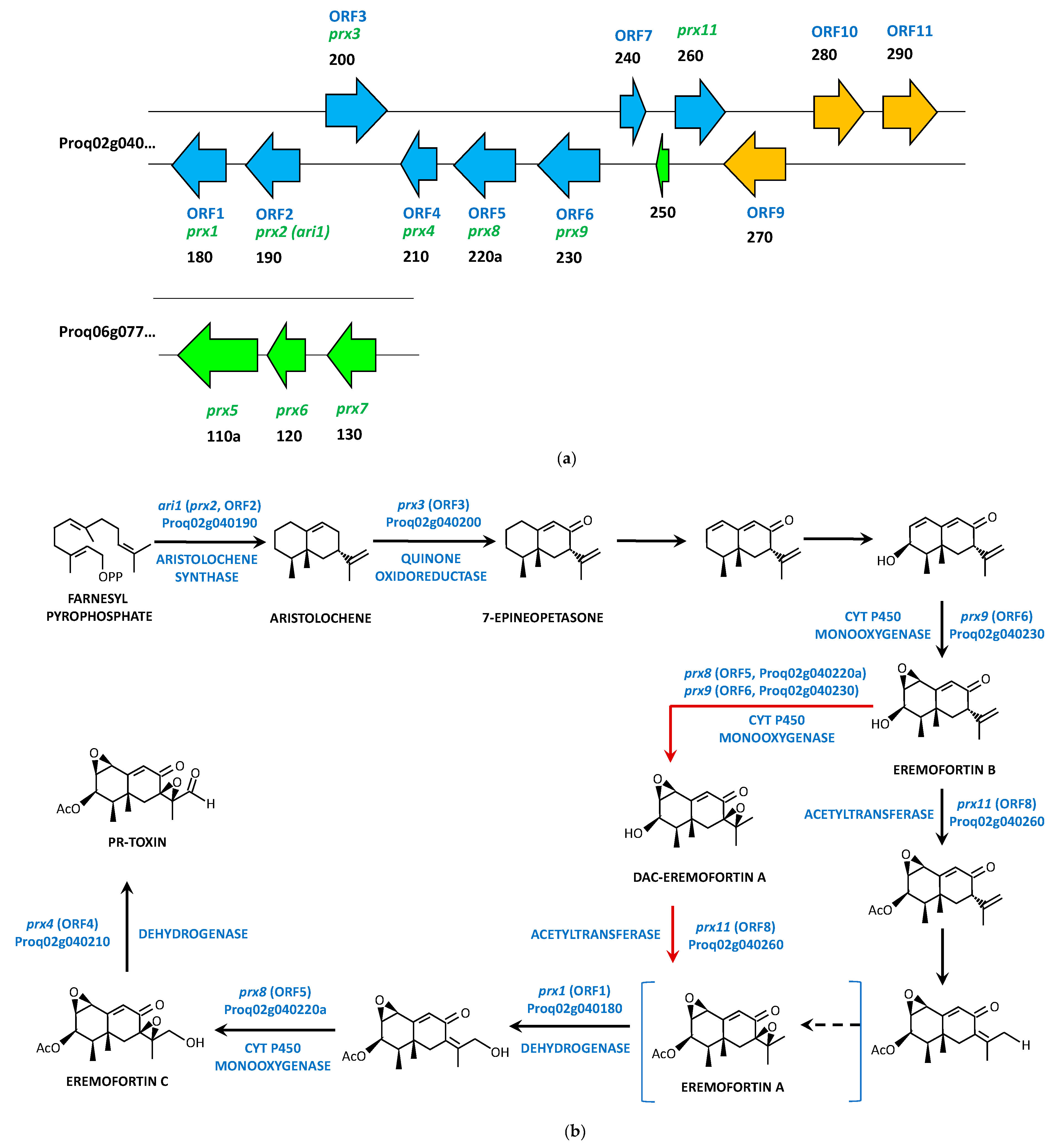
The PR-toxin is an aristolochene-derived bicyclic sesquiterpenoid compound of the eremophilane class [27] synthesized by the aristolochene synthase (encoded by ari1) from farnesyldiphosphate [59,60]. The ari1 gene was cloned from P. roqueforti [60] and used as reference for the screening of a phage library of P. roqueforti, which allowed the identification of four genes (prx1-prx4) of the PR-toxin biosynthetic pathway, including the ari1 gene (prx2) [61]. This structure was compared to that present in P. chrysogenum, thereby revealing that in this microorganism, the prx cluster contained 11 genes (Pc12g06260 to Pc12g06370, with Pc12g06290 being a pseudogene) [61]. Later, the availability of the genome sequence of P. roqueforti allowed the full characterization of the prx gene cluster in this filamentous fungus [34]. Orthologs to prx1 (Proq02g040180)-prx11 (Proq02g040260) were identified on scaffolds ProqFM164S02 and ProqFM164S04, since prx5 (Proq06g077110a, 2233 bp, encoding a putative MFS membrane protein of 567 amino acids), prx6 (Proq06g077120, 992 bp, encoding a putative NAD-dependent dehydrogenase of 291 amino acids), and prx7 (Proq06g077130, 1113 bp, encoding a putative NAD-dependent dehydrogenase of 295 amino acids) genes were found on a different genomic region (contig Proq06) away from prx1, prx2 (ari1), prx3, prx4, prx8, prx9, and prx11 genes (contig Proq02). In addition, no orthologs were identified for prx10 in P. roqueforti. Instead, two ORFs were found between prx9 and prx11 genes and between Proq02g040240 (ORF7) and Proq02g040250 (150 bp, annotated as 49 amino acids unnamed protein product) [34] (Figure 4a). Given this unusual distribution of the prx genes in two different genome regions in P. roqueforti, a new biosynthetic gene cluster was proposed and also included 11 ORFs, with ORF1 corresponding to the prx1 gene (Proq02g040180) (Figure 4a, Table 3). This cluster did not include the prx5, prx6, and prx7 genes and incorporated three additional genes located downstream of prx11: Proq02g040270a (ORF9), Proq02g040280 (ORF10), and Proq02g040290 (ORF 11). This new cluster was similar in terms of gene synteny and protein homologies to that present in P. chrysogenum and Penicillium camemberti. The main difference was the orientation of ORF7 (Proq02g040240) in P. roqueforti regarding the other two species [62].
The silencing of ORF1 to ORF4 (prx1-to prx4 genes) [61] and ORF5, ORF6, and ORF 8 (prx8, prx9 and prx11 genes) [62], together with the identification of new pathway intermediates (7-epineopetasone) [63], allowed the proposal of hypothetical PR-toxin biosynthetic pathways in P. roqueforti (Figure 4b), although the precise function of several enzymes encoded by the prx genes and the nature of most pathway intermediates are still unclear.
The PR-toxin is formed by three molecules of isopentenyldiphosphate (converted into farnesyl diphosphate) and from an acetyl group [64]. Farnesyl diphosphate is then cyclized to the 15-carbon molecule aristolochene by the aristolochene synthase, encoded by the ari1 (prx2, ORF2, Proq02g040190) gene [34,59,60,61,62]. Aristolochene is then converted to 7-epineopetasone via an allylic oxidation likely catalyzed by a quinone oxidoreductase encoded by prx3 (ORF3, Proq02g040200). The formation of Eremofortin B from 7-epineopetasone seems to involve one dehydrogenation (or allylic oxidation), one oxidation, and one epoxidation catalyzed by the monooxygenase encoded by prx9 (ORF6, Proq02g040230) as confirmed by gene-silencing methods [62]. Initially, it was proposed that Eremofortin B can be acetylated first by the enzyme encoded by prx11 and further oxidized, with Eremofortin A being a shunt product rather than an intermediate [63]. Later, it was suggested that Eremofortin B undergoes epoxidation by the enzymes encoded by prx8 (ORF5, Proq02g040220a) or prx9 (ORF6, Proq02g040230), thereby forming DAC-eremofortin A, the latter being acetylated by the acetyltransferase encoded by prx11 (ORF8, Proq02g040260) to form Eremofortin A [34]. Finally, it was suggested that Eremofortin A is really an intermediate in the pathway [62], which would be likely oxidized by the dehydrogenase encoded by prx1 (ORF1, Proq02g040180) and by the monooxygenase encoded by prx8 (ORF5, Proq02g040220a) to Eremofortin C [34,62] with the formation of Eremofortin D as a byproduct. The last step in the pathway consists of a dehydrogenation at C-12 of Eremofortin C, likely carried out by the protein encoded by prx4 (ORF4, Proq02g040210) to form the PR-toxin [34].

4.4. Mycophenolic Acid Biosynthetic Gene Cluster and Pathway

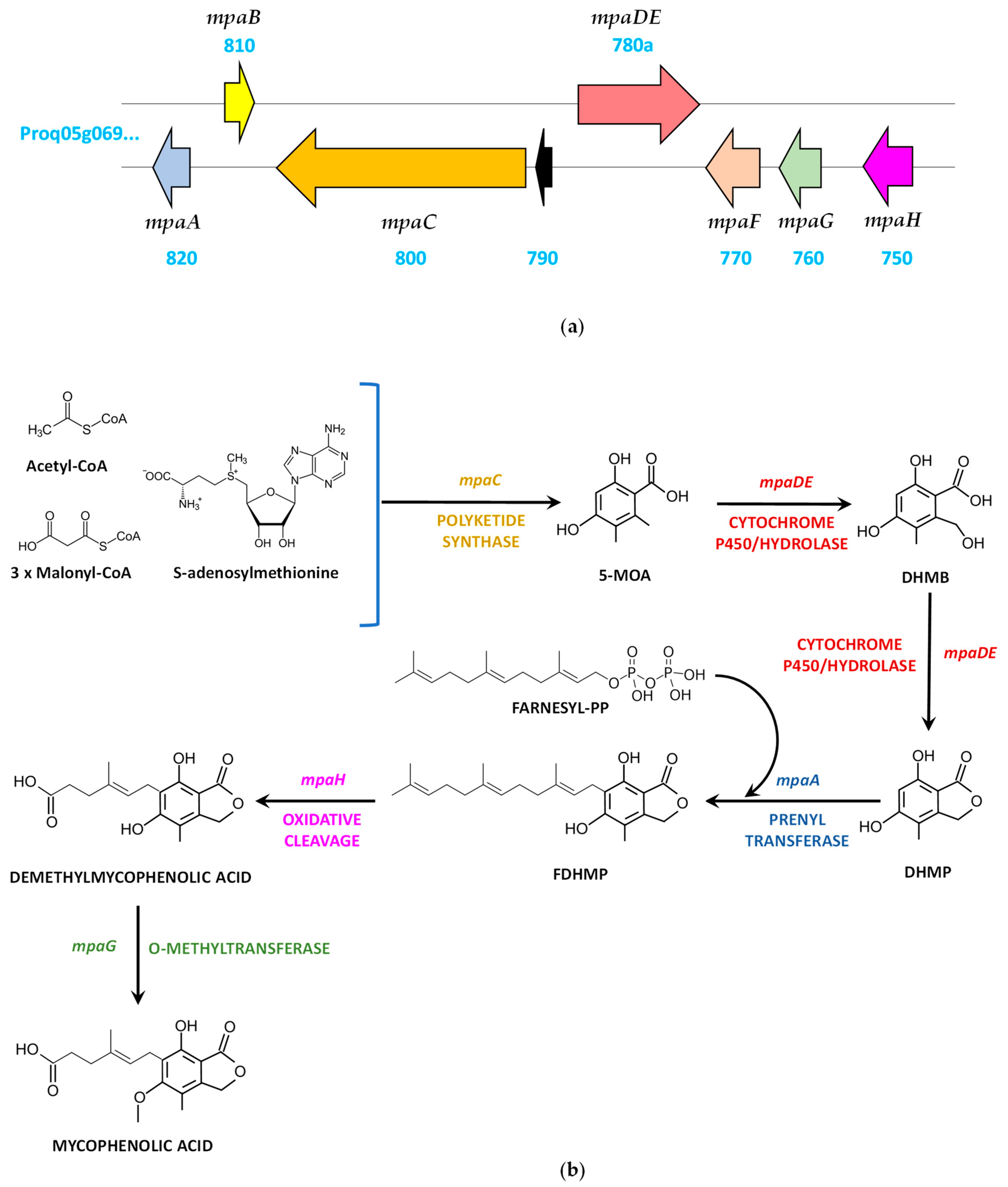
The information provided in different studies carried out in Penicillium brevicompactum, where the mycophenolic acid gene cluster comprising mpaA, mpaB, mpaC, mpaDE, mpaF, mpaG, and mpaH genes was identified [65,66,67], laid the foundations for the characterization of the mycophenolic acid biosynthetic gene cluster and pathway in P. roqueforti, which showed a similar structure [68,69] (Figure 5a, Table 4).
Starting from the mpaA gene, the cluster includes, adjacent to it and in divergent orientation, the mpaB gene, which was annotated as a 949-bp gene. However, this gene was predicted and confirmed to be larger (1397 bp) and with an additional exon at the 3′ end [68]. The next gene in the cluster is mpaC, and annotated next to it there is an ORF (Proq05g069790) that was not included in the cluster described by Del–Cid et al. [68] and Gillot et al. [69]. The next gene in the cluster is mpaDE, which was annotated as a 3710-bp ORF encoding a fusion protein of 932 amino acids made up of cytochrome P450 domain in the N-terminal region and a hydrolase domain in the C-terminal region. However, this gene was predicted and confirmed to be shorter (2929 bp; 788 bp in excess at the 5′ end) [68]. The last three genes in the cluster are mpaF, mpaG, and mpaH (Figure 5a).
Involvement of the mpa genes in the biosynthesis of mycophenolic acid in P. roqueforti was confirmed by gene-silencing experiments, which led to a reduction in the production of this metabolite after knocking down each of the seven genes [68]. These data, together with early precursor labeling and culture feeding studies [65,66,67,68,70], have led to the proposal of a biosynthetic pathway for mycophenolic acid (Figure 5b). Acetyl-CoA, malonyl-CoA and S-adenosylmethionine are the precursor molecules of mycophenolic acid. These compounds are condensed by the non-reducing polyketide synthase encoded by mpaC, thus forming 5-methylorsellinic acid. This molecule is further converted into 4,6-dihydroxy-2-(hydroxymethyl)-3-methylbenzoic acid and 5,7-dihydroxy-4-methylphthalide by sequential reactions carried out by the bifunctional cytochrome P450/hydrolase (lactone synthase) encoded by the mpaDE gene. Next, the prenyltransferase encoded by mpaA catalyzes the farnesylation of 5,7-dihydroxy-4-methylphthalide, thus forming 6-farnesyl-5,7-dihydroxy-4-methylphthalide, which undergoes oxidative cleavage of the farnesyl chain by the enzyme encoded by mpaH, thereby generating demethylmycophenolic acid. The last reaction consists of the methylation of the 5-hydroxy group of the demethylmycophenolic acid to form mycophenolic acid, a reaction that is catalyzed by the O-methyltransferase encoded by mpaG. The role of the mpaB-encoded protein remains unknown, whereas the probable inosine-5′-monophosphate dehydrogenase encoded by mpaF could confer resistance to mycophenolic acid and, in some way, also participate in the production of this metabolite, since knock-down mutants in this gene showed reduced production of mycophenolic acid [68].

4.5. Andrastin A Biosynthetic Gene Cluster and Pathway

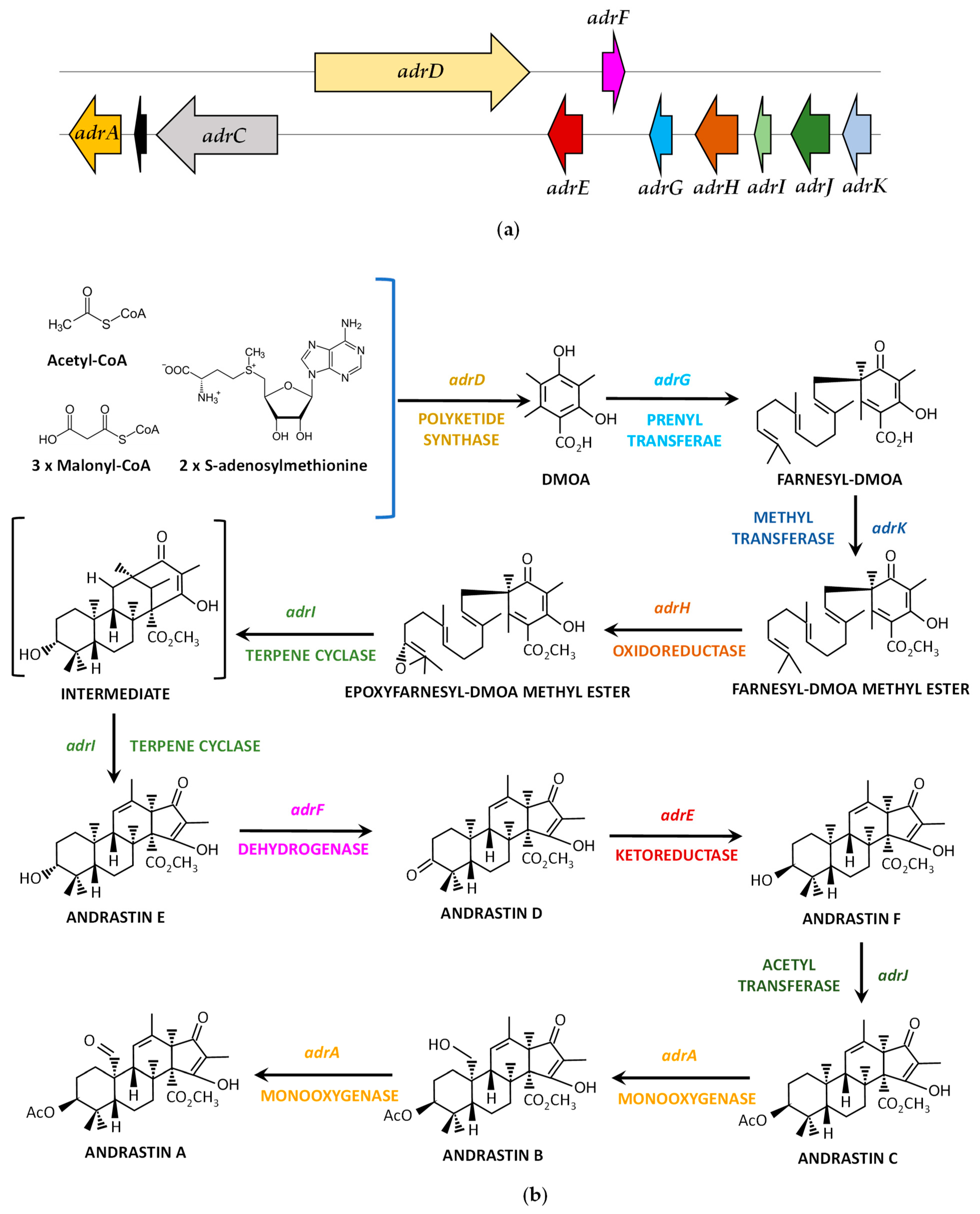
Following the information published in P. chrysogenum, where a ~30-kbp genomic cluster of 11 genes (adrA, Pc22g22820 to adrK, Pc22g22920) was proposed to encode the enzymes involved in Andrastin A biosynthesis [71], the homologous cluster was characterized in P. roqueforti [72]. The main difference with the P. chrysogenum adr cluster is the lack of the adrB gene in P. roqueforti, where only a small fragment of the 5′-end of this gene is present (representing a pseudogene), thereby comprising 10 genes in this filamentous fungus (Figure 6a, Table 5).
Despite the characterization of the adr gene cluster, the biosynthetic pathway for Andrastin A biosynthesis could not be elucidated in P. roqueforti. Initial studies in P. chrysogenum were able to hypothesize the initial steps of this pathway based on similarities of the adrD, adrG, adrK, and adrH genes with homologous genes of the fungal austinol and terretonin biosynthetic gene clusters [71]. According to these authors, the iterative Type I polyketide synthase, the prenyltransferase, the methyltransferase, and the FAD-dependent oxidoreductase would consecutively catalyze the four steps leading to the biosynthesis of epoxyfarnesyl-3,5-dimethylorsellinic acid methyl ester from acetyl CoA, malonyl CoA and S-adenosylmethionine. Next, these authors confirmed the function of the protein encoded by adrI (terpene cyclase) and were able to complete the biosynthesis of Andrastin A by heterologous co-expression of adrF, adrE, adrJ, and adrA genes in a strain of Aspergillus oryzae supplemented with Andrastin E. On the other hand, the putative role of the adrB- and adrC-encoded proteins could not be characterized in P. chrysogenum [71].
Given the high percentage of identity (ranging between 83% and 94%) shared by the orthologous adr-encoded proteins of P. chrysogenum and P. roqueforti (with exception of adrB, as indicated above), and since the 10 adr-encoded proteins are involved in the biosynthesis of andrastin A in P. roqueforti (as it was confirmed by RNA-mediated gene silencing of each of the 10 genes of the adr gene cluster [72]) a similar pathway to that proposed by P. chrysogenum [71] (Figure 6b) is likely to occur in P. roqueforti. Interestingly, the ABC transporter encoded by adrC (indicated as MFS transporter by [72]), although involved in Andrastin A production in P. roqueforti, showed no specific role in the secretion of this metabolite [72].

4.6. Annullatins D and F Biosynthetic Gene Cluster and Pathway

Prenylated salicylaldehyde derivatives are composed of a salicylaldehyde scaffold carrying a saturated or an unsaturated C7 side chain [73]. In fungi, prenylated salicylaldehyde derivatives, particularly those belonging to the group of flavoglaucins, have been isolated mainly from the Aspergillus (Eurotium) species [74,75,76,77,78]. Recently, the nine-gene biosynthetic gene cluster for flavoglaucin (the fog cluster) was identified in Aspergillus ruber [79]. Taking advantage of this information, Xiang et al. [80] used the fog cluster to search for homologues in the genome of P. roqueforti FM164. As a result, they identified a ~28-kbp genomic cluster of eleven genes in P. roqueforti, the anu cluster, which comprised genes anuA to anuK and included anuK and anuJ genes at both ends of the cluster (Figure 7a, Table 6).
Based on the putative functions of the anu genes, it was speculated that the product of this biosynthetic gene cluster should be a prenylated derivative. To identify the product, the full cluster was cloned in a plasmid and transferred to A. nidulans for heterologous expression. The resulting transformant (named A. nidulans BK08) was cultivated in a liquid medium. Broths of this transformant were extracted with ethyl acetate and analyzed by LC–MS. As a result, four new products absent in control strain were identified. The structures of these compounds were elucidated by spectroscopic techniques as Annullatin D, Annullatin F, Annullatin G, and Annullatin H [80]. Annullatins are a group of compounds that comprise 2,3-dihydrobenzofurans or aromatic polyketides derivatives and were previously identified in the entomopathogenic fungus Cordyceps annullata [81].
Further experiments, including gene deletion of anu genes in A. nidulans BK08, feeding experiments, and enzymatic characterization allowed for the reconstruction of a putative biosynthetic pathway for Annullatins D and F in P. roqueforti [80] (Figure 7b). Acetyl-CoA and malonyl-CoA are the precursor molecules of annullatins. These compounds are condensed to 2-hydroxymethyl-3-pentylphenol by the joint action of a reducing polyketide synthase (encoded by anuA), a short-chain dehydrogenases/reductase encoded by anuB, and a protein of unknown function encoded by anuC, which also seems to be a short-chain dehydrogenases/reductase [80]. Further conversion of 2-hydroxymethyl-3-pentylphenol into Annullatin E is catalyzed by the cytochrome P450 hydroxylase encoded by the anuE gene. Next, the prenyltransferase encoded by anuH introduces a dimethylallyl group from dimethylallyl diphosphate at the C-6 of phenol moiety of annullatin E thus forming annullatin J. At this point, the dihydrobenzofuran ring formation between the prenyl and the phenolic hydroxyl groups in Annullatin J would give rise to two diastereomers molecules, (2S, 9S)-annullatin H and the hypothetical (2R, 9S)-annullatin H. According to the literature, the formation of this ring could be non-enzymatic or enzymatic [73]. In the case of anu cluster, no gene has been involved in this part of the pathway [80]. After the diastereomers formation, the pathway splits into two branches. In the main branch, (2S, 9S)-annullatin H is converted to Annullatin D by two hypothetical sequential reactions carried out by the berberine bridge enzyme-like protein encoded by anuG. In the other branch, the hypothetical diastereomer (2R, 9S)-annullatin H is immediately converted to Annullatin F by the short-chain dehydrogenase/reductase encoded by anuF.
There are aspects of the anu cluster and its biosynthetic pathway that deserve to be highlighted. P. roqueforti has never been reported as a producer of annullatins. Indeed, Xiang et al. [80] did not detect annullatins in cultures of P. roqueforti FM164 grown under different conditions. These antecedents suggest that the anu cluster is silent. However, there is no gene expression data in P. roqueforti that could confirm it, but the heterologous expression of the anu cluster in A. nidulans provides indirect support to this fact (see Section 5.1).
Another interesting aspect is that the precise function of several enzymes encoded by the anu cluster and the nature of several pathway intermediates are unclear. Gene deletion experiments of anu genes in A. nidulans BK08 indicated that anuD, anuI, and anuJ are likely not involved in the annullatins biosynthesis [80]. Therefore, the function (if any) of these genes in P. roqueforti is currently unknown. Concerning pathway intermediates, several of them are hypothetical, with isomer (2R, 9S)-annullatin H, from which emerges one of the pathway branches (Figure 7b), being the most important one. It is speculated that this compound is very unstable, and for this reason, it cannot be detected [80].
Finally, there are several compounds that are produced in A. nidulans BK08 by shunt pathways, which could not be necessarily produced by natural strains of P. roqueforti. The main shunt pathway is observed after the deletion of anuE gene in A. nidulans BK08. Under these conditions, 2-hydroxymethyl-3-pentylphenol would be transformed to a hypothetical prenylated derivative by the prenyltransferase encoded by anuH. This prenylated precursor would be subsequently metabolized to Annullatin I, Annullatin B, and Annullatin A by unknown mechanisms (Figure 7b). Another shunt product, Annullatin G, would be produced from Annullatin F through an enzymatic reaction catalyzed by an unidentified endogenous enzyme from A. nidulans [80] (Figure 7b).

5. Control and Regulation of the Biosynthesis of Secondary Metabolites in P. roqueforti

The fungal secondary metabolism is controlled by a wide array of regulators, which lead directly or indirectly to the activation or repression of a given biosynthetic gene cluster. Depending on the level and extension of the effect, regulators of fungal secondary metabolism can be classified in two main groups: cluster-specific regulators and global regulators [82]. A cluster-specific regulator (also named specific transcription factor or narrow domain regulator) refers to a regulatory protein with a high similarity to a transcription factor that is encoded by a gene that is part of a specific biosynthetic gene cluster. More importantly, this transcription factor specifically regulates the expression of the other genes of the biosynthetic gene cluster [82]. On the contrary, global regulators (also named wide domain regulators) are encoded by genes located outside of a biosynthetic gene cluster and are pleiotropic regulators in fungi. Although most of these regulators are transcription factors, this category also includes signal transducer proteins and epigenetic regulators [82]. In the following sections, we will summarize the current knowledge about the regulation of biosynthetic gene clusters in P. roqueforti by cluster-specific and global regulators.

5.1. Cluster-Specific Regulators in Biosynthetic Gene Clusters Functionally Characterized in P. roqueforti

As mentioned before, the biosynthetic gene clusters for the biosynthesis of PR-toxin, Andrastin A, Roquefortine C, mycophenolic acid, annullatins, and Isofumigaclavine A have been functionally characterized in P. roqueforti [46,54,61,62,68,69,72,80]. Among them, only the gene clusters for the biosynthesis of annullatins and PR-toxin contain candidate genes for a specific transcription factor [62,80].
In the case of the biosynthetic gene cluster of Annullatin D, the functionality of the gene encoding the specific transcription factor (named anuK) was partially demonstrated [80]. As mentioned in Section 4.6, the Annullatin D biosynthetic gene cluster seems to be silent. Therefore, it was heterologously expressed in A. nidulans for its functional characterization. For this purpose, the biosynthetic gene cluster was previously cloned into plasmid pJN017, which contains the constitutive promoter of the gpdA gene. Therefore, the authors took advantage of the fact that the first gene of the biosynthetic gene cluster corresponds to anuK (Figure 7) and cloned it without the natural promoter of anuK in such a way that this gene was under the control of the gpdA promoter [80]. As a result, the production of Annullatin D, Annullatin F, and other related metabolites was achieved, as described in Section 4.6. At this point, it should be noted that beyond this result, there is no further experimental evidence confirming the role of AnuK as the transcriptional activator of the annullatin biosynthetic gene cluster in P. roqueforti. However, bioinformatic analysis supports such a role. AnuK has high similarity to FogI, a putative transcription factor found in the biosynthetic gene cluster for the biosynthesis of flavoglaucin in A. ruber [79]. In addition, AnuK contains a Zn(II)2Cys6 domain. This kind of domain is found almost exclusively in fungi and has been linked to several functions, including the regulation of the secondary metabolism [83,84].
Concerning the biosynthetic gene cluster of PR-toxin, its functional characterization in P. roqueforti was reported in two consecutive papers [61,62], where seven genes were silenced by RNAi-mediated gene-silencing technology. Unfortunately, the gene encoding the putative transcription factor of the biosynthetic gene cluster, named ORF10 (Figure 4) [62], was not analyzed. Similar to AnuK described before, ORF10 encodes a protein that contains a Zn(II)2Cys6 domain. ORF10 is highly similar to orthologous genes found in the biosynthetic gene clusters for PR-toxin in P. camemberti and P. chrysogenum [62]. Thus, the high degree of conservation of ORF10 in biosynthetic gene clusters of different fungi suggests that this gene could be functional. In the future it will be interesting to determine the role of ORF10 in the regulation of the biosynthetic gene cluster for the biosynthesis of PR-toxin in P. roqueforti.

5.2. Global Regulators of Biosynthetic Gene Clusters in P. roqueforti

Almost all the current knowledge about the regulation of biosynthetic gene clusters in P. roqueforti corresponds to global regulators. However, it should be kept in mind that in comparison with model fungi (Aspergillus spp., P. chrysogenum, etc.), this knowledge is still scarce. Indeed, as far as we know, the effect of only three global regulators on the secondary metabolism have been studied in P. roqueforti. They are detailed below.

5.2.1. The pga1 Gene Encoding for an α-Subunit of a Heterotrimeric G Protein

The first global regulator studied in P. roqueforti was a heterologous gene named pga1, which encodes an α-subunit of a heterotrimeric G protein. Heterotrimeric G proteins, particularly α-subunits, are important in fungi and have been involved mainly in the regulation of growth, asexual reproduction, and secondary metabolism [85]. García–Rico et al. [86] isolated and characterized the pga1 gene from P. chrysogenum, and they decided to perform the heterologous expression of a mutant version of this gene in P. roqueforti [87]. For this purpose, they used a plasmid containing a mutant version of pga1 (named pga1G42R), which encodes a protein where a glycine is replaced by arginine at Position 42, producing a “constitutively active” Pga1 protein that is always signaling [88]. The introduction of this “constitutive” allele in P. roqueforti produced several phenotypic effects. In the case of secondary metabolism, García–Rico et al. [87] measured the production of Roquefortine C during 30 days in a strain of P. roqueforti containing the pga1G42R allele and observed drastic changes in the production of this secondary metabolite as compared with the wild-type strain. Namely, the strain containing pga1G42R increased production of the mycotoxin, reaching 0.7 μg/mg of dry mycelium at day 18, whereas the wild-type strain produced 0.4 μg/mg of dry mycelium at the same day. Interestingly, the levels of Roquefortine C were higher in the pga1G42R strain throughout a culture period ranging between 10–30 days [87]. These results suggested that pga1 has a positive effect on the production of Roquefortine C in P. roqueforti [87]. As mentioned above, these experiments were performed using a heterologous gene from P. chrysogenum. Therefore, the role of the native pga1 gene on the secondary metabolism of P. roqueforti remains to be tested.

5.2.2. The sfk1 Gene Encoding for Suppressor of Four-Kinase 1 Protein

Another global regulator influencing the biosynthesis of secondary metabolites in P. roqueforti is Sfk1 (Suppressor of four-kinase 1), a transmembrane protein located on the plasma membrane that was originally described in Saccharomyces cerevisiae [89]. In this yeast, Sfk1 physically interacts with Stt4, an important phosphatidylinositol 4-kinase involved in the phosphoinositide second messenger’s pathway [90]. Stt4 must be localized to the plasma membrane to fulfill its role. Hence, it is thought that its interaction with Sfk1 would allow its proper subcellular localization [91]. In addition, Sfk1 is essential for the retention of ergosterol in the plasma membrane of the yeast [92]. The mammalian homologue of Sfk1 was also studied [93,94]. The role of this homologue is essentially similar to that described in yeast, although it does not have a direct role in mammals in the localization of the phosphatidylinositol 4-kinase to the plasma membrane [93]. In addition, it has been suggested that in mammals, Sfk1 would also be a negative regulator of transbilayer movement of phospholipids in plasma membrane [94].
The interest in studying Sfk1 in P. roqueforti emerged from a previous work by Gil-Durán and co-workers [95]. These authors performed suppression subtractive hybridization (SSH) experiments, looking for downstream genes regulated by Pga1. Among the sequences obtained from these experiments, the full cDNA of sfk1 gene was found. Considering this antecedent, Torrent and co-workers [96] decided to analyze the role of this gene in P. roqueforti. For this purpose, they performed the RNA-mediated gene-silencing of sfk1 and measured several biological properties in the transformants. In the case of secondary metabolites, they observed that the knock-down of sfk1 produced a drastic decrease in the production of Roquefortine C, Andrastin A, and mycophenolic acid in P. roqueforti [96]. These results suggest that sfk1 is a positive regulator of the production of these three secondary metabolites in P. roqueforti.

5.2.3. The pcz1 Gene Encoding a Protein with a Zn(II)2Cys6 Domain

The last global regulator of secondary metabolism studied in P. roqueforti is that encoded by pcz1 (Penicillium C6 zinc-finger protein 1), a gene whose role in fungi was unknown until recent time. This gene encodes a protein containing a Zn(II)2Cys6 domain [95]. As in the case of sfk1, pcz1 was also obtained from SSH experiments performed on P. roqueforti [95]. Since there are orthologues of this gene in all ascomycetes analyzed so far [95], it seemed interesting to determine its role on the secondary metabolism of P. roqueforti. For this purpose, two kinds of P. roqueforti strains were obtained: strains overexpressing pcz1 [97] and strains where pcz1 was knocked-down by RNA-silencing technology [95]. Using both types of strains, the production of Roquefortine C, Andrastin A, and mycophenolic acid was measured in comparison to the wild-type strain [97]. In the case of mycophenolic acid, a clear effect was found. Strains overexpressing pcz1 showed higher titers of mycophenolic acid than the wild-type strain, which correlated with higher levels of the expression of key genes from the mycophenolic acid biosynthetic gene cluster [97]. On the contrary, strains where pcz1 was knocked down produced lower levels of the mycophenolic acid as compared to wild-type fungus in concomitance with lower levels of the expression of genes from its biosynthetic gene cluster [97]. These results indicate that pcz1 exerts a positive control of the production of mycophenolic acid and suggest that this effect is mediated by modifying the transcriptional status of the biosynthetic gene cluster of mycophenolic acid.
Unlike mycophenolic acid, important reductions in the production of Roquefortine C and Andrastin A were observed in both types of strains [97]. These results were confirmed by measuring the level of transcription of key genes from Roquefortine C and Andrastin A biosynthetic gene clusters [97]. Thus, the effect of pcz1 on these metabolites was unexpected and difficult to interpret. Considering these results, an indirect effect of pcz1 on the production of Roquefortine C and Andrastin A was suggested, and some hypotheses were proposed. The first one was related to competition for the use of sharing substrates. These three compounds require acetyl CoA directly or indirectly for their biosynthesis. Therefore, the overexpression of the mycophenolic acid pathway (produced in turn by the overexpression of pcz1) would use more efficiently acetyl-CoA, in detriment of the production of Andrastin A and Roquefortine C. This would produce a “negative loop” in the expression of Andrastin A and Roquefortine C biosynthetic gene clusters, thereby resulting in a low production of these compounds. The second hypothesis was the alteration of the normal balance of unknown regulators of Roquefortine C and Andrastin A because of the overexpression of pcz1 [97]. A similar phenomenon was described before in Aspergillus. In this case, it was observed that the elimination of the regulator mtfA decreased the production of two related mycotoxins: aflatoxin and sterigmatocystin [98,99]. However, the overexpression of mtfA also decreased the production of these mycotoxins because mtfA downregulates the normal expression of the transcriptional factor aflR, which is a positive activator of the biosynthesis of aflatoxin and sterigmatocystin in Aspergillus [98,99]. Finally, the last hypothesis suggested the existence of a regulatory mechanism not fully understood, known as regulatory cross-talk [97]. Regulatory cross-talk refers to an inter-regulation amongst different and non-related biosynthetic gene clusters in fungi [100]. Regulatory cross-talk was originally described in A. nidulans, where the overexpression of a gene encoding a NRPS of the so-called inp biosynthetic gene cluster resulted in the unexpected activation of the non-related biosynthetic gene cluster for the biosynthesis of asperfuranone [101].
In the literature, there are several other examples of regulatory cross-talk in different fungi [100]. In P. roqueforti, regulatory cross-talk has been described in the case of biosynthetic gene clusters encoding PR-toxin and mycophenolic acid [61]. More precisely, during the experiments of the functional characterization of the PR-toxin biosynthetic gene cluster [61], it was noticed that the down regulation of several genes of this biosynthetic gene cluster by RNAi-mediated silencing technology largely increased the production of mycophenolic acid. It is important to highlight that PR-toxin and mycophenolic acid are unrelated secondary metabolites, so this result can hardly be due to an imbalance of shared substrates. PR-toxin is a bicyclic sesquiterpene that belongs to eremophilane. Its core structures is aristolochene, a sesquiterpene produced from farnesyl diphosphate [28], whereas mycophenolic acid is a meroterpenoid whose core structure, 5-methylorsellinic acid (5-MOA), is formed by a polyketide synthase. 5-MOA is subjected to several further modifications (including prenylation) to yield mycophenolic acid [102]. Thus, both compounds are synthesized by entirely different biosynthetic pathways in P. roqueforti [62,68].
Considering the previous observation of regulatory cross-talk in P. roqueforti, Rojas–Aedo and co-workers [97] suggested that the unexpected result in the regulation of mycophenolic acid, Andrastin A and Roquefortine C by pcz1 in this fungus, could be due to the existence of regulatory cross-talk between the biosynthetic gene clusters of these metabolites. Hence, when pcz1 is subjected to genetic manipulation, a complex network of regulatory cross-talk between these biosynthetic gene clusters is triggered. Depending on the case, the overproduction or reduction in the levels of some compounds is observed. However, this remains as a hypothesis that requires further experimental support.

5.3. Concluding Remarks on Regulation of Secondary Metabolism in P. roqueforti

In comparison with other fungi, our current knowledge about the regulation of the biosynthesis of secondary metabolites in P. roqueforti is poor, which should encourage fungal biologists to pay more attention to this interesting topic. As mentioned above, to date, barely one specific regulator of a biosynthetic gene cluster has been indirectly characterized in P. roqueforti [80], and some important global regulators, such as CreA, LaeA, PacC, or AreA have not yet been analyzed in this fungus. This is unlike in other fungi, where their role in the control of secondary metabolism has been largely demonstrated. For example, CreA exerts carbon catabolic repression on penicillin biosynthesis and the expression of the pcbAB (the gene encoding the first enzyme of the penicillin pathway) in P. chrysogenum [103], while PacC exerts pH-dependent control on the production of patulin and the expression of its BGC in P. expansum [104]. In the case of LaeA, its influence on the secondary metabolism of several Penicillium species has been documented [82]. Concerning the regulator of the nitrogen metabolism AreA, it has been involved in the control of the production of secondary metabolites in P. chrysogenum and P. griseofulvum [82]. Interestingly, the areA gene homologue has been studied in P. roqueforti [105], but its role on secondary metabolism has not been addressed in this fungus.

6. Future Challenges and Perspectives in the Study of Secondary Metabolism in P. roqueforti

P. roqueforti provides cheese with characteristic organoleptic properties. These properties have been associated to the potent proteolytic and lipolytic activities of this fungus [20,21,106]. In the case of the secondary metabolites produced by P. roqueforti, it remains to be determined if they have any role in the organoleptic properties of the cheeses. It is known that P. roqueforti inoculates secondary metabolites to cheese in which it grows [42,107]. Therefore, one of the challenges to be addressed in this fungus is whether these secondary metabolites confer organoleptic properties.
It has been estimated that the number of fungal species in nature is between 2- and 11-million species, of which around 150,000 are formally described taxa [108]. Therefore, most of the biosynthetic potential of fungi is yet to be discovered. On the other hand, genomic analyses performed on known fungal species indicate that 80% of their metabolic potential remains unknown [2]. In Penicillium species, a large proportion of its biosynthetic gene clusters, in some cases up to an astonishing 90%, have not been connected yet to any molecule [8]. These data indicate that in fungi, including Penicillium species, secondary metabolism and biosynthetic gene clusters have been hitherto barely investigated. This is also true for P. roqueforti. Recent analyses of several genomes from P. roqueforti revealed that depending on the strain, they contain between 34–37 biosynthetic gene clusters [10,109]. As it has been detailed in this review, to date, only six of these biosynthetic gene clusters have been experimentally linked to secondary metabolites produced by P. roqueforti. Therefore, the rest of the biosynthetic gene clusters (between 28–31, depending on the strain, representing around 80% of the total biosynthetic gene clusters of P. roqueforti), have not been hitherto associated to any known compound. It would be also interesting in the future to study the expression of those biosynthetic gene clusters when P. roqueforti or related species are grown on cheese as a substrate.
One of the most interesting research questions to be addressed in P. roqueforti is the assignment of a biosynthetic gene cluster of unknown function to a specific secondary metabolite. To answer this question, the coupling of microbiological and molecular biology techniques with chemical methodologies is necessary [110,111,112,113]. This strategy requires tools for efficient genetic manipulation of the fungal biosynthetic gene clusters of interest. Gene deletion has been the gold standard for this purpose. Gene deletion can be performed by gene targeting; that is, the replacement of a gene of a biosynthetic gene cluster with a non-functional version of the same gene through random events of homologous recombination [110,114,115]. Unfortunately, in filamentous fungi, gene targeting is a cumbersome process. In most filamentous fungi, DNA integration is mainly driven by non-homologous recombination, whereas homologous recombination events occur at frequencies between 1–5%, or even less [116,117]. As a result, successful gene-targeted deletion events occur at such a low frequency that it makes this technique impractical for many fungi [118]. In the case of P. roqueforti, there is only one report of successful gene deletion, [119] which, to the best of our knowledge, has not been replicated by other researchers.
The lack of efficient tools for gene knock-out in P. roqueforti has led to most of the studies of its biosynthetic gene clusters having been done by using RNA-mediated gene-silencing (or gene knock-down). Gene silencing affects gene functionality by reducing its expression but does not affect gene structure (the gene remains intact within the fungal genome) [120,121]. Gene silencing has high efficiency in fungi [121,122]. In some cases, up to 95% of reduced gene expression has been observed, thus yielding phenotypes close to knock-out strains [68,123,124]. Due to its simplicity (this technique only requires the expression of a very small double-stranded RNA molecule [121,122]), gene silencing allows the quick analysis of several genes of a biosynthetic gene cluster at the same time. Considering all these advantages, RNA-mediated gene-silencing has become the most useful technique for the study of biosynthetic gene clusters in P. roqueforti [46,54,61,62,68,69,72].
The scenario described above is now changing thanks to the development of CRISPR–Cas9 systems. In past years, several CRISPR–Cas9 systems dedicated to filamentous fungi have been developed and successfully applied to inactivate biosynthetic gene clusters, thus opening new horizons in the research of fungal secondary metabolism [114,125]. Recently, Seekles and co-workers [126] developed the first CRISPR–Cas9 system dedicated to P. roqueforti, which has already been successfully applied to the inactivation of genes in this fungus [126,127]. We believe that the availability of this CRISPR/Cas9 system will be a decisive boost for the study of biosynthetic gene clusters, as well as regulatory genes in P. roqueforti. This technique must be the starting point to the “synthetic biology era” in this fungus, thereby allowing the re-programming of P. roqueforti to produce novel secondary metabolites in ways that have not yet been explored.

Author Contributions

All authors contributed to the review conceptualization, wrote, reviewed, and edited the manuscript, and prepared the figures. All authors have read and agreed to the published version of the manuscript.

Funding

This work was funded by Fondo Nacional de Desarrollo Científico y Tecnológico, FONDECYT, from the government of Chile, grant number 1211832.

Institutional Review Board Statement

Not applicable.

Informed Consent Statement

Not applicable.

Data Availability Statement

Not applicable.

Acknowledgments

R.C. acknowledges the support of DICYT-USACH. The authors would like to dedicate this article, in memoriam, to our beloved colleague Francisco Fierro, who recently passed away.

Conflicts of Interest

The authors declare no conflict of interest.

References

  1. Keller, N.P. Fungal secondary metabolism: Regulation, function and drug discovery. Nat. Rev. Microbiol. 2019, 17, 167–180. [Google Scholar] [CrossRef]
  2. Hautbergue, T.; Jamin, E.L.; Debrauwer, L.; Puel, O.; Oswald, I.P. From genomics to metabolomics, moving toward an integrated strategy for the discovery of fungal secondary metabolites. Nat. Prod. Rep. 2018, 35, 147–173. [Google Scholar] [CrossRef] [PubMed]
  3. Abdel-Razek, A.S.; El-Naggar, M.E.; Allam, A.; Morsy, O.M.; Othman, S.I. Microbial natural products in drug discovery. Processes 2020, 8, 470. [Google Scholar] [CrossRef] [Green Version]
  4. Fierro, F.; Vaca, I.; Castillo, N.I.; García-Rico, R.O.; Chávez, R. Penicillium chrysogenum, a vintage model with a cutting-edge profile in biotechnology. Microorganisms 2022, 10, 573. [Google Scholar] [CrossRef] [PubMed]
  5. Sawant, A.M.; Vamkudoth, K.R. Biosynthetic process and strain improvement approaches for industrial penicillin production. Biotechnol. Lett. 2022, 44, 179–192. [Google Scholar] [CrossRef]
  6. Frisvad, J.C.; Smedsgaard, J.; Larsen, T.O.; Samson, R.A. Mycotoxins, drugs and other extrolites produced by species in Penicillium subgenus Penicillium. Stud. Mycol. 2004, 49, 201–241. [Google Scholar]
  7. Grijseels, S.; Nielsen, J.C.; Randelovic, M.; Nielsen, J.; Nielsen, K.F.; Workman, M.; Frisvad, J.C. Penicillium arizonense, a new, genome sequenced fungal species, reveals a high chemical diversity in secreted metabolites. Sci. Rep. 2016, 6, 35112. [Google Scholar] [CrossRef] [Green Version]
  8. Nielsen, J.C.; Grijseels, S.; Prigent, S.; Ji, B.; Dainat, J.; Nielsen, K.F.; Frisvad, J.C.; Workman, M.; Nielsen, J. Global analysis of biosynthetic gene clusters reveals vast potential of secondary metabolite production in Penicillium species. Nat. Microbiol. 2017, 2, 17044. [Google Scholar] [CrossRef]
  9. Grijseels, S.; Nielsen, J.C.; Nielsen, J.; Larsen, T.O.; Frisvad, J.C.; Nielsen, K.F.; Frandsen, R.J.N.; Workman, M. Physiological characterization of secondary metabolite producing Penicillium cell factories. Fungal Biol. Biotechnol. 2017, 4, 8. [Google Scholar] [CrossRef] [Green Version]
  10. Coton, E.; Coton, M.; Hymery, N.; Mounier, J.; Jany, J.-L. Penicillium roqueforti: An overview of its genetics, physiology, metabolism and biotechnological applications. Fungal Biol. Rev. 2020, 34, 59–73. [Google Scholar] [CrossRef]
  11. Thom, C. Fungi in cheese ripening: Camembert and Roquefort. USDA Bureau Anim. Industry Bull. 1906, 82, 1–39. [Google Scholar]
  12. Boysen, M.; Skouboe, P.; Frisvad, J.; Rossen, L. Reclassification of the Penicillium roqueforti group into three species on the basis of molecular genetic and biochemical profiles. Microbiology 1996, 142, 541–549. [Google Scholar] [CrossRef] [PubMed] [Green Version]
  13. Frisvad, J.C.; Samson, R.A. Polyphasic taxonomy of Penicillium subgenus Penicillium—A guide to identification of food and air-borne terverticillate Penicillia and their mycotoxins. Stud. Mycol. 2004, 49, 1–73. [Google Scholar]
  14. Houbraken, J.; Kocsubé, S.; Visagie, C.M.; Yilmaz, N.; Wang, X.C.; Meijer, M.; Kraak, B.; Hubka, V.; Bensch, K.; Samson, R.A.; et al. Classification of Aspergillus, Penicillium, Talaromyces and related genera (Eurotiales): An overview of families, genera, subgenera, sections, series and species. Stud. Mycol. 2020, 95, 5–169. [Google Scholar] [CrossRef] [PubMed]
  15. Ropars, J.; López-Villavicencio, M.; Dupont, J.; Snirc, A.; Gillot, G.; Coton, M.; Jany, J.L.; Coton, E.; Giraud, T. Induction of sexual reproduction and genetic diversity in the cheese fungus Penicillium roqueforti. Evol. Appl. 2014, 7, 433–441. [Google Scholar] [CrossRef]
  16. Albillos, S.M.; García-Estrada, C.; Martín, J.-F. Spanish blue cheeses: Functional metabolites. In Cheese: Types, Nutrition and Consumption; Foster, R.D., Ed.; Nova Science Publishers, Inc.: New York, NY, USA, 2011; pp. 89–105. [Google Scholar]
  17. Moreau, C. Le Penicillium roqueforti, morphologie, physiologie, interet en industrie fromagere, mycotoxines. Lait 1980, 60, 254–271. [Google Scholar] [CrossRef] [Green Version]
  18. Labbe, M.; Serres, J.-P. Chroniques du Roquefort: De la préhistoire à l′aube de l′ère industrielle; Graphi Imprimeur: La Primaube, France, 2004. [Google Scholar]
  19. Labbe, M.; Serres, J.-P. Chroniques du Roquefort: Des hommes, des entreprises, des marques, période modern; Graphi Imprimeur: La Primaube, France, 2009. [Google Scholar]
  20. McSweeney, P.L.H.; Sousa, M.J. Biochemical pathways for the production of flavour compounds in cheeses during ripening: A review. Lait 2000, 80, 293–324. [Google Scholar] [CrossRef]
  21. Collins, Y.F.; McSweeney, P.L.H.; Wilkinson, M.G. Lipolysis and free fatty acid catabolism in cheese: A review of current knowledge. Int. Dairy J. 2003, 13, 841–866. [Google Scholar] [CrossRef]
  22. Deetae, P.; Bonnarme, P.; Spinnler, H.E.; Helinck, S. Production of volatile aroma compounds by bacterial strains isolated from different surface-ripened French cheeses. Appl. Microbiol. Biotechnol. 2007, 76, 1161–1171. [Google Scholar] [CrossRef] [PubMed]
  23. Ismaiel, A.A.; Ahmed, A.S.; El-Sayed, E.S.R. Optimization of submerged fermentation conditions for immunosuppressant mycophenolic acid production by Penicillium roqueforti isolated from blue-molded cheeses: Enhanced production by ultraviolet and gamma irradiation. World J. Microbiol. Biotechnol. 2014, 30, 2625–2638. [Google Scholar] [CrossRef]
  24. Silva, T.P.; Souza, L.O.; Reis, N.S.; Assis, S.A.; Ferreira, M.L.O.; Oliveira, J.R.; Aguiar-Oliveira, E.; Franco, M. Cultivation of Penicillium roqueforti in cocoa shell to produce and characterize its lipase extract. Rev. Mex. Ing. Quim. 2017, 16, 745–756. [Google Scholar]
  25. de Almeida Antunes Ferraz, J.L.; Souza, L.O.; Soares, G.A.; Coutinho, J.P.; de Oliveira, J.R.; Aguiar-Oliveira, E.; Franco, M. Enzymatic saccharification of lignocellulosic residues using cellulolytic enzyme extract produced by Penicillium roqueforti ATCC 10110 cultivated on residue of yellow mombin fruit. Bioresour. Technol. 2018, 248, 214–220. [Google Scholar] [CrossRef]
  26. Wei, R.D.; Still, P.E.; Smalley, E.B.; Schnoes, H.K.; Strong, F.M. Isolation and partial characterization of a mycotoxin from Penicillium roqueforti. Appl. Microbiol. 1973, 25, 111–114. [Google Scholar] [CrossRef] [PubMed]
  27. Wei, R.D.; Schnoes, H.K.; Hart, P.A.; Strong, F.M. The structure of PR toxin, a mycotoxin from Penicillium roqueforti. Tetrahedron 1975, 31, 109–114. [Google Scholar] [CrossRef]
  28. Dubey, M.K.; Aamir, M.; Kaushik, M.S.; Khare, S.; Meena, M.; Singh, S.; Upadhyay, R.S. PR toxin—Biosynthesis, genetic regulation, toxicological potential, prevention and control measures: Overview and challenges. Front. Pharmacol. 2018, 9, 288. [Google Scholar] [CrossRef] [PubMed] [Green Version]
  29. Chang, S.C.; Lu, K.L.; Yeh, S.F. Secondary metabolites resulting from degradation of PR toxin by Penicillium roqueforti. Appl. Environ. Microbiol. 1993, 59, 981–986. [Google Scholar] [CrossRef] [Green Version]
  30. Chang, S.C.; Yeh, S.F.; Li, S.Y.; Lei, W.Y.; Chen, M.Y. A novel secondary metabolite relative to the degradation of PR toxin by Penicillium roqueforti. Curr. Microbiol. 1996, 32, 141–146. [Google Scholar] [CrossRef]
  31. Moreau, S.; Gaudemer, A.; Lablache-Combier, A.; Biguet, J. Metabolites de Penicillium roqueforti: PR toxine et metabolites associes. Tetrahedron Lett. 1976, 11, 833–834. [Google Scholar] [CrossRef]
  32. Moreau, S.; Cacan, M.; Eremofortin, C. A new metabolite obtained from Penicillium roqueforti cultures and from biotransformation of PR toxin. J. Org. Chem. 1977, 42, 2632–2634. [Google Scholar] [CrossRef] [PubMed]
  33. Moreau, S.; Biguet, J.; Lablache-Combier, A.; Baert, M.; Delfosse, C. Structures et stereochimie des sesquiterpenes de Penicillium roqueforti pr toxine et eremofortines a, b, c, d, e. Tetrahedron. 1980, 36, 2989–2997. [Google Scholar] [CrossRef]
  34. García-Estrada, C.; Martín, J.F. Biosynthetic gene clusters for relevant secondary metabolites produced by Penicillium roqueforti in blue cheeses. Appl. Microbiol. Biotechnol. 2016, 100, 8303–8313. [Google Scholar] [CrossRef]
  35. Ohmomo, S.; Sato, T.; Utagawa, T.; Abe, M. Isolation of festuclavine and three new indole alkaloids, roquefortine A, B and C from the cultures of Penicillium roqueforti. Agr. Biol. Chem. 1975, 39, 1333–1334. [Google Scholar] [CrossRef] [Green Version]
  36. Ohmomo, S.; Utagawa, T.; Abe, M. Identification of roquefortine C produced by Penicillium roqueforti. Agr. Biol. Chem. 1977, 41, 2097–2098. [Google Scholar] [CrossRef]
  37. Scott, P.M.; Merrien, M.A.; Polonsky, J. Roquefortine and isofumigaclavine A, metabolites from Penicillium roqueforti. Experientia 1976, 32, 140–142. [Google Scholar] [CrossRef]
  38. Ohmomo, S.; Oguma, K.; Ohashi, T.; Abe, M. Isolation of a new indole alkaloid, roquefortine D, from the cultures of Penicillium roqueforti. Agr. Biol. Chem. 1978, 42, 2387–2389. [Google Scholar] [CrossRef] [Green Version]
  39. Nielsen, K.F.; Sumarah, M.W.; Frisvad, J.C.; Miller, J.D. Production of metabolites from the Penicillium roqueforti complex. J. Agric. Food Chem. 2006, 54, 3756–3763. [Google Scholar] [CrossRef] [PubMed]
  40. Bentley, R. Mycophenolic acid: A one hundred year odyssey from antibiotic to immunosuppressant. Chem. Rev. 2000, 100, 3801–3826. [Google Scholar] [CrossRef] [PubMed]
  41. O′Brien, M.; Nielsen, K.F.; O′Kiely, P.; Forristal, P.D.; Fuller, H.T.; Frisvad, J.C. Mycotoxins and other secondary metabolites produced in vitro by Penicillium paneum Frisvad and Penicillium roqueforti Thom isolated from baled grass silage in Ireland. J. Agric. Food Chem. 2006, 54, 9268–9276. [Google Scholar] [CrossRef] [PubMed] [Green Version]
  42. Nielsen, K.F.; Dalsgaard, P.W.; Smedsgaard, J.; Larsen, T.O. Andrastins A-D, Penicillium roqueforti metabolites consistently produced in blue-mold-ripened cheese. J. Agric. Food Chem. 2005, 53, 2908–2913. [Google Scholar] [CrossRef] [PubMed]
  43. Wang, J.P.; Yu, J.; Shu, Y.; Shi, Y.X.; Luo, P.; Cai, L.; Ding, Z.T. Peniroquesines A-C: Sesterterpenoids possessing a 5-6-5-6-5-fused pentacyclic ring system from Penicillium roqueforti YJ-14. Org. Lett. 2018, 20, 5853–5856. [Google Scholar] [CrossRef]
  44. Wang, J.P.; Shu, Y.; Hu, J.T.; Liu, R.; Cai, X.Y.; Sun, C.T.; Gan, D.; Zhou, D.J.; Mei, R.F.; Ding, H.; et al. Roquefornine A, a sesterterpenoid with a 5/6/5/5/6-fused ring system from the fungus Penicillium roqueforti YJ-14. Org. Chem. Front. 2020, 7, 1463–1468. [Google Scholar] [CrossRef]
  45. Wang, J.P.; Shu, Y.; Liu, R.; Gan, J.L.; Deng, S.P.; Cai, X.Y.; Hu, J.T.; Cai, L.; Ding, Z.T. Bioactive sesterterpenoids from the fungus Penicillium roqueforti YJ-14. Phytochemistry 2021, 187, 112762. [Google Scholar] [CrossRef] [PubMed]
  46. Kosalková, K.; Domínguez-Santos, R.; Coton, M.; Coton, E.; García-Estrada, C.; Liras, P.; Martín, J.F. A natural short pathway synthesizes roquefortine C but not meleagrin in three different Penicillium roqueforti strains. Appl. Microbiol. Biotechnol. 2015, 99, 7601–7612. [Google Scholar] [CrossRef] [PubMed]
  47. Ali, H.; Ries, M.I.; Nijland, J.G.; Lankhorst, P.P.; Hankemeier, T.; Bovenberg, R.A.; Vreeken, R.J.; Driessen, A.J. A branched biosynthetic pathway is involved in production of roquefortine and related compounds in Penicillium chrysogenum. PLoS ONE 2013, 8, e65328. [Google Scholar] [CrossRef] [PubMed] [Green Version]
  48. Cheeseman, K.; Ropars, J.; Renault, P.; Dupont, J.; Gouzy, J.; Branca, A.; Abraham, A.L.; Ceppi, M.; Conseiller, E.; Debuchy, R.; et al. Multiple recent horizontal transfers of a large genomic region in cheese making fungi. Nat. Commun. 2014, 5, 2876. [Google Scholar] [CrossRef] [Green Version]
  49. García-Estrada, C.; Ullán, R.V.; Albillos, S.M.; Fernández-Bodega, M.Á.; Durek, P.; von Döhren, H.; Martín, J.F. A single cluster of coregulated genes encodes the biosynthesis of the mycotoxins roquefortine C and meleagrin in Penicillium chrysogenum. Chem. Biol. 2011, 18, 1499–1512. [Google Scholar] [CrossRef] [Green Version]
  50. Ries, M.I.; Ali, H.; Lankhorst, P.P.; Hankemeier, T.; Bovenberg, R.A.; Driessen, A.J.; Vreeken, R.J. Novel key metabolites reveal further branching of the roquefortine/meleagrin biosynthetic pathway. J. Biol. Chem. 2013, 288, 37289–37295. [Google Scholar] [CrossRef] [Green Version]
  51. Barrow, K.D.; Colley, P.W.; Tribe, D.E. Biosynthesis of the neurotoxin alkaloid roquefortine. J. Chem. Soc. Chem. Commun. 1979, 5, 225–226. [Google Scholar] [CrossRef]
  52. Gorst-Allman, C.P.; Steyn, P.S.; Vleggaar, R. The biosynthesis of roquefortine. An investigation of acetate and mevalonate incorporation using high field n.m.r. spectroscopy. J. Chem. Soc. Chem. Commun. 1982, 12, 652–653. [Google Scholar] [CrossRef]
  53. Martín, J.F.; Liras, P. Evolutionary formation of gene clusters by reorganization: The meleagrin/roquefortine paradigm in different fungi. Appl. Microbiol. Biotechnol. 2016, 100, 1579–1587. [Google Scholar] [CrossRef]
  54. Fernández-Bodega, Á.; Álvarez-Álvarez, R.; Liras, P.; Martín, J.F. Silencing of a second dimethylallyltryptophan synthase of Penicillium roqueforti reveals a novel clavine alkaloid gene cluster. Appl. Microbiol. Biotechnol. 2017, 101, 6111–6121. [Google Scholar] [CrossRef]
  55. Martín, J.F.; Álvarez-Álvarez, R.; Liras, P. Clavine alkaloids gene clusters of Penicillium and related fungi: Evolutionary combination of prenyltransferases, monooxygenases and dioxygenases. Genes 2017, 8, 342. [Google Scholar] [CrossRef]
  56. Wallwey, C.; Li, S.M. Ergot alkaloids: Structure diversity, biosynthetic gene clusters and functional proof of biosynthetic genes. Nat. Prod. Rep. 2011, 28, 496–510. [Google Scholar] [CrossRef] [PubMed]
  57. Robinson, S.L.; Panaccione, D.G. Heterologous expression of lysergic acid and novel ergot alkaloids in Aspergillus fumigatus. Appl. Environ. Microbiol. 2014, 80, 6465–6472. [Google Scholar] [CrossRef] [Green Version]
  58. Gerhards, N.; Li, S.M. A bifunctional old yellow enzyme from Penicillium roqueforti is involved in ergot alkaloid biosynthesis. Org. Biomol. Chem. 2017, 15, 8059–8071. [Google Scholar] [CrossRef]
  59. Hohn, T.M.; Plattner, R.D. Purification and characterization of the sesquiterpene cyclase aristolochene synthase from Penicillium roqueforti. Arch. Biochem. Biophys. 1989, 272, 137–143. [Google Scholar] [CrossRef]
  60. Proctor, R.H.; Hohn, T.M. Aristolochene synthase. isolation, characterization, and bacterial expression of a sesquiterpenoid biosynthetic gene (Ari1) from Penicillium roqueforti. J. Biol. Chem. 1993, 268, 4543–4548. [Google Scholar] [CrossRef] [PubMed]
  61. Hidalgo, P.I.; Ullán, R.V.; Albillos, S.M.; Montero, O.; Fernández-Bodega, M.Á.; García-Estrada, C.; Fernández-Aguado, M.; Martín, J.F. Molecular characterization of the PR-toxin gene cluster in Penicillium roqueforti and Penicillium chrysogenum: Cross talk of secondary metabolite pathways. Fungal Genet. Biol. 2014, 62, 11–24. [Google Scholar] [CrossRef]
  62. Hidalgo, P.I.; Poirier, E.; Ullán, R.V.; Piqueras, J.; Meslet-Cladière, L.; Coton, E.; Coton, M. Penicillium roqueforti PR toxin gene cluster characterization. Appl. Microbiol. Biotechnol. 2017, 101, 2043–2056. [Google Scholar] [CrossRef] [PubMed]
  63. Riclea, R.; Dickschat, J.S. Identification of intermediates in the biosynthesis of PR toxin by Penicillium roqueforti. Angew. Chem. Int. Ed. Engl. 2015, 54, 12167–12170. [Google Scholar] [CrossRef]
  64. Chalmers, A.A.; de Jesus, A.E.; Gorst-Allman, C.P.; Steyn, P.S. Biosynthesis of PR toxin by Penicillium roqueforti. J. Chem. Soc. Perkin. 1 1981, 10, 2899–2903. [Google Scholar] [CrossRef]
  65. Regueira, T.B.; Kildegaard, K.R.; Hansen, B.G.; Mortensen, U.H.; Hertweck, C.; Nielsen, J. Molecular basis for mycophenolic acid biosynthesis in Penicillium brevicompactum. Appl. Environ. Microbiol. 2011, 77, 3035–3043. [Google Scholar] [CrossRef] [PubMed] [Green Version]
  66. Hansen, B.G.; Mnich, E.; Nielsen, K.F.; Nielsen, J.B.; Nielsen, M.T.; Mortensen, U.H.; Larsen, T.O.; Patil, K.R. Involvement of a natural fusion of a cytochrome P450 and a hydrolase in mycophenolic acid biosynthesis. Appl. Environ. Microbiol. 2012, 78, 4908–4913. [Google Scholar] [CrossRef] [Green Version]
  67. Zhang, W.; Cao, S.; Qiu, L.; Qi, F.; Li, Z.; Yang, Y.; Huang, S.; Bai, F.; Liu, C.; Wan, X.; et al. Functional characterization of MpaG′, the O-methyltransferase involved in the biosynthesis of mycophenolic acid. Chembiochem 2015, 16, 565–569. [Google Scholar] [CrossRef] [PubMed]
  68. Del-Cid, A.; Gil-Durán, C.; Vaca, I.; Rojas-Aedo, J.F.; García-Rico, R.O.; Levicán, G.; Chávez, R. Identification and functional analysis of the mycophenolic acid gene cluster of Penicillium roqueforti. PLoS ONE 2016, 11, e0147047. [Google Scholar] [CrossRef] [Green Version]
  69. Gillot, G.; Jany, J.L.; Dominguez-Santos, R.; Poirier, E.; Debaets, S.; Hidalgo, P.I.; Ullán, R.V.; Coton, E.; Coton, M. Genetic basis for mycophenolic acid production and strain-dependent production variability in Penicillium roqueforti. Food Microbiol. 2017, 62, 239–250. [Google Scholar] [CrossRef]
  70. Hansen, B.G.; Genee, H.J.; Kaas, C.S.; Nielsen, J.B.; Regueira, T.B.; Mortensen, U.H.; Frisvad, J.C.; Patil, K.R. A new class of IMP dehydrogenase with a role in self-resistance of mycophenolic acid producing fungi. BMC Microbiol. 2011, 11, 202. [Google Scholar] [CrossRef] [Green Version]
  71. Matsuda, Y.; Awakawa, T.; Abe, I. Reconstituted biosynthesis of fungal meroterpenoid andrastin A. Tetrahedron 2013, 69, 8199–8204. [Google Scholar] [CrossRef]
  72. Rojas-Aedo, J.F.; Gil-Durán, C.; Del-Cid, A.; Valdés, N.; Álamos, P.; Vaca, I.; García-Rico, R.O.; Levicán, G.; Tello, M.; Chávez, R. The biosynthetic gene cluster for andrastin A in Penicillium roqueforti. Front. Microbiol. 2017, 8, 813. [Google Scholar] [CrossRef] [Green Version]
  73. Ran, H.; Li, S.M. Fungal benzene carbaldehydes: Occurrence, structural diversity, activities and biosynthesis. Nat. Prod. Rep. 2021, 38, 240–263. [Google Scholar] [CrossRef]
  74. Lin, L.B.; Gao, Y.Q.; Han, R.; Xiao, J.; Wang, Y.M.; Zhang, Q.; Zhai, Y.J.; Han, W.B.; Li, W.L.; Gao, J.M. Alkylated salicylaldehydes and prenylated indole alkaloids from the endolichenic fungus Aspergillus chevalieri and their bioactivities. J. Agric. Food Chem. 2021, 69, 6524–6534. [Google Scholar] [CrossRef] [PubMed]
  75. Zhong, W.; Chen, Y.; Wei, X.; Wang, J.; Zhang, W.; Wang, F.; Zhang, S. Salicylaldehyde derivatives from a marine-derived fungus Eurotium sp. SCSIO F452. J. Antibiot. 2021, 74, 273–279. [Google Scholar] [CrossRef]
  76. Li, D.L.; Li, X.M.; Li, T.G.; Dang, H.Y.; Proksch, P.; Wang, B.G. Benzaldehyde derivatives from Eurotium rubrum, an endophytic fungus derived from the mangrove plant Hibiscus tiliaceus. Chem. Pharm. Bull. 2008, 56, 1282–1285. [Google Scholar] [CrossRef] [Green Version]
  77. Ishikawa, Y.; Morimoto, K.; Hamasaki, T. Flavoglaucin, a metabolite of Eurotium chevalieri, its antioxidation and synergism with tocopherol. J. Am. Oil Chem. Soc. 1984, 61, 1864–1868. [Google Scholar] [CrossRef]
  78. Gao, J.; León, F.; Radwan, M.M.; Dale, O.R.; Husni, A.S.; Manly, S.P.; Lupien, S.; Wang, X.; Hill, R.A.; Dugan, F.M.; et al. Benzyl derivatives with in vitro binding affinity for human opioid and cannabinoid receptors from the fungus Eurotium repens. J. Nat. Prod. 2011, 74, 1636–1639. [Google Scholar] [CrossRef] [PubMed] [Green Version]
  79. Nies, J.; Ran, H.; Wohlgemuth, V.; Yin, W.B.; Li, S.M. Biosynthesis of the prenylated salicylaldehyde flavoglaucin requires temporary reduction to salicyl alcohol for decoration before reoxidation to the final product. Org. Lett. 2020, 22, 2256–2260. [Google Scholar] [CrossRef]
  80. Xiang, P.; Kemmerich, B.; Yang, L.; Li, S.M. Biosynthesis of annullatin D in Penicillium roqueforti implies oxidative lactonization between two hydroxyl groups catalyzed by a BBE-like enzyme. Org. Lett. 2022, 24, 6072–6077. [Google Scholar] [CrossRef]
  81. Asai, T.; Luo, D.; Obara, Y.; Taniguchi, T.; Monde, K.; Yamashita, K.; Oshima, T. Dihydrobenzofurans as cannabinoid receptor ligands from Cordyceps annullata, an entomopathogenic fungus cultivated in the presence of an HDAC inhibitor. Tetrahedron Lett. 2012, 53, 2239–2243. [Google Scholar] [CrossRef]
  82. El Hajj Assaf, C.; Zetina-Serrano, C.; Tahtah, N.; Khoury, A.E.; Atoui, A.; Oswald, I.P.; Puel, O.; Lorber, S. Regulation of secondary metabolism in the Penicillium genus. Int. J. Mol. Sci. 2020, 21, 9462. [Google Scholar] [CrossRef]
  83. Chang, P.K.; Ehrlich, K.C. Genome-wide analysis of the Zn(II)2Cys6 zinc cluster-encoding gene family in Aspergillus flavus. Appl. Microbiol. Biotechnol. 2013, 97, 4289–4300. [Google Scholar] [CrossRef]
  84. García-Estrada, C.; Domínguez-Santos, R.; Kosalková, K.; Martín, J.-F. Transcription factors controlling primary and secondary metabolism in filamentous fungi: The β-lactam paradigm. Fermentation 2018, 4, 47. [Google Scholar] [CrossRef] [Green Version]
  85. Moon, H.; Han, K.H.; Yu, J.H. Upstream regulation of development and secondary metabolism in Aspergillus species. Cells 2022, 12, 2. [Google Scholar] [CrossRef] [PubMed]
  86. García-Rico, R.O.; Martín, J.F.; Fierro, F. The pga1 gene of Penicillium chrysogenum NRRL 1951 encodes a heterotrimeric G protein alpha subunit that controls growth and development. Res. Microbiol. 2007, 158, 437–446. [Google Scholar] [CrossRef] [PubMed]
  87. García-Rico, R.O.; Chávez, R.; Fierro, F.; Martín, J.F. Effect of a heterotrimeric G protein alpha subunit on conidia germination, stress response, and roquefortine C production in Penicillium roqueforti. Int. Microbiol. 2009, 12, 123–129. [Google Scholar] [PubMed]
  88. García-Rico, R.O.; Martín, J.F.; Fierro, F. Heterotrimeric Gα protein Pga1 from Penicillium chrysogenum triggers germination in response to carbon sources and affects negatively resistance to different stress conditions. Fungal Genet. Biol. 2011, 48, 641–649. [Google Scholar] [CrossRef]
  89. Audhya, A.; Emr, S.D. Stt4 PI 4-kinase localizes to the plasma membrane and functions in the Pkc1-mediated MAP kinase cascade. Dev. Cell. 2002, 2, 593–605. [Google Scholar] [CrossRef] [Green Version]
  90. Audhya, A.; Foti, M.; Emr, S.D. Distinct roles for the yeast phosphatidylinositol 4-kinases, Stt4p and Pik1p, in secretion, cell growth, and organelle membrane dynamics. Mol. Biol. Cell 2000, 11, 2673–2689. [Google Scholar] [CrossRef] [Green Version]
  91. Voelker, D.R. Protein and lipid motifs regulate phosphatidylserine traffic in yeast. Biochem. Soc. Trans. 2005, 33, 1141–1145. [Google Scholar] [CrossRef]
  92. Kishimoto, T.; Mioka, T.; Itoh, E.; Williams, D.E.; Andersen, R.J.; Tanaka, K. Phospholipid flippases and Sfk1 are essential for the retention of ergosterol in the plasma membrane. Mol. Biol. Cell. 2021, 32, 1374–1392. [Google Scholar] [CrossRef]
  93. Chung, J.; Nakatsu, F.; Baskin, J.M.; De Camilli, P. Plasticity of PI4KIIIα interactions at the plasma membrane. EMBO Rep. 2015, 16, 312–320. [Google Scholar] [CrossRef]
  94. Mioka, T.; Fujimura-Kamada, K.; Mizugaki, N.; Kishimoto, T.; Sano, T.; Nunome, H.; Williams, D.E.; Andersen, R.J.; Tanaka, K. Phospholipid flippases and Sfk1p, a novel regulator of phospholipid asymmetry, contribute to low permeability of the plasma membrane. Mol. Biol. Cell. 2018, 29, 1203–1218. [Google Scholar] [CrossRef] [PubMed]
  95. Gil-Durán, C.; Rojas-Aedo, J.F.; Medina, E.; Vaca, I.; García-Rico, R.O.; Villagrán, S.; Levicán, G.; Chávez, R. The pcz1 gene, which encodes a Zn(II)2Cys6 protein, is involved in the control of growth, conidiation, and conidial germination in the filamentous fungus Penicillium roqueforti. PLoS ONE 2015, 10, e0120740. [Google Scholar] [CrossRef] [PubMed] [Green Version]
  96. Torrent, C.; Gil-Durán, C.; Rojas-Aedo, J.F.; Medina, E.; Vaca, I.; Castro, P.; García-Rico, R.O.; Cotoras, M.; Mendoza, L.; Levicán, G.; et al. Role of sfk1 Gene in the filamentous fungus Penicillium roqueforti. Front. Microbiol. 2017, 8, 2424. [Google Scholar] [CrossRef] [PubMed] [Green Version]
  97. Rojas-Aedo, J.F.; Gil-Durán, C.; Goity, A.; Vaca, I.; Levicán, G.; Larrondo, L.F.; Chávez, R. The developmental regulator Pcz1 affects the production of secondary metabolites in the filamentous fungus Penicillium roqueforti. Microbiol. Res. 2018, 212–213, 67–74. [Google Scholar] [CrossRef]
  98. Ramamoorthy, V.; Dhingra, S.; Kincaid, A.; Shantappa, S.; Feng, X.; Calvo, A.M. The putative C2H2 transcription factor MtfA is a novel regulator of secondary metabolism and morphogenesis in Aspergillus nidulans. PLoS ONE 2013, 8, e74122. [Google Scholar] [CrossRef] [PubMed] [Green Version]
  99. Zhuang, Z.; Lohmar, J.M.; Satterlee, T.; Cary, J.W.; Calvo, A.M. The master transcription factor mtfA governs aflatoxin production, morphological development and pathogenicity in the fungus Aspergillus flavus. Toxins 2016, 8, 29. [Google Scholar] [CrossRef] [PubMed] [Green Version]
  100. Sheridan, K.J.; Dolan, S.K.; Doyle, S. Endogenous cross-talk of fungal metabolites. Front. Microbiol. 2015, 5, 732. [Google Scholar] [CrossRef] [Green Version]
  101. Bergmann, S.; Funk, A.N.; Scherlach, K.; Schroeckh, V.; Shelest, E.; Horn, U.; Hertweck, C.; Brakhage, A.A. Activation of a silent fungal polyketide biosynthesis pathway through regulatory cross talk with a cryptic nonribosomal peptide synthetase gene cluster. Appl. Environ Microbiol. 2010, 76, 8143–8149. [Google Scholar] [CrossRef] [Green Version]
  102. Zhang, W.; Du, L.; Qu, Z.; Zhang, X.; Li, F.; Li, Z.; Qi, F.; Wang, X.; Jiang, Y.; Men, P.; et al. Compartmentalized biosynthesis of mycophenolic acid. Proc. Natl. Acad. Sci. USA 2019, 116, 13305–13310. [Google Scholar] [CrossRef] [Green Version]
  103. Cepeda-García, C.; Domínguez-Santos, R.; García-Rico, R.O.; García-Estrada, C.; Cajiao, A.; Fierro, F.; Martín, J.F. Direct involvement of the CreA transcription factor in penicillin biosynthesis and expression of the pcbAB gene in Penicillium chrysogenum. Appl. Microbiol. Biotechnol. 2014, 98, 7113–7124. [Google Scholar] [CrossRef] [Green Version]
  104. Chen, Y.; Li, B.; Xu, X.; Zhang, Z.; Tian, S. The pH-responsive PacC transcription factor plays pivotal roles in virulence and patulin biosynthesis in Penicillium expansum. Environ. Microbiol. 2018, 20, 4063–4078. [Google Scholar] [CrossRef] [PubMed]
  105. Gente, S.; Poussereau, N.; Fèvre, M. Isolation and expression of a nitrogen regulatory gene, nmc, of Penicillium roqueforti. FEMS Microbiol. Lett. 1999, 175, 291–297. [Google Scholar] [CrossRef]
  106. Chavez, R.; Fierro, F.; Rico, R.O.G.; Laich, F. Mold-fermented foods: Penicillium spp. as ripening agents in the elaboration of cheese and meat products. In Mycofactories; Leitão, A.L., Ed.; Bentham Science Publishers: Emirate of Sharjah, United Arab Emirates, 2011; pp. 73–98. [Google Scholar]
  107. Fontaine, K.; Passerò, E.; Vallone, L.; Hymery, N.; Coton, M.; Jany, J.L.; Mounier, J.; Coton, E. Occurrence of roquefortine C, mycophenolic acid and aflatoxin M1 mycotoxins in blue-veined cheeses. Food Control 2015, 47, 634–640. [Google Scholar] [CrossRef]
  108. Phukhamsakda, C.; Nilsson, R.H.; Bhunjun, C.S.; de Farias, A.R.G.; Sun, Y.R.; Wijesinghe, S.N.; Raza, M.; Bao, D.F.; Lu, L.; Tibpromma, S.; et al. The numbers of fungi: Contributions from traditional taxonomic studies and challenges of metabarcoding. Fungal Divers. 2022, 114, 327–386. [Google Scholar] [CrossRef]
  109. van der Nest, M.A.; Chávez, R.; De Vos, L.; Duong, T.A.; Gil-Durán, C.; Ferreira, M.A.; Lane, F.A.; Levicán, G.; Santana, Q.C.; Steenkamp, E.T.; et al. IMA genome—F14: Draft genome sequences of Penicillium roqueforti, Fusarium sororula, Chrysoporthe puriensis, and Chalaropsis populi. IMA Fungus 2021, 12, 5. [Google Scholar] [CrossRef] [PubMed]
  110. Kjærbølling, I.; Mortensen, U.H.; Vesth, T.; Andersen, M.R. Strategies to establish the link between biosynthetic gene clusters and secondary metabolites. Fungal Genet. Biol. 2019, 130, 107–121. [Google Scholar] [CrossRef] [PubMed]
  111. Tran, P.N.; Yen, M.R.; Chiang, C.Y.; Lin, H.C.; Chen, P.Y. Detecting and prioritizing biosynthetic gene clusters for bioactive compounds in bacteria and fungi. Appl. Microbiol. Biotechnol. 2019, 103, 3277–3287. [Google Scholar] [CrossRef] [PubMed] [Green Version]
  112. Lyu, H.N.; Liu, H.W.; Keller, N.P.; Yin, W.B. Harnessing diverse transcriptional regulators for natural product discovery in fungi. Nat. Prod. Rep. 2020, 37, 6–16. [Google Scholar] [CrossRef]
  113. Soldatou, S.; Eldjarn, G.H.; Huerta-Uribe, A.; Rogers, S.; Duncan, K.R. Linking biosynthetic and chemical space to accelerate microbial secondary metabolite discovery. FEMS Microbiol. Lett. 2019, 366, fnz142. [Google Scholar] [CrossRef] [Green Version]
  114. Qiao, Y.M.; Yu, R.L.; Zhu, P. Advances in targeting and heterologous expression of genes involved in the synthesis of fungal secondary metabolites. RSC Adv. 2019, 9, 35124–35134. [Google Scholar] [CrossRef] [Green Version]
  115. Wang, S.; Chen, H.; Tang, X.; Zhang, H.; Chen, W.; Chen, Y.Q. Molecular tools for gene manipulation in filamentous fungi. Appl. Microbiol. Biotechnol. 2017, 101, 8063–8075. [Google Scholar] [CrossRef] [PubMed]
  116. Kück, U.; Hoff, B. New tools for the genetic manipulation of filamentous fungi. Appl. Microbiol. Biotechnol. 2010, 86, 51–62. [Google Scholar] [CrossRef] [PubMed]
  117. Mei, Y.Z.; Zhu, Y.L.; Huang, P.W.; Yang, Q.; Dai, C.C. Strategies for gene disruption and expression in filamentous fungi. Appl. Microbiol. Biotechnol. 2019, 103, 6041–6059. [Google Scholar] [CrossRef]
  118. Weld, R.J.; Plummer, K.M.; Carpenter, M.A.; Ridgway, H.J. Approaches to functional genomics in filamentous fungi. Cell Res. 2006, 16, 31–44. [Google Scholar] [CrossRef] [PubMed] [Green Version]
  119. Goarin, A.; Silar, P.; Malagnac, F. Gene replacement in Penicillium roqueforti. Curr. Genet. 2015, 61, 203–210. [Google Scholar] [CrossRef]
  120. Chang, S.S.; Zhang, Z.; Liu, Y. RNA interference pathways in fungi: Mechanisms and functions. Annu. Rev. Microbiol. 2012, 66, 305–323. [Google Scholar] [CrossRef] [Green Version]
  121. Dang, Y.; Yang, Q.; Xue, Z.; Liu, Y. RNA interference in fungi: Pathways, functions, and applications. Eukaryot. Cell 2011, 10, 1148–1155. [Google Scholar] [CrossRef] [Green Version]
  122. Majumdar, R.; Rajasekaran, K.; Cary, J.W. RNA Interference (RNAi) as a potential tool for control of mycotoxin contamination in crop plants: Concepts and considerations. Front. Plant Sci. 2017, 8, 200. [Google Scholar] [CrossRef] [Green Version]
  123. Caribé dos Santos, A.C.; Sena, J.A.; Santos, S.C.; Dias, C.V.; Pirovani, C.P.; Pungartnik, C.; Valle, R.R.; Cascardo, J.C.; Vincentz, M. dsRNA-induced gene silencing in Moniliophthora perniciosa, the causal agent of witches′ broom disease of cacao. Fungal Genet. Biol. 2009, 46, 825–836. [Google Scholar] [CrossRef]
  124. Rappleye, C.A.; Engle, J.T.; Goldman, W.E. RNA interference in Histoplasma capsulatum demonstrates a role for alpha-(1,3)-glucan in virulence. Mol. Microbiol. 2004, 53, 153–165. [Google Scholar] [CrossRef]
  125. Wang, Q.; Coleman, J.J. Progress and challenges: Development and implementation of CRISPR/Cas9 technology in filamentous fungi. Comput. Struct. Biotechnol. J. 2019, 17, 761–769. [Google Scholar] [CrossRef] [PubMed]
  126. Seekles, S.J.; Teunisse, P.P.P.; Punt, M.; van den Brule, T.; Dijksterhuis, J.; Houbraken, J.; Wösten, H.A.B.; Ram, A.F.J. Preservation stress resistance of melanin deficient conidia from Peritomies variotii and Penicillium roqueforti mutants generated via CRISPR/Cas9 genome editing. Fungal Biol. Biotechnol. 2021, 8, 4. [Google Scholar] [CrossRef] [PubMed]
  127. Punt, M.; Seekles, S.J.; van Dam, J.L.; de Adelhart Toorop, C.; Martina, R.R.; Houbraken, J.; Ram, A.F.J.; Wösten, H.A.B.; Ohm, R.A. High sorbic acid resistance of Penicillium roqueforti is mediated by the SORBUS gene cluster. PLoS Genet. 2022, 18, e1010086. [Google Scholar] [CrossRef] [PubMed]
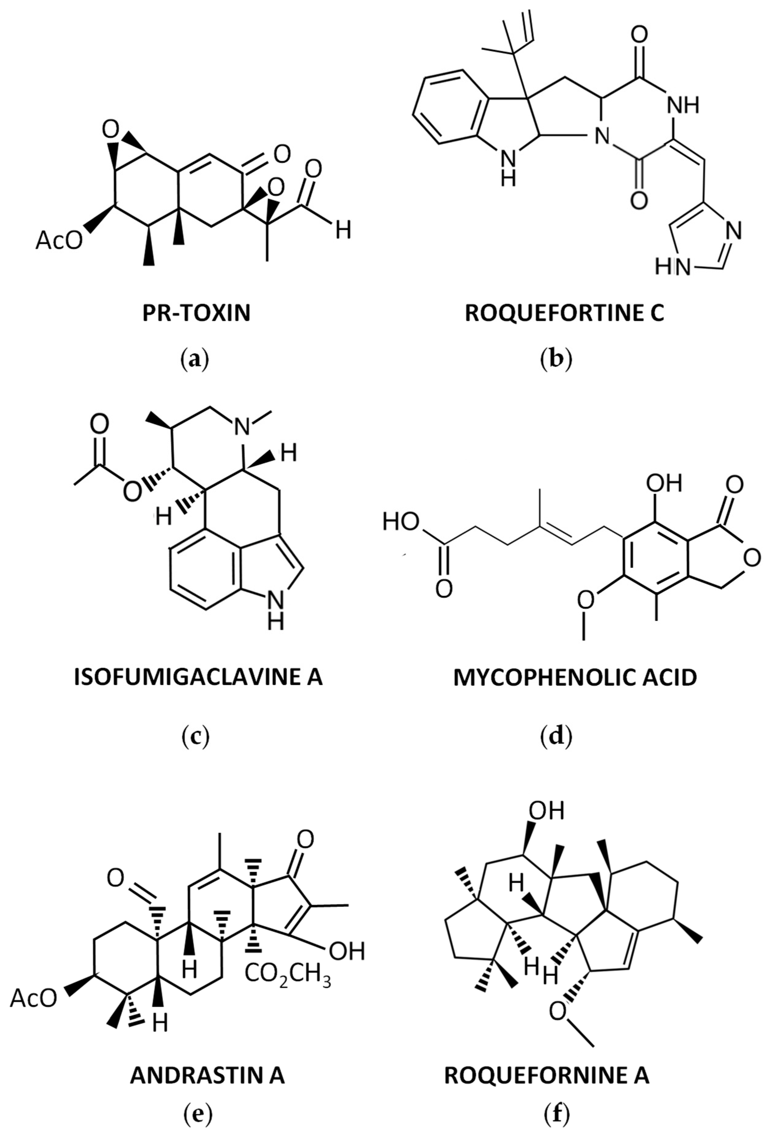
Figure 1. Representative secondary metabolites produced by P. roqueforti: (a) PR-toxin; (b) Roquefortine C; (c) Isofumigaclavine A; (d) Mycophenolic acid; (e) Andrastin A; (f) Roquefornine A.
Jof 09 00459 g001
Figure 2. Biosynthesis of Roquefortine C/meleagrin: (a) Roquefortine C biosynthetic gene cluster in P. roqueforti. The pseudogene is indicated as a black box; (b) Roquefortine C/meleagrin biosynthetic pathway in P. roqueforti and P. chrysogenum. The late reactions only present in P. chrysogenum are included within a shaded box, whereas side reactions suggested to involve the gmt-encoded methyltransferase and leading to the formation of minor methylated derivatives (e.g., roquefortine F) in P. roqueforti, are indicated between blue parentheses. Adapted from [46].
Jof 09 00459 g002
Figure 3. Biosynthesis of isofumigaclavine A in P. roqueforti: (a) Isofumigaclavine A biosynthetic gene cluster A and B in P. roqueforti. Those genes encoding proteins with uncharacterized role in this biosynthetic pathway (Proq02g028380 and Proq02g028400) are indicated with black arrows; (b) Isofumigalavine A biosynthetic pathway in P. roqueforti. The festuclavine synthase activity corresponds to the activity of two duplicated proteins encoded by ifgF1 and igF2 (represented as ifgF). The ifgH gene encoding the festuclavine hydroxylase is not located in Cluster A or B, and it is denoted with an asterisk. DMA-PP (dimethylallyl pyrophosphate); PP (pyrophosphate). Adapted from [54,55].
Jof 09 00459 g003
Figure 4. Biosynthesis of PR-toxin: (a) PR-toxin biosynthetic gene cluster in P. roqueforti. The cluster initially described by García–Estrada and Martín [34] includes genes represented as green (contig Proq06) and blue arrows contig (Proq02), whereas the cluster later described by Hidalgo et al., 2017 includes genes represented as blue and orange arrows (ORF 1 to ORF 11). The nomenclature of each gene, according to its position in each contig, is also indicated in black color; (b) Proposed biosynthetic pathway for PR-toxin in P. roqueforti. The alternative steps for the transformation of Eremofortin B to Eremofortin A are indicated with red arrows. Eremofortin A has been suggested to be either a shunt product or an intermediate (in blue brackets) adapted from [34,61,62].
Jof 09 00459 g004
Figure 5. Biosynthesis of mycophenolic acid: (a) Mycophenolic acid biosynthetic gene cluster in P. roqueforti.; (b) Proposed biosynthetic pathway for mycophenolic acid. 5-MOA (5-methylorsellinic acid); DHMB (4,6-dihydroxy-2-(hydroxymethyl)-3-methylbenzoic acid); DHMP (5,7-dihydroxy-4-methylphthalide); FDHMP (6-farnesyl-5,7-dihydroxy-4-methylphthalide); PP (pyrophosphate). Adapted from [34,67].
Jof 09 00459 g005
Figure 6. Biosynthesis of Andrastin A: (a) Andrastin A biosynthetic gene cluster in P. roqueforti. The pseudogene is indicated as a black box; (b) Proposed biosynthetic pathway for Andrastin A in P. chrysogenum. Likely, this pathway is similar in P. roqueforti. DMOA (3,5-dimethylorsellinic acid). Adapted from [71,72].
Jof 09 00459 g006
Figure 7. Biosynthesis of Annullatins D and F: (a) Annullatin biosynthetic gene cluster in P. roqueforti. The functions of anuD, anuI, and anuG remain unknown. The anuK gene, encoding a hypothetical transcription factor, is discussed below; (b) Proposed biosynthetic pathway for Annullatins D and F in P. roqueforti. Hypothetical intermediates are indicated between parentheses. Shunt pathways observed in Aspergillus nidulans are included within red boxes. Adapted from [80].
Jof 09 00459 g007
Table 1. Roquefortine C biosynthetic gene cluster in P. roqueforti.
GeneORF NameSize (bp)ProteinSize (aa)Proposed Function
gmtProq01g0227601058Methyltransferase332Formation of methylated derivatives
Pseudogene_____
rpt (dmaW)Proq01g0227701337Reverse prenyltransferase425Addition of an isopentenyl group to the cyclopeptide
rdhProq01g0227801638Dehydrogenase517Dehydrogenation of the cyclopeptide
rdsProq01g0227907200Nonribosomal peptide synthetase2363Formation of the cyclopeptide from L-histidine and L-tryptophan
Table 2. Isofumigaclavine A biosynthetic gene clusters (A) and (B) in P. roqueforti.
Gene (A)ORF NameSize (bp)ProteinSize (aa)Proposed Function
ifgEProq05g069260848Short-chain dehydrogenase/reductase (CDM36673)261Formation of chanoclavine I aldehyde
ifgF1Proq05g069270992Festuclavine synthase (CDM36674)287Contribution to the formation of festuclavine
ifgDProq05g0692801464Catalase (CDM36675)466Contribution to the formation of chanoclavine I
ifgBProq05g0692901087SAM-dependent methyltransferase (CDM36676)340Formation of N-methyl-dimethylallyltryptophan
ifgCProq05g0693001953FAD oxidase (CDM36677)629Contribution to the formation of chanoclavine I
ifgAProq05g0693101507Dimethylallyltryptophan synthase (CDM36678)462Formation of dimethylallyltryptophan
Gene (B)ORF NameSize (bp)ProteinSize (aa)Proposed Function
ifgGProq02g0283701128FMN-containing “old yellow enzyme” aldolase-type protein (CDM30151)375Contribution to the formation of festuclavine
-Proq02g028380861Phytanoyl-CoA dioxygenase (CDM30152)286Unknown
ifgIProq02g0283901455acetyltransferase (CDM30153)484Conversion of isofumigaclavine B to isofumigaclavine A
-Proq02g028400511Unnamed protein product (CDM30154)121Unknown
ifgF2Proq02g028410996Festuclavine synthase (CDM30155)287Contribution to the formation of festuclavine
Table 3. PR-toxin biosynthetic gene cluster in P. roqueforti as proposed by [62].
GeneORF NameSize (bp)ProteinSize (aa)Proposed Function
prx1 (ORF1)Proq02g0401801072Short-chain dehydrogenase340Oxidation of eremofortin A
prx2 (ari1) (ORF2)Proq02g0401901129Aristolochene synthase342Biosynthesis of aristolochene
prx3 (ORF3)Proq02g0402001716Quinone oxidase521Formation of 7-epineopetasone
prx4 (ORF4)Proq02g040210990Short chain alcohol dehydrogenase329Dehydrogenation of eremofortin C
prx8 (ORF5)Proq02g040220a1827Cytochrome P450 monooxygenase540Oxidation of intermediates
prx9 (ORF6)Proq02g0402301792Cytochrome P450 monooxygenase579Oxidation of intermediates
ORF7Proq02g040240613Outer membrane protein, beta-barrel186Unknown
prx11 (ORF8)Proq02g0402601416Acetyltransferase471Acetylation of intermediates
ORF9Proq02g040270a1823Cytochrome P450 monooxygenase509Oxidation of non-activated hydrocarbons
ORF10Proq02g0402801437Transcriptional regulator458Regulation of the gene cluster transcription
ORF11Proq02g0402901650Cytochrome P450 monooxygenase480Oxidation of non-activated hydrocarbons
Table 4. Mycophenolic acid biosynthetic gene cluster in P. roqueforti.
GeneORF NameSize (bp)ProteinSize (aa)Proposed Function
mpaAProq05g0698201126Prenyltransferase325Farnesylation of 5,7-dihydroxy-4-methylphthalide
mpaBProq05g069810949 (1397)Protein of unknown function298 (427)Unknown
mpaCProq05g0698007715Non-reducing polyketide synthase2477Formation of 5-methylorsellinic acid from acetyl-CoA, malonyl-CoA and S-adenosylmethionine
-Proq05g069790523Protein of unknown function116Unknown
mpaDEProq05g069780a3710 (2929)Fusion protein (Cytochrome P450/hydrolase)932 (852)Sequential hydroxylation/lactonization of 5-methylorsellinic acid
mpaFProq05g0697701708Inosine-5′-monophosphate dehydrogenase like526Role in self-resistance to mycophenolic acid and in the production of this compound
mpaGProq05g0697601250O-methyltransferase398Methylation of demethylmycophenolic acid to form mycophenolic acid
mpaHProq05g0697501479Unnamed protein product433Oxidative cleavage of the farnesyl chain to form demethylmycophenolic acid
Table 5. Andrastin A biosynthetic gene cluster in P. roqueforti.
GeneORF NameSize (bp)ProteinSize (aa)Proposed Function
adrAProq04g0628201746Cytochrome P450 monooxygenase508Consecutive oxidations for the formation of Andrastin A from Andrastin C
adrCProq04g0628304585ABC transporter1452Unknown (somehow involved in the production of Andrastin A)
adrDProq04g0628407973Polyketide synthase2495Formation of 3,5-dimethylorsellinic acid from acetyl-CoA, malonyl-CoA and S-adenosylmethionine
adrEProq04g0628501249Ketoreductase336Formation of Andrastin F from Andrastin D
adrFProq04g062860a952Short-chain dehydrogenase256Formation of Andrastin D from Andrastin E
adrGProq04g062870951Prenyltransferase316Farnesylation of 3,5-dimethylorsellinic acid
adrHProq04g062880a1633FAD-dependent oxidoreductase476Formation of epoxyfarnesyl-3,5-dimethylorsellinic acid methyl ester
adrIProq04g062890790Terpene cyclase245Formation of Andrastin E epoxyfarnesyl-3,5-dimethylorsellinic acid methyl ester
adrJProq04g0629001554Acetyltransferase496Formation of Andrastin C from Andrastin F
adrKProq04g0629101050Methyltransferase278Methylation of farnesyl-3,5-dimethylorsellinic acid
Table 6. Annullatins D and F biosynthetic gene cluster in P. roqueforti.
GeneORF NameSize (bp)ProteinSize (aa)Proposed Function
anuKProq03g0541401246Transcription factor (77.4% identity with FogI)261Unknown (transcriptional activation of the anu genes)
anuAProq03g0541506704Reducing polyketide synthase2130Formation of 2-hydroxymethyl-3-pentylphenol from acetyl-CoA and malonyl-CoA
anuBProq03g054160a1458Short-chain dehydrogenase/reductase283Formation of 2-hydroxymethyl-3-pentylphenol
anuCProq03g054170276Protein with 44.3% identity with the short-chain dehydrogenase/reductase FogB91Unknown (Formation of 2-hydroxymethyl-3-pentylphenol)
anuDProq03g0541801288Short-chain dehydrogenase381Unknown
anuEProq03g0541901772Cytochrome P450 hydroxylase490Formation annullatin E from 2-hydroxymethyl-3-pentylphenol into
anuFProq03g054200937Short-chain dehydrogenase/reductase276Formation of annullatin F
anuGProq03g0542101797Berberine bridge enzyme-like protein509Formation of annullatin D
anuHProq03g0542201512Prenyltransferase451Addition of a dimethylallyl group to annullatin E (formation of annullatin J) and formation of other prenylated derivatives
anuIProq03g054230759Short-chain dehydrogenase/reductase252Unknown
anuJProq03g0542402587Aromatic ring hydroxylating dehydrogenase818Unknown
Disclaimer/Publisher’s Note: The statements, opinions and data contained in all publications are solely those of the individual author(s) and contributor(s) and not of MDPI and/or the editor(s). MDPI and/or the editor(s) disclaim responsibility for any injury to people or property resulting from any ideas, methods, instructions or products referred to in the content.