Article

Molecular Regulatory Mechanism of the Iron-Ion-Promoted Asexual Sporulation of Antrodia cinnamomea in Submerged Fermentation Revealed by Comparative Transcriptomics

1
College of Food Science and Engineering, Yangzhou University, Yangzhou 225009, China
2
Jiangsu Provincial Key Construction Laboratory of Probiotics Preparation, Huaiyin Institute of Technology, Huaian 223003, China
3
Jiangsu Key Laboratory of Dairy Biotechnology and Safety Control, Yangzhou University, Yangzhou 225009, China
*
Author to whom correspondence should be addressed.
J. Fungi 2023, 9(2), 235; https://doi.org/10.3390/jof9020235
Submission received: 19 December 2022 / Revised: 29 January 2023 / Accepted: 8 February 2023 / Published: 10 February 2023

Abstract

:
Antrodia cinnamomea is a precious edible and medicinal fungus with activities of antitumor, antivirus, and immunoregulation. Fe2+ was found to promote the asexual sporulation of A. cinnamomea markedly, but the molecular regulatory mechanism of the effect is unclear. In the present study, comparative transcriptomics analysis using RNA sequencing (RNA-seq) and real time quantitative PCR (RT-qPCR) were conducted on A. cinnamomea mycelia cultured in the presence or absence of Fe2+ to reveal the molecular regulatory mechanisms underlying iron-ion-promoted asexual sporulation. The obtained mechanism is as follows: A. cinnamomea acquires iron ions through reductive iron assimilation (RIA) and siderophore-mediated iron assimilation (SIA). In RIA, ferrous iron ions are directly transported into cells by the high-affinity protein complex formed by a ferroxidase (FetC) and an Fe transporter permease (FtrA). In SIA, siderophores are secreted externally to chelate the iron in the extracellular environment. Then, the chelates are transported into cells through the siderophore channels (Sit1/MirB) on the cell membrane and hydrolyzed by a hydrolase (EstB) in the cell to release iron ions. The O-methyltransferase TpcA and the regulatory protein URBS1 promote the synthesis of siderophores. HapX and SreA respond to and maintain the balance of the intercellular concentration of iron ions. Furthermore, HapX and SreA promote the expression of flbD and abaA, respectively. In addition, iron ions promote the expression of relevant genes in the cell wall integrity signaling pathway, thereby accelerating the cell wall synthesis and maturation of spores. This study contributes to the rational adjustment and control of the sporulation of A. cinnamomea and thereby improves the efficiency of the preparation of inoculum for submerged fermentation.

1. Introduction

Antrodia cinnamomea (syn. Antrodia camphorata) is a precious edible and medicinal fungus that belongs to phylum Basidiomycetes, family Polyporaceae, and genus Antrodia [1]. A. cinnamomea presents various biological activities, such as hepatoprotective, antitumor, antioxidant, antiviral, antivasodilation, hypoglycemic, immunoregulatory, and gut microbiota regulatory [2,3,4,5]. The main bioactive compounds of A. cinnamomea include triterpenoids (such as antcins A, antcins B, antcins C, antcins H, antcins K, methylantcinate, and sulphurenic acid), polysaccharides, ubiquinone derivatives (such as antroquinonol, antroquinonol B, antroquinonol C, antroquinonol D, antroquinonol L, antroquinonol M, and 4-acetyantroquinonol B), maleic and succinic acid derivatives (such as antrodins A, antrodins B, antrodins C, antrodins D, and antrodins E), and benzene derivatives [4,6,7,8]. What is more, some compounds, such as antcins C, antcins K, antrodins B, antrodins C, and 4-acetyantroquinonol B, with excellent bioactivities from A. cinnamomea, have not been found in other edible and medicinal fungi so far [8,9].
A. cinnamomea has a huge market demand due to its outstanding biological activities and medicinal values. However, the wild fruiting bodies of A. cinnamomea are scarce and extremely expensive. Thus, the large-scale artificial culture of A. cinnamomea becomes necessary and important [10]. At present, the main techniques for the artificial culture of A. cinnamomea are basswood culture, plate culture, solid-state fermentation, and submerged fermentation [10,11]. Among them, the components of the fruiting bodies obtained from the basswood culture are the most similar to those of the wild fruiting bodies of A. cinnamomea, but the basswood must be from Cinnamomum kanehirae Hay, which is the only natural host of A. cinnamomea and is very expensive. In addition, the production periods of basswood culture are 1–5 years, which causes a high cost of time and materials. The production periods of plate culture are 2–4 months, and its yield of A. cinnamomea fruiting body is low, which cause low production efficiency by plate culture. The production periods of solid-state fermentation for A. cinnamomea are 1–2 months, and the A. cinnamomea mycelia obtained from solid-state fermentation cannot be separated from the culture medium, which cause a low content of the active compounds in the products of solid-state fermentation. Thus, the submerged fermentation becomes the most popular artificial culture method for A. cinnamomea because of the advantages of short fermentation period (10–14 days), high production efficiency, and easy to large-scale application [10,11,12].
Mycelium inoculation is usually used in the traditional submerged fermentation processes for A. cinnamomea [11,12], whereas mycelium inoculation in the submerged fermentation of A. cinnamomea presents some disadvantages, such as the difficult control of the quality and amount of inoculum (i.e., mycelia), and the poor synchronization of mycelial growth during fermentation, resulting in poor batch stability. Nevertheless, spore inoculation can solve these problems well [10]. However, the large-scale production of A. cinnamomea through submerged fermentation still has problems, such as the tedious and time-consuming preparation of inoculum (i.e., asexual spores) due to low sporulation, which severely limits the efficiency and benefit of A. cinnamomea production through submerged fermentation [11].
The sporulation of filamentous fungi is affected by various factors, such as nutrition, light, pH, and metal ions [13,14,15]. Among these factors, iron ions play an important role in the growth and development of most filamentous fungi [16,17]. For example, adding 56 µg/mL of iron could promote the growth and sporulation capacity of Cylindrocarpon destructans [18]. The growth rate and sporulation capacity of Phanerochaete chrysosporium could be significantly improved by the appropriate concentration of iron ions [19]. Iron balance is beneficial for promoting and enhancing the germination and infectivity of Beauveria bassiana spores [20].
Nonetheless, further studies on the molecular regulatory mechanisms of the iron-ion-promoted asexual sporulation of filamentous fungi remain lacking. What is more, there are no reports on iron ions promoting the asexual sporulation of A. cinnamomea in submerged fermentation and its mechanism so far. In the present study, comparative transcriptomics analysis using RNA-seq and RT-qPCR was conducted on A. cinnamomea mycelia cultured in the presence or absence of Fe2+. Then, the molecular regulatory mechanism underlying the Fe2+-promoted asexual sporulation of A. cinnamomea was revealed by combining relevant reports.

2. Materials and Methods

2.1. Strain

A. cinnamomea strains (No. ATCC 200183) were purchased from the American Type Culture Collection (Manassas, VA, USA).

2.2. Submerged Fermentation of A. cinnamomea

A. cinnamomea was cultured in accordance with the method reported by Li et al. [21]. In brief, the spore concentration (i.e., sporulation) of inoculum was counted and calculated using a hemocytometer under an optical microscope, then the A. cinnamomea was cultured in the fermentation medium (20.0 g/L glucose, 4.0 g/L yeast extract powder, 3.0 g/L KH2PO4, and 1.5 g/L MgSO4, with the initial pH of 4.5) at 26 °C and 150 r/min for 10–11 days, with an inoculum size of 1.0 × 10⁶ spores/mL.

2.3. Effects of Different Concentrations of Fe2+ on the Sporulation and Biomass of A. cinnamomea

FeCl2·4H2O was used to prepare the Fe2+ mother liquor, with a concentration of 1 mol/L. The corresponding volume of the Fe2+ mother liquor was added into the culture medium to adjust the Fe2+ concentrations of the culture medium to 0.05, 0.1, 0.2, 0.3, 0.4, and 0.5 mmol/L. Then, the culture medium was inoculated and cultured in accordance with the method described in Section 2.2. Samples were taken on days 6–11 to determine sporulation and biomass [22]. Medium with the same volume of deionized water added was regarded as the control group.

2.4. Sample Preparation for RNA-Seq

The fermentation broth of A. cinnamomea cultured in the presence or absence of Fe2+ was prepared in accordance with the method reported by Li et al. [23]. In brief, the A. cinnamomea mycelia pellets cultured in the presence or absence of Fe2+ (marked as “Fe” or “CK” group, respectively) for 7, 8, and 9 days were collected by filtering with four layers of gauze, then washed with Tris-EDTA (Ethylene Diamine Tetraacetic Acid) buffer solution (pH 8.0) and snap-frozen by liquid nitrogen. Three biological replicates were taken at each time point of each group.

2.5. RNA-Seq and Bioinformatics Analysis

The mycelial samples were sent to Beijing Novogene Biotechnology Co., Ltd. (Beijing, China) for high-throughput sequencing using an Illumina HiSeq™ 2500 (San Diego, CA, USA). The low-quality reads in the raw data were removed by FASTP software (version 0.19.7) with the parameters of “-g -q 5 -u 50 -n 15 -l 150” to obtain the qualified reads (i.e., clean reads). Then, the clean reads were used for de novo assembly by Trinity [24] software to obtain the transcript database. Finally, the unigene database (Supplementary Table S1) of A. cinnamomea was obtained after being compared against the genome database of A. cinnamomea (Accession number: GCA_000766995.1, NCBI).
The differentially expressed genes (DEGs) were obtained with the H-Cluster algorithm by Corset (https://code.google.com/p/corset-project/ accessed on 14 February 2022) [25]. The annotation and functional analysis of the DEGs were conducted using the NCBI database (https://www.ncbi.nlm.nih.gov/ accessed on 22 February 2022), GO database (http://geneontology.org/ accessed on 25 February 2022), and KEGG database (http://www.genome.jp/kegg/ accessed on 3 March 2022).

2.6. RT-qPCR Analysis

RT-qPCR analysis was performed in accordance with the reported method [23] using the same samples for RNA-seq and the 18S ribosomal ribonucleic acid (rRNA) sequence of A. cinnamomea as the internal reference. The A. cinnamomea mycelium samples cultured in the absence of Fe2+ were used as control. The RT-qPCR primer sequences of the related genes are shown in Table 1.

2.7. Statistical Analysis of Data

At least three replicates were prepared for each experimental group. Data were presented as mean ± standard deviation (SD). One-way analysis of variance (ANOVA) was carried out using SPSS PASW Statistics Version 18.0 (Palo Alto, CA, USA), and the significant difference level was set as p < 0.05. The principal component analysis (PCA) was performed based on the “Fragments Per Kilobase of exon model per Million mapped fragments” (FPKM) values of different groups with the “ggplot2” package of R (Version 3.0.3). For the differentially expressed gene analysis, it was considered as a significant difference when the fold change of genetic expression was more than 1.25 times (up-regulation, log2 fold change ≥ 0.32) or less than 0.80 times (down-regulation, log2 fold change ≤ −0.32).

3. Results and Discussion

3.1. Effects of Fe2+ on the Sporulation Capacity of A. cinnamomea

As shown in Figure 1, the addition of 0.1–0.2 mmol/L Fe2+ markedly promoted the sporulation and growth of A. cinnamomea in submerged fermentation. The addition of 0.1 mmol/L Fe2+ had the most significant effect and increased the maximum sporulation of A. cinnamomea by 72.39%. However, when Fe2+ was added at concentrations exceeding 0.4 mmol/L, it had an obvious inhibitory effect on the growth of A. cinnamomea and dramatically inhibited sporulation. Therefore, Fe2+ can significantly promote the sporulation and growth of A. cinnamomea when its addition concentration is strictly controlled.

3.2. RNA-Seq and Statistical Analysis

3.2.1. Preparation of Sequencing Samples

Figure 1 shows that A. cinnamomea started to produce spores rapidly on day 7. What is more, A. cinnamomea was in the early stage of rapid sporulation on day 8 and was in the middle stage of rapid sporulation on day 9. In other words, days 7, 8, and 9 were the beginning, early, and middle stages of the rapid sporulation of A. cinnamomea in submerged fermentation, respectively. Therefore, our sequencing samples were A. cinnamomea mycelia incubated for 7, 8, and 9 days in the culture medium without Fe2+ (referred to as “CK_7d”, “CK_8d”, and “CK_9d”, respectively) and A. cinnamomea mycelia incubated for 7, 8, and 9 days in the culture medium with 0.1 mmol/L Fe2+ (referred to as “Fe_7d”, “Fe_8d”, and “Fe_9d”, respectively) for a total of six groups. Three biological replicates were taken for each group. For example, three biological replicates of the “CK_7d” group were recorded as “CK_7d_1”, “CK_7d_2”, and “CK_7d_3”, and the rest may be deduced by analogy. A total of 18 samples were used for RNA-seq.

3.2.2. Statistical Analysis of Sample Repeatability and DEGs

On the basis of the expression of unigenes, which are represented by FPKM values (Supplementary Table S2), in various samples, we performed principal component analysis (PCA) (Figure 2A) and DEG analysis (Figure 2B) to investigate the consistency of the three biological repetitions and DEG distribution in the three sample groups.
From the statistical analysis, it was found that the three biological replicates in each group are concentrated, and the different groups can be clearly distinguished (Figure 2B), which indicated that the three biological replicates of each groups had good biological repeatability and the genes were expressed with significant differences between different groups. In addition, the distributions of the maximum, median, and minimum of all biological repetition samples were basically consistent, and gene expression was largely dispersed in different groups (Figure 2C), which verified the good biological repeatability of the replicates and significant differences between the groups again. What is more, it was shown that Fe2+ exerted a global impact on gene expression in A. cinnamomea, and the change in gene expression exhibited certain trends on the basis of incubation time and sample treatment (Figure 2A). In particular, some gene clusters were significantly up-regulated (color changed from blue to red) or significantly down-regulated (color changed from red to blue) compared with the control group after adding Fe2+ (Figure 2A), indicating that Fe2+ significantly affected the expression of some genes, and these gene are probably involved in iron ions promoting the asexual sporulation of A. cinnamomea. In sum, the results in Figure 2 indicate that the design of the sequencing sample is reasonable and that the sequencing quality is perfect.

3.2.3. Enrichment Analysis of DEGs

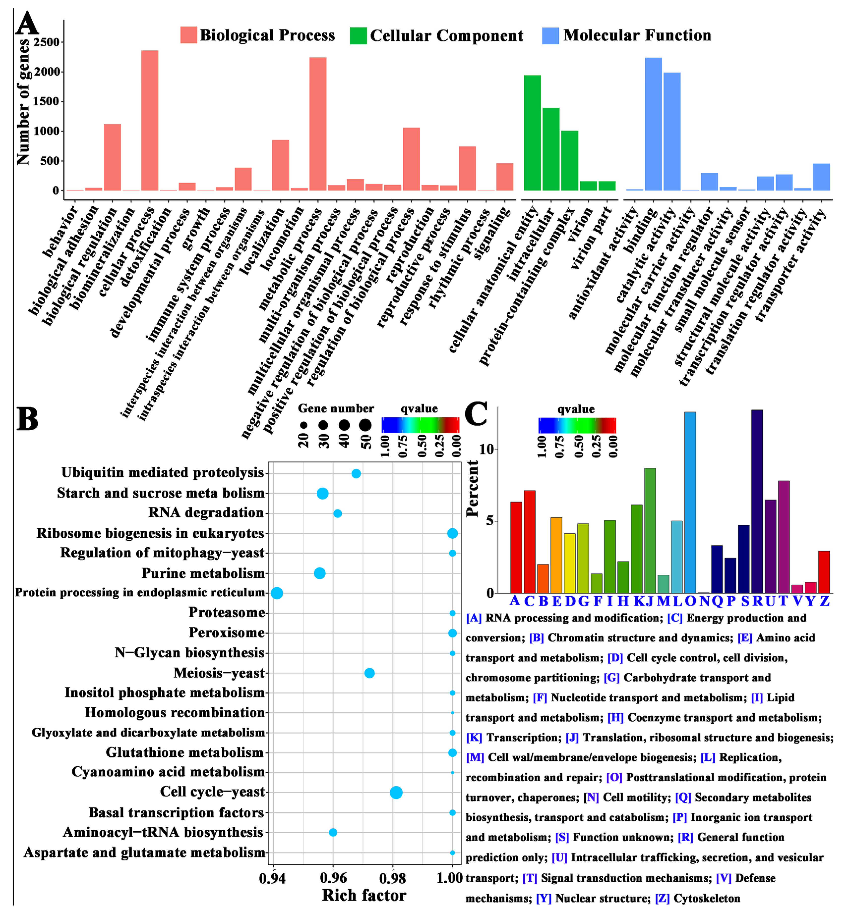
The GO, KOG, and KEGG enrichment analyses of DEGs (Figure 3, Supplementary Table S3) revealed that the DEGs were involved in the cellular process, the developmental process, the reproductive process, transcription regulator activity, transporter activity, cell wall integrity (CWI), cell cycle control, cell division, signal transduction, amino acid metabolism, transport and catabolism, cell growth and death, sensory systems, and membrane transport. The CWI signaling pathway was reported to be closely related to cell wall synthesis and fungal sporulation in phytopathogenic fungi [26,27]. Serine metabolism is involved in the promotion of the sporulation of Aspergillus fumigatus by iron ions [28]. As inferred through functional classification, these genes may be related to the iron-ion-mediated asexual sporulation of A. cinnamomea and require further bioinformatics analysis.

3.3. Bioinformatic Analysis

First, on the basis of our previous study [21], we successfully obtained 18 genes related to the FluG-mediated asexual sporulation signaling pathway of A. cinnamomea from the unigene database, namely, sfaD, fluG, velB, flbA, ganB, veA, vosA, fadA, flbC, pkaA, nsdD, flbD, wetA, abaA, flbB, stuA, brlA, and sfgA. Subsequently, on the basis of the relevant references [26,28,29,30,31,32,33,34,35,36,37,38,39,40,41], we obtained 24 genes (Table 2) that may be related to the Fe2+-mediated asexual sporulation of A. cinnamomea among the DEGs (Supplementary Table S4), namely, mirB, ftrA, hapX, sreA, fetC, bck1, uvt, urbs1, sit, fre, slt2, ssiG 06045, feoB, tpcA, nrps, nps2, nps4, clpP, mkk1, sidA, yvmB, fur, estB, and wsc1 (Table 2).
Iron, a trace element, is an indispensable factor for the growth of filamentous fungi. However, the molecular regulatory mechanism underlying the induction of the asexual sporulation of filamentous fungi by iron ions remains unclear. In A. fumigatus, reductive iron assimilation (RIA) includes the ferric reductase FreB, the ferroxidase FetC, and the iron permease FtrA [42,43]. Iron permease widely exists in organisms and belongs to the ferredoxin-dependent family [43]. Most fungal species, including siderophore producers or nonproducers, have at least one homologous iron permease gene [44].
Nonreductive iron assimilation works through the synthesis and utilization of siderophores. In Aspergillus nidulans, the deletion of the sidA gene results in the inability to synthesize siderophores [26]. Nonribosomal peptide synthetase (NRPS) is a multifunctional protein that can biosynthesize small peptides and is responsible for the synthesis of low-molecular-weight secondary metabolites. Among NRPSs, nps2 is structurally conserved and functionally stable in many filamentous fungi and is closely related to the biosynthesis of extracellular and intracellular siderophores [29]. nps4 plays an important role in the surface hydrophobicity of pathogenic bacteria and the formation of conidia [29]. In A. fumigatus, mirb transports the chelates of siderophores and Fe2+ into cells that are then hydrolyzed by hydrolase EstB for Fe2+ release [30,31]. Iron ion assimilation is thus achieved.
Fungi possess two transcriptional regulatory systems for jointly regulating iron homeostasis: the basic region-leucine zipper (bZIP)-type transcription factor hap and the GATA-type zinc finger transcription factor sre [32]. A negative feedback regulatory system is formed on the basis of these two factors to jointly regulate iron homeostasis [33]. The transcription factor hapX can regulate the iron acquisition and conidial infection ability of Beauveria bassiana in accordance with the concentration of iron ions [45]. HapX also affects the growth, sporulation, and conidial germination, and maintains iron homeostasis in fungi [46,47,48]. The zinc finger transcription factor sre is involved in the regulation of siderophore synthesis and iron assimilation in fungi. For example, the transcription factor sreA is involved in the transcriptional activation of genes related to the siderophore synthesis pathway in A. fumigatus [49]. SRE1 is involved in siderophore synthesis in Histoplasma capsulatum [34].
Moreover, the increase in biofilm synthesis by A. fumigatus induced by a low-iron condition is dependent on the CWI pathway because the receptor Wsc1 on the cell membrane in the CWI pathway is involved in the perception of biofilm synthesis [35,50]. In B. bassiana, bck1, mkk1, and slt2 in the CWI pathway can not only maintain cell integrity but can also positively regulate growth, sporulation, and host infectivity [26,51]. The deletion of yvmB inhibits the sporulation of Bacillus subtilis and leads to the disruption of the proteins involved in iron transport [36]. The protein encoded by the uvt3277 gene can transport iron ions [52]. In Ustilago maydis, the deletion of the urbs1 gene inhibits siderophore biosynthesis and limits iron assimilation [41]. The cmSIT1 gene is associated with siderophore-mediated transport [37].

3.4. RT-qPCR Analysis

By comparing the expression levels (FPKM values) of the genes shown in Table 2 in the sequencing samples, the bck1, mkk1, and slt2 genes in the CWI pathway and the flbD and abaA genes in the FluG-mediated sporulation signaling pathway were found to exhibit a dramatic increment after Fe2+ treatment. Thus, RT-qPCR was adopted to further analyze and verify the expression patterns of these five genes in the samples. As shown in Figure 4, compared with those in the CK group, the expression levels of the bck1, mkk1, and slt2 genes had increased by 3–18 times and the expression levels of the flbD and abaA genes had increased by 9–21 times in A. cinnamomea mycelia treated with Fe2+. Therefore, these five genes were speculated to play a key regulatory role in the iron-ion-promoted asexual sporulation of A. cinnamomea.

3.5. Model of the Signaling Pathway of the Iron-Ion-Promoted Asexual Sporulation of A. cinnamomea

In fungi, the iron ion is a cofactor of many enzymes. The regulation of the transmembrane assimilation of iron is a primary and important way to maintain intracellular iron homeostasis [28]. For this reason, fungi have evolved a variety of iron assimilation methods. These iron assimilation methods are generally divided into two categories: high-affinity iron assimilation and low-affinity iron assimilation [28]. The high-affinity iron assimilation method is divided into two types: RIA and siderophore-mediated iron assimilation (SIA) [53].
In A. fumigatus, iron assimilation through RIA first involves the reduction of trivalent iron ions into divalent iron ions with improved solubility under the action of ferric reductase [42]. Then, these ions are transported into the cell by the high-affinity protein complex formed by the feroxidase FetC and the iron transporter permease FtrA [54]. The process of SIA in A. fumigatus is as follows: First, siderophores are secreted outside to chelate the iron in the external environment [35]. Then, iron ions are transported into the cells through the siderophore channel (Sit1/MirB) on the cell membrane; finally, the chelates are hydrolyzed by hydrolases (such as EstB), then Fe2+ is released [31]. The feroxidase fetC, the iron transporter permease ftrA, and several siderophore-synthesis-related genes in A. cinnamomea were successfully matched. Thus, we believe that RIA and SIA simultaneously exert effects on iron assimilation by A. cinnamomea.
The CWI signaling pathway, which is a conserved pathway in fungi, plays an important role in the growth and reproduction of fungi and the response to environmental stress [27]. The upstream of the CWI signaling pathway includes the baroreceptor WSC1, which is located on the cell membrane and is involved in cell wall reinforcement and repair in the responses to environmental stress [26,35]. In addition, the WSC1 receptor can respond to the concentration of iron ions [35,55]. In phytopathogenic fungi, the CWI signaling pathway is not only involved in the regulation of cell wall synthesis but also in the regulation of sporulation and pathogenicity [27]. The expression of the bck1, mkk1, and slt2 genes in the CWI signaling pathway dramatically increased in the A. cinnamomea mycelial samples cultured with Fe2+ compared with that in the control (Figure 4). Therefore, in A. cinnamomea, the Wsc1 receptor on the cell membrane is believed to respond to the concentration of Fe2+ and ultimately promotes the expression of relevant genes in the CWI signaling pathway, thereby accelerating the cell wall synthesis and maturation of asexual spores.
In A. fumigatus and A. nidulans, two key transcription factors are involved in the regulation of cellular iron balance. They are the GATA-type zinc finger transcription factor SreA and the CCAAT-type bZip transcription factor HapX [42,43]. Both of the two factors can respond to the concentration of iron ions and transcriptionally inhibit each other [32,33]. When iron ions are sufficient in the environment, the expression of sreA is upregulated, which inhibits the transcription of hapX and the expression of genes related to iron ion assimilation, including the RIA and SIA pathways, and promotes the consumption of iron ions [32]. By contrast, when iron ions are deficient in the environment, then the expression of hapX is upregulated, which inhibits the transcription of sreA and promotes the expression of genes related to iron ion assimilation [33]. In the FluG-mediated sporulation signaling pathway in A. cinnamomea, FlbB, similar to HapX, is a bZip-type transcription factor, and BrlA, similar to SreA, is a transcription factor with two zinc finger structures. Moreover, the expression of the downstream genes of flbB and brlA (flbd and abaA) in A. cinnamomea mycelial samples cultured with Fe2+ had increased sharply compared with that in the control (Figure 4). Therefore, the target of HapX is speculated to be flbd and has the same function as FlbB, i.e., promoting the expression of flbd, and the target of SreA is abaA and has the same function as BrlA, i.e., promoting the expression of abaA.
Fur is also a regulatory factor that regulates the balance of intracellular iron ion concentration with multiple regulatory modes for the transcription of target genes. Fur has been reported to not only regulate the expression of genes related to iron ion transport but also to inhibit the expression of some other genes, such as flbB [56]. The typical regulation model for Fur in eukaryotes is as follows: When intracellular iron ions are sufficient, Fur combines with Fe2+, and the formed Fur–Fe2+ complex can bind to Fur–boxDNA in the upstream of the promoter to inhibit the expression of iron assimilation genes by inhibiting the binding of RNA polymerase. When intracellular iron ions are deficient, Fe2+ dissociates from Fur, leading to the reduction in the DNA-binding ability of Fur and activating the expression of the genes related to iron assimilation [38,39,54,55,56].
Nps2 is involved in the formation of spores in fungi. For example, the Alternaria alternate strain with the nps2 deletion produces more spores earlier than the wild-type strain [57]. In the pathogenic bacterium Cochliobolus heterostrophus, NPS4 plays an important role in virulence and conidia morphogenesis [40]. The O-methyltransferase TpcA can regulate the expression of hapx in A. fumigatus [58]. In Ustilago maydis, the deletion of the urbs1 gene inhibits siderophore biosynthesis and places siderophore-mediated iron assimilation outside the reach of regulation [41].
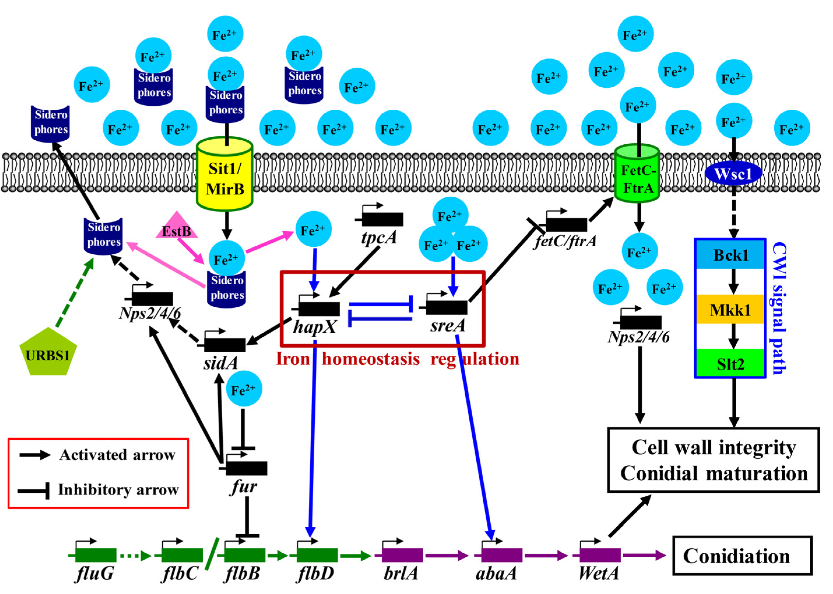
To sum up, the Fe2+- and FluG-mediated signal pathway of the asexual sporulation of A. cinnamomea was predicted (Figure 5) as follows: A. cinnamomea obtains iron ions through RIA and SIA. In RIA, ferrous iron ions are directly transported into cells by the high-affinity protein complex formed by a feroxidase (FetC) and an iron transporter permease (FtrA). In SIA, siderophores are secreted outside to chelate Fe2+ in the extracellular environment. Then, the chelates are transported into cells through the siderophore channels (Sit1/MirB) on the cell membrane and hydrolyzed by a hydrolase (EstB) in the cell to release Fe2+. The free siderophores can be extracellularly secreted again. Subsequently, HapX and SreA respond to the intercellular concentration of Fe2+. When iron ions are sufficient, the expression of sreA is upregulated, which inhibits the expression of hapX, fetC, and ftrA, thereby inhibiting iron assimilation. When iron ions are deficient, the expression of hapX is upregulated, which inhibits the expression of sreA and the consumption of iron. Furthermore, HapX and SreA affect flbD and abaA, respectively, and promote their expression.
The ferric uptake transcriptional regulator Fur can inhibit the expression of the flbB gene and further inhibit sporulation. However, when intercellular iron ion concentrations increase, Fe2+ combines with Fur to form the Fe2+–Fur complex to inhibit the expression of fur and alleviate the inhibition of flbB, which indirectly promotes sporulation. In addition, Fe2+ acts on the Wsc1 baroreceptor and promotes the expression of relevant genes in the CWI signaling pathway through a series of cascade reactions, thereby accelerating the cell wall synthesis and maturation of the asexual spores of A. cinnamomea. The O-methyltransferase TpcA can promote the expression of hapX then promote the synthesis of siderophores. The regulatory protein URBS1 can directly or indirectly regulate the synthesis of siderophores.

4. Conclusions

In the present study, comparative transcriptomics was used to reveal the molecular regulatory mechanisms underlying the asexual sporulation of A. cinnamomea promoted by Fe2+ in submerged fermentation. RIA and SIA were found to function in A. cinnamomea. In RIA, the ferroxidase FetC and the iron transporter permease FtrA directly transported Fe2+ into cells. In SIA, Fe2+ ions were transported into cells by siderophores through siderophore channels and released by the hydrolase EstB. Then, the transcription factors HapX and SreA responded to the concentration of Fe2+ and affected the expression of flbD and abaA in the FluG-mediated central signaling pathway, thereby promoting the sporulation of A. cinnamomea in submerged fermentation. However, the functions of relevant genes need to be further verified by more technical means, which is the focus and direction of the following research. Nevertheless, the present study contributes to improving the preparation efficiency of the inoculum (asexual spores) of A. cinnamomea in submerged fermentation, which saves production costs and improves production efficiency. For example, this method is easy to operate and inexpensive and is thus of great value for development and application. In addition, it provides a reference for further research on the molecular regulatory mechanisms of iron-ion-promoted asexual sporulation in other filamentous fungi.

Supplementary Materials

The following supporting information can be downloaded at: https://www.mdpi.com/article/10.3390/jof9020235/s1, Table S1: The unigene database of Antrodia cinnamome by RNA-seq; Table S2: The FPKM values of the unigenes from RNA-seq; Table S3: The obtained differentially expressed genes (DEGs) of enrichment analyses. Table S4: The DEGs from pairwise comparison and their functioin annotations.

Author Contributions

Funding acquisition, H.L.; investigation, H.L. and Y.S.; writing—original draft, Y.S.; data curation and validation, J.D.; software and visualization, L.J.; conceptualization and methodology, X.Z.; resources, Z.Y. and X.Z.; writing—review and editing, H.L. and L.J.; supervision and project administration, Z.Y. All authors have read and agreed to the published version of the manuscript.

Funding

This work was financially supported by the National Natural Science Foundation of China (Grant numbers: 32001661), and the Natural Science Foundation of Jiangsu Province, China (Grant number: BK20190890).

Institutional Review Board Statement

Not applicable.

Informed Consent Statement

Not applicable.

Data Availability Statement

Data are contained within the article or Supplementary Materials.

Conflicts of Interest

The authors declare no conflict of interest.

References

  1. Chen, C.L.; Li, W.C.; Chuang, Y.C.; Liu, H.C.; Huang, C.H.; Lo, K.Y.; Chen, C.Y.; Chang, F.M.; Chang, G.A.; Lin, Y.L.; et al. Sexual crossing, chromosome-level genome sequences, and comparative genomic analyses for the medicinal mushroom Taiwanofungus camphoratus (Syn. Antrodia Cinnamomea, Antrodia Camphorata). Microbiol. Spectr. 2022, 10, e02032-21. [Google Scholar] [CrossRef] [PubMed]
  2. Xu, X.Y.; Geng, Y.; Xu, H.X.; Ren, Y.; Liu, D.Y.; Mao, Y. Antrodia camphorata-derived antrodin C inhibits liver fibrosis by blocking TGF-Beta and PDGF signaling pathways. Front. Mol. Biosci. 2022, 9, 835508. [Google Scholar] [CrossRef] [PubMed]
  3. Huang, T.F.; Wang, S.W.; Lai, Y.W.; Liu, S.C.; Chen, Y.J.; Hsueh, T.Y.; Lin, C.C.; Lin, C.H.; Chung, C.H. 4-Acetylantroquinonol B suppresses prostate cancer growth and angiogenesis via a VEGF/PI3K/ERK/mTOR-dependent signaling pathway in subcutaneous xenograft and in vivo angiogenesis Models. Int. J. Mol. Sci. 2022, 23, 1446. [Google Scholar] [CrossRef] [PubMed]
  4. Ganesan, N.; Baskaran, R.; Velmurugan, B.K.; Thanh, N.C. Antrodia cinnamomea—An updated minireview of its bioactive components and biological activity. J. Food Biochem. 2019, 43, e12936. [Google Scholar] [CrossRef]
  5. Lu, C.L.; Li, H.X.; Zhu, X.Y.; Luo, Z.S.; Rao, S.Q.; Yang, Z.Q. Regulatory effect of intracellular polysaccharides from Antrodia cinnamomea on the intestinal microbiota of mice with antibiotic-associated diarrhea. Qual. Assur. Saf. Crops Foods 2022, 14, 124–134. [Google Scholar] [CrossRef]
  6. Li, H.X.; Wang, J.J.; Lu, C.L.; Gao, Y.J.; Gao, L.; Yang, Z.Q. Review of bioactivity, isolation, and identification of active compounds from Antrodia cinnamomea. Bioengineering 2022, 9, 494. [Google Scholar] [CrossRef]
  7. Wang, C.; Zhang, W.; Wong, J.H.; Ng, T.; Ye, X. Diversity of potentially exploitable pharmacological activities of the highly prized edible medicinal fungus Antrodia camphorata. Appl. Microbiol. Biotechnol. 2019, 103, 7843–7867. [Google Scholar] [CrossRef]
  8. Lu, M.C.; El-Shazly, M.; Wu, T.Y.; Du, Y.C.; Chang, T.T.; Chen, C.F.; Hsu, Y.M.; Lai, K.H.; Chiu, C.P.; Chang, F.R.; et al. Recent research and development of Antrodia cinnamomea. Pharmacol. Therapeut. 2013, 139, 124–156. [Google Scholar] [CrossRef]
  9. Liu, X.; Yu, S.; Zhang, Y.; Zhang, W.; Zhong, H.; Lu, X.; Guan, R. A review on the protective effect of active components in Antrodia camphorata against alcoholic liver injury. J. Ethnopharmacol. 2023, 300, 115740. [Google Scholar] [CrossRef]
  10. Lu, Z.M.; He, Z.; Li, H.X.; Gong, J.S.; Geng, Y.; Xu, H.Y.; Xu, G.H.; Shi, J.S.; Xu, Z.H. Modified arthroconidial inoculation method for the efficient fermentation of Antrodia camphorata ATCC 200183. Biochem. Eng. J. 2014, 87, 41–49. [Google Scholar] [CrossRef]
  11. Li, H.X.; Lu, Z.M.; Geng, Y.; Gong, J.S.; Zhang, X.J.; Shi, J.S.; Xu, Z.H.; Ma, Y.H. Efficient production of bioactive metabolites from Antrodia camphorata ATCC 200183 by asexual reproduction-based repeated batch fermentation. Bioresour. Technol. 2015, 194, 334–343. [Google Scholar] [CrossRef]
  12. Zhang, B.B.; Guan, Y.Y.; Hu, P.F.; Chen, L.; Xu, G.R.; Liu, L.; Cheung, P.C.K. Production of bioactive metabolites by submerged fermentation of the medicinal mushroom Antrodia cinnamomea: Recent advances and future development. Crit. Rev. Biotechnol. 2019, 39, 541–554. [Google Scholar] [CrossRef] [PubMed]
  13. Lin, E.S.; Wang, C.C.; Sung, S.C. Cultivating conditions influence lipase production by the edible basidiomycete Antrodia cinnamomea in submerged culture. Enzym. Microb. Technol. 2006, 39, 98–102. [Google Scholar] [CrossRef]
  14. Klingen, I.; Holthe, M.P.; Westrum, K.; Suthaparan, A.; Torp, T. Effect of light quality and light-dark cycle on sporulation patterns of the mite pathogenic fungus Neozygites floridana (Neozygitales: Entomophthoromycota), a natural enemy of Tetranychus urticae. J. Invertebr. Pathol. 2016, 137, 43–48. [Google Scholar] [CrossRef]
  15. Zhao, X.; Fan, Y.; Xiang, M.; Kang, S.; Wang, S.; Liu, X. DdaCrz1, a C2H2-type transcription factor, regulates growth, conidiation, and stress resistance in the nematode-trapping fungus Drechslerella dactyloides. J. Fungi 2022, 8, 750. [Google Scholar] [CrossRef]
  16. Wang, F.; Lu, Y.Y.; Liu, M.M.; Xiao, S.Q.; Gao, Y.B.; Yuan, M.Y.; Xue, C.S. Effects of iron on the asexual reproduction and major virulence factors of Curvularia lunata. Eur. J. Plant Pathol. 2020, 157, 497–507. [Google Scholar] [CrossRef]
  17. Da Silva Hellwig, A.H.; Pagani, D.M.; Rios, I.d.S.; Ribeiro, A.C.; Zanette, R.A.; Scroferneker, M.L. Influence of iron on growth and on susceptibility to itraconazole in Sporothrix spp. Med. Mycol. 2021, 59, 400–403. [Google Scholar] [CrossRef] [PubMed]
  18. Rahman, M.; Punja, Z.K. Influence of iron on cylindrocarpon root rot development on ginseng. Phytopathology 2006, 96, 1179–1187. [Google Scholar] [CrossRef]
  19. Canessa, P.; Munoz-Guzman, F.; Vicuna, R.; Larrondo, L.F. Characterization of PIR1, a GATA family transcription factor involved in iron responses in the white-rot fungus Phanerochaete chrysosporium. Fungal Genet. Biol. 2012, 49, 626–634. [Google Scholar] [CrossRef]
  20. Jirakkakul, J.; Wichienchote, N.; Likhitrattanapisal, S.; Ingsriswang, S.; Yoocha, T.; Tangphatsornruang, S.; Wasuwan, R.; Cheevadhanarak, S.; Tanticharoen, M.; Amnuaykanjanasin, A. Iron homeostasis in the absence of ferricrocin and its consequences in fungal development and insect virulence in Beauveria bassiana. Sci. Rep. 2021, 11, 19624. [Google Scholar] [CrossRef]
  21. Li, H.X.; Lu, Z.M.; Zhu, Q.; Gong, J.S.; Geng, Y.; Shi, J.S.; Xu, Z.H.; Ma, Y.H. Comparative transcriptomic and proteomic analyses reveal a flug-mediated signaling pathway relating to asexual sporulation of Antrodia camphorata. Proteomics 2017, 17, 1700256. [Google Scholar] [CrossRef]
  22. Li, H.X.; Ji, D.; Luo, Z.S.; Ren, Y.L.; Lu, Z.M.; Yang, Z.Q.; Xu, Z.H. Comparative transcriptomic analyses reveal the regulatory mechanism of nutrient limitation-induced sporulation of Antrodia cinnamomea in submerged fermentation. Foods 2022, 11, 2715. [Google Scholar] [CrossRef] [PubMed]
  23. Chen, Y.; Zhu, J.; Ying, S.H.; Feng, M.G. Three mitogen-activated protein kinases required for cell wall integrity contribute greatly to biocontrol potential of a fungal entomopathogen. PLoS ONE 2014, 9, e87948. [Google Scholar] [CrossRef] [PubMed]
  24. Grabherr, M.G.; Haas, B.J.; Yassour, M.; Levin, J.Z.; Thompson, D.A.; Amit, I.; Adiconis, X.; Fan, L.; Raychowdhury, R.; Zeng, Q.; et al. Full-length transcriptome assembly from RNA-Seq data without a reference genome. Nat. Biotechnol. 2011, 29, 644–652. [Google Scholar] [CrossRef]
  25. Davidson, N.M.; Oshlack, A. Corset: Enabling differential gene expression analysis for de novo assembled transcriptomes. Genome Biol. 2014, 15, 410. [Google Scholar] [CrossRef] [PubMed]
  26. Chen, Y.; Zhu, J.; Ying, S.H.; Feng, M.G. The GPI-anchored protein Ecm33 is vital for conidiation, cell wall integrity, and multi-stress tolerance of two filamentous entomopathogens but not for virulence. Appl. Microbiol. Biotechnol. 2014, 98, 5517–5529. [Google Scholar] [CrossRef] [PubMed]
  27. Haas, H. Fungal siderophore metabolism with a focus on Aspergillus fumigatus. Nat. Prod. Rep. 2014, 31, 1266–1276. [Google Scholar] [CrossRef] [PubMed]
  28. Eisendle, M.; Oberegger, H.; Zadra, I.; Haas, H. The siderophore system is essential for viability of Aspergillus nidulans: Functional analysis of two genes encoding l-ornithine N 5-monooxygenase (sidA) and a non-ribosomal peptide synthetase (sidC). Mol. Microbiol. 2003, 49, 359–375. [Google Scholar] [CrossRef]
  29. Kim, K.-H.; Cho, Y.; La Rota, M.; Cramer, R.A., Jr.; Lawrence, C.B. Functional analysis of the Alternaria brassicicola non-ribosomal peptide synthetase gene AbNPS2 reveals a role in conidial cell wall construction. Mol. Plant Pathol. 2007, 8, 23–39. [Google Scholar] [CrossRef]
  30. Kragl, C.; Schrettl, M.; Abt, B.; Sarg, B.; Lindner, H.H.; Haas, H. EstB-mediated hydrolysis of the siderophore triacetylfusarinine C optimizes iron uptake of Aspergillus fumigatus. Eukaryot. Cell 2007, 6, 1278–1285. [Google Scholar] [CrossRef] [Green Version]
  31. Raymond-Bouchard, I.; Carroll, C.S.; Nesbitt, J.R.; Henry, K.A.; Pinto, L.J.; Moinzadeh, M.; Scott, J.K.; Moore, M.M. Structural requirements for the activity of the MirB ferrisiderophore transporter of Aspergillus fumigatus. Eukaryot. Cell 2012, 11, 1333–1344. [Google Scholar] [CrossRef]
  32. Schrettl, M.; Beckmann, N.; Varga, J.; Heinekamp, T.; Jacobsen, I.D.; Jochl, C.; Moussa, T.A.; Wang, S.; Gsaller, F.; Blatzer, M.; et al. HapX-mediated adaption to iron starvation is crucial for virulence of Aspergillus fumigatus. PLoS Pathog. 2010, 6, e1001124. [Google Scholar] [CrossRef]
  33. Schrettl, M.; Kim, H.S.; Eisendle, M.; Kragl, C.; Nierman, W.C.; Heinekamp, T.; Werner, E.R.; Jacobsen, I.; Illmer, P.; Yi, H.; et al. SreA-mediated iron regulation in Aspergillus fumigatus. Mol. Microbiol. 2008, 70, 27–43. [Google Scholar] [CrossRef] [PubMed]
  34. Hwang, L.H.; Seth, E.; Gilmore, S.A.; Sil, A. SRE1 regulates iron-dependent and -independent pathways in the fungal pathogen Histoplasma capsulatum. Eukaryot. Cell 2012, 11, 16–25. [Google Scholar] [CrossRef] [PubMed]
  35. Bom, V.L.; de Castro, P.A.; Winkelstroter, L.K.; Marine, M.; Hori, J.I.; Ramalho, L.N.; dos Reis, T.F.; Goldman, M.H.; Brown, N.A.; Rajendran, R.; et al. The Aspergillus fumigatus sitA phosphatase homologue is important for adhesion, cell wall integrity, biofilm formation, and virulence. Eukaryot. Cell 2015, 14, 728–744. [Google Scholar] [CrossRef] [PubMed]
  36. Randazzo, P.; Aubert-Frambourg, A.; Guillot, A.; Auger, S. The MarR-like protein PchR (YvmB) regulates expression of genes involved in pulcherriminic acid biosynthesis and in the initiation of sporulation in Bacillus subtilis. BMC Microbiol. 2016, 16, 190. [Google Scholar] [CrossRef]
  37. Sun, X.; Zhao, Y.; Jia, J.; Xie, J.; Cheng, J.; Liu, H.; Jiang, D.; Fu, Y. Uninterrupted expression of CmSIT1 in a sclerotial parasite Coniothyrium minitans leads to reduced growth and enhanced antifungal ability. Front. Microbiol. 2017, 8, 2208. [Google Scholar] [CrossRef] [PubMed]
  38. Yang, X.W.; He, Y.; Xu, J.; Xiao, X.; Wang, F.P. The regulatory role of ferric uptake regulator (Fur) during anaerobic respiration of Shewanella piezotolerans WP3. PLoS ONE 2013, 8, e75588. [Google Scholar] [CrossRef] [PubMed]
  39. Peng, Y.J.; Hou, J.; Zhang, H.; Lei, J.H.; Lin, H.Y.; Ding, J.L.; Feng, M.G.; Ying, S.H. Systematic contributions of CFEM domain-containing proteins to iron acquisition are essential for interspecies interaction of the filamentous pathogenic fungus Beauveria bassiana. Environ. Microbiol. 2022, 24, 3693–3704. [Google Scholar] [CrossRef] [PubMed]
  40. Lee, B.N.; Kroken, S.; Chou, D.Y.; Robbertse, B.; Yoder, O.C.; Turgeon, B.G. Functional analysis of all nonribosomal peptide synthetases in Cochliobolus heterostrophus reveals a factor, NPS6, involved in virulence and resistance to oxidative stress. Eukaryot. Cell 2005, 4, 545–555. [Google Scholar] [CrossRef] [Green Version]
  41. An, Z.; Zhao, Q.; McEvoy, J.; Yuan, W.M.; Markley, J.L.; Leong, S.A. The second finger of Urbs1 is required for iron-mediated repression of sid1 in Ustilago maydis. Proc. Natl. Acad. Sci. USA 1997, 94, 5882–5887. [Google Scholar] [CrossRef]
  42. Schrettl, M.; Haas, H. Iron homeostasis-Achilles’ heel of Aspergillus fumigatus? Curr. Opin. Microbiol. 2011, 14, 400–405. [Google Scholar] [CrossRef]
  43. Haas, H. Iron—A key nexus in the virulence of Aspergillus fumigatus. Front. Microbiol. 2012, 3, 28. [Google Scholar] [CrossRef] [PubMed]
  44. Stanford, F.A.; Matthies, N.; Cseresnyes, Z.; Figge, M.T.; Hassan, M.I.A.; Voigt, K. Expression patterns in reductive iron assimilation and functional consequences during phagocytosis of Lichtheimia corymbifera, an emerging cause of mucormycosis. J. Fungi 2021, 7, 272. [Google Scholar] [CrossRef]
  45. Peng, Y.J.; Wang, J.J.; Lin, H.Y.; Ding, J.L.; Feng, M.G.; Ying, S.H. HapX, an indispensable bZIP transcription factor for iron acquisition, regulates infection Iinitiation by orchestrating conidial oleic acid homeostasis and cytomembrane functionality in mycopathogen Beauveria bassiana. mSystems 2020, 5, e00695-20. [Google Scholar] [CrossRef]
  46. Hortschansky, P.; Eisendle, M.; Al-Abdallah, Q.; Schmidt, A.D.; Bergmann, S.; Thon, M.; Kniemeyer, O.; Abt, B.; Seeber, B.; Werner, E.R.; et al. Interaction of HapX with the CCAAT-binding complex—A novel mechanism of gene regulation by iron. EMBO J. 2007, 26, 3157–3168. [Google Scholar] [CrossRef] [PubMed]
  47. Lopez-Berges, M.S.; Capilla, J.; Turra, D.; Schafferer, L.; Matthijs, S.; Jochl, C.; Cornelis, P.; Guarro, J.; Haas, H.; Di Pietro, A. HapX-mediated iron homeostasis is essential for rhizosphere competence and virulence of the soilborne pathogen Fusarium oxysporum. Plant Cell 2012, 24, 3805–3822. [Google Scholar] [CrossRef] [PubMed]
  48. Emri, T.; Sumegi-Gyori, V.M.; Pall, K.; Gila, B.C.; Pocsi, I. Effect of the combinatorial iron-chelation and oxidative stress on the growth of Aspergillus species. Res. Microbiol. 2022, 173, 103969. [Google Scholar] [CrossRef]
  49. Oberegger, H.; Schoeser, M.; Zadra, I.; Abt, B.; Haas, H. SREA is involved in regulation of siderophore biosynthesis, utilization and uptake in Aspergillus nidulans. Mol. Microbiol. 2001, 41, 1077–1089. [Google Scholar] [CrossRef]
  50. Nazik, H.; Sass, G.; Ansari, S.R.; Ertekin, R.; Haas, H.; Deziel, E.; Stevens, D.A. Novel intermicrobial molecular interaction: Pseudomonas aeruginosa quinolone signal (PQS) modulates Aspergillus fumigatus response to iron. Microbiology 2020, 166, 44–55. [Google Scholar] [CrossRef] [PubMed]
  51. Wang, J.; Chen, J.; Hu, Y.; Ying, S.H.; Feng, M.G. Roles of six Hsp70 genes in virulence, cell wall integrity, antioxidant activity and multiple stress tolerance of Beauveria bassiana. Fungal Genet. Biol. 2020, 144, 103437. [Google Scholar] [CrossRef] [PubMed]
  52. Valiante, V.; Macheleidt, J.; Foge, M.; Brakhage, A.A. The Aspergillus fumigatus cell wall integrity signaling pathway: Drug target, compensatory pathways, and virulence. Front. Microbiol. 2015, 6, 325. [Google Scholar] [CrossRef]
  53. Eisendle, M.; Schrettl, M.; Kragl, C.; Muller, D.; Illmer, P.; Haas, H. The intracellular siderophore ferricrocin is involved in iron storage, oxidative-stress resistance, germination, and sexual development in Aspergillus nidulans. Eukaryot. Cell 2006, 5, 1596–1603. [Google Scholar] [CrossRef] [PubMed]
  54. Moore, M.M. The crucial role of iron uptake in Aspergillus fumigatus virulence. Curr. Opin. Microbiol. 2013, 16, 692–699. [Google Scholar] [CrossRef]
  55. Albarouki, E.; Deising, H.B. Infection structure-specific reductive iron assimilation is required for cell wall integrity and full virulence of the maize pathogen Colletotrichum graminicola. Mol. Plant-Microbe Interact. 2013, 26, 695–708. [Google Scholar] [CrossRef]
  56. Rouault, T.; Klausner, R. Regulation of Iron Metabolism in Eukaryotes. Curr. Top. Cell. Regul. 1997, 35, 1–19. [Google Scholar] [CrossRef] [PubMed]
  57. Voss, B.; Kirschhofer, F.; Brenner-Weiss, G.; Fischer, R. Alternaria alternata uses two siderophore systems for iron acquisition. Sci. Rep. 2020, 10, 3587. [Google Scholar] [CrossRef]
  58. Blachowicz, A.; Chiang, A.J.; Romsdahl, J.; Kalkum, M.; Wang, C.C.C.; Venkateswaran, K. Proteomic characterization of Aspergillus fumigatus isolated from air and surfaces of the international space station. Fungal Genet. Biol. 2019, 124, 39–46. [Google Scholar] [CrossRef]
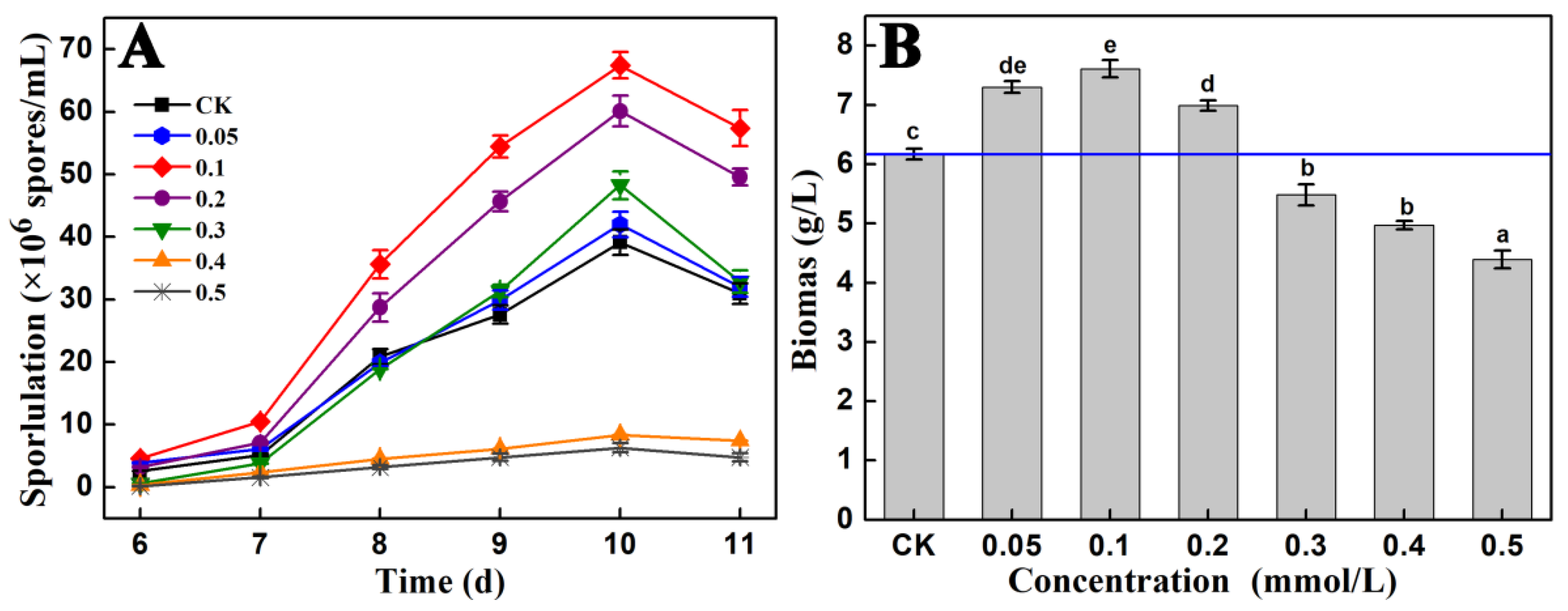
Figure 1. Effects of different concentrations of Fe2+ on the sporulation (A) and biomass (B) of A. cinnamomea in submerged fermentation. Note: “CK”, no metal ion was added in the culture medium; “0.05”, the concentration of Fe2+ in the culture medium is 0.05 mmol/L; “0.1, 0.2, 0.3, 0.4, and 0.5” are similar to “0.05 mmol/L Fe2+”. The A. cinnamomea mycelia cultured for 10 days at 26 °C and 150 r/min with inoculum size of 1.0 × 10⁶ spores/mL were used to measure the biomass; Different letters in (B) indicate significant differences at the level of 0.05 (p < 0.05).
Jof 09 00235 g001
Figure 2. Statistical analysis of the RNA-seq samples. Note: (A): Cluster analysis of gene expression; (B): principal component (PC) analysis; (C): distribution of the FPKM values of genes in all samples.
Jof 09 00235 g002
Figure 3. GO (A), KEGG (B), and KOG (C) enrichment analyses of DEGs.
Jof 09 00235 g003
Figure 4. Expression levels of genes involve in CWI pathway (A) and FluG-mediated patthway (B) in the mycelium samples of A. cinnamomea. Note: “mRNA”, messenger RNA; “CK”, the mycelia cultured absence of Fe2+; “Fe”, the mycelia cultured in the presence of 0.1 mmol/L of Fe2+. The 18S rRNA gene of A. cinnamomea was taken as the internal reference.
Jof 09 00235 g004
Figure 5. Model diagram of the Fe2+- and FluG-mediated signal pathway of the asexual sporulation of A. cinnamomea in submerged fermentation. Note: “green arrow”, upstream developmental activation pathway; “purple arrow”, central developmental pathway; “blue arrow”, iron homeostasis regulator pathway.
Jof 09 00235 g005
Table 1. Primers used for RT-qPCR.
Gene NameUpstream Primer (5′→3′)Downstream Primer (5′→3′)Product (bp)
flbDAATGTCTGAAGGTCGTGATGCCGCCGTATCGTTAGCCGTATGG126
abaATGTGCGAGTGCGGAGACCGTAGACGACGGACAGGAGGAC116
bck1GTCAACAGTATAGATATGCGTCAACAGTATAGATATGC127
mkk1CATAAAGGTCTTCGCTATCATAAAGGTCTTCGCTAT165
slt2ATCTCCTTTAGAAGACATCATCTCCTTTAGAAGACATC103
18S rRNAGCTGGTCGCTGGCTTCTTAGCGCTGGCTCTGTCAGTGTAG123
Table 2. Genes that may be related to the Fe2+-mediated asexual sporulation of A. cinnamomea among DEGs.
Unigene IDGenome IDGene NameAccession NumberE ValueScore
Cluster-140.3091ACg001255mirBNC_007196.11 × 10−19198
Cluster-140.1965ACg005881ftrANC_007198.15 × 10−1182
Cluster-140.3564ACg006970hapXNC_007198.14 × 10−18183
Cluster-140.2153ACg000929sreANC_007198.16 × 10−1289
Cluster-140.3137ACg005708fetCNC_032094.12 × 10−16138
Cluster-140.2788ACg000854bck1NW_007930838.18 × 10−16156
Cluster-140.3618ACg001175uvtKJ158162.12 × 10−17164
Cluster-140.2669ACg007032urbs1NC_026479.17 × 10−16154
Cluster-140.3500ACg005433sitMF447899.16 × 10−14102
Cluster-140.2357ACg001741freNC_007197.14 × 10−14100
Cluster-140.4088ACg006852slt2AEU60018.12 × 10−90326
Cluster-140.2081ACg007003ssiG 06045XP_001593123.16 × 10−18189
Cluster-140.1623ACg002353feoBNC_000913.33 × 10−16145
Cluster-140.3137ACg005708tpcANW_020939752.11 × 10−18175
Cluster-140.3451ACg003216nrpsKIM81356.102771
-ACg008442nps2NC_031953.17 × 10−1184
Cluster-140.3206ACg002074nps4KY471559.11 × 10−25268
Cluster-140.4587ACg007029clpPNC_000964.33 × 10−15126
Cluster-140.4385ACg003470mkk1NW_007930837.12 × 10−1079
-ACg000676sidANC_007194.18 × 10−19202
Cluster-140.3564ACg006969yvmBNC_020507.12 × 10−18179
Cluster-140.132ACg001175furNC_016845.13 × 10−1497
Cluster-140.2003ACg007734estBNC_007196.15 × 10−16149
Cluster-140.2389ACg007303wsc1NC_007198.13 × 10−16146
Note: “Unigene ID” is the code of the unigene generated in the process of software assembly; “Genome ID” is the code corresponding to the gene matched with the unigene in the A. cinnamomea genome (ACg); “-” indicates an unmatched gene in the A. cinnamomea genome database; “accession number” is the NCBI number of the protein matched with a unigene in the local protein database; “E value”, “Homology”, and “Coverage” are used to describe the matching between a unigene and the corresponding protein in the local protein database. If the E value is low, then homology and coverage are high, and the matching degree is high. It was considered as a successful match when the E value ≤ 10−6.
Disclaimer/Publisher’s Note: The statements, opinions and data contained in all publications are solely those of the individual author(s) and contributor(s) and not of MDPI and/or the editor(s). MDPI and/or the editor(s) disclaim responsibility for any injury to people or property resulting from any ideas, methods, instructions or products referred to in the content.