Article

Molecular Systematics and Taxonomic Analyses of Three New Wood-Inhabiting Fungi of Hyphoderma (Hyphodermataceae, Basidiomycota)

1
College of Biodiversity Conservation, Southwest Forestry University, Kunming 650224, China
2
Yunnan Key Laboratory of Gastrodia and Fungal Symbiotic Biology, Zhaotong University, Zhaotong 657000, China
3
Key Laboratory for Forest Resources Conservation and Utilization in the Southwest Mountains of China, Ministry of Education, Southwest Forestry University, Kunming 650224, China
4
Office of Management and Protection, Green Peacock Provincial Nature Reserve, Dali 671000, China
*
Authors to whom correspondence should be addressed.
J. Fungi 2023, 9(11), 1044; https://doi.org/10.3390/jof9111044
Submission received: 22 September 2023 / Revised: 18 October 2023 / Accepted: 23 October 2023 / Published: 24 October 2023
(This article belongs to the Special Issue Fungal Diversity in Various Environments, 2nd Edition)

Abstract

:
In this present study, three new wood-inhabiting fungal taxa, Hyphoderma niveomarginatum, H. sordidum and H. weishanense, are proposed. Hyphoderma niveomarginatum is characterized by the ceraceous basidiomata having a smooth, cracking hymenial surface and the presence of the moniliform cystidia and ellipsoid basidiospores (7–9 × 3.5–5 µm). Hyphoderma sordidum is characterized by its resupinate basidiomata with a smooth hymenial surface with the fimbriate margin, the presence of the tubular cystidia and ellipsoid basidiospores (3–4.5 × 2–3 µm). Hyphoderma weishanense differs in its membranous basidiomata with a slightly buff to buff hymenial surface and the presence of broadly ellipsoid basidiospores (4.5–8.5 × 4–7 µm). Sequences of ITS+nLSU+mt-SSU+RPB1+RPB2 genes were used for the phylogenetic analyses using three methods. The ITS+nLSU+mt-SSU+RPB1+RPB2 analysis of the genus Hyphoderma indicated that the 3 new species of Hyphoderma were nested into genus Hyphoderma, in which H. niveomarginatum formed a single group and then grouped with H. membranaceum and H. sinense; H. sordidum was a sister to H. nudicephalum; and H. weishanense closely grouped with H. crystallinum.

1. Introduction

Fungi are eukaryotic microorganisms that play fundamental ecological roles as decomposers and mutualists of plants and animals, in which they drive carbon cycling in the forest ecosystem, mediate the mineral nutrition of plants and alleviate carbon limitations of other soil organisms [1]. Wood-inhabiting fungi are an ecologically important branch of the tree of life, inferred from their distinct and diverse characteristics [2]. Taxa from the family Hyphodermataceae are continuously reported, with the frequent inclusion of data from DNA sequences and by employing both fresh material and cultures, and mycologists re-collect historic taxa and their types to accomplish taxonomy and molecular systematics in this family Hyphodermataceae [3,4].
The genus Hyphoderma Wallr. (1833: 576) belongs to the family Hyphodermataceae (Polyporales, Basidiomycota), typified by H. setigerum (Fr.) Donk. (1957: 15), and represents one of the important genera among wood-inhabiting fungi [4]. The genus is characterized by resupinate to effuse-reflexed basidiomata with ceraceous consistency, and smooth to tuberculate, grandinioid or odontioid hymenial surfaces, a monomitic hyphal system (rarely dimitic) with clamp connections on generative hyphae, the presence or not of cystidia, suburniform to subcylindrical and cylindrical basidia, and ellipsoid to subglobose, smooth, thin-walled basidiospores [5]. Based on the Index Fungorum (www.indexfungorum.org; accessed on 3 September 2023), the genus Hyphoderma has 206 specific and registered names. Currently, one hundred and thirteen species have been accepted worldwide [6,7,8,9,10,11,12,13].
This pioneering research for the phylogenetic analysis process of the genus Hyphoderma was just the prelude to the molecular systematics period [9,10,11,12,13,14]. The phylogenetic research revealed that all Hyphoderma taxa clustered into the different groups in phylogenetic trees at the class level based on the molecular phylogenetic methods, in which the result indicated that H. praetermissum (P. Karst.) J. Erikss. & Å. Strid and Resinicium bicolor (Alb. & Schwein.) Parmasto were grouped together, while the other Hyphoderma species, Hypochnicium J. Erikss, and several other species formed a separate branch [14]. The phylogeny of Hyphoderma showed that two species H. obtusum J. Erikss. and H. setigerum nested into the family Meruliaceae Rea and formed a sister taxon to Hypochnicium polonense (Bres.) Å. Strid [15]. The phylogenetical relationships among the closely related taxa in Hyphoderma were determined and a new species was proposed, H. macaronesicum Tellería, M. Dueñas, Beltrán-Tej., Rodr-Armas & M.P. Martín [16]. The research comprising the representative sequences of the H. setigerum complex showed that H. pinicola Yurch. & Sheng H. Wu represented a fifth species in this complex of this genus Hyphoderma [17]. The research of the family-level classification of the order Polyporales indicated that four Hyphoderma species grouped into the residual polyporoid clade, belonging to the family Hyphodermataceae, in which they grouped with three related genera in the family Meripilaceae as Meripilus P. Karst., Physisporinus P. Karst. and Rigidoporus Murrill [18].
During the investigations of the wood-inhabiting fungi, we collected three new Hyphoderma taxa from Yunnan Province, China, that could not be assigned to any described species of the order Polyporales. We present the morphological characteristics and multi-gene phylogenetic analyses with nLSU, ITS, mt-SSU, RPB2 and RPB1 that support the three species in the genus Hyphoderma.

2. Materials and Methods

2.1. Sample Collection and Herbarium Specimen Preparation

Fresh fruiting bodies of fungi growing on angiosperm branches were collected from the Lincang, Qujing and Dali of Yunnan Province, China. The samples were photographed in situ and fresh macroscopic details were recorded. Photographs were recorded using a Jianeng 80D camera (Tokyo, Japan). All of the photos were stacked and merged using Helicon Focus Pro 7.7.5 software. Specimens were dried in an electric food dehydrator at 40 °C, and then sealed and stored in an envelope bag and deposited in the herbarium of the Southwest Forestry University (SWFC), Kunming, Yunnan Province, China.

2.2. Morphology

Macromorphological descriptions were based on field notes and photos captured in the field and lab. Color terminology followed Petersen [19]. Micromorphological data were obtained from the dried specimens following observation under a light microscope [20]. The following abbreviations were used: KOH = 5% potassium hydroxide water solution, CB = cotton clue, CB– = acyanophilous, IKI = Melzer’s reagent, IKI– = both inamyloid and indextrinoid, L = mean spore length (arithmetic average for all spores), W = mean spore width (arithmetic average for all spores), Q = variation in the L/W ratios between the specimens studied and n = a/b (number of spores (a) measured from a given number (b) of specimens).

2.3. DNA Extraction and Sequencing

The EZNA HP Fungal DNA Kit (Omega Biotechnologies Co., Ltd., Kunming, China) was used to extract DNA from the dried specimens. The ITS region was amplified with the primer pair ITS5/ITS4 [21], the nLSU region with the primer pair LR0R/LR7 [22], the mt-SSU region with the primer pair MS1/MS2 [21], the RPB1 region with the primer pair RPB1-Af/RPB1-Cf [23] and the RPB2 region with the primer pair bRPB2-6F/bRPB2-7.1R [24]. The PCR procedure for ITS, nLSU, mt-SSU, RPB1 and RPB2 followed a previous study [22]. All of the newly generated sequences were deposited in GenBank (Table 1).

2.4. Phylogenetic Analyses

The sequences were aligned in MAFFT version 7 using the G-INS-i strategy [28]. The alignment was manually adjusted using AliView version 1.27 [29]. The sequence alignments were deposited in TreeBase (ID 30751; (accessed on 8 September 2023)). Diplomitoporus crustulinus (Bres.) Domański were assigned as an outgroup to root trees following a previous study analysis [13].
Maximum parsimony (MP), maximum likelihood (ML) and Bayesian Inference (BI) analyses were applied to the three combined datasets. The phylogenetic analysis method was adopted by Zhao and Wu [30]. MP analysis was performed in PAUP* version 4.0b10 [31]. All of the characteristics were equally weighted and gaps were treated as missing data. Trees were inferred using the heuristic search option with TBR branch swapping and 1000 random sequence additions. Max-trees were set to 5000, branches of zero length were collapsed and all most-parsimonious trees were saved. Clade robustness was assessed using bootstrap (BT) analysis with 1000 replicates [32]. Descriptive tree statistics tree length (TL), the consistency index (CI), the retention index (RI), the rescaled consistency index (RC) and the homoplasy index (HI) were calculated for each most-parsimonious tree generated. ML was inferred using RAxML-HPC2 through the Cipres Science Gateway (www.phylo.org (accessed on 13 September 2023)) [33]. Branch support (BS) for ML analysis was determined using 1000 bootstrap replicates and evaluated under the gamma model.
MrModeltest 2.3 [34] was used to determine the best-fit evolution model for each dataset for Bayesian inference (BI), which was performed using MrBayes 3.2.7a with a GTR+I+G model of DNA substitution and a gamma distribution rate variation across sites [35]. Four Markov chains were run twice from a random starting tree, over 10 million generations of the dataset (Figure 1), and the tree was sampled every 1000 generations. The first one fourth of all generations were discarded as burn-in. The majority rule consensus tree of all remaining trees was calculated. Branches were considered as significantly supported if they received a maximum likelihood bootstrap value (BS) >70%, maximum parsimony bootstrap value (BT) >70% or Bayesian posterior probabilities (BPPs) >0.95.

3. Results

3.1. Molecular Phylogeny
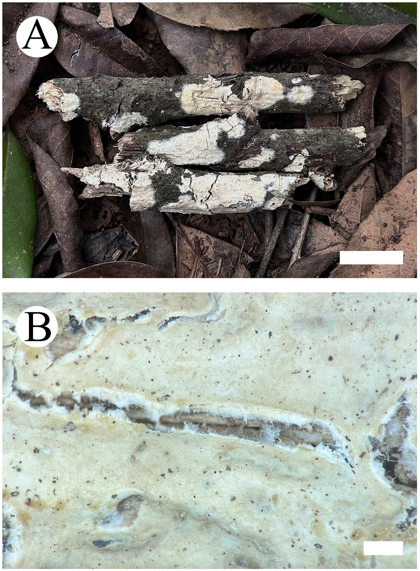
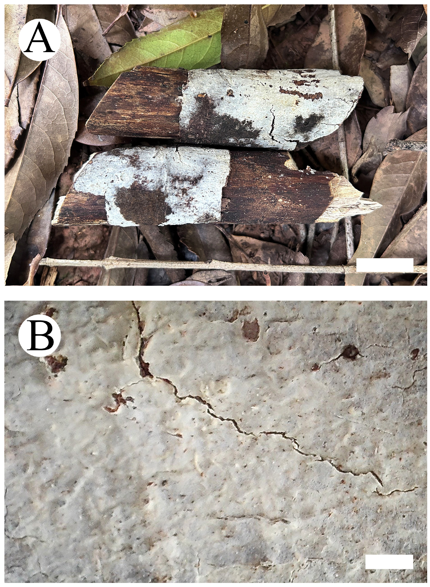
The dataset based on ITS+nLSU+mt-SSU+RPB1+RPB2 (Figure 1) comprises sequences from 60 fungal specimens representing 39 species. The alignment length of this dataset is 5675 characters, of which 2964 characters are constant, 1380 characters are variable with no information and 1331 characters have no information. Maximum parsimony analysis yielded three equally parsimonious trees (TL = 6130, CI = 0.5863, HI = 0.4137, RI = 0.6036, RC = 0.3539). Bayesian analysis and ML analysis resulted in a similar topology as MP analysis with an average standard deviation of split frequencies of 0.025189 (BI), and the effective sample size (ESS) across the two runs is double the average ESS (avg ESS) = 2823.
The phylogram based on the ITS+nLSU+mt-SSU+RPB1+RPB2 rDNA gene regions (Figure 1) indicated that three new species grouped into genus Hyphoderma, in which H. niveomarginatum grouped with two taxa, H. membranaceum C.L. Zhao & Q.X. Guan and H. sinense C.L. Zhao & Q.X. Guan, and then closely grouped with H. transiens (Bres.) Parmasto, H. amoenum (Burt) Donk and H. fissuratum C.L. Zhao & X. Ma. Hyphoderma sordidum clustered with H. nudicephalum Gilb. & M. Blackw. Hyphoderma weishanense grouped with H. crystallinum C.L. Zhao & Q.X. Guan, and then clustered with H. variolosum Boidin, Lanq. & Gilles, H. marginatum Z.Y. Duan & C.L. Zhao, H. medioburiense (Burt) Donk, H. assimile (H.S. Jacks. & Dearden) Donk and H. subsetigerum Sheng H. Wu.

3.2. Taxonomy

Hyphoderma niveomarginatum Y. Yang & C.L. Zhao, sp. nov. Figure 2 and Figure 3.
MycoBank no.: 849948.
Holotype—China, Yunnan Province, Lincang, Yun County, Dumu Village. GPS coordinates: 24°23′ N, 101°9′ E; altitude: 1960 m asl. On fallen unidentified angiosperm branch, leg. C.L. Zhao, 20 October 2022, CLZhao 25078 (SWFC).
Etymology—niveomarginatum (Lat.): referring to the white margin of basidiomata surface.
Basidiomata—Annual, resupinate, adnate, ceraceous, odorless when fresh, and up to 4 cm long, 2 cm wide and 50–200 µm thick. Hymenial surface smooth, pale yellowish when fresh, cream on drying, cracking. Sterile margin white to cream, up to 2 mm wide.
Hyphal system—Monomitic; generative hyphae with clamp connections; colorless, thin-walled, frequently branched, interwoven, 2–4 µm in diameter; IKI–, CB–, tissues unchanged in KOH.
Hymenium—Cystidia moniliform, with a variable number of constrictions, colorless, thin-walled, 29–55.5 × 5–7 µm; basidia clavate, slightly constricted in the median to somewhat sinuous, often with oil droplets, with four sterigmata and a basal clamp connection, 22–24 × 7–8 µm; basidioles in shape similar to basidia, but slightly smaller.
Spores—Basidiospores ellipsoid, colorless, thin-walled, smooth, with irregular oil droplets inside, IKI–, CB–, (6–)7–9(–10) × (3–)3.5–5(–5.5) µm, L = 8.01 µm, W = 4.24 µm, Q = 1.90 (n = 30/1).
Notes—Hyphoderma litschaueri (Burt) J. Erikss. & Å. Strid, H. malenconii (Manjón & G. Moreno) Manjón, G. Moreno & Hjortstam, H. membranaceum, H. moniliforme (P.H.B. Talbot) Manjón, G. Moreno & Hjortstam and H. tropicum Z.Y. Duan & C.L. Zhao are similar to H. niveomarginatum by having the moniliform cystidia. However, H. litschaueri differs in its larger moniliform cystidia (60–100 × 6–8 µm) and larger subcylindrical basidiospores (9–12 µm × 3–4 µm) [36]; H. malenconii is separated from H. niveomarginatum by having the dendrohyphidia and larger subcylindrical basidiospores (12–15 × 5.5–10 µm) [37]; H. membranaceum is distinct from H. niveomarginatum by tuberculate hymenial surface and larger basidiospores (11–13.5 × 4.5–5.5 µm) [10]; H. moniliforme differs in its membranous basidiomata and having the larger moniliform cystidia (85–100 × 6–8 µm) [4]; and H. tropicum is distinguished from H. niveomarginatum by having tuberculate hymenial surface and larger basidia (29.5–38 × 4–6 µm) [13].
Hyphoderma floccosum C.L. Zhao & Q.X. Guan, H. mopanshanense C.L. Zhao, H. sinense and H. transiens are similar to H. niveomarginatum by having the ceraceous basidiomata. However, H. floccosum differs in H. niveomarginatum by farinaceous hymenial surface and two types of cystidia: septate cystidia and tubular cystidia [11]; H. mopanshanense is separated from H. niveomarginatum by porulose hymenial surface and wider thick-walled generative hyphae (4–6 µm) [9]; H. sinense is distinct from H. niveomarginatum by having two types of cystidia: encrusted cystidia and moniliform cystidia, and cylindrical to allantoid basidiospores [11]; and H. transiens is distinguished from H. niveomarginatum by having odontioid hymenophore, larger cystidia (40–95 × 7–9 µm) and cylindrical basidiospores (8.5–12 × 2.7–4.1) [38].
Hyphoderma sordidum Y. Yang & C.L. Zhao, sp. nov. Figure 4 and Figure 5.
MycoBank no.: 849949.
Holotype—China, Yunnan Province, Qujing, Zhanyi District, Yanzhu Village. GPS coordinates: 25°44′ N, 103°36′ E; altitude: 1950 m asl. On fallen unidentified angiosperm branch, leg. C.L. Zhao, 7 March 2023, CLZhao 27390 (SWFC).
Etymology—sordidum (Lat.): referring to the sordid white hymenial surface.
Basidiomata—Annual, resupinate, adnate, membranous, odorless and up to 5 cm long, 2.5 cm wide and 50–150 µm thick. Hymenial surface smooth, white to cream when fresh, cream upon drying. Sterile margin white, fimbriate, up to 1–2 mm wide.
Hyphal system—Monomitic; generative hyphae with clamp connections; colorless, thin-walled, interwoven, 1.5–2 µm in diameter; IKI–, CB–, tissues unchanged in KOH.
Hymenium—Cystidia tubular, basally widened, tapering but without sublate apex, slightly sinuous, colorless, thin-walled, 42–72.5 × 6–11 µm; basidia clavate to subcylindrical, constricted in the middle to somewhat sinuous, with four sterigmata and a basal clamp connection, 8–14 × 3–3.5 µm; basidioles in shape similar to basidia, but slightly smaller.
Spores—Basidiospores ellipsoid, colorless, thin-walled, smooth, some with irregular oil droplets inside, IKI–, CB–, (2.5–)3–4.5 × (1.5–)2–3(–4) µm, L = 3.62 µm, W = 2.35 µm, Q = 1.56–1.70 (n = 60/2).
Additional specimens examined (paratypes)—China, Yunnan Province, Qujing, Zhanyi District, Yanzhu Village. GPS coordinates: 25°44′ N, 103°36′ E; altitude: 1950 m asl. On fallen unidentified angiosperm branches, leg. C.L. Zhao, 7 March 2023, CLZhao 17908; CLZhao 27379 (SWFC).
Notes—Hyphoderma anthracophilum (Bourdot) Jülich, H. cremeoalbum (Höhn. & Litsch.) Jülich, H. multicystidium (Hjortstam & Ryvarden) Hjortstam & Tellería, H. tropicum and H. obtusiforme J. Erikss. & Å. Strid are similar to H. sordidum by having ellipsoid basidiospores. However, H. anthracophilum differs from H. sordidum by the cracked hymenophore and larger basidiospores of 6–9 × 4–6 µm [39]; H. cremeoalbum is distinct from H. sordidum by having larger basidia (30–45 × 7–9 µm) and larger basidiospores (10–14 × 5–6 µm) [5]; H. multicystidium is separated from H. sordidum by the reticulate and tomentose hymenial surface, wider generative hyphae (2–3 µm) and larger basidiospores (8–10 × 4.5–5 µm) [40]; H. obtusiforme differs in having wider generative hyphae (3–4 µm), larger basidia (30–40 × 6–8 µm) and larger basidiospores (10–12 × 5–6 µm) [38]; and H. tropicum is distinguished from H. sordidum by having tuberculate hymenial surface, moniliform cystidia and larger basidiospores (6.5–7.5 × 3–4 µm) [13].
Hyphoderma crystallinum, H. marginatum, H. membranaceum, H. moniliforme and H. tenuissimum C.L. Zhao & Q.X. Guan are similar to H. sordidum by having membranous basidiomata. However, H. crystallinum differs in H. sordidum by hymenial surface with scattered nubby crystals, larger basidia (21.5–31 × 6–8.5 µm) and larger allantoid basidiospores (11–14.5 × 4–5.5 µm) [10]; H. marginatum is separated from H. sordidum by having cracking hymenial surface, cylindrical cheilocystidia and larger basidiospores (9–10 × 3.5–4.5 µm) [13]; H. membranaceum is distinct from H. sordidum by having cracking hymenial surface, moniliform cystidia and larger basidiospores (11–13.5 × 4.5–5.5 µm) [10]; H. moniliforme differs from H. sordidum by having cracking hymenial surface, larger basidia (20–30.5 × 6–7.5 µm) and larger basidiospores (6–9 × 3–4.5 µm) [4]; and H. tenuissimum is distinguished from H. sordidum by having tuberculate to minutely grandinioid hymenial surface, larger cylindrical cystidia (50–220 × 6.5–13 µm) and larger cylindrical basidiospores (7–10.5 × 3–4.5 µm) [12].
Hyphoderma weishanense Y. Yang & C.L. Zhao, sp. nov. Figure 6 and Figure 7.
MycoBank no.: 849950.
Holotype—China, Yunnan Province, Dali, Weishan County, Qinghua Town, Green Peacock Reserve. GPS coordinates: 24°52′ N, 100°12′ E; altitude: 1550 m asl. On fallen unidentified angiosperm branch, leg. C.L. Zhao, 18 July 2022, CLZhao 22403 (SWFC).
Etymology—weishanense (Lat.): refers to the locality (Weishan) of the type specimen.
Basidiomata—Annual, resupinate, adnate, membranous when fresh, hard membranous when dry, odorless and up to 11 cm long, 4 cm wide and 50–100 µm thick. Hymenial surface smooth, white when fresh, slightly buff to buff upon drying. Sterile margin thin, white, up to 1–2 mm wide.
Hyphal system—Monomitic; generative hyphae with clamp connections; colorless, thin-walled, interwoven, 2.5–3.5 µm in diameter; IKI–, CB–, tissues unchanged in KOH.
Hymenium—Cystidia absent; basidia subcylindrical, constricted in the middle to somewhat sinuous, with four short sterigmata and a basal clamp connection, 16.5–18.7 × 4.5–7.5 µm; basidioles in shape similar to basidia, but slightly smaller.
Spores—Basidiospores broadly ellipsoid, colorless, thin-walled, smooth, IKI–, CB–, (4–)4.5–8.5(–9) × (3–)4–7(–8) µm, L = 6.25 µm, W = 5.23 µm, Q = 1.20 (n = 30/1).
Notes—Hyphoderma floccosum, H. obtusiforme, H. puerense C.L. Zhao & Q.X. Guan, H. transiens and H. tropicum are similar to H. weishanense by having the ellipsoid basidiospores. However, H. floccosum differs in H. weishanense by having ceraceous basidiomata, farinaceous hymenial surface and two types of cystidia: septate cystidia and tubular cystidia [11]; H. obtusiforme is distinguished from H. weishanense by having porulose hymenial surface, cylindrical cystidia (50–60 × 8–10 µm), larger basidia (30–40 × 6–8 µm) and larger basidiospores (10–12 × 5–6 µm) [38]; H. puerense is separated from H. weishanense by the byssoid basidiomata, thick-walled generative hyphae covered by crystals and the tubular cystidia [12]; H. transiens is distinct from H. weishanense by having the ceraceous basidiomata, odontioid hymenial surface, subcylindrical cystidia and larger basidiospores (9–13 × 3–4.5 µm) [10]; and H. tropicum differs from H. weishanense by having tubercula hymenial surface, the moniliform cystidia and larger basidia (29.5–38 × 4–6 µm) [13].
Hyphoderma anthracophilum, H. cremeoalbum, H. fissuratum, H. sibiricum (Parmasto) J. Erikss. & Å. Strid and H. tenuissimum C.L. Zhao & Q.X. Guan are similar to H. weishanense by absent cystidia. However, H. anthracophilum is separated from H. weishanense by the pale grey to isabelline hymenial surface and larger basidia (30–40 × 5–7 µm) [5]; H. cremeoalbum differs in H. weishanense by having larger basidia (30–45 × 7–9 µm) and larger basidiospores (10–14 × 5–6 µm) [5]; H. fissuratum is distinct from H. weishanense by having ceraceous basidiomata, larger basidia (24–28 × 4–4.5 µm) and cylindrical basidiospores [12]; and H. sibiricum is distinguished from H. sordidum by the small irregular patches of basidiomata and larger basidia (25–35 × 5–7 µm) [38].

4. Discussion

Based on the molecular systematics study amplifying the nLSU, ITS and RPBl genes, the family-level classification for the order Polyporales (Basidiomycota) revealed that the four taxa of Hyphoderma macaronesicum, H. medioburiense, H. mutatum (Peck) Donk and H. setigerum nested into the family Hyphodermataceae within the residual polyporoid clade [18]. In the present study, from the phylogram inferred from the ITS+nLSU+mt-SSU+RPB1+RPB2 data, three new species grouped into Hyphoderma (Figure 1), in which H. niveomarginatum grouped with two taxa, H. membranaceum and H. sinense, and then closely grouped with H. transiens, H. amoenum and H. fissuratum; H. sordidum clustered with H. nudicephalum; and H. weishanense grouped with H. crystallinum, and then grouped closely with H. variolosum, H. marginatum, H. medioburiense, H. assimile and H. subsetigerum. However, morphologically, H. membranaceum is distinct from H. niveomarginatum by membranous basidiomata and wider basidiospores (11–13.5 × 4.5–5.5 µm) [10]; H. sinense differs from H. niveomarginatum by membranous basidiomata and thick-walled generative hyphae [11]; H. transiens is separated from H. niveomarginatum by its odontioid hymenial surface and larger subcylindrical cystidia (50–70 × 6–8 µm) [36]; H. amoenum is separated from H. niveomarginatum by membranous basidiomata and larger basidiospores measuring 12–15 × 4–6 µm [41]; and H. fissuratum differs from H. niveomarginatum by leathery basidiomata and cylindrical basidiospores [9]. Hyphoderma nudicephalum is distinct from H. sordidum by having the farinaceous to odontioid hymenial surface, and conspicuous capitate cystidia with the nonincrusted apices and thick-walled generative hyphae [42]. Hyphoderma crystallinum differs from H. weishanense by the hymenial surface with scattered nubby crystals and larger, allantoid basidiospores (11–14.5 × 4–5.5 µm) [10]; H. variolosum is separated from H. weishanense by its tuberculiform hymenial surface and having the tubular cystidia [43]; H. marginatum differs from H. weishanense by the cream hymenial surface and the larger basidiospores of 9–10 × 3.5–4.5 µm [13]; H. medioburiense is distinguished from H. weishanense by the porulose hymenial surface and having the tubular cystidia [44]; H. assimile is distinct from H. weishanense by its white to pale cream basidiomata, the tubular cystidia and larger basidia (33–37 × 7–8 µm) [45]; and H. subsetigerum differs from H. weishanense by its grandinioid hymenophore with the whitish to ivory yellow hymenial surface, and the thick-walled generative hyphae [37].
Wood-inhabiting fungi are found in living trees, decorticated wood of dead tree branches and trunks as well as manufactured wood products. These fungi of the cell walls and the components within the living cells secrete various enzymes that effectively degrade cellulose, hemicellulose and lignin into simple inorganic substances, and consequently play an important role in forest ecosystems as an important group of decomposers [46]. In terms of geographical distribution and ecological importance, Hyphoderma species are an extensively studied group, mainly found on hardwood, although a few species grow on coniferous wood [10]. We believe that more species of Hyphoderma occur in subtropical and tropical Asia, since wood-inhabiting fungi play a core role in the forest; they are rich in tropical China [47,48,49,50,51,52,53,54,55]; and it is very possible that the same phenomenon exists for Hyphoderma.

Author Contributions

Conceptualization, C.Z. and H.Z.; methodology, C.Z. and Y.Y.; software, C.Z. and H.Z.; validation, C.Z., H.Z. and Y.Y.; formal analysis, C.Z. and Y.Y.; investigation, C.Z., Y.Y., Q.J., Q.L., L.C. (Lijun Cheng) and S.Y.; resources, C.Z., J.Y. and L.C. (Li Cha); writing—original draft preparation, C.Z. and Y.Y.; writing—review and editing, C.Z., H.Z. and Y.Y.; visualization, C.Z. and Y.Y.; supervision, C.Z. and H.Z.; project administration, C.Z.; funding acquisition, C.Z. and H.Z. All authors have read and agreed to the published version of the manuscript.

Funding

The research was supported by the National Natural Science Foundation of China (Project Nos. 32170004, 32160063), the Research Project of Yunnan Key Laboratory of Gastrodia and Fungal Symbiotic Biology (TMKF2023A03) and High-level Talents Program of Yunnan Province (YNQR-QNRC-2018-111).

Institutional Review Board Statement

Not applicable for studies involving humans or animals.

Informed Consent Statement

Not applicable for studies involving humans.

Data Availability Statement

Publicly available datasets were analyzed in this study. This data can be found here: (https://www.ncbi.nlm.nih.gov/; https://www.mycobank.org/page/Simple%20names%20search; http://purl.org/phylo/treebase, submission ID 30751; accessed on 7 September 2023).

Conflicts of Interest

The authors declare no conflict of interest.

References

  1. Tedersoo, L.; Bahram, M.; Põlme, S.; Kõljalg, U.; Yorou, N.S.; Wijesundera, R.; Ruiz, L.V.; Vasco-Palacios, A.M.; Thu, P.Q.; Suija, A.; et al. Global diversity and geography of soil fungi. Science 2014, 346, 1256688. [Google Scholar] [CrossRef] [PubMed]
  2. James, T.Y.; Stajich, J.E.; Hittinger, C.T.; Rokas, A. Toward a fully resolved fungal tree of life. Annu. Rev. Microbiol. 2020, 74, 291–313. [Google Scholar] [CrossRef] [PubMed]
  3. Wijayawardene, N.; Hyde, K.D.; Dai, D.; Sánchez-García, M.; Goto, B.; Saxena, R.; Erdoğdu, M. Outline of Fungi and fungus-like taxa-2021. Mycosphere 2022, 13, 53–453. [Google Scholar] [CrossRef]
  4. Yurchenko, E.; Wu, S.H. Hyphoderma moniliforme and H. nemorale (Basidiomycota) newly recorded from China. Mycosphere 2015, 6, 113–121. [Google Scholar] [CrossRef]
  5. Bernicchia, A.; Gorjón, S.P. Fungi Europaei 12: Corticiaceae s.l.; Edizioni Candusso: Salamanca, Spain, 2010; pp. 1–1007. [Google Scholar]
  6. Wu, S.H.; Nilsson, H.R.; Chen, C.T.; Yu, S.Y.; Hallenberg, N. The white-rotting genus Phanerochaete is polyphyletic and distributed throughout the phleboid clade of the Polyporales (Basidiomycota). Fungal Divers. 2010, 42, 107–118. [Google Scholar] [CrossRef]
  7. Baltazar, J.M.; Silveira, R.M.B.; Rajchenberg, M. Type studies of J. Rick’s corticioid homobasidiomycetes (Agaricomycetes, Basidiomycota) housed in the Herbarium Anchieta (PACA). Phytotaxa 2016, 255, 101–132. [Google Scholar] [CrossRef]
  8. Martín, M.P.; Zhang, L.F.; Fernández-López, J.; Dueñas, M.; Rodríguez-Armas, J.L.; Beltrán-Tejera, E.; Telleria, M.T. Hyphoderma paramacaronesicum sp. nov. (Meruliaceae, Polyporales, Basidiomycota), a cryptic lineage to H. macaronesicum. Fungal Syst. Evol. 2018, 2, 57–68. [Google Scholar] [CrossRef]
  9. Ma, X.; Huang, R.X.; Zhang, Y.; Zhao, C.L. Hyphoderma fissuratum and H. mopanshanense spp. nov. (Polyporales) from southern China. Mycoscience 2021, 62, 36–41. [Google Scholar] [CrossRef]
  10. Guan, Q.X.; Zhao, C.L. Taxonomy and Phylogeny of the Wood-Inhabiting Fungal Genus Hyphoderma with Descriptions of Three New Species from East Asia. J. Fungi 2021, 7, 308. [Google Scholar] [CrossRef]
  11. Guan, Q.X.; Zhao, C.L. Two new corticioid species: Hyphoderma sinense and H. floccosum spp. nov. (Hyphodermataceae, Polyporales) from southern China. Mycosystema 2021, 40, 447–461. [Google Scholar] [CrossRef]
  12. Guan, Q.X.; Li, Y.F.; Zhao, C.L. Morphological and phylogenetic evidence for recognition of two new species of Hyphoderma (Basidiomycota) from southern China, with a key to all Chinese Hyphoderma. MycoKeys 2021, 83, 145–160. [Google Scholar] [CrossRef] [PubMed]
  13. Duan, Z.Y.; Guan, Q.X.; Luo, K.Y.; Zhao, C.L. Morphological and molecular identification of three new resupinate species of Hyphoderma (Hyphodermataceae, Agaricomycetes) from East Asia. Phytotaxa 2023, 599, 1–19. [Google Scholar] [CrossRef]
  14. Langer, E. Phylogeny of Non-Gilled and Gilled Basidiomycetes: DNA Sequence Inference, Ultrastructure and Comparative Morphology; Habilitationsschrift, Universität Tübingen: Tübingen, Germany, 2002. [Google Scholar]
  15. Larsson, K.H. Re-thinking the classification of corticioid fungi. Mycol. Res. 2007, 111, 1040–1063. [Google Scholar] [CrossRef] [PubMed]
  16. Telleria, M.T.; Dueñas, M.; Beltrán-Tejera, E.; Rodríguez-Armas, J.L.; Martín, M.P. A new species of Hyphoderma (Meruliaceae, Polyporales) and its discrimination from closely related taxa. Mycologia 2012, 104, 1121–1132. [Google Scholar] [CrossRef]
  17. Yurchenko, E.; Wu, S.H. Hyphoderma pinicola sp. nov. of H. setigerum complex (Basidiomycota) from Yunnan, China. Bot. Stud. 2014, 55, 71–78. [Google Scholar] [CrossRef] [PubMed]
  18. Justo, A.; Miettinen, O.; Floudas, D.; Ortiz-Santana, B.; Sjökvist, E.; Lindner, D.; Nakasone, K.; Niemelä, T.; Larsson, K.-H.; Ryvarden, L.; et al. A revised family-level classification of the Polyporales (Basidiomycota). Fungal Biol. 2017, 121, 798–824. [Google Scholar] [CrossRef]
  19. Petersen, J.H. Farvekort. The Danish Mycological Society’s Colour-Chart; Foreningen til Svampekundskabens Fremme: Greve, Denmark, 1996; pp. 1–6. [Google Scholar]
  20. Dai, Y.C.; Cui, B.K.; Si, J.; He, S.H.; Hyde, K.D.; Yuan, H.S.; Liu, X.Y. Dynamics of the worldwide number of fungi with emphasis on fungal diversity in China. Mycol. Prog. 2015, 14, 62. [Google Scholar] [CrossRef]
  21. White, T.J.; Bruns, T.; Lee, S.; Taylor, J. Amplification and Direct Sequencing of Fungal Ribosomal RNA Genes for Phylogenetics. In PCR Protocols: A Guide to Methods and Applications; Innis, M.A., Gelfand, D.H., Sninsky, J.J., White, T.J., Eds.; Academic Press: San Diego, CA, USA, 1990. [Google Scholar]
  22. Vilgalys, R.; Hester, M. Rapid genetic identification and mapping of enzymatically amplified ribosomal DNA from several Cryptococcus species. J. Bacteriol. 1990, 172, 4238–4246. [Google Scholar] [CrossRef]
  23. Matheny, P.B.; Liu, Y.J.; Ammirati, J.F.; Hallet, B.D. Using RPBI sequences to improve phylogenetic inference among mushrooms (Inocybe, Agaricales). Am. J. Bot. 2002, 89, 688–698. [Google Scholar] [CrossRef]
  24. Matheny, P.B. Improving phylogenetic inference of mushrooms with RPB1 and RPB2 nucleotide sequences (Inocybe, Agaricales). Mol. Phylogenet. Evol. 2005, 35, 1–20. [Google Scholar] [CrossRef]
  25. Vu, D.; Groenewald, M.; Vries, M.; Gehrmann, T.; Stielow, B.; Eberhardt, U.; Al-Hatmi, A.; Groenewald, J.Z.; Cardinali, G.; Houbraken, J.; et al. Large-scale generation and analysis of filamentous fungal DNA barcodes boosts coverage for kingdom Fungi and reveals thresholds for fungal species and higher taxon delimitation. Stud. Mycol. 2019, 92, 135–154. [Google Scholar] [CrossRef] [PubMed]
  26. Floudas, D.; Hibbett, D.S. Revisiting the taxonomy of Phanerochaete (Polyporales, Basidiomycota) using a four gene dataset and extensive ITS sampling. Fungal Biol. 2015, 119, 679–719. [Google Scholar] [CrossRef] [PubMed]
  27. Nilsson, R.H.; Hallenberg, N.; Nordén, B.; Maekawa, N.; Wu, S.H. Phylogeography of Hyphoderma setigerum (Basidiomycota) in the Northern Hemisphere. Mycol. Res. 2003, 107, 645–652. [Google Scholar] [CrossRef] [PubMed]
  28. Katoh, K.; Rozewicki, J.; Yamada, K.D. MAFFT online service: Multiple sequence alignment, interactive sequence choice and visualization. Brief. Bioinform. 2019, 20, 1160–1166. [Google Scholar] [CrossRef] [PubMed]
  29. Larsson, A. AliView: A fast and lightweight alignment viewer and editor for large data sets. Bioinformatics 2014, 30, 3276–3278. [Google Scholar] [CrossRef] [PubMed]
  30. Zhao, C.L.; Wu, Z.Q. Ceriporiopsis kunmingensis sp. nov. (Polyporales, Basidiomycota) evidenced by morphological characters and phylogenetic analysis. Mycol. Prog. 2017, 16, 93–100. [Google Scholar] [CrossRef]
  31. Swofford, D.L. PAUP*: Phylogenetic Analysis Using Parsimony (*and Other Methods); Version 4.0b10; Sinauer Associates: Sunderland, MA, USA, 2002. [Google Scholar]
  32. Felsenstein, J. Confidence intervals on phylogenetics: An approach using bootstrap. Evolution 1985, 39, 783–791. [Google Scholar] [CrossRef]
  33. Miller, M.A.; Pfeiffer, W.; Schwartz, T. The CIPRES Science Gateway: Enabling High-Impact Science for Phylogenetics Researchers with Limited Resources. Assoc. Comput. Mach. 2012, 39, 1–8. [Google Scholar] [CrossRef]
  34. Nylander, J.A.A. MrModeltest v2; Program Distributed by the Author; Evolutionary Biology Centre, Uppsala University: Uppsala, Sweden, 2004. [Google Scholar]
  35. Ronquist, F.; Huelsenbeck, J.P. MRBAYES 3: Bayesian phylogenetic inference under mixed models. Bioinformatics 2003, 19, 1572–1574. [Google Scholar] [CrossRef]
  36. Eriksson, J.; Ryvarden, L. The Corticiaceae of North Europe; Fungiflora: Oslo, Norway, 1975; Volume 3, pp. 288–546. [Google Scholar]
  37. Wu, S.H. New species of Hyphoderma from Taiwan. Mycologia 1997, 89, 132–140. [Google Scholar] [CrossRef]
  38. Parmasto, E. Conspectus Systematis Corticiacearum; Institutum Zoologicum et Botanicum Academiae Scientiarum RPSS Estonicae: Tartu, Estonia, 1968. [Google Scholar]
  39. Jülich, W. The genera of the Hyphodermoideae (Corticiaceae). Persoonia 1974, 8, 59–97. [Google Scholar]
  40. Tellería, M.T. Annotated list of the Corticiaceae, sensu lato (Aphyllophorales, Basidiomycotina), for Peninsular Spain and Balearic Islands. In Bibliotheca Mycologica; Schweizerbart Science Publishers: Stuttgart, Germany, 1990; Volume 135, pp. 1–152. [Google Scholar]
  41. Donk, M.A. Notes on resupinate Hymenomycetes IV. Fungus 1957, 27, 1–29. [Google Scholar]
  42. Gilbertson, R.L.; Blackwell, M. Some new or unusual corticioid fungi from the Gulf Coast region. Mycotaxon 1988, 33, 375–386. [Google Scholar]
  43. Boidin, J.; Gilles, G. Basidiomycètes Aphyllophorales de l’Île de La Réunion. XVI. Les genres Hyphoderma, Hyphodermopsis, Chrysoderma nov. gen. et Crustoderma. Cryptogam. Mycol. 1991, 12, 97–132. [Google Scholar]
  44. Eriksson, J. Studies in the Heterobasidiomycetes and Homobasidiomycetes—Aphyllophorales of Muddus National Park in North Sweden; AB Lundequistska Bokhandeln: Uppsala, Sweden, 1958; Volume 16, pp. 1–172. [Google Scholar]
  45. Manjón, J.L.; Moreno, G. Estudios sobre Aphyllophorales. III. Fructificaciones en Abies pinsapo Boiss. Cryptog. Mycol. 1983, 4, 145–156. [Google Scholar]
  46. Dai, Y.C. A revised checklist of corticioid and hydnoid fungi in China for 2010. Mycoscience 2011, 52, 69–79. [Google Scholar] [CrossRef]
  47. Liu, Z.B.; Wu, Y.D.; Zhao, H.; Lian, Y.P.; Wang, Y.R.; Wang, C.G.; Mao, W.L.; Yuan, Y. Outline, Divergence Times, and Phylogenetic Analyses of Trechisporales (Agaricomycetes, Basidiomycota). Front. Microbiol. 2022, 13, 818358. [Google Scholar] [CrossRef]
  48. Luo, K.Y.; Zhao, C.L. Morphology and multigene phylogeny reveal a new order and a new species of wood-inhabiting basidiomycete fungi (Agaricomycetes). Front. Microbiol. 2022, 13, 970731. [Google Scholar] [CrossRef]
  49. Qu, M.-H.; Wang, D.-Q.; Zhao, C.-L. A Phylogenetic and Taxonomic Study on Xylodon (Hymenochaetales): Focusing on Three New Xylodon Species from Southern China. J. Fungi 2022, 8, 35. [Google Scholar] [CrossRef]
  50. Shen, H.W.; Bao, D.F.; Bhat, D.J.; Su, H.Y.; Luo, Z.L. Lignicolous freshwater fungi in Yunnan Province, China: An overview. Mycology 2022, 13, 119–132. [Google Scholar] [CrossRef]
  51. Wu, F.; Tohtirjap, A.; Fan, L.F.; Zhou, L.W.; Alvarenga, R.L.M.; Gibertoni, T.B.; Dai, Y.C. Global Diversity and Updated Phylogeny of Auricularia (Auriculariales, Basidiomycota). J. Fungi 2021, 7, 933. [Google Scholar] [CrossRef] [PubMed]
  52. Wu, F.; Yuan, Y.; Chen, J.J.; Cui, B.K.; Zhou, M.; Dai, Y.C. Terrestriporiaceae fam. nov., a new family of Russulales (Basidiomycota). Mycosphere 2020, 11, 2755–2766. [Google Scholar] [CrossRef]
  53. Xiao, Y.P.; Wang, Y.B.; Hyde, K.D.; Eleni, G.; Sun, J.Z.; Yang, Y.; Meng, J.; Yu, H.; Wen, T.C. Polycephalomycetaceae, a new family of clavicipitoid fungi segregates from Ophiocordycipitaceae. Fungal Divers. 2023, 120, 1–76. [Google Scholar] [CrossRef]
  54. Zhao, C.L.; Qu, M.H.; Huang, R.X.; Karunarathna, S.C. Multi-Gene Phylogeny and taxonomy of the wood-rotting fungal genus Phlebia sensu lato (Polyporales, Basidiomycota). J. Fungi 2023, 9, 320. [Google Scholar] [CrossRef]
  55. Zhou, M.; Dai, Y.C.; Vlasák, J.; Yuan, Y. Molecular Phylogeny and Global Diversity of the Genus Haploporus (Polyporales, Basidiomycota). J. Fungi 2021, 7, 96. [Google Scholar] [CrossRef]
Figure 1. Maximum parsimony strict consensus tree illustrating the phylogeny of three new species and related species in Hyphoderma within Polyporales based on ITS+nLSU+mt-SSU+RPB1+RPB2 sequences. The branch is labeled with a maximum likelihood lead value greater than 70%, a reduced lead value greater than 50% and a Bayesian posterior probability greater than 0.95. The new species are in bold/green, the holotypes superscript “T”.
Jof 09 01044 g001
Figure 2. Hyphoderma niveomarginatum (holotype): basidiomata on the substrate (A), macroscopic characteristics of hymenophore (B). Bars: (A) = 2 cm and (B) = 1 mm.
Jof 09 01044 g002
Figure 3. Microscopic structures of Hyphoderma niveomarginatum (holotype): a section of the hymenium (A), basidia and basidioles (B), cystidia (C), basidiospores (D). Bars: (AD) = 10 µm.
Jof 09 01044 g003
Figure 4. Hyphoderma sordidum (holotype): basidiomata on the substrate (A), microscopic characteristics of hymenophore (B). Bars: (A) = 1 cm and (B) = 1 mm.
Jof 09 01044 g004
Figure 5. Microscopic structures of Hyphoderma sordidum (holotype): a section of the hymenium (A), basidia and basidioles (B), cystidia (C), basidiospores (D). Bars: (AD) = 10 µm.
Jof 09 01044 g005
Figure 6. Hyphoderma weishanense (holotype): basidiomata on the substrate (A), microscopic characteristics of hymenophore (B). Bars: (A) = 2 cm and (B) = 1 mm.
Jof 09 01044 g006
Figure 7. Microscopic structures of Hyphoderma weishanense (holotype): a section of the hymenium (A), basidia and basidioles (B), basidiospores (C). Bars: (AC) = 10 µm.
Jof 09 01044 g007
Table 1. List of species, specimens and GenBank accession numbers of sequences used in this study. The new species are in bold, the holotypes superscript “T”.
Species NameSample No.GenBank Accession No.References
ITSnLSURPB1RPB2mt-SSU
Diplomitoporus crustulinusFD-137KP135299KP135211KP134883 [18]
Hyphoderma amoenumUSO 286622HE577030 [16]
H. assimileCBS:125852MH863808MH875272 [25]
H. cremeoalbumNH 11538 (GB)DQ677492DQ677492 [15]
H. cremeoalbumCLZhao 17007OM985716OM985753 OQ706819[13]
H. crystallinumCLZhao 9338 TMW917161MW913414 [10]
H. crystallinumCLZhao 9374MW917162MW913415 [10]
H. definitumNH 12266 (GB)DQ677493DQ677493 [15]
H. fissuratumCLZhao 6731MT791331MT791335 OQ706806[9]
H. fissuratumCLZhao 6726 TMT791330MT791334 OQ706805[9]
H. floccosumCLZhao 17129 TMW301683MW293733 OQ706826[11]
H. floccosumCLZhao 17215MW301687MW293735 OQ706829[11]
H. granuliferum5273JN710545JN710545 JN710673[17]
H. incrustatumKHL6685 AY586668 [17]
H. litschaueriNH 7603 (GB)DQ677496DQ677496 [15]
H. litschaueriFP-101740-SpKP135295KP135219KP134868KP134965 [13]
H. macaronesicumMA:Fungi 90388KC984327KF150025 KF181122 Unpublished
H. macaronesicumTFC:Mic 15115HE577011KF150050 KF181118 [17]
H. marginatumCLZhao 3404 TOM985717OM985754 [13]
H. medioburienseFD-335KP135298KP135220KP134869KP134966 [26]
H. membranaceumCLZhao 5844MW917167MW913420 OQ706797[10]
H. membranaceumCLZhao 6971 TMW917168MW913421 OQ706807[10]
H. microporoidesCLZhao 6857 TMW917169MW913422 [10]
H. microporoidesCLZhao 8695MW917170MW913423 [10]
H. moniliformeWu 0211-42 TKC928282 [4]
H. moniliformeWu 0211-46KC928284KC928285 [4]
H. mopanshanenseCLZhao 6498 TMT791329MT791333 [9]
H. mopanshanenseCLZhao 6449OM985720OM985759 OQ706803[13]
H. nemoraleTNM F3931KJ885183KJ885184 [4]
H. nemoraleWu 9508-14 TKC928280KC928281 [4]
H. niveomarginatumCLZhao 25078 TOR141728OR506179 OR543992 Present study
H. nudicephalumWu9307_29AJ534269 [27]
H. nudicephalumCLZhao 17839OM985721OM985760 OQ706835[13]
H. obtusiformeKHL1464JN572909 [17]
H. obtusiformeKHL11105JN572910 [17]
H. obtusumJS17804 AY586670 [17]
H. occidentaleKHL 8477 (GB)DQ677499DQ677499 [15]
H. paramacaronesicumMA:Fungi 87736KC984399 [8]
H. paramacaronesicumMA:Fungi 87737KC984405 [8]
H. pinicolaTNM F13637 TKJ885181KJ885182 [17]
H. pinicolaWu 0108-36KC928278KC928279 [17]
H. prosopidisARIZ HHB 8479HE577029 [4]
H. puerenseCLZhao 9476 TMW443045 [12]
H. puerenseCLZhao 9583MW443046MW443051 [12]
H. roseocremeumNH10545 AY586672 [17]
H. setigerumFCUG 1200AJ534273 [27]
H. setigerumFCUG 1688 TAJ534272 [27]
H. sinenseCLZhao 7963MW301679MW293730 [11]
H. sinenseCLZhao 17811 TMW301682MW293732 [11]
H. sordidumCLZhao 27379OR141731 OR507165Present study
H. sordidumCLZhao 27390 TOR141732OR506180OR520149 OR507166Present study
H. subsetigerumHHB11620GQ409521 [17]
H. tenuissimumCLZhao 7221 TMW443049MW443054 OQ706809[12]
H. tenuissimumCLZhao 16210MW443050MW443055 [12]
H. transiensNH 12304 (GB)DQ677504DQ677504 [15]
H. tropicumCLZhao 17308 TOM985727OM985768 [13]
H. variolosumCBS:734.91MH862320MH873992 [25]
H. variolosumCBS:735.91MH862321MH873993 [25]
H. weishanenseCLZhao 22403 TOR141727OR506181 Present study
H. yunnanenseCLZhao 8845 TOM985769 OQ706811[13]
Disclaimer/Publisher’s Note: The statements, opinions and data contained in all publications are solely those of the individual author(s) and contributor(s) and not of MDPI and/or the editor(s). MDPI and/or the editor(s) disclaim responsibility for any injury to people or property resulting from any ideas, methods, instructions or products referred to in the content.

