Article

A New Genotype of Trichophyton quinckeanum with Point Mutations in Erg11A Encoding Sterol 14-α Demethylase Exhibits Increased Itraconazole Resistance

Department of Dermatology, Jena University Hospital, Friedrich Schiller University, D-07747 Jena, Germany
*
Author to whom correspondence should be addressed.
J. Fungi 2023, 9(10), 1006; https://doi.org/10.3390/jof9101006
Submission received: 22 August 2023 / Revised: 8 September 2023 / Accepted: 10 October 2023 / Published: 12 October 2023
(This article belongs to the Special Issue Dermatophytes and Dermatophytoses, 2nd Edition)

Abstract

:
Trichophyton quinckeanum, the causative agent of mouse favus, has been responsible for several infections of animal owners in recent years and showed an infection peak around 2020 in Jena, Thuringia. The isolated T. quinckeanum strains from Thuringia differ in some positions of the ITS region compared to strains from the IHEM collection as well as to Trichophyton schoenleinii. All T. quinckeanum strains of the new genotype show up to a 100-fold increased itraconazole resistance as measured by microplate laser nephelometry (MLN) assays. Analysis of genes involved in Trichophyton indotineae azole resistance, such as Erg1, which encodes squalene epoxidase, and Erg11B, one of two copies of the sterol 14-α demethylase gene, show a 100% identity between the two T. quinckeanum genotypes. In contrast, Erg11A fragments differ in 15-nucleotide positions between both T. quinckeanum genotypes, resulting in the unique amino acid substitution Ala256Ser in resistant strains. The new T. quinckeanum genotype may have evolved through interspecies mating. Mating type analysis showed a nearly 100% identity of the minus type MAT1-1-1 fragment for all T. quinckeanum isolates. The closely related Trichophyton schoenleinii belongs to the plus mating type and has 100% identical fragments of Erg1 and Erg11B. Erg11A protein sequences of T. schoenleinii and T. quinckeanum showed increased diversity.

1. Introduction

Trichophyton quinckeanum is the causative agent of favus in mice. H. I. Quincke, who was honored as the species’ namesake, presumed that several fungi could cause the clinical symptoms of favus in humans [1]. Favus is a condition that most commonly affects the scalp in humans, where fungal hyphae invade the skin around hair follicles and form a cup-shaped, yellow scab. Quincke’s excellent drawings of microscopic analyses of the described ‘α-fungus’, which he found in human favus, show typical micro- and macroconidia [1]. He also infected animals such as mice with the ‘α-fungus’ and was able to successfully trigger the disease in these mice with the typical favus symptoms [1]. Today, Trichophyton schoenleinii is the causative agent of most cases of favus in humans [2], which was first described by J. L. Schoenlein and R. Remak [2] and corresponds to the ‘γ-fungus’ description of H. I. Quincke [1]. Nevertheless, rare cases of human favus due to T. quinckeanum have sometimes been identified [3]. Phylogenetic analyses showed that both species are closely related [4]; however, the morphology of T. quinckeanum and T. schoenleinii differs significantly; the latter is incapable of producing micro- or macroconidia and the mycelium displays chandelier- or antler-like hyphae [2].
Favus in humans has been gradually disappearing in Europe and is more prevalent in Middle East and Africa, although the trend is also declining [2]. However, resurgence of favus in humans is possible [5], particularly if people do not have access to or avoid medical treatment. Interestingly, the number of patients presenting with T. quinckeanum infections has increased significantly in recent years in the outpatient clinic of the Department of Dermatology at Jena University Hospital [6]. Here, infections caused typically tinea corporis or tinea capitis [6]. Severe tinea capitis due to the new T. quinckeanum subtype has been mainly observed in young children, but not in adults or adolescents, where tinea corporis was the typical diagnosis [6]. Several of the infected individuals were cat owners [6]. Two of the investigated T. quinckeanum fungal cultures were isolated from animal hair of the pet owners’ cats. The infection peak in 2020 correlated with a peak in the field mouse population (Microtus arvalis) in agriculture in the Jena area, according to reports from the Thuringia ministry of agriculture (TLLLR) [7].
Genetic analysis of the T. quinckeanum strains isolated in Jena showed that they differ in few positions in the ITS region compared to the neotype strain IHEM 13697 [8]. Strains with this new ITS pattern were also observed in other regions of Germany [9,10]. Recently, T. quinckeanum strains of this new ITS genotype were isolated in the Czech Republic [11]. First isolates were detected in 2016, and increased numbers of strains were reported up to 2020. Cats and dogs often transmit the disease to their owners [11]. A recent Swiss study showed that cats with dermatomycosis are more likely to be infected with T. quinckeanum when kept outdoors and thus have the opportunity to hunt mice [12]. The comparison of epidemiological studies is often hampered by name changes and reclassifications of dermatophytes. For example, the reference strain CBS 318.56 of the Swiss study mentioned was first classified as T. mentagrophytes [12] and later reclassified as T. quinckeanum [8].
In recent decades, changes in species designation based on morphological and molecular datasets have become more frequent. The species identification debate surrounding T. quinckeanum started with analysis of mating behavior [13,14]. In the late 1990s, the species name was changed into T. mentagrophytes due to the mating behavior [15,16]. However, it was then renamed to T. quinckeanum in 2017 [8]. Weitzman and Padhye demonstrated the ability of T. quinckeanum to produce viable ascospore progeny with both T. simii isolate and T. benhamiae [14]. Interestingly, all T. quinckeanum isolates of this study belonged to the minus mating type [14]. The combination of phylogenetic data based on a range of DNA sequences from multiple genes as well as morphological and physiological data sets allows the identification of new species or subtypes. An example of this is T. benhamiae var. luteum, classified according to the new classification system [17] and formerly referred to as the yellow subtype of T. benhamiae [18]. Another example of a recently identified new species is T. indotineae [19,20], a novel human pathogen that exhibits multiple drug resistances to allylamines and azoles [21,22,23]. How these new genotype species evolved when clonal reproduction appears to be the main mode of reproduction in Trichophyton species is an open question and part of the current debate [24]. So far, Metin and Heitman have described clonal reproduction of anthropophilic species like T. rubrum or T. interdigitale, while sexual reproduction is believed to be restricted to zoophilic strains such as T. mentagrophytes or T. benhamiae [24]. Hence, it is important to elucidate how the new subtype of T. quinckeanum evolved and why we did not recover its old subtype in our patients.
The objective of this study was to investigate the sensitivity of the collected T. quinckeanum strains from Thuringia that show differences in the ITS sequence compared to the IHEM strains as well as Trichophyton schoenleinii towards common antifungal azoles. Additionally, analysis of genes involved in azole resistance, such as Erg1, Erg11A and Erg11B, was performed to identify point mutations responsible for amino acid substitutions, which could account for the sensitivity differences to azoles. Finally, yet importantly, mating type analysis was carried out for the T. quinckeanum isolates to determine whether the new genotype may have evolved through interspecies mating.

2. Materials and Methods

2.1. Strains and Growth Conditions

T. quinckeanum strains from the Jena University Hospital (UKJ) were stored as glycerol stock solutions at −80 °C and cultured on dermasel agar and Sabouraud Dextrose (2% w/v Dextrose, SDA) agar plates. For microscopy, strains were cultivated on Takashio agar [25,26]. IHEM strains were purchased from the Belgian Coordinated Collection of Microorganisms BCCM/IHEM (Brussels, Belgium). Strain ATCC 46950, originally classified as T. schoenleinii [27], was obtained from the American Type Culture Collection ATCC (Manassas, VA, USA). T. schoenleinii UKJ 1317/12 was a gift of H.-J. Tietz, head of the Mycoclinic Berlin. Source information on the isolates is provided in Table 1.

2.2. Plate Assay for Resistance Estimation of T. schoenleinii and T. quinckeanum

T. schoenleinii does not form micro- or macroconidia; therefore, pieces of mycelium were transferred to liquid or solid media. Methods based on optical density measurements were not suitable because large particles lead to an inhomogeneous solution, which influences the measurements results. An agar-based resistance assay was developed for Aspergillus fumigatus [30] and was adapted to estimate resistance pattern of T. schoenleinii. Small plates were filled with an 8 mL SDA agar. One plate was used as a control without antifungal compounds, and three further plates contained increasing concentrations of fluconazole (0.4 µg/mL; 4 µg/mL; 40 µg/mL) or itraconazole (0.005 µg/mL; 0.05 µg/mL; 0.5 µg/mL). Concentration ranges were estimated from the results of T. quinckeanum. The plates were inoculated with small pieces (around 0.5 × 1 cm2) of T. schoenleinii or T. quinckeanum mycelium obtained from pre-cultivated plates grown for three to eight weeks. The pieces were excised from the growth front zone. Growth patterns were assessed after 5 to 7 days and compared to growth on SDA control plates without antifungal agents. Positive results were obtained when mycelium was able to grow from the transferred agar piece on antifungal-containing agar plates similar to the control plate without the addition of antifungal compounds. Negative results were obtained if the mycelial growth was restricted to the transferred agar piece. An intermediate reaction was classified as growth within the restrictive agar plate being distinctly reduced in comparison to the control.

2.3. Microplate Laser Nephelometry Assays

The MLN assays allow the recording of growth curves in each single well of a 96-well microplate. The analyses were performed as previously described [23,31,32] over 120 h at 30 °C with continuous orbital shaking and hourly measurements employing the NEPHELOstar Galaxy (BMG Labtech, Ortenberg, Germany). Spores were collected from pre-cultivated SDA or Dermasel agar plates pre-cultivated between three and eight weeks at room temperature. The spores were suspended in a 5 mL sterile, isotonic NaCl solution (9 g/L; Fresenius Kabi, Bad Homburg, Germany) and the suspension was then filtered through a cell strainer with a mesh size of 40 µm (Greiner Bio-One, Frickenhausen, Germany) to remove mycelial debris. Spore concentration was estimated by counting spores using disposable counting chambers (type Neubauer improved; Carl Roth GmbH, Karlsruhe, Germany), and suspensions were adjusted to a final concentration of 2 × 10³ spores/mL. A total of 100 µL of the adjusted spore solution was added to each well, which resulted in approximately 200 spores per well. Adjusted spore solutions were also plated on SDA agar to evaluate spore viability and allow determination of numbers of colony-forming units. The antifungal compounds itraconazole, voriconazole, sertaconazole-nitrate, terbinafin and nystatin were solved in DMSO and stored as stocks as described [23]. Ciclopirox olamine was prepared as previously reported [32]. Amorolfin (EP standard, Sigma Aldrich GmbH, Taufkirchen, Germany) was stored as a stock solution of 1 mg/mL in DMSO and clotrimazole (PHR Standard, Sigma Aldrich) was stored as a stock of 5 mg/mL in DSMO. Antifungal stocks were diluted in a liquid SDA medium in 1:2 increments, reducing the concentration by half in each case. As positive control SDA media without antifungals (100 µL per well) was added to the spore solution, blanks of each type were included as negative controls with the spore solution being replaced by the SDA medium. For each strain, three technical and two biological replicates were obtained. The concentration of a 90% growth inhibition (MIC90) of the antifungals was calculated from the growth curves over 120 h as described previously [23]. The following antifungal concentration ranges were used: itraconazole (1 µg/mL to 0.0005 µg/mL), voriconazole (2 µg/mL to 0.008 µg/mL), sertaconazole-nitrate (8 µg/mL to 0.13 µg/mL), clotrimazole (1 µg/mL to 0.016 µg/mL), terbinafin (0.05 µg/mL to 0.0008 µg/mL), amorolfin (0.13 µg/mL to 0.002 µg/mL), nystatin (40 µg/mL to 0.63 µg/mL) and ciclopirox olamine (32 µg/mL to 0.5 µg/mL).

2.4. DNA Isolation and DNA Amplification
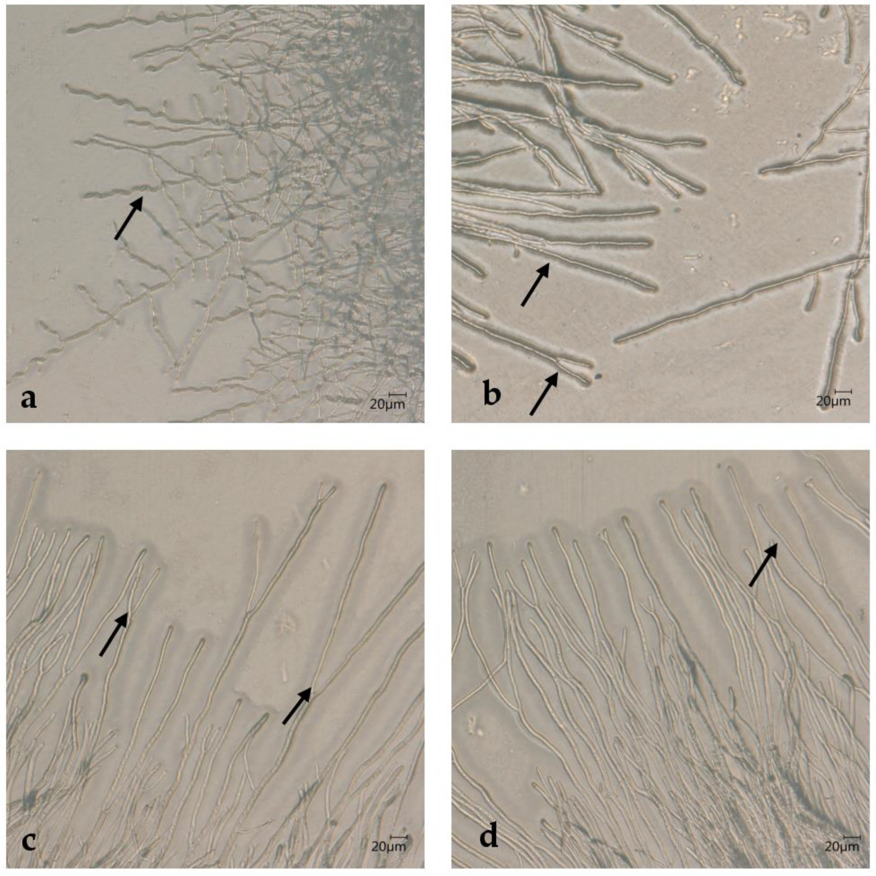
T. schoenleinii and ATCC strain 46950 DNA were isolated from fungal cultures grown in a SD broth in Erlenmeyer flasks under shaking (120 rpm) at room temperature to enhance the mycelial material. The mycelium was harvested using Miracloth (Merck Kg, Darmstadt, Germany) filter screens and treated with liquid nitrogen using a mortar and a pestle. T. quinckeanum DNA was obtained from cultures of at least two weeks of age on SD agar. Sufficient amounts of microconidia were formed within this period. For further DNA preparation, the Qiagen GmbH (Hilden, Germany) DNA mini kit was used according to the manufacturer’s instructions. Protease K treatment at 55 °C was limited to one hour for the liquid culture preparations but was performed overnight for mycelium forming microconidia. Erg1, Erg11A and Erg11B fragments were amplified as previously described [23]. ITS regions were sequenced to correctly identify all strains using primer V9G [33] and LSU266 [34]. The phylogenetic tree based on the Neighbor Joining method [35] (Figure S1) visualized the relationship of T. quinckeanum and T. schoenleinii genotypes. Mating type analysis was performed as described [26]. Primers specially designed for amplification of T. quinckeanum fragments are listed in Table S1. Sequence data were stored in GenBank; GenBank Acc No. OQ536505–OQ536519 for Erg1 fragments; GenBank Acc. No. OQ536520–OQ536534 for Erg11A; GenBank Acc. No. OQ536535- OQ536549 for Erg11B, GenBank Acc. No. OQ536550–OQ536561, OQ536564 for the MAT1-1-1 encoding α-box transcription factor and GenBank Acc. No. OQ536562, OQ536563 for the MAT1-1-2 encoding HMG box transcription factor. ITS sequence data GenBank Acc. No. for T. quinckeanum were as previously described [6]. Strains and related GenBank Acc. No. are listed in Table S2.

2.5. Microscopy

Microscopy was performed with the Keyence digital microscope on mycelium-grown Takashio agar plates [25,26]. A piece of agar medium, about 0.5–1 cm wide and 3–4 cm long, was taken from of the petri dish and covered with sterile cellophane foil before starting fungal cultivation to improve the contrast for microscopic images. Microscopic images were obtained directly from grown culture of the plates without damaging the mycelium.

3. Results

3.1. Resistant Phenotypes of T. quinckeanum Strains

MLN was used to determine the 90% minimal inhibitory concentration (MIC90) for the antifungal agents (Table 2). Similar MIC90 values were found for most antifungals (Table 2). In contrast, an increase in itraconazole MIC90 values was observed when T. quinckeanum IHEM strains and UKJ collection strains were compared. IHEM and UKJ strains represent different T. quinckeanum ITS genotypes. All UKJ strains showed values in the range of 0.09–0.45 µg/mL for the MIC90 of itraconazole (Table 2) and represented one genotype. UKJ isolates 1953/19 and 1254/20 were isolated from cat hair provided by the infected pet owners. This showed how important it is that, in addition to treating pet owners, the infected pets should also be treated by a veterinarian. IHEM strains showed MIC90 values for itraconazole in the range of 0.003–0.07 µg/mL and represented a different genotype (Table 2). A comparison of the results for both T. quinckeanum genotypes exhibited an up to 100-fold increase in values for itraconazole (Table 2). Cut-off values have not yet been defined. Nevertheless, a cut-off of 0.08 µg/mL would separate both genotypes in sensitive and resistant strains. IHEM strain analysis allows a glimpse into the past, as these genotypes were isolated in different years and in different parts of the world. F. Blank of Philadelphia, USA isolated IHEM strain 13570 from a dog and strain 13697 from a mouse [4,14], respectively, named No. 19 (syn. CDC X395) and No. 10. (syn. CDC X393). Strain IHEM 13697 represents the neotype isolate of T. quinckeanum [8]. The IHEM 13572 strain was isolated from a rodent in Adelaide, Australia in 1964. Strain IHEM 26522 is a subculture of CBS 318.56, which causes deep trichophytosis in humans, and it was isolated in 1955 by J. Zoon in the Netherlands.
All T. quinckeanum strains demonstrated high resistance to fluconazole, and it was not possible to determine MIC90 values with the MLN assay. A fluconazole concentration of 160 µg/mL was not able to sufficiently inhibit the growth of T. quinckeanum strains. and the MLN assay failed.
To understand the relationship between the two genotypes of T. quinckeanum and the close relative T. schoenleinii, two T. schoenleinii strains were also analyzed. IHEM strain 13515 was a human isolate from Casablanca, Morocco (obtained in 1966) [4] and UKJ strain 1317/12 was a gift of H.-J. Tietz isolated approximately 1972. ITS data confirm the species identification. Strain ATCC 46950 was isolated in Iraq from depilated human hair and identified as T. schoenleinii [36]. Genetic analysis showed that ATCC 46950 belongs to the T. mentagrophytes complex [4]. However, T. schoenleinii strains IHEM 13515 and UKJ 1317/12 as well as T. mentagrophytes strain ATCC 46950 form typical ‘stag antler’ hyphae as shown in Figure 1. Therefore, the morphology of ATCC 46950 closely resembles that of T. schoenleinii. ‘Antler’ hyphae are equal in length in both hyphal branches and often have an arc of about 45°. Interestingly, only a few hyphae of this type formed in IHEM 13515 (Figure 1a), while almost all hyphae observed in ATCC 46950 belonged to this type (Figure 1c,d). UKJ 1317/12 and ATCC 46950 showed high morphological similarities. Hyphae of T. mentagrophytes ATCC 46950 showed a coordinated growth front in large parts of the colony (Figure 1d).
Since T. schoenleinii and T. mentagrophytes strain ATCC 46950 do not form spores, an agar-based method was used for the analysis of azole resistance. The method is suitable to compare data of T. schoenleinii strains with T. quinckeanum strains, as shown in Table 3. T. schoenleinii strains showed increased sensitivity against fluconazole compared to the two genotypes of T. quinckeanum. The itraconazole behavior of T. schoenleinii resembled more the sensitive type of T. quinckeanum IHEM strains. The highest sensitivity to both azoles was found for T. mentagrophytes strain ATCC 46950. However, the agar-based method did not reach the accuracy of the MLN assay and produced more of an estimate.

3.2. Gene Analyses for Putative Association with Resistance Phenotypes

Azoles interact with sterol 14-α demethylases (Erg11); therefore, the two copies of Erg11, labeled A and B, were candidate genes to be analyzed. Point mutations in the gene for squalene epoxidase Erg1 of T. indotineae were shown to be associated with an increase in azole resistance [23,31]. When comparing the sequences of Erg11B and Erg1 of the T. quinckeanum ITS genotypes including T. schoenleinii, no nucleotide exchanges within the aligned fragments were observed (Table 4). The sequence of the neotype strain IHEM 13697 [8] was used as reference sequence. DNA sequence gaps were not counted in statistical analysis (Table 4). The results for Erg11A showed a higher level of nucleotide exchanges between the two ITS genotypes of T. quinckeanum compared to T. schoenleinii and the T. quinckeanum IHEM strains (Table 4).
Interestingly, Erg11A protein sequence alignments differ from the results obtained at the nucleotide level. Only two amino acid positions differed in the Erg11A protein alignments of both T. quinckeanum genotypes, whereas four amino acid exchanges were found in the protein sequences of IHEM T. quinckeanum and T. schoenleinii. A comparison of IHEM T. quinckeanum sequences with T. simii also revealed four amino acid exchanges. The Phe61Ser mutation is unique for IHEM T. quinckeanum strains compared to UKJ T. quinckeanum, T. schoenleinii and T. simii, where a Phe codon was found in this position. The UKJ T. quinckeanum Ala254Ser mutation differs from the Ala codon found in IHEM T. quinckeanum, T. schoenleinii and T. simii at this protein sequence position. Specific to T. schoenleinii were exchanges at Asp69Asn, Pro215His and Met306Ile. T. simii exhibited specific exchanges at Val316Ala, Val389Ile and Pro420Leu of the Erg11A protein sequence. Amino acids positions were deduced from the complete gene sequences decoded from the T. schoenleinii genome [36]. Interestingly, the T. schoenleinii-specific Pro215His mutation is part of the F-F’ motif, which is necessary for the function of Erg11 proteins [37].

3.3. T. quinckeanum and T. schoenleinii Represent Opposite Mating Types

Mating type genes were partially amplified as previously described [26] and sequenced. Results show that all T. quinckeanum strains belong to the minus mating type representing the MAT1-1-1 gene [38] of the α-box transcription factor. Sequence information of the eight UKJ strains and the four IHEM strains of T. quinckeanum indicate the presence of the MAT1-1-1 allele (Table 4). Interestingly, T. schoenleinii strains represent the opposite mating type, and the Mat1-2-1 allele of the HMG-box transcription factor [34] was detected (Table 4). Other T. quinckeanum UKJ strains were analyzed using PCR and agarose gel electrophoresis (Figure S2). Thirty-six additionally analyzed T. quinckeanum UKJ strains were also of the minus mating type; so far, no isolate was identified that belongs to the opposite mating type. T. mentagrophytes strain ATCC 46950 also features the minus mating type (Table 4). Nevertheless, 39 nucleotide exchanges were counted when compared to T. quinckeanum IHEM 13697 sequences (Table 4).

4. Discussion

Both T. quinckeanum genotypes differ in the azole resistance pattern, in particular for itraconazole. It is likely that selection for azole-resistant strains began decades ago. Nowadays, sensitive strains of the IHEM genotype seem to have disappeared from the fungal population. Azoles belonging to the group of imidazole and triazole, for example, fluconazole and tebuconazole, have been widely used in agriculture for crop protection [39]. The risk of cross-resistance developing in non-phytopathogenic fungi has already been described for Aspergillus fumigatus [40], but also for other putative human fungal pathogens [39]. In accordance, the global emergence of antifungal resistance has been identified as an urgent public health threat, which requires a One Health approach [41,42]. Azole exposure of field mice in agriculture may drive selection pressure for azole resistance of zoophilic fungi of animals living in agricultural environments. There is a direct link between the environment and wellbeing [42], and increased awareness is needed for the crossing points of agriculture, animals and humans affecting our health [41]. There is also great danger when such a zoophilic Trichophyton species changes its host preference, as has happened in the past and as was the case, for example, with T. indotineae.
Putative genes involved in azole resistance only show diversification of Erg11A, which encodes one sterol 14α-demethylase. Other genes like Erg11B and Erg1 feature no differences in nucleotide sequences when comparing sensitive and resistant T. quinckeanum strains.
The high number of silent point mutations found in Erg11A of the new T. quinckeanum genotype strains do not support the hypothesis of sole clonal reproduction. Since the T. quinckeanum strains of both genotypes belong to the same mating type, the question arises as to which species could serve as a mating partner. The imbalance in the mating type distribution was demonstrated in this study for both the T. quinckeanum strains of the IHEM genotype [14] and for the new T. quinckeanum genotype. Nevertheless, T. quinckeanum inter-species mating [14] was observed with ascopore isolates of T. benhamiae var. benhamiae (according to the new classification system [17]) and with T. simii [14]. Interestingly, meiotic F1 progeny of T. quinckeanum with T. benhamiae var. benhamiae behaves in mating type distribution as expected from Mendelian rules [14]. Mating of T. quinckeanum and T. simii, in contrast, produces unusual F1 recombinants in which only one mating type is dominant, leading to an imbalance in the mating type distribution [14]. A similar phenomenon of unusual recombinants leading to an unbalanced distribution of mating types was observed in crosses between T. europaeum (formerly white subtype of T. benhamiae) and T. benhamiae var. benhamiae [26].
Phylogenetic analyses of several genes showed that T. quinckeanum and T. simii are closely related, while the T. benhamiae complex represents a somewhat more distant relation [4,8]. They also showed the close relationship of T. schoenleinii and T. quinckeanum, which could also be considered as conspecifics. Both species belong to opposite mating types, and perhaps T. schoenleinii appeared as a mating partner of T. quinckeanum in the past. However, for the new T. quinckeanum strains, both T. schoenleinii and T. simii cannot be the donor of the Erg11A gene. So far, no genome information is available for the unknown donor species. The environmental conditions of the unknown donor species must be very similar to those of T. quinckeanum, which explains the high similarity of the Erg11A protein sequences, while many silent point mutations were detected on the nucleotide level. The phylogenetic distance of T. quinckeanum and the donor is comparable to that of T. simii.
Metin and Heitman [24] postulated that anthropophilic Trichophyton strains consist of one mating type, while zoophilic strains consist of two mating types. Interestingly, even when both mating types belong to closely related species, like T. europaeum and T. japonicum (previously described as white subtypes of T. benhamiae), no recombinants are detected from natural obtained isolates [17]. Nevertheless, both species are able to produce viable ascospores [18,26]. The phenomenon of recombinants not following Mendelian rules explains the mating type imbalance and preferential parental genotypes of meiotic ascospores. In a postulated mating system in which recombinants are nearly suppressed, parental genotypes recreated by meiosis and clonal reproduction is mimicked. Meiosis is a powerful tool to remove defective genes from the genome and is important for successful adaptation to new environmental conditions [43,44,45]. Suppression of recombinants from closely related species enhances speciation and helps adaptation when subtypes prefer different host species. The ability to also mate with more distant related species that produces F1 progeny according to Mendelian rules could be understand as a putative exit strategy to avoid extinction if host population decreases.
In conclusion, our study highlights the importance of monitoring trends in dermatophyte species and emergence of resistance. New genotypes of dermatophytes may evolve by mating and due to selection pressure. A limitation of the study is the fact that it is single-centered, and only T. quinckeanum strains from Thuringia are examined. Hence, the occurrence of this new genotype could be interpreted as a local phenomenon. However, recent reports from other regions [9,10,11] clearly indicate a wider distribution and a general trend for the selection towards this new, azole-resistant genotype of T. quinckeanum. Hence, treatment of infections with these strains will likely present a challenge to clinicians for managing dermatophytosis in the future, which is especially troublesome in the light of the development of severe Tinea capitis infections (Kerion celsi) in children. In accordance with the call made by Fisher et al. to establish a global network including policymakers, funders, researchers, antifungal producers and product users [42], the next step for elucidating the distribution of the new genotype of T. quinckeanum and its antifungal resistance could be implementation of a national study.

Supplementary Materials

The following supporting information can be downloaded at: https://www.mdpi.com/article/10.3390/jof9101006/s1, Figure S1: Phylogenetic tree of ITS sequences of T. quinckeanum and related species using the Neighbor Joining method; Figure S2: Mating type analysis of T. quinckeanum strains; Table S1: Adjusted primer list for amplification of T. quinckeanum fragments; Table S2: GenBank Acc. No. of sequenced DNA fragments. References [4,6,26,35,46,47,48,49] are cited in the supplementary materials.

Author Contributions

Conceptualization, C.W. and A.B.; methodology, P.W. and A.B.; software, A.B.; validation, P.W., A.B. and C.W.; formal analysis, P.W. and A.B.; investigation, P.W. and A.B.; resources, C.W.; data curation, P.W. and A.B.; writing—original draft preparation, A.B.; writing—review and editing, C.W. and P.W.; visualization, P.W. and A.B.; supervision, J.T.; project administration, J.T.; funding acquisition, J.T. All authors have read and agreed to the published version of the manuscript.

Funding

This research received no external funding.

Data Availability Statement

Sequence data were submitted to GenBank and are publicly available. Detailed information is presented in Table S2.

Acknowledgments

We thank Hans-Jürgen Tietz for donation of a collected strain of Trichophyton schoenleinii.

Conflicts of Interest

The authors declare no conflict of interest.

References

  1. Quincke, H.I. Ueber Favuspilze. Arch. Exp. Pathol. Pharmakol. 1886, 22, 62–67. [Google Scholar] [CrossRef]
  2. Ilkit, M. Favus of the scalp: An overview and update. Mycopathologia 2010, 170, 143–154. [Google Scholar] [CrossRef] [PubMed]
  3. García-Sánchez, M.S.; Pereiro, M.; Pereiro, M.M.; Toribio, J. Favus due to Trichophyton mentagrophytes var. quinckeanum. Dermatology 1997, 194, 177–179. [Google Scholar] [CrossRef] [PubMed]
  4. Beguin, H.; Pyck, N.; Hendrickx, M.; Planard, C.; Stubbe, D.; Detandt, M. The taxonomic status of Trichophyton quinckeanum and T. interdigitale revisited: A multigene phylogenetic approach. Med. Mycol. 2012, 50, 871–882. [Google Scholar] [CrossRef]
  5. Iwasa, K.; Ogawa, K.; Azukizawa, H.; Tanabe, H.; Iwanaga, T.; Anzawa, K.; Mochizuki, T.; Asada, H. Revival. of favus in Japan caused by Trichophyton schoenleinii. J. Dermatol. 2019, 46, 347–350. [Google Scholar] [CrossRef] [PubMed]
  6. Gregersen, D.M.; Burmester, A.; Ludriksone, L.; Darr-Foit, S.; Hipler, U.-C.; Wiegand, C. Renaissance of mouse favus. Retrospective analysis of Trichophyton quinckeanum infections at Jena University Hospital in the period 2015–2020. Hautarzt 2021, 72, 847–854. [Google Scholar] [CrossRef] [PubMed]
  7. Erntebericht 2020. Available online: https://infrastruktur-landwirtschaft.thueringen.de/fileadmin/Landwirtschaft/07_Pflanzliche_Erzeugung_Gartenbau_und_Sonderkulturen/Erntebericht_2020.pdf (accessed on 18 August 2022).
  8. De Hoog, G.S.; Dukik, K.; Monod, M.; Packeu, A.; Stubbe, D.; Hendrickx, M.; Kupsch, C.; Stielow, J.B.; Freeke, J.; Göker, M.; et al. Toward a novel multilocus phylogenetic taxonomy for the dermatophytes. Mycopathologia 2017, 182, 5–31. [Google Scholar] [CrossRef]
  9. Uhrlaß, S.; Schroedl, W.; Mehlhorn, C.; Krüger, C.; Hubka, V.; Maier, T.; Gräser, Y.; Paasch, U.; Nenoff, P. Molecular epidemiology of Trichophyton quinckeanum—A zoophilic dermatophyte on the rise. J. Dtsch. Dermatol. Ges. 2018, 16, 21–32. [Google Scholar] [CrossRef]
  10. Nenoff, P.; Uhrlaß, S.; Bethge, A.; Pöge, A.; Krüger, C.; Kohl, M. Tinea capitis profunda durch Trichophyton quinckeanum. Derm. Prakt. Dermatol. 2018, 1, 12–23. [Google Scholar]
  11. Lysková, P.; Dobiáš, R.; Čmoková, A.; Kolařík, M.; Hamal, P.; Šmatláková, K.; Hušek, J.; Mencl, K.; Mallátová, N.A.; Poláčková, Z.; et al. An outbreak of Trichophyton quinckeanum zoonotic infections in the Czech Republic transmitted from Cats and Dogs. J. Fungi 2021, 7, 684. [Google Scholar] [CrossRef] [PubMed]
  12. Drouot, S.; Mignon, B.; Fratti, M.; Roosje, P.; Monod, M. Pets as the main source of two zoonotic species of the Trichophyton mentagrophytes complex in Switzerland, Arthroderma vanbreuseghemii and Arthroderma benhamiae. ESVD ACVD 2008, 20, 13–18. [Google Scholar] [CrossRef]
  13. Ajello, L.; Bostick, L.; Cheng, S.-H. The relationship of Trichophyton quinckeanum to Trichophyton mentagrophytes. Mycologia 1968, 60, 1185–1189. [Google Scholar] [CrossRef] [PubMed]
  14. Weitzman, I.; Padhye, A.A. Is Arthroderma simii the perfect state of Trichophyton quinckeanum? Sabouraudia 1976, 14, 65–74. [Google Scholar] [CrossRef] [PubMed]
  15. Weitzman, I.; Summerbell, R.C. The dermatophytes. Clin. Microbiol. Rev. 1995, 8, 240–259. [Google Scholar] [CrossRef] [PubMed]
  16. Gräser, Y.; Kuijpers, A.F.A.; Presber, W.; De Hoog, S.B. Molecular taxonomy of Trichophyton mentagrophytes and T. tonsurans. Med. Mycol. 1999, 37, 315–330. [Google Scholar] [CrossRef]
  17. Čmoková, A.; Kolařík, M.; Dobiáš, R.; Hoyer, L.L.; Janouškovcová, H.; Kano, R.; Kuklová, I.; Lysková, P.; Machová, L.; Maier, T.; et al. Resolving the taxonomy of emerging zoonotic pathogens in the Trichophyton benhamiae complex. Fungal Divers. 2020, 104, 333–387. [Google Scholar] [CrossRef]
  18. Symoens, F.; Jousson, O.; Packeu, A.; Fratti, M.; Staib, P.; Mignon, B.; Monod, M. The dermatophyte species Arthroderma benhamiae: Intraspecies variability and mating behavior. J. Med. Microbiol. 2013, 62, 377–385. [Google Scholar] [CrossRef] [PubMed]
  19. Nenoff, P.; Verma, S.B.; Uhrlaß, S.; Burmester, A.; Gräser, Y. A clarion call for preventing taxonomical errors of dermatophytes using the example of the novel Trichophyton mentagrophytes genotype VIII uniformly isolated in the Indian epidemic of superficial dermatophytosis. Mycoses 2019, 62, 6–10. [Google Scholar] [CrossRef] [PubMed]
  20. Tang, C.; Kong, X.; Ahmed, S.A.; Thakur, R.; Chowdhary, A.; Nenoff, P.; Uhrlass, S.; Verma, S.B.; Meis, J.F.; Kandemir, H.; et al. Taxonomy of the Trichophyton mentagrophytes/T. interdigitale species complex harboring the highly virulent, multiresistant genotype T. indotineae. Mycopathologia 2021, 186, 315–326. [Google Scholar] [CrossRef] [PubMed]
  21. Singh, A.; Masih, A.; Khurana, A.; Singh, P.K.; Gupta, M.; Hagen, F.; Meis, J.F.; Chowdhary, A. High terbinafine resistance in Trichophyton interdigitale isolates in Delhi, India harbouring mutations in the squalene epoxidase gene. Mycoses 2018, 61, 477–484. [Google Scholar] [CrossRef] [PubMed]
  22. Ebert, A.; Monod, M.; Salamin, K.; Burmester, A.; Uhrlaß, S.; Wiegand, C.; Hipler, U.C.; Krüger, C.; Koch, D.; Wittig, F.; et al. Alarming India-wide phenomenon of antifungal resistance in dermatophytes: A multicentre study. Mycoses 2020, 63, 717–728. [Google Scholar] [CrossRef] [PubMed]
  23. Burmester, A.; Hipler, U.-C.; Elsner, P.; Wiegand, C. Point mutations in the squalene epoxidase erg1 and sterol 14-α demethylase erg11 gene of T. indotineae isolates indicate that the resistant mutant strains evolved independently. Mycoses 2021, 65, 97–102. [Google Scholar] [CrossRef]
  24. Metin, B.; Heitman, J. She loves me, she loves me not: On the dualistic asexual/sexual nature of dermatophyte fungi. Mycopathologia 2020, 185, 87–101. [Google Scholar] [CrossRef] [PubMed]
  25. Takashio, M. Is Arthroderma behamiae the perfect state of Trichophyton mentagrophytes? Sabouraudia 1972, 10, 122–127. [Google Scholar] [CrossRef] [PubMed]
  26. Burmester, A.; Hipler, U.-C.; Elsner, P.; Wiegand, C. Mating analyses of Trichophyton benhamiae offspring reveals linkage of genetic markers used in taxonomy. Med. Mycol. 2019, 57, 885–892. [Google Scholar] [CrossRef] [PubMed]
  27. Guirges, S.Y. Viability of Trichophyton schoenleinii in epilated hairs. Sabouraudia 1981, 19, 155–156. [Google Scholar] [CrossRef] [PubMed]
  28. IHEM Species Details. Available online: https://bccm.belspo.be/catalogues/ihem-species-details?NUM=IHEM (accessed on 6 September 2023).
  29. CBS 318.56 Species Details. Available online: https://wi.knaw.nl/fungal_table (accessed on 6 September 2023).
  30. Guinea, J.; Verweij, P.E.; Meletiadis, J.; Mouton, J.W.; Barchiesi, F.; Arendrup, M.C. How to: EUCAST recommendations on the screeningprocedure E.Def 10.1 for the detection of azole resistance in Aspergillus fumigatus isolates using four-well azole-containing agar plates. Clin. Microbiol. Infect. 2019, 25, 681–687. [Google Scholar] [CrossRef] [PubMed]
  31. Burmester, A.; Hipler, U.-C.; Uhrlaß, S.; Nenoff, P.; Singal, A.; Verma, S.B.; Elsner, P.; Wiegand, C. Indian Trichophyton mentagrophytes squalene epoxidase erg1 double mutants show high proportion of combined fluconazole and terbinafine resistance. Mycoses 2020, 63, 1175–1180. [Google Scholar] [CrossRef]
  32. Fink, S.; Burmester, A.; Hipler, U.-C.; Neumeister, C.; Götz, M.R.; Wiegand, C. Efficacy of antifungal agents against fungal spores: An in vitro study using microplate laser nephelometry and an artificially infected 3D skin model. MicrobiologyOpen 2022, 11, e1257. [Google Scholar] [CrossRef] [PubMed]
  33. De Hoog, G.S.; van den Ende, A.H.G.G. Molecular diagnostics of clinical strains of filamentous basidiomycetes. Mycoses 1998, 41, 183–189. [Google Scholar] [CrossRef] [PubMed]
  34. Heidemann, S.; Monod, M.; Gräser, Y. Signature polymorphisms in the internal transcribed spacer region relevant for the differentiation of the zoophilic and anthropophilic strains of Trichophyton interdigitale and other species of T. mentagrophytes sensu lato. Br. J. Dermatol. 2010, 162, 282–295. [Google Scholar] [CrossRef]
  35. Saitou, N.; Nei, M. The neighbor-joining method: A new method for reconstructing phylogenetic trees. Mol. Biol. Evol. 1987, 4, 406–425. [Google Scholar]
  36. Ge, L.-Y.; Liu, J.; Zheng, H.-L.; Mei, H.; Liang, G.-Z.; Liu, W.-D. Comprehensive genome and transcriptome analysis of the dermatophyte Trichophyton schoenleinii reveals the candidate pathogenic genes. Mycoses 2021, 64, 624–633. [Google Scholar] [CrossRef] [PubMed]
  37. Rosam, K.; Monk, B.C.; Lackner, M. Sterol 14α-demethylase ligand-binding pocket-mediated acquired and intrinsic azole resistance in fungal pathogens. J. Fungi 2021, 7, 1. [Google Scholar] [CrossRef]
  38. Burmester, A.; Shelest, E.; Glöckner, G.; Heddergott, C.; Schindler, S.; Staib, P.; Heidel, A.; Felder, M.; Petzold, A.; Szafranski, K.; et al. Comparative and functional genomics provide insights into the pathogenicity of dermatophytic fungi. Genome Biol. 2011, 12, R7. [Google Scholar] [CrossRef] [PubMed]
  39. Azevedo, M.-M.; Faria-Ramos, I.; Cruz, L.C.; Pina-Vaz, C.; Rodrigues, A.G. Genesis of Azole Antifungal Resistance from Agriculture to Clinical Settings. J. Agric. Food Chem. 2015, 63, 7463–7468. [Google Scholar] [CrossRef]
  40. Meis, J.; Chowdhary, A.; Rhodes, J.L.; Fisher, M.C.; Verweij, P.E. Clinical implications of globally emerging azole resistance in Aspergillus fumigatus. Phil. Trans. R. Soc. B 2016, 371, 20150460. [Google Scholar] [CrossRef] [PubMed]
  41. Langfeldt, A.; Gold, J.A.W.; Chiller, T. Emerging fungal infections: From the fields to the clinic, resistant Aspergillus fumigatus and dermatophyte species: A one health perspective on an urgent public health problem. Curr. Clin. Microbiol. Rep. 2022, 9, 46–51. [Google Scholar] [CrossRef] [PubMed]
  42. Fisher, M.C.; Alastruey-Izquierdo, A.; Berman, J.; Bicanic, T.; Bignell, E.M.; Bowyer, P.; Bromley, M.; Brüggemann, R.; Garber, G.; Cornely, O.A.; et al. Tackling the emerging threat of antifungal resistance to human health. Nat. Rev. Microbiol. 2022, 20, 557–571. [Google Scholar] [CrossRef] [PubMed]
  43. Ohkura, H. Meiosis: An overview of key differences from mitosis. Cold Spring Harb. Perspect. Biol. 2015, 7, a015859. [Google Scholar] [CrossRef] [PubMed]
  44. Nieuwenhuis, B.P.S.; James, T.Y. The frequency of sex in fungi. Phil. Trans. R. Soc. B 2016, 371, 20150540. [Google Scholar] [CrossRef] [PubMed]
  45. Billiard, S.; Pez-Villavicencio, M.L.; Hood, M.E.; Giraud, T. Sex, outcrossing and mating types: Unsolved questions in fungi and beyond. J. Evol. Biol. 2012, 25, 1020–1038. [Google Scholar] [CrossRef] [PubMed]
  46. Okonechnikov, K.; Golosova, O.; Fursov, M.; Ugene Team. Unipro UGENE: A unified bioinformatics toolkit. Bioinformatics 2012, 28, 1166–1167. [Google Scholar] [CrossRef] [PubMed]
  47. Phylogenetic Tree Online Tool. Available online: https://www.ncbi.nlm.nih.gov/blast/treeview (accessed on 5 June 2023).
  48. Baert, F.; Stubbe, D.; D’hooge, E.; Packeu, A.; Hendrickx, M. Updating the taxonomy of dermatophytes of the BCCM/IHEM collection according to the new standard: A phylogenetic approach. Mycopathologia 2020, 185, 161–168. [Google Scholar] [CrossRef] [PubMed]
  49. Suh, S.-O.; Grosso, K.M.; Carrion, M.E. Multilocus phylogeny of the Trichophyton mentagrophytes species complex and the application of matrix-assisted laser desorption/ionization-time-of-flight (MALDI-TOF) mass spectrometry for the rapid identification of dermatophytes. Mycology 2018, 110, 118–130. [Google Scholar] [CrossRef] [PubMed]
Figure 1. Morphology of T. schoenleinii strains IHEM 13515 (a) and UKJ 1317/12 (b) as well as T. mentagrophytes strain ATCC 46950 (c,d). ‘Antler’ hyphae show equal length of both hyphal branches and represent the main type of hyphae formed by UKJ 1317/12 and ATCC 46950 (bd). Only a few examples of this type were visible for IHEM 13515 (a). Arrows mark typical ‘stag antler’ hyphae.
Jof 09 01006 g001
Table 1. Source information for strains used in MLN or plate assays to determine fungal resistance.
SpeciesStrain 1Synonyms 1Collected at YearCountryHostAge, SexDiagnosis
T. quinckeanumUKJ 1505/12 2012GermanyHuman6 y, femaleTinea corporis
UKJ 621/13 2013GermanyHumanmaleTinea corporis
UKJ 1547/14 2014GermanyHuman7 y, femaleTinea capitis
UKJ 1905/19 2019GermanyHuman25 y, femaleTinea manum
UKJ 1953/19 2019GermanyCat Infected hairs
UKJ 1254/20 2020GermanyCat Infected hairs
UKJ 1506/20 2020GermanyHuman69 y, femaleTinea corporis
UKJ 1891/20 2020GermanyHuman21 y, femaleTinea corporis
IHEM 13570CDC X395Before 1968 2USADog Favus 4
IHEM 13572 1964AustraliaRodent Favus 4
IHEM 13697CDC X393Before 1968 2USAMouse Favus 4
IHEM 26522CBS 318.561955NetherlandsHuman Deep trichophytosis 4
T. schoenleiniiIHEM 13515 1966MoroccoHuman Favus 4
UKJ 1317/12 ap. 1972
T. mentagrophytesATCC 46950 Before 1980 3IraqHuman Infected epilated hairs
1 The prefix UKJ indicates that strains belong to strain collection of Jena University Hospital. IHEM-designated strains were obtained from Belgian Coordinated Collection of Microorganisms BCCM (Belgium), and ATCC-named strains were obtained from American Type Culture Collection (USA). The abbreviation CBS stands for CBS-KNAW Culture Collection, Westerdijk Fungal Biodiversity Institute (Utrecht, The Netherlands), and CDC refers to a former numbering by the Centers for Disease Control and Prevention (Atlanta, GA, USA) as cited here [4,13]. 2 The strains’ descriptions were first published [13] with reference to them being a gift from F. Blank to the authors without a specific collection date. 3 The capability of infected epilated hairs leading to fungal cultures were studied over a time range of four years by the author before submission to the strain collection [27]. 4 Information as obtained from IHEM or CBS strain descriptions [28,29].
Table 2. MIC90 values of T. quinckeanum strains.
StrainItrVorSerCltTerAmoNysCic
UKJ 1505/120.45 ± 0.130.71 ± 0.042.78 ± 0.660.42 ± 0.020.0038 ± 0.00060.021 ± 0.00416.4 ± 1.97.5 ± 1.0
UKJ 621/130.12 ± 0.0010.70 ± 0.103.81 ± 0.110.39 ± 0.010.0030 ± 0.00050.053 ± 0.000415.6 ± 0.57.4 ± 0.8
UKJ 1547/140.29 ± 0.110.59 ± 0.061.88 ± 0.220.25 ± 0.020.0030 ± 0.00020.026 ± 0.00111.0 ± 1.56.4 ± 0.9
UKJ 1905/190.11 ± 0.0040.55 ± 0.042.03 ± 0.010.20 ± 0.0040.0030 ± 0.00020.027 ± 0.00110.0 ± 0.28.7 ± 2.4
UKJ 1953/190.25 ± 0.130.99 ± 0.281.60 ± 0.230.22 ± 0.060.0030 ± 0.00050.019 ± 0.00118.9 ± 2.66.4 ± 0.3
UKJ 1254/200.11 ± 0.020.88 ± 0.011.48 ± 0.340.18 ± 0.060.0026 ± 0.00020.018 ± 0.000419.4 ± 0.97.5 ± 1.5
UKJ 1506/200.34 ± 0.0611.03 ± 0.151.49 ± 0.130.25 ± 0.030.0029 ± 0.00040.020 ± 0.000110.5 ± 0.37.0 ± 0.9
UKJ 1891/200.091 ± 0.020.47 ± 0.061.96 ± 0.440.22 ± 0.010.0028 ± 0.00020.034 ± 0.0019.9 ± 0.66.7 ± 0.9
IHEM 135700.0033 ± 0.00040.11 ± 0.0090.81 ± 0.160.06 ± 0.020.0026 ± 0.00030.014 ± 0.00015.4 ± 0.44.9 ± 1.8
IHEM 135720.070 ± 0.0070.53 ± 0.101.88 ± 0.170.26 ± 0.020.0036 ± 0.00070.024 ± 0.00410.1 ± 3.17.9 ± 0.9
IHEM 136970.0030 ± 0.000070.17 ± 0.0030.67 ± 0.190.08 ± 0.010.0016 ± 0.00010.015 ± 0.0014.1 ± 0.612.5 ± 0.4
IHEM 265220.019 ± 0.0010.62 ± 0.071.66 ± 0.130.22 ± 0.040.0020 ± 0.00020.025 ± 0.00210.8 ± 0.77.1 ± 0.3
The following antifungal compounds were analyzed by microplate laser nephelomtric (MLN) assay: itraconazole (Itr), voriconazole (Vor), sertaconazolenitrate (Ser), clotrimazole (Clt), terbinafin (Ter), amorolfin (Amo), nystatin (Nys) and clicopirox olamine (Cic). All UKJ strains represent the new genotype, whereas all IHEM strains represent the old genotype. Values denote average of antifungal concentration in µg/mL and standard deviation.
Table 3. Growth on SDA plates doped with increasing concentrations of fluconazole (Flu) or itraconazole (Itr).
SpeciesStrain *Flu
0.4 µg/mL
Flu
4 µg/mL
Flu
40 µg/mL
Itr
0.005 µg/mL
Itr
0.05 µg/mL
Itr
0.5 µg/mL
T. quinckeanumUKJ 1505/12n.t.++n.t.++
UKJ 621/13n.t.++n.t.++
UKJ 1547/14n.t.++n.t.++
UKJ 1905/19n.t.++n.t.++
UKJ 1953/19n.t.++n.t.++
UKJ 1254/20n.t.++n.t.++
UKJ 1506/20n.t.++n.t.++
UKJ 1891/20n.t.++n.t.++
IHEM 13570n.t.++n.t.+/−
IHEM 13572n.t.++n.t.+/−
IHEM 13697n.t.++n.t.+/−
IHEM 26522n.t.++n.t.+/−
T. schoenleiniiUKJ 1317/12+++
IHEM 13515+++
T. mentagrophytesATCC 46950+
* If the mycelium was able to grow into the agar plate, the result was scored as positive (+), and if mycelium remained confided to the transferred small region, the growth was scored as negative (−). When small amounts of mycelium showed limited growth on the azole-containing agar, the reaction was labeled intermediate (+/−). Concentrations not tested were marked as n.t. (not tested).
Table 4. Multiple gene fragment alignments of T. quinckeanum and T. schoenleinii strains.
SpeciesStrain Alignment in bp *Erg11A
1509 bp
Erg11B
1722 bp
Erg1
1420 bp
Mat1-1-1
571 bp
Mat1-2-1
1121 bp
T. quinckeanumUKJ 1505/1215001absent
UKJ 621/1315001absent
UKJ 1547/1415001absent
UKJ 1905/1915001absent
UKJ 1953/1915001absent
UKJ 1254/2015001absent
UKJ 1506/2015001absent
UKJ 1891/2015001absent
IHEM 135700001absent
IHEM 135721001absent
IHEM 136970000absent
IHEM 265220001absent
T. schoenleiniiUKJ 1317/12800absent0
HEM 13515900absent0
CMCC (F)T2s800absent0
T. simiiHEM 44202030227absent
T. mentagrophytesATCC 4695075666539absent
* IHEM neotype strain 13697 was selected as the reference sequence. Numbers represent amounts of nucleotide exchanges found in alignments of the different strains compared to the reference sequence. IHEM strain 13515 chosen as reference for the plus mating type specific fragment Mat1-2-1. Genomic data from T. schoenleinii CMCC (F)T2s [36] confirm species identification of IHEM 13570 and UKJ 1317/12. Data from T. simii IHEM 4420 were obtained from a bioproject of GenBank Acc. No. PRJNA656715.
Disclaimer/Publisher’s Note: The statements, opinions and data contained in all publications are solely those of the individual author(s) and contributor(s) and not of MDPI and/or the editor(s). MDPI and/or the editor(s) disclaim responsibility for any injury to people or property resulting from any ideas, methods, instructions or products referred to in the content.

