Next Article in Journal
A Relationship Prediction Method for Magnaporthe oryzae–Rice Multi-Omics Data Based on WGCNA and Graph Autoencoder
Next Article in Special Issue
Correction: Silva-Filho et al. Eoscyphella luciurceolata gen. and sp. nov. (Agaricomycetes) Shed Light on Cyphellopsidaceae with a New Lineage of Bioluminescent Fungi. J. Fungi 2023, 9, 1004
Previous Article in Journal
A New Genotype of Trichophyton quinckeanum with Point Mutations in Erg11A Encoding Sterol 14-α Demethylase Exhibits Increased Itraconazole Resistance
Previous Article in Special Issue
New Species of Bioluminescent Mycena Sect. Calodontes (Agaricales, Mycenaceae) from Mexico
 
 
Correction published on 13 December 2023, see J. Fungi 2023, 9(12), 1189.
Article

Eoscyphella luciurceolata gen. and sp. nov. (Agaricomycetes) Shed Light on Cyphellopsidaceae with a New Lineage of Bioluminescent Fungi

by
Alexandre G. S. Silva-Filho
1,
Andgelo Mombert
2,
Cristiano C. Nascimento
1,
Bianca B. Nóbrega
3,4,
Douglas M. M. Soares
4,
Ana G. S. Martins
5,
Adão H. R. Domingos
5,
Isaias Santos
5,
Olavo H. P. Della-Torre
5,
Brian A. Perry
6,
Dennis E. Desjardin
7,
Cassius V. Stevani
3,4,* and
Nelson Menolli, Jr.
1,*
1
IFungiLab, Departamento de Ciências da Natureza e Matemática (DCM), Subárea de Biologia (SAB), Instituto Federal de Educação, Ciência e Tecnologia de São Paulo (IFSP), Campus São Paulo (SPO), São Paulo 01109-010, SP, Brazil
2
Independent Researcher, 25640 Corcelle-Mieslot, France
3
Departamento de Bioquímica, Instituto de Química, Universidade de São Paulo, São Paulo 05508-000, SP, Brazil
4
Departamento de Química Fundamental, Instituto de Química, Universidade de São Paulo, São Paulo 05508-000, SP, Brazil
5
Instituto de Pesquisa da Biodiversidade (IPBio), Iporanga 18330-000, SP, Brazil
6
Department of Biological Sciences, California State University, East Bay, Hayward, CA 94542, USA
7
Department of Biology, San Francisco State University, San Francisco, CA 94132, USA
*
Authors to whom correspondence should be addressed.
J. Fungi 2023, 9(10), 1004; https://doi.org/10.3390/jof9101004
Submission received: 17 July 2023 / Revised: 22 August 2023 / Accepted: 6 October 2023 / Published: 12 October 2023 / Corrected: 13 December 2023
(This article belongs to the Special Issue New Perspectives on Fungal Bioluminescence)

Abstract

:
During nocturnal field expeditions in the Brazilian Atlantic Rainforest, an unexpected bioluminescent fungus with reduced form was found. Based on morphological data, the taxon was first identified as belonging to the cyphelloid genus Maireina, but in our phylogenetic analyses, Maireina was recovered and confirmed as a paraphyletic group related to genera Merismodes and Cyphellopsis. Maireina filipendula, Ma. monacha, and Ma. subsphaerospora are herein transferred to Merismodes. Based upon morphological and molecular characters, the bioluminescent cyphelloid taxon is described as the new genus Eoscyphella, characterized by a vasiform to urceolate basidiomata, subglobose to broadly ellipsoid basidiospores, being pigmented, weakly to densely encrusted external hyphae, regularly bi-spored basidia, unclamped hyphae, and an absence of both conspicuous long external hairs and hymenial cystidia. Phylogenetic analyses based on ITS rDNA and LSU rDNA support the proposal of the new genus and confirm its position in Cyphellopsidaceae. Eoscyphella luciurceolata represents a new lineage of bioluminescent basidiomycetes with reduced forms.

1. Introduction

Agaricomycetes forms a large and diverse group that includes the mushroom-forming fungi and produces the most complex basidiomata forms, such as gilled mushrooms, boletes, polypores, and puffballs [1]. Some species of gilled mushroom are well known and stand out for their natural light emission with a luciferin/luciferase chemical reaction [2,3]. The bioluminescent fungi are morphologically well characterized and typically known for their gilled or poroid basidiomata within the order Agaricales [4]. The known bioluminescent mushrooms are distributed in tropical and temperate regions, where they grow on moist decaying wood or leaves [4].
The first reports describing light emission with fungi were written in the 19th century by J. F. Heller [5]. In the 20th century, approximately 50 species of fungi related to light emission were described [6,7,8,9,10,11,12,13,14,15,16,17,18,19,20,21]. These species were evaluated and revised by Desjardin et al. [4], who recovered 64 valid names of bioluminescent mushrooms. In recent years, the number of known species has increased substantially [22,23,24,25,26,27,28,29], with approximately 110 bioluminescent fungi currently recognized [30].
Desjardin et al. [4,24] proposed four molecular lineages to accommodate the bioluminescent species: Armillaria, Mycenoid, Omphalotus, and Lucentipes. The Armillaria lineage is represented by species of the genus Armillaria (Fr.) Staude, which is phylogenetically positioned in the Physalacriaceae Corner [31]. These species are popularly called Honey Mushrooms and represent saprotrophic or forest tree root pathogens [32]. The Mycenoid is the most diverse lineage, with species known in the genus Mycena (Pers.) Roussel sensu lato, Filoboletus Henn. (manipularis group), Panellus P. Karst. (Panellus/Dictyopanus species), Roridomyces Rexer, and Resinomycena Redhead & Singer, all anchored in the family Mycenaceae Overeem [4,25]. Bioluminescent species of the Mycenoid lineage exhibit wide phenotypic variation; the majority produce small mushrooms with lamellate hymenophores, like most Mycena species, whilst other have poroid hymenophores like those in Filoboletus, and pleurotoid species with poroid or lamellate hymenophores are represented by the genus Panellus [4]. The Omphalotus lineage is well represented by species of the genera Neonothopanus R.H. Petersen & Krisai and Omphalotus Fayod plus Nothopanus eugrammus (Mont.) Singer [=Pleurotus eugrammus (Mont.) Dennis] and Pleurotus decipiens Corner [4]. Omphalotus and Neonothopanus are phylogenetically positioned in Omphalotaceae Bresinsky, while the phylogenetic position of N. eugrammus and P. decipiens has not yet been confirmed [4,33]. Omphalotus and Neonothopanus are saprobic, forming large and conspicuous agaricoid mushrooms, some popularly known as jack-o’-lantern mushrooms in Europe and North America [34]. Gerronema viridilucens Desjardin, Capelari & Stevani and Mycena lucentipes Desjardin, Capelari & Stevani (the Lucentipes clade) form an independent lineage of bioluminescent fungi with uncertain phylogenetic position at the family level [24,35].
Agaricomycetes also includes species that produce reduced forms such as the cyphelloid fungi, comprised primarily of saprobic species, producing minute barrel-, cup-, bowl-, or tube-shaped basidiomata with smooth and even hymenophores [36,37,38,39,40]. The cyphelloid fungi were first grouped in the artificial family Cyphellaceae Burnett [41]. The name Porotheleaceae Murrill was later related to tubular and discoid Hymenomycetes [42]. Some authors, including Cooke [43], used this classification for the reduced forms. However, the polyphyletic status of the cyphelloid fungi, with multiple lineages in the order Agaricales, has already been elucidated in previous molecular phylogenetic studies [31,44,45,46,47,48,49,50].
The diversity of cyphelloid fungi includes roughly 120 taxa that have been classified in approximately 40 widely accepted genera [44,51], with additional new taxa recently described [50,52,53,54,55,56]. It is estimated that the number of cyphelloid fungi distributed worldwide could reach nearly 400 to 500 species [45,53,57].
Currently, Cyphellopsidaceae Jülich and Niaceae Jülich are the names related to the Nia clade [45,58]. Cyphellopsidaceae is the most diverse family and the largest lineage of cyphelloid forms confirmed with molecular data [45]. The genera Calathella D.A.Reid. Cyphellopsis Donk, Merismodes Earle (abbreviated here as Me.), and Woldmaria W.B. Cooke were previously classified in Cyphellopsidaceae [58] and typified with Cyphellopsis anomala (Pers.) Donk. Niaceae was erected in the same work [58] to accommodate the genus Nia R.T. Moore & Meyers, typified by the marine species Nia vibrissa R.T. Moore & Meyers. Binder et al. [59] placed N. vibrissa in the euagaric clade and Hibbett and Binder [60] confirmed its placement in the euagaric clade along with two additional marine basidiomycetes, Calathella mangrovei E.B.G. Jones & Agerer and Halocyphina villosa Kohlm. & E. Kohlm. Bodensteiner et al. [45] recognized in the Nia clade the cyphelloid genera Calathella, Cyphellopsis, Flagelloscypha Donk, Halocyphina Kohlm. & E. Kohlm., Lachnella Fr., Merismodes, and Woldmaria, as well as the corticioid genus Dendrothele Höhn. & Litschn. Finally, Maireina W.B. Cooke (abbreviated here as Ma.) has had its phylogenetic position confirmed in the Nia clade (=Cyphellopsidaceae) [54,55]. In the Mycobank and the Index Fungorum databases, the names Digitatispora Doguet, Flagelloscypha, Halocyphina, Lachnella, Maireina, Merismodes, Nia, Peyronelina P.J. Fisher, J. Webster & D.F. Kane, and Woldmaria are still classified in the family Niaceae. The name Cyphellopsidaceae was legitimized over Niaceae by Knudsen and Vesterholt [61], although the name Niaceae is still being used by some authors (e.g., [62]).
During one of many nocturnal expeditions into the Atlantic Rainforest in the state of São Paulo (Brazil), in the same area where 12 bioluminescent species have already been described or recorded [22,23,24,25], an unusual bioluminescent fungus with cyphelloid form was discovered by co-authors of this work. The aims of this study are as follows: (i) confirm the phylogenetic position and classification of all known bioluminescent fungi based on molecular data; (ii) identify, based on morphology and molecular data, the new bioluminescent fungi with reduced form; and (iii) provide the phylogenetic placement of Maireina monacha to better understand its relationship with related genera. Based on molecular analyses, Maireina is considered a synonym of Merismodes and is herein amended. Maireina filipendula Læssøe, Ma. monacha (Speg.) W.B. Cooke, and Ma. subsphaerospora Mombert are transferred to Merismodes, and the new bioluminescent cyphelloid taxon from Brazil is described in the new genus Eoscyphella gen. nov., within Cyphellopsidaceae. Eoscyphella luciurceolata represents a new lineage of bioluminescent basidiomycetes with cyphelloid form.

2. Materials and Methods

2.1. Collecting Area

2.1.1. Brazilian Site of the New Luminescent Taxon

Basidiomata of the new bioluminescent taxon were collected during expeditions to the Atlantic Rainforest in the municipality of Eldorado, state of São Paulo, Brazil. More specifically, at a 546 m altitude and 500 m west of the entrance to the “Caverna do Diabo” (Devil’s Cave) State Park at coordinates 24°38′14.0100” S and 48°24′37.6812” W. The climate there is classified as humid subtropical, and the mean annual temperatures are usually between 20 and 22 °C and have a high pluviometric index, with average annual rainfall ranging from 1500 to 2000 mm [63]. The forest type is Dense Ombrophilous Forest, which is mainly composed of the Angiosperm families Annonaceae Juss., Euphorbiaceae Juss., Lauraceae Juss., Melastomataceae Juss., Moraceae Gaudich., Myrtaceae Juss., Rubiaceae Juss., and Sapotaceae Juss. [64,65].

2.1.2. French Site of Maireina monacha

The Butte de la Garenne is located in the Cantal department in Southern-Central France. The site is covered with a calcareous beech forest of approximately one hectare, and a pubescent oak forest in the remaining area [66].

2.2. Morphological Analyses

Macroscopic features were recorded from fresh material. Color names and codes follow Kornerup and Wanscher [67]. Micromorphological analyses were performed using the methodology of Bodensteiner [53]. Basidiospores were measured in lateral view using 5% KOH. Basidiospore statistics include the following: xm = arithmetic mean of basidiospore length × basidiospore width (±standard deviation) for n basidiospores measured in a single specimen; xr = range of basidiospore means; Q = quotient of basidiospore length by basidiospore width in any one basidiospore, indicated as a range of variation in n basidiospores measured; Qm = mean of Q-values in a single specimen; n = number of basidiospores measured per specimen; and s = number of specimens studied. Distilled water was used in order to visualize crystals in skeletal hyphae, whilst Melzer’s reagent was used to test amyloid/dextrinoid reactions. The Brazilian specimens were deposited at the Fungarium IFungi (FIFUNGI) from the IFungiLab at the “Instituto Federal de Educação, Ciência e Tecnologia de São Paulo (IFSP)”, Brazil, and the European specimens are housed at the “Muséum National d’Histoire Naturelle” (P), France ([68], 2023, continuously updated).

2.3. Molecular Methods

Entire basidiomata were homogenized in lysis tubes with magnetic beads for three cycles of 2 min in SpeedMill Plus (Analytik, Jena, Germany) in an AP1 buffer, and the genomic DNA was extracted using the Qiagen Dneasy® Plant Mini Kit (Germantown, MD, USA) according to the manufacturer’s instructions. Primer pairs ITS1-F/ITS4 and LR0R/LR5 were used to amplify and sequence the ITS rDNA region and the LSU rDNA gene, respectively [44,69]. Sequencing reactions were conducted at Macrogen (Seoul, Republic of Korea).

2.4. Phylogenetic Analyses

The newly generated sequences were assembled and edited in Sequencher TM v5.0 software (Gene Codes Corporation, Ann Arbor, MI, USA) and were deposited in GenBank (codes in the tree and in Supplementary Table S1). Three new ITS rDNA and two novel LSU r DNA sequences were generated in this study. Three distinct datasets were constructed: one composed only of the LSU rDNA sequences, one only with ITS rDNA sequences, and a third including the ITS rDNA + LSU rDNA sequences. To assemble the LSU rDNA dataset, our generated sequences were submitted to the BLASTn algorithm at NCBI (GenBank, https://blast.ncbi.nlm.nih.gov/Blast.cgi, accessed on 1 May 2023) to retrieve similar sequences. Other sequences of cyphelloid basidiomycetes, including those generated by Bodensteiner et al. [45], Læssøe et al. [54], Baltazar et al. [48], Karasiński et al. [56], and Vizzini et al. [70], were downloaded and included in the dataset. Existing sequences of known bioluminescent fungi were also downloaded from GenBank to compose a final dataset that includes all known bioluminescent and cyphelloid lineages. The LSU rDNA sequence most likely misnamed G. viridilucens (EF514207), which is available on GenBank and was used in the phylogenetic analyses by Vizzini et al. [70] and Na et al. [71], is 99.7% identical to sequence M. lucentipes (DED7828) [72]. For this reason, we excluded the sequence EF514207 from our phylogenetic analyses and included a new one of G. viridilucens (DED7822), originally from type locality and morphologically described and confirmed [22]. For the ITS rDNA dataset, sequences of species belonging to Cyphellopsidaceae were retrieved from GenBank and then used to recover similar sequences using the BLASTn algorithm. The combined ITS+LSU rDNA dataset was constructed to focus primarily on the Merismodes clade. Boletus griseiceps B. Feng, Y.Y. Cui, J.P. Xu & Zhu L. Yang; Boletus subviolaceofuscus B. Feng, Y.Y. Cui, J.P. Xu & Zhu L. Yang; and Fistulinella ruschii Magnago were used as an outgroup in the LSU rDNA dataset. Two sequences of Cunninghammyces Stalpers were used as an outgroup in the ITS rDNA dataset, and sequences of Acanthocorticium brueggemannii Baltazar, Gorjón & Rajchenb were used for the combined analyses.
Our datasets were aligned using MAFFT v.7 under the E-INS-i criteria [73]. Seaview v.4 was used to visualize the alignment [74]. To compute the best-fit model of nucleotide evolution, the ITS rDNA alignment was subdivided into three partitions: ITS1, 5.8S, and ITS2. Maximum Likelihood analyses were performed in RAxML v8.2.X [75]. The most appropriate nucleotide substitution models were selected with BIC (Bayesian Information Criterion) using jModelTest 2v.1.6 [76]. Bayesian inferences (BI) were performed using MrBayes 3.1.2, performing 2 × 10⁷ MCMC generations, sampling one tree every 1 × 10³ generations [77]. The jModelTest 2v.1.6, RAxML v8.2.X, and MrBayes 3.1.2 software were implemented in CIPRES Science Gateway 3.1 [78]. Trees were visualized and rooted in FigTree v.1.4.4 and the final tree figures were completed in CorelDRAW Grafics Suit 2021. A node was considered significantly supported if it received bootstrap (BS) ≥ 70% and Bayesian posterior probability (BPP) ≥ 0.95.

3. Results

3.1. Phylogenetic Results

3.1.1. LSU rDNA Dataset

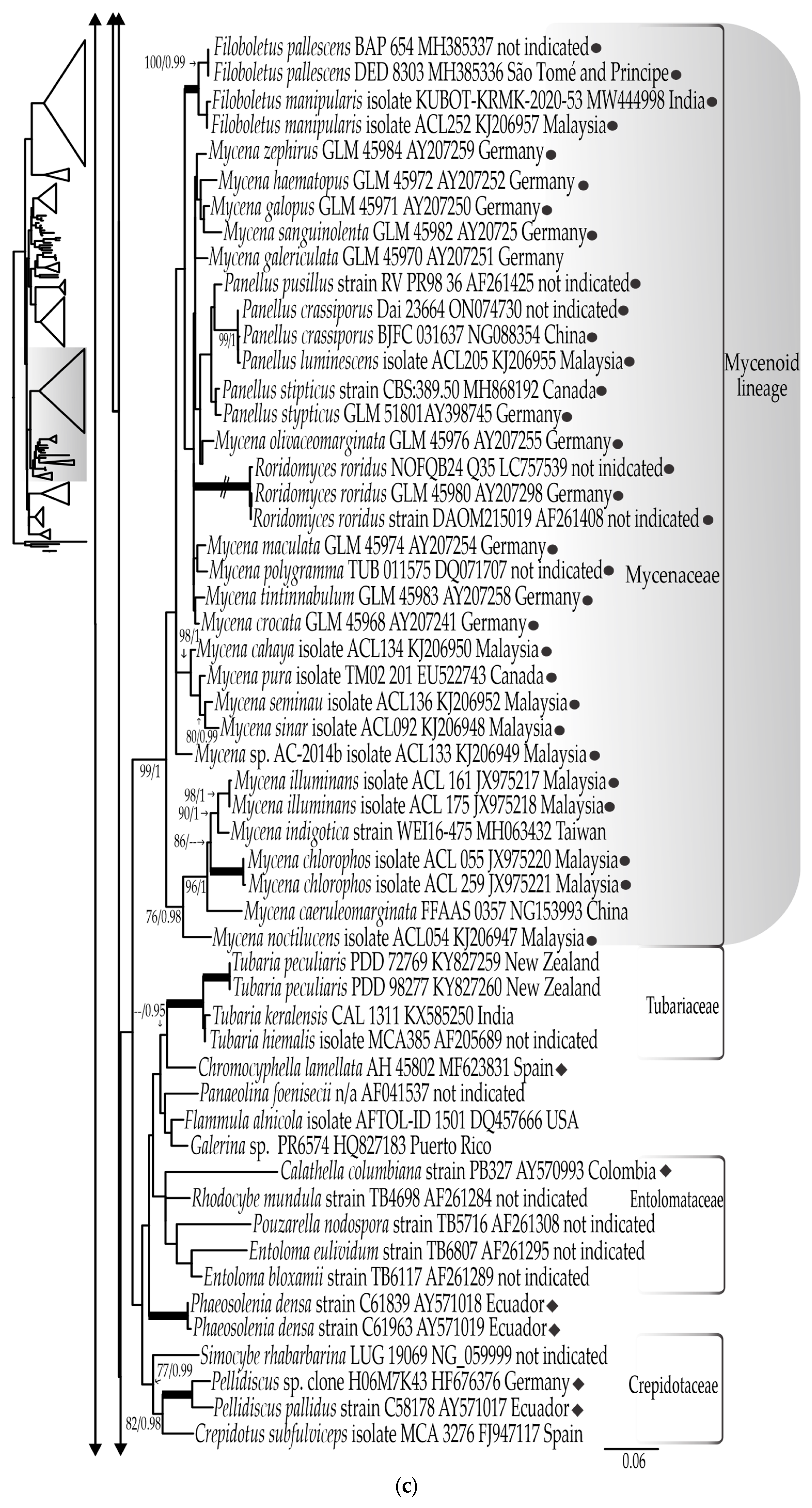
The final LSU rDNA dataset contains 206 sequences (including 2 that are newly generated), consisting of 1051 nucleotide sites, including gaps. The most appropriate evolutionary model estimated was TrN+I+G. The bootstrapping criteria from the ML analyses stopped after 350 replicates. Both the RAxML analysis and Bayesian inference yielded similar tree topologies. The LSU rDNA tree generated from the ML analysis, including bootstrap values and posterior probabilities, is shown in four parts (Figure 1a–d).
The family Cyphellopsidaceae, represented by 52 sequences, forms a well-supported clade (100% BS, 1.0 BPP) (Figure 1a) that harbors the largest number of cyphelloid genera hitherto confirmed with molecular data: Akenomyces G. Arnaud, Calathella (represented by Calathella gayana (Lév.) Agerer), Flagelloscypha, Halocyphina, Lachnella, Maireina, Merismodes, Nia, Eoscyphella gen. nov., Pseudolasiobolus Agerer, and Woldmaria.
The new proposed genus Eoscyphella formed a well-supported clade (100% BS, 1.0 BPP) sister to the cyphelloid genus Woldmaria. The LSU rDNA sequences of Eoscyphella and Woldmaria are from 92.6% to 92.7% similar. Sequences of taxa of the genus Maireina were represented in our analyses by Ma. filipendula, Ma. subsphaerosphora, and Ma. monacha (type species of Maireina), with the latter sampled, sequenced, and identified by Mombert [55] and fully described and epityfied in this study. Maireina formed a paraphyletic group, but represents a monophyletic clade when including sequences of Merismodes anomala (Pers.) Singer (as = Cyphellopsis anomala) and Me. fasciculata (Schwein.) Earle, with the latter being the type species of the genus. The clade formed with Maireina and Merismodes is well supported (75% BS, 0.99 BPP).
In addition to Cyphellopsidaceae, our LSU rDNA analyses recovered another 11 lineages of cyphelloid fungi (Figure 1a–d). Several cyphelloid genera are recovered in distinct well-supported clades that correspond to at least five well-delimited families: in Cyphellaceae (96% BS, 1.0 BPP), the genus Cyphella Fr. (100% BS, 1.0 BPP); in Crepidotaceae (S. Imai) Singer (77% BS, 0.99 BPP), the genus Pellidiscus Donk (100% BS, 1.0 BPP); in Marasmiaceae Roze ex Kühner (98% BS, 1.0 BPP), the genus Amyloflagellula Singer; in Phyllotopsidaceae Locquin ex Olariaga, Huhtinen, Læssøe, J.H. Petersen & K. Hansen (94% BS, 1.0 BPP), the genus Cyphelloporia Karasiński, L. Nagy, Szarkándi, Holec & Kolařík (100% BS, 1.0 BPP); and in Porotheleaceae (94% BS, 0.99 BPP), the genus Stromatoscypha Donk (100% BS, 1.0 BPP). The cyphelloid genus Stigmatolemma Kalchbr. clusters with sequences of Resupinatus alboniger (Pat.) Singer (MK278432) and Resupinatus conspersus (Pers.) Thorn, Moncalvo & Redhead (AY570994) in a well-supported clade (91% BS, 1.0 BPP) that is sister (unsupported) to the well-supported clade (78% BS, 0.99 BP) formed with sequences of the cyphelloid genus Calyptella Quél. (Figure 1b).
Additionally, sequences of the cyphelloid genera Henningsomyces Kuntze and Rectipilus Agerer are resolved in two phylogenetically distant clades: the Henningsomyces/Rectipilus/Acanthocorticium clade (100% BS, 1.0 BPP) that is sister (86% BS, 1.0 BPP) to Cyphellopsidaceae (Figure 1a), and in the clade of Phyllotopsidaceae sensu Olariaga et al. [77] and Karasiński et al. [56], forming a well-supported clade (99% BS, 1.0 BPP) with Cyphelloporia representatives (Figure 1b). Other cyphelloid taxa, such as Calathella columbiana Agerer (AY570993), Chromocyphella lamellata G. Moreno & Olariaga (MF623831), and Phaeosolenia densa (Berk.) W.B. Cooke (AY571018, AY571019) formed independent lineages with no clear relationship to other known lineages (Figure 1c).
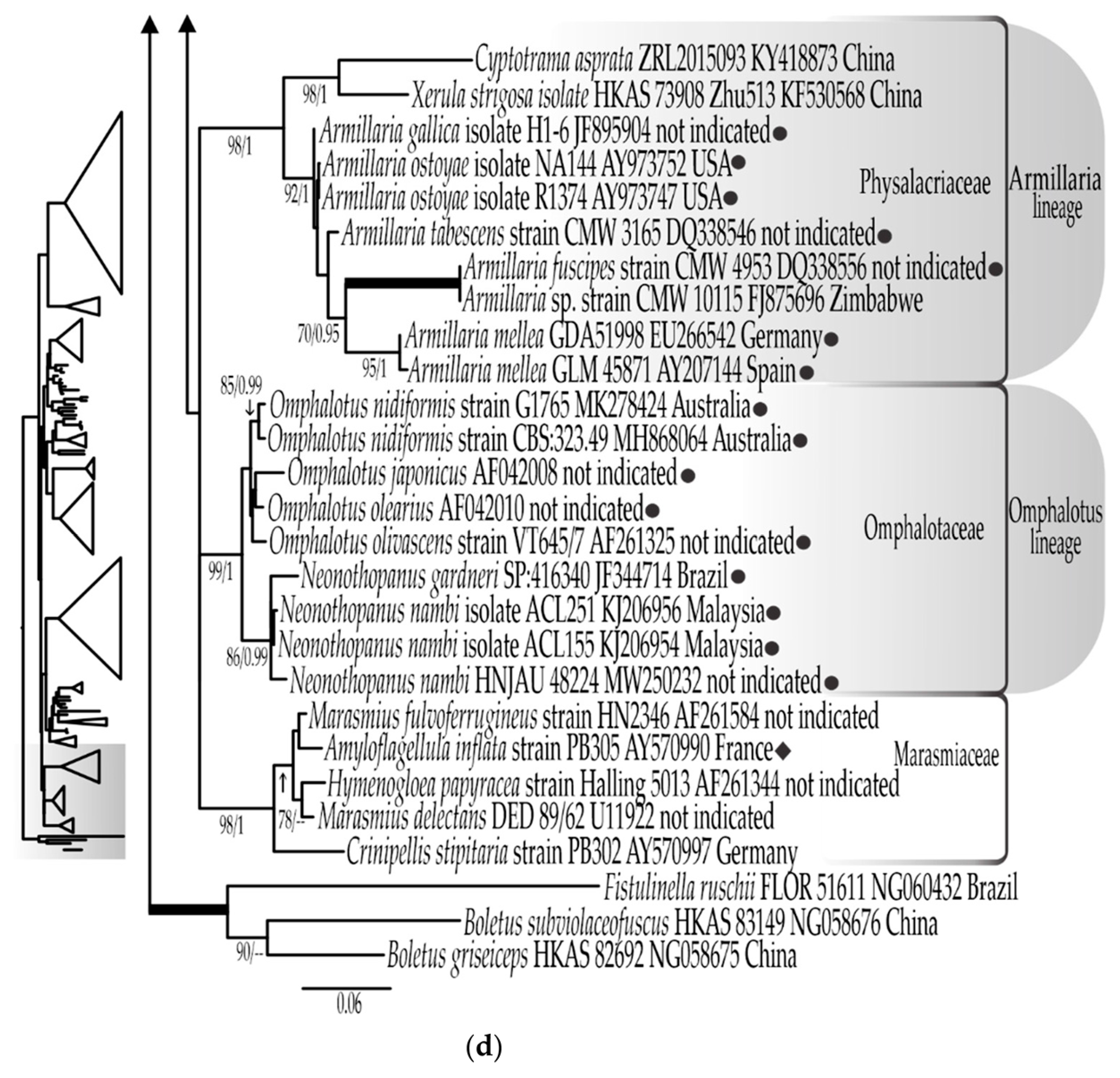
The four bioluminescent lineages sensu Desjardin et al. [4] are represented (Figure 1a–d). The Mycenoid lineage is the largest and forms a monophyletic group in a well-supported (99% BS, 1.0 BPP) clade (family Mycenaceae) represented in our analyses (Figure 1c) by 25 species of the genera Filoboletus, Mycena, Panellus, and Roridomyces. The Armillaria lineage (Figure 1d) is here represented by five species of the genus Armillaria that clustered in a well-supported (92% BS, 1.0 BPP) clade sister to sequences of Cyptotrama asprata (Berk.) Redhead & Ginns (KY418873) and Xerula strigosa Zhu L. Yang, L. Wang & G.M. Muell. (KF305680) within Physalacriaceae (97% BS, 1.0 BPP). The Omphalotus lineage is represented (Figure 1d) by six species of Omphalotus and Neonothopanus that form a well-supported (98% BS, 1.0 BPP) clade corresponding to Omphalotaceae. The Lucentipes clade forms a well-supported (0.99 BPP) independent lineage (Figure 1b) that contains, in addition to Gerronema viridilucens (EF514207) and Mycena lucentipes (OR343215), sequences identified as Atheniela rutila Q. Na & Y.P. Ge, (NG153951), Mycopan scabripes (Murrill) Redhead, Moncalvo & Vilgalys (MK278154), Hydropus trichoderma (Joss.) Singer (MK278158), and Mycena cf. quiniaultensis Kauffman (EU681183). A fifth bioluminescent lineage is composed of the proposed new genus Eoscyphella, represented by two sequences of Eoscyphella luciurceolata sp. nov. (Figure 1a).

3.1.2. ITS rDNA Dataset

The final ITS rDNA dataset has 44 sequences (including 3 that are newly generated), consisting of 1051 nucleotide sites, including gaps. The best evolutionary models estimated for each part of the alignments were ITS1: TPM2uf+G, 5.8S: TPM2+G, and ITS2: HKY+G. The bootstrapping criteria from the ML analysis stopped after 300 replicates. Both the RAxML analysis and Bayesian inference yielded similar tree topologies. The ITS rDNA tree generated from the ML analysis, including bootstrap and posterior probabilities, is shown in Figure 2.
The family Cyphellopsidaceae (100% BS, 1.0 BPP) is represented by 42 sequences, with no representatives of Woldmaria nor Peyronelina due to lack of available sequences. Eoscyphella luciurceolata sp. nov. and the non-bioluminescent Eoscyphella sp. formed a well-supported clade (84% BS, 1.0 BPP), sister to (but not supported) a clade that contains sequences of Dendrothele microspora (H.S. Jacks. & P.A. Lemke) P.A. Lemke, Dendrothele incrustans (P.A. Lemke) P.A. Lemke, and Dendrothele griseocana (Bres.) Bourdot & Galzin (Figure 2). The genus Maireina, represented by the same species as in the LSU rDNA analyses, is again confirmed as paraphyletic with the ITS rDNA data. However, as in the nLSU analyses, the included Maireina sequences form a monophyletic and well-supported clade (91% BS, 1.9 BPP) when including sequences of Merismodes anomala, Me. fasciculata, and Merismodes sp. (MZ919217).

3.1.3. Combined LSU rDNA + ITS rDNA Dataset

The final combined LSU rDNA plus ITS rDNA dataset contains 20 ITS rDNA and 16 LSU rDNA sequences (including 5 generated as part of this study) for 21 terminals, and consists of 1806 nucleotide sites, including gaps. The most appropriate evolutionary models estimated for each part of the alignments were ITS1: TPM2uf+G, 5.8S: TPM2, ITS2: TPM2uf+G, and LSU: TIM3+G.
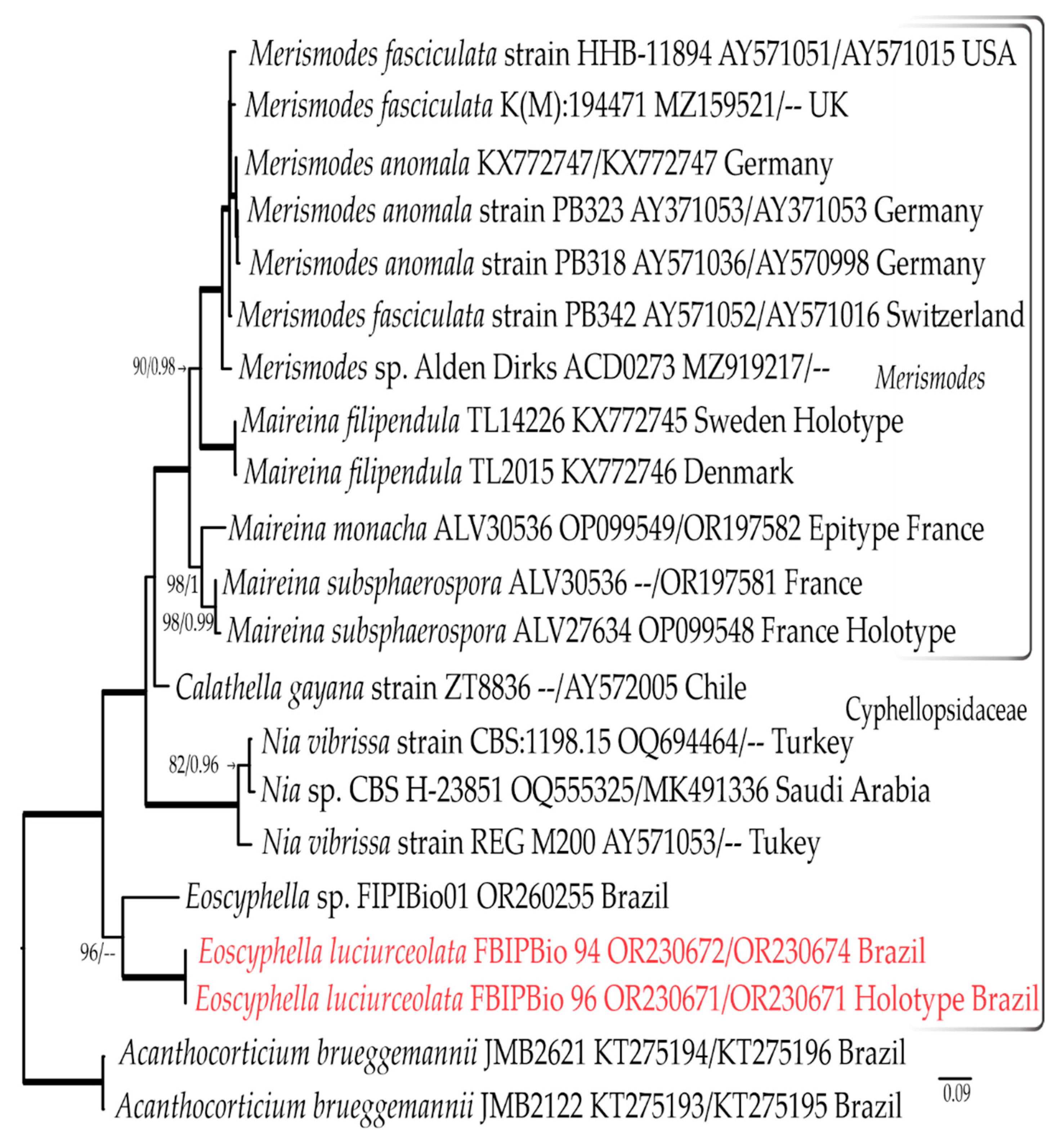
The bootstrapping criteria from the ML analysis stopped after 50 replicates. The most likely tree generated with the ML analysis is shown in Figure 3. The family Cyphellopsidaceae (100% BS, 1.0 BPP) is represented by 19 terminals, with emphasis on the Merismodes clade (100% BS, 1.0 BBP), represented by 12 terminals of Maireina and Merismodes. Consistent with the other previous analyses, both the Bayesian inference and ML analysis recover Maireina as a paraphyletic group (Figure 3).

3.2. Taxonomic Part

From molecular phylogenetic results, we consider Maireina a synonym of Merismodes (=Cyphellopsis), supporting the taxonomic concept of Knudsen and Vesterholt [61], who considered Maireina, Cyphellopsis, and Phaeocyphellopsis W.B. Cooke synonyms of Merismodes. We herein propose the combination of Ma. monacha, Ma. filipendula, and Ma. subsphaerophora into Merismodes, as well as the description of the genus Eoscyphella to accommodate the novel bioluminescent cyphelloid species from Brazil.
  • Merismodes Earle, Bulletin of the New York Botanical Garden 5: 406 (1909) emend. Silva-Filho & Menolli
=Cyphellopsis Donk, Mededelingen van de Nederlandse Mycologische Vereeniging 18–20: 128 (1931).
=Maireina W.B. Cooke, Beihefte zur Sydowia 4: 83 (1961).
=Phaeocyphellopsis W.B. Cooke, Beihefte zur Sydowia 4: 119 (1961).
=Pseudodasyscypha Velen., Novitates mycologicae: 167 (1939).
Original diagnosis [79]: Not pultrecent, densely connate-cespitose: pileus fleshy, irregular: lamellae reduced to obscure folds: spores white or hyaline: veil none: stipe irregular, the bases fused.
Emended description: Basidiomata gregarious or scattered. Receptacle cyphelloid, cupulate to tubular, sessile or pendant; outside covered with yellow brown to brown hairs, hymenium pale, whitish. Subiculum absent or developed. External hyphae thick-walled, not branched, straight, attenuated to spiraled towards the distal end, yellow to brown pigmented, sometimes with apical ends colorless, tips incrusted or smooth, obtuse to inflated, inamyloid to slightly dextrinoid. Trama gelatinous or non-gelatinous. Basidiospores subglobose, ellipsoid, cylindrical, allantoid or subfusiform, smooth, thin-walled, hyaline, inamyloid. Basidia cylindrical to clavate, four-spored, occasionally two-spored. Cystidia absent or rarely present. Clamp connections present or absent.
Notes: After the very brief protologue, Knudsen and Vesterholt [61] included in their description of Merismodes include some morphological characteristics of the genera Maireina, Cyphellopsis, and Phaeocyphellopsis. In our emendation, we include additional distinctive morphological characteristics of the species recently described [52,53,54] and of Maireina based on Bodensteiner [57]. In all our analyses, the genus Maireina is resolved as paraphyletic, forming a well-supported monophyletic lineage with the sequences of Mersimodes included. Based on these results and those of previous investigators [61], we consider Maireina a synonym of the latter genus and propose an amendment. The name Merismodes, proposed in 1909 [79], has priority against Maireina erected in 1961 [43]. Thus, to better accommodate the Maireina species sampled in our analyses (which includes sequences from holotype material), we propose the combination of Ma. filipendula and Ma. subsphaerosphora in Merismodes. Additionally, a recently collected sample of Ma. monacha (type species of Maireina) from France (same country locality of the holotype) was also included in our analyses. The taxon is herein re-analyzed and confirmed in Merismodes and an epitype is designated.
  • Merismodes monacha (Speg.) Silva-Filho, Mombert & Menolli comb. nov.
  • MycoBank: MB 849402
Basionym: Cyphella monacha Speg., Michelia 2 (7): 303 (1881).
Cyphellopsis monacha (Speg.) D.A. Reid, Kew Bulletin 17: 297 (1963).
Maireina monacha (Speg.) W.B. Cooke, Beihefte zur Sydowia 4: 90 (1961).
=Cyphella bresadolae Grélet, Bulletin de la Société Mycologique de France 38: 174 (1922).
=Cyphella bresadolae var. gregaria (Syd. & P. Syd.) Pilát, Annales Mycologici 23: 162 (1925).
=Cyphella bresadolae var. leochroma (Bres.) Grélet, Bulletin de la Société Mycologique de France 38: 174 (1922).
=Cyphella bresadolae var. tephroleuca (Bres.) Grélet, Bulletin de la Société Mycologique de France 38: 174 (1922).
=Merismodes bresadolae (Grélet) Singer, The Agaricales in modern taxonomy. 3rd ed. J. Cramer, Lehre, Vaduz: 665 (1975).
=Cyphella gregaria Syd. & P. Syd., Hedwigia 39(3): 116 (1900).
=Cyphella leochroma Bres., Fungi Tridentini II (fasc. 14): 99, Table 211, f. 1 (1900).
=Cyphella obscura Roum., Fungi selecti gallici exsiccati. Michelia II, Cent. 20, no. 1905 (1882).
=Cyphella sydowii Bres., in SYDOW H, Mycotheca Marchica. Cent. 38, no. 3706 (1892).
=Cyphella tephroleuca Bres., Fungi Tridentini II (fasc. 11–13): 57, Table 166, f. 2 (1898).
=Maireina marginata (McAlpine) W.B. Cooke, Sydowia, Annales Mycologici, Beiheft 4: 89 (1962).
Macro- and micro-morphological description: Cooke [43].
Material examined: FRANCE, Cantal. St-Santin-de-Maurs, on a still-attached dead twig of Cornus sanguinea L., 28 June 2021. Leg. A. Mombert., ALV30536 [PC0142589, Epitype here designated! (validated identifier: MBT 204394)].
Habitat and known distribution: On bark of dead branch of Cornus sanguinea in oak forest in France, but also Acer campestre L. (Aceraceae), Berberis vulgaris L. (Berberidaceae), Bupleurum fruticosum L. (Apiacaceae), Hieracium umbellatum L. (Asteraceae), Cytisus sp., Genista tinctoria L., Sarothamnus scoparius (L.) Link (Fabaceae), Lonicera sp. (Caprifoliaceae), Prunus amygdalus Batsch, P. persica (L.) Batsch (Rosaceae), Quercus mongolica Fisch. ex Ledeb. (Fagaceae) [57]. Distributed in Europe and Oceania [43].
Notes: Our specimen agrees with the description of Me. monacha presented by Cooke (ref. [43], as Ma. monacha), who analyzed authentic material of all names included here as synonyms, including the types of Cyphella obscura Roum. and Cyphella sydowii Bres. According to Cooke [43], Me. monacha is characterized by brown receptacles with long hairs around the cup edge and at the hymenial surface, elongate to cylindrical basidiospores, four-spored basidia, and cylindrical, yellowish brown to brown external hyphae with paler apices. Although our material has had slightly broader receptacles (1.5–3 mm diam.) and basidia (9.0–110 µm diam.) than reported by Cooke [43] (receptacles, 0.5–1 mm diam.; basidia, 5.5–8.0 µm diam.), other macro- and micromorphological characteristics are sufficient for the identification of this sample as Ma. monacha sensu Cooke [43] and Bodensteiner [57]. Merismodes monacha was originally described from samples collected in France but it has a distribution recorded in many European countries, including Germany, Austria, Italy, the Czech Republic, Hungary, the United Kingdom, and one record from Australia [43]. The holotype of Cyphella monacha Speg. [anon. s.n. (Fung. Gall. 768) Spegazzini s.n.] was deposited at the New York Botanical Garden Herbarium (NY). Considering the complete morphological and molecular data recovered from our sample that is from a region close to the type locality, we decided to designate the voucher ALV30536 as epitypus.
  • Merismodes filipendula (Læssøe) Silva-Filho & Menolli comb. nov.
MycoBank: MB 849405
Basionym: Maireina filipendula Læssøe, Karstenia 56 (1): 40 (2016).
Macro- and micro-morphological description: see Læssøe et al. [54].
  • Merismodes subsphaerospora (Mombert) Silva-Filho, Mombert & Menolli comb. nov.
MycoBank: MB 849406
Basionym: Maireina subsphaerospora Mombert, Bulletin Mycologique et Botanique Dauphiné-Savoie 246: 38 (2022).
Macro- and micro-morphological description: see Mombert [55].
  • Eoscyphella Silva-Filho, Stevani & Menolli gen. nov.
MycoBank: MB 849403
Etymology: Eos = light of day; the goddess of dawn (Greek); cyphella (from kyfos in Greek) = shape of a cup, something hollow. The prefix “Eos” is in reference to the light emitted by the bioluminescent basidiomata of the type species. Additionally, the Roman equivalent refers to Eosforos as Lucifer, which is the entity’s name that was later considered into Christianity as the devil, and it also refers to the name of the protected area (Devil’s Cave State Park) near where the specimens of the type species were found. The name cyphella is a reference to the genus Cyphella and to the cyphelloid body form.
Type species: Eoscyphella luciurceolata Silva-Filho, Stevani & Desjardin (described below).
Diagnosis: Eoscyphella is morphologically similar to Merismodes and Woldmaria but differs from Woldmaria in lacking conspicuous long hairs in the receptacle, subglobose to broadly ellipsoid basidiospores, regularly bi-spored basidia, and unclamped hyphae; and from Merismodes by the absence of conspicuous hairs in the receptacle, absence of cystidia, regularly bi-spored basidia, and the characteristic external hyphae that are always pigmented and encrusted at the tips.
Notes: Eoscyphella, typified here using Eoscyphella luciurceolata sp. nov., represents a new lineage of bioluminescent fungi. It is supported with phylogenetic data (Figure 1a, Figure 2 and Figure 3) and morphological characteristics, including the absence of conspicuous long hairs on the receptacle, subglobose to broadly ellipsoid basidiospores, regularly bi-spored basidia, the absence of clamp connections, and the consistent presence of pigmented and encrusted external hyphae. An additional collection (FIPBIO 01) of a related non-bioluminescent cyphelloid species was found in the same region of the type species. The ITS rDNA sequence data (OR260255) resolves this taxon as sister to E. luciurceolata and suggests that it represents an additional species of Eoscyphella (Figure 2 and Figure 3). The presence of a second species indicates that Eoscyphella is likely a non-monospecific genus that includes both bioluminescent and non-bioluminescent members. Until additional material of the non-bioluminescent taxon can be collected to confirm these initial observations, we prefer to leave it undescribed.
MycoBank: MB 849404
Etymology: Luci = light (Latin); urceolus = diminutive of urceus “pitcher” (Latin), in reference to urceolate shape of the receptacle. Since bioluminescent and non-bioluminescent species occur in the genus, the prefix “Luci” is here applied to differentiate this new species from putative non-bioluminescent ones.
Holotype: BRAZIL, São Paulo state, Eldorado, approximately 500 m west of the entrance to the “Caverna do Diabo” (Devil’s Cave) State Park, but still in the buffered conservation area, on a single “fumeiro” tree (Solanum swartzianum Roem. & Schult.), 24°38′14.0100″ S and 48°24′37.6812″ W, alt. 546 m, 22 March 2023, FBIPBio 96.20230322, leg. Isaias Santos, Adão Henrique Rosa Domingos, Olavo H. P. Della-Torre (FIFUNGI0001, holotype!) GenBank [ITS rDNA]: OR230671, [LSU rDNA]: OR230673.
Diagnosis: Eoscyphella luciurceolata differs from other known species of cyphelloid fungi by the following combination of characters: receptacle vasiform to urceolate without conspicuous long hairs; external hyphae cylindrical, sinuous, coiled to conspicuously spiraled, pigmented, weakly to densely incrusted overall, less so near their tips, with small globular crystals; basidiospores subglobose to ovoid or broadly ellipsoid; basidia cylindrical to subclavate, 2-spored (rarely 4-spored); hymenial cystidia absent; clamp connections absent.
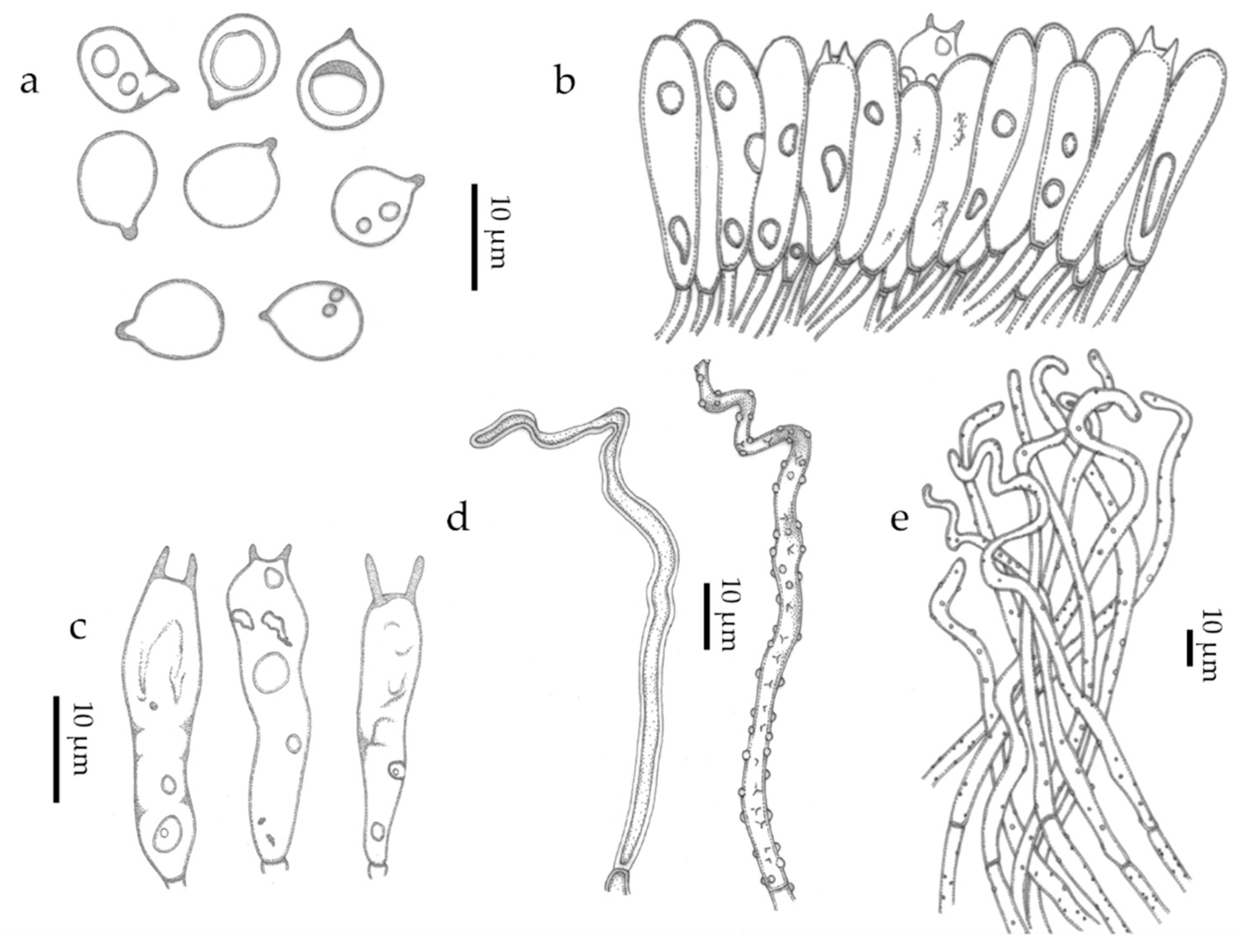
Basidiomata scattered (Figure 5 and Figure 6). Receptacle 0.3–0.5 mm tall, 0.2–0.3 mm diam, vasiform to urceolate, sessile (astipitate), with distinct opening; external surface dull, dry, felted to appressed-pubescent, conspicuous long hairs absent, pale yellow (2A3) to greyish yellow (2B3, 4B5) or greyish orange (5B4), white (1A–B1) near the distal opening (Figure 5c and Figure 6b; subiculum absent; hymenial surface greyish yellow (2C4), smooth. External hyphae 60–128 × 2.0–4.0 μm, cylindrical, sinuous to coiled, yellowish brown to brownish orange in water or KOH, weakly to densely incrusted overall, less so near the tips, with small globular crystals, thick-walled (0.5–1.5 um thick), thinner near the tip, inamyloid, non-gelatinous, unclamped; terminal cells narrowed towards the tip to 1.5–2.0 µm diam, tips hyaline to pale yellowish brown, obtuse to subacute, those at the margin of the pore hyaline and conspicuously spiraled (Figure 7c–e and Figure 8d–e); dendrohyphidia absent. Trama composed of an interwoven layer of irregularly cylindrical to inflated, short-celled hyphae 3.0–9.5 µm diam, hyaline to pale yellowish brown, much-branched, non-incrusted, non-gelatinous, thin- to thick-walled (0–0.5 µm thick), unclamped (Figure 7c). Subhymenial layer composed of cylindrical hyphae 3.0–4.0 μm diam, hyaline, thin- to thick-walled (0.5–1.5 µm thick), unclamped. Basidiospores (6.5–)7.5–9.5 × (5.5–)6.5–8(–9.5) μm [xm = 8.56 ± 0.13 × 7.35 ± 0.29 µm, xr = 8.5–8.7 × 7.1–7.6 µm, Q = 1.0–1.6, Qm = 1.18 ± 0.05, n = 60, s = 3], subglobose to ovoid or broadly ellipsoid, predominantly subglobose, smooth, hyaline, inamyloid, sometimes one- or two-guttulate, hilar appendix up to 1 μm long, thin- or thick-walled (0.5–1.0 µm) at maturity (Figure 7a and Figure 8a). Basidia 22–32 × 7.0–10.0 μm, cylindrical to subclavate, two-spored, rarely four-spored, hyaline, sometimes with refringent contents, unclamped; sterigmata up to 12 μm long (Figure 7b and Figure 8b). Basidioles subclavate (Figure 7c and Figure 8b). Hymenial cystidia absent (Figure 7c). Clamp connections absent in all tissues examined. Bioluminescence: emitting yellowish green light only in a narrow band around the pore margin of the receptacle; water droplets likely magnify the light (Figure 5 and Figure 6).
Additional specimens examined: BRAZIL, São Paulo state, Eldorado, exact same location, and tree described above, 2 August 2023, FBIPBio 93.20220802, leg. Isaias Santos, Adão Henrique Rosa Domingos, Olavo H. P. Della-Torre (FIFUNGI00249, Paratype!); ibid, 20 September 2022, FBIPBio 94.20220920 (FIFUNGI00250, Paratype!) GenBank [ITS rDNA]: OR230672, [LSU rDNA] OR230674.
Habitat and known distribution: On bark of “fumeiro” tree (Solanum swartzianum) in the Atlantic Rainforest, southern Brazil. Known only from the type locality.
Notes: When morphologically compared with other cyphelloid species, Maireina spiralis (Coker) W.B. Cooke has external hyphae with spiral tips, differing from E. luciurceolata in the clamped hyphae and with longer (11–15 µm long) ellipsoid basidiospores [43]. Maireina afibulata Bodensteiner and Ma. pseudochracea W.B. Cooke do not produce clamp connection, but the first has smaller basidiospores (5–6(–6.5) × 3–4 μm) and both have straight external hyphae and produce smaller basidia (6–23 × 5–6.5 µm in Ma. afibulata; 17.5 × 5.8 µm in Ma. pseudochracea) with four sterigma [43,53].

4. Discussion

The morphological delimitation of Merismodes, Cyphellopsis, and Maireina has been the cause of debates about the morphological limits of these genera [43,51,57,62,80,81,82,83]. Reid [81] considered Cyphellopsis and Maireina as synonyms and suggested that the depth of the cavity that lined the hymenium is a character insufficient for the separation of Cyphellopsis (=Maireina) and Merismodes. Singer [83] synonymized the genus Cyphellopsis and Maireina with Merismodes and listed both Maireina and Cyphellopsis as sections. The first broad research on cyphelloid fungi based on molecular phylogenetic analyses resolved Merismodes and Cyphellopsis as a monophyletic group, recognizing them as a single genus [45]. Another broad study of Maireina without molecular data led Bodensterner [53,57] to recognize the genus Maireina as an independent lineage from Merismodes and Cyphellopsis. Knudsen and Vesterholt [61] recognized Cyphellopsis, Maireina, and Phaeocyphellopsis as synonyms of Merismodes, providing a broad description for the genus. The first works to describe new species of Maireina based in-part on molecular data are those of Læssøe et al. [54] and Mombert [55]. In both, the sequences of Maireina clustered with Merismodes and Cyphellopsis in a large clade, making it possible to determine the phylogenetic position within Cyphellopsidaceae. Our phylogenetic analyses in separate and combined LSU rDNA and ITS rDNA recognized Merismodes, Cyphellopsis, and Maireina as a monophyletic group, supporting the proposal of Knudsen and Vesterholt [61] for a broad morphological concept of Merismodes. The samples and sequences of Me. monacha, type species of Maireina, first studied by Mombert [55] were extremely important for the recognition and the phylogenetic positioning of the genus Maireina. Although the sequences are not of the holotype specimen, the collection is from a region very close to the type locality, and the morphological description agrees with the complete redescription presented by Cooke [43].
Our cyphelloid bioluminescent samples were initially identified within the morphological concept of Maireina sensu Bodensteiner [53,57]. However, our phylograms (Figure 1a, Figure 2 and Figure 3) showed a phylogenetic distance between E. luciurceolata and Me. monacha, which are only 90.6% to 90.7% similar in the LSU rDNA and 64.6% to 65.9% similar in the ITS rDNA. Eoscyphella is closely related to the genus Woldmaria in our analyses, but the included taxa are 7.3% to 7.4% divergent in their LSU rDNA sequences, a high value considering a similarity threshold of around 96.91% to discriminate genera using LSU rDNA in Basidiomycota [84]. These data and results support the proposition of a new cyphelloid genus and distinct molecular lineage. Additionally, Eoscyphella is also morphologically well delimited with receptacles that lack conspicuous long hairs, subglobose to broadly ellipsoid basidiospores, frequently bi-spored basidia, unclamped hyphae, and weakly to densely incrusted overall external hyphae, which are always pigmented and encrusted at the tips.
Regarding the cyphelloid genera within Agaricomycetes, our LSU rDNA analyses retrieved 11 lineages of cyphelloid fungi and the phylogenetic relationship of the cyphelloid genera agrees with recent phylogenetic studies [45,50,56]. However, we highlight that sequences of the collection PB327 named as Calathella columbiana appear in different positions and for this reason were excluded from the combined analyses: in the ITS rDNA tree within Cyphellopsidaceae (Figure 2), and in the LSU rDNA tree (Figure 1c) in a clade close to representatives of Entolomataceae. Additionally, Phaeosolenia densa (Berk.) W.B. Cooke was shown by Bodensteiner et al. [45] to be close to the genus Tubaria (W.G. Sm.) Gillet, whilst in our analyses, it forms an isolated clade without support (Figure 1c).
Desjardin et al. [4] performed the second review of bioluminescent fungi worldwide, referring 64 luminescent species into three lineages, Armillaria, Mycenoid, and Omphalotus, indicating that Gerronema viridilucens and Mycena lucentipes do not belong to the Mycenoid lineage. Later, Desjardin et al. [24] referred G. viridilucens and M. lucentipes to a new and unnamed lineage, which was later named the Lucentipes lineage by Oliveira et al. [35]. Our LSU rDNA phylogram (Figure 1b) shows and confirms G. viridilucens plus M. lucentipes as a separate bioluminescent lineage. The Eoscyphella lineage is here recognized as a new and fifth bioluminescent lineage in Cyphellopsidaceae (Figure 1a).
From previous phylogenetic analyses, G. viridilucens has been proposed within Porotheleaceae [70,71]. However, our LSU rDNA phylogram (Figure 1b) showed Porotheleaceae, represented by type species of the genus Hydropus [Hydropus fuliginarius (Batsch) Singer, AF261368], forming a well-supported clade (94%, BS, 0.99 BPP) that harbors most of the species of Gerronema Singer, except G. viridilucens, which clustered with sequences of M. lucentipes, Atheniella rutilla (NG153951), Hydropus trichoderma (MK278154), Mycena cf. quiniaultensis (EU681183), and Mycopan scabripes (MK278154) in a clade phylogenetically distant from Porotheleaceae. Vizzini et al. [70] showed the genera Acanthocorticium Baltazar, Gorjón & Rajchenb.; Athelia Pers.; Atheniella Redhead, Moncalvo, Vilgalys, Desjardin & B.A. Perry; Baeospora Singer; Calyptella; Campanophyllum Cifuentes & R.H. Petersen; Cheimonophyllum Singer; Chondrostereum Pouzar; Cyphella; Granulobasidium Jülich; Gloeostereum S. Ito & S. Imai; Mycopan Redhead, Moncalvo & Vilgalys; Pleurella E. Horak; Henningsomyces; and Rectipilus as part of the Henningsomyces/Rectipius/Acanthocorticium clade, with all accommodated in Cyphellaceae. Due to the close relationship between Atheniella and Mycopan, most of the genera of Cyphellaceae sensu Vizzini et al. [70] were included in our phylogeny in order to confirm the phylogenetic position of G. viridilucens plus Mycena lucentipes. However, in our LSU rDNA analyses (Figure 1a,b), Cyphellaceae sensu Vizzini et al. [70] was retrieved as a polyphyletic group, with representatives grouped into five different clades. In the LSU rDNA phylogram (Figure 1b) Cyphellaceae can be well represented by the clade with sequences of Cyphella digitalis (Alb. & Schwein.) Fr. (AY29293175 and AY635771), Cheimonophyllum candidissimum (Sacc.) Singer (DQ457654), and Campanophyllum proboscideum (Fr.) Cifuentes & R.H. Petersen (AY230866). Thus, it is confirmed that G. viridilucens and Mycena lucentipes are positioned neither in Porothelleaceae nor in Cyphellaceae.

5. Conclusions

Our systematic study confirms the findings of previous studies regarding the existence of multiple bioluminescent lineages in Agaricales. All bioluminescent fungi have currently been described in suborder Marasmiineae Aime, Dentinger & Gaya. The newly described Eoscyphella luciurceolata was confirmed from molecular phylogenies in the family Cyphellopsidaceae, currently accepted within the suborder Schizophyllineaeae Aime, Dentinger & Gaya [62]. Additionally, our study reveals a new lineage within a group primarily consisting of reduced forms. Fungal bioluminescence engages in a cyclical process of biosynthesis known as the Caffeic Acid Cycle (CAC), which relies on a sequence of four consecutive enzymes: hispidin synthase (HispS), hispidin-3-hydroxylase (H3H), luciferase (Luz), and caffeylpyruvate hydrolase (CPH) [3]. At present, there are limited genomic data concerning bioluminescent fungi in the existing literature [85], with the majority originating from the Mycenoid and Armillaria lineages. By identifying this recently discovered bioluminescent lineage and uncovering the sequences of the hisps, h3h, luz, and cph genes, there is potential for enhancing our understanding of the evolutionary progression of the bioluminescent trait within the fungal kingdom.
A high diversity of bioluminescent fungi has been discovered in Brazil, with 23 species (including our new described species) reported theretofore, see [86]. In the Brazilian Atlantic Rainforest, new species of bioluminescent fungi have been described or reported, e.g., [87], with emphasis to the southwestern portion of the state of São Paulo, the same area where E. luciurceolata was found and where another 12 species of Mycenoid and Lucentipes lineage taxa have already been described or reported [22,23,24,25]. Even so, new bioluminescent samples collected at the same area are currently in the process of molecular and morphological characterization and may represent taxonomic novelties, demonstrating that the Atlantic Rainforest in the southwestern region of the São Paulo state is one of the most studied areas of bioluminescent fungi and may represent a biodiversity hot spot for these organisms.

Supplementary Materials

The following supporting information can be downloaded at: https://www.mdpi.com/2309-608X/9/10/1004/s1, Table S1: List of specimens; culture, herbarium access number, isolate, strain, or voucher collection (V); and GenBank accession numbers.

Author Contributions

Conceptualization, A.G.S.S.-F., C.V.S. and N.M.J.; Resource, A.M., B.B.N., B.A.P., C.C.N., D.E.D., D.M.M.S., A.G.S.M., A.H.R.D., I.S. and O.H.P.D.-T.; Writing: A.G.S.S.-F., C.V.S., D.E.D., B.A.P. and N.M.J.; and Funding, C.V.S. and N.M.J. All authors have read and agreed to the published version of the manuscript.

Funding

This work was supported by “Fundação de Amparo à Pesquisa do Estado de São Paulo” (FAPESP) under grant numbers 2018/15677-0 (N.M.J.), 2021/09109-1 (A.G.S.S.-F.), 2020/16000-3 (B.B.N.), 2019/12605-0 and 2022/14964-0 (D.M.M.S.), and 2017/22501-2 (C.V.S.), and by the Brazilian National Council for Scientific and Technological Development (CNPq) under grant numbers 314236/2021-0 (N.M.J.) and 303525/2021-5 (C.V.S.). This work was also supported with funding from the Office of Naval Research Global through grant ONRG N62909-17-1-2023 to C.V.S. and D.E.D.

Institutional Review Board Statement

Not applicable.

Informed Consent Statement

Not applicable.

Data Availability Statement

The DNA sequence data obtained in this study were deposited at GenBank. The accession numbers can be found in the trees and in Supplementary Table S1. This study is according to the Brazilian legislation on access to biodiversity and is registered in the “Sistema Nacional de Gestão do Patrimônio Genético e do Conhecimento Tradicional Associado” (SisGen #A5A80A7).

Acknowledgments

The authors thank Alexandro Andrade and Jefferson Góis for the initial taxonomic support, Suzana Ehlin Martins for the taxonomic identification of the host plant, Dimitrios Floudas for suggesting the Greek prefix of the new generic name and also for helping to compose the etymology of the new genus, Carlos Roberto Silva Moraes (also known as Duco) for granting us access to the collection site, and the anonymous reviewers for improvements to the original manuscript. We are also grateful to “Fundação Florestal” and “Secretaria do Meio Ambiente do Estado de São Paulo” for the collection licenses (process #260108-010.245/2017).

Conflicts of Interest

The authors declare no conflict of interest.

References

  1. Hibbett, D.S.; Bauer, R.; Binder, M.; Giachini, A.J.; Hosaka, K.; Justo, A.; Larsson, E.; Larsson, K.H.; Lawrey, J.D.; Miettinenm, O.; et al. (Eds.) Systematics and Evolution. In The Mycota; Springer: Berlin/Heidelberg, Germany, 2014; Volume 7A. [Google Scholar]
  2. Kaskova, Z.M.; Dorr, F.A.; Petushkov, V.N.; Purtov, K.V.; Tsarkova, A.S.; Rodionova, N.S.; Mineev, K.S.; Guglya, E.B.; Kotlobay, A.; Baleeva, N.S.; et al. Mechanism and color modulation of fungal bioluminescence. Sci. Adv. 2017, 3, e1602847. [Google Scholar] [CrossRef]
  3. Kotlobay, K.; Sarkisyan, K.S.; Mokrushina, Y.A.; Marcet-Houben, M.; Serebrovskaya, E.O.; Markina, N.M.; Somermeyer, L.G.; Gorokhovatsky, A.Y.; Vvedensky, A.; Purtov, K.V.; et al. A genetically encodable bioluminescent system from fungi. Proc. Natl. Acad. Sci. USA 2018, 115, 12728–12732. [Google Scholar] [CrossRef] [PubMed]
  4. Desjardin, D.E.; Oliveira, A.G.; Stevani, C.V. Fungi bioluminescence revisited. Photochem. Photobiol. Sci. 2008, 7, 170–182. [Google Scholar] [CrossRef] [PubMed]
  5. Heller, J.F. Ueber das Leuchten im Pflanzen und Tierreiche. Pathol. Chem. Mikr 1853, 6, 44–54, 81–90, 121–137, 161–166, 201–216, 241–251. [Google Scholar]
  6. Buller, A.H.R. The bioluminescence of Panus stipticus. Res. Fungi 1924, 3, 357–431. [Google Scholar]
  7. Bothe, F. Über das Leuchten verwesender Blätter und seine Erreger. Planta 1931, 14, 752–765. [Google Scholar] [CrossRef]
  8. Buller, A.H.R. Omphalia flavida, a gemmiferous and luminous leaf-spot fungus. Res. Fungi 1934, 6, 397–443. [Google Scholar]
  9. Wassink, W.C. Luminescence in fungi. In Bioluminescence in Action; Academic Press: New York, NY, USA, 1978. [Google Scholar]
  10. Corner, E.J.H. Descriptions of two luminous tropical agarics (Dictyopanus and Mycena). Mycologia 1950, 42, 423–443. [Google Scholar] [CrossRef]
  11. Kobayasi, Y. Contributions to the luminous fungi from Japan. J. Hattori Bot. Lab. 1951, 5, 1–6. [Google Scholar]
  12. Josserand, M. Sur la luminescence de “Mycena rorida” en Europe occidentale. Bull. Mens. Soc. Linn. Lyon. 1953, 22, 99–102. [Google Scholar] [CrossRef]
  13. Corner, E.J.H. Further descriptions of luminous agarics. Trans. Br. Myco Soc. 1954, 37, 256–271. [Google Scholar] [CrossRef]
  14. Haneda, Y. Luminous organisms of Japan and the Far East. In The Luminescence of Biological Systems; American Association for the Advancement of Science: Washington, DC, USA, 1955. [Google Scholar]
  15. Bigelow, H.E.; Miller, O.K., Jr.; Thiers, H.D. A new species of Omphalotus. Mycotaxon 1976, 3, 363–372. [Google Scholar]
  16. Horak, E. Mycena rorida (Fr.) Quel. and related species from the Southern Hemisphere. Ber. Schweiz. Bot. Ges. 1978, 88, 20–29. [Google Scholar]
  17. Zang, M. Some new species of higher fungi from Xizang (Tibet) of China. Acta Bot. Yunnan 1979, 1, 101–105. [Google Scholar]
  18. Corner, E.J.H. The agaric genera Lentinus, Panus and Pleurotus. Nova Hedwig. Beih. 1981, 69, 1–169. [Google Scholar]
  19. Treu, R.; Agerer, A. Culture characteristics of some Mycena species. Mycotaxon 1990, 38, 279–309. [Google Scholar]
  20. Maas Geesteranus, R.A. Verhandelingen. In Mycenas of the Northern Hemisphere; Afdeling natuurkunde: Amsterdam, The Netherlands, 1992. [Google Scholar]
  21. Li, J.; Hu, X.A. New species of Lampteromyces from Hunan. Acta Sci. Nat. Univ. Norm. Hunan 1993, 16, 188–189. [Google Scholar]
  22. Desjardin, D.E.; Capelari, M.; Stevani, C.V. A new bioluminescent agaric from Sao Paulo, Brazil. Fung. Div. 2005, 18, 9–14. [Google Scholar]
  23. Desjardin, D.E.; Capelari, M.; Stevani, C.V. Bioluminescent Mycena species from São Paulo, Brazil. Mycologia 2007, 99, 317–331. [Google Scholar] [CrossRef]
  24. Desjardin, D.E.; Perry, B.A.; Lodge, D.J.; Stevani, C.V.; Nagasawa, E. Luminescent Mycena: New and noteworthy species. Mycologia 2010, 102, 459–477. [Google Scholar] [CrossRef]
  25. Desjardin, D.E.; Perry, B.A.; Stevani, C.V. New luminescent mycenoid fungi (Basidiomycota, Agaricales) from São Paulo state, Brazil. Mycologia 2016, 108, 1165–1174. [Google Scholar]
  26. Aravindakshan, D.M.; Kumar, T.K.A.; Manimohan, P. A new bioluminescent species of Mycena sect. Exornatae from Kerala State, India. Mycosphere 2012, 3, 556–561. [Google Scholar] [CrossRef]
  27. Shih, Y.S.; Chen, C.Y.; Lin, W.W.; Kao, H.W. Mycena kentingensis, a new species of luminous mushroom in Taiwan, with reference to its culture method. Mycol. Prog. 2013, 13, 429–435. [Google Scholar] [CrossRef]
  28. Chew, A.L.C.; Desjardin, D.E.; Tan, Y.S.; Musa, M.Y.; Sabaratnam, V. Bioluminescent fungi from Peninsular Malaysia-a taxonomic and phylogenetic overview. Fung. Div. 2015, 70, 149–187. [Google Scholar] [CrossRef]
  29. Cortés-Pérez, A.; Desjardin, D.E.; Perry, B.A.; Ramírez-Cruz, V.; Ramírez-Guillén, F.; Villalobos-Arámbula, A.R.; Rockefeller, A. New species and records of bioluminescent Mycena from Mexico. Mycologia 2019, 111, 1–20. [Google Scholar] [CrossRef] [PubMed]
  30. Huei-Mien, K.; Isheng, J.T. Understanding and using fungal bioluminescence—Recent progress and future perspectives. Curr. Opin. Green Sustain. Chem. 2022, 33, 100570. [Google Scholar]
  31. Matheny, P.B.; Curtis, J.M.; Hofstetter, V.; Aime, M.C.; Moncalvo, J.M.; Ge, Z.W.; Yang, Z.L.; Slot, J.C.; Ammirati, J.F.; Baroni, T.J.; et al. Major clades of Agaricales: A multilocus phylogenetic overview. Mycologia 2006, 98, 982–995. [Google Scholar] [CrossRef]
  32. Mihail, J.D.; Bruhn, J.N. Dynamics of bioluminescence by Armillaria gallica, A. mellea and A. tabescens. Mycologia 2007, 99, 341–350. [Google Scholar] [CrossRef]
  33. Kirchmair, M.; Morandell, S.; Stolz, D.; Poder, R.; Sturmbauer, C. Phylogeny of the genus Omphalotus based on nuclear ribosomal DNA sequences. Mycologia 2004, 96, 1253–1260. [Google Scholar] [CrossRef]
  34. Vanden Hoek, T.L.; Erickson, T.; Hryhorczuk, D.; Narasimhan, K. Jack o’lantern mushroom poisoning. Ann. Emerg. Med. 1991, 20, 559–561. [Google Scholar] [CrossRef]
  35. Oliveira, A.G.; Desjardin, D.E.; Perry, B.A.; Stevani, C.V. Evidence that a single bioluminescent system is shared by all known bioluminescent fungal lineages. Photochem. Photobiol. Sci. 2012, 11, 848–852. [Google Scholar] [CrossRef] [PubMed]
  36. Agerer, R. Cyphelloide Pilze aus Teneriffa. Nova Hedwig. 1978, 30, 295–342. [Google Scholar] [CrossRef]
  37. Agerer, R. Typusstudien an cyphelloiden Pilzen IV. Lachnella Fr. s.l. Mitt. Bot. Staatss München 1983, 19, 163–334. [Google Scholar]
  38. Donk, M.A. The generic names proposed for Hymenomycetes—I. “Cyphellaceae”. Reinwardtia 1951, 1, 199–220. [Google Scholar]
  39. Donk, M.A. Notes on “Cyphellaceae.” I. Persoonia 1959, 1, 25–110. [Google Scholar]
  40. Donk, M.A. A reassessment of the Cyphellaceae. Acta Bot. Neerl. 1966, 15, 95–101. [Google Scholar] [CrossRef]
  41. Burnett, G.T. Outlines of Botany: Including a General History of the Vegetable Kingdom, in Which Plants Are Arranged According to the System of Natural Affinities; Nabu Press: Charleston, WV, USA, 1835. [Google Scholar]
  42. Murrill, W.A. Notes and brief articles. A new family of Hymenomycetes. Mycologia 1916, 8, 52–56. [Google Scholar] [CrossRef]
  43. Cooke, W.B. The cyphellaceous fungi. A study in the Porotheleaceae. Beih. Sydowia 1961, 4, 1–144. [Google Scholar]
  44. Moncalvo, J.-M.; Vilgalys, R.; Redhead, S.A.; Johnson, J.E.; James, T.Y.; Aime, M.C.; Hofstetter, V.; Verduin, S.J.W.; Larsson, E.; Baroni, T.J.; et al. One hundred and seventeen clades of euagarics. Mol. Phylogenet Evol. 2002, 23, 357–400. [Google Scholar] [CrossRef]
  45. Bodensteiner, P.; Binder, M.; Moncalvo, J.M.; Agerer, R.; Hibbett, D.S. Phylogenetic relationships of cyphelloid homobasidiomycetes. Mol. Phylogenet Evol. 2004, 33, 501–515. [Google Scholar] [CrossRef] [PubMed]
  46. Binder, M.; Hibbett, D.S.; Larsson, K.H.; Larsson, E.; Langer, E.; Langer, G. The phylogenetic distribution of resupinate forms across the major clades of mushroom-forming fungi (Homobasidiomycetes). Syst. Biodivers. 2005, 3, 113–157. [Google Scholar] [CrossRef]
  47. Thorn, R.G.; Moncalvo, J.M.; Redhead, S.A.; Lodge, D.J.; Martín, M.P. A new poroid species of Resupinatus from Puerto Rico, with a reassessment of the cyphelloid genus Stigmatolemma. Mycologia 2005, 97, 1140–1151. [Google Scholar] [CrossRef]
  48. Baltazar, J.M.; Gorjón, S.P.; Pildain, M.B.; Rajchenberg, M.; da Silveira, M.B. Acanthocorticium brueggemanii, a new corticioid genus and species related to cyphelloid fungi in the euagarics clade (Agaricales, Basidiomycota). Botany 2015, 93, 453–463. [Google Scholar] [CrossRef]
  49. Lucas, A.; Dentinger, B.T.M. Rectipilus afibulatus a new cyphelloid mushroom (Agaricales) from Great Britain. Kew Bull. 2015, 70, 1–6. [Google Scholar] [CrossRef]
  50. Moreno, G.; Prieto, M.; Esteve-Raventós, F.; Olariaga, I. Phylogenetic assessment of Chromocyphellaceae (Agaricineae, Basidiomycota) and a new lamellate species of Chromocyphella. Mycologia 2017, 109, 578–587. [Google Scholar]
  51. Singer, R. The Agaricales in Modern Taxonomy, 4th ed.; Koeltz Scientific Books: Königstein, Germany, 1986. [Google Scholar]
  52. Handa, T.; Harada, Y. Flagelloscypha japonica: A new species of minute basidiomycete (Niaceae) from Japan. Mycoscience 2005, 46, 265–267. [Google Scholar] [CrossRef]
  53. Bodensteiner, P. Maireina afibulata and M. attenuatipilis, new members of the cyphelloid genus Maireina (Basidiomycota, Agaricomycetes). Mycol. Prog. 2007, 6, 221–228. [Google Scholar] [CrossRef]
  54. Læssøe, T.; Davey, M.L.; Petersen, J.H. A new species of Maireina on Filipendula ulmaria. Karstenia 2016, 56, 39–46. [Google Scholar] [CrossRef]
  55. Mombert, A. Maireina subsphaerospora (Niaceae, Agaricomycetes), un nouveau champignon cyphelloïde découvert en France. Bull. Mycol. Bot. Dauphiné-Savoie 2022, 246, 37–42. [Google Scholar]
  56. Karasiński, D.; László, G.N.; Szarkándi, J.G.; Dvořák, D.; Kolařík, M.; Holec, J. Cyphelloporia bialoviesensis (Fungi, Agaricales)—A new genus and species for a giant cyphelloid fungus from Białowieża virgin forest in Poland. Phytotaxa 2023, 589, 119–136. [Google Scholar] [CrossRef]
  57. Bodensteiner, P.; Maireina, W.B. Cooke. Morphologisch-Anatomische Untersuchungen an Einer Gattung Cyphelloider Homobasidiomyceten. Master’s Thesis, Fakultät für Biologie der Ludwig Maximilians Universität München, München, Germany, 2006. [Google Scholar]
  58. Jülich, W. Higher taxa of Basidiomycetes. Biblioth Mycol. 1982, 85, 1–485. [Google Scholar]
  59. Binder, M.; Hibbett, D.S.; Molitoris, H.P. Phylogenetic relationships of the marine gasteromycete Nia vibrissa. Mycologia 2001, 93, 679–688. [Google Scholar] [CrossRef]
  60. Hibbett, D.S.; Binder, M. Evolution of marine mushrooms. Biol. Bull. 2001, 201, 319–322. [Google Scholar] [CrossRef]
  61. Knudsen, H.; Vesterholt, J. Funga Nordica, 2nd ed.; Nordsvamp: Copenhagen, Denmark, 2012. [Google Scholar]
  62. Kalichman, J.; Kirk, P.M.; Matheny, P.B. A compendium of generic names of agarics and Agaricales. Taxon 2020, 69, 425–447. [Google Scholar] [CrossRef]
  63. Peel, M.C.; Finlayson, B.L.; McMahon, T.A. Updated world map of the Köppen-Geiger climate classification. Hydrol. Earth Syst. Sci. 2007, 11, 1633–1644. [Google Scholar] [CrossRef]
  64. Oliveira Filho, A.; Fontes, M.A. Patterns of floristic differentiation among Atlantic Forests in southeastern Brazil, and the influence of climate. Biotropica 2000, 32, 793–810. [Google Scholar] [CrossRef]
  65. Aidar, M.P.M.; Godoy, J.R.L.; Bergmann, J.; Joly, C.A. Atlantic Forest succession over calcareous soil, Parque Estadual Turístico do Alto Ribeira–PETAR, SP. Rev. Bras. Bot. 2001, 24, 455–469. [Google Scholar] [CrossRef]
  66. Van Vooren, N.; Estival, E.; Hairaud, M.; Mombert, A.; Priou, J.-P. Ascomycètes d’Auvergne. Bull. Mycol. Bot. Dauphiné-Savoie 2022, 245, 5–24. [Google Scholar]
  67. Kornerup, A.; Wanscher, J.H. Methuen Handbook of Colour, 3rd ed.; Methuen Eyre: London, UK, 1978. [Google Scholar]
  68. Thiers, B.; New York Botanical Garden’s Virtual Herbarium. Index Herbariorum: A Global Directory of Public Herbaria and Associated Staff. Available online: http://sweetgum.nybg.org/science/ih/ (accessed on 1 May 2023).
  69. White, T.J.; Bruns, T.; Lee, S.; Taylor, J. Amplification and direct sequencing of fungal ribosomal RNA genes for phylogenetics. In PCR Protocols: A Guide to Methods and Applications; Innis, M.A., Gelfand, D.H., White, T.J., Eds.; Academic Press: San Diego, CA, USA, 1990; pp. 315–322. [Google Scholar]
  70. Vizzini, A.; Consiglio, G.; Marchetti, M.; Borovička, J.; Campo, E.; Cooper, J.; Lebeuf, R.; Ševčíková, H. New data in Porotheleaceae and Cyphellaceae: Epitypification of Prunulus scabripes Murrill, the status of Mycopan Redhead, Moncalvo & Vilgalys and a new combination in Pleurella Horak emend. Mycol. Prog. 2022, 21, 44. [Google Scholar]
  71. Na, Q.; Hu, Y.; Zeng, H.; Song, Z.; Ding, H.; Cheng, X.; Ge, Y. Updated taxonomy on Gerronema (Porotheleaceae, Agaricales) with three new taxa and one new record from China. Mycokeys 2022, 89, 87–120. [Google Scholar] [CrossRef] [PubMed]
  72. Brian, A.P.; Department of Biological Sciences, California State University East Bay, Hayward, CA, USA; The Sequence EF514207 of G. viridilucens represents M. lucentipes. Personal communication, 2023.
  73. Katoh, K.; Standley, D.M. MAFFT multiple sequence alignment software version 7: Improvements in performance and usability. Molec Biol. Evol. 2013, 30, 772–780. [Google Scholar] [CrossRef]
  74. Gouy, M.; Guindon, S.; Gascuel, O. SeaView version 4: A multiplatform graphical user interface for sequence alignment and phylogenetic tree building. Molec Biol. Evol. 2010, 27, 221–224. [Google Scholar] [CrossRef]
  75. Stamatakis, A. RaxML Version 8: A tool for phylogenetic analysis and post-analysis of large phylogenies. Bioinformatics 2014, 30, 1312–1313. [Google Scholar] [CrossRef] [PubMed]
  76. Darriba, D.; Taboada, G.l.; Doall, O.R.; Posada, D. jModelTest 2: More models, new heuristics and parallel computing. Nat. Methods 2012, 9, 772. [Google Scholar] [CrossRef]
  77. Ronquist, F.; Huelsenbeck, J.P. MrBayes version 3.0: Bayesian phylogenetic inference under mixed models. Bioinformatics 2003, 19, 1572–1574. [Google Scholar] [CrossRef] [PubMed]
  78. Miller, M.A.; Pfeiffer, W.; Schwartz, T. Creating the CIPRES Science Gateway for inference of large phylogenetic trees. In Proceedings of the Gateway Computing Environments Workshop (GCE), New Orleans, LA, USA, 14 November 2010. [Google Scholar]
  79. Earle, F.S. The Genera of North American Gill Fungi; Bulletin of the New York Botanical Garden: New York, NY, USA, 1909. [Google Scholar]
  80. Olariaga, I.; Huhtinen, S.; Læssøe, T.; Petersen, J.H.; Hansen, K. Phylogenetic origins and family classification of typhuloid fungi, with emphasis on Ceratellopsis, Macrotyphula and Typhula (Basidiomycota). Stud. Mycol. 2020, 96, 155–184. [Google Scholar] [CrossRef]
  81. Reid, D.A. Notes on some fungi in Michigan—I. “Cyphellaceae”. Persoonia 1964, 3, 97–154. [Google Scholar]
  82. Agerer, R.; Prillinger, H.-J.; Noll, H.-P. Studien zur Sippenstruktur der Gattung Cyphellopsis—I. Darstellung zweier Ausgangssippen. Z. Mykol. 1980, 46, 177–207. [Google Scholar]
  83. Singer, R. The Agaricales in Modern Taxonomy, 3rd ed.; J. Cramer: Lehre, Germany, 1975. [Google Scholar]
  84. Vu, D.; Groenewald, M.; de Vries, M.; Gehrmann, T.; Stielow, B.; Eberhardt, U.; Al-Hatmi, A.; Groenewald, J.Z.; Cardinali, G.; Houbraken, J.; et al. Large scale generation and analysis of filamentous fungal DNA barcodes boosts coverage for kingdom fungi and reveals thresholds for fungal species and higher taxon delimitation. Stud. Mycol. 2019, 92, 135–154. [Google Scholar] [CrossRef] [PubMed]
  85. Ke, H.M.; Lee, H.H.; Lin, C.I.; Liu, Y.C.; Lu, M.R.; Hsieh, J.A.; Chang, C.C.; Wu, P.H.; Lu, M.J.; Li, J.Y.; et al. Mycena genomes resolve the evolution of fungal bioluminescence. Proc. Natl. Acad. Sci. USA 2020, 117, 31267–31277. [Google Scholar] [CrossRef]
  86. Oliveira, J.J.S.; Vargas-Isla, R.; Cabral, T.S.; Cardoso, J.S.; Andriolli, F.S.; Rodrigues, D.P.; Ikeda, T.; Clement, C.R.; Ishikawa, N.K. The Amazonian luminescent Mycena cristinae sp. nov. from Brazil. Mycoscience 2021, 62, 395–405. [Google Scholar] [CrossRef] [PubMed]
  87. Borges, M.E.A. Diversidade de fungos bioluminescentes do gênero Mycena (Basidiomycota, Mycenaceae) da Mata Atlântica catarinense, Santa Catarina, Brasil. Master’s Thesis, Universidade Federal de Santa Catarina, Florianópolis, Santa Catarina, Brasil, 2020. [Google Scholar]
Figure 1. (ad) The ML phylogeny of representative collections of Agaricomycetes based on complete LSU rDNA. Voucher/strain/clone or herbarium number as well as GenBank accession numbers and country of origin follow taxon name. Cyphelloid species are noted with the symbol ◆ and bioluminescent species with the symbol ⚫. The new species is highlighted in red, and the luminescent lineages are in gray. Thicker lines represent branches with maximum bootstrap values and posterior probabilities (100% BS, 1.0 BPP). Bootstrap values and Bayesian posterior probabilities are indicated if they are equal to or greater than 70%, and 0.95, respectively. The scale bar represents the expected number of nucleotide changes per site.
Jof 09 01004 g001aJof 09 01004 g001bJof 09 01004 g001cJof 09 01004 g001d
Figure 2. ML phylogeny of collections of Cyphellopsidaceae representatives based on complete ITS rDNA. The new species is highlighted in red. Voucher/strain/clone or herbarium number as well as GenBank accession numbers and country of origin follow taxon name. Thicker lines represent branches with maximum bootstrap values and posterior probabilities (100% BS, 1.0 BPP). Bootstrap values and Bayesian posterior probabilities are indicated if they are equal to or greater than 70%, and 0.95, respectively. The scale bar represents the expected number of nucleotide changes per site.
Jof 09 01004 g002
Figure 3. ML phylogeny of Cyphellopsidaceae focusing on collections of Merismodes representatives based on combined ITS rDNA and LSU rDNA. The new species is highlighted in red. Voucher/strain/clone or herbarium number as well as GenBank accession numbers and country of origin follow taxon name. Thicker lines represent branches with maximum bootstrap values and posterior probabilities (100% BS, 1.0 BPP). Bootstrap values and Bayesian posterior probabilities are indicated if they are equal to or greater than 70%, and 0.95, respectively. The scale bar represents the expected number of nucleotide changes per site.
Jof 09 01004 g003
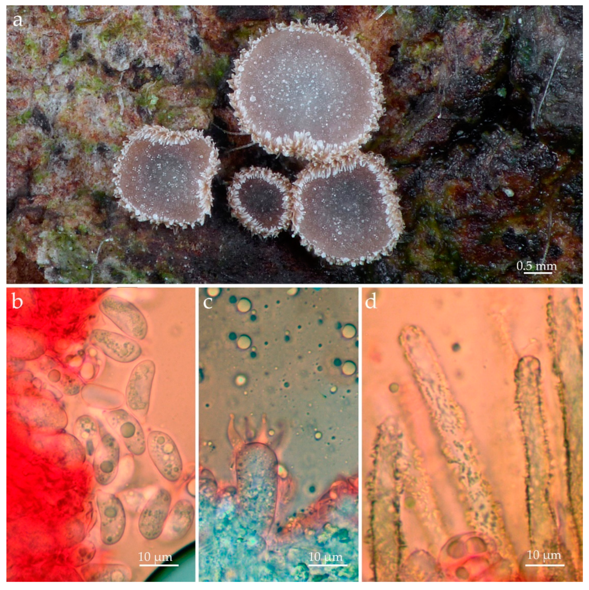
Figure 4. Merismodes monacha (ALV30536, Epitype–PC0142589). (a) Basidiomata in situ; (b) basidiospores; (c) basidium; (d) external hyphae. Photos by Andgelo Mombert.
Jof 09 01004 g004
Figure 5. Eoscyphella luciurceolata basidiomata in light (above) and dark (below). (ac) On the bark of “fumeiro” tree (Solanum swartzianum). Note that only dry mushrooms, whose margin is adorned with water droplets, emit light. FBIPBio 93.20220802 (Paratype–FIFUNGI00249). Photos by Adão Henrique Rosa Domingos.
Jof 09 01004 g005
Figure 6. Eoscyphella luciurceolata basidiomata in light (above) and dark (below) on removed bark of “fumeiro” tree (Solanum swartzianum). Note that mushrooms are in wetter conditions and all of them emit light. (a) A dried mushroom is shown next to a scalpel blade to demonstrate its size; (b) FBIPBio 93.20220802 (Paratype–FIFUNGI00249). Photos by Adão Henrique Rosa Domingos and Isaias Santos.
Jof 09 01004 g006
Figure 7. Eoscyphella luciurceolata (FBIPBio 96.20230322, holotype–FIFUNGI0001). (a) Basidiospores; (b) basidia; (c) hymenium and external surface; (d,e) external hyphae. Photos by Alexandre G. S. Silva-Filho and Cristiano C. Nascimento.
Jof 09 01004 g007
Figure 8. Eoscyphella luciurceolata (FBIPBio 96.20230322, holotype-FIFUNGI0001. (a) Basidiospores; (b) hymenium with basidia and basidioles; (c) basidia; (d,e) external hyphae. Drawings: original by Alexandre G. S. Silva-Filho and inked by K. Sousa.
Jof 09 01004 g008
Disclaimer/Publisher’s Note: The statements, opinions and data contained in all publications are solely those of the individual author(s) and contributor(s) and not of MDPI and/or the editor(s). MDPI and/or the editor(s) disclaim responsibility for any injury to people or property resulting from any ideas, methods, instructions or products referred to in the content.