Article

Screening of Antibacterial Activity of Some Resupinate Fungi, Reveal Gloeocystidiellum lojanense sp. nov. (Russulales) against E. coli from Ecuador

1
Microbial Systems Ecology and Evolution MS2E, Biodiversity of Tropical Ecosystems BIETROP Research Groups, Departamento de Ciencias de la Salud, Departamento de Ciencias Biológicas y Agropecuarias, Universidad Técnica Particular de Loja, San Cayetano Alto s/n, Loja 1101608, Ecuador
2
Département de Biologie, Ecologie et Evolution, Université de Liège, Quai Van Beneden, BE-4000 Liège, Belgium
3
Mycothèque de l’Université Catholique de Louvain (MUCL, BCCM), Earth and Life Institute–Microbiology (ELIM), Université Catholique de Louvain, Croix du Sud 2 bte L7.05.06, BE-1348 Louvain-la-Neuve, Belgium
4
Instituto Nacional de Biodiversidad INABIO, Quito 17078976, Ecuador
*
Author to whom correspondence should be addressed.
J. Fungi 2023, 9(1), 54; https://doi.org/10.3390/jof9010054
Submission received: 15 November 2022 / Revised: 9 December 2022 / Accepted: 22 December 2022 / Published: 29 December 2022
(This article belongs to the Section Fungal Evolution, Biodiversity and Systematics)

Abstract

:
Bacterial resistance to antibiotics is a serious public health problem that needs new antibacterial compounds for control. Fungi, including resupinated fungi, are a potential source to discover new bioactive compounds efficient again to bacteria resistant to antibiotics. The inhibitory capacity against the bacterial species was statistically evaluated. All the species (basidiomata and strains) were molecularly characterized with the ITS1-5.8S-ITS2 barcoding marker. The strains Ceraceomyces sp., Fuscoporia sp., Gloeocystidiellum sp., Oliveonia sp., Phanerochaete sp., and Xenasmatella sp. correspond to resupinate Basidiomycetes, and only the strain Hypocrea sp. is an Ascomycete, suggesting contamination to the basidiome of Tulasnella sp. According to the antagonistic test, only the Gloeocystidiellum sp. strain had antibacterial activity against the bacterial species Escherichia coli of clinical interest. Statistically, Gloeocystidiellum sp. was significantly (<0.001) active against two E. coli pathotypes (O157:H7 and ATCC 25922). Contrarily, the antibacterial activity of fungi against other pathotypes of E. coli and other strains such as Serratia sp. was not significant. The antibacterial activity between 48 and 72 h increased according to the measurement of the inhibition halos. Because of this antibacterial activity, Gloeocystidiellum sp. was taxonomically studied in deep combined morphological and molecular characterization (ITS1-5.8S-ITS2; partial LSU D1/D2 of nrDNA). A new species Gloeocystidiellum lojanense, a resupinate and corticioid fungus from a tropical montane rainforest of southern Ecuador, with antibacterial potential against E. coli, is proposed to the science.

1. Introduction

Pathogenic microorganisms (e.g., bacteria, fungi, and viruses) that cause infections are a serious public health problem that is increasing considerably, mainly by the high rate of genetic changes, resistance mechanisms, or wrong and excessive use of antimicrobials [1,2,3]. In addition, bacterial resistance to antibiotics increases infection rates (i.e., Gram-negative bacteria, 61.3%; Gram-positive bacteria, 34.8%; yeasts, 2%; and other pathogens, 1.9%) mainly in developing countries [2,3,4,5].
The Enterobacteriaceae family is the largest and most heterogeneous group of Gram-negative bacteria of clinical importance [6]. Within this group, genera such as Citrobacter, Enterobacter, Escherichia, Klebsiella, Proteus, Serratia, Shigella, and Salmonella are the most frequent causes of human infections [7,8,9]. About 80% of infections, including urinary tract infections, pneumonia, diarrhea, meningitis, sepsis, and endotoxic shock, among others, are caused by Enterobacteriaceae [5,9,10,11,12].
On the other hand, according to the reviewed literature, an alarming decrease in the discovery of antibiotics has been observed during recent decades [13,14], which probably involves factors such as the lack of interest from the pharmaceutical industry [15], as well as the lack of resources intended for research and bioprospecting of organisms [16]. In this sense, the need arises to look for new molecules or proteins with antibacterial properties, such as synthetic or natural compounds, investigated mainly in plants and very poorly in fungi [17].
Fungi constitute a promising group of interest for the search for bioactive compounds [18], in addition to being a highly diverse group of organisms, with an estimated 1.5 to 5 million species in the world [19], of which only a small proportion of <100,000 species have been described, according to Baldrian et al. [20]. This group of organisms is able to adapt to and survive extreme conditions in several ecosystems [21]. This characteristic may be due to the production of a wide range of bioactive compounds [22]. For example, filamentous fungi, mainly Ascomycetes (e.g., Aspergillus, Cladosporium, Fusarium, Penicillium notatum), are able to produce enzymes, microbial biomass, and secondary metabolites, including antibiotics (e.g., fusidic acid, cephalosporin, and penicillin), that are applied for the treatment of various infectious diseases [18,23,24]. Likewise, Basidiomycete fungi generate a large number of metabolites that have demonstrated antibacterial, antifungal, antiviral, cytotoxic, and hallucinogenic capacities [25,26].
Among the fungi, the macro Basidiomycetes Lentinus edodes, followed by species within the genera Boletus, Ganoderma, and Lepista are promising candidates for the search for compounds with antibiotic activity against Gram-positive and Gram-negative bacteria [25]. Likewise, other fungi with resupinate and corticioid characteristics, such as Perenniporia spp. and Antrodia spp. in the order Polyporales, have been evaluated for antimicrobial activity [26]. However, there is still a low number of prospective mushroom studies, especially in neotropical areas such as Ecuador, where these organisms are still poorly cataloged [27,28,29,30], characterized morphologically and molecularly [31,32,33,34], or even more chemically characterized by evaluating the metabolites that they generate [35]. However, mushrooms with medicinal properties, from an ethnomycological point of view, have been reported in Ecuador [36,37,38,39].
Most research focused on fungal antibacterial compounds has been conducted mainly from macrofungi due to their ease of characterization and cultivation [25]. However, the study of fungi that are almost imperceptible to the naked eye, such as “resupinates”, which are distributed in several taxonomic groups within Homobasidomycetes and Heterobasidomycetes, has been left aside [40]. Resupinated fungi exhibit diverse basidiomata forms (e.g., corticoid, trechisporoid, jaapiaid, poliporoid, russuloid, Heimenoquetide, and Cantharelloides) [40] and fulfill ecological roles, such as saprotrophs (decomposing organic matter) [41,42], symbionts (forming mycorrhizae) [31,43], or parasites of insects or plants [44]. The integration of molecular, morphological [32], and biochemical [35] characterization has allowed the discovery of new species with different biotechnological potentials [45].
Therefore, this research aims to evaluate the antibacterial potential of seven fungal strains isolated from resupinate basidiomata from a tropical montane forest (Podocarpus National Park (PNP)) in southern Ecuador, against bacteria of clinical interest, such as Escherichia coli, Serratia sp., and Klebsiella sp. We describe Gloeocystidiellum lojanense as a new species to science, integrating a morphological and molecular (ITS1-5.8S-ITS2; partial LSU D1/D2 nrDNA) characterization, and we report for the first time the antibacterial activity of G. lojanense against the Gram-negative bacteria Escherichia coli.

2. Materials and Methods

2.1. Study Area

Seven resupinate basidiomata were collected in the Podocarpus National Park (PNP) Cajanuma sector, Loja, Ecuador, between 2500 and 3000 m a.s.l. (Figure 1), in a tropical montane rainforest with a characteristic flora structure with dominant trees of the canopy generally exhibit twisted or tortuous trunks and branches, presenting smaller, leathery leaves [46,47,48]. The specimens are deposited in the fungarium of the Herbarium of the Universidad Técnica Particular de Loja HUTPL(F), under the codes [HUTPL(F)2162, 2166, 2181, 2183, 2187, 2198, 2203]. The pure strains isolated from the basidiomes were obtained by sporulation on two plates of potato dextrose agar (PDA) + chloramphenicol 1%. The strains were kept in the HUTPL annexed strain collection, under the codes [HUTPL(F)550, 551, 552, 553, 554, 555, 556].

2.2. Morphological Analysis

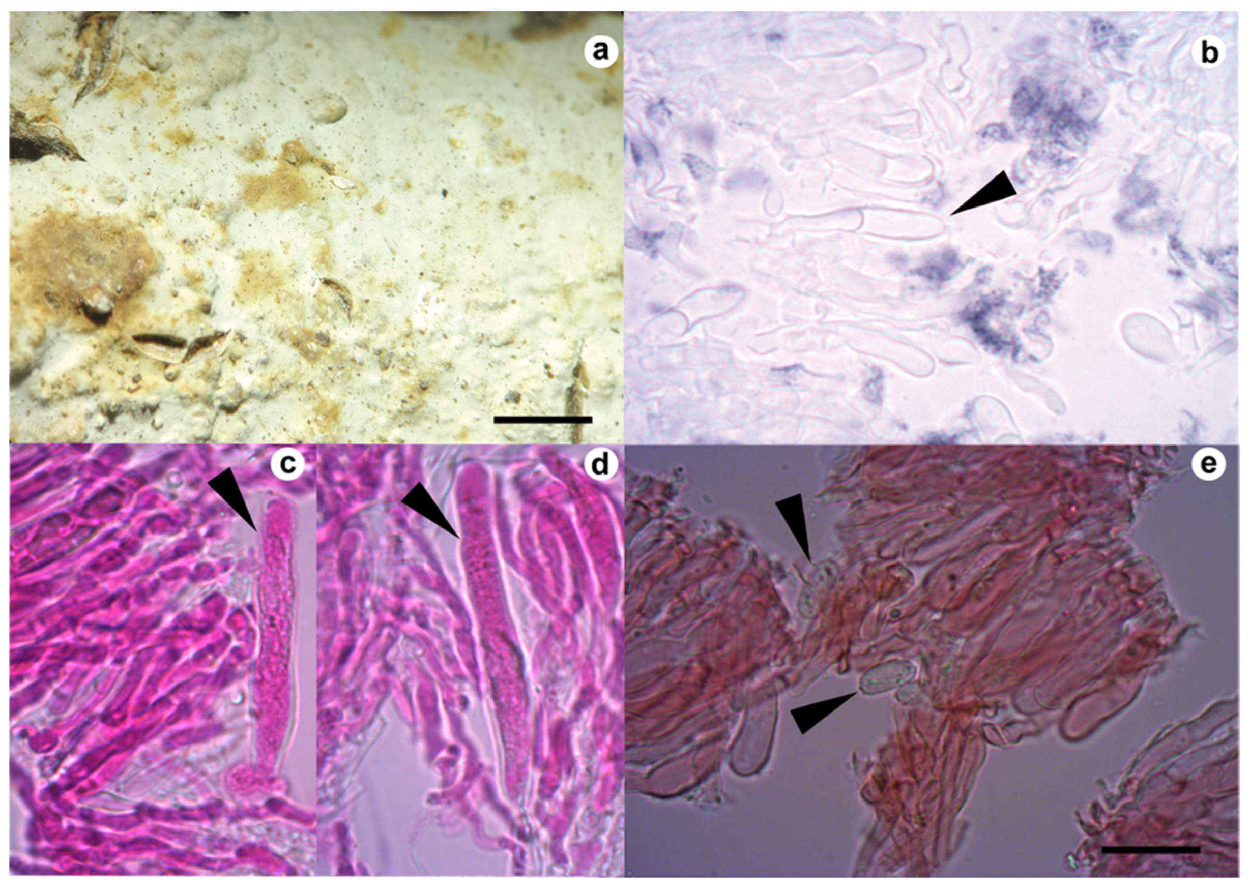
Only Gloeocystidiellum sp., after positive antibacterial activity, was analyzed morphologically in detail. In order to perform the analysis, freehand sections were made with a razor blade under a stereomicroscope (Stemi Carl Zeiss). The microscopic procedure followed Xing et al. [49]. The preparations of the sections were with phloxine 1% and decolorization with 10% potassium hydroxide (KOH) solution and Congo Red 1%. The amyloid reaction was evaluated with Melzer’s reagent (Figure 2). A sulfoaldehyde (SA) reaction to detect a sulfuric reaction of gloecystidia was performed with sulfuric acid + vanillin (Sigma-Aldrich). Observations were under a light microscope (CX31, Olympus) at 100× magnification. A detailed illustration (Figure 3) of the specimen was performed by hand using a scale (1 × 1 cm2 = 5 × 5 µm2) and later revision of the taxonomic key for the genus available in Wu, Larsson, and Ryvarden [50,51]. Color codes are based on the online server https://encycolorpedia.es/ (11 July 2022) [52].

2.3. Molecular Analysis

DNA was isolated from fresh basidiomata using the Phire Plant Direct Master Mix PCR Kit (Thermo Scientific™) and subsequently from pure fungal strains with the InnuPREP DNA Kit (Analytik-jena™) according to the manufacturer’s instructions. For the polymerase chain reaction (PCR), the ITS1 (5′CCGTAGGTGAACCTGCGG3′) and NL4 (5′GGTCCGTGTTTCAAGACGG3′) primers were used [53] to amplify the internal transcribed spacer (ITS) region and a partial sequence of nuclear large subunits (LSUs). The reactions were carried out under the following conditions: initial denaturation (98 °C, 5 min), followed by 40 cycles with denaturation (98 °C, 10 s), hybridization (55 °C, 10 s), extension (72 °C, 30 s), and final extension (72 °C, 5 min). Final reaction volume was 20 µL, including 1 µL of the extracted DNA for each reaction. The PCR product was purified with the PureLink™ PCR Purification Kit (Thermo Scientific™) and sequenced at Macrogen Inc. (Seoul, Korea), with the same set of primers used for PCR amplification. All sequences corresponding to the basidiomata, and strains were subjected to a BLAST search against the GenBank database (https://www.ncbi.nlm.nih.gov) as well as the UNITE database (https://unite.ut.ee).
Only the sequences corresponding to Gloeocystidiellum sp. were assembled and edited using Lasergene 7 (DNAStar, Madison, WI, USA). The sequences obtained in this study were submitted to the NCBI nucleotide database under the accession numbers presented in Table 1. The alignment of the sequences obtained and reference sequences downloaded from GenBank (Table 1) was performed in the program MAFFT 7 [54] under the GINSI algorithm. Alignments were visualized in PhyDE software [55] in order to check for ambiguities, especially at the tails, for a manual adjustment if required. The alignments were analyzed by means of a neighbor-joining (NJ) approach using a Kimura two-parameter (K2P) with 1000 bootstrap repetitions (BS) and a maximum likelihood using the general time-reversible (GTR) method with 1000 bootstrap repetitions. Both analyses were performed with MEGA 11 software [56].
Finally, four phylogenetic trees were calculated: the first includes 28 sequences for the region (ITS1-5.8S-ITS2) (Figure S1), the second tree includes 36 sequences for the partial LSU region (D1/D2) (Figure S2), the third tree corresponds to 31 concatenated sequences (ITS1-5.8S-ITS2 and partial LSU D1/D2) (Figure 4), and the fourth tree includes 34 sequences that correspond only to the ITS1 region (Figure 5) due to the requirement of comparing three short sequences (JQ7345551, JQ7169401, JQ7345541) described in Chile by Gorjón and Hallenberg [57]. All the phylogenetic trees presented in this study were inferred using a maximum likelihood approach.
Interspecific variation between sequences from specimen HUTPL(F)2181 and close phylogenetic species was evaluated by pairwise distances for the ITS1-5.8S-ITS2 regions and partial LSU D1/D2 in the MEGA 11 software with a Kimura two-parameter distance [58] by the “partial deletion” and “complete deletion” method.
Additionally, sequences from seven strains of various Gloeocystidiellum spp. (MUCL33964, MUCL33965, MUCL33966, MUCL33967, MUCL33968, MUCL35247, MUCL35248) were included in the phylogenetic analyses. These strains are maintained in the collection of the Mycothèque de l’Université catholique de Louvain (MUCL/BCCM), Louvain-la-Neuve, Belgium.

2.4. Antibacterial Activity

2.4.1. Tested Bacterias

Ten Gram-negative bacterial strains of clinical interest were used in this study to evaluate the antibacterial activity of seven strains from resupinated fungi. These strains correspond to six different species: Enterobacter aerogenes; Enterobacter cloacae; Escherichia coli, including two pathotypes (uropathogenic Escherichia coli (UPEC), phylogenetic group (GF) A; GF B2; O157:H7) and the certified strain E. coli ATCC 25922 GF: B2; Klebsiella pneumoniae (ATCC BAA-1706); Pseudomonas aeruginosa; and Serratia sp. All the strains are available in the strain collection of the Laboratory of Cultures and Conservation of Microorganisms–UTPL, and were previously characterized with standard biochemical tests and API20E tests.

2.4.2. Antibacterial Activity Assay

Antibacterial activity was determined using the agar disc diffusion method by the Kirby–Bauer method as described in CLSI M100 [59]. All bacterial strains were grown on Trypticase Soy Agar (TSA, DIFCO) for 24 h at 37 °C. Subcultured colonies were suspended in 0.85% saline, followed by 0.5 McFarland density adjustment (1.5 × 10⁸ CFU/mL) by spectrophotometry.
The antibacterial tests were carried out by antagonism in a Petri dish (9 cm diameter) with Mueller–Hinton agar. Discs (0.5 cm diameter) of PDA medium colonized with the strains replaced similar space (three replicates) in the medium with bacterial growth. All plates were prepared and incubated at 37 °C for 24–72 h. Antibacterial activity was recorded as diameter (mm) of the zone of inhibition formed around the fungal disc (Figure S1). Commercial antibiotic discs for susceptibility testing based on CLSI reports [59,60,61] were also used as positive controls. Antibiotics correspond to 30 µg of cefepime for all strains, except for K. pneumoniae and uropathogenic E. coli phylogenetic group B2, in which 10 µg of imipenem was used as a positive control due to its bacterial resistance. Each Petri dish with bacterial growth is considered as one test, carried out in triplicate.

2.5. Data Analysis

Statistical analysis was performed by calculating the average ( X ¯ ) of the three repetitions of each test. Prior to the analysis, the dependent variable “inhibition” was transformed to a logarithm in base 2 to fulfill the assumption of normality (p > 0.05). Two-way analysis of variance to test the effects of Gloeocystidiellum sp. strain (the only fungus with antibacterial activity) and event on bacterial inhibition was used. Finally, a post-hoc Tukey test to check differences in inhibition between pairs of bacteria was performed. All analyses were carried out in R Studio software (RStudio, Inc., Boston, MA, USA) [62].

3. Results

Six of seven strains identify as five Basidiomycetes, and one ascomycete under six orders (Table 2) was not characterized taxonomically because they did not present antibacterial activity. On the other hand, Gloeocystidiellum sp., which presents positive antibacterial activity against E. coli, is taxonomically described as a new species for science. The BLAST identity percent found between 87 and 97 suggests a possibility to find some new species for the science in this tropical forest.

3.1. Taxonomy

Gloeocystidiellum lojanense A. Jaramillo, D. Cruz & C. Decock., sp. nov. (Figure 2 and Figure 3).
ETYMOLOGY: The specific epithet refers to Loja province (Ecuador) where the species was found for the first time.
BASIDIOMA—resupinate, ceraceous, or subceraceous. Hymenial surface is bright grayish white (#ecf0d4) and slightly light yellow (#b0ac6e), extending smoothly and slightly tuberculate over the substrate up to about 15 cm2; bounded margins.
HOLOTYPE—South America, Ecuador, Loja Province, and Canton, Podocarpus National Park, Cajanuma sector, alt. c. 2900 m, on fallen decomposing branch of unknown tree, 23 February 2021. A. Jaramillo [HUTPL(F)2181].
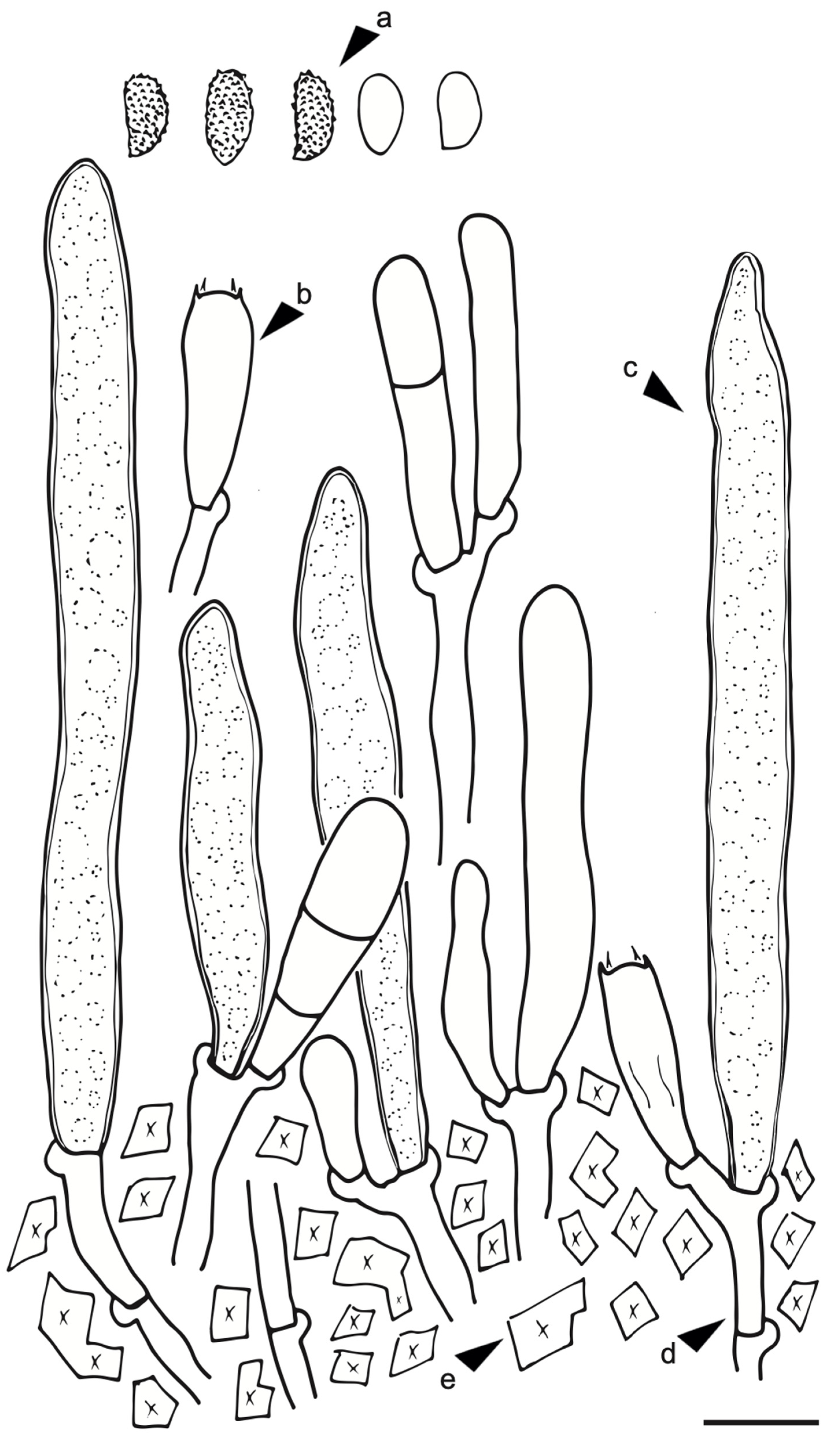
Microscopic Structure. Hyphal system monomitic: generative hyphae 3–4 µm diameter, septate, thin- to thick-walled hyaline with clamp connection with calcium oxalate crystals, gloeocystidia abundant, tubular or cylindrical, slightly tapering towards the apices, thick-walled throughout, up to approximately 80–90 μm long, 7–8 μm wide, the protoplasmic content granular and yellowish in KOH, blackish in SA (positive reaction), basidia clavate 25–35 long × 5–6 μm wide, transversely septate at the basal zone, with four sterigmata, basidiospores hyaline ellipsoid and slightly verrucose, thin-walled, 6.5–8 μm long × 3.4–4.5 μm wide, and slightly amyloid under Meltzer’s reagent (Figure 2 and Figure 3).
Gloeocystidiellum lojanense is morphologically related to Gloeocystidiellum formosanum, G. compactum, G. aspellum, and G. rajchenbergii [50,57]. These species are closely similar as far as their morphology is concerned, especially the basidiospores and gloeocystidia features (Table 3); additionally, all of them present an amyloid reaction in Meltzer’s reagent. Microscopically, the shape and size (Table 3) of basidiospores and gloeocystidia in G. lojanense are indistinguishable from those of its allied species listed above. Although the size of the basidiospores has been considered as the main characteristic to define species in this group [50,57,63,64,65], it is evident that they can overlap each other, causing taxonomic confusion, as has already been indicated in other groups of fungi (e.g., Tulasnella spp.), where molecular data allowed revealing some cryptic morphological species [32].
Another reason to propose G. lojanense as a new neotropical species is their biogeographic and ecological location, as has been described in other Gloeocystidiellum species [64,65]. G. lojanense was found in a tropical montane rainforest in southern Ecuador at 2900 m asl; different to the places in eastern Asia (Taiwan, China) or Patagonia Andean of Chile, about 600 to 1250 m asl, reported to the closest morphological species [50,57,66].

3.2. Phylogenetic Hypothesis for Gloeocystidiellum lojanense

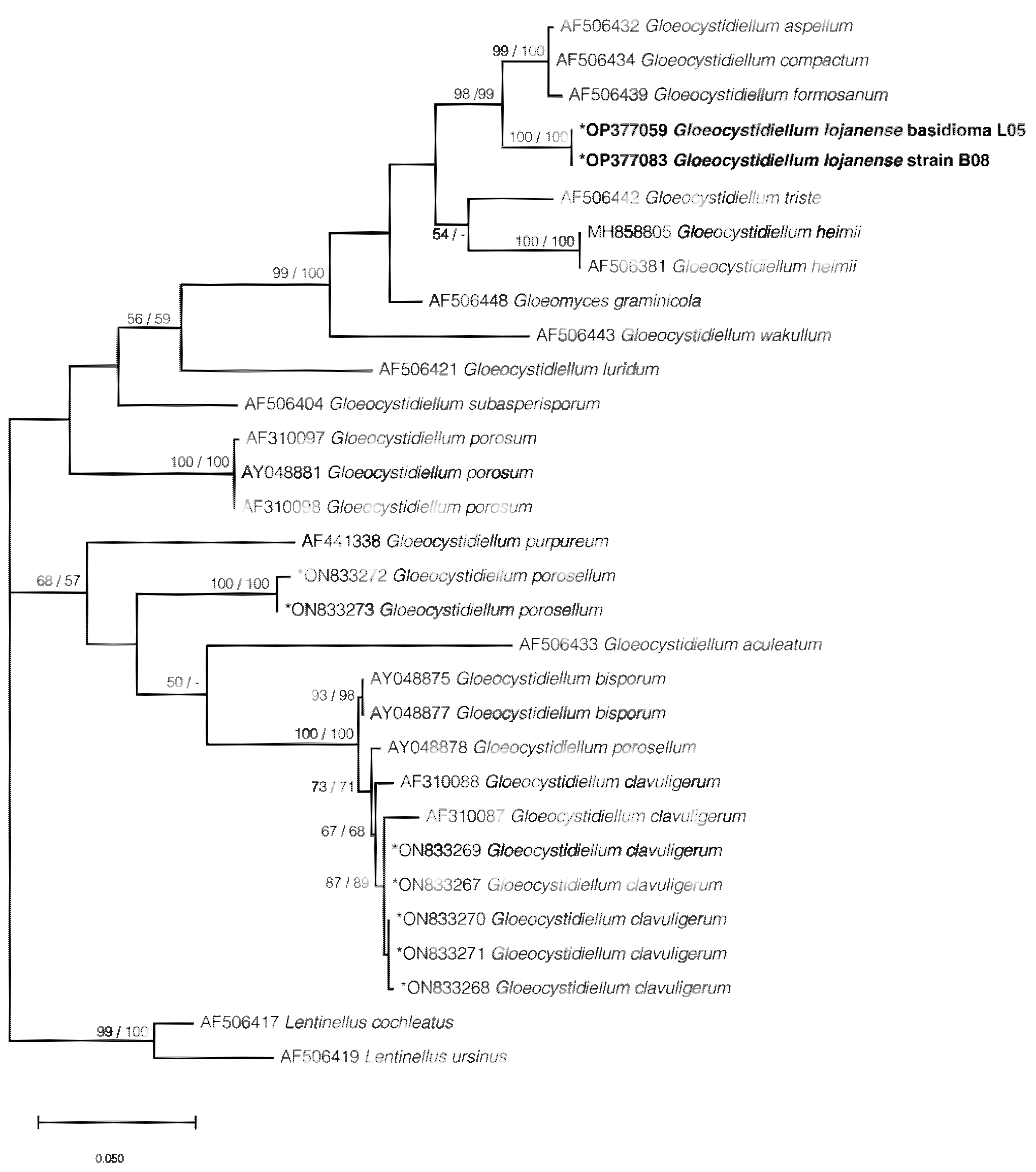
The phylogenetic results, based on the ITS or the concatenated data sets (Figure 4 and Figure 5; Figures S2 and S3) showed that G. lojanense form an independent well-supported clade (respectively, 96 and 100% bootstrap) that is here interpreted as a new phylogenetic species. Gloeocystidiellum lojanense forms a sister clade (Figure 5, BS 98/99% for ML/NJ) to the clade gathering G. aspellum, G. compactum, and G. formosatum from several countries (Table 1). A similar topology if observed is when the ITS1 region (63/56% BS for ML/NJ), including the sequence from the species G. rajchenbergii, is analyzed.
The phylogenetic tree obtained from the concatenated dataset (ITS1-5.8S-ITS2 and partial LSU D1/D2 regions) (Figure 4) is consistent with the phylogenies from independent analyses for the ITS1 (Figure 5) or ITS1-5.8S-ITS2 (341 bp) regions (Figure S1) and LSU D1/D2 partial (471 bp) (Figure S3), where G. lojanense is grouped as a sister group with the species G. aspellum, G. compactum, G. formosatum, and G. rajchenbergii but remains as an independent clade with BS values higher than 95/97% for ML/NJ.
The resulting interspecific groups are totally supported when the sequences were analyzed by complete deletion and partial deletion; for example, the range for the ITS1-5.8-ITS2 region is between 6.45% and 6.90% (Table 4), ITS1 is between 2.94 and 3.69%, and partial LSU D1/D2 is 0.81% (Table 4).

3.3. Antibacterial Activity

Gloeocystidiellum lojanense HUTPL(F)550 was the unique strains tested by inhibiting the bacterial strains of Escherichia coli and Serratia sp. (Table 5). The other six strains (Table 2) were not further charactrized because they were totally inactive against the bacterias tested.
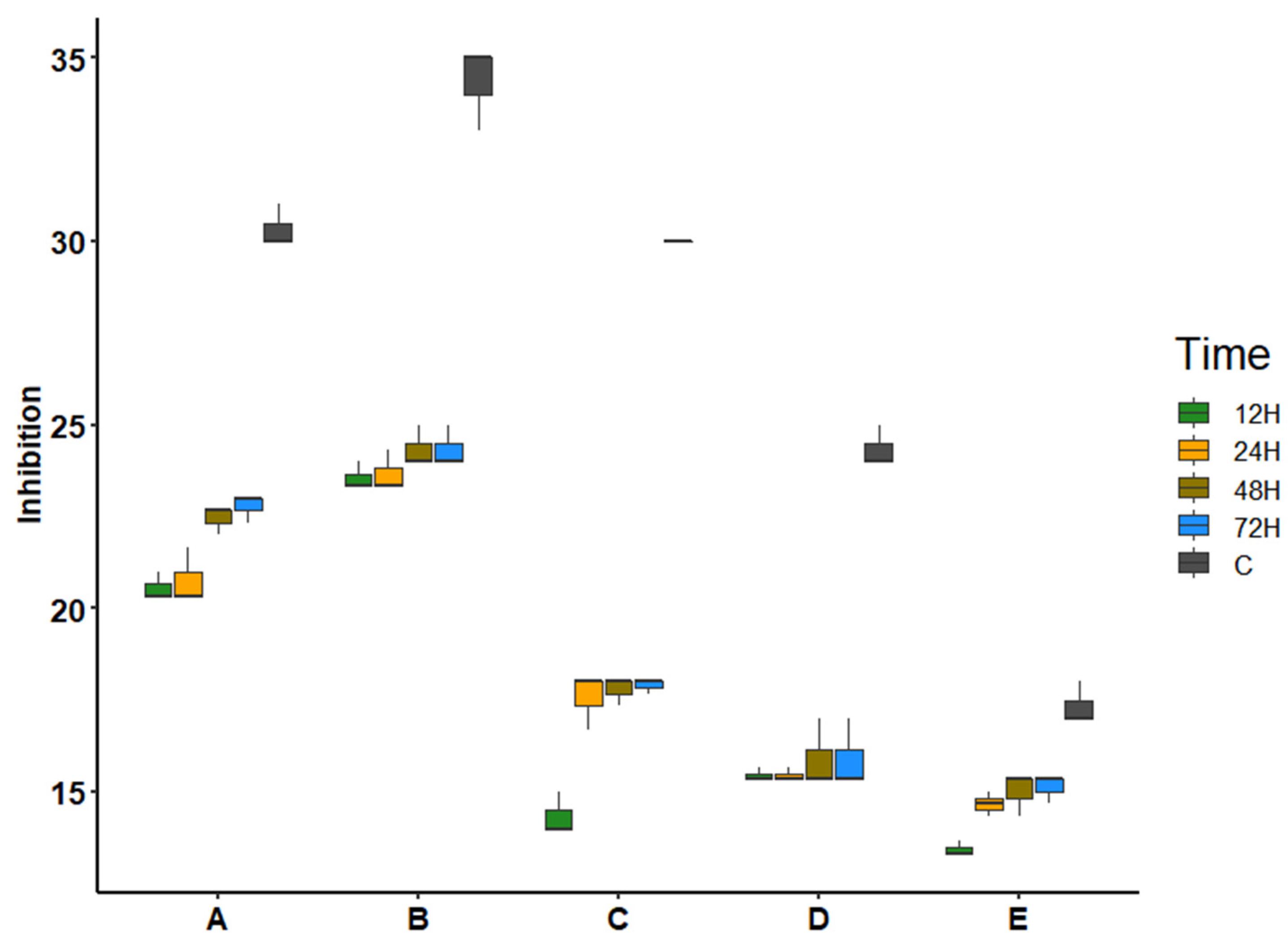
Gloeocystidiellum lojanense was observed to be more effective at inhibiting mainly two pathotypes of E. coli (i.e., Escherichia coli ATCC 25922 and Escherichia coli O157:H7). The inhibition halos generated for these strains were larger in size (between 22.78 and 24.33 mm in diameter (Table 5) and statistically significant (<0.001) versus the other tests (Figure 6, Table 6). However, the inhibition halos generated by G. lojanense did not exceed the halos generated from the positive controls (30.33–34.33 mm in diameter) for the strains Escherichia coli ATCC 25922 and Escherichia coli O157: H7. Inhibition halos varied according to the incubation time (Table 5).
The bacteria Enterobacter aerogenes, Enterobacter cloacae, Klebsiella pneumoniae, Klebsiella pneumoniae ATCC BAA-1706, and Pseudomonas aeruginosa did not exhibit inhibitory activity by G. lojanense.
The box plot clearly indicates that Gloeocystidiellum lojanense presents greater inhibition in the bacteria Escherichia coli ATCC 25922 (22.44/22.78 mm) and Escherichia coli O157:H7 (24.33 mm), increasing during 48 and 72 hours, compared to Escherichia coli. UPEC FG: A, Escherichia coli UPEC FG: B2 and Eb, EGA, and Serratia sp., which were more stable over time (Figure 6).
The analysis of variance indicated that the fungal species, time, as well as their interaction between the two factors, significantly affect the inhibition of the bacteria, showing significant values of each factor (<0.001) (Table 6).

4. Discussion

Gloeocystidiellum lojanense represents a species new to science from a tropical montane rainforest in southern Ecuador. This species is phylogenetically supported, forming an independent clade with BS values greater than 96% for the ITS1-5.8S-ITS2 and partial LSU regions, analyzed independently or concatenated. This species presents an interspecific genetic divergence for the ITS1-5.8S-ITS2 region greater than 6.45% with respect to the other species. This interspecific divergence of 6.45% would generate a barcode gap with respect to the 3% or 4% thresholds of intraspecific variability studied for fungi [32,67].
Morphologically and molecularly, G. lojanense is suggested as a sister group to the clade comprising G. formosanum, G. compactum, G. aspellum, and G. rajchenbergii [57]. All these species are practically indistinguishable morphologically, mainly due to the overlap of shapes and sizes of basidiospores and gloeocystidia. This problem, discussed as “cryptic species”, is already well known to other groups such as Tulasnella spp. [32], where several morphologically cryptic species were delimited genetically. Other species (i.e., Gloeocystidiellum clavuligerum and G porosum) are morphologically [63,64,65] closely related to G. lojanense but genetically distant (>6.45% ITS1-5.8S-ITS2), forming a different clade (Figure 4). Additionally, the interspecific genetic difference for the ITS1-5.8S-ITS2 region is greater than 6.45%, exceeding the so-called barcode gap discussed by [32].
Gloeocystidiellum lojanense exhibited antibacterial activity against the four strains of the E. coli species, with E. coli ATCC 25922 and E. coli O157:H7 (Figure 6) showing greater inhibition than the other E. coli strains. This inhibitory action may be due to the fact that these E. coli pathotypes are less virulent and resistant [68]. Other pathotypes (e.g., Escherichia coli UPEC FG: A, Escherichia coli UPEC FG: B2) are reported to be resistant to antibiotics [69], observed in slight size changes in the inhibition halos. Likewise, G. lojanense did not significantly inhibit the growth of Serratia sp. This species is also known to have a high bacterial resistance to antibiotics [70], as evidenced in the positive control with a smaller size of the inhibition halo. Suay et al. [71] present a study indicating that the species Gloeocystidiellum porosum has antibacterial activity against Pseudomonas aeruginosa, Serratia marcescens, and Staphylococcus aureus, which is why a further exploration of this group of fungi is required.
Antibacterial inhibition increased between 48 and 72 h, probably because the fungal metabolites exhibiting these antibacterial properties were generated secondarily by nutrient depletion, or simply by recognition of the bacterial foreign agent [72]. Six different strains of resupinated fungi within (Amylocorticiales, Cantharellales, Hymenochaetales, Polyporales, Russulales) and one Hypocrea sp. (Hypocreales) did not exhibit inhibition activity against any bacterial strain tested. The Gloeocystidiellum sp. (active against bacteria) and Xenasmatella sp. (inactive against bacteria) share the same order Russulales but differ in antibacterial activity. Many studies report different fungi species in the same order such as this study (e.g., Polyporales, Russulales) but positive for bacteria inhibitions [25,26]. Probably, the antibacterial activity of fungi is restricted at the species level by specific genetic adaptations [73] requiring further analysis of the gene expression of these fungi.
Statistically, the antibacterial activity of G. lojanense is significant mainly for two pathotypes of E. coli (i.e., E. coli ATCC 25922 and E. coli O157:H7). However, our analysis requires an evaluation of the minimum inhibitory concentration by obtaining its extracts, as has already been evaluated for other fungi [74].
Ecuador, considered within the megadiverse countries worldwide [75,76,77], has as consequence a great diversity of fungi [27,29,33,34] that require integrative taxonomic research to discover new species such as G. lojanense with bioactive potential and applications in several sectors such as human health.

Supplementary Materials

The following supporting information can be downloaded at: https://www.mdpi.com/article/10.3390/jof9010054/s1, Figure S1: Antibacterial activity assay flowchart; Figure S2: Maximum Likelihood phylogenetic tree for the ITS1-5.8-ITS2 region for sequence positioning corresponding to Gloeocystidiellum lojanense sp. nov. Bar = number of expected substitutions per position. * Sequences generated in this study; Figure S3: Maximum likelihood phylogenetic tree for the LSU region for sequence positioning corresponding to Gloeocystidiellum lojanense sp. nov. Bar = number of expected substitutions per position. * Sequences generated in this study.

Author Contributions

A.J.-R., D.C. and C.D. contributed to the conceptualization and methodology of this study. A.J.-R. and D.C. carried out sampling. Á.B. performed the statistical analyses. J.P.S. and G.C. performed strong critical revisions of the manuscript and contributed with general comments. All authors have read and agreed to the published version of the manuscript.

Funding

This research was funded by Académie de Recherche et d’enseignement supérieur (ARES), Université de Liège, Université Catholique de Louvain and Universidad Técnica Particular de Loja, grant number.

Data Availability Statement

All data are included in this document.

Acknowledgments

The authors are very grateful to Blga. Luisa Espinosa and Blgo. Jhoan Peña Biology, alumni at Universidad Técnica Particular de Loja, who helped during sampling and laboratory work.

Conflicts of Interest

The authors declare no conflict of interest.

References

  1. Mir, M.; Rasool, U.; Aisha, S.; Alshehri, B.; Hamadani, S. Human pathogenic microbes (bacterial and fungal) and associated diseases. Hum. Pathog. Microbes 2022, 1–30. [Google Scholar] [CrossRef]
  2. Lagadinou, M.; Onisor, M.; Rigas, A.; Musetescu, D.; Gkentzi, D.; Assimakopoulos, S.; Panos, G.; Marangos, M. Antimicrobial properties on non-antibiotic drugs in the era of increased bacterial resistance. Antibiotics 2020, 9, 107. [Google Scholar] [CrossRef] [Green Version]
  3. Zhang, F.; Cheng, W. The mechanism of bacterial resistance and potential bacteriostatic strategies. Antibiotics 2022, 11, 1215. [Google Scholar] [CrossRef]
  4. Mena, Y.; Ramírez, L.; Arango, Á. Actividad antibacteriana y antioxidante del hongo Phanerochaete spp. Inf. Tecnol. 2021, 32, 69–78. [Google Scholar] [CrossRef]
  5. Pérez Guerrero, P.; Galán Sánchez, F.; Gutiérrez Saborido, D.; Guerrero Lozano, I. Infecciones por enterobacterias. Med.-Programa Form. Médica Contin. Acreditado 2014, 11, 3276–3282. [Google Scholar] [CrossRef]
  6. Janda, J.; Abbott, S. The changing face of the family Enterobacteriaceae (Order: “Enterobacterales”): New members, taxonomic issues, geographic expansion, and new diseases and disease syndromes. Clin. Microbiol. Rev. 2021, 34, e00174-20. [Google Scholar] [CrossRef] [PubMed]
  7. Staley, Z.; He, D.; Edge, T. Persistence of fecal contamination and pathogenic Escherichia coli O157:H7 in snow and snowmelt. J. Great Lakes Res. 2017, 43, 248–254. [Google Scholar] [CrossRef]
  8. Uprety, S.; Dangol, B.; Nakarmi, P.; Dhakal, I.; Sherchan, S.; Shisler, J.; Jutla, A.; Amarasiri, M.; Sano, D.; Nguyen, T. Assessment of microbial risks by characterization of Escherichia coli presence to analyze the public health risks from poor water quality in Nepal. Int. J. Hyg. Environ. Health 2020, 226, 113484. [Google Scholar] [CrossRef]
  9. Morosini, M.; del Campo, R. Urinary tract infections and antimicrobial resistance. Rev. Clín. Esp. 2019, 219, 149–150. [Google Scholar] [CrossRef]
  10. Centers for Disease Control and Prevention (CDC). “Vital signs: Carbapenem-resistant Enterobacteriaceae.” MMWR Morb. Mortal. Wkly. Rep. 2013, 62, 165–170.
  11. Monteiro, A.; Cardoso, J.; Guerra, N.; Ribeiro, E.; Viegas, C.; Cabo Verde, S.; Sousa-Uva, A. Exposure and health effects of bacteria in healthcare units: An overview. Appl. Sci. 2022, 12, 1958. [Google Scholar] [CrossRef]
  12. Shen, S.; Qu, X.; Zhang, W.; Li, J.; Lv, Z. Infection against infection: Parasite antagonism against parasites, viruses and bacteria. Infect. Dis. Poverty 2019, 8, 1–12. [Google Scholar] [CrossRef]
  13. Pulingam, T.; Parumasivam, T.; Gazzali, A.; Sulaiman, A.; Chee, J.; Lakshmanan, M.; Chin, C.; Sudesh, K. Antimicrobial resistance: Prevalence, economic burden, mechanisms of resistance and strategies to overcome. Eur. J. Pharm. Sci. 2022, 170, 106103. [Google Scholar] [CrossRef]
  14. Morrill, H.; Pogue, J.; Kaye, K.; LaPlante, K. Treatment options for carbapenem-resistant Enterobacteriaceae infections. Open Forum Infect. Dis. 2015, 2, ofv050. [Google Scholar] [CrossRef] [Green Version]
  15. Ciabuschi, F.; Lindahl, O. The decline of innovation in the antibiotics industry and the global threat of antibiotic resistance: When entrepreneurial efforts are not enough. Entrep. Ind. Life Cycle 2018, 205–229. [Google Scholar] [CrossRef]
  16. Singh, D.; Thapa, S.; Mahawar, H.; Kumar, D.; Geat, N.; Singh, S. Prospecting potential of endophytes for modulation of biosynthesis of therapeutic bioactive secondary metabolites and plant growth promotion of medicinal and aromatic plants. Antonie Van Leeuwenhoek 2022, 115, 699–730. [Google Scholar] [CrossRef]
  17. Kamaruzaman, N.; Mohd Noor, N.; Radin Mohamed, R.; Al-Gheethi, A.; Ponnusamy, S.; Sharma, A.; Vo, D. Applicability of bio-synthesized nanoparticles in fungal secondary metabolites products and plant extracts for eliminating antibiotic-resistant bacteria risks in non-clinical environments. Environ. Res. 2022, 209, 112831. [Google Scholar] [CrossRef]
  18. Hyde, K.; Tennakoon, D.; Jeewon, R.; Bhat, D.; Maharachchikumbura, S.; Rossi, W.; Leonardi, M.; Lee, H.; Mun, H.; Houbraken, J.; et al. Fungal diversity notes 1036–1150: Taxonomic and phylogenetic contributions on genera and species of fungal taxa. Fungal Divers. 2019, 96, 1–242. [Google Scholar] [CrossRef]
  19. Purvis, A.; Hector, A. Getting the measure of biodiversity. Nature 2000, 405, 212–219. [Google Scholar] [CrossRef]
  20. Baldrian, P.; Větrovský, T.; Lepinay, C.; Kohout, P. High-throughput sequencing view on the magnitude of global fungal diversity. Fungal Divers. 2021, 114, 539–547. [Google Scholar] [CrossRef]
  21. Sidorova, I.I.; Velikanov, L.L. Bioactive substances of agaricoid basidiomycetes and their possible role in regulation of myco- and microbiota structure in soils of forest ecosystems. I. Antibiotic activity of water extracts from basidioms of several dominant agaricoid basidiomycetes. Mikol. Fitopatol. 2000, 34, 11–17. [Google Scholar]
  22. Kumar, V.; Prasher, I. Antimicrobial potential of endophytic fungi isolated from Dillenia indica L. and identification of bioactive molecules produced by Fomitopsis meliae (Undrew.) Murril. Nat. Prod. Res. 2022, 36, 1–5. [Google Scholar] [CrossRef] [PubMed]
  23. Alkhulaifi, M.; Awaad, A.; AL-Mudhayyif, H.; Alothman, M.; Alqasoumi, S.; Zain, S. Evaluation of antimicrobial activity of secondary metabolites of fungi isolated from Sultanate Oman soil. Saudi Pharm. J. 2019, 27, 401–405. [Google Scholar] [CrossRef] [PubMed]
  24. Karwehl, S.; Stadler, M. Exploitation of fungal biodiversity for discovery of novel antibiotics. Curr. Top. Microbiol. Immunol. 2016, 303–338. [Google Scholar] [CrossRef]
  25. Venturini, M.; Rivera, C.; Gonzalez, C.; Blanco, D. Antimicrobial activity of extracts of edible wild and cultivated mushrooms against foodborne bacterial strains. J. Food Prot. 2008, 71, 1701–1706. [Google Scholar] [CrossRef]
  26. Winnie, C.S.; Stephen, A.I.; Josphat, C.M. Antimicrobial activity of basidiomycetes fungi isolated from a Kenyan tropical forest. Afr. J. Biotechnol. 2019, 18, 112–123. [Google Scholar] [CrossRef] [Green Version]
  27. Batallas-Molina, R.; Moya-Marcalla, G.F.; Navas Muñoz, D. Listado de la colección de hongos (Ascomycota y Basidiomycota) del Herbario Nacional del Ecuador (QCNE) del Instituto Nacional de Biodiversidad (INABIO). Av. Cienc. Ing. 2021, 12, 38–71. [Google Scholar]
  28. Cruz, D.; Suárez, J.; Piepenbring, M. Morphological revision of Tulasnellaceae, with two new species of Tulasnella and new records of Tulasnella spp. for Ecuador. Nova Hedwig. 2016, 102, 279–338. [Google Scholar] [CrossRef]
  29. Gates, G.; Goyes, P.; Gundogdu, F.; Cruz, J.; Ratkowsky, D. Small plot surveying reveals high fungal diversity in the Ecuadorian Amazon–A case study. Curr. Res. Environ. Appl. Mycol. 2021, 11, 16–36. [Google Scholar] [CrossRef]
  30. Læssøe, T.; Petersen, J.H. Svampelivet på ækvator. Svampe 2008, 58, 1–53. [Google Scholar]
  31. Cruz, D.; Suárez, J.; Kottke, I.; Piepenbring, M.; Oberwinkler, F. Defining species in Tulasnella by correlating morphology and nrDNA ITS-5.8S sequence data of basidiomata from a tropical Andean forest. Mycol. Prog. 2011, 10, 229–238. [Google Scholar] [CrossRef]
  32. Cruz, D.; Suárez, J.; Kottke, I.; Piepenbring, M. Cryptic species revealed by molecular phylogenetic analysis of sequences obtained from basidiomata of Tulasnella. Mycol. Progress 2014, 106, 708–722. [Google Scholar] [CrossRef] [PubMed]
  33. Toapanta-Alban, C.; Ordoñez, M.; Barnes, C.; Blanchette, R. Taxonomy of the major rhizomorphic species of the “Melanopus group” within Polyporaceae in Yasuní National Park, Ecuador. PLoS ONE 2021, 16, e0254567. [Google Scholar] [CrossRef] [PubMed]
  34. Toapanta-Alban, C.; Ordoñez, M.; Blanchette, R. New findings on the biology and ecology of the Ecuadorian Amazon fungus Polyporus leprieurii var. yasuniensis. J. Fungi 2022, 8, 203. [Google Scholar] [CrossRef]
  35. Sárközy, A.; Kúsz, N.; Zomborszki, Z.; Csorba, A.; Papp, V.; Hohmann, J.; Vanyolos, A. Isolation and characterization of chemical constituents from the Poroid Medicinal Mushroom Porodaedalea chrysoloma (Agaricomycetes) and their antioxidant activity. Int. J. Med. Mushrooms 2020, 22, 125–131. [Google Scholar] [CrossRef] [PubMed]
  36. Gamboa-Trujillo, P.; Wartchow, F.; Cerón-Martinez, C.; Andi, D.; Uwinjin, P.; Grefa, G.; Entza, M.; Chimbo, E.; Chimbo, J.; Payaguaje, J.; et al. Edible mushrooms of Ecuador: Consumption, myths and implications for conservation. Ethnobot. Res. Appl. 2019, 18, 1–15. [Google Scholar] [CrossRef] [Green Version]
  37. Andrade, J.M.; Malagón, O.; Piepenbring, M.; Armijos, C. Etnomicología y valor nutricional de macrohongos silvestres de la comunidad indígena Saraguro en el Sur del Ecuador. Boletin Soc. Micológica Madr. 2012, 36, 193–201. [Google Scholar]
  38. Benítez, Á.; Cruz, D.; Vega, M.; González, L.; Jaramillo, N.; López, F.; Aguirre, Z. Briófitos y hongos (liquenizados y no liquenizados) del Parque Universitario Francisco Vivar Castro, Loja, Ecuador. Bosques Latid. Cero 2021, 11, 1–18. [Google Scholar] [CrossRef]
  39. Kuhar, F.; Furci, G.; Drechsler-Santos, E.; Pfister, D. Delimitation of Funga as a valid term for the diversity of fungal communities: The Fauna, Flora & Funga proposal (FF&F). IMA Fungus 2018, 9, A71–A74. [Google Scholar] [CrossRef] [Green Version]
  40. Binder, M.; Hibbett, D.; Larsson, K.; Larsson, E.; Langer, E.; Langer, G. The phylogenetic distribution of resupinate forms across the major clades of mushroom-forming fungi (Homobasidiomycetes). Syst. Biodivers. 2005, 3, 113–157. [Google Scholar] [CrossRef]
  41. Piepenbring, M.; López, F.; Cáceres, O. Colaboradores escondidos–La Importancia de los Hongos en los Ecosistemas Información para Educación Ambiental. Puente Biol. 2019, 8, 57–91. [Google Scholar]
  42. Hallenberg, N.; Ryberg, M.; Nilsson, R.; Wood, A.; Wu, S. Pseudolagarobasidium (Basidiomycota): On the reinstatement of a genus of parasitic, saprophytic, and endophytic resupinate fungi. Botany 2008, 86, 1319–1325. [Google Scholar] [CrossRef] [Green Version]
  43. Mosquera-Espinosa, A.; Bayman, P.; Prado, G.; Gómez-Carabalí, A.; Otero, J. The double life of Ceratobasidium: Orchid mycorrhizal fungi and their potential for biocontrol of Rhizoctonia solani sheath blight of rice. Mycologia 2013, 105, 141–150. [Google Scholar] [CrossRef]
  44. Sánchez-García, M.; Ryberg, M.; Khan, F.; Varga, T.; Nagy, L.; Hibbett, D. Fruiting body form, not nutritional mode, is the major driver of diversification in mushroom-forming fungi. Proc. Natl. Acad. Sci. USA 2020, 117, 32528–32534. [Google Scholar] [CrossRef] [PubMed]
  45. Silva, S.; Melo, B.; Santos, M.; Rios, R.; Santos, C.; Júnior, K.; Maranhão, F.; Santos, T.; Fraga, A. Endophytic fungi: Benefits for plants and biotechnological potential. Res. Soc. Dev. 2022, 11, e9211427008. [Google Scholar] [CrossRef]
  46. Homeier, J.; Werner, F.A.; Gradstein, S.R. Potential vegetation and floristic composition of Andean forests in South Ecuador, with a focus on the RBSF. In Gradients in a Tropical Mountain Ecosystem of Ecuador: Ecological Studies; Beck, E., Bendix, J., Kottke, I., Makeschin, F., Mosandl, R., Eds.; Springer: Berlin/Heidelberg, Germany, 2008; pp. 87–100. [Google Scholar] [CrossRef]
  47. Homeier, J.; Hertel, D.; Camenzind, T.; Cumbicus, N.L.; Maraun, M.; Martinson, G.O.; Poma, L.N.; Rillig, M.C.; Sandmann, D.; Scheu, S.; et al. Tropical Andean forests are highly susceptible to nutrient inputs-rapid effects of experimental N and P addition to an Ecuadorian montane forest. PLoS ONE 2012, 7, e47128. [Google Scholar] [CrossRef]
  48. Bendix, J.; Rollenbeck, R.; Göttlicher, D.; Cermak, J. Cloud occurrence and cloud properties in Ecuador. Clim. Res. 2006, 30, 133–147. [Google Scholar] [CrossRef] [Green Version]
  49. Xing, J.-H.; Song, J.; Decock, C.; Cui, B.-K. Morphological characters and phylogenetic analysis reveal a new species within the Ganoderma lucidum complex from South Africa. Phytotaxa 2016, 226, 115–224. [Google Scholar] [CrossRef]
  50. Wu, S.H. Studies on Gloeocystidiellum sensu lato (Basidiomycotina) in Taiwan. Mycotaxon 1996, 58, 1–68. [Google Scholar]
  51. Larsson, K.H.; Ryvarden, L. Corticioid fungi of Europe. Fungiflora 2021, 1, 253–257. [Google Scholar]
  52. Esquemas de Color Hexagonal, Pesquisa de Pintura, Paletas de Colores y Conversions. Available online: https://encycolorpedia.es/ (accessed on 11 July 2022).
  53. White, T.; Bruns, T.; Lee, S.; Taylor, J. Amplification and direct sequencing of fungal ribosomal RNA genes for phylogenetics. PCR Protoc. 1990, 18, 315–322. [Google Scholar]
  54. Katoh, K. MAFFT: A novel method for rapid multiple sequence alignment based on fast Fourier transform. Nucleic Acids Res. 2002, 30, 3059–3066. [Google Scholar] [CrossRef] [PubMed] [Green Version]
  55. Müller, K.; Quandt, D.; Müller, J.; Neinhuis, C. PhyDE-Phylogenetic Data Editor. Program Distributed by the Authors, Version 10.0. 2005. Available online: https://www.phyde.de (accessed on 2 May 2022).
  56. Tamura, K.; Stecher, G.; Kumar, S. MEGA11: Molecular Evolutionary Genetics Analysis Version 11. Mol. Biol. Evol. 2021, 38, 3022–3027. [Google Scholar] [CrossRef] [PubMed]
  57. Gorjón, S.; Hallenberg, N. Some new species and a first checklist of corticioid fungi (Basidiomycota) from Chile. Mycol. Prog. 2012, 12, 185–192. [Google Scholar] [CrossRef]
  58. Kimura, M. A simple method for estimating evolutionary rates of base substitutions through comparative studies of nucleotide sequences. J. Mol. Evol. 1980, 16, 111–120. [Google Scholar] [CrossRef]
  59. CLSI. Performance Standards for Antimicrobial Susceptibility Testing; CLSI Supplement M100; Clinical and Laboratory Standards Institute: Wayne, PA, USA, 2022; ISBN 978-1-68440-135-2. [Google Scholar]
  60. CLSI. Performance Standards for Antimicrobial Disk Susceptibility Tests; CLSI Standard M02; Clinical and Laboratory Standards Institute: Wayne, PA, USA, 2018; ISBN 1-56238-835-5. [Google Scholar]
  61. CLSI. Methods for Dilution Antimicrobial Susceptibility Test for Bacteria that Grow Aerobically; CLSI Standard M07; Clinical and Laboratory Standards Institute: Wayne, PA, USA, 2018; ISBN 1-56238-837-1. [Google Scholar]
  62. RStudio Team. RStudio: Integrated Development for R; RStudio, PBC: Boston, MA, USA, 2020. [Google Scholar]
  63. Larsson, E.; Hallenberg, N. Species delimitation in the Gloeocystidiellum porosum-clavuligerum complex inferred from compatibility studies and nuclear rDNA sequence data. Mycologia 2001, 93, 907–914. [Google Scholar] [CrossRef]
  64. Larsson, E.; Larsson, K.H. Phylogenetic relationships of russuloid basidiomycetes with emphasis on aphyllophoralean taxa. Mycologia 2003, 95, 1037–1065. [Google Scholar] [CrossRef]
  65. Telleria, M.; Dueñas, M.; Melo, I.; Beltrán-Tejera, E.; Rodríguez-Armas, J.; Salcedo, I.; Martín, M. Gloeocystidiellum kenyense in Azores and Madeira. Mycotaxon 2012, 119, 337–343. [Google Scholar] [CrossRef]
  66. Miller, S.; Larsson, E.; Larsson, K.; Verbeken, A.; Nuytinck, J. Perspectives in the new Russulales. Mycologia 2006, 98, 960–970. [Google Scholar] [CrossRef]
  67. Nilsson, R.; Kristiansson, E.; Ryberg, M.; Hallenberg, N.; Larsson, K. Intraspecific ITS variability in the kingdom fungi as expressed in the international sequence databases and its implications for molecular species identification. Evol. Bioinform. 2008, 4, 193–201. [Google Scholar] [CrossRef]
  68. Shah, C.; Baral, R.; Bartaula, B.; Shrestha, L. Virulence factors of uropathogenic Escherichia coli (UPEC) and correlation with antimicrobial resistance. BMC Microbiol. 2019, 19, 204. [Google Scholar] [CrossRef] [PubMed] [Green Version]
  69. Miranda-Estrada, L.; Ruíz-Rosas, M.; Molina-López, J.; Parra-Rojas, I.; González-Villalobos, E.; Castro-Alarcón, N. Relationship between virulence factors, resistance to antibiotics and phylogenetic groups of uropathogenic Escherichia coli in two locations in Mexico. Enferm. Infecc. Microbiol. Clin. 2017, 35, 426–433. [Google Scholar] [CrossRef] [PubMed]
  70. Iguchi, A.; Nagaya, Y.; Pradel, E.; Ooka, T.; Ogura, Y.; Katsura, K.; Kurokawa, K.; Oshima, K.; Hattori, M.; Parkhill, J.; et al. Genome evolution and plasticity of Serratia marcescens, an important multidrug-resistant nosocomial pathogen. Genome Biol. Evol. 2014, 6, 2096–2110. [Google Scholar] [CrossRef] [PubMed] [Green Version]
  71. Suay, I.; Arenal, F.; Asensio, F.J.; Basilio, A.; Angeles Cabello, M.; Teresa Díez, M.; García, J.B.; González del Val, A.; Gorrochategui, J.; Hernández, P.; et al. Screening of basidiomycetes for antimicrobial activities. Antonie Van Leeuwenhoek 2000, 78, 129–140. [Google Scholar] [CrossRef]
  72. Rosa, L.; Machado, K.; Jacob, C.; Capelari, M.; Rosa, C.; Zani, C. Screening of Brazilian basidiomycetes for antimicrobial activity. Memórias Instituto Oswaldo Cruz 2003, 98, 967–974. [Google Scholar] [CrossRef] [Green Version]
  73. Kohler, A.; Kuo, A.; Nagy, L.G.; Morin, E.; Barry, K.W.; Buscot, F.; Canbäck, B.; Choi, C.; Cichocki, N.; Clum, A.; et al. Convergent losses of decay mechanisms and rapid turnover of symbiosis genes in mycorrhizal mutualists. Nat. Genet. 2015, 47, 410–415. [Google Scholar] [CrossRef]
  74. Barros, L.; Calhelha, R.; Vaz, J.; Ferreira, I.; Baptista, P.; Estevinho, L. Antimicrobial activity and bioactive compounds of Portuguese wild edible mushrooms methanolic extracts. Eur. Food Res. Technol. 2006, 225, 151–156. [Google Scholar] [CrossRef]
  75. Jørgensen, P.M.; León-Yánez, S. (Eds.) Catalogue of the vascular plants of Ecuador. Monogr. Syst. Bot. Mo. Bot. Gard. 1999, 75, 1–1182. [Google Scholar]
  76. Megadiverse Countries Definition. Biodiversity A-Z. Available online: https://www.biodiversitya-z.org/content/megadiverse-countries (accessed on 17 September 2022).
  77. Hawksworth, D.; Lücking, R. Fungal diversity revisited: 2.2 to 3.8 million species. Fungal Kingd. 2017, 5, 79–95. [Google Scholar] [CrossRef]
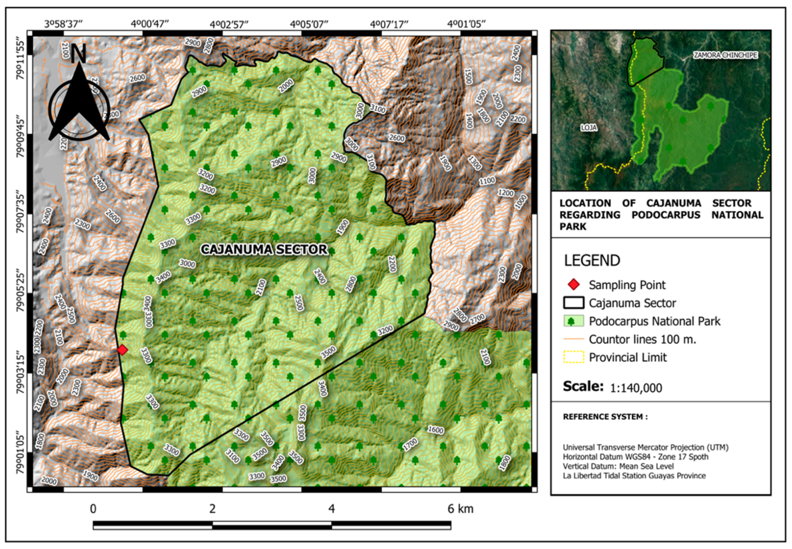
Figure 1. Location of sampling area in Cajanuma within the Podocarpus National Park (PNP); located in the provinces of Loja and Zamora Chinchipe, Ecuador.
Jof 09 00054 g001
Figure 2. Gloeocystidiellum lojanense: (a). basidiome; (b). gloecystidias (black arrowhead) in Meltzer’s reagent; (c,d). gloecystidias (black arrowheads) with granular protoplasmic content in Phloxine; (e). basidiospores in Congo Red. Scale bar = 10 μm except (a) 1 cm.
Jof 09 00054 g002
Figure 3. Gloeocystidiellum lojanense sp. nov. coll. [HUTPL(F)2181] (holotype): (a). basidiospores verrucose and basidiospores smooth (immature); (b). basidia clavate; (c). gloecystidia; (d). hyphae with clamps; (e). calcium oxalate. Scale bars = 10 μm.
Jof 09 00054 g003
Figure 4. Concatenated maximum likelihood phylogenetic tree (ITS1-5.8S-ITS2 and partial LSU D1/D2) for sequence positioning corresponding to Gloeocystidiellum lojanense sp. nov. Values on the nodes correspond to ML and NJ, respectively. Bar = number of substitutions expected per position. * Sequences generated in this study. Sequences and accession numbers of the new species are in bold.
Jof 09 00054 g004
Figure 5. Maximum likelihood phylogenetic tree for the ITS1 region for sequence positioning corresponding to Gloeocystidiellum lojanense sp. nov. and positioning of Gloeocystidiellum rajchenbergii. Values on the nodes correspond to ML and NJ, respectively. Bar = number of expected substitutions per position. * Sequences generated in this study. Sequences and accession numbers of the new species are in bold.
Jof 09 00054 g005
Figure 6. Box plot of the effects of bacteria and time on inhibition: (A): Escherichia coli ATCC 25922; (B): Escherichia coli O157:H7; (C): Escherichia coli UPEC phylogroup A; (D): Escherichia coli UPEC phylogroup B2; (E): Serratia sp.
Jof 09 00054 g006
Table 1. GenBank sequences reference applied in molecular analyses in this study.
TaxonHerbarium/CollectionProvenanceGenBankSequence Region
Gloeocystidiellum aculeatumGB/2647TaiwanAF265546ITS, nuLSU
Gloeocystidiellum aculeatum * GB/2647TaiwanAF506433ITS, nuLSU
Gloeocystidiellum aspellum * GB/LIN625TaiwanAF506432ITS, nuLSU
Gloeocystidiellum aspellumHE4262ChinaKY860460nuLSU
Gloeocystidiellum aspellumHE2041ChinaKY860461nuLSU
Gloeocystidiellum aspellumHE1773ChinaKY860462nuLSU
Gloeocystidiellum bisporumCBS/961.96SwedenAY048875ITS, nuLSU
Gloeocystidiellum bisporumGB/KHL4700SwedenAY048876ITS
Gloeocystidiellum bisporumGB/KHL11135NorwayAY048877ITS, nuLSU
Gloeocystidiellum clavuligerumHHB1046USA, Mich.AF310087ITS, nuLSU
Gloeocystidiellum clavuligerumGB/NH11185Spain, TenerifeAF310088ITS, nuLSU
Gloeocystidiellum clavuligerumHE3313ChinaKY860441nuLSU
Gloeocystidiellum clavuligerumMUCL/33964FranceON833267ITS, nuLSU
Gloeocystidiellum clavuligerumMUCL/33965FranceON833268ITS, nuLSU
Gloeocystidiellum clavuligerumMUCL/33966FranceON833269ITS, nuLSU
Gloeocystidiellum clavuligerumMUCL/33967FranceON833270ITS, nuLSU
Gloeocystidiellum clavuligerumMUCL/33968FranceON833271ITS, nuLSU
Gloeocystidiellum compactumNMNS/WU880615-21TaiwanAF506434ITS, nuLSU
Gloeocystidiellum formosanumNMNS/WU9404-19TaiwanAF506439ITS, nuLSU
Gloeocystidiellum friesiiCBS/323.66FranceMH870446nuLSU
Gloeocystidiellum graminicola * GB/WU9210-12TaiwanAF506448ITS, nuLSU
Gloeocystidiellum heimiiLY/CBS321.66C. African Rep.AF506381ITS, nuLSU
Gloeocystidiellum heimiiLY/CBS321.66C. African Rep.MH858805ITS, nuLSU
Gloeocystidiellum kenyenseTFC/15278PortugalFR878082ITS
Gloeocystidiellum kenyenseTFC/15309PortugalFR878083ITS
Gloeocystidiellum kenyenseMA/Fungi80408PortugalFR878084ITS
Gloeocystidiellum leucoxanthaCBS/454.86USAAF287860nuLSU
Gloeocystidiellum lojanense * HUTPL(F)/2181EcuadorOP377059ITS, nuLSU
Gloeocystidiellum lojanenseHUTPL(F)/550EcuadorOP377083ITS, nuLSU
Gloeocystidiellum luridumHK9808SwedenAF506421ITS, nuLSU
Gloeocystidiellum porosellumHJM8851SwedenAY048878ITS, nuLSU
Gloeocystidiellum porosellumMUCL/35247FranceON833272ITS, nuLSU
Gloeocystidiellum porosellumMUCL/35248FranceON833273ITS, nuLSU
Gloeocystidiellum porosumCBS/51085NetherlandsAF310097ITS, nuLSU
Gloeocystidiellum porosumCBS/27154FranceAF310098ITS, nuLSU
Gloeocystidiellum porosumGB/EB990923SwedenAY048881ITS, nuLSU
Gloeocystidiellum porosumWU1608-176TaiwanLC430908nuLSU
Gloeocystidiellum purpurem * GB/WU9310-45SwedenAF441338ITS, nuLSU
Gloeocystidiellum rajchenbergii * GB/NH16358ChileJQ716940ITS1
Gloeocystidiellum rajchenbergiiGB/NH16348ChileJQ734554ITS1
Gloeocystidiellum rajchenbergiiGB/NH16353ChileJQ734555ITS1
Gloeocystidiellum subasperisporumGB/KHL8695NorwayAF506404ITS, nuLSU
Gloeocystidiellum tristeGB/KHL10334Puerto RicoAF506442ITS, nuLSU
Gloeocystidiellum wakullumO/OSLO-930107TanzaniaAF506443ITS, nuLSU
Lentinellus cochleatusGB/KGN96-09-28SwedenAF506417ITS, nuLSU
Lentinellus ursinusGB/EL73-97USA, N.CAF506419ITS, nuLSU
* Holotypes. Acronyms correspond to: CBS, Centraalbureau voor Schimmelcultures; GB, Herbarium University Gothenburg; HHB, H. H. Burdsall; HK, H. Knudsen; HUTPL(F), Herbario (Fungario) UTPL; LY, Université Claude Bernard Lyon 1; NMNS, National Museum of Natural Science; MA, Real Jardín Botánico; MUCL, Mycothèque UCLouvain; O, Oslo University; TFC, Universidad de La Laguna. Accession numbers to new sequences obtained in this study are in bold.
Table 2. BLAST similarity of basidiomata and fungal strains analyzed in this study.
CodeSourceClosest Identified RelativeQuery Cover (%)Identity (%)Culture From FructificationOrderClosest Identified RelativeQuery Cover (%)Identity (%)
HUTPL(F)2181BasidiomeGloeocystidiellum sp. 9396HUTPL(F)550RussulalesGloeocystidiellum sp.9495.93
HUTPL(F)2162BasidiomeTulasnella sp.9895.53HUTPL(F)551HypocrealesHypocrea sp.9897
HUTPL(F)2166BasidiomeOliveonia sp.9994.2HUTPL(F)552CantharellalesOliveonia sp.9793.79
HUTPL(F)2183BasidiomePhanerochaete sp.9998.11HUTPL(F)553PolyporalesPhanerochaete sp.9897.93
HUTPL(F)2187BasidiomeCeraceomyces sp.9089.1HUTPL(F)554AmylocorticialesCeraceomyces sp.8988.9
HUTPL(F)2198BasidiomeFuscoporia sp.9790.12HUTPL(F)555HymenochaetalesFuscoporia sp.9789.57
HUTPL(F)2203BasidiomeXenasmatella sp.9697.21HUTPL(F)556RussulalesXenasmatella sp.9492.75
Table 3. Comparison of size measurements between representative structures of Gloeocystidiellum lojanense versus the most morphologically related species into the genus Gloeocystidiellum, according the data from Donk [50].
SpeciesBasidiospores (L)Basidiospores (W)Gloeocystidia (L)Gloecystidia (W)Basidia (L)Basidia (W)
Gloeocystidiellum rajchenbergii6–73–3.540–705.5–7.515–205–5.5
Gloeocystidiellum formosanum6.5–7.52.8–3.21006–1216–225–6.5
Gloeocystidiellum compactum5.8–6.53–3.41005–1017–275–6
Gloeocystidiellum aspellum7–83.5–41505–1025–356–7
Gloeocystidiellum lojanense6.5–83.4–4.580–907–825–355–6
Note: L: long, W: wide. The measurements are expressed in µm. Data obtained from the new species in this study are in bold.
Table 4. Interspecific variation percentages between Gloeocystidiellum lojanense and phylogenetically sister species.
SpeciesGloeocystidiellum lojanense
ITS1ITS1-5.8S-ITS2Partial LSU (D1/D2)
Gloeocystidiellum aspellum3.69/3.56%6.90/7.02%0.81/0.78%
Gloeocystidiellum compactum2.94/2.81%6.45/6.59%0.81/0.78%
Gloeocystidiellum formosanum2.94/2.81%6.45/6.59%0.81/0.78%
Gloeocystidiellum rajchenbergii2.94/2.81%--
Other species6.78–21.16/6.39–21.77%8.72–33.23/9.45–28.19%1.36–5.05/1.85–5.75%
Note: Genetic difference values correspond to complete deletion and (/) partial deletion analysis parameters, respectively. (-) Unpublished data.
Table 5. Antibacterial activity per diameter (mm) of inhibition halos generated by Gloeocystidiellum lojanense against the bacterial strains under study.
Inhibition Zone Diameter (mm)
Time (h)
Pathogenic BacteriaC12244872
Enterobacter aerogenes25.67 ± 0.58----
Enterobacter cloacae26.00 ± 0.00----
Escherichia coliATCC 25922 FG: B230.33 ± 0.5820.56 ± 0.3820.78 ± 0.7722.44 ± 0.3822.78 ± 0.38
Escherichia coliO157:H734.33 ± 1.1523.56 ± 0.3823.67 ± 0.5824.33 ± 0.5824.33 ± 0.58
Escherichia coli UPEC FG: A30.00 ± 0.0014.33 ± 0.5817.56 ± 0.7717.78 ± 0.3817.89 ± 0.19
Escherichia coli UPEC FG: B224.33 ± 0.5815.44 ± 0.1915.44 ± 0.1915.89 ± 0.9615.89 ± 0.96
Klebsiella pneumoniae21.00 ± 0.00----
Klebsiella pneumoniae ATCC BAA-170620.00 ± 0.00----
Pseudomonas aeruginosa28.67 ± 0.58----
Serratia sp.17.33 ± 0.5813.44 ± 0.1914.67 ± 0.3315.00 ± 0.5815.11 ± 0.38
The symbol “-” = without activity; C = positive control; SD standard deviation is indicated by ± (n = 9).
Table 6. Two-factor analysis of variance results.
FactorsdfSSMSFp-Value
G. lojanense42.36290.5907836<0.001
Time41.52260.3807538.7<0.001
G. lojanense: Time160.22840.014320.2<0.001
Residuals500.03530.0007
df: degrees of freedom, ss: sum of squares, ms: mean squares, f: f statistic.
Disclaimer/Publisher’s Note: The statements, opinions and data contained in all publications are solely those of the individual author(s) and contributor(s) and not of MDPI and/or the editor(s). MDPI and/or the editor(s) disclaim responsibility for any injury to people or property resulting from any ideas, methods, instructions or products referred to in the content.