Article

Lignin-Modifying Enzymes in Scedosporium Species

by
Wilfried Poirier
*,
Jean-Philippe Bouchara
and
Sandrine Giraud
University of Angers, University of Brest, IRF (Infections Respiratoires Fongiques), SFR ICAT 4208, CEDEX 9, 49933 Angers, France
*
Author to whom correspondence should be addressed.
J. Fungi 2023, 9(1), 105; https://doi.org/10.3390/jof9010105
Submission received: 24 November 2022 / Revised: 6 January 2023 / Accepted: 9 January 2023 / Published: 12 January 2023

Abstract

:
Scedosporium species are usually soil saprophytes but some members of the genus such as S. apiospermum and S. aurantiacum have been regularly reported as causing human respiratory infections, particularly in patients with cystic fibrosis (CF). Because of their low sensitivity to almost all available antifungal drugs, a better understanding of the pathogenic mechanisms of these fungi is mandatory. Likewise, identification of the origin of the contamination of patients with CF may be helpful to propose prophylactic measures. In this aim, environmental studies were conducted demonstrating that Scedosporium species are abundant in human-made environments and associated with nutrient-rich substrates. Although their natural habitat remains unknown, there is accumulated evidence to consider them as wood-decaying fungi. This study aimed to demonstrate the ability of these fungi to utilize lignocellulose compounds, especially lignin, as a carbon source. First, the lignolytic properties of Scedosporium species were confirmed by cultural methods, and biochemical assays suggested the involvement of peroxidases and oxidases as lignin-modifying enzymes. Scedosporium genomes were then screened using tBLASTn searches. Fifteen candidate genes were identified, including four peroxidase and seven oxidase genes, and some of them were shown, by real-time PCR experiments, to be overexpressed in lignin-containing medium, thus confirming their involvement in lignin degradation.

1. Introduction

Fungi are key players in the degradation of lignocellulosic biomass. Besides cellulose and hemicelluloses (mainly xylans), lignin is a macromolecule providing strength as well as rigidity to the plant cell wall and protecting its two polysaccharidic partners against microbial attack by hydrolytic enzymes. Meanwhile, the degradation of cellulose and hemicellulose involves glycosidases (mainly cellobiohydrolases, cellulose cellobiosidases and glycosidases, or xylanases and xylohydrolases, respectively), degradation of lignin is more complex as it is a polyphenolic macromolecule resulting from the polymerization of three types of monolignols (paracoumarylic, coniferylic and sinapylic acids). As a consequence, whereas many microorganisms are able to use cellulose and hemicellulose as the carbon source, only a few groups of fungi are able to degrade lignin, among which the wood-decaying Basidiomycetes, also termed “white-rot fungi”, are the most efficient. They mineralize the complex lignin polymer by the synergistic action of several extracellular enzymes, such as peroxidases and laccases [1].
During the past few decades, the metabolic pathways and the enzymatic mechanisms involved in lignin degradation by Basidiomycetes have been extensively studied [2,3]. Conversely, little is known about the lignocellulose degradation by Ascomycetes. Wood decay by some Ascomycetes was first observed in 1954 by Savory [4,5]. These fungi were designated as “soft-rot fungi” due to their ability to degrade cellulose in notable amounts compared to lignin. Nevertheless, several studies demonstrated that some Ascomycetes may also attack lignin. They are able to mineralize lignin or lignin model compounds, but to a much lower extent than the wood-inhabiting white-rot fungi [6,7,8,9,10,11]. Laccases seem to be key elements in the degradation process. Indeed, in most of the studies, these fungi have been reported to produce laccases whereas no peroxidase activity was detected. Moreover, whereas lignin degradation is considered as secondary metabolism in white-rot fungi, the Sordariomycete Phialemonium inflatum (formerly Paecilomyces inflatus) mineralizes lignin during its initial growth stage, thus during primary metabolism [12].
The Scedosporium species (formerly known as the asexual state of Pseudallescheria species) are soil saprophytic filamentous fungi that are known to determine severe disseminated infections in immunocompromised patients by inhalation of some airborne spores, but also to colonize the respiratory tract of immunocompetent individuals with chronic pulmonary disease. They are notably the second filamentous fungi in terms of frequency in the respiratory tract of patients with cystic fibrosis. These Sordariomycetes exhibit a low susceptibility to current antifungal drugs, and a better understanding of their pathogenic mechanisms is required to identify new therapeutic targets. Likewise, a better knowledge of the origin of the contamination of patients should allow us to propose prophylactic measures. In this context, several environmental studies have been performed which demonstrated that Scedosporium species are mostly found in polluted environments and anthropogenic areas such as agricultural soils, gardens, wastewaters, playgrounds or city parks [13,14,15]. Nevertheless, the natural habitat of these fungi remains to be determined. They are capable to grow in environments with a high osmotic pressure or in poorly aerated soils, to tolerate high salt concentrations [16], and to degrade hydrocarbons. In addition, there is now accumulated evidence to consider them as wood-decaying fungi.
Since it has been reported that filamentous fungi may use the lignin modifying pathway to degrade complex molecules [17,18,19], this work aimed to analyze the lignocellulolytic properties of Scedosporium species.

2. Materials and Methods

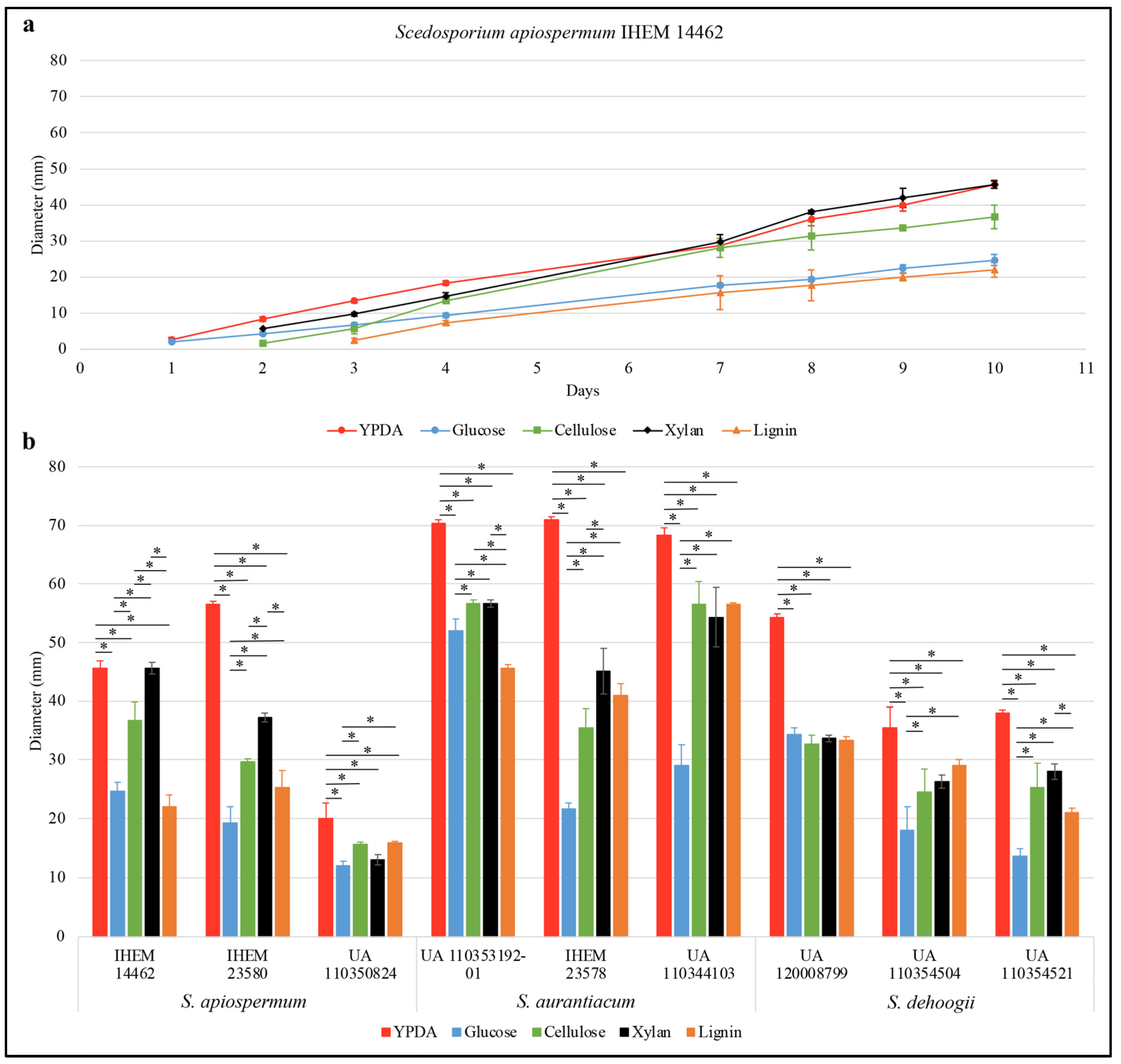
2.1. Isolates and Culture Conditions

The study was conducted on three Scedosporium species. Three isolates per species (including one whole-genome sequenced) were used: S. apiospermum IHEM 14462, IHEM 23580 and UA 110350824; S. aurantiacum IHEM 23578, UA 100353192-01 and UA 110344103; and S. dehoogii UA 120008799, UA 110354504 and UA 110354521. Isolates were maintained by regular passages on YPDA plates (containing in g per liter: yeast extract, 5; peptone, 10; dextrose, 20; agar, 20; and chloramphenicol, 0.5).
Growth studies were carried out in triplicate on a synthetic agar-based medium derived from the Scedo-Select III selective culture medium [20] and containing in g per liter: carbon source, 0.9; ammonium sulphate, 5; potassium dihydrogenophosphate, 1.25; magnesium sulphate, 0.625; agar, 20; and chloramphenicol, 0.5. Different compounds were compared in this culture medium as the unique carbon source: glucose for control conditions, xylan (SERVA Electrophoresis GmbH, Heidelberg, Germany), cellulose (Sigma-Aldrich, Saint Louis, MI, USA) or lignin (Sigma-Aldrich). Mycelium from a 14-day-old YPDA culture was collected using a sterile needle and inoculated by central pricking. Plates were incubated at 37 °C and growth was evaluated by measuring the diameter of the colonies every day for ten days. Results were compared with those obtained on the rich and commonly used medium, YPDA. For statistical analyses, an ANOVA–Tukey test was performed to compare growth at 10 days within each culture conditions, * p-value < 0.05.
For other experiments, isolates were grown on Potato Dextrose Agar (Conda, Madrid, Spain) plates at 37 °C for seven days to enhance sporulation. Conidia were harvested from colonies by aseptically scraping the plates in water and filtration through Miracloth® mesh filter (Merck, Darmstad, Germany) to remove the hyphae, and finally enumerated by hemocytometer counts. To evaluate the impact of the carbon source on the expression level of the target genes, a short duration of incubation (the same for all carbon sources) was needed to avoid degradation of mRMAs. As the germination kinetic vary according to the carbon source, a normalization of the germination step therefore was performed by preincubation of the conidia in YPD medium for 24 h in order to obtain germ tubes. Briefly, 2 × 10⁷ conidia were inoculated in 50 mL of YEPD medium (containing in g per liter: yeast extract, 5; peptone, 10; dextrose, 20; and chloramphenicol, 0.5). After a 24 h-incubation at 37 °C with agitation (120 rpm), germ tubes collected on 11-μm pore-size nylon filters were inoculated in 50 mL of derived Scedo-Select III broth (containing the same components as the agar-based culture medium, except agar) with the appropriate carbon source, which was then incubated with agitation (120 rpm) at 37 °C during 4 h for RNA extraction or 24 h for enzymatic assays.

2.2. Enzymatic Assays

Culture supernatants were collected after 24 h of incubation and preserved by freeze-drying before lyophilization. Lyophilizates were resuspended in 1 mL of saline and used for two-point enzymatic assays. For each enzymatic assay, three independent experiments were realized and all reactions were carried out in duplicate. Protein concentration was determined by fluorimetry using Qubit. The specific activity corresponds to the number of international enzyme units (i.e., µmoles of substrates converted per unit time) per mg of total proteins. According to the data distribution, statistical analysis was performed using a Student’s t-test or Mann–Whitney test.

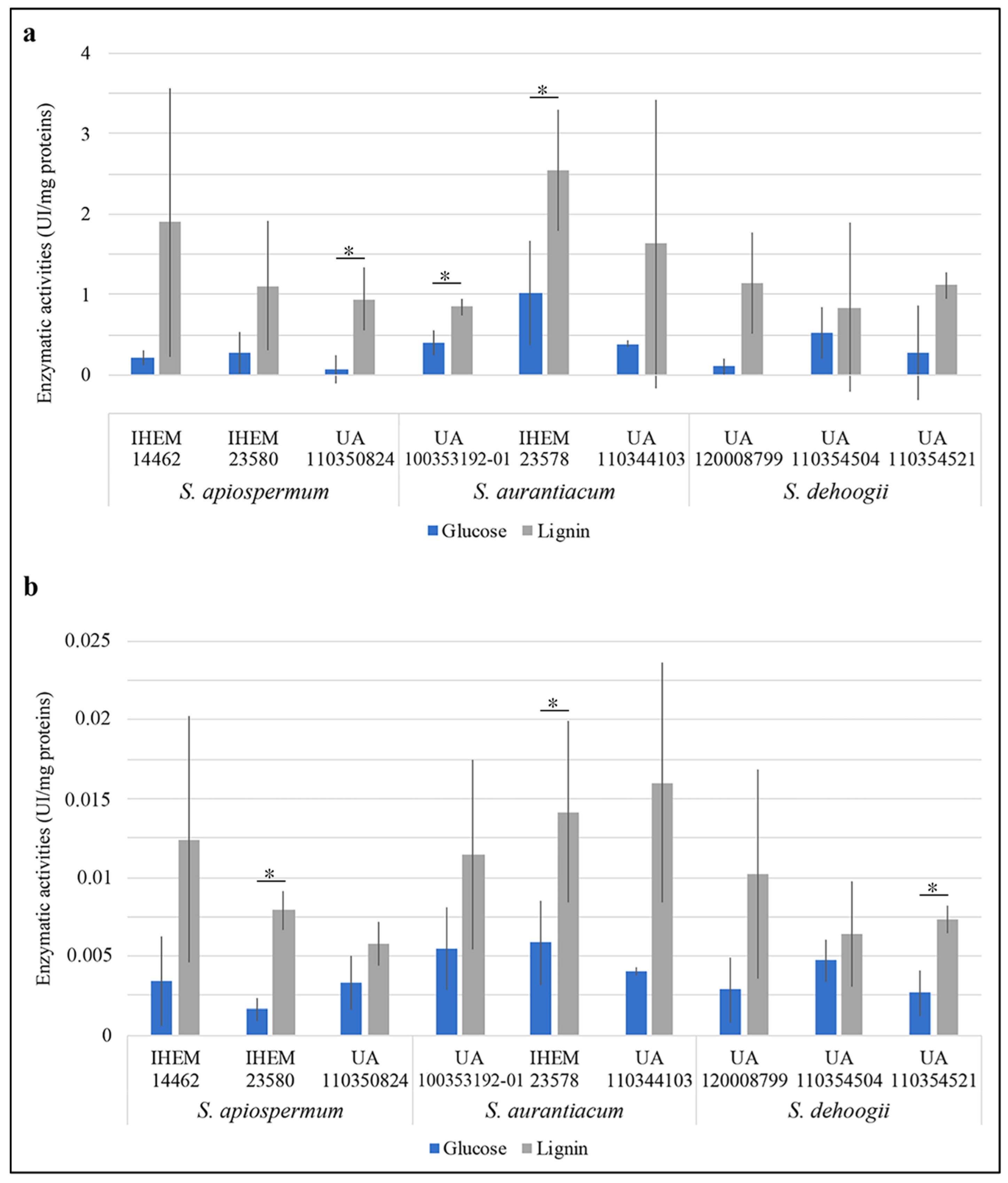
2.2.1. Detection of Peroxidase Activity

Detection of peroxidase activity was performed using the O-PhenyleneDiamine (OPD; Sigma-Aldrich) oxidation test [21]. Two different controls were used: (i) a control lyophilizate obtained from a fungus-free culture medium submitted to the same incubation time and temperature; (ii) a control lyophilizate supplemented with 2.5 μL of horseradish peroxidase-conjugated antibodies (0.5 ng/µL).
The reaction mixture contained OPD (0.75 mg/mL final concentration), 5 μL of hydrogen peroxide 30%, 12.5 μL of lyophilizate and an appropriate volume of 0.1 M citric acid/sodium phosphate buffer pH 5 to reach a final volume of 250 µL. The mix was incubated at room temperature for 5 min, and the reaction was stopped with 62.5 µL of 0.2 M sulfuric acid. OPD oxidation yielded a soluble end-product which was yellow-orange in color. The absorbance was determined at 490 nm. The extinction coefficient used was 1.578 mM−1cm−1, as described previously [22].

2.2.2. Detection of Oxidase Activity

The ability of the isolates to produce extracellular oxidases was quantified using 2-2′-Azinobis(3-ethylBenzoThiazoline-6-Sulphonic acid) (ABTS; Sigma-Aldrich) as described by Shrestha et al. [23]. ABTS working solution consisted in 0.1 mM ABTS and hydrogen peroxide 0.0075% in 0.1 M citric acid/sodium phosphate buffer pH 5. The reaction solution was composed by equal volumes (i.e., 150 μL) of ABTS working solution and samples. A negative control (obtained from a fungus-free culture medium) and a positive control (the same supplemented with a suitable amount of Aspergillus laccase from Sigma-Aldrich) were included in each series of tests. All reactions were incubated at 37 °C for 20 min. The oxidation of ABTS was followed by measurement of the absorption at 420 nm. The molar extinction coefficient used was 36 mM−1cm−1.

2.3. Genome Mining

A literature review was performed to identify lignocellulolytic fungi and then focused on lignin-modifying enzymes. Orthologs of the genes identified by this way were searched in Scedosporium genomes (JOWA01000000 for S. apiospermum, JUDQ01 for S. aurantiacum and PGIR00000000.1 for S. dehoogii) through tBLASTn analysis (https://blast.ncbi.nlm.nih.gov/Blast.cgi, accessed on 10 January 2023). Only results with an E-value < 1 × 10⁻⁶ on at least 40% of the query sequence were considered. Gene organization (exon/intron) was determined using HMMER program on EnsemblFungi (https://fungi.ensembl.org/hmmer/index.html, accessed on 10 January 2023) and validated with Augustus (https://bioinf.uni-greifswald.de/augustus/, accessed on 10 January 2023).
For classification analysis, all peroxidase sequences were collected from PeroxiBase (http://peroxibase.toulouse.inra.fr, accessed on 10 January 2023) [24], and multicopper oxidase (MCO) sequences from the NCBI GenBank [25]. The Geneious software package [26] was used for the phylogenetic study. An alignment was created using the default settings for multiple alignments. Phylogenetic trees were constructed by the neighbor-joining method. Bootstrapping was carried out with 500 replications.

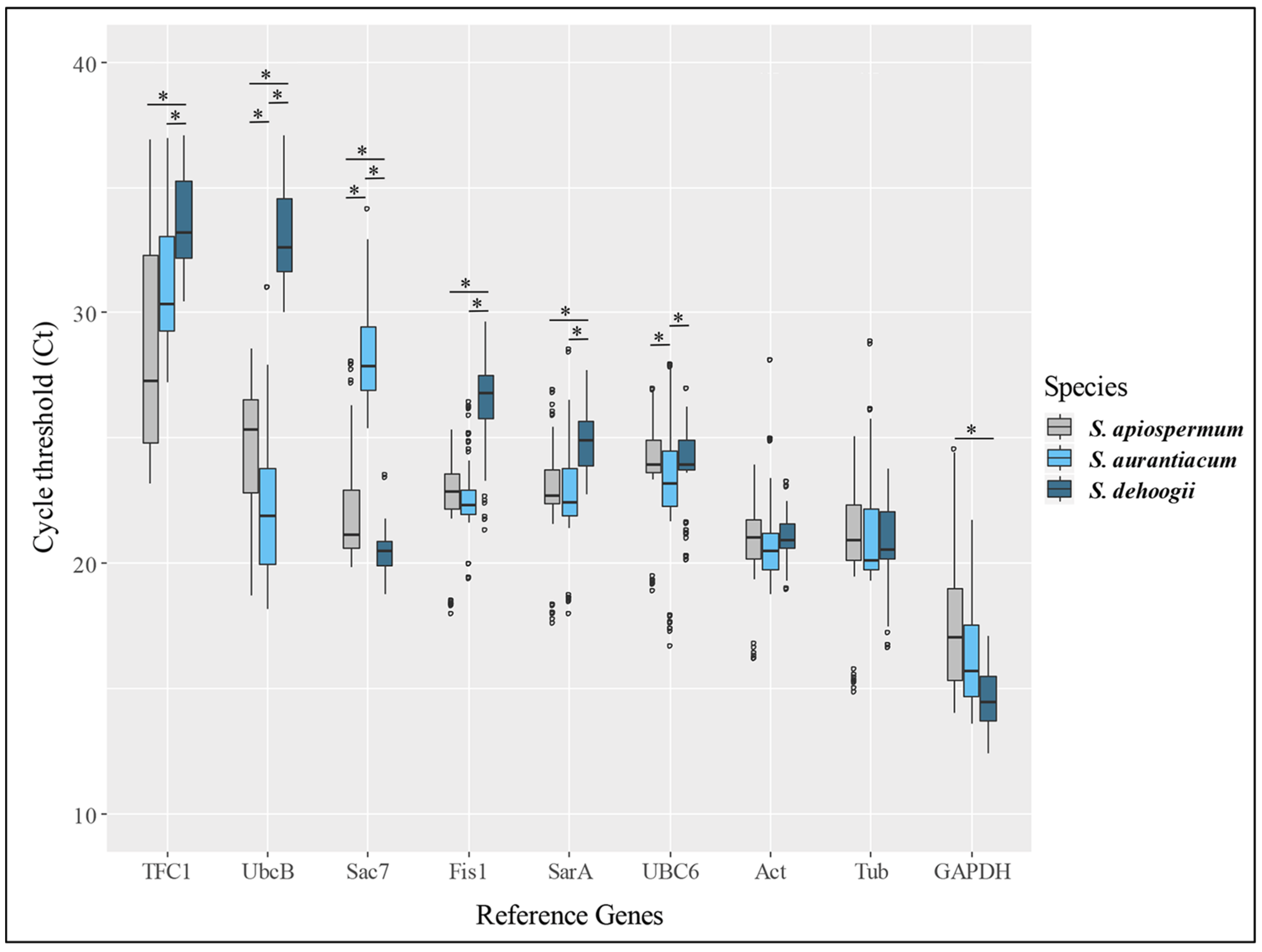
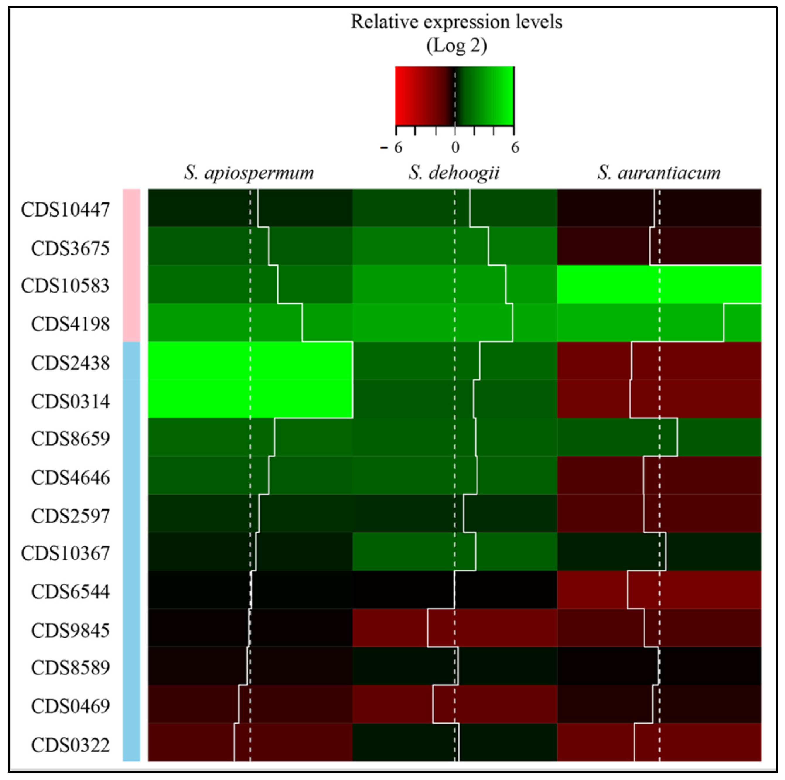
2.4. RNA Isolation, Reverse Transcription and Real-Time Quantitative PCR
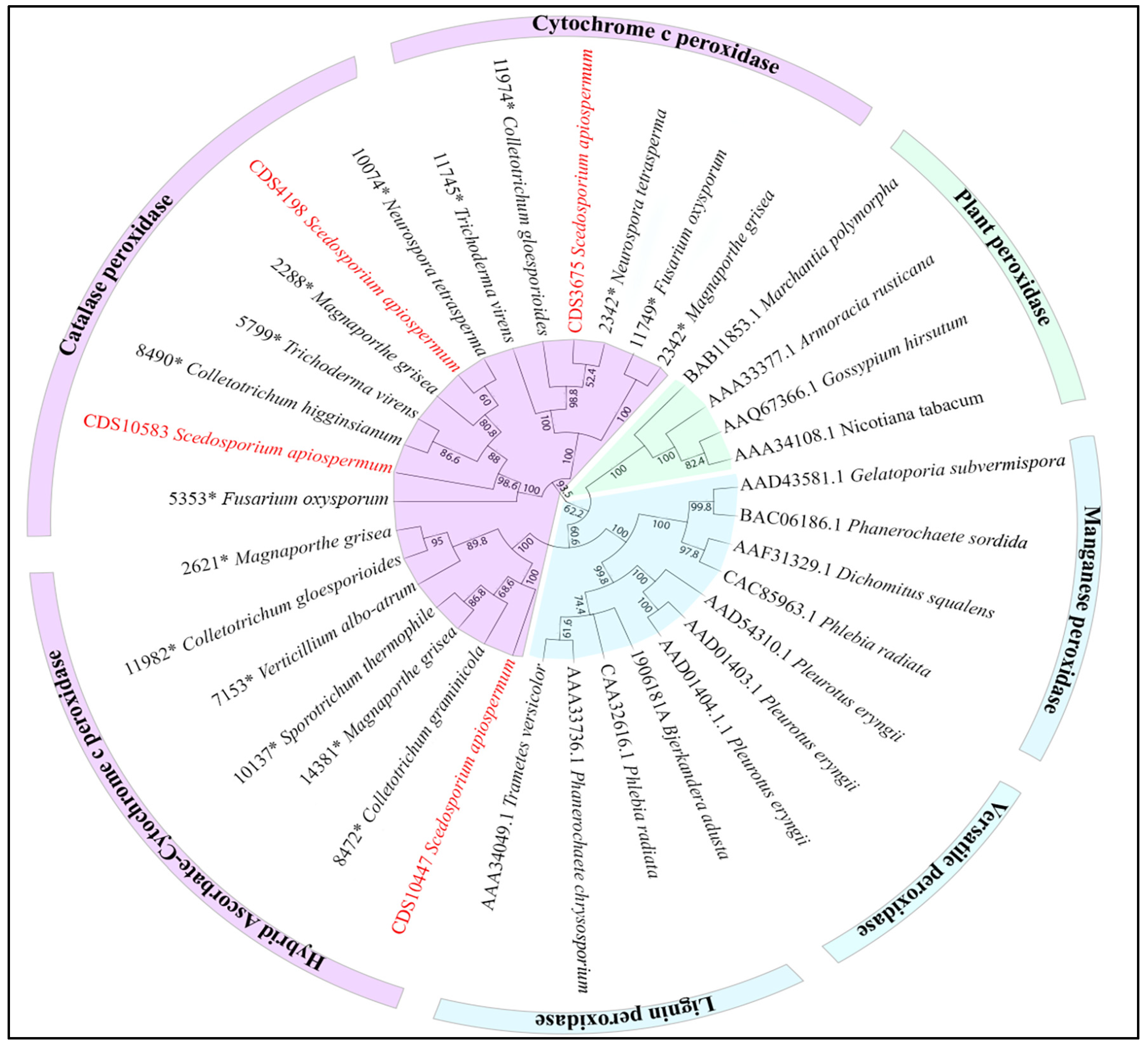
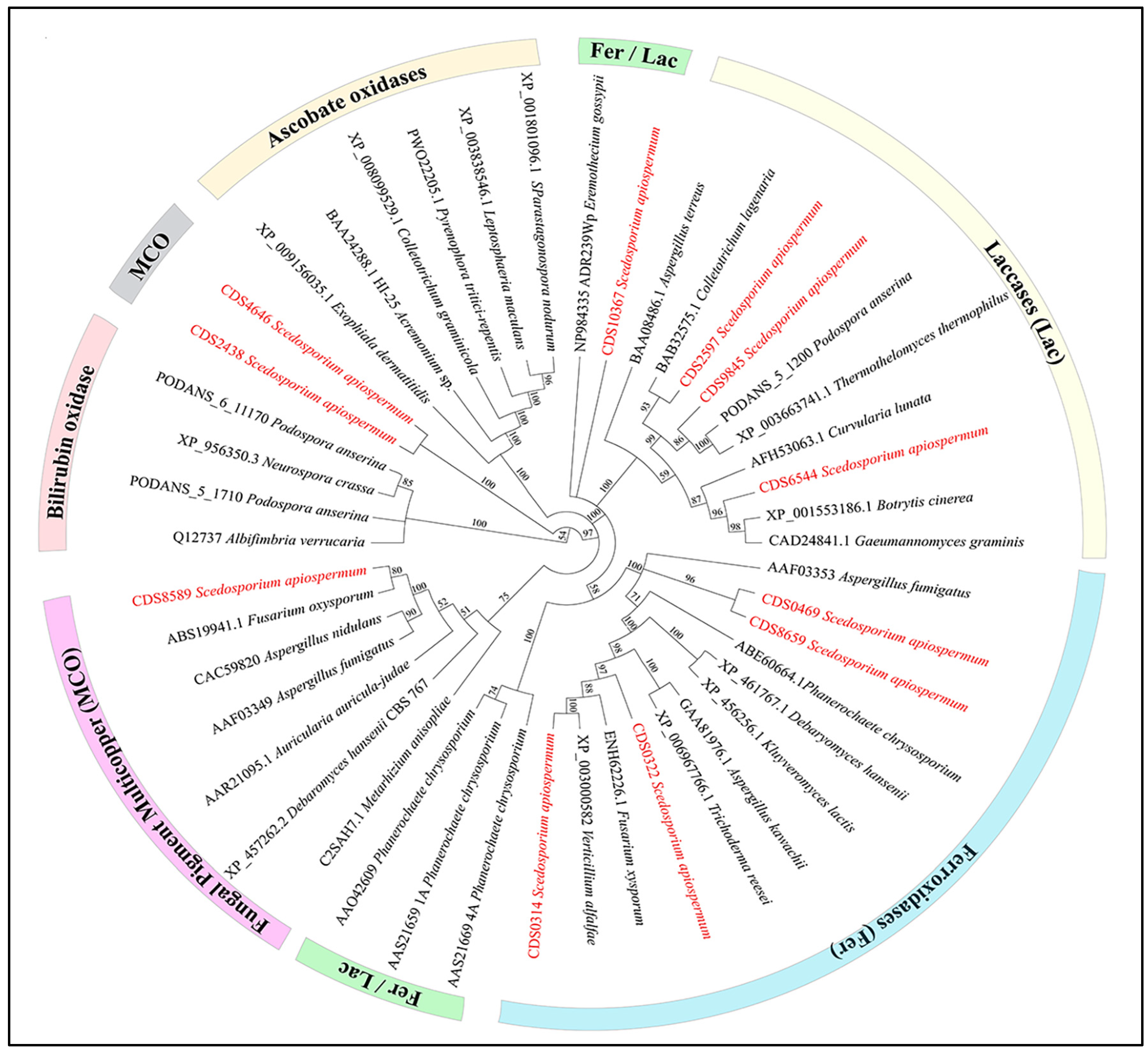
Fungal cells were ground in liquid nitrogen with a mortar and pestle. Total RNAs were extracted using the NucleoSpin® RNA plant kit (Macherey-Nagel, Dueren, Germany), according to the manufacturer’s instructions. RNA samples were then treated with 2 U of RNase-free DNase I (AmbionTM Life Technologies, Carlsbad, CA, USA), following the manufacturer’s recommendations. RNA quantity and quality were evaluated by Qubit dosage and agarose-gel electrophoresis, respectively. Complementary DNA (cDNA) was synthetized from 500 ng total RNA using High Capacity cDNA Reverse Transcription kit (Applied Biosystems, Foster City, CA, USA), and random primers, according to the protocol supplied by the manufacturer. Thereafter, cDNA samples were 10-fold diluted and used as template for real-time quantitative PCR (qPCR). PCR reactions were performed in a final volume of 12.5 μL containing FAST SyBR®Green PCR Master Mix (Applied Biosystems), 200 nM of each primer (Integrated DNA Technologies Inc., Leuven, Belgium) and 2 μL of diluted cDNA. Primers used for qPCR experiments and PCR efficiencies are compiled in Tables S1 and S2 (Supplementary Materials), respectively. qPCR reactions were carried out on a StepOnePlusTM thermocycler (Applied Biosystems) with the following amplification program: 95 °C for 20 s, 40 cycles of 95 °C for 3 s, 60 °C for 30 s. Melting curve analysis (95 °C for 15 s and stepwise annealing from 60 to 95 with 0.3 °C increments) was performed immediately after the amplification. For each gene, fold changes relative to standard condition (i.e., in the presence of glucose as the carbon source) were calculated with the delta–delta Ct method [27,28]. Two reference genes were selected among nine candidates on the basis of their stable expression (validated by an ANOVA–Tukey statistical test) whatever the culture conditions and the species considered [29,30,31,32]. For each point, three biological replicates and two technical replicates were performed and a variation in expression of a given gene was considered significant if the log2 fold change ± standard deviation was <−1 or >1.

3. Results

Figure 1a illustrates the results obtained for S. apiospermum IHEM 14462 isolate. The fungus was not able to develop in the absence of any carbon source (data not shown), and it grew more slowly on a lignin-containing synthetic medium compared to the other culture media (Figure 1a and Figure S1). In addition, for most of the strains, the mean diameter of the colonies after ten days of incubation was significantly higher in the presence of cellulose or xylan compared to the results obtained in the presence of lignin or, strikingly, of glucose (Figure 1b). This suggests that these Scedosporium species are able to assimilate lignocellulosic compounds, including lignin. Therefore, we decided to further investigate this property. During the last few decades, the ability to attack lignin has been found to depend on two main types of enzymatic activities: peroxidase and laccase. In order to quantify these activities, enzymatic assays were performed using OPD and ABTS as substrates for peroxidase and laccase activities, respectively.
There was an important inter-experimental variability (possibly explained by the fact the analysis was conducted during the early phase of hyphal growth—after 24 h of incubation) leading to important standard deviations. Significant differences were observed for three of the strains tested regarding the peroxidase activity and three other strains for laccase activities (Figure 2). Nevertheless, for all the species both enzymatic activities tend to increase in the presence of lignin as carbon source.
Genes reported in the literature as encoding such enzymes were used to screen the genome of S. apiospermum strain IHEM 14462 by tBLASTn searches. Fifteen putative genes were identified, four encoding members of the peroxidase family and the others encoding genes belonging to the MCO superfamily. Among these genes, three were annotated as encoding uncharacterized or hypothetical proteins (SAPIO_CDS2597, SAPIO_CDS8589 and SAPIO_CDS9845) and three others were considered by Augustus as pseudogenes (SAPIO_CDS2438, SAPIO_CDS4198 and SAPIO_CDS4646) because of the lack of introns. To assess whether the genes predicted in silico were actually involved in lignin metabolism, we studied their expression in Scedosporium cells grown for 4 h in a lignin-containing synthetic liquid medium and in control conditions. In this aim, gene structure, especially the position of intronic sequences, was verified when possible and forward/reverse primers were designed from distinct exons (Table S1). PCR efficiency was evaluated and considered as acceptable within a range of 80% to 120% (Table S2).
As the 2−∆∆CT method was used to analyze the relative gene expression, the first step was to select reference genes. Among the nine genes considered as the most interesting for qPCR studies in filamentous fungi [32], seven showed significant differences between the three Scedosporium species tested and two did not show significant variations whatever the culture conditions (Figure 3). Thus, the expression of actin and β-tubulin genes was used for normalization.
Similar expression patterns were obtained for S. apiospermum and S. dehoogii (Figure 4). All peroxidase genes, except SAPIO_CDS10447, were overexpressed compared to control conditions suggesting direct or indirect involvement in lignin degradation. In S. aurantiacum, only two of these genes (SAPIO_CDS4198 and SAPIO_CDS10583) were overexpressed. Considering the MCO superfamily, only SAPIO_CDS8659 was overexpressed in all species. Three other genes were also overexpressed but only in two species S. apiospermum and S. dehoogii: SAPIO_CDS2438, SAPIO_CDS0314 and SAPIO_CDS4646. Finally, SAPIO_CDS10367 was overexpressed only in S. dehoogii.

4. Discussion

This study reports for the first time the ability of Scedosporium species to use each component of lignocellulose (i.e., lignin, cellulose and xylan) independently as a carbon source. Even if results should be confirmed on natural lignin (kraft lignin was used in our experiments), they suggest that the Scedosporium species participates in biomass degradation, an essential role in the Earth’s carbon cycle. Two of the three ecological groups of lignocellulolytic fungi, the white- and brown-rot fungi, mainly belong to Basidiomycetes whereas wood-decaying Ascomycetes are usually included in soft-rot fungi. Basidiomycetes seem to be the only organism capable of an efficient lignin mineralization using specific class II peroxidases (lignin, manganese or versatile peroxidases). In contrast, little is known about the capability of Ascomycetes to attack the complex structure of lignin polymers. In a previous work, we demonstrate that the Scedosporium species displays all the genetic equipment needed for the intracellular degradation of phenolics’ compounds. These lower funneling pathways degrade aromatic compounds, such as those resulting from the extracellular degradation of lignin [33].
Results from our study are a first step for classifying Scedosporium species among the soft-rot fungi. Further characterization of the peroxidase arsenal analyzed in this study should allow definitive exclusion of these Ascomycetes from white-rot fungi. All Scedosporium peroxidases identified by genome mining belonged to class I peroxidase (Figure 5): two, encoded by SAPIO_CDS4198 and SAPIO_CDS10583, were catalase peroxidases, whereas the others belonged to the cytochrome c peroxidase (SAPIO_CDS3675) or the hybrid ascorbate-cytochrome c peroxidase (SAPIO_CDS10447) subclasses. The presence of a C-terminal WSC (cell wall integrity and stress response component) domain allows us to classify the latter among the hybrid-type B peroxidases [34].
Peroxidase and oxidase activities were detected for the three Scedosporium species studied, and they were shown to be markedly increased when Scedosporium isolates were cultivated in the presence of lignin, suggesting their involvement in lignin degradation. Previous studies suggested that Ascomycetes mainly used oxidases as lignolytic enzymes. Five out of the eleven putative genes of the MCO superfamily were overexpressed in at least one Scedosporium species, including two encoding unclassified related-MCO proteins, SAPIO_CDS4646 and SAPIO_CDS2438. The increased gene expression of the canonical MCO of FET3 subfamily SAPIO_CDS0314, the Abr-1-like protein encoded by SAPIO_CDS8659 and the “ferroxidase/laccase” encoded by SAPIO_CDS10367 suggests a link between the lignolytic properties and iron metabolism (Figure 6).
In brown-rot fungi, lignin chemical changes involving small oxidants are largely documented, the Fenton system being the best-known candidate for production of oxidant molecules. Fe2+ reacts with H2O2 to produce water, Fe3+ and usually a hydroxyl radical [35]. The use of extracellular reactive oxygen species (ROS) for lignolytic activity of fungi has been described since the middle of the last century. The mechanism has not been specified, but previous studies evidenced the involvement of ROS production in the attack of lignin in soft-rot fungi, and mineralization of synthetic lignin was decreased by the specific inhibition of hydroxyl radical production [35,36]. Proteins overexpressed in Scedosporium species in lignin-containing medium suggest that such mechanisms may be implicated. Nevertheless, complementary experiments (functional genomic studies and structural analysis of lignin by-products) are required to understand the role and functions of these proteins as well as the chemical reactions involved.
Although the natural habitat of Scedosporium species remains to be determined, indirect evidence suggest an association of the fungi with wood. Several strains of the genus Scedosporium were recovered from submerged woods in estuarian or marine coasts [37,38], from forest soils or wood [16,39] but also from xylophagous insects [40,41,42]. Recent works on Ascomycetes showed that these fungi use the same enzymatic arsenal to degrade lignocellulose and organic pollutants such as aromatic hydrocarbons [17,18,19]. The lignolytic ability of Scedosporium species may also explain their occurrence in polluted environments. In addition, a link between catabolism of hydrocarbons and fungal virulence, suggested for human fungal pathogens [43], was demonstrated in plant pathogenic fungi. For instance, Martins et al. [44] reported an increase in the pathogenic potential in human of some fungi upon exposure to aromatic chlorinated compounds. In plants, Sclerotinia sclerotiorum metabolizes salicylate, a key antifungal defense component of the host [45] and the virulence of Botrytis cinerea on grape is correlated at least in part with its ability to metabolize stilbene-type phytoalexins [46]. A better understanding of Scedosporium species adaptation to their environment and especially polluted environments may allow us to identify interesting pathways as targets for the development of more potent antifungal drugs.

Supplementary Materials

The following supporting information can be downloaded at: https://www.mdpi.com/article/10.3390/jof9010105/s1, Table S1. Primers used in this study; Table S2. PCR efficiency according to Scedosporium species and the primer pair used; Figure S1. Kinetics of growth of Scedosporium aurantiacum UA 100353192-01 (A) and Scedosporium dehoogii IHEM 120008799 (B) on a synthetic agar-based medium Scedo-Select III containing a unique carbon source: glucose for control conditions, lignin, cellulose or xylan.

Author Contributions

W.P. performed the experiments and participated in the writing of the draft; J.-P.B. provided the funding and was responsible of the final version of the paper; S.G. designed and supervised the study and participated in the writing of the draft. All authors have read and agreed to the published version of the manuscript.

Funding

During this work, Wilfried Poirier benefited from a doctoral contract funded by the University of Angers (France), which is gratefully acknowledged.

Institutional Review Board Statement

Although results were presented in part during the 20th international workshop on International Society for Human and Animal Mycology that was held in Amsterdam, Netherlands (on June 2018); the work reported here is original.

Informed Consent Statement

Not applicable.

Data Availability Statement

All the genome sequences used are publicly available and the WGS ID were provided in the manuscript.

Conflicts of Interest

The authors declare no conflict of interest.

References

  1. Hatakka, A. Biodegradation of lignin. In Biopolymers. Biology, Chemistry, Biotechnology, Applications. Vol 1. Lignin, Humic Substances and Coal; Wiley-Blackwell: New York, NY, USA, 2001; pp. 129–180. [Google Scholar]
  2. Martínez, A.T.; Speranza, M.; Ruiz-Dueñas, F.J.; Ferreira, P.; Camarero, S.; Guillén, F.; Martínez, M.J.; Gutiérrez, A.; del Río, J.C. Biodegradation of lignocellulosics: Microbial, chemical, and enzymatic aspects of the fungal attack of lignin. Int. Microbiol. 2005, 8, 195–204. [Google Scholar]
  3. Ruiz-Dueñas, F.J.; Martínez, Á.T. Microbial degradation of lignin: How a bulky recalcitrant polymer is efficiently recycled in nature and how we can take advantage of this. Microb. Biotechnol. 2009, 2, 164–177. [Google Scholar] [CrossRef] [Green Version]
  4. Savory, J.G. Damage to wood caused by microorganisms. J. Appl. Bacteriol. 1954, 17, 213–218. [Google Scholar] [CrossRef]
  5. Savory, J.G. Breakdown of timber by Ascomycetes and Fungi Imperfecti. Ann. Appl. Biol. 1954, 41, 336–347. [Google Scholar] [CrossRef]
  6. Duncan, C.G.; Eslyn, W.E. Wood-decaying Ascomycetes and Fungi Imperfecti. Mycologia 1966, 58, 642. [Google Scholar] [CrossRef]
  7. Ferraz, A.; Baeza, J.; Duránt, N. Softwood biodegradation by an Ascomycete Chrysonilia sitophila (TFB 27441 strain). Lett. Appl. Microbiol. 1991, 13, 82–86. [Google Scholar] [CrossRef]
  8. Norris, D.M. Degradation of C-labeled lignins and C-labeled aromatic acids by Fusarium solani. Appl. Environ. Microbiol. 1980, 40, 376–380. [Google Scholar] [CrossRef] [Green Version]
  9. Rodríguez, A.; Carnicero, A.; Perestelo, F.; de la Fuente, G.; Milstein, O.; Falcón, M.A. Effect of Penicillium chrysogenum on lignin transformation. Appl. Environ. Microbiol. 1994, 60, 2971–2976. [Google Scholar] [CrossRef] [Green Version]
  10. Rodriguez, A.; Perestelo, F.; Carnicero, A.; Regalado, V.; Perez, R.; Fuente, G.; Falcon, M.A. Degradation of natural lignins and lignocellulosic substrates by soil-inhabiting Fungi Imperfecti. FEMS Microbiol. Ecol. 1996, 21, 213–219. [Google Scholar] [CrossRef]
  11. Liers, C.; Ullrich, R.; Steffen, K.T.; Hatakka, A.; Hofrichter, M. Mineralization of 14C-labelled synthetic lignin and extracellular enzyme activities of the wood-colonizing Ascomycetes Xylaria hypoxylon and Xylaria polymorpha. Appl. Microbiol. Biotechnol. 2006, 69, 573–579. [Google Scholar] [CrossRef]
  12. Kluczek-Turpeinen, B.; Tuomela, M.; Hatakka, A.; Hofrichter, M. Lignin degradation in a compost environment by the Deuteromycete Paecilomyces inflatus. Appl. Microbiol. Biotechnol. 2003, 61, 374–379. [Google Scholar] [CrossRef]
  13. Rougeron, A.; Schuliar, G.; Leto, J.; Sitterlé, E.; Landry, D.; Bougnoux, M.-E.; Kobi, A.; Bouchara, J.-P.; Giraud, S. Human-impacted areas of France are environmental reservoirs of the Pseudallescheria boydii/Scedosporium apiospermum species complex. Environ. Microbiol. 2015, 17, 1039–1048. [Google Scholar] [CrossRef]
  14. Kaltseis, J.; Rainer, J.; De Hoog, G.S.; Kaltseis, J.; Rainer, J.; De Hoog, G.S. Ecology of Pseudallescheria and Scedosporium species in human-dominated and natural environments and their distribution in clinical samples. Med. Mycol. 2009, 47, 398–405. [Google Scholar] [CrossRef] [Green Version]
  15. Harun, A.; Gilgado, F.; Chen, S.C.-A.; Meyer, W. Abundance of Pseudallescheria/Scedosporium species in the Australian urban environment suggests a possible source for scedosporiosis including the colonization of airways in cystic fibrosis. Med. Mycol. 2010, 48, S70–S76. [Google Scholar] [CrossRef]
  16. de Hoog, G.S.; Marvin-Sikkema, F.D.; Lahpoor, G.A.; Gottschall, J.C.; Prins, R.A.; Guého, E. Ecology and physiology of the emerging opportunistic fungi Pseudallescheria boydii and Scedosporium prolificans. Mycoses 1994, 37, 71–78. [Google Scholar] [CrossRef]
  17. Cajthaml, T.; Svobodová, K. Biodegradation of aromatic pollutants by ligninolytic fungal strains. In Microbial Degradation of Xenobiotics; Singh, S.N., Ed.; Environmental Science and Engineering; Springer: Berlin/Heidelberg, Germany, 2012; pp. 291–316. ISBN 978-3-642-23788-1. [Google Scholar]
  18. Cajthaml, T. Biodegradation of endocrine-disrupting compounds by ligninolytic fungi: Mechanisms involved in the degradation: Ligninolytic fungi degrade EDCs via various ways. Environ. Microbiol. 2015, 17, 4822–4834. [Google Scholar] [CrossRef]
  19. Korniłłowicz-Kowalska, T.; Rybczyńska, K. Screening of microscopic fungi and their enzyme activities for decolorization and biotransformation of some aromatic compounds. Int. J. Environ. Sci. Technol. 2015, 12, 2673–2686. [Google Scholar] [CrossRef] [Green Version]
  20. Pham, T.; Giraud, S.; Schuliar, G.; Rougeron, A.; Bouchara, J.-P. Scedo-Select III: A new semi-selective culture medium for detection of the Scedosporium apiospermum species complex. Med. Mycol. 2015, 53, 512–519. [Google Scholar] [CrossRef]
  21. Naranjo, L.; Urbina, H.; Sisto, A.D.; Leon, V. Isolation of autochthonous non-white rot fungi with potential for enzymatic upgrading of Venezuelan extra-heavy crude oil. Biocatal. Biotransform. 2007, 25, 341–349. [Google Scholar] [CrossRef] [Green Version]
  22. Romano, M.; Baralle, F.E.; Patriarca, P. Expression and characterization of recombinant human eosinophil peroxidase: Impact of the R286H substitution on the biosynthesis and activity of the enzyme. Eur. J. Biochem. 2000, 267, 3704–3711. [Google Scholar] [CrossRef]
  23. Shrestha, P.; Joshi, B.; Joshi, J.; Malla, R.; Sreerama, L. Isolation and physicochemical characterization of laccase from Ganoderma lucidum-CDBT1 isolated from its native habitat in Nepal. BioMed Res. Int. 2016, 2016, 3238909. [Google Scholar] [CrossRef] [PubMed]
  24. Passardi, F.; Theiler, G.; Zamocky, M.; Cosio, C.; Rouhier, N.; Teixera, F.; Margis-Pinheiro, M.; Ioannidis, V.; Penel, C.; Falquet, L.; et al. PeroxiBase: The peroxidase database. Phytochemistry 2007, 68, 1605–1611. [Google Scholar] [CrossRef] [PubMed]
  25. Hoegger, P.J.; Kilaru, S.; James, T.Y.; Thacker, J.R.; Kues, U. Phylogenetic comparison and classification of laccase and related multicopper oxidase protein sequences. FEBS J. 2006, 273, 2308–2326. [Google Scholar] [CrossRef] [PubMed]
  26. Kearse, M.; Moir, R.; Wilson, A.; Stones-Havas, S.; Cheung, M.; Sturrock, S.; Buxton, S.; Cooper, A.; Markowitz, S.; Duran, C.; et al. Geneious Basic: An integrated and extendable desktop software platform for the organization and analysis of sequence data. Bioinformatics 2012, 28, 1647–1649. [Google Scholar] [CrossRef] [Green Version]
  27. Livak, K.J.; Schmittgen, T.D. Analysis of relative gene expression data using real-time quantitative PCR and the 2−ΔΔCT method. Methods 2001, 25, 402–408. [Google Scholar] [CrossRef]
  28. Pfaffl, M.W. A new mathematical model for relative quantification in real-time RT-PCR. Nucleic Acids Res. 2001, 29, e45. [Google Scholar] [CrossRef]
  29. Vandesompele, J.; Preter, K.D.; Roy, N.V.; Paepe, A.D. Accurate normalization of real-time quantitative RT-PCR data by geometric averaging of multiple internal control genes. Genome Biol. 2002, 3, RESEARCH0034. [Google Scholar] [CrossRef] [Green Version]
  30. Bohle, K.; Jungebloud, A.; Göcke, Y.; Dalpiaz, A.; Cordes, C.; Horn, H.; Hempel, D.C. Selection of reference genes for normalisation of specific gene quantification data of Aspergillus niger. J. Biotechnol. 2007, 132, 353–358. [Google Scholar] [CrossRef]
  31. Teste, M.-A.; Duquenne, M.; François, J.M.; Parrou, J.-L. Validation of reference genes for quantitative expression analysis by real-time RT-PCR in Saccharomyces cerevisiae. BMC Mol. Biol. 2009, 10, 99. [Google Scholar] [CrossRef] [Green Version]
  32. Llanos, A.; François, J.; Parrou, J.-L. Tracking the best reference genes for RT-qPCR data normalization in filamentous fungi. BMC Genom. 2015, 16, 71. [Google Scholar] [CrossRef] [Green Version]
  33. Poirier, W.; Ravenel, K.; Bouchara, J.P.; Giraud, S. Lower funneling pathways in Scedopsorium species. Front. Microbiol. 2021, 12, 630753. [Google Scholar] [CrossRef] [PubMed]
  34. Zámocký, M.; Gasselhuber, B.; Furtmüller, P.G.; Obinger, C. Turning points in the evolution of peroxidase–catalase superfamily: Molecular phylogeny of hybrid heme peroxidases. Cell. Mol. Life Sci. 2014, 71, 4681–4696. [Google Scholar] [CrossRef] [PubMed] [Green Version]
  35. Hatakka, A.; Hammel, K.E. Fungal biodegradation of lignocelluloses. In Industrial Applications; Hofrichter, M., Ed.; Springer: Berlin/Heidelberg, Germany, 2011; pp. 319–340. ISBN 978-3-642-11457-1. [Google Scholar]
  36. Regalado, V.; Perestelo, F.; Rodríguez, A.; Carnicero, A.; Sosa, F.J.; De la Fuente, G.; Falcón, M.A. Activated oxygen species and two extracellular enzymes: Laccase and aryl-alcohol oxidase, novel for the lignin-degrading fungus Fusarium proliferatum. Appl. Microbiol. Biotechnol. 1999, 51, 388–390. [Google Scholar] [CrossRef]
  37. Kirk, P.W. A comparison of saline tolerance and sporulation in marine and clinical isolates of Allescheria boydii Shear. Mycopath. Mycol. Appl. 1967, 33, 65–75. [Google Scholar] [CrossRef]
  38. Azevedo, E.; Caeirao, M.F.; Rebelo, R.; Barata, M. Biodiversity and characterization of marine mycota from Portuguese waters. Anim. Biodivers. Conserv. 2011, 34, 205–215. [Google Scholar] [CrossRef]
  39. Gilgado, F.; Cano, J.; Gené, J.; Guarro, J. Molecular phylogeny of the Pseudallescheria boydii species complex: Proposal of two new species. J. Clin. Microbiol. 2005, 43, 4930–4942. [Google Scholar] [CrossRef] [Green Version]
  40. Nirma, C.; Eparvier, V.; Stien, D. Antifungal agents from Pseudallescheria boydii SNB-CN73 isolated from a Nasutitermes sp. termite. J. Nat. Prod. 2013, 76, 988–991. [Google Scholar] [CrossRef]
  41. Wu, Q.; Jiang, N.; Bo Han, W.; Ning Mei, Y.; Ming Ge, H.; Kai Guo, Z.; Seik Weng, N.; Xiang Tan, R. Antibacterial epipolythiodioxopiperazine and unprecedented sesquiterpene from Pseudallescheria boydii, a beetle (Coleoptera)-associated fungus. Org. Biomol. Chem. 2014, 12, 9405–9412. [Google Scholar] [CrossRef]
  42. Rojas-Jiménez, K.; Hernández, M. Isolation of fungi and bacteria associated with the guts of tropical wood-feeding Coleoptera and determination of their lignocellulolytic activities. Int. J. Microbiol. 2015, 2015, 285018. [Google Scholar] [CrossRef] [Green Version]
  43. Prenafeta-Boldú, F.X.; Summerbell, R.; Sybren de Hoog, G. Fungi growing on aromatic hydrocarbons: Biotechnology’s unexpected encounter with biohazard? FEMS Microbiol. Rev. 2006, 30, 109–130. [Google Scholar] [CrossRef] [Green Version]
  44. Martins, C.; Varela, A.; Leclercq, C.C.; Núñez, O.; Větrovský, T.; Renaut, J.; Baldrian, P.; Silva Pereira, C. Specialisation events of fungal metacommunities exposed to a persistent organic pollutant are suggestive of augmented pathogenic potential. Microbiome 2018, 6, 208. [Google Scholar] [CrossRef] [PubMed]
  45. Penn, C.D.; Daniel, S.L. Salicylate degradation by the fungal plant pathogen Sclerotinia sclerotiorum. Curr. Microbiol. 2013, 67, 218–225. [Google Scholar] [CrossRef] [PubMed]
  46. Sbaghi, M.; Jeandet, P.; Bessis, R.; Leroux, P. Degradation of stilbene-type phytoalexins in relation to the pathogenicity of Botrytis cinerea to grapevines. Plant Pathol. 1996, 45, 139–144. [Google Scholar] [CrossRef]
Figure 1. (a) Kinetics of growth of Scedosporium apiospermum IHEM 14462 on a synthetic agar-based medium Scedo-Select III containing a unique carbon source: glucose for control conditions, lignin, cellulose or xylan. (b) Colony diameter (mm) after 10 days of culture on agar-based media according to the carbon source. Fungal culture collections: IHEM, clinical strains; and UA, clinical or environmental strains. For statistical analyses, an ANOVA–Tukey test was performed to compare growth at 10 days within each culture conditions, * p-value < 0.05.
Jof 09 00105 g001
Figure 2. (a) Peroxidase and (b) oxidase activities (UI/mg proteins) of culture supernatants collected after 24 h of incubation. For each enzymatic assay, three independent experiments were realized and all reactions were carried out in duplicate. According to the data distribution, statistical analysis was performed using a Student’s t-test or Mann–Whitney test, * p-value < 0.05.
Jof 09 00105 g002
Figure 3. Variations in the expression of several reference genes in the different culture conditions studied. For each gene, an ANOVA–Tukey test was performed to analyze the variations between Scedosporium species. * p-value < 0.01; ns: non-significant. Outliers are represented by an empty circle. TFC1: transcription protein on polymerase III promoters; UbcB: ubiquitin carrier protein; Sac7: rho guanosine triphosphatase activator; Fis1: mitochondrial membrane fission protein; SarA: guanosine triphosphate-binding protein; UBC6: protein catabolic process; Act: Actin; Tub: β-tubulin; GAPDH: D-glyceraldehyde-3-phosphate dehydrogenase.
Jof 09 00105 g003
Figure 4. Relative expression level (Log2) of candidate genes in the presence of lignin. For each species, results correspond to the average of RT-qPCR analysis performed on three different isolates. Pink: peroxidases; blue: oxidases. CDS: coding sequence.
Jof 09 00105 g004
Figure 5. Classification of candidate enzymes among fungal peroxidases. An alignment of the protein sequences was created using the default settings for multiple alignments. Phylogenetic trees were constructed by the neighbor-joining method with cladogram presentation. Bootstrapping was carried out with 500 replications. Class I peroxidases are colored in pink, class II in blue and class III in green. Genes studied within Scedosporium species are marked in red. * Identification number from RedoxBase.
Jof 09 00105 g005
Figure 6. Classification of candidate enzymes among fungal MCO. An alignment of the protein sequences was created using the default settings for multiple alignments. Phylogenetic trees were constructed by the neighbor-joining method with cladogram presentation. Bootstrapping was carried out with 500 replications. Ferroxidases (Fer) are colored in blue, Laccases (Lac) in light yellow, Ferroxidases/Laccases (Fer/Lac) in green, Ascorbate oxidases in light orange, Multicopper oxidases (MCO) in grey, Bilirubin oxidase in dark orange and Fungal pigment multicopper in purple. Genes studied within Scedosporium species are marked in red.
Jof 09 00105 g006
Disclaimer/Publisher’s Note: The statements, opinions and data contained in all publications are solely those of the individual author(s) and contributor(s) and not of MDPI and/or the editor(s). MDPI and/or the editor(s) disclaim responsibility for any injury to people or property resulting from any ideas, methods, instructions or products referred to in the content.

