Article

Yeast Diversity in the Qaidam Basin Desert in China with the Description of Five New Yeast Species

1
College of Life Science and Agricultural Engineering, Nanyang Normal University, 1638 Wolong Road, Nanyang 473061, China
2
State Key Laboratory of Mycology, Institute of Microbiology, Chinese Academy of Sciences, Beijing 100101, China
3
College of Life Sciences, University of Chinese Academy of Sciences, No.19 (A) Yuquan Road, Shijingshan District, Beijing 100049, China
4
China General Microbiological Culture Collection Center, Institute of Microbiology, Chinese Academy of Sciences, Beijing 100101, China
*
Authors to whom correspondence should be addressed.
J. Fungi 2022, 8(8), 858; https://doi.org/10.3390/jof8080858
Submission received: 2 August 2022 / Revised: 12 August 2022 / Accepted: 13 August 2022 / Published: 16 August 2022
(This article belongs to the Special Issue Women in Mycology)

Abstract

:
The Qaidam Basin is the highest and one of the largest and driest deserts on Earth. It is considered a mars analog area in China. In contrast to numerous studies concerning its geology, geophysical, and chemistry, relatively few studies have reported microbial diversity and distribution in this area. Here, we investigated culturable yeast diversity in the northeast Qaidam Basin. A total of 194 yeast strains were isolated, and 12 genera and 21 species were identified, among which 19 were basidiomycetous yeasts. Naganishia albida, N. adeliensis, and Filobasidium magnum were the three most dominant species and were distributed in thirteen samples from eight locations. Five new species (Filobasidium chaidanensis, Kondoa globosum, Symmetrospora salmoneus, Teunia nitrariae, and Vishniacozyma pseudodimennae) were found and described based on ITS and D1D2 gene loci together with phenotypic characteristics and physiochemical analysis. Representative strains from each species were chosen for the salt-tolerant test, in which species showed different responses to different levels of NaCl concentrations. Further, the strain from soil can adapt well to the higher salt stress compared to those from plants or lichens. Our study represents the first report of the yeast diversity in the Qaidam Basin, including five new species, and also provides further information on the halotolerance of yeasts from the saline environment in mars analog.

1. Introduction

Mars analogs are places on Earth that possess one or more geological, geochemical, and geomorphic features similar to those found on Mars, either current or past [1,2]. The possibility of the emergence of life on the early Mars stage has been shown due to the presence of liquid water, carbon, electron donors, and acceptors that can drive microbial metabolism [3,4,5,6,7,8]. Due to the expensive and long-term dependent organization of space exploration, terrestrial analogs can serve as models for studies on the habitability of Mars, which has been a priority of Mars science investigations [5]. Generally, the terrestrial analogs on Earth are characterized by extreme environmental conditions, such as hyper-arid desert, saline or hypersaline habitat, thermal acidic spring, and cryogenic and dry Antarctica [9,10,11,12,13,14,15]. The Atacama Desert in Chile, the Mars Desert Research Station in Utah, USA, and Rio Tinto in Huelva, Southwestern Spain are among the famous and well-studied Mars analog environments. Special issues of International Journal of Astrobiology (‘Astrobiology field research in Moon/Mars analog environments’ in 2011), Life (‘Planetary Exploration: Habitats and Terrestrial Analog’ in 2014), Antonie van Leeuwenhoek (‘Microbiology of the Atacama Desert’ in 2018), and Environmental Microbiology (‘Ecophysiology of Extremophiles’ in 2021) have been published for systematical studies on the Mars analog environments on Earth.
Microbial composition and ecology in different Mars analogs have drawn attention due to their ecological and astrobiological importance. Microbiological studies in Mars analogs performed in the past decades have contributed to our better understanding of the strategy and spatiotemporal limit of life on Earth [16,17,18,19,20,21,22,23]. However, these studies mostly focused on bacteria [18,20,24,25], and the diversity and ecology of fungi in Mars analogs remain largely unknown.
Yeasts are unicellular or dimorphic fungi with a stable free-living unicellular state during their life cycles [26]. Yeasts are considered ubiquitous and have been found in highly diversified substrates and environments, including extremely dry or cold habitats [27]. An increasing number of studies have shown that yeasts can inhabit the Antarctic, glacier, acidic, hypersaline, hyper-arid, and desert areas [28,29,30,31,32,33,34]. Nevertheless, the occurrence and diversity of yeasts in extreme environments have not been fully investigated.
The Qaidam Basin is located in the North Tibetan Plateau, China, and is the highest desert in terms of altitude and one of the largest and driest deserts on Earth. It has an average elevation of ~2800 m and is considered a terrestrial analog site of Mars [12]. This area harbors Mars-like extreme environments, e.g., hyper-arid, salt deposits, and high-UV, and similar mineral compositions and geomorphological structures to terrains on Mars [12,35]. The geology, geophysiology, and chemistry of the Qaidam Basin have been well characterized [36,37,38], and microbial diversities focusing on bacteria and archaea in different parts of this area using culture-independent methods have been performed in recent years [39,40,41]. However, the fungal diversity in the Qaidam Basin remains to be revealed.
In this study, we collected soil, soil crust, plant, and lichen samples in the northeast Qaidam Basin in Qinghai province and investigated the occurrence and diversity of yeasts in these samples using a culture-based approach. A total of 21 species, including five new species, were identified from the isolated yeast strains. In addition, the salt-tolerant ability of the representative yeast strains isolated was tested to evaluate their biotechnological potential. To our knowledge, this is the first report of the diversity of culturable yeasts within the Qaidam Basin. The aim of this study is to characterize the yeast community inhabiting this specific environment and identify potential salt-tolerant yeasts that could be of importance for astrobiology and biotechnology.

2. Materials and Methods
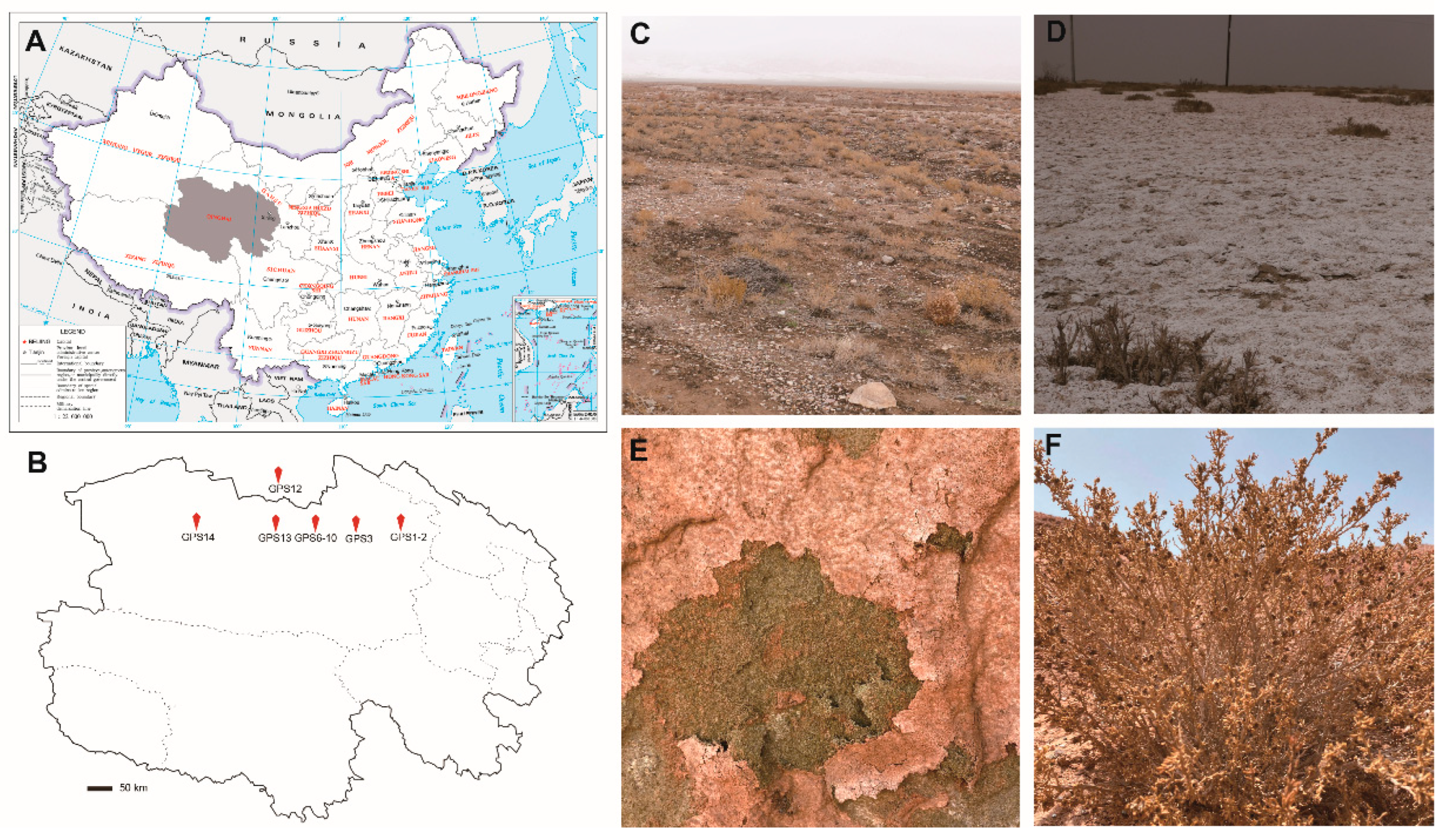
2.1. Sampling Sites

A total of 27 samples, including 19 surface soil and soil crust samples and eight whole plant tissue and lichen samples, were selected from Delingha city in the Qaidam Basin located in Qinghai province in April 2021 (Figure 1 and Table 1). Samples were sealed in sterilized plastic bags and transported to the laboratory at ambient temperatures.

2.2. Soil Chemical Analyses

The soil was suspended 1:5 (w/v) in sterile water and shaken at 180 rpm at 20 °C for 30 min. Soil pH and salinity were determined using the HQ40d Portable Meter (Hach Company, Loveland, CO, USA) (Table 1).

2.3. Yeast Isolation

All samples were processed immediately after returning to the laboratory. Approximately 2 g of soil, soil crust, and fragmented plant and lichen tissue samples were suspended in 10 mL of sterile water and shaken at 180 rpm for 24 h. Aliquots of 0.2 mL of each suspension were spread on potato dextrose agar (PDA, w/v, 20% potato infusion, 2% glucose, 2% agar), 1/2 PDA, and 1/10 PDA plates supplemented with 500 mg L−1 of ampicillin and 500 mg L−1 of streptomycin sulfate. The plates were incubated at 17 °C and 25 °C, respectively, for 3–7 d. All the yeast colony morphotypes were isolated, purified, and preserved in 20% glycerol at −80 °C for further study.

2.4. Molecular Phylogenetic Analysis

Genomic DNA was extracted from yeast cells that were actively growing on a YPD medium (w/v, 1% yeast extract, 2% peptone, 2% glucose, 2% agar) following the protocol described by Makimura et al. [42]. The ITS region (including the 5.8 S rRNA gene) and D1/D2 domain of the LSU rRNA gene were amplified and sequenced using the primers ITS1 and NL4 as described previously [43].
Sequences were inspected and assembled using the SeqMan program in the Lasergene 7 software package (DNASTAR Inc., Madison, WI, USA). They were then aligned using MAFFT version 7 using the G-INS-I algorithm [44]. The alignment was manually edited and trimmed, and minor gaps in all alignments were deleted. Maximum likelihood (ML) and Bayesian analyses were performed for separate and combined gene sequences using RAxML-HPC V.8 [45] and MrBayes 3.2.1 [46] on the CIPRES Science Gateway (http://www.phylo.org (accessed on 23 January 2022)), respectively. ML analysis was implemented with the rapid bootstrap algorithm with 1000 replicates and a subsequent search for the best-scoring tree using the GTRGAMMA model. The best-fit evolution model of separate and combined nucleotide sequences was determined using the Bayesian Information Criterion (BIC) jModeltest [47]. Five million generations were run with four Markov chains, sampling every 5000 generations.
Branches that received bootstrap values (BP) for Maximum likelihood and Bayesian posterior probabilities (BPP) greater than or equal to 70% and 0.95, respectively, were considered as significantly supported. The GenBank accession numbers for the sequences of the ITS region, LSU rRNA gene D1/D2 domain used in this study are listed in Supplementary Table S1. The alignments and trees were deposited in TreeBase (www.treebase.org (accessed on 1 March 2022), No. 29479).

2.5. Phenotypical Characterization

The phenotypic and physiological characters were examined according to standard methods used in yeast taxonomy [48]. The potential sexual cycles of all new species were investigated using CMA (w/v, 2.5% corn meal infusion, 2% agar), PDA, V8 (w/v, 10% V8 juice, 2% agar), and YCB (w/v, 1.17% yeast carbon base, 2% agar). A loopful of cells of each test strain was incubated singly or mixed on an agar plate and incubated at 17 °C for one month. The ballistoconidium-forming activity of all new species was observed by the inverted-plate method [49] on CMA at 17 °C. After 3 to 14 d, the glass slide containing the discharged spores was removed for examination under the microscope.

2.6. Salinity Tolerance Analysis

Salinity tolerance tests were performed on the representative strains of each species using spot plates. All the growth experiments were conducted at 20 °C in YPD agar plates supplemented with 0%, 3%, 6%, 9%, 12%, and 15% (w/v) NaCl, respectively. Briefly, strains were precultured in 3 mL YPD broth for 48 h at 20 °C with shaking at 180 rpm. Optical density (OD) at 600 was measured, and the cell suspension was adjusted to OD 0.8 before a ten-fold dilution series was carried out. An aliquot of 4 μL of each dilution of cell suspension was then spotted onto YPD agar plates supplemented with different concentrations of NaCl and incubated for five days at 20 °C.

2.7. Statistical Analysis

Standard statistical analyses were performed in the R project (v3.3.1) [50]. The statistical significance was calculated using the ANOVA or Kruskal–Wallis test executed by the aov function and kruskal.test function of R package multcomp and FSA, respectively.

3. Results

3.1. Yeast Diversity

Yeast strains were isolated from 10 of the 19 soil samples and six of the eight plant and lichen samples collected from Delingha city in the Qaidam Basin. The soil samples with more than 34.32‰ salinity and the soil crust samples were yeast negative (Table 1). A total of 194 yeast strains were isolated, and the abundance of yeasts varied depending on the substrate, incubation temperature, and medium (Figure 2). A total of 153 and 41 yeast strains were isolated from the plant and lichen tissues and soil samples, respectively. The PDA, 1/2 PDA, and 1/10 PDA agar produced 95, 68, and 31 yeast strains, respectively. A total of 91 and 103 yeast strains were isolated from the plates incubated at 17 °C and 25 °C, respectively (Supplementary Tables S2–S4). Comparative analysis indicated that both the numbers of yeast strains and species from plant and lichen tissue samples were significantly higher than those from soil samples, whereas no significant difference in the abundance and species number was observed regarding the temperature and medium used for yeast isolation (Figure 2). In addition to the shared species, different substrates, temperatures, and media harbored their own yeast species (Figure 2).
The sequences of the D1/D2 domain of the 194 isolated strains were determined. The closest relative of each strain was recognized based on a blast search against the GenBank database using the BLASTn tool. The strains were classified to the species level based on the threshold of >99% sequence identity with the type strain of a described species in the ITS region or D1/D2 domain [51,52,53]. A total of 21 species belonging to 12 genera were identified from the isolated strains. Among the species identified, 19 species were basidiomycetous yeasts, including Dioszegia hungarica, Filobasidium magnum, Kondoa sorbi, Naganishia adeliensis, N. albida, Papiliotrema laurentii, Teunia korlaensis, Udeniomyces puniceus, Vishniacozyma dimennae, V. tephrensis, V. victoriae, and eight undescribed species, i.e., Fibulobasidium sp., Filobasidium sp., Kondoa sp., Symmetrospora sp., Teunia sp., and Vishniacozyma sp. (Supplementary Table S2), and two were ascomycetous yeast species, namely Diutina catenulata and Pichia kudriavzevii. The most abundant yeast genus was Naganishia, followed by Filobasidium and Teunia. The Naganishia species, viz. N. albida and N. adeliensis, accounted for 39.2% of the total number of isolates. To a lesser extent was the genus Filobasidium, of which an undescribed species, Filobasidium sp. formed the majority of the isolates. The strains belonging to the genus Teunia accounted for 9.8% of the total yeast strains isolated, and half belonged to an undescribed species of the genus. On the whole, 24.7% of the isolated strains belonged to undescribed species, and most of these were isolated from plant samples (Supplementary Table S2). Among the 21 species identified, three species, i.e., Filobasidium magnum, Naganishia adeliensis, and N. albida, were the most dominant species, which occurred in seven to eight samples collected from different locations, while thirteen species, i.e., Dioszegia hungarica, Fibulobasidium sp., Kondoa sorbi, Kondoa globosum sp. nov., Kondoa sp. Teunia nitrariae sp. nov., Vishniacozyma dimennae, Vishniacozyma pseudodimennae sp. nov., and Vishniacozyma tephrensis, occurred only in one sample or location (Supplementary Table S2).

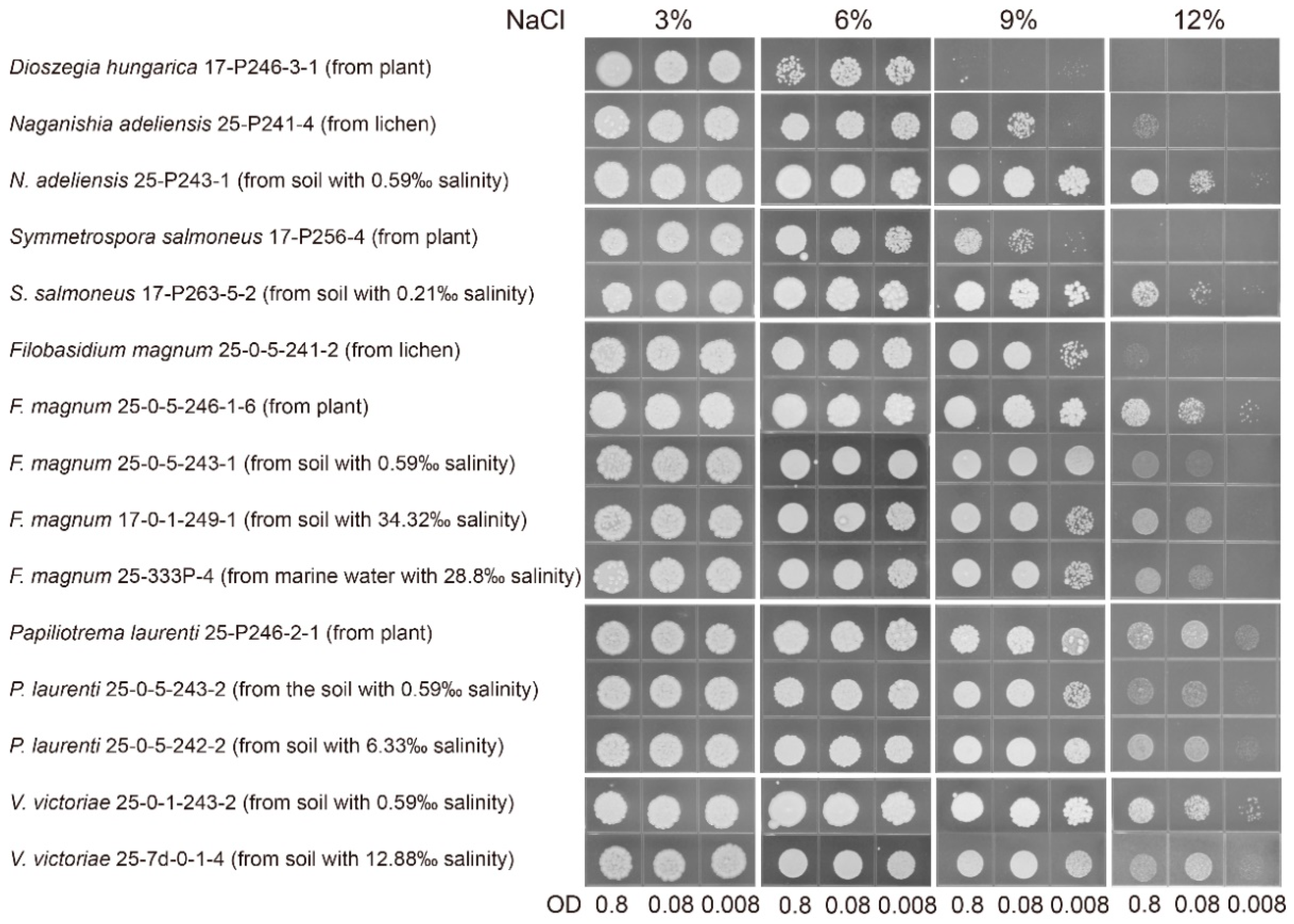
3.2. Salt-Tolerant Ability

Representative strains from lichen, plant, and soil samples were subjected to salt-tolerant ability tests using spot plate assay. Different species had different responses to concentrations of NaCl. In detail, all tested strains of 21 species identified in this study could grow well with NaCl concentrations up to 6% (Supplementary Figure S2), while no strains could grow on the plate with NaCl concentrations of 15% (data not shown). It suggested that their growth was apparently prevented when NaCl concentration increased to 15%. Four species, such as Dioszegia hugarica, Fibulobasidium sp., Kondoa globosum, and Teunia korlaensis, were particularly sensitive to 9% NaCl concentration, while the other species, especially Diutina catenulata, Filobasidium magnum, Kondoa sorbi, Naganishia adeliensis, Papiliotrema laurenti, Pichia kudriavzevii, Symmetrospora salmoneus, and Vishniacozyma victoriae, showed to be unaffected by this level of salt exposure (Supplementary Figure S2). Several strains, such as F. chaidanensis, Kondoa sp. N. albida, Teunia nitrariae, Teunia sp., Udeniomyces puniceus, Vishniacozyma dimennae, V. pseudodimennae, and V. tephrensis, showed some ability to grow at 9% NaCl concentration. In contrast, most strains appeared to be sensitive to exposure to NaCl concentration of 12%. The strains of F. magnum, P. laurenti, and V. victoriae seemed to be relatively unaffected by this level of NaCl exposure (Supplementary Figure S2). These three species exhibited a similar tendency, that is, no difference, in all tested strains in growth ability regardless of their origin from plant and soil samples with different NaCl concentrations, except for the strain of F. magnum from lichen (Figure 3). The strains of N. adeliensis and S. salmoneus from the soil sample showed better growth than those from plant or lichen thallus at 12% NaCl concentration, indicating that the strain from soil samples can adapt well to the higher salt stress.

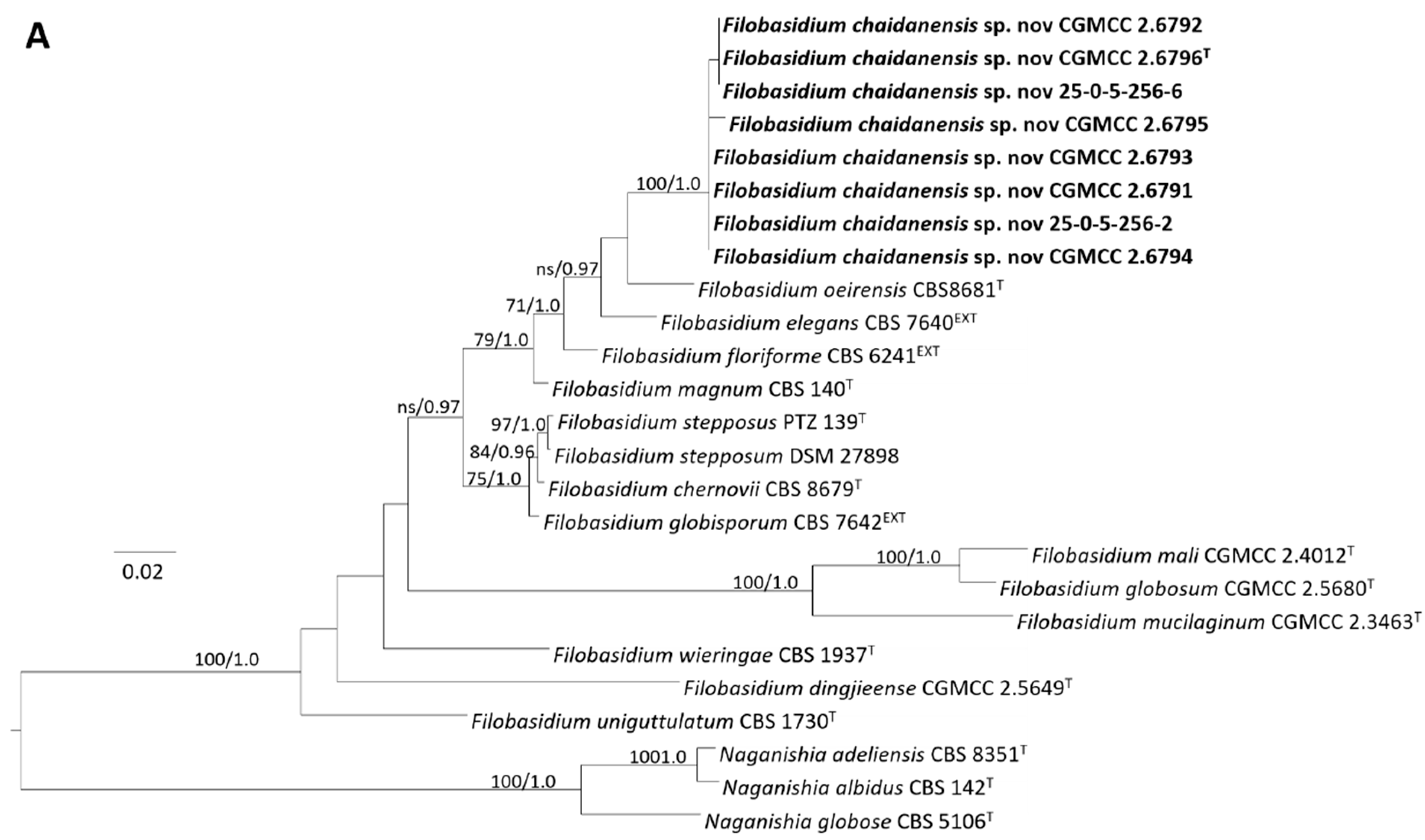
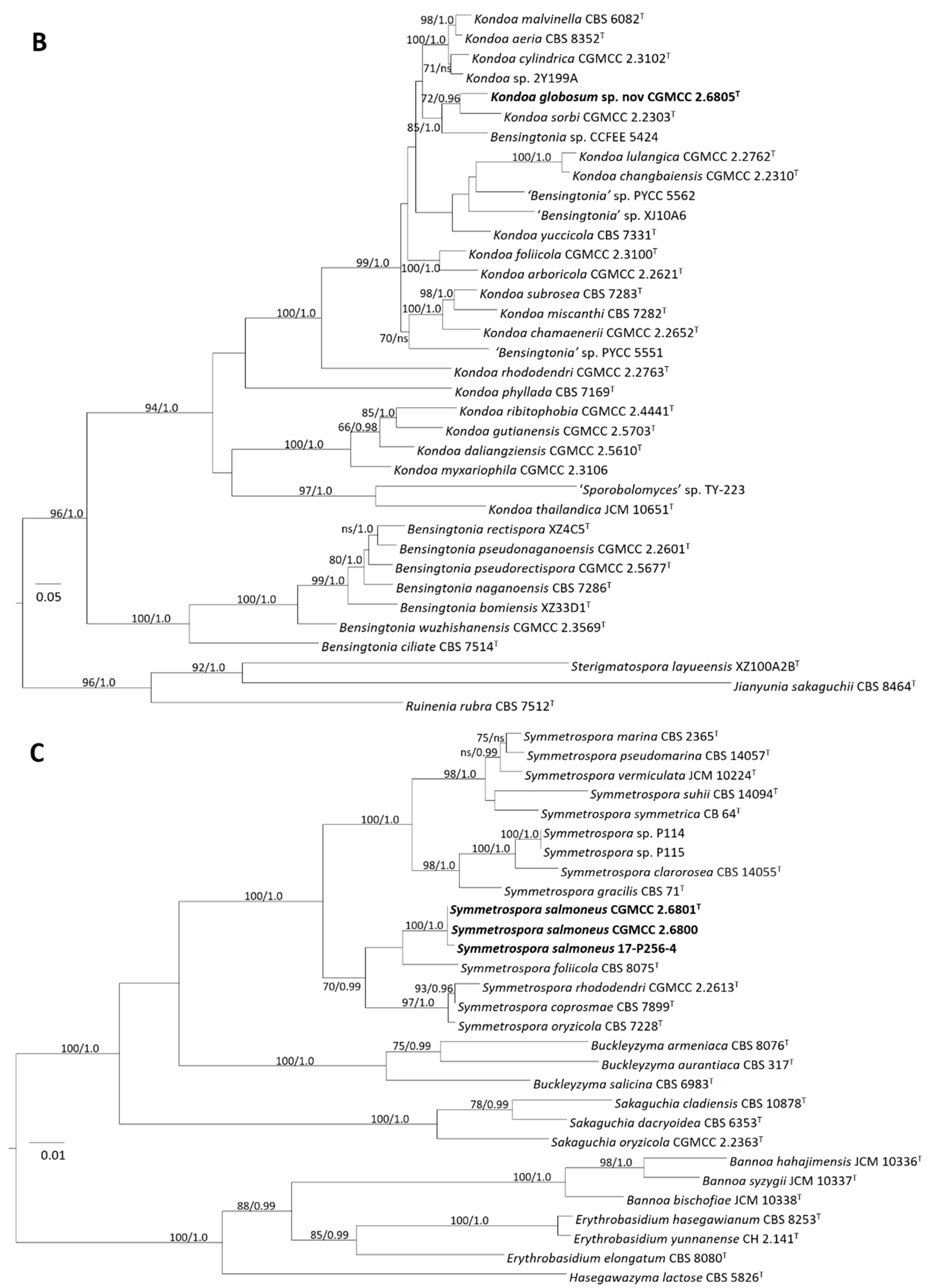
3.3. Phylogeny and Taxonomy of Novel Yeast Species
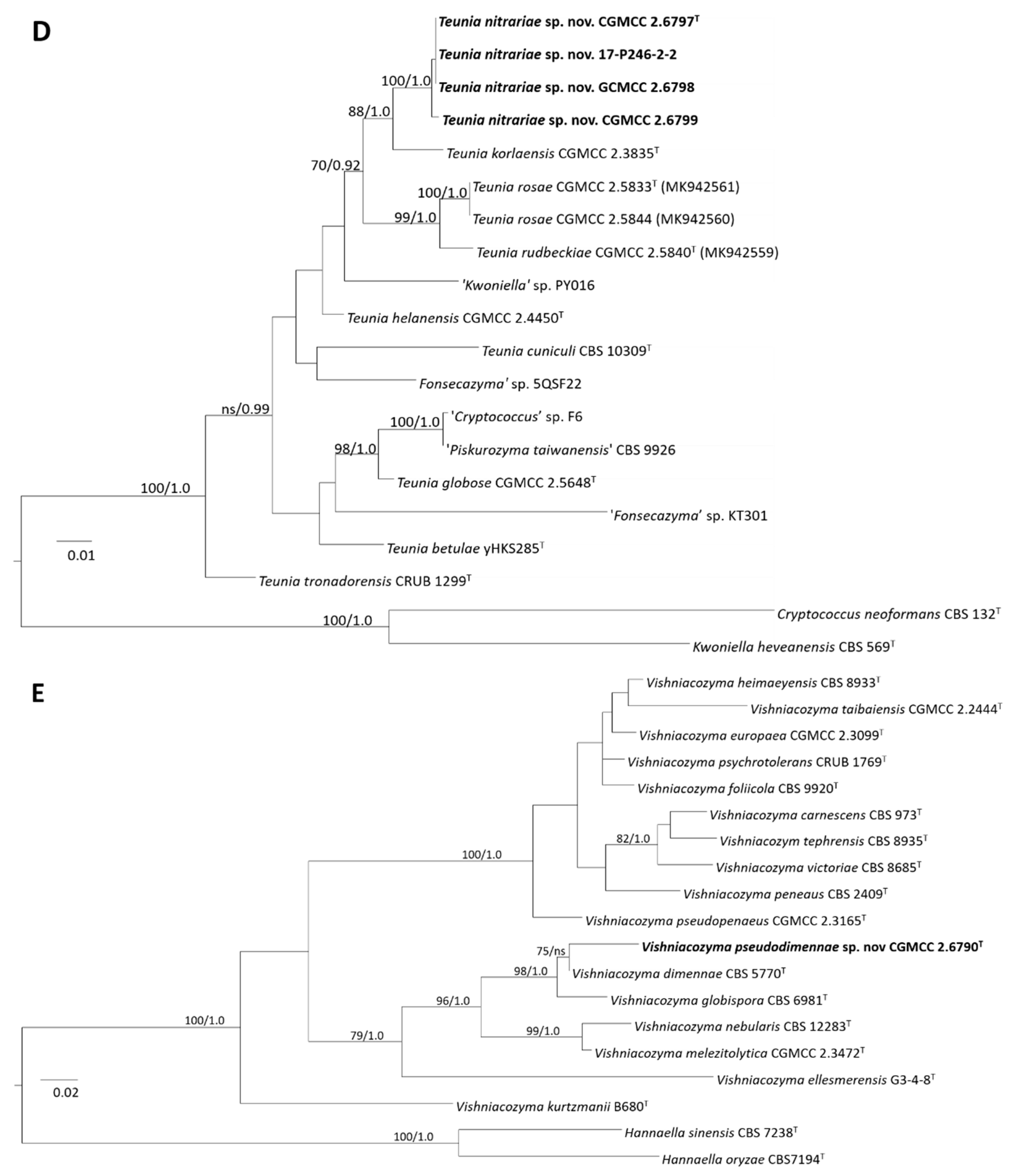
3.3.1. Phylogeny

Among the yeast strains isolated, 43 strains were unable to be identified as any species because of their significant difference from any described yeast species in the sequence of the D1/D2 domain. The ITS sequences of these strains were then determined to confirm their novelty and phylogenetic positions.
A total of 32 strains, represented by CGMCC 2.6791, CGMCC 2.6792, CGMCC 2.6793, CGMCC 2.6794, CGMCC 2.6795, CGMCC 2.6796T, 25-0-5-256-2, and 25-0-5-256-6 isolated from plant tissues formed a separate clade in the genus Filobasidium in the tree obtained from the combined ITS and D1/D2 sequences (Figure 4A). These strains possessed identical ITS and D1/D2 sequences or differed by only one nucleotide in the two regions and thus were conspecific. They differed from the type strain of closely related species F. oeirensis, F. floriforme, F. elegans, and F. magnus by seven to nine nt (~1.1–1.5%) and 26–36 nt (~4.1–5.8%) mismatches in the D1/D2 (Supplementary Figure S1A) and ITS regions, respectively. Therefore, these strains represent a new species in Filobasidium, for which the name F. chaidanensis sp. nov. was proposed.
Strain CGMCC 2.6805T was located in the Kondoa clade and closely related with K. sorbi CGMCC 2.2303T in the tree obtained from the combined ITS and D1D2 sequences (Figure 4B). The two strains differed from each other by 9 nt (~1.4%) and 55 nt (~8.4%) mismatches in the D1/D2 and ITS regions, respectively. An undescribed strain ‘Bensingtonia’ sp. CCFEE 5424 isolated from rock samples [54] and two unpublished strains ‘Pucciniomycotina’ sp. RP185 (AB727159) and RP186 (AB727160) isolated from plant materials in Japan showed close relationships with strain CGMCC 2.6805T (Supplementary Figure S1B), but they differed from the latter by more than 1% bases in the D1/D2 domain. The result suggests that strain CGMCC 2.6805T represents a novel species in the genus Kondoa, for which the name K. globosum sp. nov. was proposed.
Three strains, CGMCC 2.6800, CGMCC 2.6801T, and 17-256-4 isolated from sandy soil and plant tissues possessed similar sequences with no more than one nt difference in the ITS and D1D2 regions, indicating they are conspecific. They were clustered in the Symmetrospora clade with strong support in the tree constructed from the combined ITS and D1/D2 sequences (Figure 4C). S. foliicola CBS 8075T is closely related to these three strains but without significant support (Figure 4C and Supplementary Figure S1C). Ten to eleven nt (~1.6–1.7%) and eighteen nt (~3.1%) differences were observed in the ITS region and D1D2 domain, respectively, between these three strains and S. foliicola. S. rhododendri, S. coprosmae, and S. oryzicola were located as a sister group to the group represented by CGMCC 2.6801T and S. follicola but differed from the group CGMCC 2.6801T by 7–10 nt (~1.3–1.6%) mismatches in the D1D2 domains and more than 7.2% (44–51 bp) mismatches in the ITS regions. Thus, these three strains represent a novel Symmetrospora species, for which the name S. salmoneus sp. nov. was proposed.
Six strains, represented by strain CGMCC 2.6797T, formed a well-supported clade (Figure 4D). Four nt and one nt mismatches in the D1D2 and ITS regions, respectively, were found among these six strains indicating they were conspecific. The closest relative of the CGMCC 2.6797T group is Teunia korlaensis, but differed from the type strain of the latter by eight to nine nt (~1.3–1.5%) in the D1D2 domains and 22 nt (~3.9%) in the ITS regions, respectively. They also had a close relationship with T. rosae in the D1/D2 tree (Supplementary Figure S1D). They differed from the type strain of the latter by three to five nt (~0.5–0.8%) in the D1D2 domains but 38 to 40 nt (~6.8–7.1%) mismatches in the ITS regions, respectively. Therefore, a novel species, Teunia nitrariae, was proposed to accommodate these six strains.
Strain CGMCC 2.6790T was located in the genus Vishniacozyma. It was closely related to Vishniacozyma dimennae (Figure 4E and Supplementary Figure S1E) but differed from the type strain of V. dimennae by ten nt (~1.6%) and 19 nt (~4.0%) mismatches in the D1D2 domains and ITS regions, respectively, suggesting that CGMCC 2.6790T represents a new species in Vishniacozyma, for which the name V. pseudodimennae sp. nov. is proposed.

3.3.2. Taxonomy

Filobasidium chaidanensis X.Z. Liu, F.Y Bai, and X.Y. Wei, sp. nov. (Figure 5A)
MycoBank: MB 842980
Etymology: the specific epithet chaidanensis refers to the geographic origin of the type strain, Chaidan county, Qinghai province.
Culture characteristics: After growth on YM agar for 7 days at 20 °C, cells were globose or subglobose, 3.3–6.6 μm × 2.8–6.7 μm, and occurred singly or in pairs. Budding was polar. After 7 days of growth on YPD agar at 20 °C, the streak culture was whitish to pale greyish-cream, smooth, and butyrous. The margin was entire. In Dalmau plated culture on corn meal agar, pseudohyphae and hyphae were not formed. Sexual structures were not observed on YCB, 5% MEA, PDA, V8, and CMA agar. Ballistoconidia were not produced on corn meal agar.
Physiological and biochemical characteristics: Glucose was not fermented. Glucose, galactose (positive or weak), L-sorbose (weak or slow), sucrose, maltose, cellobiose (positive or delayed), trehalose, lactose, melibiose (weak), raffinose (positive or delayed), melezitose, inulin (weak), soluble starch, D-xylose, L-arabinose, D-arabinose (weak), D-ribose (weak), L-rhamnose, methanol (weak), ethanol (positive or delayed), glycerol (positive or delayed or weak or delayed and weak), ribitol (weak or delayed weak), galactitol (weak), D-mannitol, D-glucitol, Methyl-α-D-glucoside (positive or delayed), salicin, D-gluconate (weak), DL-lactate (weak), succinate, citrate (positive or weak), myo-inositol (weak) and xylitol (positive or delayed) were assimilated as sole carbon sources. D-glucosamine, erythritol, hexadecane, and N-Acetyl-D-glucosamine were not assimilated as sole carbon sources. Ammonium sulfate, potassium nitrate, cadaverine, L-lysine, and ethylamine (variable) were assimilated as sole nitrogen sources. Sodium nitrite was not assimilated as the sole nitrogen source. Growth in the vitamin-free medium was positive. Starch-like compounds were produced. Urease activity was positive. Diazonium Blue B reaction was positive. The maximum growth temperature was 29 °C.
Physiologically, F. chaidanensis differed from its close relatives F. oeirensis, F. floriforme, F. elegans, and F. magnus in the ability to assimilate inulin, methanol, and DL-lactate and its ability to grow in a vitamin-free medium.
Typus: China, Chaidan town, Delingha City, Qinghai province, obtained from undescribed plant tissue, April 2021, X.L. Wei, holotype CGMCC 2.6796 preserved in a metabolically inactive state. Ex-type culture has been deposited in the Japan Collection of Microorganisms (JCM) as JCM 35,461 (=25-P263-B).
Kondoa globosum X.Z. Liu, F.Y Bai, and X.Y. Wei, sp. nov. (Figure 5B)
MycoBank: MB 842983
Etymology: the specific epithet globosum refers to the globose vegetative cells of the type strain.
Culture characteristics: After growth on YM agar for 7 days at 20 °C, cells were subglobose to globose, 4.5–7.6 μm × 4.3–7.3 μm, and occurred singly or in pairs. Budding was polar. After 7 days of growth on YPD agar at 20 °C, the streak culture was cream-colored, smooth, and dull. The margin was entire. In Dalmau plated culture on corn meal agar, pseudohyphae were not formed. Hyphae were seldom formed. Sexual structures were not observed on YCB, 5% MEA, PDA, V8, and CMA agar. Ballistoconidia were not produced on corn meal agar.
Physiological and biochemical characteristics: Glucose was not fermented. Glucose, maltose, cellobiose (slow), trehalose, lactose (weak), melibiose (weak), soluble starch (delayed weak), glycerol (slow), ribitol (slow), D-mannitol (weak), salicin (weak), DL-lactate (weak), and succinate (delayed) were assimilated as sole carbon sources. Galactose, L-sorbose, sucrose, raffinose, melezitose, inulin, D-xylose, L-arabinose, D-arabinose, D-ribose, L-rhamnose, D-glucosamine, methanol, ethanol, erythritol, galactitol, D-glucitol, Methyl-α-D-glucoside, D-gluconate, citrate, myo-inositol, xylitol, hexadecane, and N-Acetyl-D-glucosamine were not assimilated as sole carbon sources. Ammonium sulfate, potassium nitrate, cadaverine, L-lysine, and ethylamine were assimilated as sole nitrogen sources. Sodium nitrite was not assimilated as the sole nitrogen source. Growth in the vitamin-free medium was positive. Starch-like compounds were not produced. Urease activity was positive. Diazonium Blue B reaction was positive. The maximum growth temperature was 28 °C.
Physiologically, K. globosum differed from its close relatives K. sorbi in its ability to assimilate potassium nitrate and ethylamine and inability to assimilate raffinose, D-xylose, L-arabinose, D-glucitol, and D-gluconate.
Typus: China, Chaidan town, Delingha City, Qinghai province, obtained from soil, April 2021, X.L. Wei, holotype CGMCC 2.6805 preserved in a metabolically inactive state. Ex-type culture has been deposited in the Japan Collection of Microorganisms (JCM) as JCM 35,467 (=17-P263-8-2).
Symmetrospora salmoneus X.Z. Liu, F.Y Bai, and X.Y. Wei, sp. nov. (Figure 5C)
MycoBank: MB842985
Etymology: The specific epithet refers to the salmon color of the colony of the strains in this species.
Culture characteristics: After growth on YM agar for 7 days at 20 °C, cells were globose and subglobose, 4.1–6.6 μm × 3.6–6.3 μm, and occurred singly or in pairs. Budding was polar. After 7 days of growth on YPD agar at 20 °C, the streak culture was salmon-pink or orange-colored, smooth, slightly wrinkled, butyrous, and semi-glossy. The margin was entire. In Dalmau plated culture on corn meal agar, pseudohyphae and hyphae were not formed. Hyphae were seldom formed when mixed strains were cultured (Figure 5D). Sexual structures were not observed on YCB, 5% MEA, PDA, V8, and CMA agar. Ballistoconidia were not produced on corn meal agar.
Physiological and biochemical characteristics: Glucose is not fermented. Glucose, galactose (weak), sucrose (weak), maltose (weak), melibiose (weak), soluble starch (weak), D-xylose (weak), L-arabinose (weak), D-ribose (delayed or delayed weak), ethanol (delayed weak), glycerol (delayed or weak), ribitol (weak), D-mannitol (variable), D-glucitol (weak), salicin (delayed weak), DL-lactate (weak), succinate (variable), and xylitol (delayed weak) are assimilated as sole carbon sources. L-sorbose, cellobiose, trehalose, lactose, raffinose, melezitose, inulin, D-arabinose, L-rhamnose, D-glucosamine, methanol, erythritol, galactitol, Methyl-α-D-glucoside, D-gluconate, citrate, myo-inositol, hexadecane, and N-Acetyl-D-glucosamine are not assimilated as sole carbon sources. Ammonium sulfate, potassium nitrate (variable), cadaverine (variable), and ethylamine (variable) are assimilated as sole nitrogen sources. Sodium nitrite and L-lysine are not assimilated as sole nitrogen sources. Growth in the vitamin-free medium is positive (weak). Starch-like compounds are not produced. Urease activity is positive. Diazonium Blue B reaction is positive. The maximum growth temperature is 29 °C.
Physiologically, S. salmoneus differed from its close relatives S. foliicola in the inability to assimilate L-sorbose, cellobiose, trehalose, melezitose, D-arabinose, D-gluconate, and sodium nitrite.
Typus: China, Chaidan town, Delingha City, Qinghai province, isolated from sand, April 2021, X.L. Wei, holotype CGMCC 2.6801 preserved in a metabolically inactive state. Ex-type culture has been deposited in the Japan Collection of Microorganisms (JCM) as JCM 35,466 (=17-P263-5-2).
Teunia nitrariae X.Z. Liu, F.Y Bai, and X.Y. Wei, sp. nov. (Figure 5E)
MycoBank: MB 842986
Etymology: The specific epithet refers to Nitraria, the plant genus from which the type strain was isolated.
Culture characteristics: After growth on YM agar for 7 days at 20 °C, cells were subglobose, globose to ovoid, 3.2–6.4 μm × 2.5–5.7 μm, and occurred singly or in pairs. Budding was polar. After 7 days of growth on YPD agar at 20 °C, the streak culture was yellowish-cream, smooth, and glossy. The margin was entire. In Dalmau plated culture on corn meal agar, pseudohyphae and hyphae were not formed. Sexual structures were not observed on YCB, 5% MEA, PDA, V8, and CMA agar. Ballistoconidia were not produced on corn meal agar.
Physiological and biochemical characteristics: Glucose was not fermented. Glucose, galactose, L-sorbose (variable), maltose (weak), cellobiose, trehalose (positive or weak), lactose (positive or slow), melibiose (weak), soluble starch (variable), D-xylose, L-arabinose, D-arabinose (variable), D-ribose (positive or weak), L-rhamnose (positive or weak), ethanol (delayed weak), glycerol (weak or delayed weak), ribitol (weak or delayed weak), galactitol (delayed), D-mannitol, D-glucitol, salicin (positive or weak), D-gluconate (positive or delayed or delayed weak), DL-lactate (delayed weak), succinate (delayed), myo-inositol (delayed weak), and xylitol (positive or delayed) were assimilated as sole carbon sources. Sucrose, raffinose, melezitose, inulin, methanol, Methyl-α-D-glucoside, citrate, D-glucosamine, erythritol, hexadecane, and N-Acetyl-D-glucosamine were not assimilated as sole carbon sources. Ammonium sulfate, potassium nitrate (variable), cadaverine, L-lysine, and ethylamine (variable) were assimilated as sole nitrogen sources. Sodium nitrite was not assimilated as the sole nitrogen source. Growth in the vitamin-free medium was positive. Starch-like compounds were produced (variable). Urease activity was positive. Diazonium Blue B reaction was positive. The maximum growth temperature was 29 °C.
Physiologically, T. nitrariae differed from its close relatives, T. korlaensis and T. rosae, in the ability to assimilate D-glucitol and L-lysine and inability to assimilate sucrose and melezitose. Thus, it should be proposed as a new species in Tuenia.
Typus: China, Delingha City, Qinghai province, obtained from plant tissue of Nitraria tangutorum, April 2021, X.L. Wei, holotype CGMCC 2.6797 preserved in a metabolically inactive state. Ex-type culture has been deposited in the Japan Collection of Microorganisms (JCM) as JCM 35,462 (=17-P246-2-1).
Vishniacozyma pseudodimennae X.Z. Liu, F.Y Bai, and X.Y. Wei, sp. nov. (Figure 5F)
MycoBank: MB 842987
Etymology: the specific epithet ‘pseudodimennae’ refers to the cell morphology and colony phenotype similar to its closely related species, V. dimennae.
Culture characteristics: After growth on YM agar for 7 days at 20 °C, cells were subglobose to ovoid, 2.6–5.6 μm × 2.4–4.3 μm, and occurred singly or in pairs. Budding was polar. After 7 days of growth on YPD agar at 20 °C, the streak culture was whitish cream, smooth, and slightly glistening. The margin was entire. In Dalmau plated culture on corn meal agar, pseudohyphae and hyphae were not formed. Sexual structures were not observed on YCB, 5% MEA, PDA, V8, and CMA agar. Ballistoconidia were not produced on corn meal agar.
Physiological and biochemical characteristics: Glucose was not fermented. Glucose, sucrose, cellobiose, trehalose (delayed weak), lactose, raffinose, soluble starch (delayed weak), D-xylose (delayed weak), D-arabinose (weak), glycerol (delayed and weak), D-glucitol (delayed and weak), salicin (delayed), D-gluconate, DL-lactate (delayed), myo-inositol (delayed), and xylitol (delayed) were assimilated as sole carbon sources. Galactose, L-sorbose, maltose, melibiose, melezitose, inulin, L-arabinose, D-ribose, L-rhamnose, D-glucosamine, methanol, ethanol, erythritol, ribitol, galactitol, D-mannitol, Methyl-α-D-glucoside, succinate, citrate, hexadecane, and N-Acetyl-D-glucosamine were not assimilated as sole carbon sources. Ammonium sulfate, potassium nitrate, cadaverine, L-lysine, and ethylamine were assimilated as sole nitrogen sources. Sodium nitrite was not assimilated as the sole nitrogen source. Growth in the vitamin-free medium was positive (weak). Starch-like compounds were not produced. Urease activity was positive. Diazonium Blue B reaction was positive. The maximum growth temperature was 30 °C.
Physiologically, V. pseudodimennae differed from its close relative V. dimennae in the ability to assimilate potassium nitrate and inability to assimilate in galactose, L-sorbose, L-arabinose, D-ribose, L-rhamnose, ethanol, ribitol, galactitol, D-mannitol, succinate, and citrate.
Typus: China, Chaidan town, Delingha City, Qinghai province, obtained from undescribed plant tissue, April 2021, X.L. Wei, holotype CGMCC 2.6790 preserved in a metabolically inactive state. Ex-type culture has been deposited in the Japan Collection of Microorganisms (JCM) as JCM 35,455 (=25-0-5-263-4).

4. Discussion

The Qaidam Basin has been proposed as the first Mars analog in China as it harbors a hyper-arid, cold, salt-deposited, and high UV environment similar to the surface of Mars, as well as Mars-like geomorphological structures and mineral composition [12]. It provides an ideal systematic model as a terrestrial analog with astrobiological and extremophile implications. This study reports the culturable yeast diversity in lichen, plants, and soils in the Qaidam Basin for the first time.
Generally, yeasts grow well in a slightly acidic environment, with the optimum pH between 4.5 to 5.5 [55]. Nevertheless, basidiomycetous yeasts, i.e., Naganishia albida, Papiliotrema laurentii, Rhodotorula glutinis, R. mucilaginosa, Sporobolomyces roseus, and Trichosporon asahii, were found to thrive at pH value above 10 [56,57]. In our study, the pH values among the sampling sites varied from 7.54 to 9.74, indicating the alkali nature of the sampling area. Yeasts were obtained in the sampling sites with pH values between 7.65 to 8.85 but not for the soil samples with pH values greater than 9. The salinity of soil samples in our study ranged from 0.16‰ to 58.51‰. Yeasts were detected in ten soil samples with salinity from 0.21‰ and 34.32‰. Meanwhile, no yeasts occurred in seven soil samples with salinity between 0.16‰ to 16.78‰, including two soil crust samples. Though pH and salinity limited the presence of yeast inhabitants to some extent, their presence did not directly correlate with these two abiotic variables. This is probably due to additional abiotic variables affecting yeast number and diversity that have not been captured in this study.
Approximately 85% of strains obtained in our study were tremellomycetous yeast species that are in good agreement with previous studies of other extreme environments [28,29,30,34,58]. Of 21 species identified in this study, six species, such as Diutina catenulata, Filobasidium magnum, Naganishia adeliensis, N. albida, Papiliotrema laurentii, and Vishniacozyma victoriae, had been reported to be resident in extreme environments, especially saline, alkali, cold or dry conditions [59]. The most frequent yeast species were assigned to Naganishia in our study in that about 39.2% of isolates were identified as N. adeliensis and N. albida. The high abundance combining frequent occurrence in more than half of the samples of these two species suggested that they are resident species in the sampling area of our study [60]. Naganishia adeliensis and N. albida are versatile extremophilic species that are frequently found in acidic, alkaline, hypersaline, cold, and arid environments [59]. These two species are found as the dominant species in hypersaline soils surrounding Urmia Lake and the dry sandy soils of the desert [30,57,61]. They are characterized as capsulated yeasts that might contribute to water retention and enhance water availability [62]. Filobasidium magnum was also detected in our study at a higher abundance and frequency (Table S2). Filobasidium magnum, which has been reported from Arctic glaciers [34,63] and plants inhabiting the Negev Desert and the Dead Sea region [64], was observed in our sampling sites in the Qaidam Basin that is characterized by hyper-arid, alkaline, and hypersaline environments. Similarly, strains of F. magnum were also plant-related with frequent isolation from plants in our study, implying this species may help plants to survive in dry areas. This was confirmed by the study where the application of F. magnum to the seed of Brassica alboglabra enhanced its metal resistance and its ability to grow in multi-metal contaminated soil [65]. We also isolated 32 strains of a novel species, Filobasidium chaidanensis sp. nov., from plants, which may own similar ecological functions as F. magnum. Though the other three species, Diutina catenulata, Papiliotrema laurentii, and Vishniacozyma victoriae, were detected in our study with low abundance and frequency, they were most commonly reported in previous studies. P. laurentii and V. victoriae displayed polyextremophilic capability, including acidophilic, alkali-tolerant, halophilic, xerotolerant, and psychrophilic aptitudes [59]. Diutina catenulata is a human pathogen that has been one of the most commonly reported in beach sand and estuarine and sea waters that represent alkali and saline environments [66,67].
Our study also yielded yeast species, namely Dioszegia hungarica, Kondoa sorbi, Pichia kudriavzevii, Teunia korlaensis, Udeniomyces puniceus, Vishniacozyma dimennae, and V. tephrensis, that have not been reported in microbial diversity research of extreme environments. Meanwhile, black yeasts, such as Hortaea werneckii, Phaeotheca triangularis, Trimmatostroma salinum, and Wallemia ichthyophaga, which are the common representatives in the hypersaline environment [59], were not found in our study. Additionally, seven undescribed species, namely Fibulobasidium sp., Kondoa globosum sp. nov., Kondoa sp., Symmetrospora salmoneus sp. nov., Teunia nitrariae sp. nov., Teunia sp., and Vishniacozyma pseudodimennae sp. nov., were found for the first time. Considering their low abundance and rare occurrence, they could be classified as transient species [60]. More impressively, most of these transient species colonized in plants, giving us an apparent indication of their role in plant growth in dry and alkaline soils. On the contrary, plants may help these organisms to survive in harsh conditions by transporting nutrition from soils. With this close association with plants, the transient species may obtain an intermedia to stabilize their colonization in harsh environments and then may evolve as a resident species with high abundance. The yeast diversity was significantly higher in the presence of vegetation in our study than that in the absence of vegetation, which has been proven in the Mars Desert Research Station in Utah, USA [17], probably indicating the important role of plants in the microbial growth and colonization.
Based on the delimitation of halophilic species that yeast strains can survive at salt concentration > 1%, such as seawater [68], the strains from the soil with salinities of 12.88‰ and 34.32‰ in our study have the potential to be halophilic species. Because they were distributed in lichen, plant, and soils with lower salinities, it is intriguing to know the extent of their ability to adapt to salinity variations, which will unravel the phenotypic scales of yeasts under extreme conditions. Along with the increasing NaCl concentration, the number of species that can grow well decreased from 17 species at 9% NaCl concentration to six species at 12% NaCl concentration and zero at 15% NaCl concentration. The four species that can grow at 12% NaCl concentration in our study, i.e., F. magnum, N. adeliensis, P. laurenti, and V. victoriae, have also been frequently found in the salty environment, and some of them from hypersaline lakes have been proved to tolerate 10% to 15% NaCl [30,59]. The intraspecies salt tolerance showed different tendencies. On the one hand, all the tested strains from lichen, plants, and soils have the same salt tolerance ability, such as F. magnum, P. laurenti, and V. victoriae. On the other hand, the adaption to salt stress should be variable inside the species. The strains of N. adeliensis and S. salmoneus from soils have extensive flexibility in changing environments compared to the strains from plants and displayed superior adaption along with elevated salinity. Previous studies indicated that halotolerant yeasts developed a number of structural and metabolic strategies to cope with the harsh environmental condition, including producing resting spores such as ascospores, teliospores, and chlamydospores [59]; adjusting the composition of a phospholipid, unsaturated fatty acid, and sterol of the yeast plasma membrane [69,70]; secreting extracellular cold-related enzymes [71] or redox enzymes to reduce the acidity [72]; synthesizing polysaccharidic capsule to prevent from drying [73]. The dynamic expression patterns of salt-dependent genes, such as ENA P-type ATPases involved in the Na+/K+ transport, Glycerol-3-phosphate dehydrogenase regulating glycerol synthesis, and BAT2, ARG4, or AGP2 involved in amino acid metabolites, has been well studied, which provide genomic-level knowledge on the mechanisms involved in the halotolerant behaviors of yeasts, especially black yeast [69,74,75]. The underlying molecular mechanisms of halotolerance in species other than black yeasts deserve further investigation. It will increase our understanding of microorganisms in the saline environment and identify potential targets of economically important yeasts. To sum up, the biodiversity in Qaidam Basin not only gives us knowledge of life limits but also reserves resources such as halotolerant yeasts that can be used in agriculture as an eco-friendly biofertilizer to elevate crop productivity under saline conditions.

5. Conclusions

This is the first study that reports on the culturable yeast diversity in Delingha city in the northeast Qaidam Basin in Qinghai province, considered to be a mars analog area in China. It contributes to the general knowledge of yeast diversity in Mars analog. A total of 194 yeast isolates assigned to 12 genera and 21 species were found from ten soil samples and six plant and lichen samples with the dominance of basidiomycetous yeasts, which reveals the underestimated importance of basidiomycetous yeasts as inhabitants of the harsh environment of the Qaidam Basin desert. Furthermore, five novel species are described in this study. A significant difference in the number of yeast strains and species from plants (including one lichen sample) and soils was observed, indicating the close association between plants and yeasts. Microorganisms can be beneficial for the growth of the plant. Representative strains from different substrates of each species found in this study were subject to salt-tolerant ability from 0% to 15% NaCl concentrations. Seventeen and five species could grow at 9% and 12% NaCl concentration, while no strains could grow on the plate with 15% NaCl concentrations. Strains from soils can adapt well to the higher salt stress compared to those from plants and lichen. More comprehensive studies on yeast distribution across a wider range of sampling sites would be clearly desirable to better comprehend the distribution, ecology, diversity, and functions of yeast in the Qaidam Basin.

Supplementary Materials

The following supporting information can be downloaded at: https://www.mdpi.com/article/10.3390/jof8080858/s1, Figure S1: The Maximum likelihood tree of (A) Filobasidium, (B) Kondoa, (C) Symmetrospora, (D) Teunia, (E) Vishniacozyma species based on D1D2 sequences; Figure S2: Growth of representative strains of all the species found in this study on YPD plates supplemented with 0%, 3%, 6%, 9%, and 12% (w/v) NaCl. Table S1: GenBank accession numbers of taxa used in phylogenetic analyses; Table S2: Occurrence and distribution of yeast species in different samples collected from the Qaidam Basin in Qinghai province. Table S3: The number of yeast strains isolated from different samples incubated at 17 °C and 25 °C; Table S4: The number of yeast strains isolated from different samples using PDA, 1/2 PDA, and 1/10 PDA media.

Author Contributions

F.-Y.B. and X.-Z.L. conceived and designed the study; X.-Y.W., X.-Z.L., H.-Y.Z. and R.-P.Z. conducted the experiments; X.-Z.L. and L.S. performed statistical analysis and figures; A.-H.L. preserved the resources; Q.-H.N. resources; X.-Z.L. wrote the manuscript draft; F.-Y.B. and X.-Z.L. revised the draft, and all authors approved the final manuscript. All authors have read and agreed to the published version of the manuscript.

Funding

This research was funded by the National Science & Technology Fundamental Resources Investigation Program of China (Grant No. 2019FY100700), National Natural Science Foundation of China (Grant No. 31670020 and 32070096), and the Youth Innovation Promotion Association of the Chinese Academy of Sciences (Grant No. 2017125).

Institutional Review Board Statement

Not applicable.

Informed Consent Statement

Not applicable.

Data Availability Statement

Publicly available datasets were analyzed in this study. All resulting alignments have been deposited in TreeBASE (http://www.treebase.org/ (accessed on 1 March 2022), accession number 29479). All newly generated sequences have been deposited in GenBank (https://www.ncbi.nlm.nih.gov/genbank/ (accessed on 28 January 2022), Table S1). All new taxa have been deposited in Mycobank (https://www.mycobank.org/ (accessed on 13 February 2022)).

Acknowledgments

The authors are very grateful to Xinli Wei for her valuable comments on improving the manuscript and offer of the soil and plant samples; Chengying Jiang for her kindly help with equipment usage.

Conflicts of Interest

The authors declare no conflict of interest. The funders had no role in the design of the study; in the collection, analyses, or interpretation of data; in the writing of the manuscript, or in the decision to publish the results.

References

  1. Farr, T.G. Terrestrial analogs to Mars: The NRC community decadal report. Planet Space Sci. 2004, 52, 3–10. [Google Scholar] [CrossRef]
  2. Léveillé, R.; Mars, A.S. Encyclopedia of Astrobiology; Gargaud, M., Irvine, W.M., Amils, R., Cleaves, H.J., Pinti, D.L., Quintanilla, J.C., Rouan, D., Spohn, T., Tirard, S., Viso, M., Eds.; Springer: Berlin/Heidelberg, Germany, 2011; p. 977. [Google Scholar]
  3. McKay, C.P.; Mancinelli, R.L.; Stoker, C.R.; Wharton, R.A., Jr. The possibility of life on Mars during a water-rich past. In Mars; Kieffer, H.H., Jakosky, B.M., Snyder, C.W., Matthews, M.S., Eds.; The University of Arizona Press: Tucson, AZ, USA, 1992; pp. 1234–1245. [Google Scholar]
  4. Brack, A. Life on Mars: A clue to life on Earth? Chem. Biol. 1997, 4, 9–12. [Google Scholar] [CrossRef]
  5. Fairén, A.; Davila, A.; Darlene, L.; Nathan, B.; Rosalba, B.; Jhony, Z.; Uceda, E.; Carol, S.; Jacek, W.; Dohm, J.; et al. Astrobiology through the ages of Mars: The study of terrestrial analogues to understand the habitability of Mars. Astrobiology 2010, 10, 821–843. [Google Scholar] [CrossRef] [PubMed]
  6. Cockell, C.S. Trajectories of Martian habitability. Astrobiology 2014, 14, 182–203. [Google Scholar] [CrossRef]
  7. Grotzinger, J.P.; Sumner, D.Y.; Kah, L.C.; Stack, K.; Gupta, S.; Edgar, L.; Rubin, D.; Lewis, K.; Schieber, J.; Mangold, N.; et al. A habitable fluvio-lacustrine environment at Yellowknife Bay, Gale Crater, Mars. Science 2014, 343, 1242777. [Google Scholar] [CrossRef]
  8. Westall, F.; Foucher, F.; Bost, N.; Bertrand, M.; Loizeau, D.; Vago, J.L.; Kminek, G.; Gaboyer, F.; Campbell, K.A.; Bréhéret, J.G.; et al. Biosignatures on Mars: What, Where, and How? Implications for the Search for Martian life. Astrobiology 2015, 15, 998–1029. [Google Scholar] [CrossRef]
  9. Mormile, M.R.; Hong, B.Y.; Benison, K.C. Molecular analysis of the microbial communities of Mars analog lakes in Western Australia. Astrobiology 2009, 9, 919–930. [Google Scholar] [CrossRef]
  10. Foing, B.H.; Stoker, C.; Zavaleta, J.; Ehrenfreund, P.; Thiel, C.; Sarrazin, P.; Blake, D.; Page, J.; Pletser, V.; Hendrikse, J.; et al. Field astrobiology research in Moon-Mars analogue environments: Instruments and methods. Int. J. Astrobiol. 2011, 10, 141–160. [Google Scholar] [CrossRef]
  11. Amils, R.; Fernández-Remolar, D.; The Ipbsl Team. Río Tinto: A Geochemical and Mineralogical Terrestrial Analogue of Mars. Life 2014, 4, 511–534. [Google Scholar] [CrossRef]
  12. Xiao, L.; Wang, J.; Dang, Y.; Cheng, Z.; Huang, T.; Zhao, J.; Xu, Y.; Huang, J.; Xiao, Z.; Komatsu, G. A new terrestrial analogue site for Mars research: The Qaidam Basin, Tibetan Plateau (NW China). Earth-Sci. Rev. 2017, 164, 84–101. [Google Scholar] [CrossRef]
  13. Bull, A.T.; Andrews, B.A.; Dorador, C.; Goodfellow, M. Introducing the Atacama Desert. Antonie Leeuwenhoek 2018, 111, 1269–1272. [Google Scholar] [CrossRef] [PubMed]
  14. Cassaro, A.; Pacelli, C.; Aureli, L.; Catanzaro, I.; Leo, P.; Onofri, S. Antarctica as a reservoir of planetary analogue environments. Extremophiles 2021, 25, 437–458. [Google Scholar] [CrossRef] [PubMed]
  15. Azua-Bustos, A.; González-Silva, C.; Fairén, A.G. The Atacama Desert in Northern Chile as an Analog Model of Mars. Front. Astron. Space Sci. 2022, 8, 810426. [Google Scholar] [CrossRef]
  16. Hallsworth, J.E.; Mancinelli, R.L.; Conley, C.A.; Dallas, T.D.; Rinaldi, T.; Davila, A.F.; Benison, K.C.; Rapoport, A.; Cavalazzi, B.; Selbmann, L.; et al. Astrobiology of life on Earth. Environ. Microbiol. 2021, 23, 3335–3344. [Google Scholar] [CrossRef]
  17. Thiel, C.; Ehrenfreund, P.; Foing, B.; Pletser, V.; Ullrich, O. PCR-based analysis of microbial communities during the EuroGeoMars campaign at Mars Desert Research Station, Utah. Int. J. Astrobiol. 2011, 10, 177–190. [Google Scholar] [CrossRef]
  18. Dorador, C.; Fink, P.; Hengst, M.; Icaza, G.; Villalobos, A.S.; Vejar, D.; Meneses, D.; Zadjelovic, V.; Burmann, L.; Moelzner, J.; et al. Microbial community composition and trophic role along a marked salinity gradient in Laguna Puilar, Salar de Atacama, Chile. Antonie Van Leeuwenhoek 2018, 111, 1361–1374. [Google Scholar] [CrossRef] [PubMed]
  19. Santiago, I.F.; Gonçalves, V.N.; Gómez-Silva, B.; Galetovic, A.; Rosa, L.H. Fungal diversity in the Atacama Desert. Antonie Leeuwenhoek 2018, 111, 1345–1360. [Google Scholar] [CrossRef]
  20. Aszalós, J.M.; Szabó, A.; Megyes, M.; Anda, D.; Nagy, B.; Borsodi, A.K. Bacterial Diversity of a High-Altitude Permafrost Thaw Pond Located on Ojos del Salado (Dry Andes, Altiplano-Atacama Region). Astrobiology 2020, 20, 754–765. [Google Scholar] [CrossRef]
  21. Bashir, A.K.; Wink, L.; Duller, S.; Schwendner, P.; Cockell, C.; Rettberg, P.; Mahnert, A.; Beblo-Vranesevic, K.; Bohmeier, M.; Rabbow, E.; et al. Taxonomic and functional analyses of intact microbial communities thriving in extreme, astrobiology-relevant, anoxic sites. Microbiome 2021, 9, 50. [Google Scholar] [CrossRef]
  22. O’Connor, B.R.W.; Fernández-Martínez, M.Á.; Léveillé, R.J.; Whyte, L.G. Taxonomic Characterization and Microbial Activity Determination of Cold-Adapted Microbial Communities in Lava Tube Ice Caves from Lava Beds National Monument, a High-Fidelity Mars Analogue Environment. Astrobiology 2021, 21, 613–627. [Google Scholar] [CrossRef]
  23. Wu, D.; Senbayram, M.; Moradi, G.; Mörchen, R.; Knief, C.; Klumpp, E.; Jones, D.L.; Well, R.; Chen, R.; Bol, R. Microbial potential for denitrification in the hyperarid Atacama Desert soils. Soil Biol. Biochem. 2021, 157, 108248. [Google Scholar] [CrossRef]
  24. Azua-Bustos, A.; Caro-Lara, L.; Vicuña, R. Discovery and microbial content of the driest site of the hyperarid Atacama Desert, Chile. Environ. Microbiol. Rep. 2015, 7, 388–394. [Google Scholar] [CrossRef] [PubMed]
  25. Hynek, B.M.; Rogers, K.L.; Antunovich, M.; Avard, G.; Alvarado, G.E. Lack of Microbial Diversity in an Extreme Mars Analog Setting: Poás Volcano, Costa Rica. Astrobiology 2018, 18, 923–933. [Google Scholar] [CrossRef] [PubMed]
  26. Boekhout, T.; Fonseca, Á.; Sampaio, J.P.; Bandoni, R.J.; Fell, J.W.; Kwon-Chung, K.J. Discussion of teleomorphic and anamorphic basidiomycetous yeasts. In The Yeasts: A Taxonomic Study; Kurtzman, C.P., Fell, J.W., Boekhout, T., Eds.; Elsevier: Amsterdam, The Netherlands, 2011; pp. 1339–1372. [Google Scholar]
  27. Groenewald, M.; Boundy-Mills, K.; Čadež, N.; Endoh, R.; Jindamorakot, S.; Pohl-Albertyn, C.; Rosa, C.A.; Turchetti, B.; Yurkov, A. Census of yeasts isolated from natural ecosystem and conserved in worldwide collections. In Yeasts in Natural Ecosystems: Diversity; Buzzini, P., Lachance, M.A., Yurkov, A., Eds.; Springer: Berlin/Heidelberg, Germany, 2017; pp. 455–476. [Google Scholar]
  28. Gadanho, M.; Libkind, D.; Sampaio, J.P. Yeast diversity in the extreme acidic environments of the Iberian Pyrite Belt. Microb. Ecol. 2006, 52, 552–563. [Google Scholar] [CrossRef]
  29. Pulschen, A.A.; Rodrigues, F.; Duarte, R.T.; Araujo, G.G.; Santiago, I.F.; Paulino-Lima, I.G.; Rosa, C.A.; Kato, M.J.; Pellizari, V.H.; Galante, D. UV-resistant yeasts isolated from a high-altitude volcanic area on the Atacama Desert as eukaryotic models for astrobiology. Microbiologyopen 2015, 4, 574–588. [Google Scholar] [CrossRef]
  30. Mokhtarnejad, L.; Arzanlou, M.; Babai-Ahari, A.; Di Mauro, S.; Onofri, A.; Buzzini, P.; Turchetti, B. Characterization of basidiomycetous yeasts in hypersaline soils of the Urmia Lake National Park, Iran. Extremophiles 2016, 20, 915–928. [Google Scholar] [CrossRef]
  31. Buzzini, P.; Turk, M.; Perini, L.; Turchetti, B.; Gunde-Cimerman, N. Yeasts in polar and subpolar habitats. In Yeasts in Natural Ecosystems: Diversity; Buzzini, P., Lachance, M.A., Yurkov, A., Eds.; Springer: Berlin/Heidelberg, Germany, 2017; pp. 331–365. [Google Scholar]
  32. Mitsuya, D.; Hayashi, T.; Wang, Y.; Tanaka, M.; Okai, M.; Ishida, M.; Urano, N. Isolation of aquatic yeasts with the ability to neutralize acidic media, from an extremely acidic river near Japan’s Kusatsu-Shirane Volcano. J. Biosci. Bioeng. 2017, 124, 43–46. [Google Scholar] [CrossRef]
  33. Sannino, C.; Tasselli, G.; Filippucci, S.; Turchetti, B.; Buzzini, P. Yeasts in nonpolar cold habitats. In Yeasts in Natural Ecosystems: Diversity; Buzzini, P., Lachance, M.A., Yurkov, A., Eds.; Springer: Berlin/Heidelberg, Germany, 2017; pp. 366–396. [Google Scholar]
  34. Luo, B.; Sun, H.; Zhang, Y.; Gu, Y.; Yan, W.; Zhang, R.; Ni, Y. Habitat-specificity and diversity of culturable cold-adapted yeasts of a cold-based glacier in the Tianshan Mountains, northwestern China. Appl. Microbiol. Biotechnol. 2019, 103, 2311–2327. [Google Scholar] [CrossRef]
  35. Anglés, A.; Li, Y. The western Qaidam Basin as a potential Martian environmental analogue: An overview. J. Geophys. Res.-Planets 2017, 122, 856–888. [Google Scholar] [CrossRef]
  36. Ding, Z.J.; Zhao, J.N.; Wang, J.; Lai, Z.P. Yardangs on Earth and implications to Mars: A review. Geomorphology 2020, 364, 107230. [Google Scholar] [CrossRef]
  37. Chen, L.; Xu, Y.; Li, B. Comparative study of the geomorphological characteristics of valley networks between Mars and the Qaidam Basin. Remote Sens. 2021, 13, 4471. [Google Scholar] [CrossRef]
  38. Liu, W.; Li, J.; Gu, W.; Santos, L.F.E.D.; Boman, J.; Zhang, X.; Tang, M.; Wang, S.; Kong, X. Chemical and hygroscopic characterization of surface salts in the Qaidam Basin: Implications for climate impacts on Planet Earth and Mars. ACS Earth Space Chem. 2021, 5, 651–662. [Google Scholar] [CrossRef]
  39. Zhong, Z.P.; Liu, Y.; Miao, L.L.; Wang, F.; Chu, L.M.; Wang, J.L.; Liu, Z.P. Prokaryotic Community Structure Driven by Salinity and Ionic Concentrations in Plateau Lakes of the Tibetan Plateau. Appl. Environ. Microbiol. 2016, 82, 1846–1858. [Google Scholar] [CrossRef] [PubMed]
  40. Cheng, Z.; Xiao, L.; Wang, H.; Yang, H.; Li, J.; Huang, T.; Xu, Y.; Ma, N. Bacterial and archaeal lipids recovered from subsurface evaporites of Dalangtan Playa on the Tibetan Plateau and their astrobiological implications. Astrobiology 2017, 17, 1112–1122. [Google Scholar] [CrossRef]
  41. Zhang, W.; Bahadur, A.; Zhang, G.; Zhang, B.; Wu, X.; Chen, T.; Liu, G. Diverse bacterial communities from Qaidam Basin of the Qinghai-Tibet Plateau: Insights into variations in bacterial diversity across different regions. Front. Microbiol. 2020, 11, 554105. [Google Scholar] [CrossRef]
  42. Makimura, K.; Murayama, S.Y.; Yamaguchi, H. Detection of a wide range of medically important fungi by the polymerase chain reaction. J. Med. Microbiol. 1994, 40, 358–364. [Google Scholar] [CrossRef]
  43. Liu, X.Z.; Wang, Q.M.; Theelen, B.; Groenewald, M.; Bai, F.Y.; Boekhout, T. Phylogeny of tremellomycetous yeasts and related dimorphic and filamentous basidiomycetes reconstructed from multiple gene sequence analyses. Stud. Mycol. 2015, 81, 1–26. [Google Scholar] [CrossRef]
  44. Katoh, K.; Standley, D.M. MAFFT multiple sequence alignment software version 7: Improvements in performance and usability. Mol. Biol. Evol. 2013, 30, 772–780. [Google Scholar] [CrossRef]
  45. Stamatakis, A. RAxML-VI-HPC: Maximum likelihood-based phylogenetic analyses with thousands of taxa and mixed models. Bioinformatics 2006, 22, 2688–2690. [Google Scholar] [CrossRef]
  46. Ronquist, F.; Teslenko, M.; van der Mark, P.; Ayres, D.L.; Darling, A.; Höhna, S.; Larget, B.; Liu, L.; Suchard, M.A.; Huelsenbeck, J.P. MrBayes 3.2: Effificient Bayesian phylogenetic inference and model choice across a large model space. Syst. Biol. 2012, 61, 539–542. [Google Scholar] [CrossRef]
  47. Posada, D. jModelTest: Phylogenetic Model Averaging. Mol. Biol. Evol. 2008, 25, 1253–1256. [Google Scholar] [CrossRef] [PubMed]
  48. Kurtzman, C.P.; Fell, J.W.; Boekhout, T.; Robert, V. Methods for isolation, phenotypic characterization and maintenance of yeasts. In The Yeasts, A Taxonomic Study; Kurtzman, C.P., Fell, J.W., Boekhout, T., Eds.; Elsevier: Amsterdam, The Netherlands, 2011; pp. 87–110. [Google Scholar]
  49. Do Carmo-Sousa, L.; Phaff, H.J. An improved method for the detection of spore discharge in the Sporobolomycetaceae. J. Bacteriol. 1962, 83, 434–435. [Google Scholar] [CrossRef]
  50. R Core Team. R: A Language and Environment for Statistical Computing; R Foundation for Statistical Computing: Vienna, Austria, 2021; Available online: https://www.R-project.org/ (accessed on 15 February 2022).
  51. Kurtzman, C.P.; Robnett, C.J. Identification and phylogeny ascomycetous yeasts from analysis of nuclear large subunit (26S) ribosomal DNA partial sequences. Antonie Leeuwenhoek 1998, 73, 331–371. [Google Scholar] [CrossRef] [PubMed]
  52. Fell, J.W.; Boekhout, T.; Fonseca, A.; Scorzetti, G.; Statzell-Tallman, A. Biodiversity and systematics of basidiomycetous yeasts as determined by large-subunit rDNA D1/D2 domain sequence analysis. Int. J. Syst. Evol. Microbiol. 2000, 50, 1351–1371. [Google Scholar] [CrossRef] [PubMed]
  53. Vu, D.; Groenewald, M.; Szöke, S.; Cardinali, G.; Eberhardt, U.; Stielow, B.; de Vires, M.; Verkleij, G.J.M.; Crous, P.W.; Boekhout, T.; et al. DNA barcoding analysis of more than 9000 yeast isolates contributes to quantitative thresholds for yeast species and genera delimitation. Stud. Mycol. 2016, 85, 91–105. [Google Scholar] [CrossRef]
  54. Selbmann, L.; Zucconi, L.; Onofri, S.; Cecchini, C.; Isola, D.; Turchetti, B.; Buzzini, P. Taxonomic and phenotypic characterization of yeasts isolated from worldwide cold rock-associated habitats. Fungal Biol. 2014, 118, 61–71. [Google Scholar] [CrossRef]
  55. Péter, G.; Takashima, M.; Čadež, N. Yeast habitats: Different but global. In Yeasts in Natural Ecosystems: Ecology; Buzzini, P., Lachance, M.A., Yurkov, A., Eds.; Springer: Berlin/Heidelberg, Germany, 2017; pp. 38–71. [Google Scholar]
  56. Lisichkina, G.A.; Babjeva, I.P.; Sorokin, D.Y. Alkalitolerant yeasts from natural biotopes. Microbiology 2003, 72, 618–620. [Google Scholar] [CrossRef]
  57. Su, J.; Ma, X.R.; Abduwaly, A.; Suo, F.; Muhtar, A.; Erkin, R. Isolation and identification of a new alkalitolerant yeast strain Trichosporon asahii var. asahii XJU-1 from spontaneous fermentation millet juice. Biotechnology 2012, 22, 55–60. [Google Scholar]
  58. Turchetti, B.; Buzzini, P.; Goretti, M.; Branda, E.; Diolaiuti, G.; D’Agata, C.; Smiraglia, C.; Vaughan-Martini, A. Psychrophilic yeasts in glacial environments of Alpine glaciers. FEMS Microbiol. Ecol. 2008, 63, 73–83. [Google Scholar] [CrossRef]
  59. Buzzini, P.; Turchetti, B.; Yurkov, A. Extremophilic yeasts: The toughest yeasts around? Yeast 2018, 35, 487–497. [Google Scholar] [CrossRef]
  60. Yurkov, A.M.; Kemler, M.; Begerow, D. Assessment of yeast diversity in soils under different management regimes. Fungal Ecol. 2012, 5, 24–35. [Google Scholar] [CrossRef]
  61. Vishniac, H.S. A multivariate analysis of soil yeasts isolated from a latitudinal gradient. Microb. Ecol. 2006, 52, 90–103. [Google Scholar] [CrossRef] [PubMed]
  62. Mestre, M.C.; Fontenla, S. Yeast communities associated with ectomycorrehizal fungi indifferent Nothofagus forests of northwestern Patagonia. Symbiosis 2021, 84, 179–193. [Google Scholar] [CrossRef]
  63. Butinar, L.; Spencer-Martins, I.; Gunde-Cimerman, N. Yeasts in high Arctic glaciers: The discovery of a new habitat for eukaryotic microorganisms. Antonie Leeuwenhoek 2007, 91, 277–289. [Google Scholar] [CrossRef]
  64. Abughosh, S.; Droby, S.; Korine, C. Seasonal and plant-dependent variations in diversity, abundance and stress tolerance of epiphytic yeasts in desert habitats. Environ. Microbiol. Rep. 2014, 6, 373–382. [Google Scholar] [CrossRef]
  65. Deng, Z.J.; Wang, W.F.; Tan, H.M.; Cao, L.X. Characterization of heavy metal-resistant endophytic yeast Cryptococcus sp. CBSB78 from rapes (Brassica chinensis) and its potential in promoting the growth of Brassica spp. in metal-contaminated soils. Water Air Soil Pollut. 2012, 223, 5321–5329. [Google Scholar] [CrossRef]
  66. Loureiro, S.T.A.; de Queiroz Cavalcanti, M.A.; Neves, R.P.; de Oliveira Passavante, J.Z. Yeasts isolated from sand and sea water in beaches of Olinda, Pernambuco state, Brazil. Braz. J. Microbiol. 2005, 36, 333–337. [Google Scholar] [CrossRef]
  67. Vogel, C.; Rogerson, A.; Schatz, S.; Laubach, H.; Tallman, A.; Fell, J. Prevalence of yeasts in beach sand at three bathing beaches in South Florida. Water Res. 2007, 41, 1915–1920. [Google Scholar] [CrossRef]
  68. Raspor, P.; Zupan, J. Yeasts in extreme environments. In Biodiversity and Ecophysiology of Yeasts; Rosa, C.A., Péter, G., Eds.; Springer: Berlin/Heidelberg, Germany, 2006; pp. 371–417. [Google Scholar]
  69. Gunde-Cimerman, N.; Plemenitaš, A. Ecology and molecular adaptions of the halophilic black yeast Hortaea werneckii. Rev. Environ. Sci. Biotechnol. 2006, 5, 323–331. [Google Scholar] [CrossRef]
  70. Gunde-Cimerman, N.; Plemenitaš, A.; Buzzini, P. Change in lipids composition and fluidity of yeast plasma membrane as response to cold. In Cold-Adapted Yeasts; Buzzini, P., Margesin, R., Eds.; Springer: Berlin/Heidelberg, Germany, 2014; pp. 225–242. [Google Scholar]
  71. Buzzini, P.; Branda, E.; Goretti, M.; Turchetti, B. Psychrophilic yeasts from worldwide glacial habitats: Diversity, adaptation strategies and biotechnological potential. FEMS Microbiol. Ecol. 2012, 82, 217–241. [Google Scholar] [CrossRef]
  72. Nguyen, V.A.T.; Senoo, K.; Mishima, T.; Hisamatsu, M. Multiple tolerance of Rhodotorula glutinis R-1 to acid, aluminum ion and manganese ion, and its unusual ability of neutralizing acidic medium. J. Biosci. Bioeng. 2001, 92, 366–371. [Google Scholar] [CrossRef]
  73. Aksenov, S.I.; Babyeva, I.P.; Golubev, V. On the mechanism of adaptation of micro-organisms to conditions of extreme low humidity. Life Sci. Space Res. 1973, 11, 55–61. [Google Scholar] [PubMed]
  74. Petrovič, U.; Gunde-Cimerman, N.; Plemenitaš, A. Cellular responses to environmental salinity in the halophilic black yeast Hortaea werneckii. Mol. Microbiol. 2002, 45, 665–672. [Google Scholar] [CrossRef] [PubMed]
  75. Lee, D.W.; Hong, C.P.; Thak, E.J.; Park, S.G.; Lee, C.H.; Lim, J.Y.; Seo, J.A.; Kang, H.A. Integrated genomic and transcriptomic analysis reveals unique mechanisms for high osmotolerance and halotolerance in Hyphopichia yeast. Environ. Microbiol. 2021, 23, 3499–3522. [Google Scholar] [CrossRef] [PubMed]
Figure 1. The locations and types of the sampling sites in the Qaidam Basin. (A) The sampling area (Qinghai province) in China, shown in grey color; (B) The locations of the sampling sites in Delingha city in Qinghai province labeled as GSP1–GPS3, GPS6–GPS10, and GPS12–GPS14; (C) Gobi desert at site GPS2; (D) Saline-alkali land near the Keluke lake at site GPS3; (E) Green sandy soil at site GPS8; (F) Typical plant at site GPS10.
Jof 08 00858 g001
Figure 2. Comparison of abundance of yeast strains (A), species (B), and unique and shared yeast species (C) isolated from different types of samples (plant/lichen and soil) at different temperatures (17 °C and 25 °C) and using different media (PDA, 1/2 PDA, and 1/10 PDA). Single asterisk (*) represents p < 0.05 and double asterisk (**) represents p < 0.01.
Jof 08 00858 g002
Figure 3. Spot plate assay for growth of representative strains of selected species on YPD medium supplemented with 0%, 3%, 6%, 9%, and 12% (w/v) NaCl for 5 days at 20 °C.
Jof 08 00858 g003
Figure 4. Phylogenetic trees constructed based on the concatenated sequences of the ITS and the LSU rDNA D1/D2 domains, depicting the positions of the new yeast species in the genera Filobasidium (A), Kondoa (B), Symmetrospora (C), Teunia (D), Vishniacozyma (E). The tree backbones were constructed using RAxML, and the numbers at each node represent bootstrap percentages (BP) of maximum likelihood from 1000 replicates and Bayesian posterior probabilities (PP). BP values ≥ 70 and PP values ≥ 0.9 are plotted on the branches of the tree. The new species are shown in bold. Scale in 0.02 substitution per nucleotide position.
Jof 08 00858 g004aJof 08 00858 g004bJof 08 00858 g004c
Figure 5. The Vegetative cells grown on YM agar for 7 days at 20 °C. (A) Filobasidium chaidanensis sp. nov. CGMCC 2.6796T; (B) Kondoa globosum sp. nov. CGMCC 2.6805T; (C) Symmetrospora salmoneus sp. nov. CGMCC 2.6801T; (D) Symmetrospora salmoneus sp. nov. CGMCC 2.6801T × CGMCC 2.6800; (E) Teunia nitrariae sp. nov. CGMCC 2.6797T; (F) Vishniacozyma pseudodimennae sp. nov. CGMCC 2.6790T.
Jof 08 00858 g005
Table 1. Geographical locations and chemical properties (mean ± SD) (n = 5) of soil samples collected.
Sampling SitesCoordinatesAltitude (m)Nature of SampleSample TypeSample No.Salinity (‰)pHPresence of Yeasts
GPS197°13′00″ E 37°20′27″ N2922Gobi desertSoil2481.15 ± 0.007.93 ± 0.01No
GPS297°08′44″ E 37°23′02″ N3041.5Gobi desert, colonized by Nitraria tangutorum and lichenSoil2450.16 ± 0.007.86 ± 0.01No
Vegetation soil2470.25 ± 0.008.10 ± 0.01Yes
Plant246//Yes
Lichen(Endocarpon)241//Yes
GPS396°51′15″ E 37°15′49″ N2813.6Saline-alkali landSoil24934.32 ± 1.138.22 ± 0.00Yes
Saline-alkali soil25050.36 ± 0.398.03 ± 0.01No
Plant251//Yes
GPS695°10′49″ E 37°34′51″ N3499Gobi desertPlant255//Yes
GPS795°09′41″ E 37°33′19″ N3370Yardang/Danxia landformsSoil2541.85 ± 0.018.85 ± 0.01Yes
Salt crystal25258.51 ± 0.557.75 ± 0.01No
GPS895°08′54″ E 37°31′09″ N3261Yardang/Danxia landformsRed soil263-81.23 ± 0.008.37 ± 0.01Yes
Sandy soil263-50.21 ± 0.017.95 ± 0.01Yes
Green sandy soil263-14.17 ± 0.027.82 ± 0.01Yes
Green soil crust263-48.69 ± 0.027.54 ± 0.01No
Yellow soil crust263-1016.78 ± 0.007.88 ± 0.00No
Plant263//Yes
GPS995°11′21″ E 37°32′25″ N3354Yardang/Danxia landformsSoil2611.84 ± 0.018.03 ± 0.00No
Rhizosphere soil2600.69 ± 0.008.34 ± 0.02Yes
Plant259//No
GPS1095°12′20″ E 37°31′54″ N3424Yardang/Danxia landformsSoil2580.26 ± 0.009.74 ± 0.00No
Rhizosphere soil2571.32 ± 0.019.01 ± 0.00No
Plant256//Yes
GPS1294°39′13″ E 38°1′36″ N2921Gobi desert, colonized by Nitraria tangutorumSoil2430.59 ± 0.007.99 ± 0.01Yes
Plant244//No
GPS1394°17′23″ E 37°57′35″ N2754Yardang landformsSaline-alkali soil26412.88 ± 0.017.65 ± 0.01Yes
GPS1493°46′19″ E 37°37′27″ N3198Sandy area and Saline-alkali landSoil2426.33 ± 0.027.87 ± 0.01Yes
Publisher’s Note: MDPI stays neutral with regard to jurisdictional claims in published maps and institutional affiliations.

