Article

Insight into the Taxonomic Resolution of the Pleosporalean Species Associated with Dead Woody Litter in Natural Forests from Yunnan, China

by
Dhanushka N. Wanasinghe
1,2,
Guang-Cong Ren
3,
Jian-Chu Xu
1,4,5,
Ratchadawan Cheewangkoon
2,* and
Peter E. Mortimer
1,4,*
1
Center for Mountain Futures, Kunming Institute of Botany, Chinese Academy of Sciences, Honghe 654400, China
2
Department of Entomology and Plant Pathology, Faculty of Agriculture, Chiang Mai University, Chiang Mai 50200, Thailand
3
Center of Excellence in Fungal Research, Mae Fah Luang University, Chiang Rai 57100, Thailand
4
Yunnan Key Laboratory for Wild Plant Resources, Department of Economic Plants and Biotechnology, Kunming Institute of Botany, Chinese Academy of Sciences, Kunming 650201, China
5
CIFOR-ICRAF China Program, World Agroforestry (ICRAF), Kunming 650201, China
*
Authors to whom correspondence should be addressed.
J. Fungi 2022, 8(4), 375; https://doi.org/10.3390/jof8040375
Submission received: 19 March 2022 / Revised: 31 March 2022 / Accepted: 4 April 2022 / Published: 6 April 2022
(This article belongs to the Topic Fungal Diversity)

Abstract

:
In the course of investigating the systematics of woody litter micromycete associates in Yunnan Province, China, we found one new species in Phaeoseptaceae, one new genus and three new species in Sulcatisporaceae from 16 specimens collected (ten collections of ascomycetous teleomorphs, four collections of hyphomycetous and two collections of coelomycetes anamorphs) from Ailaoshan, Chuxiong, Diqing, Honghe, Kunming, Lancang, Mengla and Yuxi in Yunnan Province. These taxonomic novelties were recognized with the aid of morphological comparisons and phylogenetic analyses of multiple gene sequences (non-translated loci and protein-coding regions). Pleopunctum menglaense sp. nov. is accommodated in Phaeoseptaceae (Pleosporales) based on its hyphomycetous anamorph, which is characterized by superficial sporodochia on the host surface, macronematous, mononematous, cylindrical, unbranched, aseptate, hyaline and smooth-walled conidiophores, monoblastic, terminal, hyaline conidiogenous cells, hyaline, muriform α conidia, and brown, muriform β conidia with tri-lobed wing like basal cells. Kazuakitanaka gen. nov. (type: K. yuxiensis) is introduced in Sulcatisporaceae (Massarineae, Pleosporales) for a saprobic ascomycete with teleomorphic and anamorphic (coelomycetous) features. The teleomorph possesses globose to subglobose ascomata with acentric ostiole, a peridial wall of textura angularis to textura prismatica, cylindric-clavate, pedicellate asci with an ocular chamber, and 1–2-septate, hyaline, fusiform, guttulate ascospores with a distinct mucilaginous sheath. The anamorph features pycnidial conidiomata, phialidic, ampulliform to cylindrical, hyaline conidiogenous cells and ampulliform to cylindrical, one-to-three-septate, hyaline, guttulate conidia. Loculosulcatispora was known only from its anamorph of L. thailandica. We observed the teleomorph of Loculosulcatispora hongheensis sp. nov. and amended the generic description of Loculosulcatispora accordingly. Loculosulcatispora hongheensis is characterized by globose to subglobose ascomata with a central ostiole, a peridial wall of textura angularis to globosa, branched, septate, pseudoparaphyses, clavate asci with a short pedicel and a minute ocular chamber and hyaline, fusiform, 1-septate ascospores with a thick irregular mucilaginous sheath. This study provides some insights into the diversity of fungi on dead woody litter in terrestrial habitats.

1. Introduction

Despite estimates that ~2.2–3.8 million or more fungal species exist on Earth [1], we know of only 146,150 [2], suggesting that 96% of fungal species remain unknown [3]. Some fungal groups are well researched because of their impact on human lives, while others remain seriously neglected. This could potentially distort our understanding of fungal diversity [4]. Owing to their abundance across ecosystems, the importance of fungi cannot be discounted in any region [5]. There are numerous understudied habitats that harbor abundant species, and if they were to be thoroughly studied, many new species could be found [6]. Most species of plant-associated fungi can be pathogens, endophytes, saprobes or epiphytes on a wide range of hosts that reside in terrestrial as well as aquatic habitats [7]. Particularly, microfungi play important functional roles in almost all ecosystems, functioning as decomposers that degrade dead organic materials and recycle nutrients for reuse in the ecosystem [8,9]. Given the omnipresent nature of microfungi, additional taxonomic and ecological knowledge is prerequisites to understanding microfungal biology and their environmental significance.
Ascomycota is the most species-rich phylum of Fungi, comprising more than 92,700 species [2], and Dothideomycetes is the largest and most ecologically diverse class of the phylum [10]. This class comprises saprobes, human and plant pathogens, endophytes, epiphytes, ectomycorrhizal, lichens, lichenicolous, nematode-trapping and rock-inhabiting members [11]. In the Dothideomycetes, Pleosporales is the most species-rich order, consisting of 10,142 species [2] recognized in more than 90 families and 650 genera [12]. In recent years, molecular studies coupled with morphological evidence have revealed numerous novel families, genera and species within Pleosporales [12]. Recently, many new woody litter pleosporalean lineages from terrestrial [5,11,13,14,15,16,17,18,19] or freshwater [20,21,22,23,24,25] environments have been reported in Yunnan Province, China.
At the Center for Mountain Futures (Kunming Institute of Botany), we are investigating the diversity of microfungi on woody litter [3,15,16,17,26,27]. During this survey of lignicolous ascomycetes in Yunnan, we encountered numerous undescribed pleosporalean fungi. This study aimed to document taxonomic novelties in Phaeoseptaceae and Sulcatisporaceae. We used sequence data from relevant members of Pleosporales, three functional ribosomal RNA genes, the small and large subunit of the nuclear ribosomal RNA (nc18S and nc28S), the internal transcribed spacer region (ITS) and two protein-coding genes, the second largest subunit of RNA polymerase II (rpb2) and translation elongation factor 1-alpha gene (tef1-α) to perform an in-depth phylogenetic study. We present morphological and molecular phylogenetic evidence that supports the recognition of a new species in Phaeoseptaceae and four new species in Sulcatisporaceae, including a new genus.

2. Materials and Methods

2.1. Isolates and Specimens

During our fieldwork across the Ailaoshan, Chuxiong, Diqing, Honghe, Kunming, Lancang, Mengla and Yuxi regions of Yunnan Province, China, typical black ascomata/conidiomata appearing on dead twigs were collected during both dry (January, March and April) and wet (May, June and July) seasons. In total, sixteen specimens were included in this study. The collected samples were placed in Ziploc bags and taken to the mycology laboratory of the Kunming Institute of Botany and stored inside paper envelopes. Single spore isolation was conducted, and germinated spores were handled in accordance with the methods described in Wanasinghe et al. [15]. Dried specimens (at room temperature) were preserved in the fungarium of the Cryptogams Kunming Institute of Botany, Academia Sinica (KUN-HKAS). Representative cultures were deposited in the Kunming Institute of Botany Culture Collection (KUMCC), Kunming, China and China General Microbiological Culture Collection Center (CGMCC). Nomenclatural data of fungal novelties were deposited in MycoBank [28].

2.2. DNA Extraction, PCR Amplifications and Sequencing

The extraction of genomic DNA was performed using fresh mycelia in accordance with the methods of Wanasinghe et al. [5], using the Biospin Fungus Genomic DNA Extraction Kit-BSC14S1 (BioFlux, Shanghai, China) and following manufacturer guidelines. When cultures could not be maintained for certain collected samples, DNA was extracted directly from the fruiting bodies of the fungus as outlined by Wanasinghe et al. [29]. The reference DNA for the polymerase chain reaction (PCR) was stored at 4 °C for regular use and stored at −20 °C for long-term storage.
Primers ITS5/ITS4 [30], LR0R/LR5 [31,32], NS1/NS4 [30], EF1-983F/EF1-2218R [33,34] and fRPB2-5f/fRPB2-7cR [35] were used to amplify the DNA sequences of the internal transcribed spacers (ITS), partial 28S large subunit rDNA (LSU), partial 18S small subunit rDNA (SSU), translation elongation factor 1-α (tef1), and RNA polymerase II second largest subunit (rpb2). Protocols used for PCR amplification (SSU, LSU, ITS and tef1) were used as in Wanasinghe et al. [15]. PCR amplification conditions of rpb2 were set as initial denaturation at 98 °C for 2 min, followed by 35 cycles of denaturation at 98 °C for 10 s, annealing at 52 °C for 10 s and extension at 72 °C for 20 s, with a final extension step at 72 °C for 5 min. The amplified PCR fragments were then sent to a private company for sequencing (BGI, Ltd., Shenzhen, China).

2.3. Molecular Phylogenetic Analyses

2.3.1. Sequencing and Sequence Alignment

BLAST searches using the BLASTn algorithm were performed to retrieve similar sequences from GenBank (http://www.ncbi.nlm.nih.gov, accessed on 29 December 2021) and relevant publications [36,37]. The collection/strain numbers for these sequences (Table 1) are presented in the corresponding phylogenetic trees (Figure 1 and Figure 2). All alignments were produced with the server version of MAFFT v.7 [38], checked and refined using BioEdit v.7.0.5.2 software [39].

2.3.2. Phylogenetic Analyses

The single-gene datasets were examined for topological incongruence among loci and the conflict-free alignments were concatenated into a multi-locus alignment that was subjected to maximum-likelihood (ML) and Bayesian (BI) phylogenetic analyses. The evolutionary models for Bayesian analysis and maximum-likelihood were selected independently for each locus using MrModeltest v.2.3 [40] under the Akaike Information Criterion (AIC) implemented in PAUP v.4.0b10.
The CIPRES Science Gateway platform [41] was used to perform RAxML and Bayesian analyses. ML analyses were made with RAxML-HPC2 on XSEDE v.8.2.10 [42] using the GTR+GAMMA swap model with 1000 bootstrap repetitions.
MrBayes analyses were performed setting GTR+I+G for two million generations, sampling every 1000 generations, ending the run automatically when the standard deviation of split frequencies dropped below 0.01, with a burnin fraction of 0.25. ML bootstrap values (MLB) equal to or greater than 70% and posterior probability in Bayesian statistics (BYPP) greater than 0.95 are given above each node of every tree.
Phylograms were visualized with the FigTree v1.4.0 program [43] and reorganized in Microsoft PowerPoint (2007) and Adobe Illustrator® CS5 (Version 15.0.0, Adobe®, San Jose, CA, USA). The finalized alignments and trees were deposited in TreeBASE, submission ID:29570 (http://purl.org/phylo/treebase/phylows/study/TB2:S29570, accessed on 19 March 2022).

2.4. Morphological Observations

Ascomata, conidiophores and conidia from the natural substrates were rehydrated with tap water and examined with a Motic SMZ 168 series stereo-microscope (Motic Asia, Kowloon, Hong Kong). Morphological characteristics were examined via hand sectioning of sporocarps placed on water-mounted glass slides. The following characteristics were evaluated: ascomata/conidiomata diameter, height, color and shape; width of peridium; and height and diameter of ostioles. Microscopic photography was conducted using a Nikon ECLIPSE Ni (Nikon Instruments Inc., Melville, NY, USA) compound microscope with differential interference contrast (DIC) and phase contrast (PC) illumination. Images of microscopic structures were captured with a Canon EOS 600D (Canon Inc., Ōta, Tokyo, Japan) camera. Macroscopic images of colonies were documented using an iPhone XS Max (Apple Inc., Cupertino, CA, USA) with daylight. Measurements were made with the Tarosoft (R) Image Frame Work program and images used for figures were processed with Adobe Photoshop CS6 (Adobe Systems, San Jose, CA, USA).

3. Results

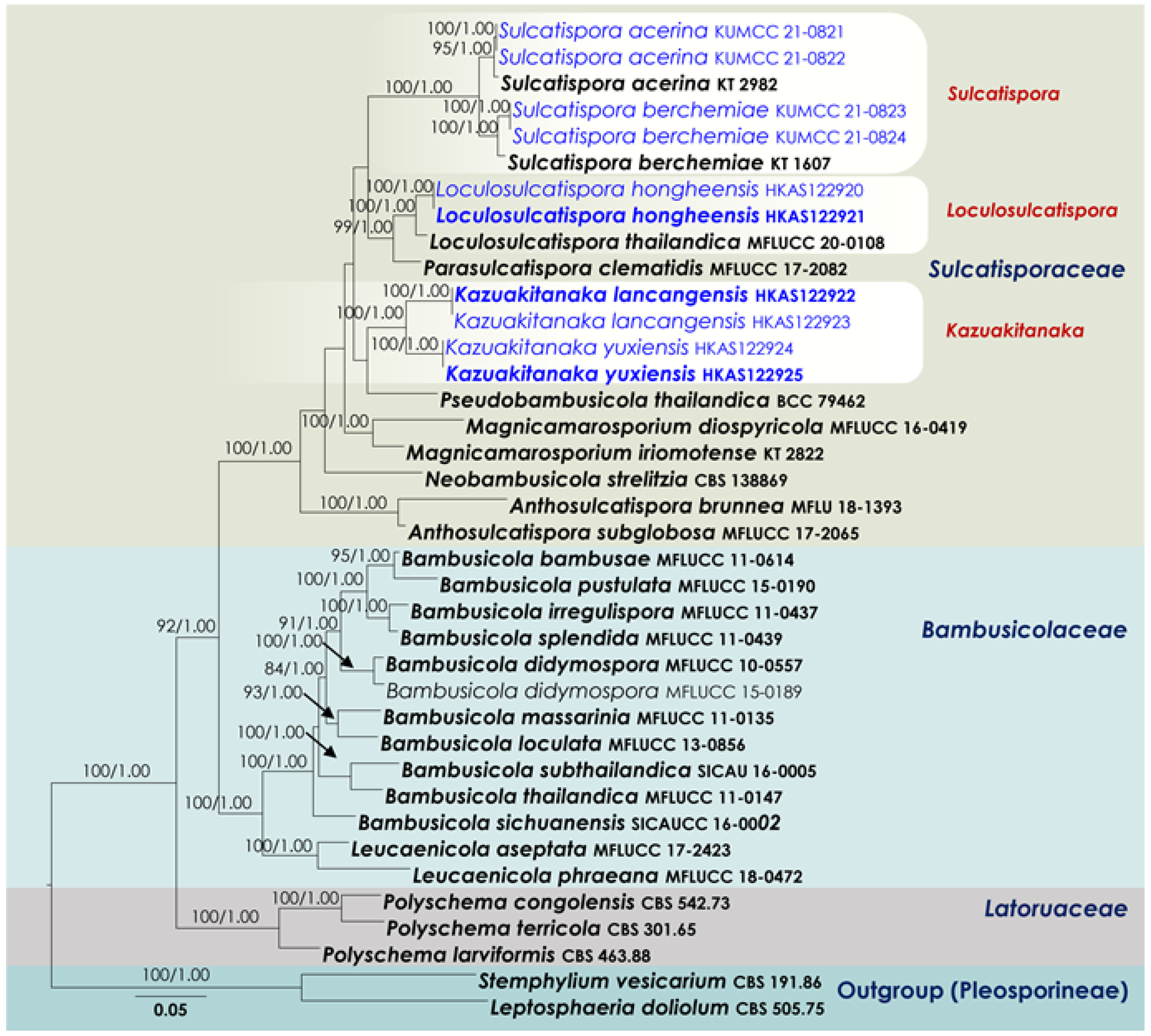
3.1. Phylogenetic Analyses
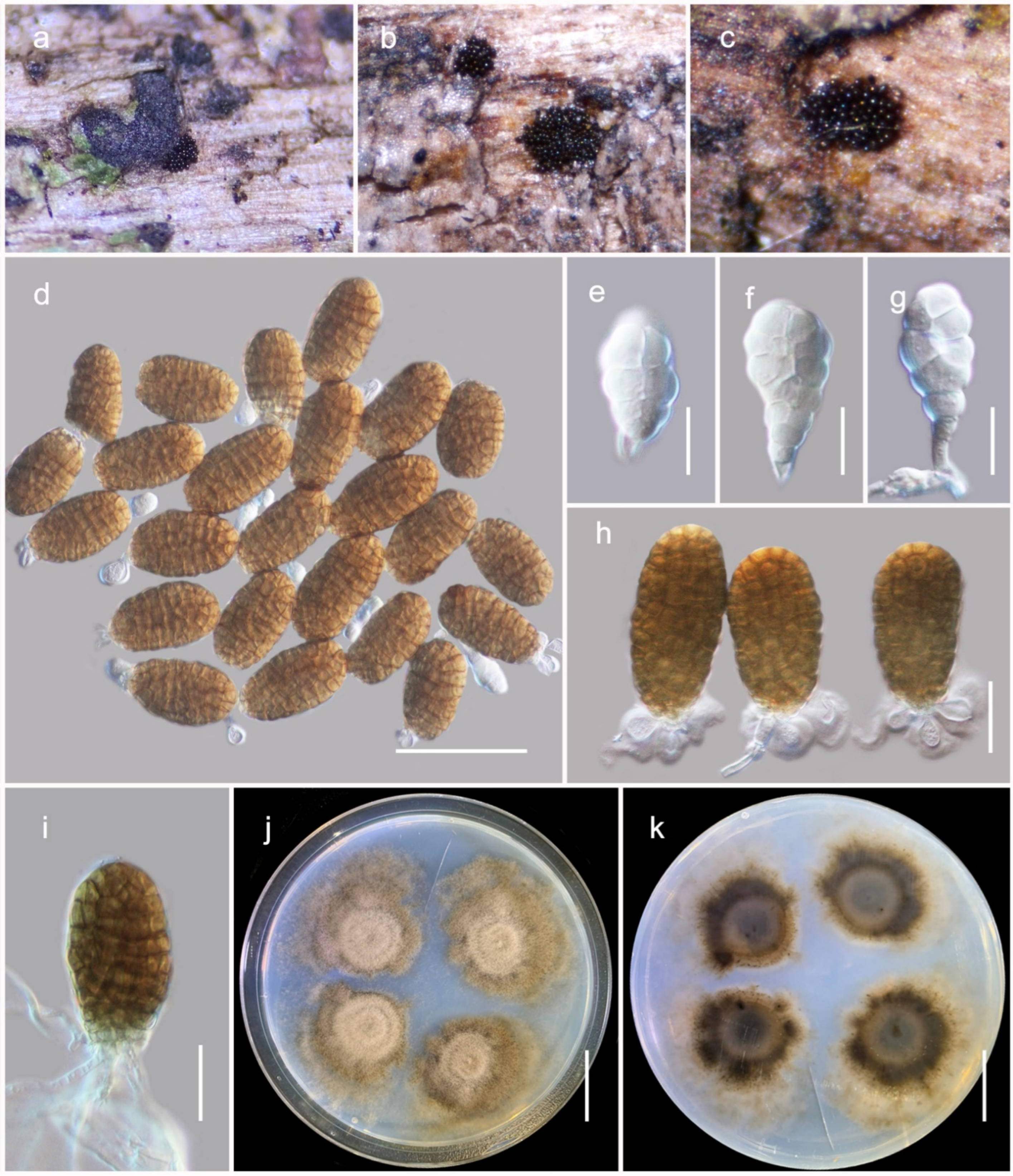
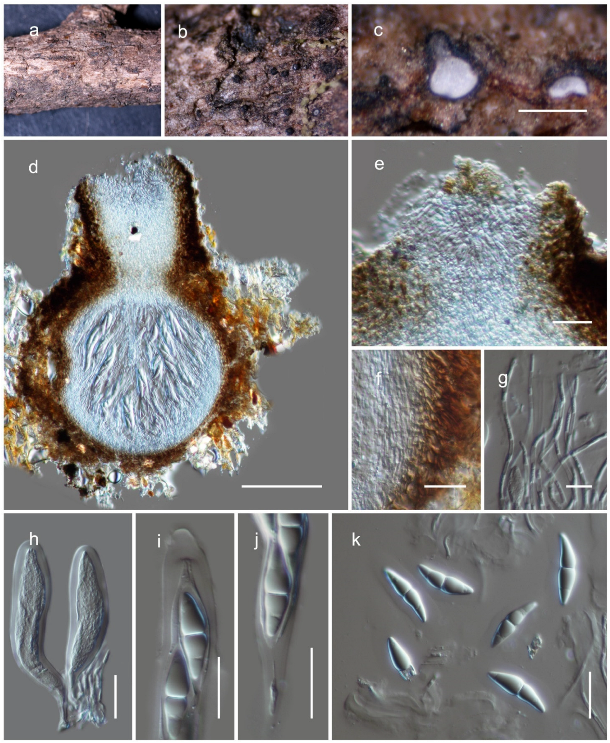
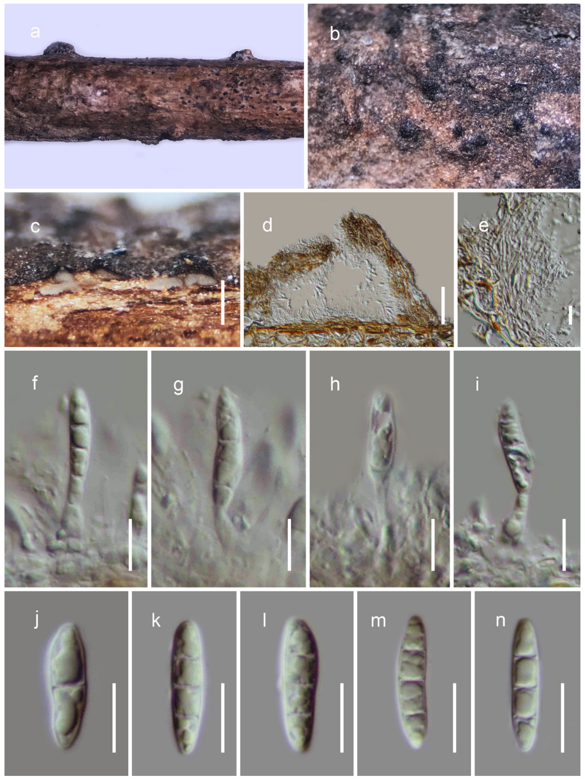
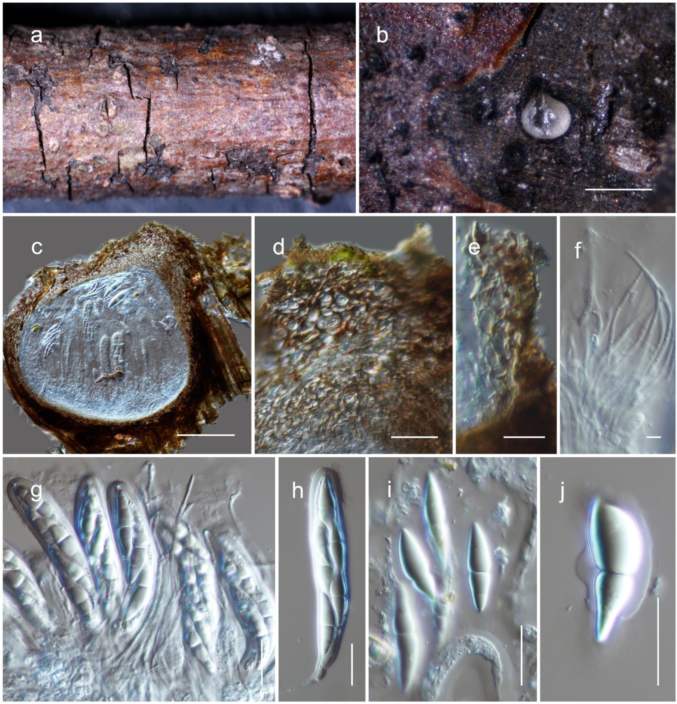
The final concatenated SSU–LSU–ITS–tef1–rpb2 alignment (Figure 1) of Phaeoseptaceae comprised 48 strains, including the outgroup taxa Gloniopsis praelonga (CBS 112415) and Hysterium angustatum (MFLUCC 16-0623). The final alignment contained a total of 4478 characters used for the phylogenetic analyses, including alignment gaps (available in TreeBASE). The RAxML analysis of the combined dataset yielded a best scoring tree with a final ML optimization likelihood value of −30565.564394. The matrix had 1930 distinct alignment patterns, with 31.15% undetermined characters or gaps. Parameters for the GTR + I + G model of the combined amplicons were as follows: estimated base frequencies, A = 0.242181, C = 0.257106, G = 0.272525, T = 0.228188; substitution rates, AC = 1.247807, AG = 3.156565, AT = 1.478256, CG = 1.137163, CT = 7.093906, GT = 1.000; proportion of invariable sites, I = 0.397831; gamma distribution shape parameter, α = 0.56366. Bayesian analyses generated 3201 trees (average standard deviation of split frequencies: 0.009446) from which 2401 were sampled after 25% of the trees were discarded as burn-in. The alignment contained a total of 1932 unique site patterns.
The family Phaeoseptaceae (SSU, LSU, ITS, tef1 and rpb2 phylogeny) was resolved into four distinct clades with 100 MLB and 1.00 BYPP support values. Four strains of Pleopunctum (KUMCC 21-0820, KUMCC 21-0025, KUMCC 21-0026 and HKAS122915) isolated in the study formed a well-supported clade with P. clematidis (MFLUCC 17-2091), P. ellipsoideum (MFLUCC 19-0390) and P. pseudoellipsoideum (MFLUCC 19-0391). Three strains of Thyridaria macrostomoides (GKM 224N, GKM 1033, GKM 1159) constituted a strongly supported monophyly with the ex-type strain of Lignosphaeria fusispora (MFLUCC 11-0377). Two of our new strains, HKAS122916 and HKAS122917, nested with the ex-type strain of Phaeoseptum mali MFLUCC 17-2108 with 100 MLB and 1.00 BYPP statistical support. Strains of Phaeoseptum aquaticum, P. carolshearerianum, P. hydei, P. mali, P. manglicola and P. terricola grouped as a monophyletic clade with strong support values in both maximum likelihood and Bayesian analyses. Finally, the two strains of Decaisnella formosa (BCC 25616, BCC 25617) formed a basal terminal clade in Phaeoseptaceae with 100 MLB and 1.00 BYPP support values.
The Sulcatisporaceae (SSU, LSU, ITS, tef1 and rpb2 phylogeny) alignment contained 38 isolates (Figure 2), and the tree was rooted to Leptosphaeria doliolum (CBS 505.75) and Stemphylium vesicarium (CBS 191.86). The final alignment contained a total of 4931 characters used for the phylogenetic analyses, including alignment gaps (available in TreeBASE). The RAxML analysis of the combined dataset yielded a best scoring tree with a final ML optimization likelihood value of −27594.247903. The matrix had 1770 distinct alignment patterns, with 23.64% undetermined characters or gaps. Parameters for the GTR + I + G model of the combined amplicons were as follows: estimated base frequencies, A = 0.241395, C = 0.257298, G = 0.268694, T = 0.232613; substitution rates, AC = 1.256046, AG = 2.926191, AT = 1.154561, CG = 0.858049, CT = 6.686514, GT = 1.000; proportion of invariable sites, I = 0.499455; gamma distribution shape parameter, α = 0.532594. The Bayesian analyses generated 401 trees (average standard deviation of split frequencies: 0.009088) from which 301 were sampled after 25% of the trees were discarded as burn-in. The alignment contained a total of 1771 unique site patterns.
The family Sulcatisporaceae was composed of distinct lineages that correspond to the genera Anthosulcatispora, Loculosulcatispora, Magnicamarosporium, Neobambusicola, Parasulcatispora, Pseudobambusicola and Sulcatispora. Four strains (HKAS122922, HKAS122923, HKAS122924, HKAS122925) obtained in the present study formed a well-separated clade in Sulcatisporaceae, featuring both a single locus and concatenated datasets. Thus, this new lineage is presented here as the new genus Kazuakitanaka gen. nov. Two of our new isolates, KUMCC 21-0821 and KUMCC 21-0822, were grouped with the ex-type strain of Sulcatispora acerina (KT 2982) with 95 MLB and 1.00 BYPP values. Another two new strains (KUMCC 21-0823 and KUMCC 21-0824) grouped with the ex-type strain of Sulcatispora berchemiae (KT 1607), with 100 MLB and 1.00 BYPP support values. Loculosulcatispora thailandica (MFLUCC 20-0108), a type of Loculosulcatispora, constituted a monophyletic clade with two of our new isolates (HKAS122920 and HKAS122921). Neobambusicola strelitzia, Parasulcatispora clematidis and Pseudobambusicola thailandica were affiliated as monotypic genera. Two species of Magnicamarosporium, M. diospyricola (MFLUCC 16-0419) and M. iriomotense (KT 2822), grouped with 100 MLB and 1.00 BYPP support values. Anthosulcatispora brunnea (MFLU 18-1393) and A. subglobosa (MFLUCC 17-2065) formed a basal terminal clade in Sulcatisporaceae, with 100 MLB and 1.00 BYPP statistical support.

3.2. Taxonomy

Pleosporales Luttr. ex M.E. Barr, Prodromus to class Loculoascomycetes: 67 (1987).
Phaeoseptaceae Boonmee, Thambug. and K.D. Hyde, Mycosphere 9 (2): 323 (2018).
Phaeoseptum Y. Zhang, J. Fourn. and K.D. Hyde, Mycologia 105 (3): 606 (2013).
Phaeoseptum mali Phukhams. and K.D. Hyde, Asian Journal of Mycology 2 (1): 120 (2019).
Material examined: China, Yunnan, Honghe Hani and Yi Autonomous Prefecture, Kaiyuan, 23.863601° N, 103.407975° E, 1053 m, on dead woody litter, 17 March 2019, D.N. Wanasinghe (HKAS122916), ibid. Chuxiong Yi Autonomous Prefecture, Shuangbai County, 24.80527° N, 101.933887° E, 1736 m, on dead woody litter under the Fagaceae species, 25 June 2019 (HKAS122917).
Notes: Phukhamsakda et al. [44] introduced Phaeoseptum mali from the bark of fallen twigs of Malus halliana from the botanical garden of the Kunming Institute of Botany, Yunnan, China. Phaeoseptum mali fits well within the generic descriptions of Phaeoseptum based on its globose and immersed ascomata, cellular pseudoparaphyses, cylindrical-clavate, pedicellate asci, allantoid and brown muriform ascospores. In this study, we collected another two specimens of Phaeoseptum mali from Chuxiong and Honghe Prefectures in Yunnan, China on dead woody litter. Multi-gene phylogenetic analyses of combined SSU, LSU, ITS, tef1 and rpb2 DNA sequence showed that the ex-type strain of Phaeoseptum mali (MFLUCC 17-2108) and our two strains (HKAS122916 and HKAS122917) are monophyletic with 100% MLB and 1.00 BYPP support values. Morphologically, these new collections are not different from the holotype of Phaeoseptum mali.
Pleopunctum N.G. Liu, K.D. Hyde and J.K. Liu, Mycosphere 10 (1): 767 (2019).
Pleopunctum menglaense Wanas. sp. nov. (Figure 3).
MycoBank: MB843430.
Etymology: The specific epithet is derived from Mengla County, Yunnan Province, China.
Holotype: HKAS122683.
The species is a saprobe on dead twigs of forest litter in terrestrial habitats. Teleomorph: undetermined. Anamorph: hyphomycetous. Colonies on host, sporodochial, superficial, black, scattered and punctiform. The conidiophores are arising from hyaline unbranched hyphae. They are 10–20 × 2.5–3.5 μm (M = 16.7 × 3.1, n = 15), macronematous, mononematous, cylindrical, unbranched, aseptate, hyaline and smooth-walled. The conidiogenous cells 5–8 × 2.8–3.5 µm (M = 6.9 × 3.2 μm, n = 30) are monoblastic, terminal, hyaline. The conidia are dimorphic, acrogenous and solitary. The α conidia are 18–25 × 10–14 µm (M = 23.1 × 11.7 μm, n = 30), hyaline, multi-septate, muriform, spatulate to obovate, notably constricted at septa, slightly obtuse to rounded at apex, notably narrow at base, often carrying remnants of conidiogenous cells at base. The β conidia are 38–55 × 20–26 µm (M =  45.6 × 24.4 μm, n = 30), brown, muriform, ellipsoidal to oblong shaped, moderately rough-walled, and slightly constricted at septa often with a hyaline, elliptical to globose, 1-3 basal cells, 7.5–12 × 4–6 µm (M = 9 × 5.1 μm, n = 25) with a tri-lobed wing-like appearance.
Culture characteristics: the conidia germinated on PDA within 12 h and germ tubes were produced from the basal cells of conidia. The colonies on PDA reached a 5 cm diameter after 2 weeks at 25 °C. They were effused, circular to lobed, with incised margin, flat or slightly hairy, pinkish brown in the center with concentric rings and pale brown at the periphery and reverse; three distinct color zones were present, namely greyish brown at the center, pinkish brown at the middle, golden brown at the periphery.
Material examined: China, Yunnan, Xishuangbanna Dai Autonomous Prefecture, Mengla County, Wangtianshu, 21.61852° N, 101.58171° E, 747 m, on dead woody litter, 12 January 2021, D.N. Wanasinghe (HKAS122683, holotype), ex-type living culture, KUMCC 21-0826. ibid. 21.618839° N, 101.581098° E, 774 m (HKAS122684), living culture, KUMCC 21-0827.
Pleopunctum pseudoellipsoideum N.G. Liu, K.D. Hyde and J.K. Liu, Mycosphere 10 (1): 768 (2019)
Material examined: China, Yunnan, Diqing Tibetan Autonomous Prefecture, Weixi Lisu Autonomous County, Zili Village, 27.803791° N, 99.059136° E, 2250 m, on dead woody litter, 25 June 2019, L.Q. Xian (HKAS122914), living culture, KUMCC 21-0820, ibid. Kunming, Luquan Yi and Miao Autonomous County, 26.052826° N, 102.670548° E, 2629 m, on dead woody litter under the Fagaceae species, 2 July 2019, D.N. Wanasinghe (HKAS122915).
Notes: Liu et al. [45] introduced Pleopunctum pseudoellipticum from decaying wood in Guizhou Province, China as a hyphomycetous muriform spored anamorph taxon in Phaeoseptaceae. During our investigation on the diversity of woody litter microfungi in Yunnan, China, two collections were made from the Diqing and Kunming areas. Morphological characteristics of our new collections, such as conidiophores and conidia, fit well within the anamorph of Pleopunctum pseudoellipticum. In our phylogenetic study (Figure 1), the new strains (KUMCC 21-0820, HKAS122915) cluster with Pleopunctum pseudoellipticum (MFLUCC 19-0391, ex-type strain) as a monophyletic clade with 100% MLB and 1.00 BYPP support values. Comparison of ITS, LSU and tef1 sequence data reveals there is no significant difference between our new isolates and Pleopunctum pseudoellipticum. Therefore, we recognize our new isolates as additional collections of Pleopunctum pseudoellipticum, and we provide SSU and rpb2 sequence data that were not provided by Liu et al. [45].
Sulcatisporaceae Kaz. Tanaka and K. Hiray., Studies in Mycology 82: 119 (2015).
Kazuakitanaka Wanas. gen. nov.
MycoBank: MB843431.
Etymology: The generic epithet stems from the combined two words “kazuaki” and “tanaka”, referring to Kazuaki Tanaka, who introduced the family Sulcatisporaceae.
The genus comprises saprobic fungi on woody substrates in terrestrial habitats. Teleomorph: the ascomata is a solitary or gregarious, semi-immersed, erumpent through the host surface, coriaceous, dark brown to black, globose to subglobose, ostiolate. The ostiole is central, with hyaline to pale-brown pseudoparenchymatous cells. The peridium is broader at the apex and thinner at the base, comprising two strata with several layers of brown or lightly pigmented to hyaline cells building textura angularis to textura prismatica, indistinguishable from the host tissues. The hamathecium comprises many branched, septate, cellular pseudoparaphyses, located between and above the asci, embedded in a gelatinous matrix. The asci are eight-spored, bitunicate, fissitunicate, cylindric-clavate, pedicellate, and apically rounded, with an ocular chamber. The ascospores are uni- to bi-seriate, partially overlapping in lateral view, and are hyaline. They are fusiform, 1–2-septate, slightly curved, deeply constricted at the central septum, conically rounded at the ends, and smooth-walled, with a distinct mucilaginous sheath, comprising a large guttule in each cell. Anamorph: Coelomycetous. The conidiomata are pycnidial, solitary, gregarious, dark brown to black, immersed or slightly erumpent, coriaceous to carbonaceous, papillate or apapillate. The conidiomatal wall is multi-layered; the outer layers are pseudoparenchymatous, with brown-walled cells, with the innermost layer thin and hyaline. The conidiophores are reduced to conidiogenous cells. The conidiogenous cells are phialidic, ampulliform to cylindrical, determinate, hyaline, smooth-walled and formed from the inner layer of the pycnidium wall. The conidia are hyaline, one-to-three-septate, straight to curved, fusiform, with conical ends, thick-walled, smooth, with large guttules in each cell.
Type species: Kazuakitanaka yuxiensis
Kazuakitanaka yuxiensis Wanas. sp. nov. (Figure 4).
MycoBank: MB843432.
Etymology: The specific epithet is derived from Yuxi, Yunnan Province, China.
Holotype: HKAS122924.
The species is a saprobe on dead twigs of forest litter in terrestrial habitats. Teleomorph: the ascomata is 350–400 µm high, 220–260 µm diam. (M = 386.6 × 249.9 µm, n = 5), scattered to gregarious, semi-immersed to erumpent, coriaceous, dark brown to black, globose to subglobose ostiolate. The ostiole is 110–140 µm long, 90–115 µm diam. (M = 125.6 × 98.7 µm, n = 5), centrally papillate, comprising hyaline cells. The peridium is 10–15 µm wide at the base, 12–20 µm wide at the sides, broad at the apex (30–40 µm), comprising two strata, with the outer stratum composed of small, pale brown to brown, slightly flattened, thick-walled cells of textura angularis, fusing and indistinguishable from the host tissues. The inner stratum is composed of several layers with lightly pigmented to hyaline cells centrally textura angularis to textura prismatica. The hamathecium is 1–2 µm wide, branched, septate, cellular pseudoparaphyses, situated between and above the asci, embedded in a gelatinous matrix. The asci are 80–120 × 17–21 µm (M = 102.2 × 18.5 µm, n = 20), eight-spored, bitunicate, fissitunicate, cylindric-clavate, with a pedicel, and is rounded at the apex, with an ocular chamber. The ascospores are 25–32 × 7–9 µm (M = 29.1 × 7.9 µm, n = 30), uni- to bi-seriate, overlapping, and hyaline. They are fusiform, 1–2-septate. They are slightly curved, deeply constricted at the central septum, conically rounded at the ends, and smooth-walled, surrounded by a distinct mucilaginous sheath, each cell with a large guttule. Anamorph: undetermined.
Material examined: China, Yunnan, Yuxi, Xinping Yi and Dai Autonomous County, 24.09083° N, 101.935124° E, 2091 m, on dead woody litter, 25 May 2019, D.N. Wanasinghe (HKAS122924, holotype). ibid. 24.09083° N, 101.935124° E, 2095 m (HKAS122925).
Kazuakitanakalancangensis Wanas. sp. nov. (Figure 5).
MycoBank: MB843433.
Etymology: The specific epithet is derived from Lancang, Yunnan Province, China.
Holotype: HKAS122922.
It is saprobic on dead twigs of forest litter in terrestrial habitats. Teleomorph: undetermined. Anamorph: coelomycetous. The conidiomata is 130–150 µm high, 160–190 µm diam. (M = 140 × 170 µm, n = 5), pycnidial, solitary, gregarious, globose to subglobose, coriaceous, uni-loculate, dark brown to black, and immersed, with a central ostiole. The conidiomata wall is 10–20 µm wide, multi-layered, with brown-walled pseudoparenchymatous cells, with a hyaline inner most layer. The conidiophores are reduced to conidiogenous cells. The conidiogenous cells are 7–10 × 2–3.5 μm (M = 8.3 × 2.7 µm, n = 15), phialidic, ampulliform to cylindrical, determinate, hyaline, smooth-walled and formed from the inner layer of the pycnidium wall. The conidia are 17–23 × 3.7–4.7 2 µm (M = 20 × 4.2 µm, n = 30), fusiform, straight, hyaline, one-to-three-septate, not constricted at septa, tip and base rounded, thick-walled, and smooth, with numerous large guttules in each cell.
Material examined: China, Yunnan, Pure, Lancang Lahu Autonomous County, 23.164681° N, 99.969248° E, 1990 m, on dead woody litter, 11 April 2019, D.N. Wanasinghe (HKAS122922, holotype). ibid. 23.164686° N, 99.969492° E, 1975 m (HKAS122923).
Loculosulcatispora G.C. Ren and K.D. Hyde, Phytotaxa 475 (2): 70 (2020) amended
MycoBank: MB557580
The genus comprises saprobic species on dead wood or twigs. Teleomorph: the ascomata are scattered, immersed to semi-immersed, globose to subglobose, brown to dark-brown, with a central ostiole. The peridium is composed of small, pale brown to brown, thick-walled cells forming textura angularis to globosa. The hamathecium comprises branched, septate, cellular pseudoparaphyses. The asci are eight-spored, bitunicate, clavate, straight to curved, with a short pedicel. They are apically rounded, with or without an ocular chamber. The ascospores are overlapping uni- to bi-seriate, hyaline, fusiform with acute ends, 1-septate, smooth-walled, surrounded by a thick irregular mucilaginous sheath. Anamorph: see Ren et al. [36].
Type species: Loculosulcatispora thailandica G.C. Ren and K.D. Hyde, Phytotaxa 475 (2): 73 (2020).
Loculosulcatispora hongheensis Wanas. sp. nov. (Figure 6).
MycoBank: MB843434.
Etymology: The specific epithet is derived from Honghe, Yunnan Province, China.
Holotype: HKAS122920.
The species is a saprobe on dead twigs of forest litter in terrestrial habitats. Teleomorph: the ascomata is 130–490 × 120–560 (M = 260 × 280, n = 5) μm, scattered, immersed, coriaceous, globose to subglobose, brown to dark-brown, with a central ostiole. The peridium is 10–15 μm wide; the outer stratum composed of small, pale brown to brown, thick-walled cells forming textura angularis to globosa indistinguishable from the host tissues. The inner stratum is composed of hyaline cells forming textura angularis. The hamathecium is 1–2.5 µm wide, branched, septate, cellular pseudoparaphyses, situated between and above the asci, embedded in a gelatinous matrix. The asci are 60–100 × 10–18 μm (M = 84 × 13.7 μm; n = 15) μm, eight-spored, bitunicate, fissitunicate, clavate, straight to curved, with a short pedicel. They are apically rounded, with a minute ocular chamber. The ascospores are 20–30 × 4–6.5 μm (M = 25 × 5 μm, n = 30), overlapping uni- to bi-seriate, hyaline, fusiform with acute ends, 1-septate, constricted at the septum, slightly longer upper cell than lower cell, slightly curved, smooth-walled, and surrounded by a thick irregular mucilaginous sheath. Anamorph: undetermined.
Material examined: China, Yunnan, Honghe Hani and Yi Autonomous Prefecture, Mengzi, 23.18604° N, 103.413838° E, 1854 m, on dead woody litter, 16 March 2019, D.N. Wanasinghe (HKAS122920, holotype). ibid. 23.186552° N, 103.414063° E, 1866 m (HKAS122921).
Sulcatispora Kaz. Tanaka and K. Hiray., Studies in Mycology 82: 120 (2015).
Sulcatispora acerina Kaz. Tanaka and K. Hiray., Studies in Mycology 82: 120 (2015).
Material examined: China, Yunnan, Pure, Jingdong Yi Autonomous County, 24.549205° N, 101.027521° E, 2560 m, on dead woody litter, 8 June 2021, D.N. Wanasinghe (HKAS122926), living culture, KUMCC 21-0821. ibid. 24.548691° N, 101.027802° E, 2546 m (HKAS122927), living culture, KUMCC 21-0822.
Notes: Sulcatispora acerina, introduced by Tanaka et al. [46], was collected from Acer palmatum in a terrestrial habitat. Based on phylogenetic analysis of combined SSU, LSU, ITS, tef1 and rpb2 sequence data, two of our isolates, KUMCC 21-0821 and KUMCC 21-0822, were clustered with the ex-type strain of Sulcatispora acerina (KT 2982) with 95% MLB and 1.00 BYPP support (Figure 2). Our isolate resembles Sulcatispora acerina in shape and size of the ascomata, asci and ascospores. Moreover, there are no base pair differences of the SSU and LSU nucleotides and only four and seven nucleotide differences in the ITS and tef1 regions, respectively. Therefore, we recognize that our new isolates belong to Sulcatispora acerina as new records from China, providing rpb2 sequence data that were not provided by Tanaka et al. [46].
Sulcatispora berchemiae Kaz. Tanaka and K. Hiray., Studies in Mycology 82: 121 (2015).
Material examined: China, Yunnan, Pure, Jingdong Yi Autonomous County, 24.548237° N, 101.026484° E, 2516 m, on dead woody litter, 8 June 2021, D.N. Wanasinghe (HKAS122918), living culture, KUMCC 21-0823. ibid. 24.548142° N, 101.026572° E, 2510 m (HKAS122919), living culture, KUMCC 21-0824.
Notes: In the phylogenetic analysis (Figure 2), two of our new isolates (KUMCC 21-0823, KUMCC 21-0824) clustered with the type strain of Sulcatispora berchemiae (KT 1607) with high bootstrap support (100% MLB, 1.00 BYPP). These three strains share similar morphological features in ascomata, asci and ascospores. KT 1607 was collected from Japan on Berchemia racemosa, whereas KUMCC 21-0823 and KUMCC 21-0824 were collected from China on dead woody litter. Based on both morphology and molecular data, we consider our new isolates and Sulcatispora berchemiae to be conspecific. While extending the biogeography of Sulcatispora berchemiae, we also provide the rpb2 gene region for the species, which was not accounted for earlier.

4. Discussion

Hyde et al. [47] established Phaeoseptaceae in Pleosporales to accommodate Phaeoseptum. However, this family is now recognized as a group of heterogenous taxa that have diversified habitats with different types of anamorphs and diverged morphological characteristics [37]. In the recent outlines of Hongsanan et al. [10] and Wijayawardane et al. [12], only Phaeoseptum and Pleopunctum are accepted in Phaeoseptaceae. In the combined multi-gene phylogenetic analyses, the ex-type strains of Lignosphaeria diospyrosa, L. fusispora and L. thailandica also clustered in Phaeoseptaceae [48]. Therefore, Lignosphaeria should be an accepted genus within Phaeoseptaceae. Even though the putative strains of Decaisnella formosa (BCC 25616, BCC 25617) and Thyridaria macrostomoides (GKM 1033, GKM 1159, GKM 224N) are phylogenetically affiliated in Phaeoseptaceae [37,47], they are not related to any type specimens. Therefore, it is necessary to recollect and epitypify them with DNA sequence data in order to ensure correct generic placement of these two species [49,50].
Phaeoseptum was introduced by Zhang et al. [51] to accommodate P. aquaticum, and it was initially described as an accepted species in Halotthiaceae based on phylogenetic analysis of LSU sequence data. Currently, six species are accepted in Phaeoseptum viz.: P. aquaticum, P. carolshearerianum, P. hydei, P. mali, P. manglicola and P. terricola [10]. Phaeoseptum aquaticum was reported from freshwater habitats, and later Hyde et al. [47] added P. terricola from a terrestrial environment. In recent studies, Phukhamsakda et al. [44] and Wanasinghe et al. [37] collected Phaeoseptum mali and P. hydei from terrestrial habitats, respectively. Dayarathne et al. [52] showed that these species can be found in tropical coastlines, while introducing Phaeoseptum carolshearerianum and P. manglicola from mangroves. In this study, we accounted for another two records of Phaeoseptum mali from Chuxiong and Honghe Prefectures in Yunnan (China) from terrestrial habitats.
Pleopunctum, typified by P. ellipsoideum, is a hyphomycetous genus established in Phaeoseptaceae by Liu et al. [45]. Species in the genus are characterized by macronematous, mononematous conidiophores with monoblastic conidiogenous cells and oval to ellipsoidal, muriform conidia mostly with a hyaline, elliptical to globose basal cell [45]. Currently, five Pleopunctum species (viz. P. bauhiniae, P. clematidis, P. ellipsoideum, P. pseudoellipsoideum, P. thailandicum) are accepted in Species Fungorum [53]. However, Koukol and Delgado [54] argued that Pleopunctum clematidis should be a synonym of P. bauhiniae (=Hermatomyces bauhiniae). Members in the genus have appeared to be saprobic on dead twigs of deciduous hosts in terrestrial habitats [45,55,56,57]. In this study, morphological characteristics and multi-gene phylogenetic analysis of combined SSU, LSU, ITS, tef1 and rpb2 DNA sequence data reveals a new species of Pleopunctum from the dead woody litter collected in Yunnan, China. In a multi-gene (concatenated LSU, SSU, ITS, tef1 and rpb2) phylogenetic analysis, two of our strains (KUMCC 21-0025, KUMCC 21-0026) clustered with Pleopunctum species as a monophyletic clade with 100 MBP and 1.00 BYPP bootstrap support (Figure 1). Morphological features of KUMCC 21-0025 and KUMCC 21-0026 are similar to extant species of Pleopunctum in having sporodochial colonies, holoblastic, monoblastic conidiogenous cells and brown muriform conidia [45]. We introduce these two isolates as a novel species belonging to this genus, Pleopunctum menglaense. Within the Pleopunctum clade, P. menglaense strains constitute a sister lineage to P. pseudoellipsoideum (MFLUCC 19-0391, KUMCC 21-0820, HKAS122915). Pleopunctum menglaense has contrasting morphological features to P. pseudoellipsoideum by its distinct, hyaline, 1–3 basal cells in β conidia, whereas P. pseudoellipsoideum has a single basal cell in β conidia [45].
Sulcatisporaceae currently accommodates seven genera: viz. Anthosulcatispora, Loculosulcatispora, Magnicamarosporium, Neobambusicola, Parasulcatispora, Pseudobambusicola and Sulcatispora [12]. The species in this family are mostly restricted to terrestrial habitats excluding Neobambusicola strelitziae, which was found from the coastal region (Eastern Cape Province) of South Africa [58]. The conidia of anamorphs of Sulcatisporaceae can vary from hyaline, aseptate or septate (Anthosulcatispora, Loculosulcatispora, Neobambusicola, Pseudobambusicola), to pigmented phragmo-conidia (Sulcatispora) or muriform conidia (Magnicamarosporium), with or without striation [36,46,56,58,59,60]. The anamorph of Parasulcatispora is yet to be discovered. Shared characteristics within the family consist of immersed ascomata, peridium comprising pseudoparenchymatous cells with a thickened apex, cellular or trabeculate pseudoparaphyses and having cylindrical to cylindric-clavate asci with a long pedicel. However, the ascospore arrangement inside an ascus and the features of ascospores can differ between species belonging to this group. The spore arrangement can be uniseriate (Anthosulcatispora) and 2–3-seriate in others. Spores are hyaline and fusiform with conical ends that are completely surrounded by a sheath excluding Anthosulcatispora brunnea, which is brown to dark brown, oblong to ellipsoidal and lacking a sheath.
Kazuakitanaka is introduced in Sulcatisporaceae as a new genus, based on LSU, ITS, tef1 and rpb2 sequence data. Kazuakitanaka morphologically resembles Parasulcatispora and Sulcatispora with its cylindric-clavate asci and fusiform, 1-septate hyaline ascospores. However, these genera were revealed as phylogenetically distant in multi-gene phylogenetic analysis (Figure 2). The coelomycetous anamorph of Kazuakitanaka is similar to species of Neobambusicola and Pseudobambusicola by its pycnidial conidiomata, phialidic conidiogenous cells and hyaline, fusiform, multi-septate conidia. The new genus has a close phylogenetic proximity to Pseudobambusicola even though this relationship is not statistically significant, and Neobambusicola was placed in a distinct branch apart from the Kazuakitanaka clade (Figure 2). The teleomorph for these genera is still undetermined and therefore they cannot be morphologically compared with the teleomorph of the new genus.
Ren et al. [36] introduced Loculosulcatispora as a monotypic genus to accommodate L. thailandica, and it was known only from its anamorph. Loculosulcatispora thailandica was introduced as a decaying woody-based saprobe from Thailand (Chiang Rai). The genus was characterized by multilocular conidiomata, phialidic, discrete, determinate, doliiform to cylindrical, hyaline conidiogenous cells, unbranched and aseptate paraphyses, and 1-celled, oblong conidia with guttules. During our sample collections, we found a teleomorphic species characterized by globose to subglobose, immersed ascomata with a central ostiole, peridium composed of textura angularis, hamathecium comprises branched, septate, cellular pseudoparaphyses, clavate, short pedicellate asci with a minute ocular chamber, hyaline, fusiform, and 1-septate ascospores with a mucilaginous sheath. In the multi-gene phylogenetic analyses, two of the isolates of this teleomorphic species (HKAS122920, HKAS122921) constituted a monophyletic clade (100% MLB and 1.00 BYPP, Figure 2) with the type strain of Loculosulcatispora thailandica (MFLUCC 20-0108). Therefore, we introduce our new collections as Loculosulcatispora hongheensis sp. nov. and amended the generic descriptions herein to accommodate its teleomorphic characteristics. The Loculosulcatispora clade has a sister affiliation to Parasulcatispora clematidis, which is known only from its teleomorph [56]. It could also be a species in Loculosulcatispora. For us to combine these two genera here would require an extensive taxonomic reassessment and describing anamorphic morphology, which is beyond the scope of the current study..
Tanaka et al. [46] introduced Sulcatispora to accommodate S. acerina and S. berchemiae, which were collected as saprobes on Sapindaceae (Acer palmatum) and Rhamnaceae (Berchemia racemosa) hosts. The teleomorph of the genus is characterized by globose, subglobose to hemispherical and immersed to erumpent ascomata, short papillate, central ostioles with periphyses, peridium composed of several layers of compressed angular cells, trabeculate pseudoparaphyses, clavate asci with a short pedicel and fusiform, 1-septate, hyaline ascospores with a large sheath. The anamorph is characteristic with globose, pycnidial conidiomata, cylindrical and annellidic conidiogenous cells, ellipsoid, yellowish brown, multi-septate conidia with striate ornamentation. These teleomorphic characteristics are somewhat similar to those in Massarina. The most distinctive feature of Sulcatispora is the longitudinal striae on the surface of conidia. Some species in Barriopsis (e.g., B. iraniana), Dwiroopa (e.g., D. ramya), Endomelanconium (e.g., E. pini), Lasiodiplodia (e.g., L. theobromae), Mucoharknessia (e.g., M. cortaderiae), Neodeightonia (e.g., N. phoenicum) and Phaeophleospora (e.g., P. striae) are known to have such conidia [61].
In the present study, we identified five genera and eight species associated with dead woody litter in Yunnan, China. Among them, one genus and four species are new to science. Our results emphasize that Yunnan Province has not yet been properly studied and is an open field for new fungal discoveries.

Author Contributions

Conceptualization, D.N.W.; resources, P.E.M.; writing—original draft preparation, D.N.W. and G.-C.R.; writing—review and editing, P.E.M., J.-C.X. and R.C.; supervision, P.E.M., J.-C.X. and R.C.; funding acquisition, P.E.M. and R.C. All authors have read and agreed to the published version of the manuscript.

Funding

High-End Foreign Experts in the High-Level Talent 318 Recruitment Plan of Yunnan Province, 2021, CAS President’s International Fellowship Initiative (grant number 2021FYB0005), the National Science Foundation of China (NSFC) under the project codes 31861143002, 32150410362, Yunnan Provincial Science and Technology Department (grant numbers 202003AD150004 and 202101AS070045), the Postdoctoral Fund from Human Resources and Social Security Bureau of Yunnan Province and Chiang Mai University.

Institutional Review Board Statement

Not applicable.

Informed Consent Statement

Not applicable.

Data Availability Statement

The datasets generated for this study can be found in the NCBI GenBank, MycoBank and TreeBASE.

Acknowledgments

Austin G. Smith at World Agroforestry (ICRAF), Kunming Institute of Botany, China, is thanked for English editing. Lu Wen Hua and Li Qin Xian are thanked for their invaluable assistance. Binu Samarakoon is thanked for the description writing of Pleopunctum menglaense. We acknowledge Kunming Institute of Botany, Chinese Academy of Sciences for providing the laboratories and instruments for molecular work.

Conflicts of Interest

The authors declare no conflict of interest.

References

  1. Hawksworth, D.L.; Lücking, R. Fungal Diversity Revisited: 2.2 to 3.8 Million Species. Microbiol. Spectr. 2017, 5, 1–17. [Google Scholar] [CrossRef] [PubMed]
  2. Bánki, O.; Roskov, Y.; Döring, M.; Ower, G.; Vandepitte, L.; Hobern, D.; Remsen, D.; Schalk, P.; DeWalt, R.E.; Keping, M.; et al. Catalogue of Life Checklist (Version 2022-01-14); Catalogue of Life: Leiden, NL, USA, 2022. [Google Scholar] [CrossRef]
  3. Wanasinghe, D.N.; Mortimer, P.E.; Bezerra, J. Editorial: Fungal Systematics and Biogeography. Front. Microbiol. 2022, 12, 827725. [Google Scholar] [CrossRef] [PubMed]
  4. Hawksworth, D.L. The magnitude of fungal diversity: The 1.5 million species estimate revisited. Mycol. Res. 2001, 105, 1422–1432. [Google Scholar] [CrossRef] [Green Version]
  5. Wanasinghe, D.N.; Wijayawardene, N.N.; Xu, J.; Cheewangkoon, R.; Mortimer, P.E. Taxonomic novelties in Magnolia-associated pleosporalean fungi in the Kunming Botanical Gardens (Yunnan, China). PLoS ONE 2020, 15, e0235855. [Google Scholar] [CrossRef] [PubMed]
  6. Hyde, K.D.; Norphanphoun, C.; Chen, J.; Dissanayake, A.J.; Doilom, M.; Hongsanan, S.; Jayawardena, R.S.; Jeewon, R.; Perera, R.H.; Thongbai, B.; et al. Thailand’s amazing diversity: Up to 96% of fungi in northern Thailand may be novel. Fungal Divers. 2018, 93, 215–239. [Google Scholar] [CrossRef]
  7. Põlme, S.; Abarenkov, K.; Nilsson, R.H.; Lindahl, B.D.; Clemmensen, K.E.; Kauserud, H.; Nguyen, N.; Kjøller, R.; Bates, S.T.; Baldrian, P.; et al. FungalTraits: A user-friendly traits database of fungi and fungus-like stramenopiles. Fungal Divers. 2020, 105, 1–16. [Google Scholar] [CrossRef]
  8. Schmit, J.P.; Mueller, G.M. An estimate of the lower limit of global fungal diversity. Biodivers. Conserv. 2007, 16, 99–111. [Google Scholar] [CrossRef]
  9. Dighton, J. Fungi in Ecosystem Processes, 2nd ed.; CRC Press: Boca Raton, FL, USA, 2016; 434p. [Google Scholar] [CrossRef]
  10. Hongsanan, S.; Hyde, K.D.; Phookamsak, R.; Wanasinghe, D.N.; McKenzie, E.H.C.; Sarma, V.V.; Boonmee, S.; Lücking, R.; Bhat, D.J.; Liu, N.G. Refined families of Dothideomycetes: Dothideomycetidae and Pleosporomycetidae. Mycosphere 2020, 11, 1553–2107. [Google Scholar] [CrossRef]
  11. Mortimer, P.E.; Jeewon, R.; Xu, J.C.; Lumyong, S.; Wanasinghe, D.N. Morpho-phylo taxonomy of novel dothideomycetous fungi associated with dead woody twigs in Yunnan Province, China. Front. Microbiol. 2021, 12, e654683. [Google Scholar] [CrossRef]
  12. Wijayawardene, N.N.; Hyde, K.D.; Dai, D.Q.; Sánchez-García, M.; Goto, B.T.; Saxena, R.K.; Erdoğdu, M.; Selçuk, F.; Rajeshkumar, K.C.; Aptroot, A.; et al. Outline of Fungi and fungus-like taxa-2021. Mycosphere 2022, 13, 53–453. [Google Scholar] [CrossRef]
  13. Jiang, H.B.; Hyde, K.D.; Jayawardena, R.S.; Doilom, M.; Xu, J.; Phookamsak, R. Taxonomic and phylogenetic characterizations reveal two new species and two new records of Roussoella (Roussoellaceae, Pleosporales) from Yunnan, China. Mycol. Prog. 2019, 18, 577–591. [Google Scholar] [CrossRef]
  14. Thiyagaraja, V.; Hyde, K.D.; Wanasinghe, D.N.; Worthy, F.R.; Karunarathna, S.C. Addition to Melanommataceae: A new geographical record of Alpinaria rhododendri from Shangri La, China. Asian J. Mycol. 2020, 3, 335–344. [Google Scholar] [CrossRef]
  15. Wanasinghe, D.N.; Mortimer, P.E.; Xu, J. Insight into the systematics of microfungi colonizing dead woody twigs of Dodonaea viscosa in Honghe (China). J. Fungi 2021, 7, 180. [Google Scholar] [CrossRef] [PubMed]
  16. Dissanayake, L.S.; Maharachchikumbura, S.S.N.; Mortimer, P.E.; Hyde, K.D.; Kang, J.C. Acrocordiella yunnanensis sp. nov. (Requienellaceae, Xylariales) from Yunnan, China. Phytotaxa 2021, 487, 103–113. [Google Scholar] [CrossRef]
  17. Ren, G.C.; Wanasinghe, D.N.; Monkai, J.; Mortimer, P.E.; Hyde, K.D.; Xu, J.C.; Pang, A.; Gui, H. Novel saprobic Hermatomyces species (Hermatomycetaceae, Pleosporales) from China (Yunnan Province) and Thailand. MycoKeys 2021, 82, 57–79. [Google Scholar] [CrossRef]
  18. Yang, E.; Tibpromma, S.; Karunarathna, S.C.; Phookamsak, R.; Xu, J.; Zhao, Z.; Karunanayake, C.; Promputtha, I. Taxonomy and phylogeny of novel and extant taxa in Pleosporales associated with Mangifera indica from Yunnan, China (Series I). J. Fungi 2022, 8, 152. [Google Scholar] [CrossRef]
  19. Xie, N.; Phookamsak, R.; Jiang, H.; Zeng, Y.; Zhang, H.; Xu, F.; Lumyong, S.; Xu, J.; Hongsanan, S. Morpho-molecular characterization of five novel taxa in Parabambusicolaceae (Massarineae, Pleosporales) from Yunnan, China. J. Fungi 2022, 8, 108. [Google Scholar] [CrossRef]
  20. Su, H.; Hyde, K.D.; Maharachchikumbura, S.S.; Ariyawansa, H.A.; Luo, Z.; Promputtha, I.; Tian, Q.; Lin, C.; Shang, Q.; Zhao, Y. The families Distoseptisporaceae fam. nov., Kirschsteiniotheliaceae, Sporormiaceae and Torulaceae, with new species from freshwater in Yunnan Province, China. Fungal Divers. 2016, 80, 375–409. [Google Scholar] [CrossRef]
  21. Su, H.; Luo, Z.; Liu, X.; Su, X.; Hu, D.; Zhou, D.; Bahkali, A.H.; Hyde, K.D. Lentithecium cangshanense sp. nov. (Lentitheciaceae) from freshwater habitats in Yunnan Province, China. Phytotaxa 2016, 267, 61–69. [Google Scholar] [CrossRef] [Green Version]
  22. Su, X.; Luo, Z.; Jeewon, R.; Bhat, D.J.; Bao, D.; Li, W.; Hao, Y.; Su, H.; Hyde, K.D. Morphology and multigene phylogeny reveal new genus and species of Torulaceae from freshwater habitats in northwestern Yunnan, China. Mycol. Prog. 2018, 17, 531–545. [Google Scholar] [CrossRef]
  23. Luo, Z.; Bhat, D.J.; Jeewon, R.; Boonmee, S.; Bao, D.; Zhao, Y.; Chai, H.; Su, H.; Su, X.; Hyde, K.D. Molecular phylogeny and morphological characterization of asexual fungi (Tubeufiaceae) from freshwater habitats in Yunnan, China. Cryptog. Mycol. 2017, 38, 27–53. [Google Scholar] [CrossRef]
  24. Luo, Z.L.; Hyde, K.D.; Liu, J.K.; Bhat, D.J.; Bao, D.F.; Li, W.L.; Su, H.Y. Lignicolous freshwater fungi from China II: Novel Distoseptispora (Distoseptisporaceae) species from northwestern Yunnan Province and a suggested unified method for studying lignicolous freshwater fungi. Mycosphere 2018, 9, 444–461. [Google Scholar] [CrossRef]
  25. Huang, S.; Maharachchikumbura, S.S.; Jeewon, R.; Bhat, D.J.; Chomnunti, P.; Hyde, K.D.; Lumyong, S. Morphological and molecular taxonomy of Jahnula dianchia sp. nov. (Jahnulales) from submerged wood in Dianchi Lake, Yunnan China. Mycol. Prog. 2018, 17, 547–555. [Google Scholar] [CrossRef]
  26. Wanasinghe, D.N.; Jeewon, R.; Gareth Jones, E.; Boonmee, S.; Kaewchai, S.; Manawasinghe, I.S.; Lumyong, S.; Hyde, K.D. Novel palmicolous taxa within Pleosporales: Multigene phylogeny and taxonomic circumscription. Mycol. Prog. 2018, 17, 571–590. [Google Scholar] [CrossRef]
  27. Wanasinghe, D.N.; Bezerra, J.D.P.; Mortimer, P.E. (Eds.) Fungal Systematics and Biogeography; Frontiers Media SA: Lausanne, Switzerland, 2022; 446p. [Google Scholar] [CrossRef]
  28. Crous, P.W.; Gams, W.; Stalpers, J.A.; Robert, V.; Stegehuis, G. MycoBank: An online initiative to launch mycology into the 21st century. Stud. Mycol. 2004, 50, 19–22. [Google Scholar]
  29. Wanasinghe, D.N.; Phukhamsakda, C.; Hyde, K.D.; Jeewon, R.; Lee, H.B.; Jones, E.B.G.; Tibpromma, S.; Tennakoon, D.S.; Dissanayake, A.J.; Jayasiri, S.C.; et al. Fungal diversity notes 709–839: Taxonomic and phylogenetic contributions to fungal taxa with an emphasis on fungi on Rosaceae. Fungal Divers. 2018, 89, 1–236. [Google Scholar] [CrossRef]
  30. White, T.J.; Bruns, T.; Lee, S.J.W.T.; Taylor, J.W. Amplification and direct sequencing of fungal ribosomal RNA genes for phylogenetics. PCR Protoc. Appl. 1990, 18, 315–322. [Google Scholar] [CrossRef]
  31. Rehner, S.A.; Samuels, G.J. Taxonomy and phylogeny of Gliocladium analysed from nuclear large subunit ribosomal DNA sequences. Mycol. Res. 1994, 98, 625–634. [Google Scholar] [CrossRef]
  32. Vilgalys, R.; Hester, M. Rapid genetic identification and mapping of enzymatically amplified ribosomal DNA from several Cryptococcus species. J. Bacteriol. 1990, 172, 4238–4246. [Google Scholar] [CrossRef] [Green Version]
  33. Rehner, S.A.; Buckley, E.A. Beauveria phylogeny inferred from nuclear ITS and EF1-α sequences: Evidence for cryptic diversification and links to Cordyceps teleomorphs. Mycologia 2005, 97, 84–98. [Google Scholar] [CrossRef]
  34. Liu, Y.J.; Whelen, S.; Hall, B.D. Phylogenetic relationships among ascomycetes evidence from an RNA polymerase II subunit. Mol. Biol. Evol. 1999, 16, 1799–1808. [Google Scholar] [CrossRef] [PubMed]
  35. Sung, G.H.; Sung, J.M.; Hywel-Jones, N.L.; Spatafora, J.W. A multi-gene phylogeny of Clavicipitaceae (Ascomycota, Fungi): Identification of localized incongruence using a combinational bootstrap approach. Mol. Phylogenet. Evol. 2007, 44, 1204–1223. [Google Scholar] [CrossRef] [PubMed]
  36. Ren, G.; Wanasinghe, D.N.; Wei, D.; Monkai, J.; Yasanthika, E.; Gui, H.; Mortimer, P.E.; Xu, J.; Hyde, K.D. Loculosulcatispora thailandica gen. et sp. nov. (Sulcatisporaceae), saprobic on woody litter in Thailand. Phytotaxa 2020, 475, 67–78. [Google Scholar] [CrossRef]
  37. Wanasinghe, D.N.; Mortimer, P.E.; Senwanna, C.; Cheewangkoon, R. Saprobic Dothideomycetes in Thailand: Phaeoseptum hydei sp. nov., a new terrestrial ascomycete in Phaeoseptaceae. Phytotaxa 2020, 449, 149–163. [Google Scholar] [CrossRef]
  38. Katoh, K.; Rozewicki, J.; Yamada, K.D. MAFFT online service: Multiple sequence alignment, interactive sequence choice and visualization. Brief. Bioinform. 2019, 20, 1160–1166. [Google Scholar] [CrossRef] [Green Version]
  39. Hall, T.A. BioEdit: A user-friendly biological sequence alignment editor and analysis program for Windows 95/98/NT. Nucl. Acids Symp. Ser. 1999, 41, 95–98. [Google Scholar]
  40. Nylander, J.A.A.; Wilgenbusch, J.C.; Warren, D.L.; Swofford, D.L. AWTY (are we there yet?): A system for graphical exploration of MCMC convergence in Bayesian phylogenetics. Bioinformatics 2008, 24, 581–583. [Google Scholar] [CrossRef] [Green Version]
  41. Miller, M.A.; Pfeiffer, W.; Schwartz, T. Creating the CIPRES Science Gateway for inference of large phylogenetic trees. In Proceedings of the 2010 Gateway Computing Environments Workshop (GCE), New Orleans, LA, USA, 14 November 2010; pp. 1–8. [Google Scholar] [CrossRef] [Green Version]
  42. Stamatakis, A. RAxML version 8: A tool for phylogenetic analysis and post-analysis of large phylogenies. Bioinformatics 2014, 30, 1312–1313. [Google Scholar] [CrossRef]
  43. Rambaut, A. FigTree Version 1.4.0. Available online: http://tree.bio.ed.ac.uk/software/figtree/ (accessed on 14 March 2022).
  44. Phukhamsakda, C.; Jeewon, R.; McKenzie, E.H.C.; Xu, J.C. Morphology and phylogeny of Phaeoseptum mali sp. nov. (Phaeoseptaceae, Pleosporales) on bark of Malus halliana. Asian J. Mycol. 2019, 2, 118–128. [Google Scholar] [CrossRef]
  45. Liu, N.G.; Hyde, K.D.; Bhat, D.J.; Jumpathong, J.; Liu, J.K. Morphological and phylogenetic studies of Pleopunctum gen. nov. (Phaeoseptaceae, Pleosporales) from China. Mycosphere 2019, 10, 757–775. [Google Scholar] [CrossRef]
  46. Tanaka, K.; Hirayama, K.; Yonezawa, H.; Sato, G.; Toriyabe, A.; Kudo, H.; Hashimoto, A.; Matsumura, M.; Harada, Y.; Kurihara, Y.; et al. Revision of the Massarineae (Pleosporales, Dothideomycetes). Stud. Mycol. 2015, 82, 75–136. [Google Scholar] [CrossRef] [PubMed] [Green Version]
  47. Hyde, K.D.; Chaiwan, N.; Norphanphoun, C.; Boonmee, S.; Camporesi, E.; Chethana, K.W.T.; Dayarathne, M.C.; De Silva, N.I.; Dissanayake, A.J.; Ekanayaka, A.H.; et al. Mycosphere notes 169–224. Mycosphere 2018, 9, 271–430. [Google Scholar] [CrossRef]
  48. Hyde, K.D.; Jeewon, R.; Chen, Y.J.; Bhunjun, C.S.; Calabon, M.S.; Jiang, H.B.; Lin, C.G.; Norphanphoun, C.; Sysouphanthong, P.; Pem, D.; et al. The numbers of fungi: Is the descriptive curve flattening? Fungal Divers. 2020, 103, 219–271. [Google Scholar] [CrossRef]
  49. Abdel-Wahab, M.A.; Jones, E.B.G. Decaisnella formosa sp. nov. (Ascomycota, Massariaceae) from an Australian sandy beach. Can. J. Bot. 2003, 81, 598–600. [Google Scholar] [CrossRef]
  50. Mugambi, G.K.; Huhndorf, S.M. Molecular phylogenetics of Pleosporales: Melanommataceae and Lophiostomataceae recircumscribed (Plesporomycetidae, Dothideomycetes, Ascomycota). Stud. Mycol. 2009, 64, 103–121. [Google Scholar] [CrossRef] [PubMed]
  51. Zhang, Y.; Fournier, J.; Phookamsak, R.; Bahkali, A.H.; Hyde, K.D. Halotthiaceae fam. nov. (Pleosporales) accommodates the new genus Phaeoseptum and several other aquatic genera. Mycologia 2013, 105, 603–609. [Google Scholar] [CrossRef] [PubMed]
  52. Dayarathne, M.C.; Jones, E.B.G.; Maharachchikumbura, S.S.N.; Devadatha, B.; Sarma, V.V.; Khongphinitbunjong, K.; Chomnunti, P.; Hyde, K.D. Morpho-molecular characterization of microfungi associated with marine based habitats. Mycosphere 2020, 11, 1–188. [Google Scholar] [CrossRef]
  53. Species Fungorum. Available online: http://www.speciesfungorum.org/Names/Names.asp (accessed on 8 March 2022).
  54. Koukol, O.; Delgado, G. Why morphology matters: The negative consequences of hasty descriptions of putative novelties in asexual ascomycetes. IMA Fungus 2021, 12, 26. [Google Scholar] [CrossRef]
  55. Hyde, K.D.; Tennakoon, D.S.; Jeewon, R.; Bhat, D.J.; Maharachchikumbura, S.S.; Rossi, W.; Leonardi, M.; Lee, H.B.; Mun, H.Y.; Houbraken, J.; et al. Fungal diversity notes 1036–1150: Taxonomic and phylogenetic contributions on genera and species of fungal taxa. Fungal Divers. 2019, 96, 1–242. [Google Scholar] [CrossRef]
  56. Phukhamsakda, C.; McKenzie, E.H.C.; Phillips, A.J.L.; Jones, E.B.G.; Bhat, D.J.; Stadler, M.; Bhunjun, C.S.; Wanasinghe, D.N.; Thongbai, B.; Camporesi, E.; et al. Microfungi associated with Clematis (Ranunculaceae) with an integrated approach to delimiting species boundaries. Fungal Divers. 2020, 102, 1–203. [Google Scholar] [CrossRef]
  57. Boonmee, S.; Wanasinghe, D.N.; Calabon, M.S.; Huanraluek, N.; Chandrasiri, S.K.U.; Jones, E.B.G.; Rossi, W.; Leonardi, M.; Singh, S.K.; Rana, S.; et al. Fungal diversity notes 1387–1511: Taxonomic and phylogenetic contributions on genera and species of fungal taxa. Fungal Divers. 2021, 111, 1–335. [Google Scholar] [CrossRef] [PubMed]
  58. Crous, P.W.; Wingfield, M.J.; Schumacher, R.K.; Summerell, B.A.; Giraldo, A.; Gené, J.; Guarro, J.; Wanasinghe, D.N.; Hyde, K.D.; Camporesi, E.; et al. Fungal planet description sheets: 281–319. Persoonia 2014, 33, 212–289. [Google Scholar] [CrossRef] [PubMed]
  59. Phukhamsakda, C.; Bhat, D.J.; Hongsanan, S.; Tibpromma, S.; Yang, J.B.; Promputtha, I. Magnicamarosporium diospyricola sp. nov. (Sulcatisporaceae) from Thailand. Mycosphere 2017, 8, 512–520. [Google Scholar] [CrossRef]
  60. Rupcic, Z.; Chepkirui, C.; Hernández-Restrepo, M.; Crous, P.W.; Luangsa-Ard, J.J.; Stadler, M. New nematicidal and antimicrobial secondary metabolites from a new species in the new genus, Pseudobambusicola thailandica. MycoKeys 2018, 33, 1–23. [Google Scholar] [CrossRef] [PubMed] [Green Version]
  61. Wijayawardene, N.N.; Hyde, K.D.; Wanasinghe, D.N.; Papizadeh, M.; Goonasekara, I.D.; Camporesi, E.; Bhat, D.J.; McKenzie, E.H.C.; Phillips, A.J.L.; Diederich, P.; et al. Taxonomy and phylogeny of dematiaceous coelomycetes. Fungal Divers. 2016, 77, 1–316. [Google Scholar] [CrossRef]
Figure 1. RAxML tree based on a combined dataset of a partial SSU, LSU, ITS, tef1 and rpb2 DNA sequence analysis in Phaeoseptaceae. Bootstrap support values for ML (MLB) equal to or greater than 70% and Bayesian posterior probabilities (BYPP) equal to or greater than 0.95 are shown as MLB/BYPP above the nodes. The new isolates are in blue. Species names given in bold indicate ex-type and ex-paratype strains. The scale bar represents the expected number of nucleotide substitutions per site.
Jof 08 00375 g001
Figure 2. RAxML tree based on a combined dataset of a partial SSU, LSU, ITS, tef1 and rpb2 DNA sequence analysis in Sulcatisporaceae. Bootstrap support values for ML (MLB) equal to or greater than 70% and Bayesian posterior probabilities (BYPP) equal to or greater than 0.95 are shown as MLB/BYPP above the nodes. The new isolates are in blue. Species names given in bold indicate ex-type and ex-paratype strains. The scale bar represents the expected number of nucleotide substitutions per site.
Jof 08 00375 g002
Figure 3. The anamorph of Pleopunctum menglaense (HKAS122683, holotype). (ac) Colonies on host surface; (d) β conidia with basal cells; (e,f) α conidia showing remnant of conidiogenous cells at base; (g) α conidia with the conidiophore;(h) β conidia with distinct basal cells; (i) germinating conidium; (j,k) colonies on PDA after 21 days. Scale bars: (d) 50 μm; (eg) 10 μm; (h,i) 20 μm; (j,k) 2 cm.
Jof 08 00375 g003
Figure 4. The teleomorph of Kazuakitanaka yuxiensis (HKAS122924, holotype). (a,b) Ascomata on dead woody twigs; (c,d) vertical section of ascomata; (e) closeup of ostiole; (f) peridium; (g) pseudoparaphyses; (hj) asci; (k) ascospores. Scale bars: (c) 500 µm; (d) 100 µm; (e,f,hk) 20 µm; (g) 10 µm.
Jof 08 00375 g004
Figure 5. Kazuakitanaka lancangensis (HKAS122922, holotype). (a,b) Conidiomata on host surface; (c,d) sections through conidiomata; (e) conidioma wall; (fi) conidiogenous cells and developing conidia; (jn) conidia. Scale bars: (c) 500 µm; (d) 50 μm; (e) 15 μm; (fn) 10 μm.
Jof 08 00375 g005
Figure 6. Loculosulcatispora hongheensis (HKAS122920, holotype). (a) Dead wood host substrate; (b,c) section of ascoma; (d); close-up of ostiole; (e) peridium; (f) pseudoparaphyses; (g,h) asci; (i,j) ascospores. Scale bars: (b) 500 µm; (c) 100 μm; (d,g,h) 20 μm; (e,i,j) 10 μm; (f) 5 μm.
Jof 08 00375 g006
Table 1. Taxa used in the phylogenetic analyses and their corresponding GenBank numbers. Isolates/sequences in bold were isolated/sequenced in the present study.
SpeciesStrain 1GenBank Accession Numbers
SSULSUITStef1rpb2
Alfoldia vorosiiCBS 145501 TMK589346MK589354JN859336MK599320-
Amorocoelophoma cassiaeMFLUCC 17-2283 TNG_065775NG_066307NR_163330MK360041MK434894
Angustimassarina acerinaMFLUCC 14-0505 TNG_063573KP888637NR_138406KR075168-
Angustimassarina populiMFLUCC 13-0034 TNG_061204KP888642KP899137KR075164-
Angustimassarina quercicolaMFLUCC 14-0506 TNG_063574KP888638KP899133KR075169-
Anthosulcatispora brunneaMFLU 18-1393 T-MH644791MH644792--
Anthosulcatispora subglobosaMFLUCC 17-2065 TMT226705MT214592MT310636MT394649MT394706
Bambusicola bambusaeMFLUCC 11-0614 TJX442039JX442035JX442031KP761722KP761718
Bambusicola didymosporaMFLUCC 10-0557 TKU872110KU863105KU940116KU940188KU940163
Bambusicola didymosporaMFLUCC 15-0189KU872111KU863106KU940117KU940189KU940164
Bambusicola irregulisporaMFLUCC 11-0437 TJX442040JX442036JX442032KP761723KP761719
Bambusicola loculataMFLUCC 13-0856 TKP761735KP761729KP761732KP761724KP761715
Bambusicola massariniaMFLUCC 11-0135 TKU872115KU863111KU940122KU940192KU940169
Bambusicola pustulataMFLUCC 15-0190 TKU872112KU863107KU940118KU940190KU940165
Bambusicola sichuanensisSICAUCC 16-0002 TMK253528MK253532MK253473MK262828MK262830
Bambusicola splendidaMFLUCC 11-0439 TJX442042JX442038JX442034KP761726KP761717
Bambusicola subthailandicaSICAU 16-0005 TMK253529MK253533MK253474MK262829MK262831
Bambusicola thailandicaMFLUCC 11-0147 TKU872113KU863108KU940119KU940191KU940166
Crassiclypeus aquaticusCBS 143643 TLC312472LC312530LC312501LC312559LC312588
Decaisnella formosaBCC 25616GQ925833GQ925846-GU479851-
Decaisnella formosaBCC 25617GQ925834GQ925847-GU479850-
Flabellascoma cycadicolaCBS 143644 TLC312473LC312531LC312502LC312560LC312589
Forliomyces uniseptataMFLUCC 15-0765 TNG_061234NG_059659NR_154006KU727897-
Gloniopsis praelongaCBS 112415FJ161134FJ161173-FJ161090FJ161113
Guttulispora crataegiMFLUCC 13-0442 TKP899125KP888639KP899134KR075161-
Halotthia posidoniaeBBH 22481GU479752GU479786---
Hysterium angustatumMFLUCC 16-0623GU397359FJ161180-FJ161096MH535875
Kazuakitanaka lancangensisHKAS122922 TON009088ON009104ON009120ON009263-
Kazuakitanaka lancangensisHKAS122923ON009089ON009105ON009121ON009264-
Kazuakitanaka yuxiensisHKAS122924ON009092ON009108ON009124ON009267ON009290
Kazuakitanaka yuxiensisHKAS122925 TON009093ON009109ON009125ON009268ON009291
Leptoparies palmarumCBS 143653 TLC312485LC312543LC312514LC312572LC312601
Leptosphaeria doliolumCBS 505.75 TGU296159GU301827NR_155309GU349069KY064035
Leucaenicola aseptataMFLUCC 17-2423 TNG_065776MK347963NR_163332MK360059MK434891
Leucaenicola phraeanaMFLUCC 18-0472 TNG_065784MK348003MK347785MK360060MK434867
Lignosphaeria fusisporaMFLUCC 11-0377 T-KP888646 NR_164233--
Loculosulcatispora thailandicaMFLUCC 20-0108 TMT383968MT383964MT376742MT380476MT380475
Loculosulcatispora hongheensisHKAS122920ON009090ON009106ON009122ON009265ON009288
Loculosulcatispora hongheensisHKAS122921 TON009091ON009107ON009123ON009266ON009289
Magnicamarosporium diospyricolaMFLUCC 16-0419 TNG_065102KY554212NR_153486KY554209KY554208
Magnicamarosporium iriomotenseKT 2822 TAB797219AB807509AB809640AB808485-
Mauritiana rhizophoraeBCC 28866GU371832GU371824-GU371817-
Misturatosphaeria aurantiacinotataGKM 1238 T-NG_059927-GU327761-
Neobambusicola strelitziaCBS 138869 T-KP004495NR_137945MG976037-
Parasulcatispora clematidisMFLUCC 17-2082 T-MT214593MT310637MT394650-
Phaeoseptum aquaticumCBS 123113 T-JN644072 KY940803--
Phaeoseptum carolshearerianumNFCCI-4221 TMK307816MK307813MK307810MK309874MK309877
Phaeoseptum carolshearerianumNFCCI-4384MK307818MK307815MK307812MK309876MK309879
Phaeoseptum hydeiMFLUCC 17-0801 TMT240624MT240623MT240622MT241506-
Phaeoseptum maliHKAS122916ON009082ON009098ON009114ON009257ON009282
Phaeoseptum maliHKAS122917ON009083ON009099ON009115ON009258ON009283
Phaeoseptum maliMFLUCC 17-2108 T-MK625197MK659580MK647990-
Phaeoseptum manglicolaNFCCI-4666 TMK307817MK307814MK307811MK309875MK309878
Phaeoseptum terricolaMFLUCC 10-0102 T MH105780 MH105779 MH105778MH105781 -
Pleopunctum ellipsoideumMFLUCC 19-0390 T MK804514 MK804517 MK804512MK828510-
Pleopunctum pseudoellipsoideumKUMCC 21-0820ON009084ON009100ON009116ON009259ON009284
Pleopunctum pseudoellipsoideumHKAS122915ON009085ON009101ON009117ON009260ON009285
Pleopunctum pseudoellipsoideumMFLUCC 19-0391 T-MK804518MK804513MK828511-
Pleopunctum thailandicumMFLUCC 21-0039 T-MZ198896MZ198894MZ172461-
Pleopunctum menglaenseKUMCC 21-0025ON009086ON009102ON009118ON009261ON009286
Pleopunctum menglaenseKUMCC 21-0026 TON009087ON009103ON009119ON009262ON009287
Polyschema congolensisCBS 542.73 T-EF204502MH860770-EF204486
Polyschema larviformisCBS 463.88 T-EF204503---
Polyschema terricolaCBS 301.65 TEF204519EF204504NR_160100-EF204487
Pseudoaurantiascoma kenyenseGKM 1195 T-NG_059928-GU327767-
Pseudobambusicola thailandicaBCC 79462-MG926560MG926559MG926562MG926561
Pseudolophiostoma cornisporumCBS 143654 TLC312486LC312544LC312515LC312573LC312602
Ramusculicola thailandicaMFLUCC 13-0284 TKP899131KP888647KP899141KR075167-
Sporormurispora atraphaxidisMFLUCC 17-0742 TNG_061296NG_059880NR_157546--
Stemphylium vesicariumCBS 191.86 TDQ247812DQ247804MH861935DQ471090KC584471
Sulcatispora acerinaKUMCC 21-0821ON009096ON009112ON009128ON009271ON009294
Sulcatispora acerinaKUMCC 21-0822ON009097ON009113ON009129ON009272ON009295
Sulcatispora acerinaKT 2982 TLC014605LC014610LC014597LC014615-
Sulcatispora berchemiaeKT 1607 TAB797244AB807534AB809635AB808509-
Sulcatispora berchemiaeKUMCC 21-0823ON009094ON009110ON009126ON009269ON009292
Sulcatispora berchemiaeKUMCC 21-0824ON009095ON009111ON009127ON009270ON009293
Sulcosporium thailandicumMFLUCC 12-0004 TKT426564KT426563MG520958--
Teichospora melanommoidesCBS 140733 T-KU601585NR_154632KU601610-
Teichospora pusillaCBS 140731 T-KU601586NR_154633KU601605-
Teichospora rubriostiolataCBS 140734 T-KU601590NR_154634KU601609KU601599
Thyridaria macrostomoidesGKM 1033-GU385190 -GU327776 -
Thyridaria macrostomoidesGKM 1159-GU385185 -GU327778 -
Thyridaria macrostomoidesGKM 224N-GU385191 -GU327777 -
Vaginatispora appendiculataMFLUCC 16-0314 TKU743219KU743218KU743217KU743220-
Westerdykella ornataCBS 379.55GU296208GU301880AY943045GU349021-
1 Ex-type strains are mentioned with superscripted “T”.
Publisher’s Note: MDPI stays neutral with regard to jurisdictional claims in published maps and institutional affiliations.

