Article

Natural Populations from the Phytophthora palustris Complex Show a High Diversity and Abundance of ssRNA and dsRNA Viruses

1
Phytophthora Research Centre, Department of Forest Protection and Wildlife Management, Faculty of Forestry and Wood Technology, Mendel University in Brno, Zemědělská 3, 613 00 Brno, Czech Republic
2
Department of Genetics and Agrobiotechnology, Faculty of Agriculture and Technology, University of South Bohemia in České Budějovice, Na Sádkách 1780, 370 05 České Budějovice, Czech Republic
*
Author to whom correspondence should be addressed.
J. Fungi 2022, 8(11), 1118; https://doi.org/10.3390/jof8111118
Submission received: 29 August 2022 / Revised: 17 October 2022 / Accepted: 19 October 2022 / Published: 24 October 2022

Abstract

:
We explored the virome of the “Phytophthora palustris complex”, a group of aquatic specialists geographically limited to Southeast and East Asia, the native origin of many destructive invasive forest Phytophthora spp. Based on high-throughput sequencing (RNAseq) of 112 isolates of “P. palustris” collected from rivers, mangroves, and ponds, and natural forests in subtropical and tropical areas in Indonesia, Taiwan, and Japan, 52 putative viruses were identified, which, to varying degrees, were phylogenetically related to the families Botybirnaviridae, Narnaviridae, Tombusviridae, and Totiviridae, and the order Bunyavirales. The prevalence of all viruses in their hosts was investigated and confirmed by RT-PCR. The rich virus composition, high abundance, and distribution discovered in our study indicate that viruses are naturally infecting taxa from the “P. palustris complex” in their natural niche, and that they are predominant members of the host cellular environment. Certain Indonesian localities are the viruses’ hotspots and particular “P. palustris” isolates show complex multiviral infections. This study defines the first bi-segmented bunya-like virus together with the first tombus-like and botybirna-like viruses in the genus Phytophthora and provides insights into the spread and evolution of RNA viruses in the natural populations of an oomycete species.

1. Introduction

Global trade has broken down the natural distribution ranges of species by enabling the long-distance movement of living organisms (including plant pests and pathogens) around the world and their establishment in new territories [1,2,3,4]. Alien invasive tree pathogens are recognized world-wide as a rising hazard to biodiversity and ecosystem functioning [2,5,6]. Therefore, the development of successful management strategies requires a profound understanding of the biology, epidemiology, and pathways of spread of the target organism [7]. Although challenging, identifying the original geographical and ecological niche of a given pathogen can not only help to predict the places where it may survive, but also identify natural antagonists, such as hyperparasites, which should be more frequent in the native range of their natural hosts than elsewhere [8,9].
A considerable proportion of known destructive alien forest and crop pathogens are oomycetes belonging to the genus Phytophthora, a group of eukaryotes phylogenetically related to brown algae, diatoms, and other Stramenopiles [10], which share much of their ecological niches, lifestyle, basic structural features, and virulence strategies with fungi [11]. Prominent examples of devastating emerging diseases include “chestnut ink disease” (caused by Phytophthora cinnamomi and P. × cambivora), “sudden oak death” in Europe, “sudden larch death” in North America (both caused by P. ramorum), and “Port-Orford-cedar (POC) root disease” (caused by P. lateralis) (reviewed in [6]). Recent surveys in natural ecosystems and population genetic studies demonstrated that many globally invasive Phytophthora pathogens are indigenous to Southeast and East Asia, indicating the region is an important center of origin of this genus [12,13,14,15,16,17]. Both Southeast and East Asia are hotspots of plant diversity due to their variety of geology, geomorphology, macroclimate, and orographic climates, their complex paleoclimatic history, the repeated immigration of plant species from northern latitudes, and the temporary connection of a multitude of islands to the mainland during glacial periods in the Pleistocene followed by interglacial separations [18]. Due to their coevolution with phylogenetically related tree species, Phytophthora species in yet unsurveyed regions, which do not cause serious diseases in their native ecological niches, could pose a risk to forests elsewhere [16]. One of these potential pathogens might be Phytophthora sp. Palustris, which was first detected alongside P. cinnamomi and Phytophthora palmivora in a Taiwanese subtropical lowland swamp forest on the Hengchun Peninsula in 2013 [14]. This taxon is only distantly related to its next relatives in Phytophthora Clade 9, including P. sp. 9 Hennops from river systems in South Africa, P. virginiana, and P. sp. lagoariana [14]. Between 2017 and 2019, isolates from several closely related Phytophthora taxa were obtained from rivers, mangroves, and ponds, and natural forests in subtropical and tropical areas in Indonesia and Japan. Collectively, the taxa from this “P. palustris” complex appear to be aquatic specialists without causing any apparent tree or plant disease [14]; T. Jung and M. Horta Jung, unpublished results. To facilitate readability, in the following text the “P. palustris” complex is referred to as “P. palustris”.
Virus research in forest sciences has been historically driven by the goal of understanding how viruses produce hypovirulence (decrease in sporulation and growth) on important tree pathogens (fungi and oomycetes) and how they could be efficiently applied as biological control agents (BCAs) [19,20]. However, the majority of fungal and oomycete viruses (at this moment, both known as mycoviruses) produce cryptic infections and do not seem to generate phenotypic alterations in their hosts (reviewed in [21]). How viruses influence their host behaviour and, in consequence, their host populations must be a way of adaptation to modulate their own transmission rates [22,23] and/or must derive from complex environment–host–mycovirus interactions [24], because viruses and their hosts are part of a holobiont in an ecosystem rather than living in isolation [23]. Thus, studying the populations of a tree pathogen and its viruses in undisturbed natural forests (endemic ecological niches) [19], where both are naturally embedded, could lead to a better understanding of the virus effect on host behaviour, virus–host coevolution patterns, and the impact of biotic and abiotic factors on the virus distribution and transmission [25,26]. Moreover, mycoviruses replicate only in the cytoplasm of their hosts. A majority of them do not have any extracellular stages and are transmitted inside cells through hyphal anastomosis and heterokaryosis (lateral or horizontal transmission), or via spores (vertical or serial transmission) [27,28]. Such mechanisms indicate that the evolution of mycoviruses may mirror that of their hosts [29]. In general, host diversification is the result of geographical separation and ecological adaptation, and viral communities can provide an insight into the history of dispersion, with a particular relevance to the establishment of new populations [30]. Mycoviruses can contribute to the identification of the invasion history of tree and plant pathogens. Cryphonectria hypovirus 1, which infects the chestnut blight fungus C. parasitica, was revealed to have been introduced in Europe along with its fungal host. It rapidly colonized the expanding host population [20]. For Ustilago maydis virus H1 (UmvH1), infecting the pathogen of maize Ustilago maydis, a similar scenario was detected, namely that the virus was spread together with the fungus from Mexico to the United States [30]. The conifer pathogen Gremmeniella abietina (biotype A) was introduced together with its gammapartitivirus from Europe to the United States [31]. In addition, Hymenoscyphus fraxineus mitovirus 1 (HfMV1) confirmed the hypothesis that only two (mitovirus-carrying) H. fraxineus individuals were brought into Europe from Japan [32].
An increasing number of viruses have lately been described from the genus Phytophthora. Single virus infections, but also, very often, multiple infections, seem to be common. Double-stranded (ds) RNA viruses related to the families Totiviridae, Megabirnaviridae, and the proposed “Fusagraviridae” and “Ustiviridae”; positive (+) single-stranded (ss) RNA viruses related to Narnaviridae and Endornaviridae; and negative (−) ssRNA viruses related to the order Bunyavirales have been described in Phytophthora species, including P. infestans, P. cactorum, P. condilina, P. castaneae, P. ramorum, and a species infecting Asparagus officinalis in Japan [33,34,35,36,37,38,39,40]. In addition, recent studies have demonstrated the endogenization of a giant virus in the genome of P. parasitica (syn. P. nicotianae) [41]. The latest advances in Phytophthora virus research show relatedness to viruses found in other oomycete genera, including Halophytophthora, Plasmopara, and Pythium [42,43,44,45,46,47], as well as to fungi dwelling in a variety of ecosystems, plants, algae, and aquatic and soil invertebrates [33,46]. Nonetheless, the catalogue of oomycete viromes remains largely incomplete and only a few oomycete species have been investigated yet. Studying metatranscriptomic and metagenomic data offers a unique opportunity to study the virus diversity and abundance of these organisms, which are ubiquitous in marine, freshwater, and terrestrial environments.
Our major goals were (i) to study the virus diversity and abundance in an endemic Phytophthora species complex without reported global distribution; and (ii) to investigate the potential for these viruses to spread by comparing the virus community structure of the different “Phytopththora palustris” populations. By answering these questions, we can gain general insights into the epidemiological relevance of Phytophthora viruses in forests.

2. Materials and Methods

2.1. “Phytophthora palustris” Sampling and Isolation

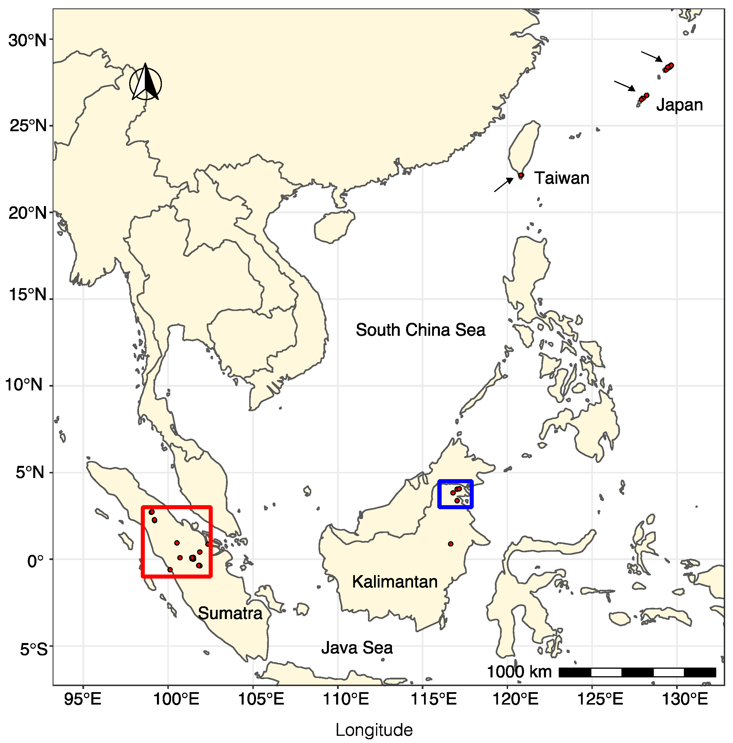
Randomly selected rivers and streams were sampled during several Phytophthora surveys in Taiwan, Japan, and the Kalimantan and Sumatra islands in Indonesia during the years 2013, 2018, and 2019 (Table S1). Forest rivers, rivers outside of a forest, mangroves, forest swamp soils, nursery ditches, and ponds were sampled. Naturally fallen leaves floating on the waterbodies were collected and directly plated onto selective PARPNH-agar [14]. Soil sampling and isolation methodology using young Fagaceae leaves as baits was performed, according to Jung et al. [14].
The complete list of “P. palustris” isolates used for this study and details of their sampling localities, dates, and collectors are given in Supplementary Table S1.

2.2. RNA Extraction

The total RNA of 112 isolates of “P. palustris” was purified from approximately 100 mg of fresh mycelium using an RNAzol® RT Column Kit [48] and treated with a TURBO DNA-free™ Kit (Thermo Fisher Scientific, Waltham, MA, USA). RNA quantity was checked in a Qubit® 2.0 Fluorometer (Thermo Fisher Scientific, Waltham, MA, USA). RNA quality was checked by Tape Station 4200 (Agilent), resulting in an RNA integrity number (RIN) of at least 7. Ten pools of RNA were prepared according to the provenance of “P. palustris” isolates and RNA quality (Table S1). Pool JP-TW contained RNA from seven Japanese and five Taiwanese isolates; both JP1 and JP2 contained nine RNAs from Japanese isolates; KA1, KA2, and KA3 enclosed, respectively, 12, 10, and 8 RNAs from Kalimantan isolates; and pools SU1, SU2, SU3, and SU4 enclosed, respectively, 12, 12, 14, and 14 RNAs from Sumatran isolates.

2.3. RNA Library Preparation and Sequencing

Approximately 1 μg of total RNA eluted in RNase-free water was sent to SEQme s.r.o (Dobris, Czech Republic) for RNA library construction and deep sequencing. The rRNA was depleted with an NEBNext rRNA Depletion Kit (Human/Mouse/Rat) and constructed with an NEBNext Ultra II Directional RNA Library Prep Kit for Illumina and NEBNext Multiplex Oligos for Illumina (Unique Dual Index Primer Pairs) (NEB, Ipswich, MA, USA). Library QC was assessed in an Agilent Bioanalyzer 2100 High sensitivity DNA Kit. A KAPA Library Quantification Kit for Illumina platform was used for absolute, qPCR-based quantification of the Illumina libraries flanked by the P5 and P7 flow cell oligo sequences. Libraries underwent paired-end (PE) (2 × 150 nt) sequencing on a NovaSeq6000 (DS-150) (Illumina, San Diego, CA, USA) using a NovaSeq S4 v1.5 reagent kit. An “in-lane” PhiX control spike was included in each lane of the flow cell.

2.4. De Novo Virus Assembly and Detection Workflow

Raw data were automatically processed by the BaseSpace cloud interface (Illumina) in default settings. The basecalling, adapter clipping, and quality filtering were carried out using Bcl2fastq v2.20.0.422 Conversion Software (Illumina). The quality of raw reads was checked using FastQC (v0.11.9) (https://www.bioinformatics.babraham.ac.uk/projects/fastqc/, accessed on 28 August 2022) and MultiQC (v1.9) (https://multiqc.info/, accessed on 28 August 2022). The raw data were cleaned from low-quality reads (quality Phred score cutoff: <20), and adapter sequences and very short sequences (≤25 bp) were removed for both reads before sequence pairing using Trim Galore (0.6.4_dev) (https://github.com/FelixKrueger/TrimGalore, accessed on 28 August 2022). The trimmed reads were aligned to host reference sequence using BWA mem (v0.7.17-r1188) (https://github.com/lh3/bwa, accessed on 28 August 2022) with default settings. Since the real host genome is not available, the genome of Phytophthora parasitica was randomly chosen and used as a reference, but two closer genomes became recently available (Phytophthora quininea strain Ex-type BL 54 and Phytophthora macrochlamydospora strain Ex-type BL). The unmapped reads were extracted and converted to fastq format using SAMTOOLS 1.7. (v1.7) (https://github.com/samtools/, accessed on 28 August 2022) and BEDTOOLS 1.7. (v2.29.0) (https://github.com/arq5x/bedtools2). These reads were used as input to the de novo assembly step using SPAdes genome assembler (v3.11.1) (https://github.com/kbaseapps/kb_SPAdes, accessed on 28 August 2022). The contigs for each sample were aligned using BLAST+ (v2.9.0+, BLASTn and BLAST) [49] to viral protein reference, non-redundant viral protein, host genome, mitochondrion, Phytophthora spp., rDNA, Boseq sp AS-1, bacterial genome, and cds sequences. All contigs with >79.9% similarity to host were eliminated.
Consequent analyses, including the determination of the final virus sequences, primer design, detection of open reading frames (ORF), protein translation, pairwise (pw) sequence comparison (PASC), and read number calculation, were performed using the platform Geneious Prime® 2021.0.4. and 2022.0.1. For the calculation of the coverage depth, we used the following formula: (Total reads mapped to the final identified virus * average read length)/virus genome or contig length).

2.5. Genetic Variability Analyses and Conserved Domains

Pairwise identities of the nucleotide and amino acid sequences were obtained after aligning the viral nucleotide and amino acid sequences by MAFFT V1.4.0 and calculated in Geneious Prime® 2021.0.4. In order to search for conserved domains within the putative viral proteins, the NCBI CDD-search tool was used (https://www.ncbi.nlm.nih.gov/Structure/cdd/wrpsb.cgi, accessed on 28 August 2022).

2.6. Phylogenetic Trees

Maximum likelihood (ML) phylogenetic trees were constructed using a rapid bootstrapping algorithm [50] in RAxML-HPC v.8 on XSEDE, conducted in CIPRES Science Gateway [51]. Tree search was enabled under the GAMMA model to avoid thorough optimization of the best scoring ML tree at the end of the run. The Jones–Taylor–Thornton (JTT) model was chosen as a substitution model for proteins. Bootstrapping was configured with the recommended parameters given by CIPRES Science Gateway. The resulting data were visualized using the software FIGTREE version 1.4.4.

2.7. Rapid Amplification of cDNA Ends (RACE) and Confirmation of Viruses’ Occurrence by Direct Reverse-Transcription Polymerase Chain Reaction (RT-PCR)

To confirm the length of the RNA 1 of botybirna-like virus 2 and RNA 1 and 2 of bunya-like virus 11 (see Results), we used the SMARTer RACE 5′/3′ KIT (TAKARABIO USA, Inc., Mountain View, CA, USA), as described in [46], and following the producer’s instructions. The occurrence of each identified virus was confirmed by direct reverse-transcription polymerase chain reaction (RT-PCR) with specific primers using total RNA as a template. The screening did not differentiate among variants, and the variant-level prevalence was not studied. A High-Capacity cDNA Reverse Transcription Kit (Applied Biosciences, Park Ave, NY, USA) was used for the cDNA synthesis. PCRs were performed with a Hot Start Taq 2× Master Mix (New England BioLabs, Ipswich, MA, USA) including 25 µL Master Mix, 1 µL of each primer (10 mM), and 4 µL of cDNA in a total volume of 50 µL. RT-PCR products were visualized using gel electrophoresis (120 V; 60 min). Analyzed fragments were separated on 1.5% agarose gel prepared with a TBE 1X buffer (Merck KGaA, Gernsheim, Germany) and stained by Ethidium bromide (SIGMA-Aldrich, Steinheim, Germany). PCR products showing the amplicons of expected length were purified and sequenced by GATC BioTech (Eurofins; Konstanz, Germany) by both directions with the primers used for PCR amplification. All the primers used for the partial amplification of the RNA-dependent RNA polymerase (RdRP) of each virus (Table S2) were designed by Primer 3 2.3.7 under Geneious Prime® 2020.0.4.

2.8. Graphs and Visualizations

Maps and graphs were created based on physically measured GPS coordinates using the R version 4.0.5 [52] programming environment using the special libraries ggplot2, sf, mapplots, ggspatial, scatterpie, and ggrepel.

3. Results

3.1. Virus Identification

A total of 10 RNA libraries containing 10–15 RNAs pooled from “P. palustris” isolates from Japan, Taiwan, and Indonesia (Kalimantan and Sumatra) were constructed and sequenced (Table 1 and Table S1). After trimming and quality checking, ~5200 million reads were obtained (Table S3). Further removal of reads mapped to the host genome yielded a total of 259,086,400 reads. The number of host-mapped reads was very variable among the different pools (Table S4) but generally very high, always higher than 84%. The highest percentage was obtained in pools JP2, SU2, and SU4 (>98%) and the lowest in pool KA1 (84.41%). A total of 730,972 possible viral contigs (length > 0–25,000 bp) were obtained by de novo assembly of viral contigs (Table S5). After being aligned using BLAST+ to all the databases mentioned in the Materials and Methods section, virus contigs were finally identified to represent 52 viruses related to five virus families and orders: Totiviridae and Botybirnaviridae with dsRNA genomes; Bunyavirales with (−)ssRNA genomes; and Narnaviridae and Tombusviridae as (+)ssRNA genomes (Table 1). No DNA viruses were identified applying our pipeline.

3.2. Virus Genomic Organization and Phylogenetic Relationships

3.2.1. (−)ssRNA Viruses

Based on the BLASTX comparison, a total of 18 viral contigs with affinities with the L (large) segment of putative members of the order Bunyavirales were assembled and characterized in Sumatran and Kalimantan pools (SU1-4 and KA1-3) (Table 1). All of them enclosed a unique large open reading frame (ORF) encoding the RNA-dependent RNA polymerase (RdRP) (Figure 1a). In addition, one contig showed a high identity percentage with nucleocapsid proteins (NC) of the family Phenuiviridae. The 17 putative viruses were designated as Phytophthora palustris bunya-like viruses (PpaBLV) (Table 1). The PASC of the 17 bunya-like RdRP nucleotide (nt) and amino (aa) sequences showed an overall pw identity of 34 and 39.4%, respectively. When all the RdRP nt and aa sequences were compared, several contigs were seen to have a pw identity >90% (Tables S8 and S9); therefore, those sequences were designated as variants of the same putative virus. Thus, both PpaBLV 9 and 14 had three variants. In ICTV, there are no primary classification and delimitation criteria for genus and species in the order Bunyavirales, and PASC and phylogenetic analyses seem to be the main point of reference to name new bunyaviruses.
Conserved domains (CDD) of the Bunya_RdRP superfamily cl20265 were found in bunya-like virus contigs 1, 2, 3, 4, 5, 6, 8, 9 (variant 1), 10, 11, and 13 (Table 1). The amino acid alignment of the CDD regions (Figure S1a) showed high similarities of the premotif A and motifs A (DxxxWx), B (XGxxNxxSS), C (SDD), D (KK), and E (ExxSx) with the rest of the bunyaviruses included in the alignment. Premotif A with the three basic residues inside (K, R, and R/K) and, downstream, the glutamic acid (E), were also identified. The conserved aa triplet TPD (threonine), typical of bunyaviruses, was also detected [53]. The aa sequence of the NC of the Phytophthora palustris bunya-like virus 11 encloses a conserved motif of pfam05733, the only member of the superfamily cl05345 with a significant e-value (2.08 × 10⁻⁷) (Table 1, Figure 1a). This family consists of several Tenuivirus and Phlebovirus nucleocapsid proteins (Figure S1, Table 1). In addition to the conserved domain of Bunya_RdRP, an L-protein N-terminus (and endonuclease domain) present in the N-terminus of many bunyavirus L proteins was found at nts 7912–8166.
On the basis of the RACE analysis, we determined the possible full viral genome sequence of PpaBLV11, which comprised RNA1 (6448 nt) and RNA2 (864 nt) (Figure 1a). The RNA2 sequence is shorter than the NGS contig (ca. 1 kb) because only 2 relatively short sequences of 3′ were obtained by the RACE analysis. Moreover, the coverage of the ends of the NGS-RNA2 contig was very low and had very low quality. The 5′ and 3′ termini of RNA1 and RNA2 had 9 and 8 conserved nts, respectively (Figure 1b). A total of 9 sequential nts at the 5′ and 3′ termini of RNA1 and 8 nts in RNA 2 were complementary to each other (Figure 1c). The comparison of the 5′ and 3′ terminal nucleotides of PpaBLV11 to those of other bunyaviruses showed a high identity. Higher variation appears to occur in the 3′ terminus of the RNA2, having a C, which is lacking in the rest of the 3′ termini of the compared bunyaviruses; similarly, no complementary G is found in the 5′ terminus of PpaBLV1 RNA1.
ORF1, the unique large ORF found in RNA1, encodes a putative protein of 2084 aa (codons) and a molecular weight of 241.396 kDa (p241), and 5′ and 3′ untranslated regions (UTRs) comprising 72 and 121 nt, respectively (Figure 1a). ORF2, the only ORF found in RNA2, encodes a putative protein of 230 aa and a molecular weight of 26.459 kDa (p26).
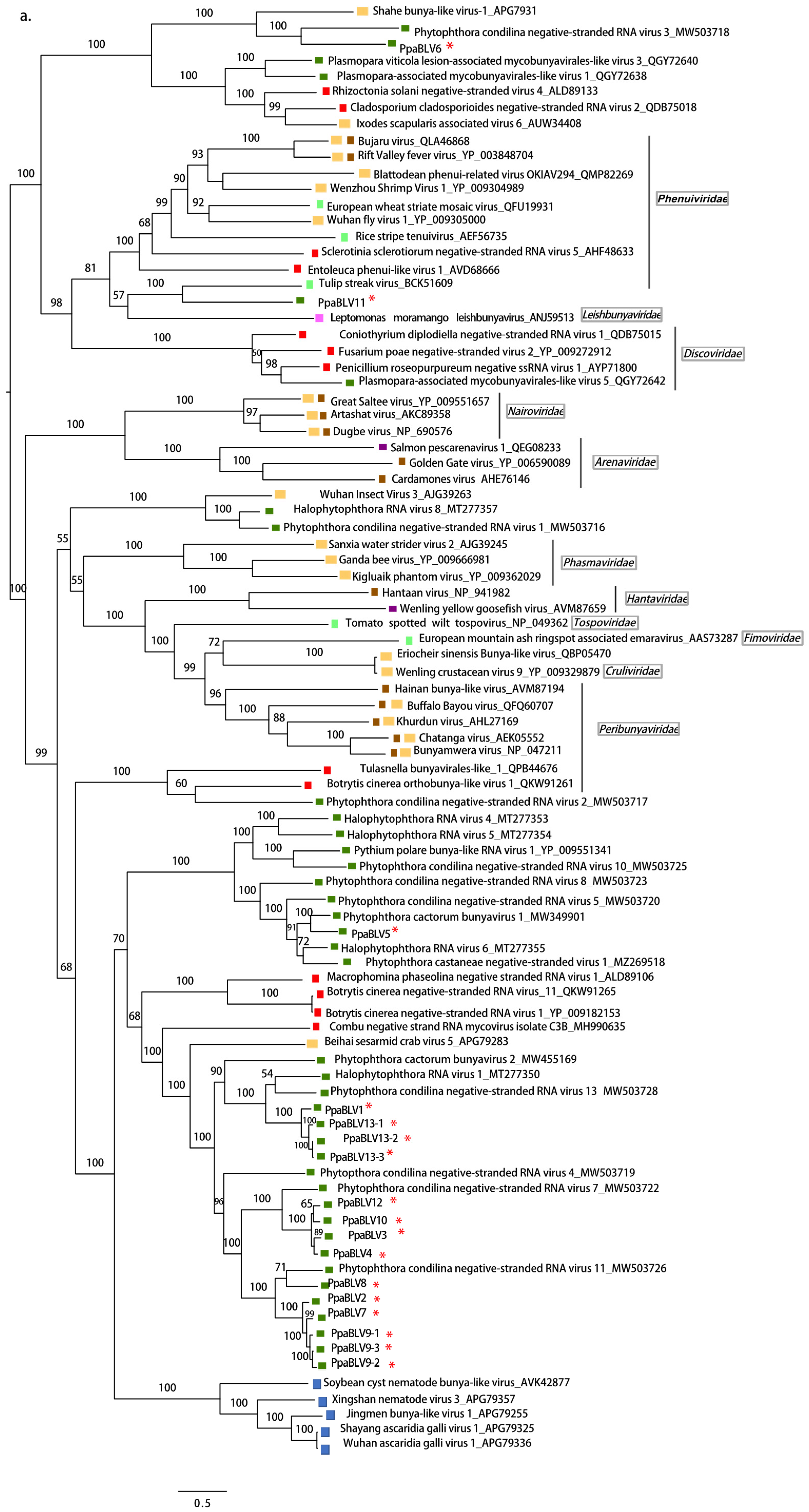
A comprehensive reconstruction of the phylogenetic relationships of “P. palustris” bunya-like viruses’ RdRP with other bunyaviruses deposited in the GenBank is shown in Figure 2a. The majority of “P. palustris” bunya-like viruses cluster together and seem to be closer to Phytophthora condilina negative-stranded RNA virus 4 (PcoNSRV4), Halophytophthora RNA virus 1 (HRV1), both viruses described in marine oomycetes collected in estuarine ecosystems in southern Portugal [29,42], and to Phytophthora cactorum virus 1 (PcBV2), described in several isolates of P. cactorum causing crown rot in strawberries in Finland [33]. PpaBLV5 appears jointly with Phytophthora cactorum bunya virus 1 [33] in a closely related cluster, which just hosts bunya-like viruses found in oomycetes. PpaBLV6 is the most distinct taxon, clustering with Phytophthora condilina negative-stranded RNA virus 6 (PcoNSRV6) [29]. These results indicate a close evolutionary relationship among bunya-like viruses, described in different species of oomycetes collected from different environments.
The NC of PpaBLV11 appears to be phylogenetically related to NCs of an insect virus (genus Phasivirus) and a plant virus, and not related to those of fungal phenuiviruses (Figure 2b).

3.2.2. (+)ssRNA Viruses

Tombus-like virus. One viral contig of 4180 bp found in the Sumatran pool SU2 appeared to have phylogenetic affinities with unclassified members of the family Tombusviridae. Although the best hit subjects in the BLASTX comparison were sequences obtained in a metatranscriptomic study from soil dominated by stands of Avena species (Table 1), other related results were Leuven tombus-like virus 3, described from larvae of predatory wasps in Belgium (e-value 8 × 10−49; identity ~32%), and Serdyukov virus (e-value 5 × 10−45; identity ~36%), reported in the ticks of Antarctic penguins [54].
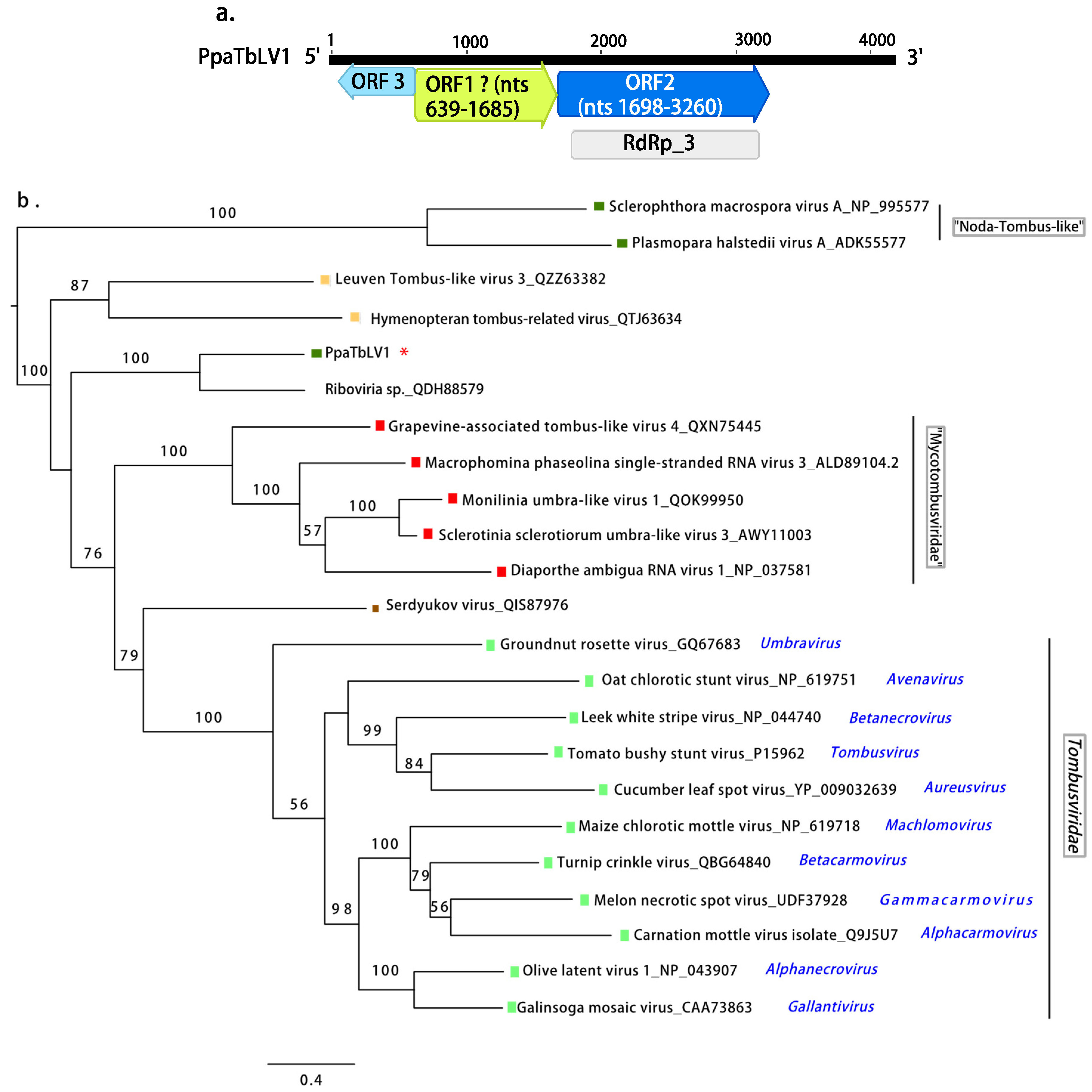
When using the standard translation code, the contig encodes two positive-sense ORFs (Figure 3a), and the ORF1 or 5′ end-proximal ORF encodes an unknown protein with no conserved domains detected. The ORF2 or 3′ end-proximal ORF encodes the RdRP. The conserved domain of the RT-like superfamily (cl2808) is located at nt positions 1797–3191 (e-value 2.11 × 10−57), particularly, the superfamily member pfam00998 (Viral RdRP_3) found in Hepatitis C virus and other plant viruses (Figure S2). A third antisense ORF (nts 635-63) is detected proximal to 5′ end and just separated by one proline (Pro) from the next ORF1. The resultant protein (191 aa) does not have any similar records when blasted to GenBank. This contig was named Phytophthora palustris tombus-like virus 1 (PpaTbLV1). The phylogenetic tree (Figure 3b) shows that PpaTbLV1 is more related to tombusviruses infecting insects rather than those infecting the obligate biotrophic oomycetes Sclerophthora macrospora [55] and Plasmopara halstedii [42].
Narna-like viruses. Virus contigs resembling members of the family Narnaviridae were abundant in all pools. After removing redundant contigs and selecting the longest ones, 10 viruses were determined. Eight of them had lengths longer than 2.5 kb and enclosed a complete single large ORF, likely representing nearly full-length virus genomes (Table 1; Figure 4a).
Two contigs contained incomplete ORFs and represented partial genomes of two potential virus isolates. No narnavirus conserved domains were detected in any of the putative virus contigs in the Conserved Domain Database (CDD) but the BLASTX search confirmed their similarity with unclassified narna-like viruses found in the fungal pathogen Eryshiphe necator and in invertebrates, and with Phytophthora infestans RNA virus 4 (Table 1). The alignment of the conserved motifs of RdRP of the narna-like viruses is shown in Figure S4. The overall PASC of the eight narna-like contig nt and RdRP aa sequences were 34.2 and 27.1%, respectively. Based on PASC in the nt and aa sequences (Tables S10 and S11) and the species demarcation criterion proposed in the ICTV for the family Narnaviridae, eight possible viruses designated as Phytophthora palustris narna-like viruses 1-8 (PpaNLV1-8) were represented in these 10 contigs. PpaNLV3 was represented by three variants as the pw identity of their aa sequences was higher than 90%.
The phylogenetic analyses, based on the aa sequences of their RdRP (Figure 4b), showed that “P. palustris” narna-like viruses are grouped in two different clusters. PpaNLV2, 3 (including all 3 variants), 4, 5, and 8 appeared in a detached group, maybe representing a different taxon within the “alphanarnavirus” clade, and closer to Saccharomycces 23S RNA narnavirus and Saccharomycces 20S RNA narnavirus. Within the clade, “betanarnaviruses” PpaNLV1 and 6 appear together with narnaviruses described in Phytophthora spp. and PpaNLV7 groups with two unclassified viruses described in an insect and in symptomatic grapevine tissue associated with the downy mildew Plasmopara viticola.

3.2.3. dsRNA Viruses

Botybirna-like viruses. Four contigs >6 kb in length enclosing one large ORF resembling the RdRP segment of unclassified botybirnaviruses present in fungi and oomycetes (Figure 5a; Table 1) were found in two pools from Sumatra (SU1, SU3) and one from Kalimantan (KA3). No second segment was detected in our data. These viral contigs were named as Phytophthora palustris botybirna-like viruses (PpaBbLV). The PASC of the nt and aa sequences of the four contigs showed high identity between them (Tables S6 and S7), with particularly high identity levels (>92%) between the Sumatran contigs. Because the ICTV does not specify virus species demarcation criteria for this family, we classified the four sequences as belonging to two viruses: three contigs represent three Sumatran variants belonging to PpaBbL1 while one represents a Kalimantan botybirna-like virus. CDD belonging to the RT-like superfamily (cl2808) and, particularly, the superfamily member pfam02123 (RdRP_4, which includes RdRPs from Luteovirus, Totivirus and Rotavirus) were also found (Figure 5a and Figure S3; Table 1). The reconstruction of the phylogenetic relationships of PpaBbLV1 and 2 with other dsRNA viruses (Figure 5b) shows that PpaBbLV1 and 2 form a novel cluster separated from other botybirnaviruses found in fungi and oomycetes, suggesting an evolutionary diversification among the mycoviruses of this family.
Toti-like viruses. Virus contigs resembling members the family Totiviridae were also abundant in all pools. After removing redundant and shorter ones, 20 contigs were characterized and named Phytophthora palustris toti-like viruses (PpaTLV). Based on the PASC (Tables S12–S14) and ICTV criterion for the species demarcation within the family Totiviridae, 14 totiviruses were described (Table 1), and those viral contigs with pw identity higher than 90% in both nt and aa sequences were considered variants of the same virus: PpaTLV1 (variants 1 and 2), PpaTLV2 (variants 1, 2, and 3), PpaTLV3 (variants 1, 2, and 3), PpaTLV4, PpaTLV5 (variants 1 and 2), PpaTLV6, 7, 8, 9, 10, 11, and PpaTLV12 (variants 1, 2, and 3). Most of the contigs consisted of ~5–6 kb sequences enclosing two ORFs. Only PpaTVL4 and 8 were shorter and contained one ORF. Conserved domains belonging to the superfamily member pfam02123 (RdRP_4) were found in the 3′-terminus proximal ORF in all the contigs (Figure 5a and Figure S3, Table 1). In addition, conserved domains of the totivirus coat superfamily, cl25797 and pfam05518, were found in the 5′-terminus proximal ORF of PpaTLV1-1, 1-2, 2-1, 2-2, 2-3, 5-1, 5-2, 6, 9, 10, and 11 (Figure S3, Table 1).
The phylogenetic relationships of PpaTLVs, reconstructed based on their aa RdRP sequences, show a high degree of evolutionary differentiation among them. Whilst PpaTLV1, 2, 5, 6, 10, and 11 appear to cluster with viruses belonging to the genus Victorivirus described in different fungi (Figure 5b), PpaTLV3 falls into the cluster with mycoviruses classified in the genus Totivirus. In a separated cluster, PpaTLV12 is phylogenetically closely related to viruses from Plasmopara viticola, Pythium, and Phytophthora spp. from diverse ecosystems, which form an oomycete-specific cluster related to Giardia canis virus (GCL). PpaTLV4, 7, and 8 cluster with unclassified toti-like viruses reported from fungi, oomycetes (including P. condilina), and diatoms.

3.3. Virus Abundance, Diversity, and Geographical Distribution

3.3.1. Virus Occurrence Based on the RT-PCR Screening

All the viruses detected by RNA-Seq were confirmed to occur in 45 isolates of “P. palustris” from 25 Sumatra and Kalimantan localities (Figure 6) after performing ca. 1800 PCRs. No virus presence was confirmed by RT-PCR in Japanese and Taiwanese isolates.
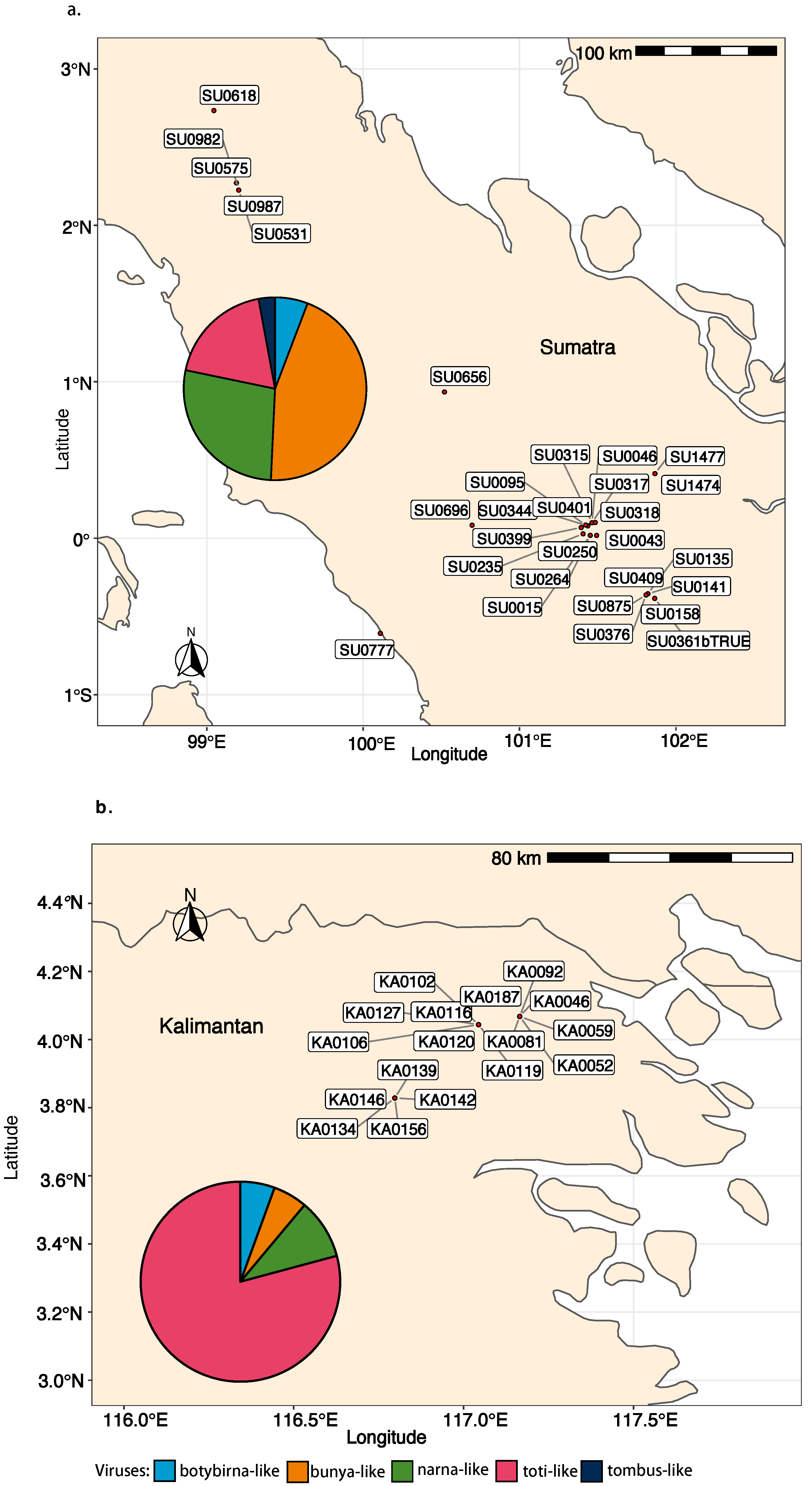
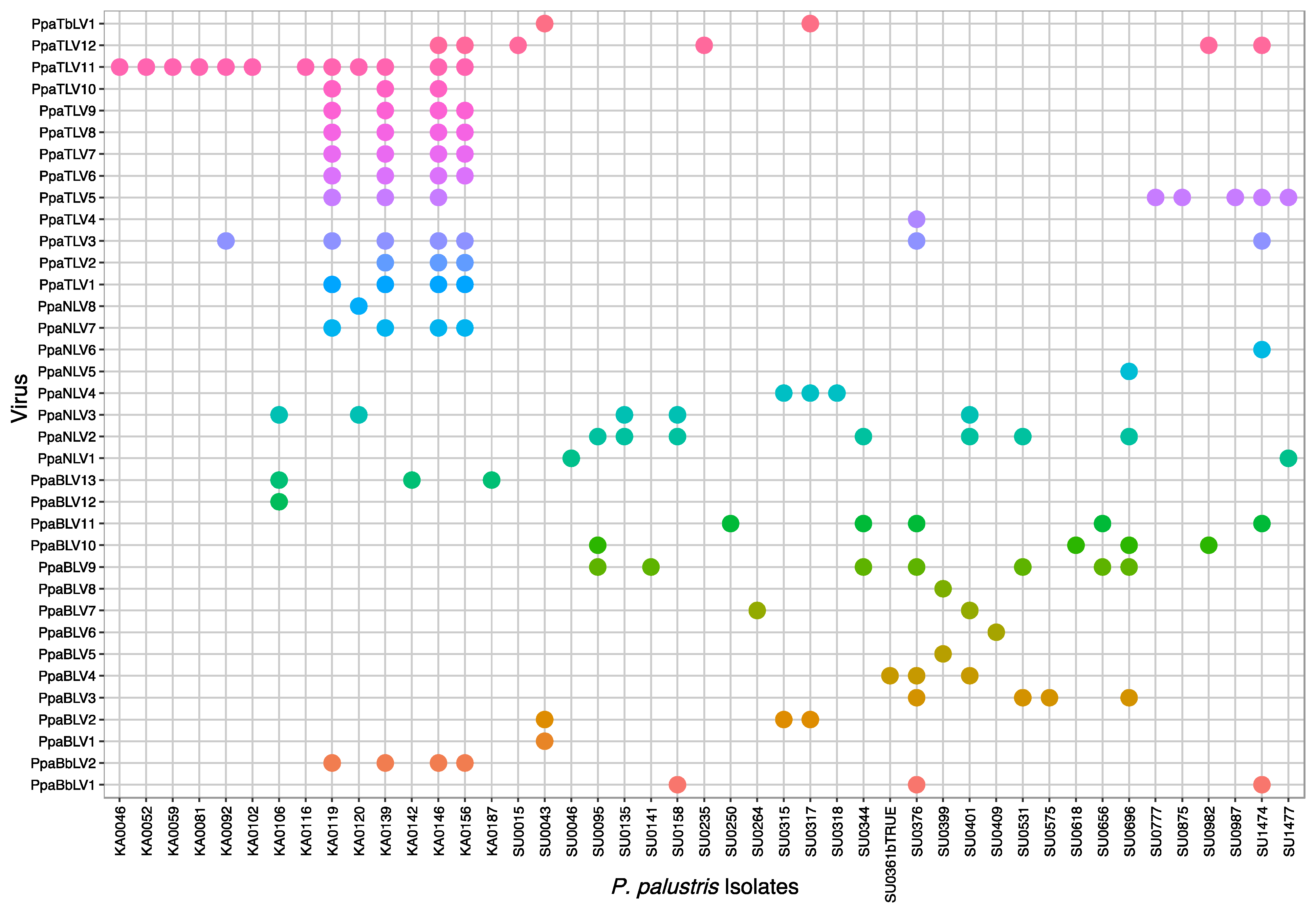
In Sumatra, 9 (SU_F11, F42, R02, R04, R05, R09, R13, R16, R29) out of 30 sampling sites (30%) did not appear to host any of the screened viruses. However, in 21 of the locations (70%), at least one virus was detected. A total of 30 “P. palustris” isolates (58%) hosted at least one virus, and 15 of them showed viral coinfections or multiviral infections. In particular, isolate SU0376 appeared to be infected by seven distinct viruses while SU1474 was infected by six viruses (Figure 7a, Figure 8, and Figure 9). In Kalimantan, isolates from four out the five sites screened (KA_R03, R05, R06, R08) contained viruses. Out of 30 “P. palustris” isolates, 15 isolates hosted at least one virus (50%), and seven of them (47%) showed coinfections and multiviral infections. “P. palustris” isolates KA0119, KA0139, KA0146, and KA0156 hosted at least 11 viruses (Figure 7b, Figure 8, and Figure 9).
The virus richness appears to be greater in Sumatra (23 viruses) than in Kalimantan (17 viruses) (Figure 8 and Figure 9). Moreover, in our samples from Sumatra, the virus family composition was more diverse (Figure 7, Figure 8, and Figure 9), as a tombus-like virus (PpaTbV1) was found in two isolates collected from two Sumatran sites (SU_R06 and R07). Conversely, with 62 and 66 confirmed viruses, respectively, total virus abundance in “P. palustris” seems to be similar in Kalimantan and Sumatra. However, virus abundance in relation to the number of isolates tested from each of the two islands showed considerable differences: Kalimantan 63/30 = 2.1, Sumatra 66/52 = 1.27. Notably, with 14 different viruses, the Kalimantan site R06 hosted the highest virus diversity of the whole study.
Comparisons of the geographical distribution of “P. palustris” viruses in Sumatra and Kalimantan show that some viruses have a wider distribution than others (Figure 8 and Figure 9). Thus, five viruses, including three toti-like viruses (PpaTLV3, PpaTLV5, and PpaTLV12) and one narna-like virus (PpaNLV3), even occurred in “P. palustris” isolates from both Sumatra and Kalimantan. PpaTLV3 was found in seven isolates collected at five sites (three in Kalimantan and two in Sumatra). PpaTLV5 was the most widely distributed virus, infecting eight isolates from six different localities (two in Kalimantan and four in Sumatran). Finally, PpaTLV12 infected six isolates from five localities (one from Kalimantan and four in Sumatran). Within the same island, PpaTLV11 was present in 12 isolates from two Kalimantan localities, and PpaBLV9 infected 7 isolates obtained from seven Sumatran localities. Finally, the results show that, in general, toti-like viruses are more abundant and diverse in Kalimantan whereas bunya-like viruses are more prevalent and diverse in Sumatra.
According to the average pw identity percentages of the alignment of the amplicon sequences (variants) with the virus contig sequences obtained by NGS (Table S15), some viruses also have more genetic stability than others, independently of their occurrence in two or more “P. palustris” isolates, i.e., the three variants of botybirna-like virus 1 (PpaBbLV1) are more diverse than the four variants of PpaBbLV2.

3.3.2. Virus Read Abundance per Pool and Island
There is a large variation in the total viral read numbers between Japan, Taiwan, and Indonesia (Kalimantan and Sumatra) as well as between specific virus contigs. As shown in Figure S5, Sumatran libraries contained the highest percentage of viral reads, while Japanese and Taiwanese libraries contained the lowest. Likewise, the narna-like viruses constituted the highest read numbers (Table 1, Figure S5), with the PpaNLV 3-1, 3-2, 3-3, and 4 being clearly the most abundant viruses, occurring in libraries KA1, KA2, and SU1-SU3. Bunya-like viruses’ reads also appeared to be very abundant, in particular, the three variants of PpaBLV9 (found in libraries SU1–SU4). Interestingly, despite not having been confirmed by RT-PCR, a total of 608 reads were mapped to bunyavirus 8 (PpaBLV8) in all three libraries JP-TW, JP1, and JP2, while 68 reads were mapped to narna-like virus 8 (PpaNLV8). These reads scarcely covered these two virus sequences, repeatedly mapping short regions of those viruses. The abundance of toti-like and tombus-like viruses’ reads did not appear to be correlated with their distribution and richness. Compared to the high diversity and distribution of toti-like viruses, the number of reads for each contig was low, while the only tombus-like virus contig, which occurred mostly in SU2, had nearly 200,000 reads.
Figure 8. Plot representing the virus occurrence and diversity for each “P. palustris” isolate.
Jof 08 01118 g008
Figure 9. Plot representing the virus occurrence and diversity for each sampling site.
Jof 08 01118 g009

4. Discussion

The virus composition, abundance, and distribution discovered in our study indicate that viruses related to the families Totiviridae, Tombusviridae, Narnaviridae, and Botybirnaviridae and the order Bunyavirales are naturally infecting “P. palustris” isolates in their niches and must be readily carried by “P. palustris” propagules, revealing that they are primarily members of the host cellular environment.

4.1. Evolutionary Insights into Novel Taxa of (+)ssRNA Viruses

PpaTbLV1 is the first tombus-like virus described in a Phytophthora species, but not the first one in oomycetes. Four tombus-like viruses have been identified in Plasmopara-viticola-associated lesions on grapevines in Italy [43]. Plasmopara halstedii virus (PhV) and Sclerophthora macrospora virus A (SmV-A) have been detected in the downy mildew of sunflower Plasmopara halstedii [55] and the grass pathogen Sclerophthora macrospora [42], respectively, although PhV and SmV-A have been assigned to a new putative viral group between Nodaviridae and Tombusviridae. Based on the BLASTX and the phylogenetic analyses of this study (Figure 3b), PpaTbLV1 does not appear to be closely related to these viruses but to insect viruses and a virus found by a metagenomic approach in soils of wild oat (Avena fatua), an annual grass common in Mediterranean climates [56]. Interestingly, PpaTbLV1 is not grouped either with recognized plant tombusviruses, such as tomato bush stunt virus (TBSV) or with fungal tombus-like viruses (mycotombusviruses), such as Diaporthe RNA virus (DRV) [57]. However, PpaTbVLV1 genomic properties are closer to the latter, as shown by similar genome size (~4.2 kb), apparent absence of movement proteins typical of plant viruses, and the presence of two main ORFs encoding a hypothetical protein of unknown function and RdRP (Figure 3a). The RdRP is presumed to be expressed as a fusion product with the 5′-proximal protein via readthrough of the amber termination codon, UAG, of the upstream ORF [58]. However, PpaTbLV1 possesses GDD as the catalytic triplet in the motif-C of its RdRP (Figure S2), in contrast with a common feature in mycotombusviruses, which have GDN as the RdRP catalytic triplet [59]. Intriguingly, a smaller antisense ORF encoding an unknown protein is detected proximal to the 5′-terminus, which seems to be a unique property, not yet found in other tombus-like viruses. Thanks to high-throughput sequencing (HTS) studies, ambisense coding genomes emerge more and more commonly within RNA viruses and, particularly, in fungal viruses, such as ambiviruses [60] and “ambinarnaviruses” [61].
PpaTbLV1 was confirmed to be carried by two isolates of “P. palustris” collected in two Sumatran localities, within a distance of a few kilometers (Figure 8 and Figure 9, Table S1). Hence, it seems possible that PpaTbV1 was transported by swimming zoospores and then exchanged via anastomosis between compatible individuals of “P. palustris”. Members of the “P. palustris” complex are aquatic oomycetes, and, interestingly, several reports have identified tombusviruses in association with rivers and lakes throughout the world [62,63]. TBSV and Carnation Italian ringspot virus (CIRV), have been isolated from waterbodies draining forest areas in Northrhine-Westfalia, Germany [64]. In addition, tombus-like viruses are also hosted by unicellular protists [65], they have been associated with the holobiont of freshwater shrimp [66], and have been discovered on marine and freshwater RNA viromes using metagenomics [67].
The family Narnaviridae is undergoing an intense reconstruction, as more and more new narna-like viruses with very diverse genomic properties have been discovered lately [58]. Until recently, it was generally understood that narnaviruses had monopartite (+) ssRNA genomes enclosing solely an essential RdRp gene encoded by a single ORF. However, it has been discovered that some narnaviruses have divided RdRPs (“splipalmiviruses”), i.e., viruses hosted by deep-sea fungi, such as Aspergillus tennesseensis. Others have bi- and multi-segmented genomes (reviewed in [58]) or possess long-reverse-frame ORFs, i.e., Plasmopara-viticola-associated narnaviruses [43]. Although we cannot rule out the possibility of segmented genomes, “P. palustris” narna-like viruses seem to be phylogenetically related to traditional monopartite positive-coding narnaviruses included in both “alphanarnaviruses” and “betanarnaviruses” clades (Figure 4b). Likewise, no reverse large ORFs were detected in any of the final contigs. The phylogenetic tree and the pairwise comparisons (Tables S10 and S11) show moderate levels of genetic variability. Whilst the narna-like viruses’ richness in “P. palustris” is remarkable (eight putative viruses are described), five of them are closely related and cluster together, suggesting they might have originated from the same “P. palustris” host and gradually incorporated genetic modifications over time [68]), probably during the replication process due to random mutations and genetic adaptations to a new geographical area [69,70,71]. A common ancestor might also explain the evolutionary relationship of PpaNLV1 with a narnavirus found in Phytophthora castaneae in Vietnam [40], and PpaNLV6 with a narnavirus found in P. infestans [34]. Narnaviruses seem to be effectively transmitted throughout “P. palustris” populations and are widespread in both Sumatra and Kalimantan. A certain relation between the high narnavirus abundance and read number obtained, in particular, for PpaNLV2, 3, and 4 (Table 1, Figure S5), agrees with the frequent occurrence of these viruses on both islands.

4.2. Discovery of New dsRNA Viruses and High Abundance of Toti-Like Viruses

Kalimantan and Sumatran isolates of “P. palustris” have been found to host two putative botybirna-like viruses (PpaBbLV1 and 2). While genetically highly similar (Tables S6 and S7), each PpaBbLV occurs in several isolates and more than one site but on separate islands, PpaBbLV1 in Sumatra and PpaBbLV2 in Kalimantan (Figure 8 and Figure 9). This constitutes the first discovery of a putative botybirnavirus in a Phytophthora species. Previously, two botybirnaviruses were identified in grapevine lesions associated with Plasmopara viticola in Spain and Italy [43], and in several fungal plant pathogens, including Botrytis porri [72] and Sclerotinia sclerotiorum [73]. Botybirnaviruses have a bipartite genome, with two types of structural proteins (p85/80, p70) and an RdRP. However, only the segment enclosing the RdRP was identified in our study. Both PpaBbLV 1 and 2 appear to be phylogenetically distinct from other mycobotybirnaviruses, representing a potential new taxon. Moreover, aside from motifs I and II, the rest of the RdRP domains closely resemble those of totiviruses (Figure S3).
P. palustris” isolates are mainly infected by totivirus-related viruses. They are widespread in both Kalimantan and Sumatra (Figure 8 and Figure 9) and novel Bona-fide totiviruses, victoriviruses, and toti-like viruses show paraphyletic relationships across the phylogenetic tree and considerable genetic diversification (Figure 5b). Some viruses reside in an oomycete-specific cluster while others cluster with unclassified toti-like viruses reported from fungi, oomycetes and diatoms dwelling in a wide range of ecosystems. This result may suggest long-term coevolution between “P. palustris” and its totivirus-like viruses.

4.3. Persistence of Bunya-Like Viruses and the First Oomycete Bunyavirus with NC Protein

Similar to recent studies on marine oomycetes [33,46], high variability and abundance of the L segment (RNA 1) of putative bunyaviruses were also found in “P. palustris”, most of them phylogenetically closely related but with an overall high pairwise genetic variability that allows the description of 13 viruses with different variants. Such virus richness can be the consequence of mutation, recombination and reassortment during the course of evolution. Viral reassortment seems to be a powerful mechanism underlying the evolution of the Bunyavirales order, and it has been pointed out that most bunyaviruses described so far are actually reassortants of extant or extinct viruses [74]. Alternatively, it cannot be ruled out that the P. palustris bunya-like contig sequences would not represent true biological entities. Since bunyaviruses might be reassortants, which may sometimes be seen in their genomes as having highly conserved sequence stretches alternating with highly variable ones, putatively resulting in chimeric contigs in the de novo assembly when they are found abundantly in NGS-pools (e.g., [37]). The phylogenetic analysis (Figure 2a) shows the increasingly complex taxonomy of mycobunyaviruses with strong cluster support.
PpaBLV11 is the first bunya-like virus with bipartite genome reported in Phytophthora. It is related to phleboviruses, which seem to have a tripartite genome (three RNA segments). In this study, we only found two RNAs, RNA1 and RNA2, respectively, corresponding to the L and S segments of phleboviruses. Neither the M segment nor M segment-like contigs were detected during the NGS analysis. If PpaBLV11 has a tripartite genome, the M segment may not be considered essential. This is in accordance with the most similar virus, tulip streak virus (TuSV) [75] and fungal phleboviruses, as Lentinula edodes negative-strand RNA virus 2-HG3 (LeNSRV2-HG3) [76]. However, similar to the rest of the Phytophthora bunyaviruses, which seem to have monopartite genomes, it is more plausible that the genome description of these viruses is incomplete. The putative NC and other non-structural (Ns) associated proteins are likely not conserved enough to be detected by homology.

4.4. Virus Population Structure and Transmission Efficiency

Our study indicates that “P. palustris” viral populations adhere to a certain structure. Although the virus family composition is similar on both Kalimantan and Sumatra islands, the virus structure differs and appears to be specific to each island. In Kalimantan, toti-like viruses predominate, whereas bunya-like viruses are more prevalent in Sumatra. Furthermore, the absence of viruses in Japanese and Taiwanese “P. palustris” isolates suggests a degree of divergence regarding Indonesian members of the “P. palustris” complex and their viruses. However, it cannot be ruled out entirely that Japanese and Taiwanese isolates also host viruses. In fact, various viral reads have been mapped to PpaNV6 and PpaBLV8 in the RNA pools JP1, JP2, and TW-JP (Figure S5). If there are any viruses, they are not those found in Kalimantan and Sumatra. Since mycoviruses do not have an extracellular stage, they strongly depend on their host for dispersal, and an analogous evolutionary scenario is expected for both the host (“P. palustris”) and the hyperparasite (mycoviruses) [26]. However, due to geographical isolation, local adaptation, and drift, “P. palustris” seems to have evolved into new lineages, and the species P. sp. palustris in Japan and Taiwan is very different from all Indonesian taxa, indicating they have been separated from each other millions of years ago (T. Jung and M. Horta Jung, unpublished data), similarly to evolutionary divergent lineages of P. ramorum [16] and P. lateralis [13]. As a consequence of speciation processes in “P. palustris” hosts, the possibilities of the associated mycoviruses to coevolve and be always co-introduced with their host would significantly decrease. Multiple introduction routes do not necessarily imply that hyperparasites are always introduced along all of them [77]. Alternatively, the absence of viruses in these pools could be also due to the limited availability of similar viral sequences in GenBank; hence, a data re-analysis is highly recommended in a few years.
Our results suggest reasonable efficient viral transmission across Indonesian “P. palustris” populations. However, some “P. palustris” viruses are more widespread than others, merging in distinct Indonesian localities of the same island and even on different islands (PpaNLV3, and PpaTLV3, 4, and 12). Most likely these viruses evolved towards optimal virus–host interactions that enabled them to replicate inside their “P. palustris” hosts and move more efficiently through their host populations [77]. Members of the “P. palustris” complex are aquatic Phytophthora species and, hence, able to produce large amounts of motile chemotactic zoospores [78], which in waterbodies can be transported passively over long distances. However, “P. palustris” shares its aquatic habitat with nematodes, algae, crustaceans, and mollusks [79], with which they undoubtedly interact, and which might eventually serve as transport means of potential virus-hosting oomycete individuals. On the contrary, the lower occurrence of other viruses might be related to the lower efficiency of interspecies virus transmission, because five species of the “P. palustris” complex occur in Sumatra and Kalimantan (T. Jung and M. Horta Jung, unpublished data). This study did not observe a virus/“P. palustris” species correlation but it should be further investigated. Previous studies concerning virus transmission in P. infestans demonstrated 100% inheritance of Phytophthora infestans RNA virus 3 (PiRV3) in individual zoospores [80]. Phytophthora infestans RNA virus 2 (PiRV2) was readily transmitted by hyphal anastomosis, and by asexual reproduction through sporangia [80]. However, attempts to transfer PiRV-2 into apparently vegetatively incompatible P. infestans isolates failed.

4.5. Multiple Viral Infections at Isolate and Site Level

Particular Indonesian localities and “P. palustris” isolates appear to be big reservoirs of viruses (Figure 7, Figure 8 and Figure 9). From an ecological perspective, in those localities with higher virus diversity and abundance, the number of contact events between and among host individuals must be higher [26]. Thus, the probability of a virus-hosting individual having contact with another compatible virus-hosting “P. palustris” isolate increases with host population density. From an individual perspective, multiple viral infections are, indeed, known to occur in oomycetes in nature [23,37,40,43,46]. However, the interactions between different viruses and potential limits for the accumulation of multiple viruses in a single host remain unclear. In a native environment, long-term co-existence between virus and host should lead to a non-lethal equilibrium. Thus, the cytoplasmic exchange that takes place during fungal (and oomycete) growth promotes the accumulation of multiple viruses within a hypha, colony, or hyphal network [81]. Unlike fungi, oomycete hyphae lack septa, potentially facilitating the accumulation and exchange of viruses within and between compatible individuals. In addition, many Phytophthora species, in particular those thriving in aquatic environments, are prompted to interspecific hybridizations, which play a major role in speciation and species radiations in diverse natural ecosystems [82,83,84]. Interspecific hybridizations might enable virus transmission between different Phytophthora species.

Supplementary Materials

The following supporting information can be downloaded at: https://www.mdpi.com/article/10.3390/jof8111118/s1, Table S1. List of “Phytophthora palustris” isolates used in the present study, Table S2. Primers used for the confirmation of each virus presence in the collection of “Phytophthora palustris” isolates by RT-PCR and RACE, Table S3. Trimmed low quality and adaptor data, Table S4. Mapped reads to Phytophthora parasitica genome (used as a reference host genome), Table S5. Data of the resulting contigs obtained by the de novo assembly in each sequenced RNA library. Pairwise identities of the RdRp nucleotide (Table S6) and amino acid (Table S7) sequences of “Phytophthora palustris” botybirnaviruses, Table S8. Pairwise identities of the RdRp amino acid sequences of “Phytophthora palustris” bunyaviruses, Table S9. Pairwise identities of the RdRp nucleotide sequences of “Phytophthora palustris” bunyaviruses, Table S10. Pairwise identities of the RdRp nucleotide sequences of “Phytophthora palustris” putative narnaviruses, Table S11. Pairwise identities of the RdRp amino acid sequences of “Phytophthora palustris” narnaviruses, Table S12. Pairwise identities of the RdRp nucleotide sequences of “Phytophthora palustris” toti-like viruses, Table S13. Pairwise identities of the RdRp amino acid sequences of “Phytophthora palustris” toti-like viruses, Table S14. Pairwise identities of the CP amino acid sequences of “Phytophthora palustris” toti-like viruses, Table S15. Average pairwise (PW) identity of the alignment of the partial viral sequences (variants) with the virus contig sequences obtained by NGS, Figure S1. A. Amino acid alignment showing Bunya_RdRp conserved motifs A to E and premotif A within the RdRp of PpaBLV1-14 and selected bunyaviruses, B. Amino acid alignment showing the conserved motifs of Tenuivirus/Phlebovirus nucleocapsid protein within PpaBLV11 and selected bunyaviruses, Figure S2. Amino acid alignment showing the conserved RdRp conserved motifs of PpaTbVL1 and selected tombusviruses, Figure S3. Conserved aa sequence motifs (I–VIII) of RdRp of totiviruses and botybirnaviruses in P. palustris, Figure S4. Conserved aa sequence motifs (III–VII) of RdRp of narna-like viruses, Figure S5. Contingency graphs showing the number of viral reads per virus group obtained in each RNA library.

Author Contributions

Conceptualization, L.B. and T.J.; methodology, L.B., M.H.J., M.R. and T.J.; formal analysis, L.B. and M.R.; writing—original draft preparation, L.B.; writing—L.B. and T.J. All authors have read and agreed to the published version of the manuscript.

Funding

This research was funded by Project Phytophthora Research Centre Reg. No. CZ.02.1.01/0.0/0.0/15_003/0000453, co-financed by the Czech Ministry for Education, Youth and Sports and the European Regional Development Fund.

Institutional Review Board Statement

Not applicable.

Informed Consent Statement

Not applicable.

Data Availability Statement

Data supporting reported results are available from GenBank under accession numbers MW503714-28. Total raw data of the 10 RNA libraries are available in Sequence Read Archive (SRA) as BioProject PRJNA788458.

Acknowledgments

The authors are thankful to the bioinformatician team from SEQme s.r.o (Dobříš, Czech Republic) for their support in the RNA-Seq data processing, to Ondřej Hejna (Biotechnological Centre, Faculty of Agriculture, University of South Bohemia, Ceske Budejovice, Czech Republic) for uploading the raw data to the SRA, to Henrieta Dankova and Milica Raco (Phytophthora Research Centre, Mendel University in Brno, Brno, Czech Republic) for the subcultivation of the ”P. palustris” isolates, and to Josef Janoušek (Phytophthora Research Centre, Mendel University in Brno, Brno, Czech Republic) for sequencing of ”P. palustris” isolates for molecular identification. The authors are particularly grateful to Tun-Tschu Chang, Kai-Leen Hsueh, and Chuen-Hsu Fu (all Taiwan Forestry Research Institute, Taipeh, Taiwan); Alvaro Duran, Leonardo Oliveira, and Marthin Tarigan (all Asia Pacific Resources International Limited (APRIL), Kerinci, Indonesia); Koji Kageyama and Ayaka Hieno (both River Basin Research Centre, Gifu University, Gifu, Japan); Hayato Masuya (Forestry and Forest Products Research Institute (FFPRI), Tsukuba, Ibaraki, Japan); Seiji Uematsu (Tokyo University of Agriculture and Technology, Tokyo, Japan), Ivan Milenkovic and Tamara Corcobado (both Phytophthora Research Centre, Mendel University in Brno, Brno, Czech Republic); and Clive Brasier and Joan Webber (both Forest Research, Farnham, UK) for their invaluable assistance during the Phytophthora surveys.

Conflicts of Interest

The authors declare no conflict of interest.

References

  1. Brasier, C.M. The biosecurity threat to the UK and global environment from international trade in plants. Plant Pathol. 2008, 57, 792–808. [Google Scholar] [CrossRef]
  2. Santini, A.; Ghelardini, L.; De Pace, C.; Desprez-Loustau, M.L.; Capretti, P.; Chandelier, A.; Cech, T.; Chira, D.; Diamandis, S.; Gaitniekis, T.; et al. Biogeographical patterns and determinants of invasion by forest pathogens in Europe. New Phytol. 2013, 197, 238–250. [Google Scholar] [CrossRef] [PubMed] [Green Version]
  3. Hantula, J.; Müller, M.M.; Uusivuori, J. International plant trade associated risks: Laissez-faire or novel solutions. Environ. Sci. Policy 2014, 37, 158–160. [Google Scholar] [CrossRef]
  4. Jung, T.; Orlikowski, L.; Henricot, B.; Abad-Campos, P.; Aday, A.G.; Aguín Casal, O.; Bakonyi, J.; Cacciola, S.O.; Cech, T.; Chavarriaga, D.; et al. Widespread Phytophthora infestations in European nurseries put forest, semi-natural and horticultural ecosystems at high risk of Phytophthora diseases. For. Pathol. 2016, 46, 134–163. [Google Scholar] [CrossRef] [Green Version]
  5. Boyd, I.L.; Freer-Smith, P.H.; Gilligan, C.A.; Godfray, H.C.J. The consequence of tree pests and diseases for ecosystem services. Science 2013, 342, 1235773. [Google Scholar] [CrossRef]
  6. Jung, T.; Pérez-Sierra, A.; Durán, A.; Jung, M.H.; Balci, Y.; Scanu, B. Canker and decline diseases caused by soil- and airborne Phytophthora species in forests and woodlands. Persoonia 2018, 40, 182. [Google Scholar] [CrossRef] [Green Version]
  7. Sakai, A.K.; Allendorf, F.W.; Holt, J.S.; Lodge, D.M.; Molofsky, J.; With, K.A.; Baughman, S.; Cabin, R.J.; Cohen, J.E.; Ellstrand, N.C.; et al. The population biology of invasive species. Annu. Rev. Ecol. Syst. 2001, 32, 305–332. [Google Scholar] [CrossRef] [Green Version]
  8. Roderick, G.K.; Navajas, M. Genes in new environments: Genetics and evolution in biological control. Nat. Rev. Genet. 2003, 4, 889–899. [Google Scholar] [CrossRef]
  9. Mitchell, C.E.; Power, A.O. Release of invasive plants from fungal and viral pathogens. Nature 2003, 421, 625–627. [Google Scholar] [CrossRef]
  10. Derelle, R.; López-García, P.; Timpano, H.; Moreira, D. A phylogenomic framework to study the diversity and evolution of Stramenopiles (=Heterokonts). Mol. Biol. Evol. 2016, 33, 2890–2898. [Google Scholar] [CrossRef]
  11. Richards, T.A.; Dacks, J.B.; Jenkinson, J.M.; Thornton, C.R.; Talbot, N.J. Evolution of filamentous plant pathogens: Gene exchange across eukaryotic kingdoms. Curr. Biol. 2006, 16, 1857–1864. [Google Scholar] [CrossRef] [Green Version]
  12. Brasier, C.M.; Vettraino, A.M.; Chang, T.T.; Vannini, A. Phytophthora lateralis discovered in an old growth Chamaecyparis forest in Taiwan. Plant Pathol. 2010, 59, 595–603. [Google Scholar] [CrossRef]
  13. Brasier, C.M.; Franceschini, S.; Vettraino, A.M.; Hansen, E.M.; Green, S.; Robin, C.; Webber, J.F.; Vannini, A. Four phenotypically and phylogenetically distinct lineages in Phytophthora lateralis. Fungal Biol. 2012, 116, 1232–1249. [Google Scholar] [CrossRef]
  14. Jung, T.; Chang, T.T.; Bakonyi, J.; Seress, D.; Pérez-Sierra, A.; Yang, X.; Hong, C.; Scanu, B.; Fu, C.H.; Hsueh, K.L.; et al. Diversity of Phytophthora species in natural ecosystems of Taiwan and association with disease symptoms. Plant Pathol. 2017, 66, 194–211. [Google Scholar] [CrossRef]
  15. Jung, T.; Scanu, B.; Brasier, C.M.; Webber, J.; Milenković, I.; Corcobado, T.; Tomšovský, M.; Pánek, M.; Bakonyi, J.; Maia, C.; et al. A survey in natural forest ecosystems of Vietnam reveals high diversity of both new and described Phytophthora taxa including P. ramorum. Forests 2020, 11, 93. [Google Scholar] [CrossRef] [Green Version]
  16. Jung, T.; Jung, M.H.; Webber, J.F.; Kageyama, K.; Hieno, A.; Masuya, H.; Uematsu, S.; Pérez-Sierra, A.; Harris, A.R.; Forster, J.; et al. The destructive tree pathogen Phytophthora ramorum originates from the laurosilva forests of East Asia. J. Fungi. 2021, 7, 226. [Google Scholar] [CrossRef]
  17. Jung, T.; Milenković, I.; Corcobado, T.; Májek, T.; Janoušek, J.; Kudláček, T.; Tomšovský, M.; Nagy, Z.; Durán, A.; Tarigan, M.; et al. Extensive morphological and behavioural diversity among fourteen new and seven described species in Phytophthora Clade 10 and its evolutionary implications. Persoonia 2022, 49, 1–57. [Google Scholar] [CrossRef]
  18. Gower, D.J.; Johnson, K.G.; Richardson, J.E.; Rosen, B.R.; Ruber, L.W.S. Biotic Evolution and Environmental Change in Southeast Asia; Cambridge University Press: Cambrigde, UK, 2012; ISBN 9780511735882. [Google Scholar]
  19. Prospero, S.; Botella, L.; Santini, A.; Robin, C. Biological control of emerging forest diseases: How can we move from dreams to reality? For. Ecol. Manag. 2021, 496, 119377. [Google Scholar] [CrossRef]
  20. Rigling, D.; Prospero, S. Cryphonectria parasitica, the causal agent of chestnut blight: Invasion history, population biology and disease control. Mol. Plant Pathol. 2018, 19, 7–20. [Google Scholar] [CrossRef] [Green Version]
  21. Ghabrial, S.A.; Suzuki, N. Viruses of plant pathogenic fungi. Annu. Rev. Phytopathol. 2009, 47, 353–384. [Google Scholar] [CrossRef]
  22. Bryner, S.F.; Rigling, D.; Brunner, P.C. Invasion history and demographic pattern of Cryphonectria hypovirus 1 across European populations of the chestnut blight fungus. Ecol. Evol. 2012, 2, 3227–3241. [Google Scholar] [CrossRef] [PubMed]
  23. Lefeuvre, P.; Martin, D.P.; Elena, S.F.; Shepherd, D.N.; Roumagnac, P.; Varsani, A. Evolution and ecology of plant viruses. Nat. Rev. Microbiol. 2019, 17, 632–644. [Google Scholar] [CrossRef] [PubMed]
  24. Poisot, T.; Stouffer, D.B.; Gravel, D. Beyond species: Why ecological interaction networks vary through space and time. Oikos 2015, 124, 243–251. [Google Scholar] [CrossRef]
  25. Schoelz, J.E.; Stewart, L.R. The Role of Viruses in the Phytobiome. Annu. Rev. Virol. 2018, 5, 93–111. [Google Scholar] [CrossRef] [PubMed]
  26. French, R.K.; Holmes, E.C. An ecosystems perspective on virus evolution and emergence. Trends Microbiol. 2020, 28, 165–175. [Google Scholar] [CrossRef] [Green Version]
  27. Ihrmark, K.; Johannesson, H.; Stenström, E.; Stenlid, J. Transmission of double-stranded RNA in Heterobasidion annosum. Fungal Genet. Biol. 2022, 36, 147–154. [Google Scholar] [CrossRef]
  28. Ihrmark, K.; Stenström, E.; Stenlid, J. Double-stranded RNA transmission through basidiospores of Heterobasidion annosum. Mycol. Res. 2004, 108, 149–153. [Google Scholar] [CrossRef]
  29. Göker, M.; Scheuner, C.; Klenk, H.-P.; Stielow, J.B.; Menzel, W. Codivergence of mycoviruses with their hosts. PLoS ONE 2011, 6, e22252. [Google Scholar] [CrossRef] [Green Version]
  30. Voth, P.D.; Mairura, L.; Lockhart, B.E.; May, G. Phylogeography of Ustilago maydis virus H1 in the USA and Mexico. J. Gen. Virol. 2006, 87, 3433–3441. [Google Scholar] [CrossRef]
  31. Botella, L.; Tuomivirta, T.T.; Hantula, J.; Diez, J.J.; Jankovsky, L. The European race of Gremmeniella abietina hosts a single species of Gammapartitivirus showing a global distribution and possible recombinant events in its history. Fungal Biol. 2015, 119, 125–135. [Google Scholar] [CrossRef]
  32. Schoebel, C.N.; Botella, L.; Lygis, V.; Rigling, D. Population genetic analysis of a parasitic mycovirus to infer the invasion history of its fungal host. Mol. Ecol. 2017, 26, 2482–2497. [Google Scholar] [CrossRef]
  33. Botella, L.; Jung, T. Multiple viral infections detected in Phytophthora condilina by total and small RNA sequencing. Viruses 2021, 13, 620. [Google Scholar] [CrossRef]
  34. Cai, G.; Hillman, B.I. Phytophthora Viruses, 1st ed.; Elsevier Inc: Amsterdam, The Netherlands, 2013; Volume 86, ISBN 9780123943156. [Google Scholar]
  35. Hacker, C.V.; Brasier, C.M.; Buck, K.W. A double-stranded RNA from a Phytophthora species is related to the plant endornaviruses and contains a putative UDP glycosyltransferase gene. J. Gen. Virol. 2005, 86, 1561–1570. [Google Scholar] [CrossRef]
  36. Kozlakidis, Z.; Brown, N.A.; Jamal, A.; Phoon, X.; Coutts, R.H.A. Incidence of endornaviruses in Phytophthora taxon douglasfir and Phytophthora ramorum. Virus Genes 2010, 40, 130–134. [Google Scholar] [CrossRef]
  37. Poimala, A.; Parikka, P.; Hantula, J.; Vainio, E.J. Viral diversity in Phytophthora cactorum population infecting strawberry. Environ. Microbiol. 2021, 23, 5200–5221. [Google Scholar] [CrossRef]
  38. Uchida, K.; Sakuta, K.; Ito, A.; Takahashi, Y.; Katayama, Y.; Omatsu, T.; Mizutani, T.; Arie, T.; Komatsu, K.; Fukuhara, T.; et al. Two novel endornaviruses co-infecting a phytophthora pathogen of Asparagus officinalis modulate the developmental stages and fungicide sensitivities of the host oomycete. Front. Microbiol. 2021, 12, 633502. [Google Scholar] [CrossRef]
  39. Xu, Z.; Khalifa, M.E.; Frampton, R.A.; Smith, G.R.; McDougal, R.L.; Macdiarmid, R.M.; Kalamorz, F. Characterization of a novel double-stranded RNA virus from Phytophthora pluvialis in New Zealand. Viruses 2022, 14, 247. [Google Scholar] [CrossRef]
  40. Raco, M.; Vainio, E.; Sutela, S.; Eichmeier, A.; Hakalová, E.; Jung, T.; Botella, L. High diversity of novel viruses in the tree pathogen Phytophthora castaneae revealed by high-throughput sequencing of total and small RNA. Front. Microbiol. 2022, 13, 911474. [Google Scholar] [CrossRef]
  41. Hannat, S.; Pontarotti, P.; Colson, P.; Kuhn, M.L.; Galiana, E.; La Scola, B.; Aherfi, S.; Panabières, F. Diverse trajectories drive the expression of a giant virus in the oomycete plant pathogen Phytophthora parasitica. Front. Microbiol. 2021, 12, 1–13. [Google Scholar] [CrossRef]
  42. Grasse, W.; Spring, O. Occurrence and genetic diversity of the Plasmopara halstedii virus in sunflower downy mildew populations of the world. Fungal Biol. 2015, 119, 170–178. [Google Scholar] [CrossRef]
  43. Chiapello, M.; Rodríguez-Romero, J.; Nerva, L.; Forgia, M.; Chitarra, W.; Ayllón, M.A.; Turina, M. Putative new plant viruses associated with Plasmopara viticola -infected grapevine samples. Ann. Appl. Biol. 2020, 176, 180–191. [Google Scholar] [CrossRef] [Green Version]
  44. Shiba, K.; Hatta, C.; Sasai, S.; Tojo, M.; Ohki, T.S.; Mochizuki, T. Genome sequence of a novel partitivirus identified from the oomycete Pythium nunn. Arch. Virol. 2018, 163, 2561–2563. [Google Scholar] [CrossRef] [PubMed]
  45. Sasai, S.; Tamura, K.; Tojo, M.; Herrero, M.L.; Hoshino, T.; Ohki, S.T.; Mochizuki, T. A novel non-segmented double-stranded RNA virus from an Arctic isolate of Pythium polare. Virology 2018, 522, 234–243. [Google Scholar] [CrossRef] [PubMed]
  46. Botella, L.; Janoušek, J.; Maia, C.; Jung, M.H.; Raco, M.; Jung, T. Marine oomycetes of the genus Halophytophthora harbor viruses related to bunyaviruses. Front. Microbiol. 2020, 11, 1–13. [Google Scholar] [CrossRef] [PubMed]
  47. Fukunishi, M.; Sasai, S.; Tojo, M.; Mochizuki, T. Novel fusari- and toti-like viruses, with probable different origins, in the plant pathogenic oomycete Globisporangium ultimum. Viruses 2021, 13, 1931. [Google Scholar] [CrossRef]
  48. Chomczynski, P.; Wilfinger, W.; Kennedy, A.; Rymaszewski, M.; Mackey, K. RNAzol® RT: A new single-step method for isolation of RNA. Nat. Methods 2010, 7, 4–5. [Google Scholar] [CrossRef]
  49. Brister, J.R.; Ako-Adjei, D.; Bao, Y.; Blinkova, O. NCBI viral genomes resource. Nucleic Acids Res. 2015, 43, D571–D577. [Google Scholar] [CrossRef] [Green Version]
  50. Stamatakis, A. RAxML-VI-HPC: Maximum likelihood-based phylogenetic analyses with thousands of taxa and mixed models. Bioinformatics 2006, 22, 2688–2690. [Google Scholar] [CrossRef] [Green Version]
  51. Miller, M.A.; Pfeiffer, W.; Schwartz, T. Creating the CIPRES Science Gateway for inference of large phylogenetic trees. In Proceedings of the Gateway Computing Environments Workshop (GCE), New Orleans, LA, USA, 14 November 2010; pp. 1–8. [Google Scholar]
  52. R Core Team. R: A Language and Environment for Statistical Computing; R Foundation for Statistical Computing: Vienna, Austria, 2021; Available online: https://www.R-project.org/ (accessed on 28 August 2022).
  53. Muller, R.; Poch, O.; Delarue, M.; Bishop, D.H.L.; Bouloy, M. Rift valley fever virus L segment: Correction of the sequence and possible functional role of newly identified regions conserved in RNA-dependent polymerases. J. Gen. Virol. 1994, 75, 1345–1352. [Google Scholar] [CrossRef]
  54. Wille, M.; Eden, J.; Shi, M.; Klaassen, M.; Hurt, A.C.; Holmes, E.C. Virus–virus interactions and host ecology are associated with RNA virome structure in wild birds. Mol. Ecol. 2018, 27, 5263–5278. [Google Scholar] [CrossRef]
  55. Yokoi, T.; Yamashita, S.; Hibi, T. The nucleotide sequence and genome organization of Sclerophthora macrospora virus A. Virology 2003, 311, 394–399. [Google Scholar] [CrossRef] [Green Version]
  56. Starr, E.P.; Nuccio, E.E.; Pett-Ridge, J.; Banfield, J.F.; Firestone, M.K. Metatranscriptomic reconstruction reveals RNA viruses with the potential to shape carbon cycling in soil. Proc. Natl. Acad. Sci. USA 2019, 116, 25900–25908. [Google Scholar] [CrossRef] [Green Version]
  57. Preisig, O.; Moleleki, N.; Smit, W.A.; Wingfield, B.D.; Wingfield, M.J. A noval RNA mycovirus in a hypovirulent isolate of the plant pathogen Diaporthe ambigua. J. Gen. Virol. 2000, 81, 3107–3114. [Google Scholar] [CrossRef]
  58. Kondo, H.; Botella, L.; Suzuki, N. Mycovirus Diversity and Evolution Revealed/Inferred from Recent Studies. Annu. Rev. Phytopathol. 2022, 60, 1–30. [Google Scholar] [CrossRef]
  59. Zhou, L.; Li, X.; Kotta-Loizou, I.; Dong, K.; Li, S.; Ni, D.; Hong, N.; Wang, G.; Xu, W. A mycovirus modulates the endophytic and pathogenic traits of a plant associated fungus. ISME J. 2021, 15, 1893–1906. [Google Scholar] [CrossRef]
  60. Forgia, M.; Isgandarli, E.; Aghayeva, D.N.; Huseynova, I.; Turina, M. Virome characterization of Cryphonectria parasitica isolates from Azerbaijan unveiled a new mymonavirus and a putative new RNA virus unrelated to described viral sequences. Virology 2021, 553, 51–61. [Google Scholar] [CrossRef]
  61. Chiapello, M.; Rodríguez-Romero, J.; Ayllón, M.A.; Turina, M. Analysis of the virome associated to grapevine downy mildew lesions reveals new mycovirus lineages. Virus Evol. 2020, 6, 1–18. [Google Scholar] [CrossRef]
  62. Jones, R.A.C. Plant and insect viruses in managed and natural environments: Novel and neglected transmission pathways. Adv. Virus Res. 2018, 101, 149–187. [Google Scholar]
  63. Issaka, S.; Traoré, O.; Longué, R.D.S.; Pinel-Galzi, A.; Gill, M.S.; Dellicour, S.; Bastide, P.; Guindon, S.; Hébrard, E.; Dugué, M.J.; et al. Rivers and landscape ecology of a plant virus, Rice yellow mottle virus along the Niger Valley. Virus Evol. 2021, 7, veab072. [Google Scholar] [CrossRef]
  64. Büttner, C.; Jacobi, V.; Koenig, R. Isolation of Carnation Italian Ringspot Virus from a creek in a forested area south west of Bonn. J. Phytopathol. 1987, 118, 131–134. [Google Scholar] [CrossRef]
  65. Culley, A.I.; Lang, A.S.; Suttla, C.A. Metagenomic analysis of coastal RNA virus communities. Science 2006, 312, 1795–1798. [Google Scholar] [CrossRef]
  66. Shi, M.; Lin, X.D.; Tian, J.H.; Chen, L.J.; Chen, X.; Li, C.X.; Qin, X.C.; Li, J.; Cao, J.P.; Eden, J.S.; et al. Redefining the invertebrate RNA virosphere. Nature 2016, 540, 539–543. [Google Scholar] [CrossRef]
  67. Zhang, Y.Y.; Chen, Y.; Wei, X.; Cui, J. Viromes in marine ecosystems reveal remarkable invertebrate RNA virus diversity. Sci. China Life Sci. 2021, 2021, 1–12. [Google Scholar] [CrossRef]
  68. Li, H.; Roossinck, M.J. Genetic bottlenecks reduce population variation in an experimental RNA virus population. Society 2004, 78, 10582–10587. [Google Scholar] [CrossRef] [Green Version]
  69. Vives, M.C.; Rubio, L.; Galipienso, L.; Navarro, L.; Moreno, P.; Guerri, J. Low genetic variation between isolates of Citrus leaf blotch virus from different host species and of different geographical origins. J. Gen. Virol. 2002, 83, 2587–2591. [Google Scholar] [CrossRef]
  70. Barr, J.N.; Fearns, R. How RNA viruses maintain their genome integrity. J. Gen. Virol. 2010, 91, 1373–1387. [Google Scholar] [CrossRef]
  71. Botella, L.; Hantula, J. Description, Distribution, and Relevance of Viruses of the Forest Pathogen Gremmeniella abietina. Viruses 2018, 10, 654. [Google Scholar] [CrossRef] [Green Version]
  72. Wu, M.; Zhang, J.; Yang, L.; Li, G. RNA mycoviruses and their role in Botrytis biology. In Botrytis—The Fungus, the Pathogen and Its Management in Agricultural Systems; Fillinger, S., Elad, Y., Eds.; Springer: Cham, Switzerland, 2015; pp. 71–90. [Google Scholar]
  73. Marzano, S.-Y.L.; Nelson, B.D.; Ajayi-Oyetunde, O.; Bradley, C.A.; Hughes, T.J.; Hartman, G.L.; Eastburn, D.M.; Domier, L.L. Identification of diverse mycoviruses through metatranscriptomics characterization of the viromes of five major fungal plant pathogens. J. Virol. 2016, 90, 6846–6863. [Google Scholar] [CrossRef] [Green Version]
  74. Briese, T.; Calisher, C.H.; Higgs, S. Viruses of the family Bunyaviridae: Are all available isolates reassortants? Virology 2013, 446, 207–216. [Google Scholar] [CrossRef] [PubMed] [Green Version]
  75. Neriya, Y.; Morikawa, T.; Hamamoto, K.; Noguchi, K.; Kobayashi, T.; Suzuki, T.; Nishigawa, H.; Natsuaki, T. Characterization of tulip streak virus, a novel virus associated with the family Phenuiviridae. J. Gen. Virol. 2021, 102, 001525. [Google Scholar] [CrossRef] [PubMed]
  76. Linn, Y.H.; Fujita, M.; Sotaro, C.; Hyodo, K.; Andika, I.B.; Suzuki, N.; Kondo, H. Two novel fungal negative-strand RNA viruses related to mymonaviruses and phenuiviruses in the shiitake mushroom (Lentinula edodes). Virology 2019, 533, 125–136. [Google Scholar] [CrossRef] [PubMed]
  77. Feau, N.; Dutech, C.; Brusini, J.; Rigling, D.; Robin, C. Multiple introductions and recombination in Cryphonectria hypovirus 1: Perspective for a sustainable biological control of chestnut blight. Evol. Appl. 2014, 7, 580–596. [Google Scholar] [CrossRef] [PubMed]
  78. Jung, T.; Stukely, M.J.C.; Hardy, G.E.S.J.; White, D.; Paap, T.; Dunstan, W.A.; Burgess, T.I. Multiple new Phytophthora species from ITS Clade 6 associated with natural ecosystems in Australia: Evolutionary and ecological implications. Pers. Mol. Phylogeny Evol. Fungi 2011, 26, 13–39. [Google Scholar] [CrossRef] [Green Version]
  79. Thines, M.; Choi, Y.J. Evolution, diversity, and taxonomy of the Peronosporaceae, with focus on the genus Peronospora. Phytopathology 2016, 106, 6–18. [Google Scholar] [CrossRef] [Green Version]
  80. Cai, G.; Fry, W.E.; Hillman, B.I. PiRV-2 stimulates sporulation in Phytophthora infestans. Virus Res. 2019, 271, 197674. [Google Scholar] [CrossRef]
  81. Deakin, G.; Dobbs, E.; Bennett, J.M.; Jones, I.M.; Grogan, H.M.; Burton, K.S. Multiple viral infections in Agaricus bisporus-Characterisation of 18 unique RNA viruses and 8 ORFans identified by deep sequencing. Sci. Rep. 2017, 7, 2469. [Google Scholar] [CrossRef] [Green Version]
  82. Schardl, C.L.; Craven, K.D. Interspecific hybridization in plant-associated fungi and oomycetes: A review. Mol. Ecol. 2003, 12, 2861–2873. [Google Scholar] [CrossRef]
  83. Burgess, T.I. Molecular characterization of natural hybrids formed between five related indigenous clade 6 Phytophthora species. PLoS ONE 2015, 10, e0134225. [Google Scholar] [CrossRef] [Green Version]
  84. Jung, T.; Jung, M.H.; Scanu, B.; Seress, D.; Kovács, G.M.; Maia, C.; Pérez-Sierra, A.; Chang, T.T.; Chandelier, A.; Heungens, K.; et al. Six new Phytophthora species from ITS clade 7a including two sexually functional heterothallic hybrid species detected in natural ecosystems in Taiwan. Pers. Mol. Phylogeny Evol. Fungi 2017, 38, 100–135. [Google Scholar] [CrossRef]
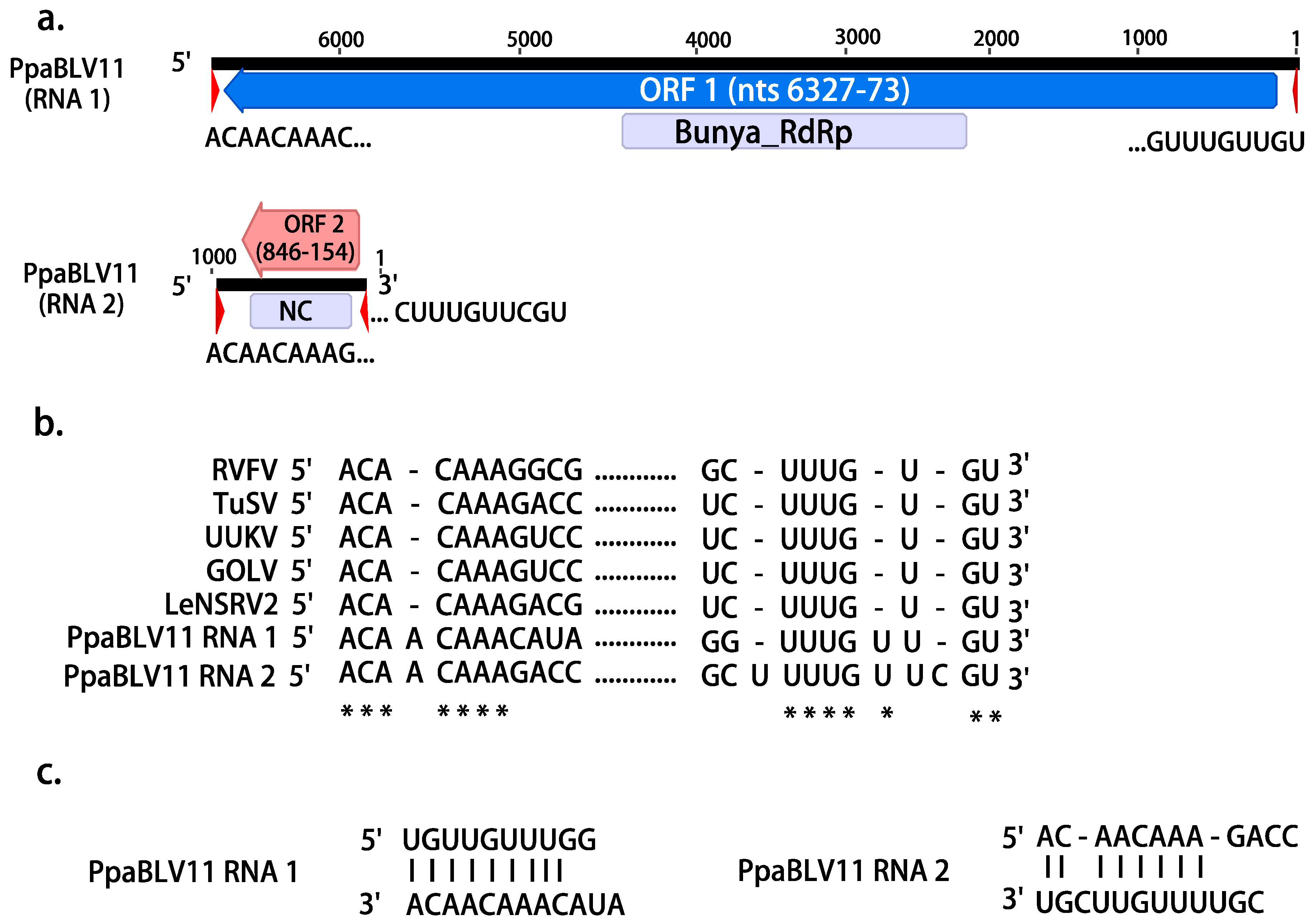
Figure 1. (a). Graphical representation of the L segment (RNA 1) and S segment (RNA 2) of PpaBLV11. (b). Alignment of 5′ and 3′ terminal sequences of PpaBLV11 RNA1 and RNA 2 with RNA1 or L segment sequences from the following viruses: Rift Valley fever virus (RVFV, DDBJ/EMBL/GenBank accession number: X56464), tulip streak virus (TuSV, LC571987), Uukuniemi virus (UUKV, D10759), Gouleako virus (GOLV, HQ541738), Lentinula edodes negative-strand RNA virus 2-HG3 (LeNSRV2, LC466007). Asterisks indicate that the nucleotides are 100% identical in all the viruses. (c). Complementary structure between the 3′ and 5′ termini in the putative PpaBLV11 genome.
Jof 08 01118 g001
Figure 2. (a) Maximum likelihood tree (RAxML) depicting the phylogenic relationship of the predicted RdRP of Phytophthora palustris bunya-like viruses with other complete RdRP belonging to related viruses from the order Bunyavirales. (b) RAxML tree with the NC of PpaBLV11 and other bunyaviruses of the family Phenuiviridae. Nodes are labeled with bootstrap support values ≥50%. Branch lengths are scaled to the expected underlying number of amino acid substitutions per site. Phytophthora palustris bunya-like viruses 1–13 (and variants) are represented by their abbreviated names (PpaBLV1-13) and indicated with a red asterisk (*). Family classification and the corresponding GenBank accession numbers are shown next to the virus names. Colorful squares represent the virus host kingdom or phylum: Jof 08 01118 i001 Fungi, Jof 08 01118 i002 Nematoda, Jof 08 01118 i003 Oomycota, Jof 08 01118 i004 Arthropoda, Jof 08 01118 i005 Plants, Jof 08 01118 i006 Mammalia, Jof 08 01118 i007 (Fishes) Chordata, Jof 08 01118 i008 Ochrophyta (Heterokonta), Jof 08 01118 i009 Excavata. Scale bars represent expected changes per site per branch.
Jof 08 01118 g002aJof 08 01118 g002b
Figure 3. (a). Graphical representation of the tombus-like virus contig (PpaTbLV1). (b). Phylogenetic analysis (RAxML) based on the predicted RdRP of Phytophthora palustris tombus-like virus 1, abbreviated and indicated by a red asterisk (*), with other complete unclassified tombus-like viruses and members of the family Tombusviridae. Nodes are labeled with bootstrap support values ≥50%. Branch lengths are scaled to the expected underlying number of amino acid substitutions per site. Tree is rooted in the midpoint. Family classification and the corresponding pBLAST accession numbers are shown next to the virus names. Colorful squares represent the virus host kingdom or phylum: Jof 08 01118 i001 Fungi, Jof 08 01118 i003 Oomycota, Jof 08 01118 i004 Arthropoda, Jof 08 01118 i010 Animalia, Jof 08 01118 i005 Plants. Scale bar = 0.4 expected changes per site per branch.
Jof 08 01118 g003
Figure 4. (a). Graphical representation of a narna-like (PpaNLV7) virus contig. (b). Phylogenetic analysis (RAxML) based on the predicted RdRP of Phytophthora palustris (+)ssRNA viruses with other complete classified and unclassified members of the families Mitoviridae, Narnaviridae, Botourmiaviridae, Leviviridae. Nodes are labeled with bootstrap support values ≥50%. Branch lengths are scaled to the expected underlying number of amino acid substitutions per site. Tree is rooted in the midpoint. Phytophthora palustris narna-like viruses are abbreviated PpaNLV1-8, and indicated with a red asterisk (*). Family classification and the corresponding pBLAST accession numbers are shown next to the virus names. Colorful squares represent the virus host kingdom or phylum: Jof 08 01118 i001 Fungi, Jof 08 01118 i003 Oomycota, Jof 08 01118 i004 Arthropoda, Jof 08 01118 i010 Animalia, Jof 08 01118 i005 Plants, Jof 08 01118 i008 Ochrophyta (Heterokonta), Jof 08 01118 i011 Apicomplexa (Protists), Jof 08 01118 i012 Bacteria. Scale bar = 0.6 expected changes per site per branch.
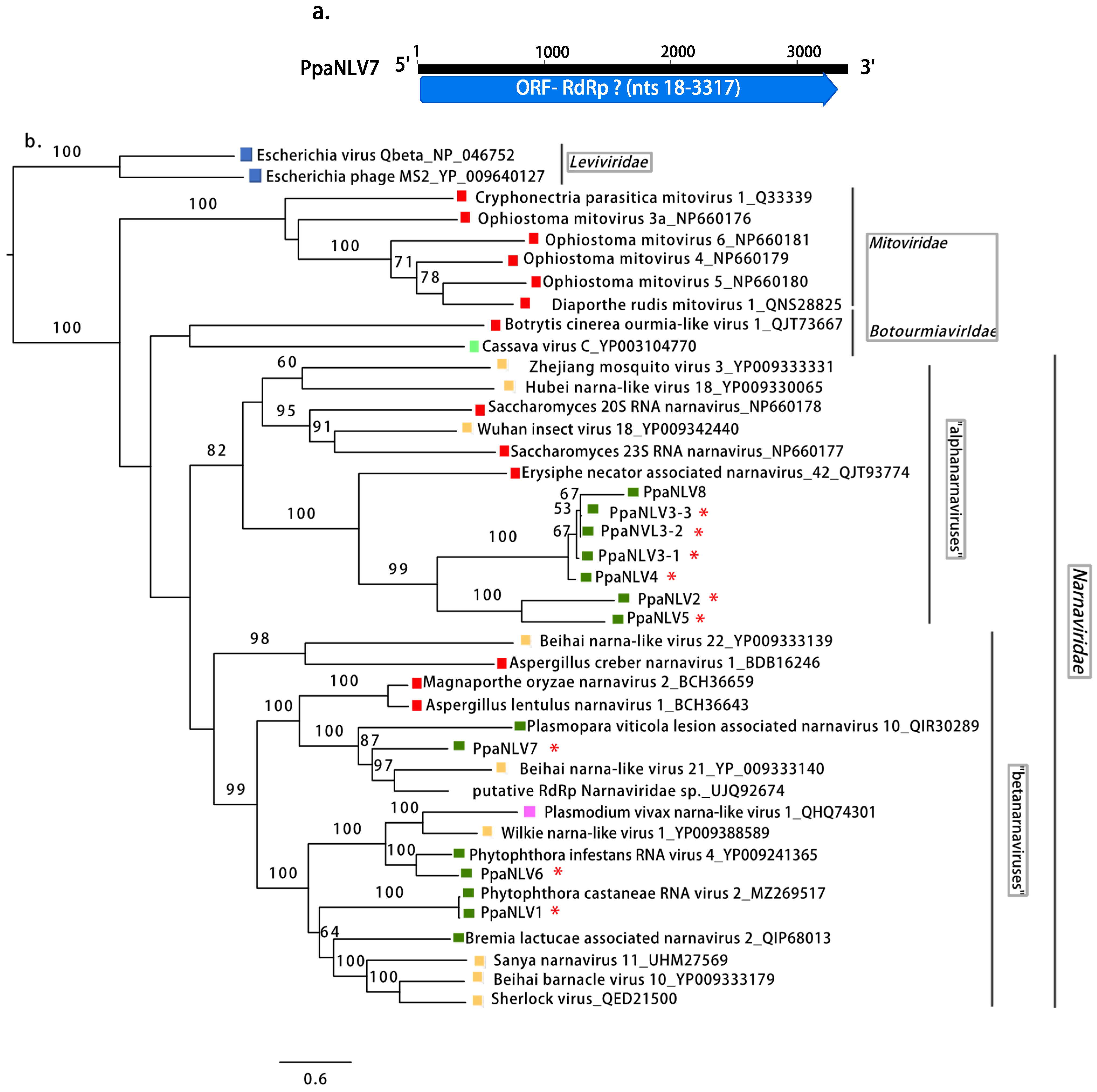
Jof 08 01118 g004
Figure 5. (a). Graphical representation of the genomes of one botybirna-like virus (PpaBbLV2) and one toti-like virus (PpaTLV1-1). (b). Phylogenetic analysis (RAxML) based on the predicted RdRP of the dsRNA viruses discovered in Phytophthora palustris with other classified and unclassified members of the families Totiviridae and Botybirnaviridae. Nodes are labeled with bootstrap support values ≥50%. Branch lengths are scaled to the expected underlying number of amino acid substitutions per site. Tree is unrooted and branches are shown in decreasing order. Variants of Phytophthora palustris toti-like and botybirna-like viruses are abbreviated PpaTLV1-12 and PpaBbLV1 and 2, respectively, and indicated with a red asterisk (*). Family classification and the corresponding pBLAST accession numbers are shown next to the virus names. Colorful squares represent the virus host kingdom or phylum: Jof 08 01118 i001 Fungi, Jof 08 01118 i003 Oomycota, Jof 08 01118 i004 Arthropoda, Jof 08 01118 i007 Chordata, Jof 08 01118 i008 Ochrophyta (Heterokonta), Jof 08 01118 i009 Excavata. Scale bar = 0.6 expected changes per site per branch.
Jof 08 01118 g005aJof 08 01118 g005b
Figure 6. Map of Southeast and East Asia showing the sampling locations in Indonesia, Taiwan, and the Japanese Okinawa and Amami islands (indicated by red dots and arrows) where “P. palustris” isolates were collected.
Jof 08 01118 g006
Figure 7. (a). Map showing the sites of the virus-hosting “P. palustris” isolates in Sumatra. (b). Map showing the sites of the virus-hosting “P. palustris” isolates in Kalimantan. The pie charts illustrate the relative frequency of each virus family/order in Sumatra and Kalimantan.
Jof 08 01118 g007
Table 1. GenBank most similar viruses to the putative “Phytophthora palustris” viruses based on BLASTX search and parameters of their genome organization.
Acronym *N N Genome L L (bp)Most Similar Virus in GenBankE-ValueI (%)QC (%)CDDCDD Position PR (bp) R
PpaBbLV1-1OL795338dsRNA6680Plasmopara viticola lesion associated botybirnavirus 18.00 × 10−2728.5316RdRp_44815–58342729
PpaBbLV1-2OL7953396680Sclerotinia sclerotiorum botybirnavirus 33.00 × 10−3127.9418RdRp_44821–58341831
PpaBbLV1-3OL7953406581Plasmopara viticola lesion associated botybirnavirus 13.00 × 10−3229.5917RdRp_44796–58096030
PpaBbLV2OL7953416514Plasmopara viticola lesion associated botybirnavirus 14.00 × 10−3229.1318RdRp_44669–58082804
PpaBLV1OL795342(−)ssRNA9042Phytophthora condilina negative-stranded RNA virus 130.048.8698Bunya_RdRp4123–5085173,414
N terminus7912–81667720
PpaBLV2OL7953439024Phytophthora condilina negative-stranded RNA virus 110.056.2997Bunya_RdRp4437–5114114,896
PpaBLV3OL7953449303Phytophthora condilina negative-stranded RNA virus 70.061.9998Bunya_RdRp4338–511172,926
PpaBLV4OL7953459302Phytophthora condilina negative-stranded RNA virus 70.056.2798Bunya_RdRp4338–582843,584
PpaBLV5OL7953468362Phytophthora cactorum bunyavirus 10.067.2797Bunya_RdRp4082–549462,939
PpaBLV6OL7953476396Phytophthora condilina negative-stranded RNA virus 30.033.9784Bunya_RdRp2305–334210,639
PpaBLV7OL7953489036Phytophthora condilina negative-stranded RNA virus 110.051.8797Bunya_RdRpn.d.47,152
PpaBLV8OL7953499737Phytophthora condilina negative-stranded RNA virus 110.059.6485Bunya_RdRp4091–511616,049
PpaBLV9-1OL7953509016Phytophthora condilina negative-stranded RNA virus 110.052.0698Bunya_RdRp4434–504559,989
PpaBLV9-2OL7953519047Phytophthora condilina negative-stranded RNA virus 110.052.4697Bunya_RdRpn.d.271,404
PpaBLV9-3OL7953529015Phytophthora condilina negative-stranded RNA virus 110.052.2998Bunya_RdRpn.d.93,848
PpaBLV10OL7953539417Phytophthora condilina negative-stranded RNA virus 70.057.2798Bunya_RdRp4451–5224146,562
PpaBLV11OL7953556448Tulip streak virus1.00 × 10−13327.6967Bunya_RdRp2443–4482223,284
OL795354878Wuhan Fly virus 11.00 × 10−0628.2954Tenui-Phenui NC210–806154,294
PpaBLV12OL7953579386Phytophthora condilina negative-stranded RNA virus 70.055.6499Bunya_RdRpn.d.29,824
PpaBLV13-1OL7953588997Phytophthora condilina negative-stranded RNA virus 130.050.6395Bunya_RdRp3982–505514,451
PpaBLV13-2OL7953598997Phytophthora condilina negative-stranded RNA virus 130.050.5895Bunya_RdRp3982–505510,073
PpaBLV13-3OL7953608995Phytophthora condilina negative-stranded RNA virus 130.050.5495Bunya_RdRp3980–50539885
PpaTbLV1OL795371(+)ssRNA4324RdRp [Riboviria sp.] QDH885791.00 × 10−13952.0933RdRp_32042–2845173,414
RNA_dep_RNAP2060–2854
PpaNV1OL795361(+)ssRNA2818Phytophthora castaneae RNA virus 20.097.0190RdRpn.d.135
PpaNV2OL7953622545Erysiphe-necator-associated narnavirus 423.00 × 10−3133.3337RdRp243,122
PpaNV3-1OL7953632725Erysiphe-necator-associated narnavirus 423.00 × 10−2130.4838RdRp138404
PpaNV3-2OL7953642719Erysiphe-necator-associated narnavirus 423.00 × 10−2231.0239RdRp206,048
PpaNV3-3OL7953652736Erysiphe-necator-associated narnavirus 422.00 × 10−2231.2838RdRp266,624
PpaNV4OL7953662761Erysiphe-necator-associated narnavirus 421.00 × 10−1933.3327RdRp916,928
PpaNV5OL7953672576Erysiphe-necator-associated narnavirus 421.00 × 10−3137.3428RdRp104,172
PpaNV6 UOL7953701344Phytophthora infestans RNA virus 44.00 × 10−14959.4597RdRp162
PpaNV7OL7953683381Beihai narna-like virus 210.037.6391RdRp125,312
PpaNV8 UOL795369916Erysiphe-necator-associated narnavirus 421.00 × 10−1151.1927RdRp46,432
PpaTLV1-1OL795372dsRNA5134Drosophila-associated totivirus 26.00 × 10−17138.6346RdRp_42958–43255030
Totivirus_coat283–2487
PpaTLV1-2OL7953735199Drosophila-associated totivirus 28.00 × 10−17138.6346RdRp_43023–43905464
Totivirus_coat348–2552
PpaTLV2-1OL7953745183Red algae totivirus 12.00 × 10−16237.5647RdRp_43026–43814044
Totivirus_coat363–2624
PpaTLV2-2OL7953755183Red algae totivirus 15.00 × 10−15238.3147RdRp_43026–43812962
Totivirus_coat363–2624
PpaTLV2-3OL7953765183Red algae totivirus 11.00 × 10−15138.2347RdRp_43026–43812504
Totivirus_coat363–2624
PpaTLV3-1OL7953774491Conidiobolus heterosporus totivirus 10.042.0349RdRp_42548–38227076
PpaTLV3-2OL7953784491Wuhan insect virus 260.041.3449RdRp_42623–38222174
PpaTLV3-3OL7953794495Wuhan insect virus 260.041.0349RdRp_42627–38263750
PpaTLV4 UOL7953801553Diatom-colony-associated dsRNA virus 17 genome type B6.00 × 10−2832.5957RdRp_441–7849026
PpaTLV5-1OL7953815188Diatom-colony-associated dsRNA virus 118.00 × 10−14938.4147RdRp_42969–43757926
Totivirus_coat486–26721546
PpaTLV5-2OL7953825199Diatom-colony-associated dsRNA virus 75.00 × 10−14839.4743RdRp_43019–4386
Totivirus_coat497–25548402
PpaTLV6OL7953835312Diatom-colony-associated dsRNA virus 111.00 × 10−9435.2237RdRp_43118–4497
Totivirus_coat389–256617,680
PpaTLV7OL7953845703Diatom-colony-associated dsRNA virus 17 genome type B2.00 × 10−3627.6727RdRp_43663–4625
PpaTLV8 UOL7953852964Diatom-colony-associated dsRNA virus 17 genome type A4.00 × 10−3527.29 RdRp_41216–21784168
PpaTLV9OL7953865268Diatom-colony-associated dsRNA virus 110.048.3952RdRp_43075–445712,384
Totivirus_coat439–2550
PpaTLV10OL7953875205Red algae totivirus 13.00 × 10−13236.3141RdRp_42979–437613,180
Totivirus_coat313–2355
PpaTLV11OL7953885408Red algae totivirus 16.00 × 10−16540.6939RdRp_43219–458318,930
Totivirus_coat403–2424
PpaTLV12-1OL7953894825Plasmopara viticola lesion associated totivirus-like 10.046.1044RdRp_43030–37371786
PpaTLV12-2OL7953905833Plasmopara viticola lesion associated totivirus-like 16.00 × 10−17945.9636RdRp_44034–474113,412
PpaTLV12-3OL7953915833Plasmopara viticola lesion associated totivirus-like 18.00 × 10−17845.5436RdRp_44046–475315,479
n.d., not detected by CDD-search in https://www.ncbi.nlm.nih.gov/Structure/cdd/wrpsb.cgi, accessed on 28 August 2022. QC, query coverage. I, identity. P CDD position in 5′-3′ nucleotide sequence. R Total reads mapped to the final virus sequence. N GenBank accession numbers. L Final virus sequence length. U Partial segment and/or coding region. * PpaBbLV, Phytophthora palustris botybirna-like viruses; PpaTbLV, Phytophthora palustris tombus-like virus; PpaNLV, Phytophthora palustris narna-like viruses; PpaTLV, Phytophthora palustris toti-like viruses; PpaBLV, Phytophthora palustris bunya-like viruses. R Total number of reads mapped to each viral contig discovered in “P. palustris” in Indonesia.
Publisher’s Note: MDPI stays neutral with regard to jurisdictional claims in published maps and institutional affiliations.