Article

Analysis of Microbiota and Mycobiota in Fungal Ball Rhinosinusitis: Specific Interaction between Aspergillus fumigatus and Haemophilus influenza?

by
Sarah Dellière
1,2,
Eric Dannaoui
3,4,5,
Maxime Fieux
6,7,
Pierre Bonfils
6,
Guillaume Gricourt
8,
Vanessa Demontant
8,
Isabelle Podglajen
9,
Paul-Louis Woerther
3,10,
Cécile Angebault
1,3,4 and
Françoise Botterel
1,3,4,*
1
Unité de Parasitologie-Mycologie, Département de Prévention, Diagnostic et Traitement des Infections, APHP, GHU Hôpitaux Universitaires Henri-Mondor, 94010 Créteil, France
2
Unité de Parasitologie-Mycologie, Hôpital Saint-Louis, Assistance Publique des Hôpitaux de Paris, Université de Paris, 75010 Paris, France
3
UR DYNAMiC 7380, Faculté de Santé, Université Paris-Est Créteil, 94010 Créteil, France
4
UR DYNAMiC 7380, Ecole Nationale Vétérinaire d’Alfort, USC Anses, 94700 Maison-Alfort, France
5
Unité de Parasitologie-Mycologie, Département de Microbiologie, Hôpital Européen George Pompidou, APHP, Université de Paris, 75015 Paris, France
6
Département d’Otorhinolaryngologie, Hôpital Européen George Pompidou, APHP, Université de Paris, 75015 Paris, France
7
Service d’Otorhinolaryngologie, d’Otoneurochirurgie et de Chirurgie Cervico-Faciale, Centre Hospitalier Lyon Sud, Hospices Civils de Lyon, 69310 Pierre Bénite, France
8
Plate-Forme Genomiques, APHP-IMRB, GHU Hôpitaux Universitaires Henri-Mondor, UPEC, 94010 Créteil, France
9
Unité de Bactériologie, Département de Microbiologie, Hôpital Européen George Pompidou, APHP, Université de Paris, 75015 Paris, France
10
Unité de Bactériologie, Département de Prévention, Diagnostic et Traitement des Infections, APHP, GHU Hôpitaux Universitaires Henri-Mondor, 94010 Créteil, France
*
Author to whom correspondence should be addressed.
J. Fungi 2021, 7(7), 550; https://doi.org/10.3390/jof7070550
Submission received: 30 April 2021 / Revised: 2 July 2021 / Accepted: 6 July 2021 / Published: 10 July 2021
(This article belongs to the Special Issue Epidemiology of Bacteria-Fungi Interactions)

Abstract

:
Fungal ball (FB) rhinosinusitis (RS) is the main type of non-invasive fungal RS. Despite positive direct examination (DE) of biopsies, culture remains negative in more than 60% of cases. The aim of the study was to evaluate the performance/efficacy of targeted metagenomics (TM) to analyze microbiota and mycobiota in FB and find microbial associations. Forty-five sinus biopsies from patients who underwent surgery for chronic RS were included. After DE and culture, DNA was extracted, then fungal ITS1–ITS2 and bacterial V3–V4 16S rDNA loci were sequenced (MiSeqTM Illumina). Operational taxonomic units (OTUs) were defined via QIIME and assigned to SILVA (16S) and UNITE (ITS) databases. Statistical analyses were performed using SHAMAN. Thirty-eight patients had FB and seven had non-fungal rhinosinusitis (NFRS). DE and culture of FB were positive for fungi in 97.3 and 31.6% of patients, respectively. TM analysis of the 38 FB yielded more than one fungal genus in 100% of cases, with Aspergillus in 89.5% (34/38). Haemophilus was over-represented in FB with >1000 reads/sample in 47.3% (18/38) compared to NFRS (p < 0.001). TM allowed fungal identification in biopsies with negative culture. Haemophilus was associated with FB. Pathogenesis could result from fungi–bacteria interactions in a mixed biofilm-like structure.

1. Introduction

Chronic rhinosinusistis (CRS) is a complex and multifactorial condition, associated with a high prevalence rate of 10.9% [1]. Because of the overlap in symptoms between CRS, acute rhinosinusitis and (non-)allergic rhinitis, CRS diagnosis can be difficult based on symptoms alone. The addition of nasal endoscopy or computed tomography (CT) scan showing signs of mucosal inflammation can help provide a reliable CRS diagnosis [2]. CRS can be classified into CRS with nasal polyps (CRSwNP) and CRS without nasal polyps (CRSsNP). In both cases, the relationship between CRS and asthma is indisputable [3]. Indeed, the prevalence of asthma is around 25% in patients with CRS compared to 5% in the general population [4].
Fungi, which are ubiquitous in our environment, belong to the upper airway microbiota and can be associated with several forms of sinus diseases. In these situations, rather than the fungi itself, it is usually the host immune state that determines the clinical presentation [2]. Fungal CRS represent a wide spectrum of diseases ranging from allergic fungal rhinosinusitis (AFRS), mild forms secondary to colonization (saprophytic fungal infestation and fungal ball), to chronic granulomatous and invasive fungal rhinosinusitis (IFRS). AFRS is a subset of CRSwNP characterized by the presence of eosinophilic mucin with non-invasive fungal hyphae within the sinuses and a type I hypersensitivity to fungi [2]. AFRS accounts for about 5–10% of CRS cases [2]. Fungal balls (FB) are non-invasive collections of fungal mycelium obstructing sinuses, while IFRS is almost always associated with an immunocompromised state, of which diabetes (50%) and hematologic malignancy (40%) account for 90% [5]. Other than the immune state, the distinction between invasive and non-invasive fungal CRS depends on the presence or absence of fungal hyphae in the mucosa [1]. FB is a condition that affects mostly adults in their fifth and sixth decade [5,6]. Although FB diagnosis can be incidental, most FB are associated with obstruction symptoms. As shown by Mensi et al., prior endodontic treatment is a risk factor for maxillary FB formation [6]. On imaging, “calcifications” and erosion of the sinus’ inner wall are the two features most predictive for FB [7]. One particularity of sinus FB is that despite fungal hyphae observed on direct examination (DE) of biopsies, mycological culture yields a fungus in less than 40% of cases [8,9,10]. This could be the result of complex microbial interactions with bacterial species that might weaken the fungus and inhibit its growth.
The microbiota could also play a pathogenic role in CRS. However, as the nose and sinuses are not sterile, causality between the microorganisms grown in sinusal cultures and CRS is difficult to establish [2]. Analysis of the bacterial (microbiota) and fungal (mycobiota) diversity over the last decade has yielded significant insight into the role of microbial communities in human diseases [11,12,13]. However, the characterization of the upper respiratory tract mycobiota in CRS has only been performed twice to our knowledge [14,15,16] and been never performed in fungal CRS and FB, specifically. Furthermore, although it has been shown that bacteria and fungi can coexist in CRS’s biofilm [17], the concomitant study of the microbiota and mycobiota by high-throughput sequencing (HTS) in this context has never been carried out. The aim of our study was to describe bacterial and fungal diversity in FB and find possible microbial intra- or trans-kingdom associations.

2. Materials and Methods

2.1. Patients and Clinical Specimens

A total of 45 samples were collected from 45 patients who underwent functional endoscopic surgery (i.e., middle meatus antrostomy or ethmoidectomy) for CRS from 2015 to 2017 at European Georges Pompidou Hospital, a teaching hospital in Paris, France. During surgery, a biopsy was performed on patients suspected of FB and a sinus aspiration was performed when no FB was present. FB rhinosinusitis (FBRS) was defined by (i) a patient addressed to the hospital for surgical removal of probable FB suspected on clinical and radiological arguments and (ii) a positive direct examination and/or culture of the surgical sample sent to the mycology department (n = 38). Non-fungal ball rhinosinusitis (i.e., controls, NFBRS) was defined by (i) patients addressed to the hospital for surgical drainage of a chronic RS with no clinical or radiological argument for a FB (n = 7). All patients’ files were reviewed by an ENT surgeon and a mycologist and were classified according to the criteria defined prior to sample processing for the metagenomic study. Patient information regarding age, clinical background, sinusitis localization and use of antifungal/antibiotic were collected and analyzed. This study complies with the ethical and legal requirements of French law (15 April 2019) and the Declaration of Helsinki. The database was officially registered with the French Data Protection Authority (Commission Nationale Informatique et Liberté) (no. 2221215). Written or verbal informed consent from all participants was waived because isolates were collected through routine clinical work and patients’ identifiable information had already been anonymized prior to analysis.

2.2. Methods

2.2.1. Direct and Histopathologic Examination and Microbiological Cultures

In the mycology department, direct examination of each sample was performed by Calcofluor-White, May–Grunwald–Giemsa and Gomori–Grocott staining. Culture was performed on 2 Sabouraud media supplemented with gentamicin and chloramphenicol (Biorad, Marnes-la-Coquette, France) incubated at 25 and 37 °C, respectively, for three weeks, one ChromAgar media (Becton Dickinson, Grenoble, France) incubated at 37 °C for 10 days and one Brain–Heart Infusion (BHI) broth incubated at 30 °C for 3 weeks. Concurrently, extra material available was stored at −80 °C before further molecular studies. When concomitantly sent to the bacteriology department (n = 21), cultures were performed on Columbia Blood agar with and without colistin and nalidixic acid, Chocolate agar PolyViteX, Drigalsky agar and Schaedler broth (bioMérieux, Lyon, France) under aerobic and anaerobic conditions and in presence of 5% CO2. Agar plates and Schaedler broth were incubated at 35 °C for 48 h and 14 days, respectively. Bacterial culture was missing for 24 samples because the samples had only been sent to the mycology department.

2.2.2. DNA Extraction Protocol for High Throughput Sequencing

Specimens were retrieved from the −80 °C samples for DNA isolation. A maximum of 250 mg of the sample was pre-treated prior to automated DNA extraction. Sample was submitted to mechanical lysis with 1.4 mm glass beads during two 60 sec cycles at 6400 rpm on a MagNA Lyser instrument (Roche, Mannheim, Germany) and enzymatic lysis with proteinase K. The remainder of the extraction process was automated on QIAsymphony with the DSP DNA midi kit (Qiagen, Hilden, Germany) with protocol VB400 default IC. Non-template DNA isolation control performed by passing PCR-grade water through the same extraction process was included. DNA extracts were stored at −20 °C until amplification.

2.2.3. Library Preparation and Sequencing

Amplicon libraries targeting the V3–V4 16S hyper-variable regions were prepared for bacteria using primers 341 and 785 without degenerated nucleotides [18]. For the ITS1 or ITS2 regions of fungi, amplicon libraries were prepared using primer pairs ITS1F/ITS2 and ITS3/ITS4, respectively [19]. Amplicon PCR, PCR products, purification and sequencing were prepared as previously described by Angebault et al. [20]. The libraries were sequenced using the MiSeq Reagent Kit V2 500-cycle kit (2 × 250) (Illumina) on an Illumina MiSeq platform (Illumina, Evry, France). Raw data are available on the NCBI website (Bioproject PRJNA 701550). One negative control per extraction batch, consisting of DNA-free water, was submitted to the same extraction protocol. Negative controls were pooled for amplification and sequencing.

2.2.4. Taxonomic Assignment, Diversity

After trimming the barcode and adapter sequences from the reads, paired-ends reads were merged, then filtered, according to Bokulich et al. [21], within QIIME2 [22]. Sequences were denoised with Deblur [23] with a length of 250 pb for 16S and 200 pb for ITS. Amplicon sequence variants (ASVs) (that will be further called operational taxonomic units, OTUs) were assigned to SILVA (v132) [24] for bacterial reads (16S) and UNITE (v8.0, released 18 November 2018) [25] for fungal reads (ITS1 and ITS2) using vsearch [26] with a minimal query coverage set to 70%. Databases Mycobank [27], FungiBank (http://fungibank.pasteur.fr, accessed on 7 July 2021), NCBI RefSeq, Greengenes [28], RDP [29] and EzBioCloud [30] (http://www.ezbiocloud.net, accessed on 7 July 2021) were used to manually assign and verify OTUs. Reads with ≥98.7 and ≥94.5% homology and 0.0 e-value were considered for identification at species and genus level, respectively [31]. Data analysis was further performed by SHAMAN (http://shaman.pasteur.fr, accessed on 7 July 2021) [32]. Rarefaction curves were computed to evaluate the quality of the taxonomic diversity assessment. Diversity indexes (Shannon, Simpson and Inverse Simpson) were calculated to compare the homogeneity of the samples in terms of microbiota and mycobiota composition.

2.2.5. Statistical Analysis

Read count normalization was performed using DESeq2 normalization method [33]. The generalized linear model (GLM) implemented in the DESeq2 R package was applied to detect differences in abundance of taxa between fungal balls and controls [34]. The latter was computed, including the patient, type of sinusitis (FB versus control) and the amplification target as main effects. Resulting p-values were adjusted according to the Benjamini and Hochberg procedure [35]. A relative abundance cut-off > 1% of total reads and >5% per sample was used for graphical display of the results. Clinical characteristics of the patients and microbiological data were reported as percentage, mean and standard deviation (SD) if variable followed a Gaussian distribution or median and interquartiles (Q1–Q3) otherwise. Univariate analyses used Fisher’s exact, Chi-2, Mann–Whitney or Student’s test depending on the variable type as appropriate. p < 0.05 (two-tailed) was considered statistically significant. Statistical analyses were performed using Prism software v8.0.

3. Results

3.1. Patient Characteristics

A total of 38 patients underwent surgery for FB removal and seven patients underwent surgical drainage of NFBRS (i.e., controls). Patients with NFBRS had nasal polyposis (n = 3), intra-sinus foreign body (suspected tumor or dental paste) (n = 3) and chronic rhinosinusitis without any specific etiology (n = 1). Mean age of the patients was 59 years (±13) and 58.3% (28/45) were female. Three patients were solid organ transplant recipients (heart, n = 2; kidney, n = 1) and had risk factors for invasive fungal disease (IFD). No patients received antibiotics or antifungals in the 7 days before surgery and sampling. Two patients with FB received antifungal treatment after surgery. The first patient had a suspicion of IFD secondarily ruled out after histologic examination reanalysis by experts. The second one was considered at risk of IFD (heart transplant) and was treated for probable IFD despite any evidence of tissue invasion during histologic examination. Table 1 shows characteristics of patients with FB compared to patients with NFBRS. No other significant differences were found between the two groups; in other words, the patients are considered comparable in age, sex, type of sinusitis, immune status and results of mycological, bacteriological or histological analyses for the remainder of the statistical analyses.

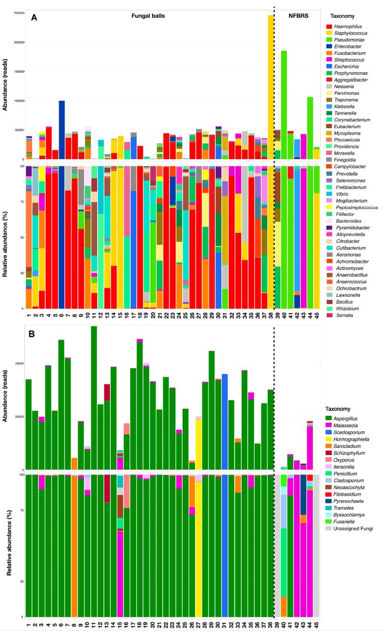
3.2. 16S, ITS1 and ITS2 Targeted Amplicon Sequencing Results

After 16S, ITS1 and ITS2 targeted amplicon sequencing, 3,115,698, 3,428,832 and 3,019,299 raw reads were obtained, respectively. The trimming process led to 1,623,694, 289,417 and 1,213,482 reads, with a median of 40,677, 1055 and 23,632 reads/sample for 16S, ITS1 and ITS2, respectively (Table 2). Finally, 671, 113 and 157 OTUs were obtained for 16S, ITS1 and ITS2. Environmental control 16S amplification yielded mostly Pseudomonas migulae, Corynebacterium sp., Anaerobacillus sp. and Ochrobactrum sp. Environmental control ITS1 and ITS2 amplification yielded Sarocladium kiliense and Byssochlamys sp. The results of environmental controls were considered as background signals and further used to detect samples potentially amplifying and sequencing background DNA only. The comparison of taxonomic distribution between environmental controls and samples was performed using a non-parametric Spearman matrix of correlation. Samples with a Spearman rank correlation coefficient above 0.7 were reanalyzed or not included in the study.

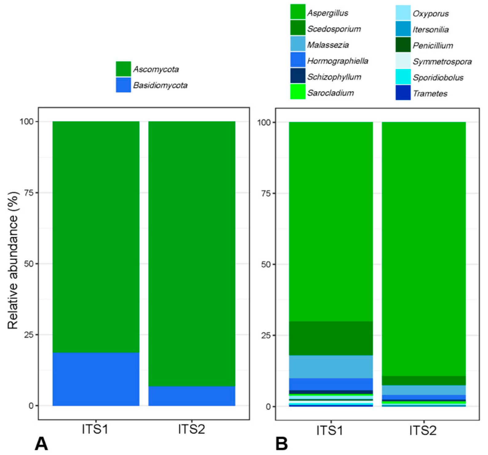
3.3. Comparison of ITS1 and ITS2 Regions for Mycobiota Analysis

Table 2 summarizes, for ITS1 and ITS2 targets, the number of reads/sample before and after trimming of raw data and the number of OTUs and taxa at genus level computed from trimmed data. Number of reads/sample prior trimming, number of OTUs/sample and number of total taxa were similar for both targets. On the contrary, the number of reads/sample after trimming varied significantly according to the ITS target with a mean of 5967 (±11,231) reads/sample for ITS1 versus 25,374 (±17,969) reads/sample for ITS2 (p < 0.001). Among FB specimens, 97.4% (37/38) produced ITS2 amplicons with at least 5000 fungal sequences. In contrast, only 26.3% (10/38) produced ITS1 amplicons with at least 5000 fungal sequences (p < 0.001). Overall, 48 and 57 taxa were identified at the genus level using ITS1 or ITS2, respectively. In the 10 most represented taxa identified with ITS1 and ITS2, eight were in agreement (Aspergillus, Scedosporium, Hormographiella, Malassezia, Schizophyllum, Oxysporus, Sarocladium and Itersonilia). Normalized relative abundances of these eight taxa are represented in Figure 1. With ITS1 analysis, the 9th and 10th most represented taxa were Penicillium and Symmetrospora (a new genus gathering species previously assigned to Rhodotorula and Sporobolomyces genera). These two taxa were found with ITS2 as the 11th and 51st most represented taxa, respectively. On the other hand, Nakaseomyces (comprising Candida glabrata and related species) was the ninth most represented taxa using ITS2 but was not detected using ITS1 and Neoascochyta (environmental and plant pathogenic fungi) was the 10th most represented taxa with ITS2 but the 24th with ITS1.
Mean α-diversity indices, Shannon, Simpson and inverse Simpson were 2.08 (±0.47), 0.41 (±0.13), 0.23 (±0.07) and 1.60 (±0.40) for ITS1 and 1.58 (±0.36), 0.21 (±0.12), 0.11 (±0.06) and 1.29 (±0.24) for ITS2. They showed a narrower diversity with ITS2 despite a higher number of OTUs (Figure S1). At the phylum level, the diversity profiles generated with primers targeting ITS1 showed an increased proportion of Basidiomycetes over Ascomycetes compare to ITS2 (p < 0.001) (Figure 1). Considering these findings, further analysis was performed on the ITS2 dataset.

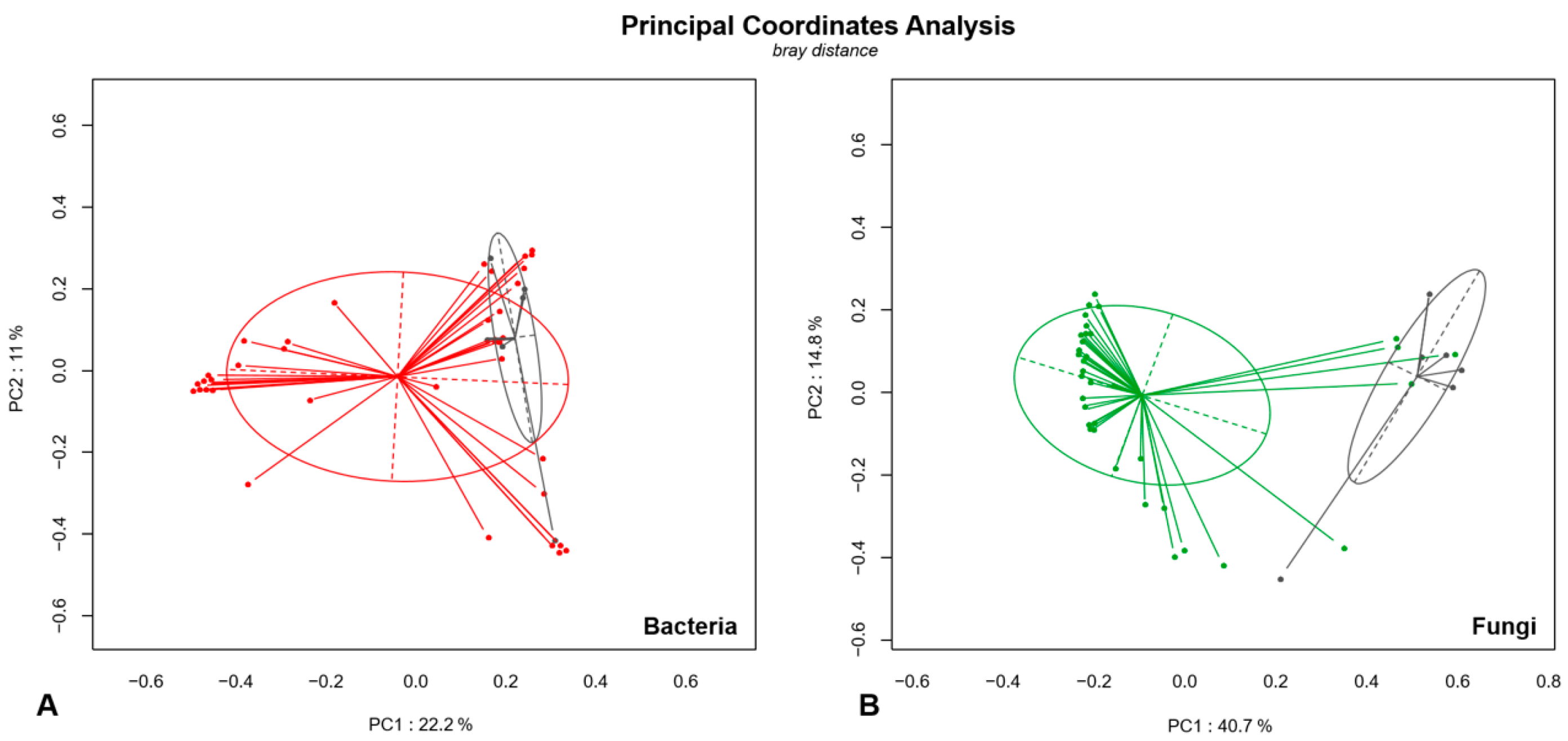
3.4. Analysis of Microbial Diversity in Fungal Ball (FB) and Non-Fungal Ball Chronic Rhinosinusitis (NFBRS)

A total of 133 bacterial and 57 fungal taxa at the genus level were found in the samples. A mean of 15 (range; 3–40) bacterial taxa and 5 (range; 1–30) fungal taxa were found per sample. Predominant bacterial taxa (relative abundance > 1% in more than 25% of samples) were Haemophilus, Pseudomonas and Staphylococcus (Figure S2A). Predominant fungal taxa were Aspergillus and Malassezia (Figure S2B). OTUs assigned to the genera Haemophilus, Pseudomonas, Staphylococcus and Streptococcus for bacteria and Aspergillus for fungi were assigned at species or section level when possible (Table S1). Among the Haemophilus genus, 86.3% of reads were Haemophilus influenzae and 13.6% could not be further assigned. Among Pseudomonas, 57.7, 23.4 and 15.6% were assigned to Pseudomonas aeruginosa, Pseudomonas mossellii (mostly represented in one patient) and Pseudomonas migulae (mostly represented in the environmental control and found at low rate in samples), respectively. Only 1.7% of Pseudomonas reads remained unassigned at the species level. Among Staphylococcus, the majority of OTUs (91.9%) were assigned as Staphylococcus aureus and 73.1% of OTUs assigned as Streptococcus belonged to the milleri group. Among Aspergillus, 80.8, 15.4 and 3.7% were further assigned to sections Fumigati, Flavi and Nidulantes, respectively.
A significant difference in bacterial (p = 0.003) and fungal (p = 0.001) microbiota between FB and NFBRS was shown by permutational multivariate ANOVA (PERMANOVA) at the genus level according to the Bray–Curtis dissimilarity metric. Results are presented as a principal coordinate analysis matrix in Figure 2. Bacterial and fungal taxonomic diversity of taxa with a relative abundance > 1% of total reads and >5% per sample are detailed in Figure 3A (read counts) and Figure 3B (relative abundance). Taxonomic diversity of all bacterial and fungal taxa is available in supplementary data (Figures S3 and S4).
Focusing on bacterial diversity, Haemophilus was the predominant taxon representing 30.3% of total bacterial reads. This taxon was present in 47.3% (18/38) of FB with over 1000 reads per samples versus 0% (0/7) in NFBRS. Regarding fungal diversity, Aspergillus was the predominant taxa in 89.5% (34/38) of FB but was present in only 1/7 (14.2%) of NFBRS. The four Aspergillus-free FB showed a majority of Sarocladium (patient 8), Hormographiella (patient 27), Scedosporium (patient 31) or a variety of 30 taxa dominated by Malassezia (patient 15). In NFBRS, very few fungal reads were detected with a mean read per sample of 6350 (±8678) compared to 30,699 (±15,703) in FB (p < 0.001). Reads were mostly assigned to Malassezia except for the substantial number of 9998 reads of Aspergillus section flavi in patient 41 (relative abundance = 81.7%). The Malassezia taxon was found in 68.2% (30/38) of FB samples and 57.1% (4/7) of NFBRS with a mean relative abundance per sample of 2.3 (range: 0–38.0%) and 38.0% (range: 0–99.3%), respectively (p = 0.23). Haemophilus and Aspergillus were confirmed to be significantly more abundant in FB than NFBRS by a generalized linear model with p < 0.001 (Figure 4). No other bacterial taxa were found to be significantly overrepresented in FB compared to NFBRS. Among FB, there were no significant differences observed with a PERMANOVA test of bacterial or fungal diversity profiles according to patient gender, localization of sinusitis and bacterial and fungal culture.

4. Discussion

Fungal and bacterial diversity of sinus samples from 45 patients with FBRS (n = 38) and NFBRS (n = 7) were analyzed by targeted metagenomics. Aspergillus and Haemophilus were the main fungal and bacterial genera identified in FBRS. From a more technical perspective, we have confirmed the superiority of using ITS2 over ITS1 as a target for the study of fungal diversity in this context. To our knowledge, this is the first study to carry out HTS of both bacterial and fungal diversity in FB by targeted metagenomics.

4.1. Targeting ITS1 or ITS2 to Study Fungal Diversity

Microbial diversity studies are greatly influenced by the method used and their results depend on the ability to extract, amplify, sequence and analyze each microbial genus [36]. The National Institutes of Health (NIH) Human Microbiome Project intends to standardize data resources and new technological approaches to enable such study to be undertaken broadly in the scientific community [37]. However, data focusing on the analysis of fungal communities remain scarce as fungi may be more difficult to manipulate and analyze, particularly due to the difficulty of extracting the fungal cell wall [20]. Controversies remain regarding the selection of markers for sequencing and that is why we intended to compare ITS1 and ITS2 sequencing in our own settings. ITS1 amplification yielded a high proportion of non-specific reads, mostly human, leaving five time less reads per sample than ITS2 amplification. This can be explained by the use of ITS1-F universal primer shown to match human DNA [38], although recommended for the identification using pure fungal DNA [39]. The lower number of reads obtained with ITS1 yielded a higher diversity than ITS2 regardless of the diversity index used. This may be due to (i) a biased analysis after normalization of a reduced number of reads and/or (ii) an actual better representation of fungal diversity with increased representation of Basidiomycota taxa. Some studies compared ITS1 and ITS2 for fungal profiles [40,41,42,43,44]. Some authors found ITS2 to be more suitable for revealing richness [40] while others reported that ITS1 was probably the best choice for the study of fungal and eukaryotic species [43]. A recent in silico study revealed that the fungal diversity and richness might be overestimated when using ITS1. Moreover, the clustering and taxonomy detected using ITS2 were more similar to those obtained with the whole ITS region rather than with ITS1 alone [44]. This can be explained by the ITS1 region having evolved more rapidly and having a more variable length than ITS2 [45]. The shorter length, lower GC content variation and greater taxonomic information content of ITS2 probably makes it more suitable than ITS1 for HTS studies of fungi. For this reason and because of a much higher number of fungal reads obtained with this marker we performed our analysis using ITS2. The latest reports, unavailable at the time we designed our study, now recommend targeting the ITS2 subregion by using the degenerated gITS7ngs and ITS4ngs primers, while we used non-degenerated ones [36].
The possibility or necessity to validate metagenomics data using an independent technique such as quantitative PCR (qPCR) is a matter of discussion in progress among the community, similarly to what is performed in transcriptomic analyses [46]. In that case, expression of the gene of interest and control genes are measured against defined standards, usually housekeeping genes’ expression, known to be consistently expressed in all cells and despite varying conditions [46]. In targeted metagenomics, qPCR could be used to quantify the fungal and bacterial biomass of species of interest (i.e., Aspergillus and Haemophilus in the present study) in order to obtain more comparable results among samples. However, because fungal species differ markedly in number of rRNA operon copies per genome, the performance of qPCR is limited [36]. However, targeting a unique copy gene would significantly decrease the sensitivity of the method. To the best of our knowledge, no such validation was carried out in a previous metagenomic study on fungal diversity.

4.2. Fungal Diversity in FBRS

Analysis of the fungal mycobiota found Aspergillus to be the predominant genus in 89.5% of FB, which correlates with previous studies, such as Morio et al.’s work, who identified Aspergillus in over 77.4% of sinus FB by targeted PCR [8,9,10]. Patients were classified as FB on clinical, radiological and surgical criteria associated with a positive direct examination, showing fungal hyphae and/or a positive culture. In FB, only one patient (patient 15) had a negative direct examination with a positive A. fumigatus culture. This 53-year-old patient had pansinusitis secondary to a suspected FB on the CT scan. Amplification of 16S yielded 98.6% of S. aureus (Figure 3A). ITS2 amplification yielded only 11,496 reads belonging to 30 fungal taxa with a majority of Malassezia reads, a difficult-to-culture commensal yeast (Figure 3B). The variety of fungal taxa may reflect transient, contaminant aerial fungi and the presence of Malassezia may reflect nasal mycobiota contamination in a sample with a low fungal biomass [47]. This patient was probably wrongly classified in the FB part. Initial criteria for FB classification should thus have been limited to a positive direct examination. Surprisingly, the Aspergillus section Flavi was found with a significant number of reads in NFBRS (patient 41). This 48-year-old patient, with a history of a right maxillary cystic lesion surgically removed at the age of 20, presented with chronic rhinosinusitis of dental origin of the same sinus. The detection of Aspergillus section Flavi in this patient using HTS may be explained by a transient or contaminant fungus in the sample. Indeed, fungal spores are found in the environment and may be inhaled or may contaminate a culture or a sample before extraction.
The small number of FB included and our choice of NFBRS as a comparison group in this study are the main limits of our study. Indeed, these NFBRS samples were addressed to the mycology lab due to a suspicion of fungal etiology, which constitutes a first bias. In addition, the sampling method for these samples was different from FB. NFBRS samples were pus aspirates and not biopsies. In a prospective study, another control could be proposed, such as sinus biopsies performed for cancer research in patients without prior suspicion of fungal infection.

4.3. Hypothesis to Explain the Poor Positive Culture Rate of FBRS Samples

Aspergillus, the predominant fungus found in FBRS, is known to grow easily on routine culture media. This raises questions regarding the absence of growth in a majority (60.6–75.0%) of FB samples in our study, as well as in previous works [8,9,10]. A first hypothesis is that interactions between Aspergillus and bacterial species could weaken the fungus and inhibit its growth. In their study, based on culture methods, Zhang et al. showed that pathogenic bacteria were associated in more than 75% of FB. They observed that the presence of Pseudomonas aeruginosa was significantly associated with altered and fragmented mycelium and that Aspergillus growth was inhibited in mixed infection [48]. They therefore hypothesized that in the presence of certain bacteria involved in FB, Aspergillus may be weakened, and its growth may be compromised in vitro. However, they did not identify a particular profile or specific bacterial taxa associated with culture-positive FB compared to culture-negative FB [48]. Of note, in non-fungal CRS and particularly in polypoid forms, S. aureus has been described as being involved in the pathogenesis of the disease. Many characteristics of S. aureus, such as its ability to regulate innate and adaptative immunity, to disrupt tissue barrier function or to promote impaired mucociliary clearance, may favor its role in CRS pathogenesis [49,50]. However, it was not specifically associated with FBRS in our study, unlike Haemophilus. Another hypothesis could be that the fungi are embedded in a biofilm-like structure, thus living in a hypometabolic state preventing secondary growth in in vitro conditions [51]. The concept of “viable, but not culturable” (VBNC) organisms has been well described in bacterial biofilms [52] and in yeasts [53,54], but never in filamentous fungi such as Aspergillus, to our knowledge. However, in mixed biofilms, histological studies on human tissue or animal models have confirmed the presence of aggregates of hyphae embedded in an extracellular matrix [55,56]. This validates the presence of Aspergillus biofilms, which, in the presence of bacteria, have recently become a topic of interest [57,58,59]. Besides the VBNC hypothesis, biofilms could also contribute to the virulence of Aspergillus by mediating adherence to host cells [55] and enhancing its resistance to antifungals and the immune system [60,61].

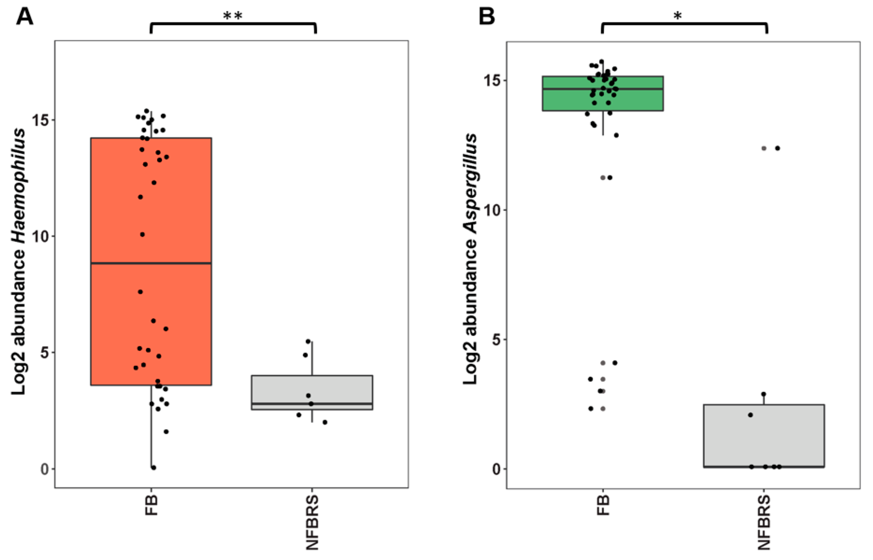
4.4. Could FBRS Result from Aspergillus–Haemophilus Microbial Interaction?

Using metagenomics, Haemophilus was found to be significantly more abundant in FB compare to NFBRS. Therefore, we hypothesize that AspergillusHaemophilus interactions could play a definite role in FB pathogenesis. Haemophilus influenzae, particularly, is known to be part of the oropharyngeal microbiota and can be responsible for otitis, sinusitis, persistent bronchitis and COPD exacerbations [62]. A longitudinal metagenomic study by Mackenzie et al. did not find a significant amount of Haemophilus in healthy bacterial sinusal microbiota. These authors did not find Haemophilus to be persistent throughout annual and seasonal changes [63]. Colonization and pathogenesis of Haemophilus on host tissue relies on different strategies. Among those, it has already been observed in vitro that Haemophilus inhibits the mucociliary clearance system [64]. This strategy could certainly benefit Aspergillus in sinuses, allowing inhaled spores to stay in airways and germinate. Except for the study led by Zhang et al. (2015), who performed bacterial cultures on FB, bacterial diversity in FB has not yet been well explored. Zhang et al. mainly found S. aureus and P. aeruginosa associated with FB, as in our study, but they did not find, as we did, an elevated prevalence of Haemophilus [48]. However, Haemophilus are bacteria that are sometimes difficult to grow, requiring specific vitamins-enriched media [62]. Working on the sequencing of three ethmoidal sinus samples, the same authors mainly found reads from Aspergillus and Haemophilus [65]. Interestingly, a study investigating allergic fungal RS (n = 4) and chronic RS (n = 7) observed polymicrobial biofilms with Haemophilus and fungal elements in 82% of sinus biopsies [17]. Although these types of sinusitis are different from FB, these results confirm the possible co-existence of Haemophilus and Aspergillus in a biofilm structure that could benefit both microorganisms in terms of persistence and growth in the sinuses. Another study from Boase et al. studied mixed biofilm formation in an in vivo model of fungal sinusitis in sheep [66]. They described a toxin inhibiting the ciliary movement of epithelial cells that allowed the formation of fungal biofilm. They also observed that fungal biofilm could form only after co-inoculation with S. aureus, Staphylococcus epidermidis or P. aeruginosa, but surprisingly not with Haemophilus. However, it was later shown that the Haemophilus ATCC 49247 strain used by Boase et al. was able to induce biofilm formation [67]. Our study does not allow us to conclude whether the co-existence of Haemophilus and Aspergillus in FB is coincidental, or if a true interaction between the two organisms takes place. It is possible that fungi and bacteria cooperate to enhance defense mechanisms toward the host and to facilitate access to nutrients by specific exchanges [52]. FISH analysis of a fine cross-section of FB with specific Aspergillus and Haemophilus probes could help confirm their co-existence, embedded in a common extracellular matrix. Then, in vitro studies would be needed to investigate possible interactions between Aspergillus and Haemophilus.

5. Conclusions

FB are specific entities among chronic rhinosinusitis. Targeted metagenomics confirms incrimination of Aspergillus spp. despite a negative culture in most cases. More interestingly, our study revealed the concomitant presence of Haemophilus in FB, underlining the importance of sending surgical samples to both clinical bacteriology and mycology laboratories regardless of the obvious fungal etiology. Our study highlights the importance of evaluating inter-kingdom interactions to improve the pathophysiological understanding of entities primarily described as solely fungal, such as sinus FB. Although correlation does not indicate causation, such results stand as a reasonable starting point to further investigate inter-kingdom interactions between Aspergillus fumigatus and Haemophilus in chronic fungal rhinosinusitis of FB type.

Supplementary Materials

The following are available online at https://www.mdpi.com/article/10.3390/jof7070550/s1, Figure S1: Fungal richness and diversity calculated based on ITS1 and ITS2, Figure S2: Heatmap of the relative abundance of major bacterial and fungal taxa, Figure S3: Bacterial diversity represented in number of reads and relative abundance for all taxa found, Figure S4: Fungal diversity represented in number of reads and relative abundance for all taxa found, Table S1: Operative taxonomic units (OTUs) assigned to Haemophilus, Pseudomonas, Staphylococcus, Streptococcus and Aspergillus identified to the species level.

Author Contributions

Conceptualization, S.D., C.A., E.D. and F.B.; methodology, S.D., C.A. and F.B; software, G.G.; validation, S.D., C.A. and F.B.; formal analysis, S.D., G.G. and C.A.; investigation, S.D., M.F., V.D. and C.A.; data curation, S.D., G.G. and C.A.; writing—original draft preparation, S.D., C.A. and F.B.; writing—review and editing, S.D., E.D., M.F., P.B., G.G., V.D., I.P., P.-L.W., C.A., F.B.; visualization, S.D., C.A. and F.B.; supervision, C.A., E.D. and F.B.; project administration, F.B. All authors have read and agreed to the published version of the manuscript.

Funding

This research received no external funding.

Institutional Review Board Statement

The database was officially registered with the French Data Protection Authority (Commission Nationale Informatique et Liberté) (No. 2221215).

Informed Consent Statement

Patient consent was waived because isolates were collected through routine clinical work and patient’s identifiable information had already been anonymized prior to analysis. The database was officially registered with the French Data Protection Authority (Commission Nationale Informatique et Liberté) (No. 2221215).

Data Availability Statement

Not applicable.

Acknowledgments

We thank Christophe Rodriguez for rigorous advice on metagenomics techniques. We are grateful to the Genotoul bioinformatics platform Toulouse Occitanie for providing help and/or computing and/or storage resources.

Conflicts of Interest

Over the past 5 years, F.B. has received grants from Astellas, payments for lectures from MSD and travel expenses from Pfizer, MSD and Astellas. E.D. has received research grants from MSD and Gilead, travel grants from Gilead, MSD, Pfizer and Astellas and speaker’s fees from Gilead, MSD and Astellas. Other authors declare that they have no competing interests.

References

  1. Pleis, J.R.; Lucas, J.W.; Ward, B.W. Summary Health Statistics for U.S. Adults: National Health Interview Survey; Vital and Health statistics. Series 10; Centers for Disease Control and Prevention: Atlanta, GA, USA, 2009; pp. 1–161.
  2. Fokkens, W.J.; Lund, V.J.; Hopkins, C.; Hellings, P.W.; Kern, R.; Reitsma, S.; Toppila-Salmi, S.; Bernal-Sprekelsen, M.; Mullol, J.; Alobid, I.; et al. European Position Paper on Rhinosinusitis and Nasal Polyps. Rhinology 2020, 58, 1–464. [Google Scholar] [CrossRef]
  3. Poddighe, D.; Brambilla, I.; Licari, A.; Marseglia, G.L. Pediatric rhinosinusitis and asthma. Respir. Med. 2018, 141, 94–99. [Google Scholar] [CrossRef]
  4. Seybt, M.W.; McMains, K.C.; Kountakis, S.E. The prevalence and effect of asthma on adults with chronic rhinosinusitis. Ear Nose Throat J. 2007, 86, 409–411. [Google Scholar] [CrossRef] [Green Version]
  5. Turner, J.H.; Soudry, E.; Nayak, J.V.; Hwang, P.H. Survival outcomes in acute invasive fungal sinusitis: A systematic review and quantitative synthesis of published evidence. Laryngoscope 2013, 123, 1112–1118. [Google Scholar] [CrossRef]
  6. Mensi, M.; Piccioni, M.; Marsili, F.; Nicolai, P.; Sapelli, P.L.; Latronico, N. Risk of maxillary fungus ball in patients with endodontic treatment on maxillary teeth: A case-control study. Oral Surg. Oral Med. Oral Pathol. Oral Radiol. 2007, 103, 433–436. [Google Scholar] [CrossRef]
  7. Raz, E.; Win, W.; Hagiwara, M.; Lui, Y.W.; Cohen, B.; Fatterpekar, G.M. Fungal Sinusitis. Neuroimaging Clin. N. Am. 2015, 25, 569–576. [Google Scholar] [CrossRef]
  8. Cabaret, O.; Toussain, G.; Abermil, N.; Alsamad, I.A.; Botterel, F.; Costa, J.-M.; Papon, J.-F.; Bretagne, S. Degradation of fungal DNA in formalin-fixed paraffin-embedded sinus fungal balls hampers reliable sequence-based identification of fungi. Med. Mycol. 2011, 49, 329–332. [Google Scholar] [CrossRef] [Green Version]
  9. Pagella, F.; Matti, E.; Bernardi, F.D.; Semino, L.; Cavanna, C.; Marone, P.; Farina, C.; Castelnuovo, P. Paranasal sinus fungus ball: Diagnosis and management. Mycoses 2007, 50, 451–456. [Google Scholar] [CrossRef] [PubMed]
  10. Morio, F.; Dannaoui, E.; Chouaki, T.; Cateau, E.; Malard, O.; Bonfils, P.; Page, C.; Dufour, X.; Cottrel, C.; Erwan, T.; et al. PCR-based detection of Aspergillus fumigatus and absence of azole resistance due to TR34/L98H in a french multicenter cohort of 137 patients with fungal rhinosinusitis. Mycoses 2018, 61, 30–34. [Google Scholar] [CrossRef] [PubMed]
  11. Cui, L.; Morris, A.; Ghedin, E. The human mycobiome in health and disease. Genome Med. BioMed. Central 2013, 5, 63. [Google Scholar] [CrossRef] [Green Version]
  12. Kumpitsch, C.; Koskinen, K.; Schöpf, V.; Moissl-Eichinger, C. The microbiome of the upper respiratory tract in health and disease. BMC Biol. 2019, 17, 87. [Google Scholar] [CrossRef] [Green Version]
  13. Tiew, P.Y.; Mac Aogain, M.; Ali, N.A.B.M.; Thng, K.X.; Goh, K.; Lau, K.J.X.; Chotirmall, S.H. The Mycobiome in Health and Disease: Emerging Concepts, Methodologies and Challenges. Mycopathologia 2020, 185, 207–231. [Google Scholar] [CrossRef]
  14. Cleland, E.J.; Bassioni, A.; Boase, S.; Dowd, S.; Vreugde, S.; Wormald, P.-J. The fungal microbiome in chronic rhinosinusitis: Richness, diversity, postoperative changes and patient outcomes. Int. Forum. Allergy Rhinology. 2014, 4, 259–265. [Google Scholar] [CrossRef]
  15. Zhao, Y.C.; Bassiouni, A.; Tanjararak, K.; Vreugde, S.; Wormald, P.-J.; Psaltis, A.J. Role of fungi in chronic rhinosinusitis through ITS sequencing. Laryngoscope 2017, 128, 16–22. [Google Scholar] [CrossRef] [PubMed]
  16. Jung, W.H.; Croll, D.; Cho, J.H.; Kim, Y.R.; Lee, Y.W. Analysis of the nasal vestibule mycobiome in patients with allergic rhinitis. Mycoses 2015, 58, 167–172. [Google Scholar] [CrossRef] [PubMed]
  17. Healy, D.Y.; Leid, J.G.; Sanderson, A.R.; Hunsaker, D.H. Biofilms with fungi in chronic rhinosinusitis. Otolaryngol. Head Neck Surg. 2008, 138, 641–647. [Google Scholar] [CrossRef]
  18. Klindworth, A.; Pruesse, E.; Schweer, T.; Peplies, J.; Quast, C.; Horn, M.; Glöckner, F.O. Evaluation of general 16S ribosomal RNA gene PCR primers for classical and next-generation sequencing-based diversity studies. Nucleic Acids Res. 2013, 41, e1. [Google Scholar] [CrossRef]
  19. White, T.J.; Bruns, T.; Lee, S.; Taylor, J. Amplification and direct sequencing of fungal ribosomal RNA genes for phylogenetics. In PCR Protocols a Guide to Methods and Applications; Academic Press: Cambridge, MA, USA, 1990; pp. 315–322. [Google Scholar]
  20. Angebault, C.; Payen, M.; Woerther, P.-L.; Rodriguez, C.; Botterel, F. Combined bacterial and fungal targeted amplicon sequencing of respiratory samples: Does the DNA extraction method matter? PLoS ONE 2020, 15, e0232215. [Google Scholar] [CrossRef] [PubMed]
  21. Bokulich, N.A.; Subramanian, S.; Faith, J.J.; Gevers, D.; Gordon, J.I.; Knight, R.; Mills, D.A.; Caporaso, J.G. Quality-filtering vastly improves diversity estimates from Illumina amplicon sequencing. Nat. Methods 2013, 10, 57–59. [Google Scholar] [CrossRef]
  22. Bolyen, E.; Rideout, J.R.; Dillon, M.R.; Bokulich, N.A.; Abnet, C.C.; Al-Ghalith, G.A.; Alexander, H.; Alm, E.J.; Arumugam, M.; Asnicar, F.; et al. Reproducible, interactive, scalable and extensible microbiome data science using QIIME. Nat. Biotechnol. 2019, 37, 852–857. [Google Scholar] [CrossRef]
  23. Amir, A.; McDonald, D.; Navas-Molina, J.A.; Kopylova, E.; Morton, J.T.; Xu, Z.; Kightley, E.P.; Thompson, L.R.; Hyde, E.R.; Gonzalez, A.; et al. Deblur Rapidly Resolves Single-Nucleotide Community Sequence Patterns. mSystems 2017, 2, e00191-16. [Google Scholar] [CrossRef] [Green Version]
  24. Quast, C.; Pruesse, E.; Yilmaz, P.; Gerken, J.; Schweer, T.; Yarza, P.; Peplies, J.; Glöckner, F.O. The SILVA ribosomal RNA gene database project: Improved data processing and web-based tools. Nucleic Acids Res. 2012, 41, D590–D596. [Google Scholar] [CrossRef]
  25. Irinyi, L.; Serena, C.; Garcia-Hermoso, D.; Arabatzis, M.; Desnos-Ollivier, M.; Vu, D.; Cardinali, G.; Arthur, I.; Normand, A.C.; Giraldo, A.; et al. International Society of Human and Animal Mycology (ISHAM)-ITS reference DNA barcoding database--the quality controlled standard tool for routine identification of human and animal pathogenic fungi. Med. Mycol. 2015, 53, 313–337. [Google Scholar] [CrossRef]
  26. Rognes, T.; Flouri, T.; Nichols, B.; Quince, C.; Mahé, F. VSEARCH: A versatile open source tool for metagenomics. PeerJ 2016, 4, e2584-22. [Google Scholar] [CrossRef]
  27. Robert, V.; Vu, D.; Amor, A.B.H.; Van de Wiele, N.; Brouwer, C.; Jabas, B.; Szoke, S.; Dridi, A.; Triki, M.; Daoud, S.B.; et al. MycoBank gearing up for new horizons. IMA Fungus 2013, 4, 371–379. [Google Scholar] [CrossRef] [PubMed]
  28. DeSantis, T.Z.; Hugenholtz, P.; Larsen, N.; Rojas, M.; Brodie, E.L.; Keller, K.; Huber, T.; Dalevi, D.; Hu, P.; Andersen, G.L. Greengenes, a Chimera-Checked 16S rRNA Gene Database and Workbench Compatible with ARB. Appl. Environ. Microbiol. 2006, 72, 5069–5072. [Google Scholar] [CrossRef] [Green Version]
  29. Cole, J.R.; Wang, Q.; Fish, J.A.; Chai, B.; McGarrell, D.M.; Sun, Y.; Brown, C.T.; Porras-Alfaro, A.; Kuske, C.R.; Tiedje, J.M. Ribosomal Database Project: Data and tools for high throughput rRNA analysis. Nucleic Acids Res. 2013, 42, D633–D642. [Google Scholar] [CrossRef] [PubMed] [Green Version]
  30. Park, S.-C.; Won, S. Evaluation of 16S rRNA Databases for Taxonomic Assignments Using a Mock Community. Genom. Inf. 2018, 16, e24. [Google Scholar] [CrossRef] [Green Version]
  31. Yarza, P.; Yilmaz, P.; Pruesse, E.; Glöckner, F.O.; Ludwig, W.; Schleifer, K.-H.; Whitman, W.B.; Euzéby, J.; Amann, R.; Rosselló-Móra, R. Uniting the classification of cultured and uncultured bacteria and archaea using 16S rRNA gene sequences. Nat. Biotechnol. 2014, 12, 635–645. [Google Scholar] [CrossRef]
  32. Volant, S.; Lechat, P.; Woringer, P.; Motreff, L.; Campagne, P.; Malabat, C.; Kennedy, S.; Ghozlane, A. SHAMAN: A user-friendly website for metataxonomic analysis from raw reads to statistical analysis. BMC Bioinform. 2020, 21, 345. [Google Scholar] [CrossRef]
  33. Love, M.I.; Huber, W.; Anders, S. Moderated estimation of fold change and dispersion for RNA-seq data with DESeq. Genome Biol. 2014, 15, 550. [Google Scholar] [CrossRef] [PubMed] [Green Version]
  34. Quereda, J.J.; Dussurget, O.; Nahori, M.-A.; Ghozlane, A.; Volant, S.; Dillies, M.-A.; Regnault, B.; Kennedy, S.; Mondot, S.; Villoing, B.; et al. Bacteriocin from epidemic Listeriastrains alters the host intestinal microbiota to favor infection. Proc. Natl. Acad. Sci. USA 2016, 113, 5706–5711. [Google Scholar] [CrossRef] [PubMed] [Green Version]
  35. Benjamini, Y.; Hochberg, Y. Controlling the false discovery rate: A practical and powerful approach to multiple testing. J. R. Stat. Soc. 1995, 57, 289–300. [Google Scholar] [CrossRef]
  36. Nilsson, R.H.; Anslan, S.; Bahram, M.; Wurzbacher, C.; Baldrian, P.; Tedersoo, L. Mycobiome diversity: High-throughput sequencing and identification of fungi. Nat. Biotechnol. 2019, 17, 95–109. [Google Scholar] [CrossRef]
  37. NIH HMP Working Group; Peterson, J.; Garges, S.; Giovanni, M.; McInnes, P.; Wang, L.; Schloss, J.A.; Bonazzi, V.; McEwen, J.E.; Wetterstrand, K.A.; et al. The NIH Human Microbiome Project. Genome Res. 2009, 19, 2317–2323. [Google Scholar]
  38. Alanio, A.; Bretagne, S. Difficulties with molecular diagnostic tests for mould and yeast infections: Where do we stand? Clin. Microbiol. Infect. 2014, 20, 36–41. [Google Scholar] [CrossRef] [Green Version]
  39. Schoch, C.L.; Seifert, K.A.; Huhndorf, S.; Robert, V.; Spouge, J.L.; Levesque, C.A.; Chen, W.; Fungal Barcoding Consortium. Fungal Barcoding Consortium Author List Nuclear ribosomal internal transcribed spacer (ITS) region as a universal DNA barcode marker for Fungi. Proc. Natl. Acad. Sci. USA 2012, 109, 6241–6246. [Google Scholar] [CrossRef] [Green Version]
  40. Bazzicalupo, A.L.; Bálint, M.; Schmitt, I. Comparison of ITS1 and ITS2 rDNA in 454 sequencing of hyperdiverse fungal communities. Fungal Ecol. 2012, 6, 102–109. [Google Scholar] [CrossRef]
  41. Blaalid, R.; Kumar, S.; Nilsson, R.H.; Abarenkov, K.; Kirk, P.M.; Kauserud, H. ITS1 versus ITS2 as DNA metabarcodes for fungi. Mol. Ecol. Resour. 2013, 13, 218–224. [Google Scholar] [CrossRef] [Green Version]
  42. Monard, C.; Gantner, S.; Stenlid, J. Utilizing ITS1 and ITS2 to study environmental fungal diversity using pyrosequencing. FEMS Microbiol. Ecol. 2012, 84, 165–175. [Google Scholar] [CrossRef] [PubMed] [Green Version]
  43. Wang, X.-C.; Liu, C.; Huang, L.; Bengtsson-Palme, J.; Chen, H.; Zhang, J.-H.; Cai, D.; Li, J.-Q. ITS1: A DNA barcode better than ITS2 in eukaryotes? Mol. Ecol. Resour. 2014, 15, 573–586. [Google Scholar] [CrossRef]
  44. Yang, R.-H.; Su, J.-H.; Shang, J.-J.; Wu, Y.-Y.; Li, Y.; Bao, D.-P.; Yao, Y.-J. Evaluation of the ribosomal DNA internal transcribed spacer (ITS), specifically ITS1 and ITS2, for the analysis of fungal diversity by deep sequencing. PLoS ONE 2018, 13, e0206428. [Google Scholar] [CrossRef] [PubMed] [Green Version]
  45. Nilsson, R.H.; Kristiansson, E.; Ryberg, M.; Hallenberg, N.; Larsson, K.-H. Intraspecific ITS variability in the kingdom fungi as expressed in the international sequence databases and its implications for molecular species identification. Evol. Bioinform. Online 2008, 4, 193–201. [Google Scholar] [CrossRef] [PubMed]
  46. Lowe, R.; Shirley, N.; Bleackley, M.; Dolan, S.; Shafee, T. Transcriptomics technologies. PLoS Comput. Biol. 2017, 13, e1005457. [Google Scholar] [CrossRef] [PubMed] [Green Version]
  47. Lusk, R.W. Diverse and Widespread Contamination Evident in the Unmapped Depths of High Throughput Sequencing Data. PLoS ONE 2014, 9, e110808. [Google Scholar] [CrossRef] [Green Version]
  48. Zhang, J.; Li, Y.; Lu, X.; Wang, X.; Zang, H.; Wang, T.; Zhou, B.; Zhang, L. Analysis of fungal ball rhinosinusitis by culturing fungal clumps under endoscopic surgery. Int. J. Clin. Exp. Med. 2015, 8, 5925–5930. [Google Scholar]
  49. Vickery, T.W.; Ramakrishnan, V.R.; Suh, J.D. The Role of Staphylococcus aureus in Patients with Chronic Sinusitis and Nasal Polyposis. Curr. Allergy Asthma Rep. 2019, 19, 1–8. [Google Scholar] [CrossRef]
  50. Poddighe, D.; Vangelista, L. Staphylococcus aureus Infection and Persistence in Chronic Rhinosinusitis: Focus on Leukocidin ED. Toxins 2020, 12, 678. [Google Scholar] [CrossRef]
  51. Ramage, G.; Rajendran, R.; Gutierrez-Correa, M.; Jones, B.; Williams, C. Aspergillus biofilms: Clinical and industrial significance. FEMS Microbiol Lett. 2011, 324, 89–97. [Google Scholar] [CrossRef] [Green Version]
  52. Flemming, H.-C.; Wingender, J.; Szewzyk, U.; Steinberg, P.; Rice, S.A.; Kjelleberg, S. Biofilms: An emergent form of bacterial life. Nat. Biotechnol. 2016, 14, 563–575. [Google Scholar] [CrossRef]
  53. Hommel, B.; Sturny-Leclere, A.; Volant, S.; Veluppillai, N.; Duchateau, M.; Yu, C.-H.; Hourdel, V.; Varet, H.; Matondo, M.; Perfect, J.R. Cryptococcus neoformans resists to drastic conditions by switching to viable but non-culturable cell phenotype. PLoS Pathogens 2019, 15, e1007945. [Google Scholar]
  54. Salma, M.; Rousseaux, S.; Sequeira-Le Grand, A.; Divol, B.; Alexandre, H. Characterization of the Viable but Nonculturable (VBNC) State in Saccharomyces cerevisiae. PLoS ONE 2013, 8, e77600. [Google Scholar] [CrossRef] [PubMed]
  55. Loussert, C.; Schmitt, C.; Prevost, M.-C.; Balloy, V.; Fadel, E.; Philippe, B.; Kauffmann-Lacroix, C.; Latgé, J.P.; Beauvais, A. In vivo biofilm composition of Aspergillus fumigatus. Cell Microbiol. 2010, 12, 405–410. [Google Scholar] [CrossRef] [PubMed]
  56. Morisse, H.; Heyman, L.; Salaün, M.; Favennec, L.; Picquenot, J.M.; Bohn, P.; Thiberville, L. In vivo molecular microimaging of pulmonary aspergillosis. Med. Mycol. 2013, 51, 352–360. [Google Scholar] [CrossRef] [PubMed] [Green Version]
  57. Melloul, E.; Roisin, L.; Durieux, M.-F.; Woerther, P.-L.; Jenot, D.; Risco, V.; Guillot, J.; Dannaoui, E.; Decousser, J.-W.; Botterel, F. Interactions of Aspergillus fumigatus and Stenotrophomonas maltophilia in an in vitro Mixed Biofilm Model: Does the Strain Matter? Front. Microbiol. 2018, 9, 2850. [Google Scholar] [CrossRef] [Green Version]
  58. Melloul, E.; Luiggi, S.; Anaïs, L.; Arné, P.; Costa, J.M.; Fihman, V.; Briard, B.; Dannaoui, E.; Guillot, J.; Decousser, J.W.; et al. Characteristics of Aspergillus fumigatus in Association with Stenotrophomonas maltophilia in an In Vitro Model of Mixed Biofilm. PLoS ONE 2016, 11, e0166325-18. [Google Scholar] [CrossRef] [PubMed]
  59. Roisin, L.; Melloul, E.; Woerther, P.-L.; Royer, G.; Decousser, J.-W.; Guillot, J.; Dannaoui, E.; Botterel, F. Modulated Response of Aspergillus fumigatus and Stenotrophomonas maltophilia to Antimicrobial Agents in Polymicrobial Biofilm. Front. Cell Infect. Microbiol. 2020, 10, CD009249-14. [Google Scholar] [CrossRef]
  60. Seidler, M.J.; Salvenmoser, S.; Muller, F.M.C. Aspergillus fumigatus Forms Biofilms with Reduced Antifungal Drug Susceptibility on Bronchial Epithelial Cells. Antimicrob. Agents Chemother. 2008, 52, 4130–4136. [Google Scholar] [CrossRef] [Green Version]
  61. Lee, M.J.; Liu, H.; Barker, B.M.; Snarr, B.D.; Gravelat, F.N.; Al Abdallah, Q.; Gavino, C.; Baistrocchi, S.R.; Ostapska, H.; Xiao, T. The Fungal Exopolysaccharide Galactosaminogalactan Mediates Virulence by Enhancing Resistance to Neutrophil Extracellular Traps. PLoS Pathogens 2015, 11, e1005187. [Google Scholar] [CrossRef]
  62. Van Eldere, J.; Slack, M.P.; Ladhani, S.; Cripps, A.W. Non-typeable Haemophilus influenzae, an under-recognised pathogen. Lancet Infect. Dis. 2014, 14, 1281–1292. [Google Scholar] [CrossRef] [Green Version]
  63. Wagner Mackenzie, B.; Chang, K.; Zoing, M.; Jain, R.; Hoggard, M.; Biswas, K.; Douglas, R.G.; Taylor, M.W. Longitudinal study of the bacterial and fungal microbiota in the human sinuses reveals seasonal and annual changes in diversity. Sci. Rep. 2019, 9, 17416. [Google Scholar] [CrossRef] [PubMed] [Green Version]
  64. Bailey, K.L.; LeVan, T.D.; Yanov, D.A.; Pavlik, J.A.; DeVasure, J.M.; Sisson, J.H.; Wyatt, T.A. Non-typeable Haemophilus influenzae decreases cilia beating via protein kinase C epsilon. Respir. Res. 2012, 13, 49. [Google Scholar] [CrossRef] [Green Version]
  65. Zhang, J.; He, S.; Li, Y.; Lv, M.; Wei, H.; Qu, B.; Zheng, Y.; Hu, C. Distinguishing the dominant species of pathogen in ethmoidal sinusitis by sequencing DNA dataset analysis. Exp. Ther. Med. 2018, 4207–4212. [Google Scholar] [CrossRef] [PubMed]
  66. Boase, S.; Jervis-Bardy, J.; Cleland, E.; Pant, H.; Tan, L.; Wormald, P.-J. Bacterial-induced epithelial damage promotes fungal biofilm formation in a sheep model of sinusitis. Int. Forum. Allergy Rhinol. 2013, 3, 341–348. [Google Scholar] [CrossRef]
  67. Wajima, T.; Anzai, Y.; Yamada, T.; Ikoshi, H.; Noguchi, N. Oldenlandia diffusa Extract Inhibits Biofilm Formation by Haemophilus influenzae Clinical Isolates. PLoS ONE 2016, 11, e0167335. [Google Scholar] [CrossRef]
Figure 1. Normalized relative abundances of fungal taxa detected by ITS1 or ITS2 targeted sequencing analysis of all samples at (A) phylum level and (B) genus level. Distribution of phyla and genera are both statistically different (p < 0.0001).
Jof 07 00550 g001
Figure 2. Principal coordinates analysis (PCoA) of bacterial (A) and fungal (B) diversity according to Bray–Curtis dissimilarity index. Fungus ball (FB) samples are represented in red/green (A/B) and non-fungal ball rhinosinusitis (NFBRS) are represented in grey. A significant difference in bacterial (p = 0.003) and fungal (p = 0.001) microbiota between FB and NFBRS was shown by permutational multivariate ANOVA (PERMANOVA) at genus level according to Bray–Curtis dissimilarity metric.
Jof 07 00550 g002
Figure 3. Bacterial (A) and fungal (B) diversity represented in number of reads and relative abundance for taxa found with a relative abundance > 1% of total reads and >5% of reads per sample.
Jof 07 00550 g003
Figure 4. Boxplot of log2 abundances of Haemophilus (A) or Aspergillus (B) taxa detected with significantly different abundances between fungus balls (FB) and non-fungal rhinosinusitis (NFBRS). * p < 0.05, ** p < 0.001.
Jof 07 00550 g004
Table 1. Characteristics of patients with fungal ball rhinosinusitis (FB) and non-fungal ball rhinosinusitis (NFBRS).
FBNFBRSp
Age (+/− SD) (years)60 (±13)55 (±14)0.45
Sex ratio H/F0.580.750.99
Typeofsinusitisn (%) 0.18
Maxillary24 (63.2)3 (42.9)
Frontal1 (2.6)1 (14.3)
Ethmoidal1 (2.6)1 (14.3)
Sphenoidal7 (18.4)0 (0)
Pansinusitis5 (13.2)2 (28.6)
Immunocompromised status2 (5.1)1 (14.3)0.41
Mycology
Positive direct examination37 (97.3)0 (0)<0.001
Positive culture12 (31.6)0 (0)0.16
Bacteriology
Culture performed14 (36.8)7 (100)<0.001
Histology
Performed26 (68.4)5 (71.4)-
Hyphae observed20 (52.6)0 (0)-
FB: fungal ball; NFBRS: non-fungal ball rhinosinusitis; NS: not significant; -: not performed.
Table 2. Reads, OTUs and taxa found with ITS1 or ITS2 targets (metagenomics targeted analysis).
ITS1ITS2p
Before trimmingMinimum reads/sample47,41826,384
Maximum reads/sample76,19667,095
Mean reads/sample129,894131,564NS
After trimmingMinimum reads/sample142
Maximum reads/sample35,81560,335
Mean reads/sample596725,374<0.001
>5000 reads/FB sample26.3% (10/38)97.4% (37/38)
OTUs produced113157NS
Mean OTUs/sample69
Taxa (genus level)4857---
OTU: operative taxonomic unit; FB: fungal balls.
Publisher’s Note: MDPI stays neutral with regard to jurisdictional claims in published maps and institutional affiliations.

