Review

Complex and Controversial Roles of Eicosanoids in Fungal Pathogenesis

by
Susana Ruiz Mendoza
1,
Daniel Zamith-Miranda
2,
Tamás Takács
3,
Attila Gacser
3,4,
Joshua D. Nosanchuk
2,* and
Allan J. Guimarães
1,*
1
Laboratório de Bioquímica e Imunologia das Micoses, Departamento de Microbiologia e Parasitologia, Instituto Biomédico, Universidade Federal Fluminense, Niterói 24210-130, RJ, Brazil
2
Departments of Medicine (Division of Infectious Diseases) and Microbiology and Immunology, Albert Einstein College of Medicine, Bronx, NY 10461, USA
3
Department of Microbiology, University of Szeged, 6726 Szeged, Hungary
4
MTA-SZTE Lendület Mycobiome Research Group, University of Szeged, 6726 Szeged, Hungary
*
Authors to whom correspondence should be addressed.
J. Fungi 2021, 7(4), 254; https://doi.org/10.3390/jof7040254
Submission received: 2 March 2021 / Revised: 19 March 2021 / Accepted: 22 March 2021 / Published: 28 March 2021
(This article belongs to the Special Issue Host Defense against Fungi)

Abstract

:
The prevalence of fungal infections has increased in immunocompromised patients, leading to millions of deaths annually. Arachidonic acid (AA) metabolites, such as eicosanoids, play important roles in regulating innate and adaptative immune function, particularly since they can function as virulence factors enhancing fungal colonization and are produced by mammalian and lower eukaryotes, such as yeasts and other fungi (Candida albicans, Histoplasma capsulatum and Cryptococcus neoformans). C. albicans produces prostaglandins (PG), Leukotrienes (LT) and Resolvins (Rvs), whereas the first two have been well documented in Cryptococcus sp. and H. capsulatum. In this review, we cover the eicosanoids produced by the host and fungi during fungal infections. These fungal-derived PGs have immunomodulatory functions analogous to their mammalian counterparts. Prostaglandin E2 (PGE2) protects C. albicans and C. parapsilosis cells from the phagocytic and killing activity of macrophages. H. capsulatum PGs augment the fungal burden and host mortality rates in histoplasmosis. However, PGD2 potentiates the effects and production of LTB4, which is a very potent neutrophil chemoattractant that enhances host responses. Altogether, these data suggest that eicosanoids, mainly PGE2, may serve as a new potential target to combat diverse fungal infections.

1. Introduction

Fungal infections are a major global threat, particularly due to their increasing prevalence in immunocompromised patients [1], the limited number of therapeutic options, their chronicity, and frequently time-consuming diagnosis [2,3]. Classical virulence factors of pathogenic fungi include the presence of urease, proteases, heat shock proteins, melanins and a polysaccharidic capsule and other structures such as α-glucans and mannans, among many others, which contribute to the spread of the pathogens and modulation of host immune responses [4]. During fungal infections the role of inflammatory mediators such as cytokines, growth factors and chemokines has been widely studied, and these products have been considered the main soluble protein mediators of host defense against pathogens. However, the role of lipid mediators during fungal infections has not been fully explored and a variety of unique lipids can also play important roles in regulating innate and adaptive immune functions [5,6,7].
Biologically active lipid mediators derive from omega-3 (n-3) and omega-6 (n-6) polyunsaturated fatty acids (PUFA) [8] and include the 20-carbon arachidonic acid (AA; (20:4, n-6)) and eicosapentaenoic acid (EPA; (20:5, n-3))-derived eicosanoids and docosahexaenoic acid (DHA; 22:6(n-3))-derived docosanoids. These PUFAs, usually obtained from dietary sources or released from membrane phospholipids upon the hydrolysis of esterified fatty acids (FAs) by phospholipase A2 (PLA2), can be oxidized by three distinct main pathways involving cyclooxygenase (COX), lipoxygenase (LOX), and heme-containing cytochrome P450 (CYTP450) oxidase or epoxygenase enzymes (Figure 1) [8]. Classic n-6 PUFA AA-derived eicosanoids participate actively during immune responses [4,9], and can be classified into the prostanoids such as prostaglandins (PGs), prostacyclin (PGI2) and thromboxanes (TXs), in addition to leukotrienes (LTs) and lipoxins [10]. In contrast with lipoxins, which are formed from AA, the pro-resolving mediators (SPMs) such as protectins, resolvins (RVs) and maresins [11] have n-3 PUFAs as their precursors, i.e., EPA and DHA [12].
An important feature about AA-derived eicosanoids is their short response time, as their formation does not require protein synthesis, due to the fact that the AA precursor is present in mammalian cell membranes and the converting enzymes are usually constitutively expressed. However, these compounds can also be produced by lower eukaryotes, including yeasts and other fungi, having an active role during infection and representing a potential class of virulence factors [4,13].
Prostaglandins (PGs) are five-carbon ring eicosanoids that are produced through the conversion of AA to prostaglandin H2 (PGH2) by the cyclooxygenase-1 and -2 enzymes (prostaglandin endoperoxide H synthases COX-1 and COX-2, respectively) [5]. Depending on the following enzymatic step, PGH2 can be modified to produce different PGs (PGF, PGD2, and PGE2), prostacyclin (PGI2) or thromboxane A2 (TXA2) [14]. They regulate numerous processes throughout the body, such as kidney function, platelet aggregation, neurotransmitter release, and modulation of inflammatory responses, where they participate, among other tasks, in thermoregulation (inducing fever) and pain [5]. PGs bind to distinct types of GPCRs (G-protein coupled receptors), consisting of DP1 (Prostaglandin D2 receptor 1) and or CRTH2 (chemoattractant receptor-homologous molecule expressed on Th2 cells; also known as DP2, PG DP2 receptor) that recognize PGD2, rhodopsin-type receptors (EP1, EP2, EP3, EP4) that recognize PGE2, FP (prostaglandin F receptor) that recognizes PGF, IP (prostacyclin receptor) that recognizes PGI2, and TP (thromboxane receptor) that recognizes TXA2 [15,16,17,18]. These GPCRs generate several second messengers and trigger distinct signal transduction pathways [19]. EP1 induces intracellular Ca2+ mobilization via the Gq protein, whereas EP2 and EP4 increase cyclic adenosine monophosphate (cAMP) production via Gs and EP3 inhibits adenyl cyclase (thus decreasing cAMP) via Gi and elicits Ca2+ mobilization and phosphoinositide 3-kinase (PI3K) activation [15,20,21,22,23]. For these reasons, they modulate the activation of protein kinase A (PKA), transcription factors such as CREB [24], and extracellular signal-regulate kinases (ERKs) as well as the expression of cytokines during the immune response [13,25,26]. PGE2 is the most studied PG, which is produced by several cells such as macrophages and fibroblasts, and has diverse effects on the regulation and activity of distinct cells [5]. For example, PGE2 can modulate the activity of professional antigen presenting cells (APCs) such as dendritic cells (DCs) and macrophages and the production of cytokines [5].
Together with PGs in the prostanoid groups, thromboxanes (TXs) are produced as a six-member ether-containing ring upon the catalysis of the thromboxane synthase (TXS), producing the intermediate TXA2 or the final synthesis product TXB2 [10]. The thromboxane receptor (T prostanoid receptor, TP) is a GPCR, with either a Gq or G12/13 coupled subunit [10,27]. TXs are produced by several types of cells such as monocytes, macrophages, epithelial, and endothelial cells as well as platelets (thrombocytes), promoting the activation/aggregation and degranulation of platelets leading to the formation of blood clots [10,28,29]. TXA2 is the most potent known vasoconstrictor, and its proinflammatory action occurs by enhancing the activation of monocytes, cytokine production, expression of leukocyte adhesion molecule, and vascular permeability [29]. TXA2 also promotes T-cell activation and proliferation, and facilitates the development of effector cytolytic T-cells [7]. For instance, TXA2 participates in the damage caused by ischemic injury and inflammation in acute stages of Trypanosoma cruzi infections [29], exacerbates acute lung injury by promoting edema formation [27] and its excessive production causes significant hyper-permeability, resulting in severe edema by disrupting the endothelial barrier via Ca2+/Rho kinase signaling [30]. In addition to these immunomodulatory functions, TXs receptors (TPs) are expressed in high levels in the thymus where they participate in the negative selection of maturing T lymphocytes [7,30].
Leukotrienes (LTs) are synthetized from AA by the enzyme 5-lipoxygenase (5-LO) and 5-lipoxygenase activating protein (FLAP) into 5-hydroperoxyeicosatetraenoic acid (5- HpETE), which is further metabolized into leukotriene A4 (LTA4), the precursor of all forms of LTs [31]. LTA4 is converted by LTA4 hydrolase (LTA4H) into leukotriene B4 (LTB4), or it can be conjugated with reduced glutathione by leukotriene C4 (LTC4) synthase to yield the cysteinyl leukotriene (CysLT) LTC4 and its derivatives [31]. LTB4 and LTC4 are exported via the specific ATP-binding cassette (ABC) transporters-1 and -4, whereas further released LTC4 is converted to leukotriene D4 (LTD4), which can undergo further conversion into leukotriene E4 (LTE4) [31,32]. LT receptors are also GPCRs located on the outer plasma membrane of resident and inflammatory cells, among other cell types. They induce the increase in intracellular Ca2+ and the reduction in intracellular cAMP levels [31,33,34]. LTB4 binds to BLT1 and BLT2 receptors, whereas the most known receptor of cysteinyl LTs is the type 1 CysLT receptor (CysLTR1), with high affinity for LTD4 and it is the target for antagonists clinically used for the management of asthma, such as Montelukast, Zafirlukast and Pranlukast [31,32,34,35]. LTs play an important role in amplifying the inflammatory responses to infection [31]. LTB4 participates in the activation and recruitment of neutrophils, macrophages, monocytes, mast cells, and T lymphocytes, while increasing phagocytosis, microbicidal activity, and generating and modulating chemokines and cytokines [31]. It is one of the main modulators of the activation and maintenance of the innate and adaptive immune response [35,36]. Fungal zymosan and peptidoglycan from Aspergillus fumigatus induce the production of LTs in the airways that contributes to the initiation of asthma and causes and exacerbates potent bronchoconstrictive effects, such as edema through vasodilation, increased vascular permeability, and enhanced recruitment of effector cells [37]. In contrast, gliotoxin from A. fumigatus suppresses the biosynthesis of LTB4 by direct interference with LTA4H activity resulting in impaired neutrophil functions [38,39,40].
Non-classical eicosanoids compose the group of specialized pro-resolving mediators (SPM) also called resolvins (Rvs) [8]. SPMs derived from EPA are designated E-series Rvs (Resolvin E1 or RvE1, RvE2 and RvE3), whereas those from DHA are referred to as D-series RVs (RvD1-6) [12,41] (Figure 1). Four further metabolites of DHA have a hydroxyl group at the 13-position and have been designated as 13-series resolvins (RvT). DHA is converted to three Rvs of which RvD1(n-3DPA) is the most abundant [8]. RVs are involved in the resolution stage of inflammation, ending the chronicity of the inflammatory process and, hence, reducing or preventing tissue damage [11,12]. RvE1 is an eicosanoid that protects human tissues from leukocyte regulated inflammatory processes [42,43,44]. RvE1 dramatically reduces dermal inflammation, peritonitis and interleukin (IL) production and inflammatory pain [45]. RvE2 can effectively reduce joint pain in arthritis [11]. RvD2 ameliorates bacterial sepsis, with RvD3 acting in later stages of resolution and RvD4 helping the clearance of apoptotic cells by skin fibroblasts [8]. In general, RvDs also block tumor necrosis factor (TNF)-α-induced IL-1β transcripts and are potent regulators of PMN infiltration in brain, skin, and peritonitis in vivo [11,12].

2. Molecular Basis of Eicosanoid Production in Fungi

The molecular background of eicosanoid biosynthesis was first revealed in mammals, with the description of three main enzymes pathways (COX, LO, and CYTP450) [46]. Eicosanoid production in yeasts was first uncovered in the early 1990′s in the non-pathogenic fungus Dipodascopsis uninucleata. Van Dyk and colleagues isolated a 20-carbon chained AA metabolite identified as 3-hydroxy-5,8,11,14- eicosatetraenoic acid (3-HETE) [47]. Later, the same oxylipid was found in other yeasts of Dipodascaceae spp. and the filamentous Mucor spp. and Rhizomucor spp. [48,49]. Noverr et al. [13] examined several pathogenic fungi for the production of eicosanoids, and each analyzed species was able to produce compounds that eluted together with mammalian PGs and LTs, in the absence and presence of exogenous AA, by either, respectively de novo or a “trans-species” mechanism with fungal phospholipases acting on host phospholipids (Figure 2) [6].
However, whole genome sequencing analyses revealed that fungi have no homologues for the abovementioned mammalian enzymes, suggesting that fungi have evolved alternative routes for the synthesis of eicosanoids [46]. Yet, the use of COX inhibitors, such as aspirin, indomethacin, and etodolac and the inhibition of the LO pathway with nordihydroguaiaretic acid inhibited eicosanoids production and clearly impacted growth of Cryptococcus neoformans and Candida albicans, offering a link between fungal growth and eicosanoid production [50,51,52].

2.1. Production of Eicosanoids by Candida albicans and Non-Albicans Species

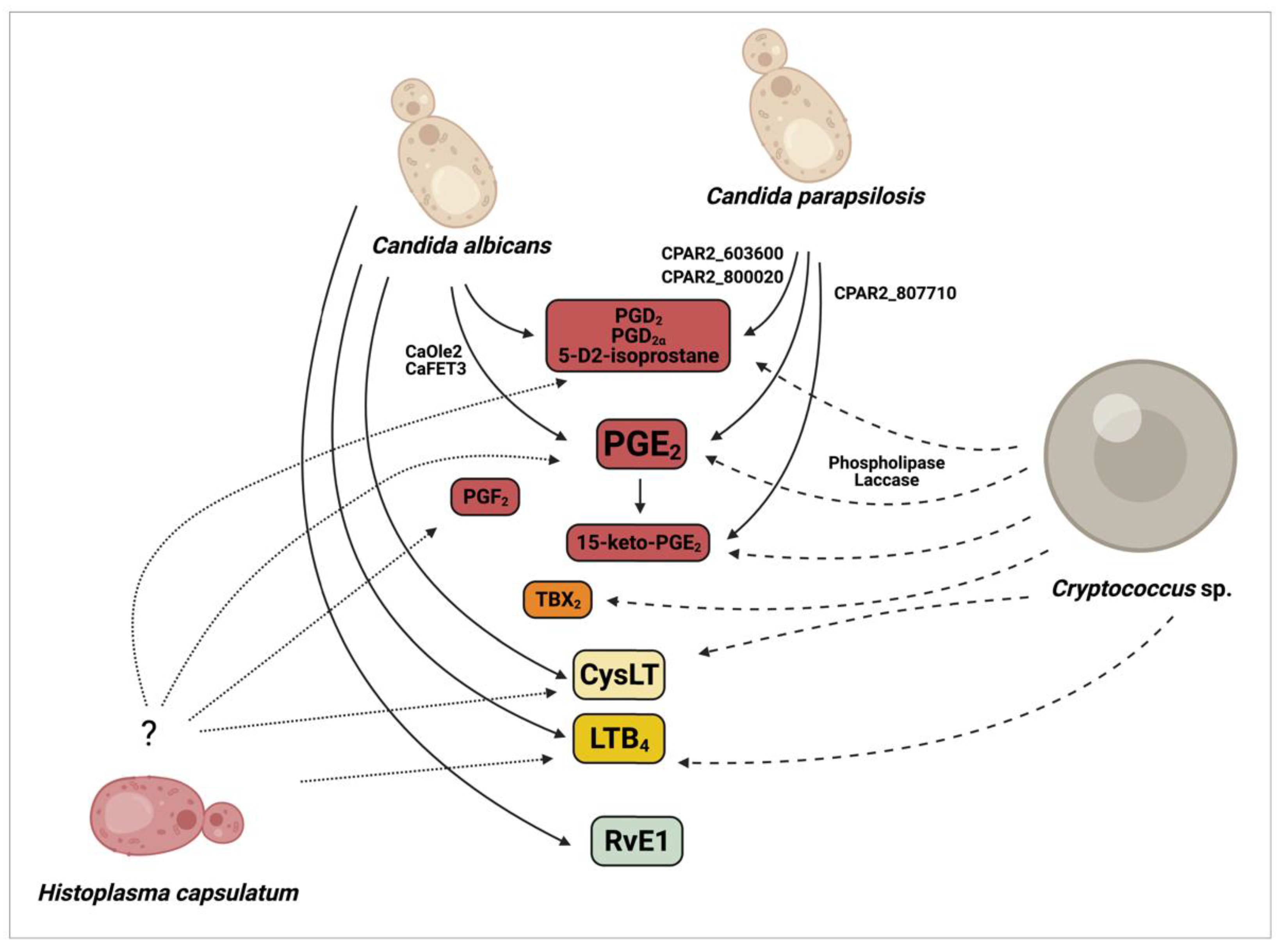
Deva et al. revealed that the opportunistic human fungal pathogen C. albicans produces 3,18-dihydroxy-5,8,11,14- eicosatetraenoic acid (3,18-di-HETE) by utilizing exogenous AA [53]. A subsequent study reported that, besides 3,18-di-HETE, C. albicans synthesizes an uncharacterized prostaglandin (PGEx) [50]. This eicosanoid was later shown to be indistinguishable from mammalian PGE2 [52]. Further investigations identified two non-COX/LO/CYTP450-related enzymes, namely the fatty acid stearyl-coenzyme A desaturase (Ole2) and the multicopper ferroxidase (Fet3), which are potentially involved in C. albicans (Ca) PGE2 biosynthesis (Figure 2) [13,52]. Homozygous deletion of both the fatty acid desaturase CaOLE2 and the multicopper oxidase CaFET3 resulted in a significant reduction in PGE2 synthesis by approximately 50–70% and 40–50%, respectively. However, PGE2 levels were still measurable in the corresponding homozygous mutant suggesting the presence of yet undiscovered PGs regulatory pathways in this species. C. albicans is also able to produce other PGs, such as PGD2 and PGD [13,52].
Besides C. albicans, several non-albicans Candida species such as C. dubliniensis, C. tropicalis, C. glabrata, and C. parapsilosis synthesize PGE2 [54,55,56], all of which are frequently associated with human fungal infections. HPLC-MS analysis of the fatty acid biosynthesis of C. parapsilosis by Grózer and colleagues revealed that this species, similar to C. albicans, is able to produce various PGs besides PGE2, and highlighted PGD2 as another major eicosanoid produced by C. parapsilosis [56]. A 2018 follow-up study with C. parapsilosis also identified an uncommon oxylipin, an autoxidative isomer of PGD2 (5-D2-IsoProstane) secreted upon incubation with exogenous AA (Figure 2) [57].
However, our knowledge of its biosynthesis is scarce [58]. A recently published study by Chakraborty and colleagues aimed to identify the molecular basis of PG production in C. parapsilosis and identified several genes involved in the process [57]. These include CPAR2_603600 (a homologue of the CaFET3), CPAR2_807710 (Acyl-CoA oxidase in S. cerevisiae, ScPOX1-3) and CPAR2_800020 (Acyl-CoA thiolase in S. cerevisiae, ScPOT1) (Figure 2). LC/MS data revealed that C. parapsilosis’ PGE2 biosynthesis is decreased by approximately 60–70% if any of these genes are disrupted. The double deletion of CPAR2_603600 and CPAR2_800020 leads to about 80% decrease in PGD2 production, suggesting their significant role in its biosynthesis. Their removal also effected the secretion of 15-keto-PGE2, a metabolite generated by the degradation of PGE2. CPAR2_807710 was shown to be most involved in 15-keto-PGE2 production. In contrast to C. albicans, the homologue of CaOLE2 has no significant role in PGE2 biosynthesis in C. parapsilosis [56].
Notably, in addition to PGs, C. albicans also utilizes AA for the biosynthesis of LTs, such as LTB4 and CysLTs (Figure 2) [13]. During Candida spp. infection, the synthesis of some LTs is altered to reduce host immune responses as a strategy for the establishment and maintenance of the infection [35]. LTB4 and CysLT production are both mediated by lipoxygenases through the production of 5-HpETE from exogenous AA [13], whereas RvE1 synthesis in C. albicans is produced from EPA [42], and some biosynthetic precursors (18-HEPE, 15-HEPE and 5-HEPE), by neutrophil 5-lipoxygenase principally, cytochrome P450 monooxygenase enzymes (CYP45), and other specific enzymes remain unknown [13,42,59]. The detailed biosynthetic pathway of LTs and RvE1 in C. albicans also remains enigmatic. Other human pathogenic non-albicans Candida species such as C. dubliniensis, C. tropicalis, and C. glabrata may also be able to produce these eicosanoids; however, this remains unconfirmed.

2.2. Production of Eicosanoids by Cryptococcus sp.

C. neoformans produces biologically active eicosanoids from exogenous sources of AA during infection, which are indistinguishable from host eicosanoids and modulate host defenses [50,51]. The major AA metabolite produced is an authentic PGD2, but the fungus is also able to produce heptadecatrienoic acid, 5-HETE, PGF2, TXB2, and PGE2 [50]. Two enzymes expressed by C. neoformans, phospholipase B1 (PLB1) and laccase (CNLAC1 gene), are believed to be associated with cryptococcal eicosanoid synthesis (Figure 2). Pharmacological enzymatic inhibition or deletion of phospholipase B1 (Δplb1) reduces secreted levels of all eicosanoids produced by C. neoformans [60,61]. In turn, deletion of laccase (Δlac1 mutants) or enzymatic inhibition by anti-lac1 antibody resulted specifically in the loss of PGE2 [51]. The addition of PGE2 was sufficient to promote growth of Δplb1 and Δlac1 in vitro and in vivo, independently of host PGE2 [60,61]. In fact, laccase is an important virulence factor for C. neoformans with a broad spectrum oxidase activity, converting polyphenolic compounds into the cell wall pigment melanin, and this polymer protects C. neoformans against oxidants, microbiocidal proteins and antifungals as well as to phagocytosis and killing by macrophages [62,63]. Additionally, recombinant laccase readily converts PGG2 into PGE2 and 15-keto-PGE2, and it is suggested as a key cryptococcal prostaglandin enzyme for this recently described unique production pathway (Figure 2) [51].

2.3. Production of Eicosanoids by Histoplasma Capsulatum

Although Histoplasma capsulatum can produce eicosanoids [13,54], further studies are necessary to dissect the pathways involved in their production and to determine whether they play a role during infection (Figure 2).

3. The Role of Eicosanoids during Fungal Infections

The production of eicosanoids by pathogenic fungi, such as C. albicans, C. dubliniensis, C. glabrata, C. tropicalis, C. neoformans, H. capsulatum and A. fumigatus is linked to the pathogenesis of each fungal infection [4,9,51,60,64,65,66]. Some fungal-derived eicosanoids can enhance both fungal colonization and induce immunomodulatory effects. Overall, fungal LTs act by enhancing the acute inflammation, whereas PGs have negative effects on innate and cellular Th1 responses against mycosis, resulting in immunological tolerance and contributing to the chronicity of fungal infections [13]. Herein, we discuss the roles of eicosanoids in three major fungal infections.

3.1. Eicosanoids in Candidiasis

Eicosanoids play an important role in both sides of the host–Candida interaction. Depending on the organ or tissue environment, host-derived PGE2 either decreases [64,67] or improves [68,69] the protective Th1 and Th17 responses that particularly may help the host restrain C. albicans at barrier surfaces and in the bloodstream.
C. albicans induces host cells to release AA from membrane phospholipids and infection-derived stimuli can also induce COX-2 expression and trigger the synthesis of PGs in various cells types [66,70]. C. albicans stimulates AA metabolism and the generation of PGE2 by synovial fibroblast, alveolar and peritoneal macrophages, and epithelial cells via stimulation of TLR2 and TLR4 [14]. Candida mannans and β-1,3-glucan induce PGE2 via stimulation of mannose receptor and dectin-1 in peripheral blood mononuclear cells, respectively [71]. PGE2 signaling stimulates Th2 and Th17 responses to yeast and limits the ability of macrophages to clear Candida sp. [71].
Although the exact role of Candida-derived eicosanoids during host–pathogen interactions is largely undiscovered, a limited number of studies are available that provide insights into how these lipid metabolites affect fungal virulence [57,67]. Many studies have pointed out the major role of host derived AA and fungi derived PGE2 in the modulation of yeast cell growth, morphogenesis, and biofilm formation in C. albicans [50,55]. In contrast, some studies focusing on the negative impact of PGE2 on yeast biology have shown that PGE2 inhibits germ tube formation by antagonizing yeast to hyphal transformation in C. albicans, which may limit tissue invasion [72].
In a previous study, the PGE2 biosynthesis associated genes OLE2, FET3, and FET31 were knocked out in C. albicans strains and the mutant’s capacity for PGE2 secretion was decreased in vitro. The authors examined the killing of the mutants by macrophages and immune-modulatory effects in vitro as well as their capacity for organ colonization ability in various mouse models of invasive candidiasis. The ole2−/− showed similar fitness and rates of hyphal formation than the wild-type (WT) counterpart. However, the gut colonizing capacity of the ole2−/− strain decreased compared to the WT strain. Besides its role in promoting colonization and survival in the mouse gut, C. albicans derived PGE2 also inhibited fungal cell internalization by phagocytes [65]. However, in CD11b+ DC and macrophage depleted mice, the WT C. albicans strain was not able to overgrow the ole2−/− strain [65], suggesting that the presence of PGE2 is beneficial for fungal growth, overcoming phagocytosis, and enhancing survival within the host.
Regarding non-albicans Candida species, the presence of AA increases biofilm formation and PGE2 production by C. glabrata, C. parapsilosis, and C. tropicalis [58]. These findings suggest that Candida spp. evolved the capacity to produce PGs, primarily PGE2, to enhance their fitness and survival within certain niches of the host that could directly promote the fungus’ pathogenesis upon a potential commensal-to-pathogenic shift event. The work of Chakraborty and colleagues suggests that fungal-derived PGs in C. parapsilosis also negatively regulate yeast cell phagocytosis and killing by macrophages, as PGs (PGE2, PGD2, and 15-keto-PGE2)-deficient C. parapsilosis cells were more susceptible to phagocytosis and killing by human peripheral blood monocyte-derived macrophages (PBMC-DM) compared to the WT strain [57]. As the virulence of PG deficient C. parapsilosis mutant strains also decreased in vivo compared to the WT strain, fungal PGs could also actively contribute to the virulence of this species.
These observations, together with other previous reports, suggest that fungi-derived prostaglandins have immunomodulatory functions analogous to their mammalian counterparts [54,73]. To further support this suggestion, another study reported that C. albicans-produced PGE2 up-regulates anti-inflammatory responses through enhancing IL-10 released by murine splenocytes. Moreover, the levels of mouse keratinocyte-derived chemokine (KC, analog to human IL-8) and other pro-inflammatory cytokines, such as TNFα, decreased after 24 h of fungal PGE2 treatment [67,71]. Fungal-derived PGE2 decreases the killing of C. albicans by intestinal macrophages, supporting the idea that fungal prostaglandins could also inhibit the killing activity of host cells.
A similar conclusion can be drawn for C. parapsilosis, where the absence of PGE2 -related genes increased the expression of pro-inflammatory cytokines such as pro-IL-1β, IL-6 and TNFα [57]. Thus, C. parapsilosis PGE2 could also negatively regulate host inflammatory responses. In C. parapsilosis, PGs production actively contributes to host cell damage, as revealed by the decreased [57] death of PBMC-DMs following infection with PG-deficient strains compared to the WT strain. C. parapsilosis PGs secretion is also suggested to contribute to organ colonization when studied in a mouse model of systemic candidiasis. However, the studied PG-related genes contributed unequally to the fungal load of each examined organ, which may suggest that the observed effect is not solely due to the presence of fungal PGE2, PGD2 and 15-keto-PGE2 [57].
LTs were also described as biologically active immunomodulatory eicosanoids [74,75]. Host-derived LTs increase capillary permeability, and activate and recruit eosinophils and neutrophils [75]. The present literature lacks information about the immunomodulatory function of fungal-derived LTs. However, a recent study showed that the amount of LTF4 increased in patients with candidemia, suggesting that LTF4 may also contribute to host responses to Candida spp. [35].
A previous study in 2007 showed that C. albicans-derived RvE1 is chemically identical to the human RvE1 [42]. When administered at low concentrations, fungal RvE1 reduced the IL-8-mediated chemotaxis of human neutrophils and also the recruitment of DCs [42,59]. In contrast, higher doses of fungal RvE1 enhanced phagocytic activity and fungicidal reactive oxygen species (ROS) production by human neutrophils against C. albicans. Interestingly, inoculation of RvE1 into mice with fungemia due to C. albicans, led to a more rapid clearance of the pathogen from the bloodstream [42]. These facts suggest that low concentrations of fungal RvE1 protects C. albicans due to the inhibition of neutrophil recruitment, although higher fungal burden (together with increased fungal RvE1 levels) could act as an alarming signal for neutrophils, which would then be able to control and restrict fungal invasion.

3.2. Eicosanoids in Cryptococcosis

C. neoformans secretes phospholipase B (PLB), which is a virulence factor. This single cryptococcal protein has three separate enzymatic activities: phospholipase B (PLB), which removes both acyl chains simultaneously from phospholipids; lysophospholipase (LPL), which removes the single acyl chain from lysophospholipids; and lysophospholipase transacylase (LPTA), which adds an acyl chain to lysophospholipids to form phospholipids [61]. Despite the lack of understanding on the structure and mechanism of action of PLB, this enzyme is involved in the survival of Cryptococci within macrophages, the destruction of lung tissue and the production of eicosanoids, which modulate phagocytic activity [61]. As mentioned, C. neoformans produces eicosanoids from exogenous AA and utilizes them to modulate the immune response favoring its own survival. For instance, LTB4 significantly reduced neutrophil recruitment in the lung vasculature of mice infected intravenously with C. neoformans, demonstrating a critical role of LTB4 in intravascular neutrophil swarming during infection [76]. The presence of CysLTs and LTB4 produced by C. neoformans strains B-3501A and H99 through the activity cryptococcal phospholipase cPLA2α and 5-LO, can contribute to fungal penetration of the blood–brain barrier in vitro and in vivo, specifically facilitating central nervous system (CNS) infection [77].
C. neoformans is also able to modulate the host inflammatory state during infection by directly manipulating host eicosanoids signaling and PGE2 is considered a mediator of cryptococcal virulence [60,78]. During macrophages infection, C. neoformans produces the dehydrogenated form of PGE2 (15-keto-PGE2) enhancing its virulence via the activation of the host nuclear transcription factor, PPAR-γ [60]. In C. neoformans infections, the use of antagonists of either EP2 or EP4 receptors improves the host defense by promoting TLR-4-mediated cytokine production, and enhancing M1 macrophage polarization followed by yeast killing [78].

3.3. Eicosanoids in Histoplasmosis

A 1992 study showed that peritoneal macrophages challenged with heat-killed H. capsulatum produce prostanoids (PGE2 and PGI2) and LTs (LTB4 and LTC4), the former being produced in a COX-dependent fashion [79]. This first observation was the stepping-stone for the study of eicosanoids in histoplasmosis. Notably, different forms of LTs and PGs are produced by the host during in vitro and in vivo challenges with H. capsulatum, but, interestingly, they commonly have opposite roles [80,81].
Sub-lethal H. capsulatum infections in mice treated with a FLAP inhibitor or in 5-LO deficient mice are fatal, suggesting that LTs are important for the host response in histoplasmosis [81,82]. Even though LTB4 and LTC4 are produced in mice infected with H. capsulatum [81], data show that administration of microspheres-associated LTB4 to 5-LO deficient mice can restore the production of cytokines and control the fungal burden [83].
Although LTB4 is an important mediator for the host response against H. capsulatum, the mechanism behind its effects is controversial. LTB4 is a very potent neutrophil chemoattractant [84], but 5-LO deficient mice and mice treated with FLAP inhibitors have lower levels of LTs and increased neutrophil recruitment when compared to their control counterparts. The increased neutrophil recruitment is followed by higher inflammatory response, an elevation of splenic fungal burdens, and 100% of mortality 14 days post-infection, even in scenarios of non-lethal H. capsulatum infections [81,82]. This suggests that features other than neutrophil chemotaxis are behind LTs’ effects during histoplasmosis. The effector mechanisms employed by macrophages are also responsive to LTs, as 5-LO deficient mice have a remarkable impairment in their ability to phagocytose non-opsonized or even IgG-opsonized H. capsulatum yeast cells, a deficiency that is bypassed by the exogenous addition of LTB4 or LTC4 [82]. Although LTs as well as PGs are usually produced at the onset of the inflammatory process, further steps in the host defense are modulated by the presence of these mediators [85]. Immunization of mice with cell-free antigens from H. capsulatum fails to confer protection in 5-LO deficient mice, possibly due to an inability to induce the recruitment of CD4+ and CD8+ cells to the lungs, and also a failure to increase the production of IFN-γ [86]. The production of LTs has an impact on events of the innate, but also of the adaptive, response during H. capsulatum infection, which modifies the outcome of the host–pathogen interaction.
The role of PGs during H. capsulatum infection is not as well studied relative to the leukotrienes. A fundamental piece of data is that the inhibition of COX-2 protects mice against lethal infection with H. capsulatum, a phenotype marked by lower fungal burden and a milder inflammatory process [80]. Curiously, when inhibiting the synthesis of prostanoids, an increase in the synthesis of LTB4 is observed, which is also beneficial to the host. The higher survival rates are associated with a decrease in neutrophil recruitment, consistent with the effects of LTs [80]. PGE2 has been associated with the deleterious effects on H. capsulatum infection [16], which correlates with the expression and activity of galectin-1 (Gal-1) [87]. Gal-1 represses the expression of PGE2 synthase, thus reducing the levels of PGE2 in H. capsulatum-infected mice. In contrast, H. capsulatum infection in Gal-1 KO mice leads to an increase in PGE2 production followed by increased fungal burden and higher mortality rates when compared to WT mice [87]. Even though PGE2 has such deleterious effects to the infected host, PGD2 has opposite effects to PGE2. The pharmacological inhibition of the endogenous production of PGD2 in H. capsulatum-infected macrophages leads to a severe inhibition of the leukocyte’s fungicidal activity, an effect that is reversed by the exogenous addition of PGD2. PGD2 also upregulates the expression of LTB4 receptor (BLT1R), potentiating the effects of LTB4 [87]. The role and mechanism of eicosanoids in the host response against H. capsulatum is still understudied, but data suggest that LTB4 and PGE2 have opposite effects in histoplasmosis by modulating the recruitment of neutrophils and the effector mechanisms of macrophages. In agreement, PGE2 also inhibits the production of hydrogen peroxide and TNF-α by monocytes, limiting the killing of Paracoccidioides brasiliensis [88]. Further studies are necessary to dissect whether other eicosanoids have a role in the infection by H. capsulatum, including ones of fungal origin and also the mechanisms involved in immune regulation.

4. Concluding Remarks

Human pathogenic fungal species such as Candida spp., C. neoformans and H. capsulatum produce eicosanoids. C. albicans utilizes exogenous AA in order to produce 3,18-di HET, LTB4, Cys-LTs, RvE1 and prostaglandins such as PGE2, PGD2, PGF2α. Non-albicans Candida species such as C. dubliniensis, C. tropicalis, C. glabrata, and C. parapsilosis also synthesize PGE2 from AA. Additionally, C. parapsilosis produces other prostaglandins such as PGD2 and 15-keto-PGE2. The exact molecular mechanisms behind the Candida-derived eicosanoid production are only uncovered in the case of PGs in C. albicans and C. parapsilosis. PGE₂ synthesis in C. albicans is regulated by OLE2, while C. parapsilosis evolved OLE2-independent PGs production pathways. This difference may explain the contrast in in vivo results: C. albicans-derived PGE2 is not required for virulence while PGs produced by C. parapsilosis influence the yeast’s capacity for host damage. Overall, the presence of fungal PGE2 has proven to be beneficial for C. albicans through increasing the ability of the pathogen to colonize the gut. Furthermore, fungal PGE2 protects C. albicans and C. parapsilosis cells from the phagocytic and killing activity of macrophages. C. albicans- derived RvE1 protects the fungus at low concentrations, whereas high concentrations expose the fungus to the host. C. neoformans produces 15-Keto-PGE2 to enhance its growth and ability to survive macrophage infection. In histoplasmosis, the inhibition of the PGs production is beneficial to the host as it favors LTB4 production, which induces a decrease in the fungal burden, mortality rates and neutrophil recruitment. PGE2 has deleterious effects on histoplasmosis, as opposed to the positive effects of PGD2, which upregulates the expression of BLT1R in H. capsulatum infected macrophages and potentiates the effects of LTB4. LTs are important for the host response, as, for example, LTB4 mediates the immune response helping to control the fungal burden. However, the mechanism behind its effect is controversial as LTB4 is a neutrophil chemoattractant and mice with lower levels of LTs have increased inflammatory responses, fungal burdens and mortality rates. However, further investigations are needed to understand the precise role of eicosanoids, mainly PGE2, during host–pathogen interactions.

5. Future Trends

The production of eicosanoids seems to be a conserved feature among several eukaryotic organisms, including filamentous and yeast fungi, protozoa and higher eukaryotes such as mammals. Independently on the organism, their biosynthetic pathways may vary considerably, as well as the full eicosanoid portfolio produced. Since pathogenic fungi are able to secrete these molecules, the exact mechanism in how they alter the microbial physiology has not been fully explored, although current research and published data have demonstrated their effects on the modulation of interactions with the host and immune responses.
Then, as it is completely plausible that fungal eicosanoids might function as virulence factors, further investigations might enable us to understand their precise role during host–pathogen interactions, as well as exploring the unique steps of the fungal eicosanoids biosynthesis, as a new potential target to combat C. albicans, C. parapsilosis, C. neoformans and H. capsulatum and possibly other fungal infections.

Author Contributions

S.R.M.; D.Z.-M.; T.T.; A.G.; J.D.N. and A.J.G. All authors have read and agreed to the published version of the manuscript.

Funding

A.J.G. was supported by grants from the Brazilian agencies Conselho Nacional de Desenvolvimento Científico e Tecnológico (CNPq, grants 311470/2018-1) and Fundação Carlos Chagas de Amparo à Pesquisa no Estado do Rio de Janeiro (E-26/202.696/2018). S.R.M. was supported by CAPES (Coordenação de Aperfeiçoamento de Pessoal de Nível Superior).

Institutional Review Board Statement

Not applicable.

Conflicts of Interest

The authors declare no conflict of interest.

Abbreviations

15-keto-PGE2Dehydrogenated form of Prostaglandin E2
3-HETE3-hydroxy-5,8,11,14- eicosatetraenoic acid
3,18-di-HETE3,18-dihydroxy-5,8,11,14- eicosatetraenoic acid
5- HpETE5-hydroperoxyeicosatetraenoic acid
5-LOEnzyme 5-lipoxygenase
AAArachidonic acid
AA (20:4, n-6)Arachidonic acid (20-carbon, 4 insaturations, omega 6 family)
ABCATP-binding cassette transporter
APCsAntigen presenting cells
BLT1Leukotriene B4 high-affinity receptor
BLT2Leukotriene B4 Low-affinity receptor
CaFET3Candida albicans multicopper ferroxidase
cAMPCyclic adenosine monophosphate
CaOLE2C. albicans fatty acid stearyl-coenzyme A desaturase
CNLAC1Cryptococcus neoformans laccase gene
CNSCentral nervous system
COXCyclooxygenase
CRTH2Chemoattractant receptor-homologous molecule expressed on Th2 cells; also known as DP2, PG DP2 receptor
CysLTCysteinyl leukotriene
CysLTR1Type 1 cysteinyl leukotriene receptor
CYTP450Cytochrome P450 oxidase
DCsDendritic cells
DHADocosahexaenoic acid
DP1Prostaglandin D2 receptor 1
EP (1-4)Rhodopsin-type receptors
EPAEicosapentaenoic acid
ERKsExtracellular signal-regulate kinases
FAsFatty acids
FLA5-lipoxygenase activating protein
FPProstaglandin F receptor
Gal-1Galectin-1
GPCRsG-protein coupled receptors
ILInterleukin
IPProstacyclin receptor
LOXLipoxygenase
LPLLysophospholipase
LPTALysophospholipase transacylase
LTLeukotriene
LTA4Leukotriene A4
LTA4HLTA4 hydrolase
LTB4Leukotriene B4
LTD4Leukotriene D4
LTE4Leukotriene E4
LTF4Leukotriene F4
LTsLeukotrienes
n-3Omega-3
n-6Omega-6
PBMC- DMPeripheral blood monocyte-derived macrophages
PGsProstaglandins
PGD2Prostaglandin D2
PGE2Prostaglandin E2
PGExUncharacterized prostaglandin
PGF2Prostaglandin F2
PGG2Prostaglandin G2
PGH2Prostaglandin H2
PGI2Prostacyclin
PI3KPhosphoinositide 3-kinase
PKAProtein kinase A
PLAPhospholipase A
PLBPhospholipase B
PMNPolymorphonuclear neutrophil
PUFAPolyunsaturated fatty acids
RvsResolvins
RvDD-series Resolvins (RvD1-6)
RvE1E-series Resolvins (RvE1-3)
RvT13-series resolvins
SPMSpecialized pro-resolving mediator
ThT-helper
TNF-αTumor necrosis factor alpha
TPThromboxane receptor
TXThromboxane
TXA2Thromboxane A2
TXSThromboxane synthase
Δlac1Laccase gene Cryptococcus neoformans mutant
Δplb1Phospholipase B1 Cryptococcus neoformans mutant

References

  1. Kanamori, H.; Rutala, W.A.; Sickbert-Bennett, E.E.; Weber, D.J. Review of Fungal Outbreaks and Infection Prevention in Healthcare Settings during Construction and Renovation. Clin. Infect. Dis. Off. Publ. Infect. Dis. Soc. Am. 2015, 61, 433–444. [Google Scholar] [CrossRef] [PubMed]
  2. Spitzer, M.; Robbins, N.; Wright, G.D. Combinatorial Strategies for Combating Invasive Fungal Infections. Virulence 2017, 8, 169–185. [Google Scholar] [CrossRef] [PubMed]
  3. Janbon, G.; Quintin, J.; Lanternier, F.; d’Enfert, C. Studying Fungal Pathogens of Humans and Fungal Infections: Fungal Diversity and Diversity of Approaches. Genes Immun. 2019, 20, 403–414. [Google Scholar] [CrossRef]
  4. Ells, R.; Kock, J.L.; Albertyn, J.; Pohl, C.H. Arachidonic Acid Metabolites in Pathogenic Yeasts. Lipids Health Dis. 2012, 11, 100. [Google Scholar] [CrossRef] [PubMed] [Green Version]
  5. Harris, S.G.; Padilla, J.; Koumas, L.; Ray, D.; Phipps, R.P. Prostaglandins as Modulators of Immunity. Trends Immunol. 2002, 23, 144–150. [Google Scholar] [CrossRef]
  6. Ghannoum, M.A. Potential Role of Phospholipases in Virulence and Fungal Pathogenesis. Clin. Microbiol. Rev. 2000, 13, 122–143, table of contents. [Google Scholar] [CrossRef]
  7. Tilley, S.L.; Coffman, T.M.; Koller, B.H. Mixed Messages: Modulation of Inflammation and Immune Responses by Prostaglandins and Thromboxanes. J. Clin. Investig. 2001, 108, 15–23. [Google Scholar] [CrossRef] [PubMed]
  8. Christie, W.W.; Harwood, J.L. Oxidation of Polyunsaturated Fatty Acids to Produce Lipid Mediators. Essays Biochem. 2020, 64, 401–421. [Google Scholar] [CrossRef]
  9. Ghannoum, M.A. Extracellular Phospholipases as Universal Virulence Factor in Pathogenic Fungi. Nihon Ishinkin Gakkai Zasshi Jpn. J. Med. Mycol. 1998, 39, 55–59. [Google Scholar] [CrossRef] [Green Version]
  10. Lehninger, A.L.; Nelson, D.L.; Cox, M.M. Lehninger Principles of Biochemistry, 4th ed.; W.H. Freeman: New York, NY, USA, 2005; ISBN 978-0-7167-4339-2. [Google Scholar]
  11. Serhan, C.N. Novel Eicosanoid and Docosanoid Mediators: Resolvins, Docosatrienes, and Neuroprotectins. Curr. Opin. Clin. Nutr. Metab. Care 2005, 8, 115–121. [Google Scholar] [CrossRef]
  12. Serhan, C.N.; Gotlinger, K.; Hong, S.; Arita, M. Resolvins, Docosatrienes, and Neuroprotectins, Novel Omega-3-Derived Mediators, and Their Aspirin-Triggered Endogenous Epimers: An Overview of Their Protective Roles in Catabasis. Prostagland. Other Lipid Mediat. 2004, 73, 155–172. [Google Scholar] [CrossRef] [PubMed]
  13. Noverr, M.C.; Toews, G.B.; Huffnagle, G.B. Production of Prostaglandins and Leukotrienes by Pathogenic Fungi. Infect. Immun. 2002, 70, 400–402. [Google Scholar] [CrossRef] [PubMed] [Green Version]
  14. Martínez-Colón, G.J.; Moore, B.B. Prostaglandin E2 as a Regulator of Immunity to Pathogens. Pharmacol. Ther. 2018, 185, 135–146. [Google Scholar] [CrossRef]
  15. Coleman, R.A.; Smith, W.L.; Narumiya, S. International Union of Pharmacology Classification of Prostanoid Receptors: Properties, Distribution, and Structure of the Receptors and Their Subtypes. Pharmacol. Rev. 1994, 46, 205–229. [Google Scholar]
  16. Pereira, P.A.T.; Assis, P.A.; Prado, M.K.B.; Ramos, S.G.; Aronoff, D.M.; de Paula-Silva, F.W.G.; Sorgi, C.A.; Faccioli, L.H. Prostaglandins D2 and E2 Have Opposite Effects on Alveolar Macrophages Infected with Histoplasma Capsulatum. J. Lipid Res. 2018, 59, 195–206. [Google Scholar] [CrossRef] [Green Version]
  17. Hardwick, J.P.; Eckman, K.; Lee, Y.K.; Abdelmegeed, M.A.; Esterle, A.; Chilian, W.M.; Chiang, J.Y.; Song, B.-J. Eicosanoids in Metabolic Syndrome. Adv. Pharmacol. 2013, 66, 157–266. [Google Scholar] [CrossRef] [Green Version]
  18. Jones, R.L. Prostanoid Receptors. In xPharm: The Comprehensive Pharmacology Reference; Elsevier: Amsterdam, The Netherlands, 2007; pp. 1–3. ISBN 978-0-08-055232-3. [Google Scholar]
  19. Sun, L.; Ye, R.D. Role of G Protein-Coupled Receptors in Inflammation. Acta Pharmacol. Sin. 2012, 33, 342–350. [Google Scholar] [CrossRef] [Green Version]
  20. Sugimoto, Y.; Narumiya, S. Prostaglandin E Receptors. J. Biol. Chem. 2007, 282, 11613–11617. [Google Scholar] [CrossRef] [Green Version]
  21. Narumiya, S.; Sugimoto, Y.; Ushikubi, F. Prostanoid Receptors: Structures, Properties, and Functions. Physiol. Rev. 1999, 79, 1193–1226. [Google Scholar] [CrossRef] [PubMed]
  22. Woodward, D.F.; Jones, R.L.; Narumiya, S. International Union of Basic and Clinical Pharmacology. LXXXIII: Classification of Prostanoid Receptors, Updating 15 Years of Progress. Pharmacol. Rev. 2011, 63, 471–538. [Google Scholar] [CrossRef] [PubMed] [Green Version]
  23. Tsuge, K.; Inazumi, T.; Shimamoto, A.; Sugimoto, Y. Molecular Mechanisms Underlying Prostaglandin E2-Exacerbated Inflammation and Immune Diseases. Int. Immunol. 2019, 31, 597–606. [Google Scholar] [CrossRef] [PubMed]
  24. Cheng, X.; Ji, Z.; Tsalkova, T.; Mei, F. Epac and PKA: A Tale of Two Intracellular CAMP Receptors. Acta Biochim. Biophys. Sin. 2008, 40, 651–662. [Google Scholar] [CrossRef] [PubMed] [Green Version]
  25. Regan, J.W. EP2 and EP4 Prostanoid Receptor Signaling. Life Sci. 2003, 74, 143–153. [Google Scholar] [CrossRef]
  26. Breyer, R.M.; Bagdassarian, C.K.; Myers, S.A.; Breyer, M.D. Prostanoid Receptors: Subtypes and Signaling. Annu. Rev. Pharmacol. Toxicol. 2001, 41, 661–690. [Google Scholar] [CrossRef]
  27. Kobayashi, K.; Horikami, D.; Omori, K.; Nakamura, T.; Yamazaki, A.; Maeda, S.; Murata, T. Thromboxane A2 Exacerbates Acute Lung Injury via Promoting Edema Formation. Sci. Rep. 2016, 6, 32109. [Google Scholar] [CrossRef] [Green Version]
  28. Altieri, D.C.; Mannucci, P.M. Thromboxane Generation by Human Monocytes Enhances Platelet Function. J. Exp. Med. 1986, 164, 1815–1820. [Google Scholar] [CrossRef] [Green Version]
  29. Ashton, A.W.; Mukherjee, S.; Nagajyothi, F.N.U.; Huang, H.; Braunstein, V.L.; Desruisseaux, M.S.; Factor, S.M.; Lopez, L.; Berman, J.W.; Wittner, M.; et al. Thromboxane A2 Is a Key Regulator of Pathogenesis during Trypanosoma Cruzi Infection. J. Exp. Med. 2007, 204, 929–940. [Google Scholar] [CrossRef]
  30. Yang, C.-W.; Unanue, E.R. Neutrophils Control the Magnitude and Spread of the Immune Response in a Thromboxane A2-Mediated Process. J. Exp. Med. 2013, 210, 375–387. [Google Scholar] [CrossRef] [Green Version]
  31. Peters-Golden, M.; Canetti, C.; Mancuso, P.; Coffey, M.J. Leukotrienes: Underappreciated Mediators of Innate Immune Responses. J. Immunol. 2005, 174, 589–594. [Google Scholar] [CrossRef] [Green Version]
  32. Morato-Marques, M.; Campos, M.R.; Kane, S.; Rangel, A.P.; Lewis, C.; Ballinger, M.N.; Kim, S.-H.; Peters-Golden, M.; Jancar, S.; Serezani, C.H. Leukotrienes Target F-Actin/Cofilin-1 to Enhance Alveolar Macrophage Anti-Fungal Activity. J. Biol. Chem. 2011, 286, 28902–28913. [Google Scholar] [CrossRef] [PubMed] [Green Version]
  33. Tager, A.M.; Luster, A.D. BLT1 and BLT2: The Leukotriene B(4) Receptors. Prostagland. Leukot. Essent. Fatty Acids 2003, 69, 123–134. [Google Scholar] [CrossRef]
  34. Kanaoka, Y.; Boyce, J.A. Cysteinyl Leukotrienes and Their Receptors; Emerging Concepts. Allergy Asthma Immunol. Res. 2014, 6, 288–295. [Google Scholar] [CrossRef]
  35. Melo, C.F.O.R.; Bachur, L.F.; Delafiori, J.; Dabaja, M.Z.; de Oliveira, D.N.; Guerreiro, T.M.; Tararam, C.A.; Busso-Lopes, A.F.; Moretti, M.L.; Catharino, R.R. Does Leukotriene F4 Play a Major Role in the Infection Mechanism of Candida Sp.? Microb. Pathog. 2020, 149, 104394. [Google Scholar] [CrossRef] [PubMed]
  36. Oliveira, S.H.P.; Canetti, C.; Ribeiro, R.A.; Cunha, F.Q. Neutrophil Migration Induced by IL-1beta Depends upon LTB4 Released by Macrophages and upon TNF-Alpha and IL-1beta Released by Mast Cells. Inflammation 2008, 31, 36–46. [Google Scholar] [CrossRef] [PubMed]
  37. Olynych, T.J.; Jakeman, D.L.; Marshall, J.S. Fungal Zymosan Induces Leukotriene Production by Human Mast Cells through a Dectin-1-Dependent Mechanism. J. Allergy Clin. Immunol. 2006, 118, 837–843. [Google Scholar] [CrossRef]
  38. König, S.; Pace, S.; Pein, H.; Heinekamp, T.; Kramer, J.; Romp, E.; Straßburger, M.; Troisi, F.; Proschak, A.; Dworschak, J.; et al. Gliotoxin from Aspergillus Fumigatus Abrogates Leukotriene B4 Formation through Inhibition of Leukotriene A4 Hydrolase. Cell Chem. Biol. 2019, 26, 524–534.e5. [Google Scholar] [CrossRef]
  39. Lee, E.K.S.; Gillrie, M.R.; Li, L.; Arnason, J.W.; Kim, J.H.; Babes, L.; Lou, Y.; Sanati-Nezhad, A.; Kyei, S.K.; Kelly, M.M.; et al. Leukotriene B4-Mediated Neutrophil Recruitment Causes Pulmonary Capillaritis during Lethal Fungal Sepsis. Cell Host Microbe 2018, 23, 121–133.e4. [Google Scholar] [CrossRef]
  40. Caffrey-Carr, A.K.; Hilmer, K.M.; Kowalski, C.H.; Shepardson, K.M.; Temple, R.M.; Cramer, R.A.; Obar, J.J. Host-Derived Leukotriene B4 Is Critical for Resistance against Invasive Pulmonary Aspergillosis. Front. Immunol. 2017, 8, 1984. [Google Scholar] [CrossRef] [Green Version]
  41. Spite, M. Deciphering the Role of N-3 Polyunsaturated Fatty Acid-Derived Lipid Mediators in Health and Disease. Proc. Nutr. Soc. 2013, 72, 441–450. [Google Scholar] [CrossRef] [PubMed] [Green Version]
  42. Haas-Stapleton, E.J.; Lu, Y.; Hong, S.; Arita, M.; Favoreto, S.; Nigam, S.; Serhan, C.N.; Agabian, N. Candida Albicans Modulates Host Defense by Biosynthesizing the Pro-Resolving Mediator Resolvin E1. PLoS ONE 2007, 2, e0001316. [Google Scholar] [CrossRef] [PubMed]
  43. Schwab, J.M.; Chiang, N.; Arita, M.; Serhan, C.N. Resolvin E1 and Protectin D1 Activate Inflammation-Resolution Programmes. Nature 2007, 447, 869–874. [Google Scholar] [CrossRef] [Green Version]
  44. Serhan, C.N. Resolution Phase of Inflammation: Novel Endogenous Anti-Inflammatory and Proresolving Lipid Mediators and Pathways. Annu. Rev. Immunol. 2007, 25, 101–137. [Google Scholar] [CrossRef] [Green Version]
  45. Sommer, C.; Birklein, F. Resolvins and Inflammatory Pain. F1000 Med. Rep. 2011, 3, 19. [Google Scholar] [CrossRef] [PubMed]
  46. Buczynski, M.W.; Dumlao, D.S.; Dennis, E.A. Thematic Review Series: Proteomics. An Integrated Omics Analysis of Eicosanoid Biology. J. Lipid Res. 2009, 50, 1015–1038. [Google Scholar] [CrossRef] [PubMed] [Green Version]
  47. van Dyk, M.S.; Kock, J.L.; Coetzee, D.J.; Augustyn, O.P.; Nigam, S. Isolation of a Novel Arachidonic Acid Metabolite 3-Hydroxy-5,8,11,14-Eicosatetraenoic Acid (3-HETE) from the Yeast Dipodascopsis Uninucleata UOFs-Y128. FEBS Lett. 1991, 283, 195–198. [Google Scholar] [CrossRef] [Green Version]
  48. Strauss, T.; Botha, A.; Kock, J.L.; Paul, I.; Smith, D.P.; Linke, D.; Schewe, T.; Nigam, S. Mapping the Distribution of 3-Hydroxylipins in the Mucorales Using Immunofluorescence Microscopy. Antonie Van Leeuwenhoek 2000, 78, 39–42. [Google Scholar] [CrossRef]
  49. Botha, A.; Kock, J.L.F.; Coetzee, D.J.; Van Dyk, M.S.; Van Der Berg, L.; Botes, P.J. Yeast Eicosanoids I. The Distribution and Taxonomic Value of Cellular Fatty Acids and Arachidonic Acid Metabolites in the Dipodascaceae and Related Taxa. Syst. Appl. Microbiol. 1992, 15, 148–154. [Google Scholar] [CrossRef]
  50. Noverr, M.C.; Phare, S.M.; Toews, G.B.; Coffey, M.J.; Huffnagle, G.B. Pathogenic Yeasts Cryptococcus Neoformans and Candida Albicans Produce Immunomodulatory Prostaglandins. Infect. Immun. 2001, 69, 2957–2963. [Google Scholar] [CrossRef] [Green Version]
  51. Erb-Downward, J.R.; Noggle, R.M.; Williamson, P.R.; Huffnagle, G.B. The Role of Laccase in Prostaglandin Production by Cryptococcus Neoformans. Mol. Microbiol. 2008, 68, 1428–1437. [Google Scholar] [CrossRef] [Green Version]
  52. Erb-Downward, J.R.; Noverr, M.C. Characterization of Prostaglandin E2 Production by Candida Albicans. Infect. Immun. 2007, 75, 3498–3505. [Google Scholar] [CrossRef] [Green Version]
  53. Deva, R.; Ciccoli, R.; Kock, L.; Nigam, S. Involvement of Aspirin-Sensitive Oxylipins in Vulvovaginal Candidiasis. FEMS Microbiol. Lett. 2001, 198, 37–43. [Google Scholar] [CrossRef] [PubMed]
  54. Noverr, M.C.; Erb-Downward, J.R.; Huffnagle, G.B. Production of Eicosanoids and Other Oxylipins by Pathogenic Eukaryotic Microbes. Clin. Microbiol. Rev. 2003, 16, 517–533. [Google Scholar] [CrossRef] [Green Version]
  55. Alem, M.A.; Douglas, L.J. Prostaglandin Production during Growth of Candida Albicans Biofilms. J. Med. Microbiol. 2005, 54, 1001–1005. [Google Scholar] [CrossRef]
  56. Grózer, Z.; Tóth, A.; Tóth, R.; Kecskeméti, A.; Vágvölgyi, C.; Nosanchuk, J.D.; Szekeres, A.; Gácser, A. Candida Parapsilosis Produces Prostaglandins from Exogenous Arachidonic Acid and OLE2 Is Not Required for Their Synthesis. Virulence 2015, 6, 85–92. [Google Scholar] [CrossRef] [PubMed] [Green Version]
  57. Chakraborty, T.; Thuer, E.; Heijink, M.; Tóth, R.; Bodai, L.; Vágvölgyi, C.; Giera, M.; Gabaldón, T.; Gácser, A. Eicosanoid Biosynthesis Influences the Virulence of Candida Parapsilosis. Virulence 2018, 9, 1019–1035. [Google Scholar] [CrossRef] [Green Version]
  58. Mishra, N.N.; Ali, S.; Shukla, P.K. Arachidonic Acid Affects Biofilm Formation and PGE2 Level in Candida Albicans and Non-Albicans Species in Presence of Subinhibitory Concentration of Fluconazole and Terbinafine. Braz. J. Infect. Dis. Off. Publ. Braz. Soc. Infect. Dis. 2014, 18, 287–293. [Google Scholar] [CrossRef] [Green Version]
  59. Arita, M.; Bianchini, F.; Aliberti, J.; Sher, A.; Chiang, N.; Hong, S.; Yang, R.; Petasis, N.A.; Serhan, C.N. Stereochemical Assignment, Antiinflammatory Properties, and Receptor for the Omega-3 Lipid Mediator Resolvin E1. J. Exp. Med. 2005, 201, 713–722. [Google Scholar] [CrossRef] [PubMed]
  60. Evans, R.J.; Pline, K.; Loynes, C.A.; Needs, S.; Aldrovandi, M.; Tiefenbach, J.; Bielska, E.; Rubino, R.E.; Nicol, C.J.; May, R.C.; et al. 15-Keto-Prostaglandin E2 Activates Host Peroxisome Proliferator-Activated Receptor Gamma (PPAR-γ) to Promote Cryptococcus Neoformans Growth during Infection. PLoS Pathog. 2019, 15, e1007597. [Google Scholar] [CrossRef] [PubMed] [Green Version]
  61. Ganendren, R.; Widmer, F.; Singhal, V.; Wilson, C.; Sorrell, T.; Wright, L. In Vitro Antifungal Activities of Inhibitors of Phospholipases from the Fungal Pathogen Cryptococcus Neoformans. Antimicrob. Agents Chemother. 2004, 48, 1561–1569. [Google Scholar] [CrossRef] [PubMed] [Green Version]
  62. Panepinto, J.C.; Williamson, P.R. Intersection of Fungal Fitness and Virulence in Cryptococcus Neoformans. FEMS Yeast Res. 2006, 6, 489–498. [Google Scholar] [CrossRef] [PubMed] [Green Version]
  63. Zhu, X.; Williamson, P.R. Role of Laccase in the Biology and Virulence of Cryptococcus Neoformans. FEMS Yeast Res. 2004, 5, 1–10. [Google Scholar] [CrossRef] [PubMed] [Green Version]
  64. Valdez, P.A.; Vithayathil, P.J.; Janelsins, B.M.; Shaffer, A.L.; Williamson, P.R.; Datta, S.K. Prostaglandin E2 Suppresses Antifungal Immunity by Inhibiting Interferon Regulatory Factor 4 Function and Interleukin-17 Expression in T Cells. Immunity 2012, 36, 668–679. [Google Scholar] [CrossRef] [PubMed] [Green Version]
  65. Tan, T.G.; Lim, Y.S.; Tan, A.; Leong, R.; Pavelka, N. Fungal Symbionts Produce Prostaglandin E2 to Promote Their Intestinal Colonization. Front. Cell. Infect. Microbiol. 2019, 9, 359. [Google Scholar] [CrossRef] [PubMed] [Green Version]
  66. Filler, S.G.; Ibe, B.O.; Luckett, P.M.; Raj, J.U.; Edwards, J.E. Candida Albicans Stimulates Endothelial Cell Eicosanoid Production. J. Infect. Dis. 1991, 164, 928–935. [Google Scholar] [CrossRef]
  67. Ma, H.; Wan, S.; Xia, C.-Q. Immunosuppressive CD11b+Ly6Chi Monocytes in Pristane-Induced Lupus Mouse Model. J. Leukoc. Biol. 2016, 99, 1121–1129. [Google Scholar] [CrossRef]
  68. Yao, C.; Sakata, D.; Esaki, Y.; Li, Y.; Matsuoka, T.; Kuroiwa, K.; Sugimoto, Y.; Narumiya, S. Prostaglandin E2-EP4 Signaling Promotes Immune Inflammation through Th1 Cell Differentiation and Th17 Cell Expansion. Nat. Med. 2009, 15, 633–640. [Google Scholar] [CrossRef] [Green Version]
  69. Chizzolini, C.; Chicheportiche, R.; Alvarez, M.; de Rham, C.; Roux-Lombard, P.; Ferrari-Lacraz, S.; Dayer, J.-M. Prostaglandin E2 Synergistically with Interleukin-23 Favors Human Th17 Expansion. Blood 2008, 112, 3696–3703. [Google Scholar] [CrossRef] [Green Version]
  70. Castro, M.; Ralston, N.V.; Morgenthaler, T.I.; Rohrbach, M.S.; Limper, A.H. Candida Albicans Stimulates Arachidonic Acid Liberation from Alveolar Macrophages through Alpha-Mannan and Beta-Glucan Cell Wall Components. Infect. Immun. 1994, 62, 3138–3145. [Google Scholar] [CrossRef] [PubMed] [Green Version]
  71. Smeekens, S.P.; van de Veerdonk, F.L.; van der Meer, J.W.M.; Kullberg, B.J.; Joosten, L.A.B.; Netea, M.G. The Candida Th17 Response Is Dependent on Mannan- and -Glucan-Induced Prostaglandin E2. Int. Immunol. 2010, 22, 889–895. [Google Scholar] [CrossRef] [Green Version]
  72. Kalo-Klein, A.; Witkin, S.S. Prostaglandin E2 Enhances and Gamma Interferon Inhibits Germ Tube Formation in Candida Albicans. Infect. Immun. 1990, 58, 260–262. [Google Scholar] [CrossRef] [Green Version]
  73. Kim, Y.-G.; Udayanga, K.G.S.; Totsuka, N.; Weinberg, J.B.; Núñez, G.; Shibuya, A. Gut Dysbiosis Promotes M2 Macrophage Polarization and Allergic Airway Inflammation via Fungi-Induced PGE2. Cell Host Microbe 2014, 15, 95–102. [Google Scholar] [CrossRef] [PubMed] [Green Version]
  74. Bernström, K.; Hammarström, S. A Novel Leukotriene Formed by Transpeptidation of Leukotriene E. Biochem. Biophys. Res. Commun. 1982, 109, 800–804. [Google Scholar] [CrossRef]
  75. Singh, R.K.; Gupta, S.; Dastidar, S.; Ray, A. Cysteinyl Leukotrienes and Their Receptors: Molecular and Functional Characteristics. Pharmacology 2010, 85, 336–349. [Google Scholar] [CrossRef] [PubMed]
  76. Sun, D.; Shi, M. Neutrophil Swarming toward Cryptococcus Neoformans Is Mediated by Complement and Leukotriene B4. Biochem. Biophys. Res. Commun. 2016, 477, 945–951. [Google Scholar] [CrossRef] [Green Version]
  77. Zhu, L.; Maruvada, R.; Sapirstein, A.; Peters-Golden, M.; Kim, K.S. Cysteinyl Leukotrienes as Novel Host Factors Facilitating Cryptococcus Neoformans Penetration into the Brain. Cell. Microbiol. 2017, 19, e12661. [Google Scholar] [CrossRef] [PubMed] [Green Version]
  78. Shen, L.; Liu, Y. Prostaglandin E2 Blockade Enhances the Pulmonary Anti-Cryptococcus Neoformans Immune Reaction via the Induction of TLR-4. Int. Immunopharmacol. 2015, 28, 376–381. [Google Scholar] [CrossRef]
  79. Wolf, J.E.; Massof, S.E.; Peters, S.P. Alterations in Murine Macrophage Arachidonic Acid Metabolism Following Ingestion of Nonviable Histoplasma Capsulatum. Infect. Immun. 1992, 60, 2559–2564. [Google Scholar] [CrossRef] [PubMed] [Green Version]
  80. Pereira, P.A.T.; Trindade, B.C.; Secatto, A.; Nicolete, R.; Peres-Buzalaf, C.; Ramos, S.G.; Sadikot, R.; Bitencourt, C.D.S.; Faccioli, L.H. Celecoxib Improves Host Defense through Prostaglandin Inhibition during Histoplasma Capsulatum Infection. Mediat. Inflamm. 2013, 2013, 950981. [Google Scholar] [CrossRef] [Green Version]
  81. Medeiros, A.I.; Sá-Nunes, A.; Soares, E.G.; Peres, C.M.; Silva, C.L.; Faccioli, L.H. Blockade of Endogenous Leukotrienes Exacerbates Pulmonary Histoplasmosis. Infect. Immun. 2004, 72, 1637–1644. [Google Scholar] [CrossRef] [Green Version]
  82. Secatto, A.; Rodrigues, L.C.; Serezani, C.H.; Ramos, S.G.; Dias-Baruffi, M.; Faccioli, L.H.; Medeiros, A.I. 5-Lipoxygenase Deficiency Impairs Innate and Adaptive Immune Responses during Fungal Infection. PLoS ONE 2012, 7, e31701. [Google Scholar] [CrossRef]
  83. Nicolete, R.; Secatto, A.; Pereira, P.A.T.; Soares, E.G.; Faccioli, L.H. Leukotriene B4-Loaded Microspheres as a New Approach to Enhance Antimicrobial Responses in Histoplasma Capsulatum-Infected Mice. Int. J. Antimicrob. Agents 2009, 34, 365–369. [Google Scholar] [CrossRef] [PubMed]
  84. Metzemaekers, M.; Gouwy, M.; Proost, P. Neutrophil Chemoattractant Receptors in Health and Disease: Double-Edged Swords. Cell. Mol. Immunol. 2020, 17, 433–450. [Google Scholar] [CrossRef] [PubMed]
  85. Samuchiwal, S.K.; Boyce, J.A. Role of Lipid Mediators and Control of Lymphocyte Responses in Type 2 Immunopathology. J. Allergy Clin. Immunol. 2018, 141, 1182–1190. [Google Scholar] [CrossRef] [PubMed] [Green Version]
  86. Medeiros, A.I.; Sá-Nunes, A.; Turato, W.M.; Secatto, A.; Frantz, F.G.; Sorgi, C.A.; Serezani, C.H.; Deepe, G.S.; Faccioli, L.H. Leukotrienes Are Potent Adjuvant during Fungal Infection: Effects on Memory T Cells. J. Immunol. 2008, 181, 8544–8551. [Google Scholar] [CrossRef] [PubMed] [Green Version]
  87. Rodrigues, L.C.; Secatto, A.; Sorgi, C.A.; Dejani, N.N.; Medeiros, A.I.; Prado, M.K.B.; Ramos, S.G.; Cummings, R.D.; Stowell, S.R.; Faccioli, L.H.; et al. Protective Effect of Galectin-1 during Histoplasma Capsulatum Infection Is Associated with Prostaglandin E2 and Nitric Oxide Modulation. Mediat. Inflamm. 2016, 2016, 5813794. [Google Scholar] [CrossRef] [Green Version]
  88. Bordon, A.P.; Dias-Melicio, L.A.; Acorci, M.J.; Calvi, S.A.; Serrão Peraçoli, M.T.; Victoriano de Campos Soares, A.M. Prostaglandin E2 Inhibits Paracoccidioides Brasiliensis Killing by Human Monocytes. Microbes Infect. 2007, 9, 744–747. [Google Scholar] [CrossRef]
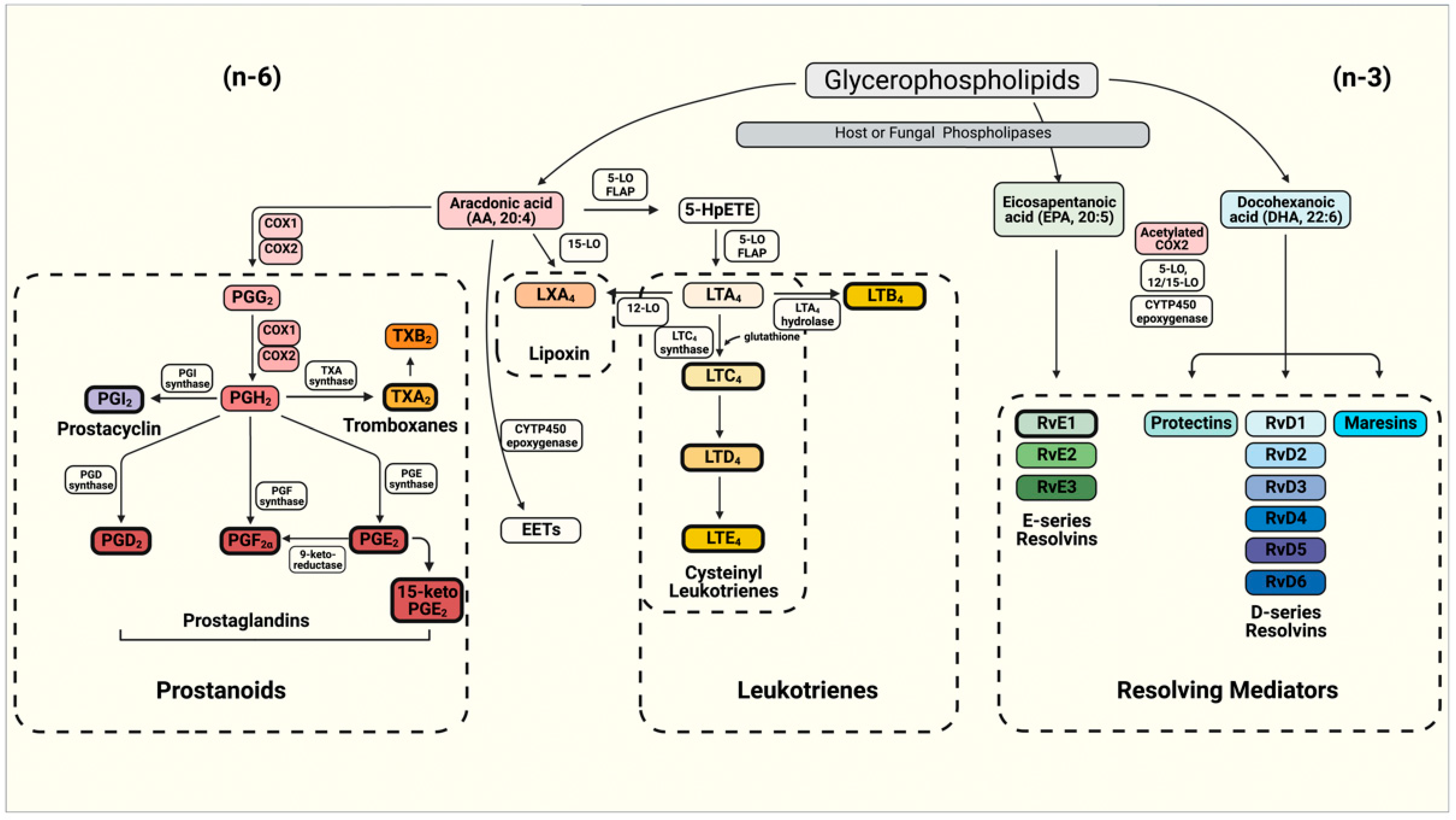
Figure 1. Schematics of the eicosanoids synthesis pathway for the production of prostanoids (Prostaglandins—PGs, Prostacyclin and Thromboxanes—TXs), Leukotrienes (LTs) and resolving mediators including D- and E-series resolvins (Rvs), protectins and maresins. The boxes depicted with bold borders illustrate the eicosanoids produced by fungi.
Jof 07 00254 g001
Figure 2. Eicosanoids production in Candida sp., Cryptococcus sp. and Histoplasma capsulatum. The figure illustrates genes involved in the synthesis of eicosanoids, with exception of H. capsulatum, with as yet undescribed genes involved. Lines and arrows indicate the eicosanoids produced by Candida sp. (solid lines), Cryptococcus sp. (dashed lines) and H. capsulatum (dotted lines).
Jof 07 00254 g002
Publisher’s Note: MDPI stays neutral with regard to jurisdictional claims in published maps and institutional affiliations.