Article

Inactivation of mrpigH Gene in Monascus ruber M7 Results in Increased Monascus Pigments and Decreased Citrinin with mrpyrG Selection Marker

1
Hubei International Scientific and Technological Cooperation Base of Traditional Fermented Foods, Huazhong Agricultural University, Wuhan 430070, China
2
College of Food Science and Technology, Huazhong Agricultural University, Wuhan 430070, China
*
Author to whom correspondence should be addressed.
J. Fungi 2021, 7(12), 1094; https://doi.org/10.3390/jof7121094
Submission received: 17 November 2021 / Revised: 16 December 2021 / Accepted: 16 December 2021 / Published: 19 December 2021

Abstract

:
Monascus pigments (MPs) have been used as food colorants for several centuries in Asian countries and are currently used around the world via Asian catering. The MPs biosynthetic pathway has been well-illustrated; however, the functions of a few genes including mrpigH in the MPs gene cluster of M. ruber M7 are still unclear. In the current study, mrpigH was disrupted in Δmrlig4ΔmrpyrG, a highly efficient gene modification system, using mrpyrG as a selection marker, and ΔmrpigHΔmrlig4ΔmrpyrG::mrpyrG and ΔmrpigHΔmrlig4ΔmrpyrG have been obtained. Subsequently, their morphologies, biomasses, MPs and citrinin (CIT) production were analyzed, respectively. These results have revealed that the deletion of mrpigH has significant effects on the morphology and growth of M. ruber M7. Moreover, compared with M. ruber M7, the yields of MPs and CIT were drastically increased and decreased in mrpigH mutants, respectively.

1. Introduction

Monascus species are famous medicinal and edible filamentous fungi used in traditional fermentation in Asian countries, such as China, Japan, and the Korean Peninsula for nearly 2000 years [1,2]. At present, their fermented products, such as Hongqu, also called red fermented rice, red yeast rice and red mold rice, are widely used as food additives and nutraceutical supplements worldwide owing to their production of abundant beneficial secondary metabolites (SMs), such as Monascus pigments (MPs), monacolin K (MK) and γ-amino butyric acid (GABA) [1,3]. However, citrinin (CIT), a nephrotoxic mycotoxin produced by some strains of Monascus spp., restricts the application of Monascus fermented products [4].
M. ruber M7, which can produce MPs and CIT, without MK, was subjected to whole-genome sequencing analysis [5]. And the functions of most genes in the MPs gene cluster of M. ruber M7 have been investigated by gene manipulation [6]. However, there are a few genes in the MPs gene cluster of M. ruber M7, such as mrpigH and mrpigI, which have not been investigated [6,7]. In 2017, Balakrishnan et al. predicted that the mppE in M. purpureus KACC (highly homologous to mrpigH in M. ruber M7) encoded a reductase which can decrease orange pigments (OPs) and red pigments (RPs) in the biosynthesis of MPs [8]. In 2019, Chen et al. also guessed that MrPigH might contribute to reducing the carbon double bond of the precursor compounds to the typical yellow pigments (YPs) monascin and ankaflavin [7].
In this study, we firstly cloned mrpigH from M. ruber M7. Subsequently, mrpigH was disrupted in the highly efficient system Δmrlig4ΔmrpyrG [9] using the mrpyrG selection marker, and two mrpigH deletion strains ΔmrpigHΔmrlig4ΔmrpyrG::mrpyrG and ΔmrpigHΔmrlig4ΔmrpyrG have been constructed. Finally, the morphologies, biomasses, MPs and CIT production of these mrpigH mutants were assessed. The results revealed that the deletion of mrpigH led to a dramatic reduction in biomass accumulation. Crucially, the inactivation of MrPigH resulted in an increase of MPs and a decrease of CIT.

2. Materials and Methods

2.1. Fungal Strains, Culture Media, and Growth Conditions

M. ruber M7 (CCAM 070120, Culture Collection of State Key Laboratory of Agricultural Microbiology, Wuhan, China), which can produce MPs and CIT, but no MK, is an original strain, which was isolated from Hongqu and preserved in our laboratory [10]. Δmrlig4ΔmrpyrG, the highly efficient gene modification system for M. ruber M7, also named as the markerless disruption strain [9] was used to generate ΔmrpigHΔmrlig4ΔmrpyrG::mrpyrG and ΔmrpigHΔmrlig4ΔmrpyrG. All strains used in this study are described in Table 1. Strains were cultivated in PDA (potato dextrose agar) or minimal medium (MM, 2.0 g NH4Cl, 1 g (NH4)2SO4, 0.5 g KCl, 0.5 g NaCl, 1.0 g KH2PO4, 0.5 g MgSO4·7H2O, 0.02 g FeSO4·7H2O, 20.0 g glucose, distilled water to 1 L, pH 5.5). When required, 10 mM uridine and/or 0.75 mg/mL 5-fluoroorotic acid (5-FOA) were added. For observation of colonial and microscopic morphologies, four different types of media, PDA, malt extract agar (MA), Czapek yeast extract agar (CYA) and 25% glycerol nitrate agar (G25N) were utilized [11]. PDA was used for the analysis of MPs and CIT production. All strains were maintained on PDA slants at 28 °C.

2.2. Cloning and Analysis of mrpigH

A pair of primers, pigH F1–pigH R1 (Table 2), was designed to amplify mrpigH using Oligo 6 software (http://www.oligo.net/, accessed on 24 March 2021). PCR was carried out to amplify mrpigH from the genome of M. ruber M7. Amino acid sequence encoded by mrpigH was predicted using Softberry’s FGENESH program (http://www.softberry. com/, accessed on 24 March 2021), and the MrPigH functional regions were analyzed using the Pfam 33.1 program (http://pfam.xfam.org/, accessed on 24 March 2021). Homology of the deduced amino acid sequence was analyzed using the BLASTP program on the NCBI website (http://blast.ncbi.nlm.nih.gov/Blast.cgi, accessed on 24 March 2021).

2.3. Construction of Deletion Cassettes and Plasmids

The genomic DNA of M. ruber M7 used for PCR was isolated as described previously [12]. The mutant strains ΔmrpigHΔmrlig4ΔmrpyrG::mrpyrG and ΔmrpigHΔmrlig4ΔmrpyrG were constructed using site-directed homologous recombination. The mrpigH gene markerless deletion cassette (5′UTR-mrpyrG-5′-1UTR-3′UTR) was constructed by seamless cloning, and shown schematically in Figure 1a. The relative primer pairs were shown in Table 2.
The 5′ and 3′ flanking regions (993 bp and 775 bp, respectively) and the 5′-1 flanking region (531 bp) of mrpigH were amplified with the primers pigHpyrG 5F–pigHpyrG 5R, pigHpyrG 3F–pigHpyrG 3R and pigHpyrG 5F-1–pigHpyrG 5R-1, respectively. The 1.28-kb mrpyrG marker cassette was amplified from M. ruber M7 genomic DNA with the primer pair pigHpyrG pyrGeF–pigHpyrG pyrGeR. Then the four amplicons (5′ and 3′ regions, 5′-1 regions and mrpyrG expression fragment) were mixed at a 1:1:1:1 molar ratio and cloned into vector pBLUE-T using the seamless cloning and assembly kit (Beijing Zoman Biotechnology). Subsequently, both the cloned DNA fragment and the pCAMBIA3300 plasmid were digested with HindIII and XbaI, and ligated by T4 DNA ligase to generate plasmid pCPGPIGH for the mrpigH knock-out harbouring mrpyrG selection marker.

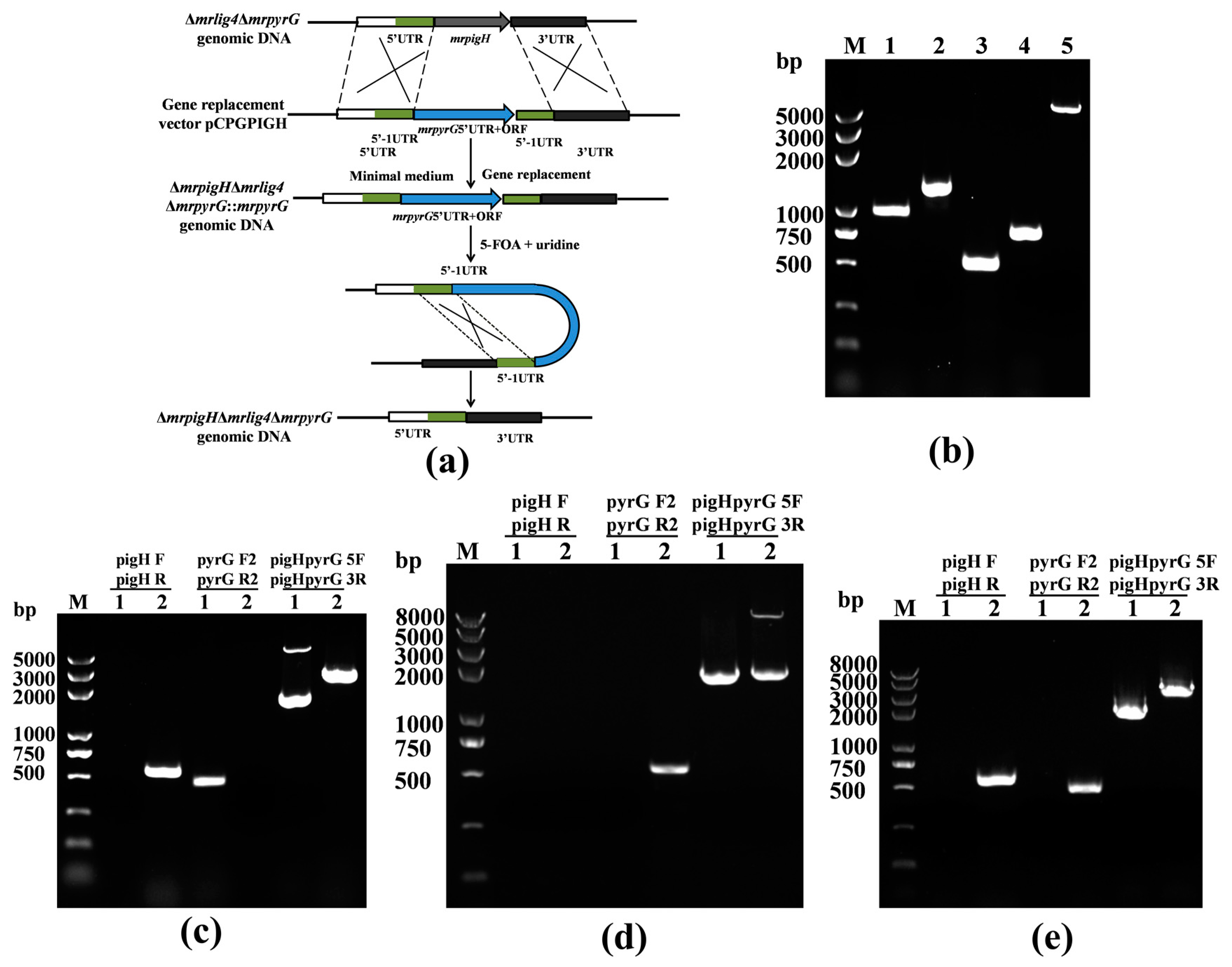
2.4. Deletion of mrpigH in Δmrlig4ΔmrpyrG Strain

The plasmid pCPGPIGH was transformed into Agrobacterium tumefaciens EHA105 using a freeze-thaw method [13]. Δmrlig4ΔmrpyrG, a markerless disruption strain, was used as a host strain to delete mrpigH with the mrpyrG recyclable marker [9]. The A. tumefaciens clones containing pCPGPIGH were incubated for transformation with Δmrlig4ΔmrpyrG to generate a mrpigH gene deletion mutant (ΔmrpigHΔmrlig4ΔmrpyrG::mrpyrG) by minimal medium without uridine/uracil. The conidia of ΔmrpigHΔmrlig4ΔmrpyrG::mrpyrG were collected and spread onto PDA with 0.75 mg/mL 5-FOA and 10 mM uridine. After incubated at 28 °C for 6 days, the surviving colonies were transferred to a new PDA under the same conditions for 4 days. The final surviving colonies were selected and verified by PCR. Selected transformants were designated as ΔmrpigHΔmrlig4ΔmrpyrG.

2.5. MPs and CIT Analyses

M. ruber M7 can produce MPs and CIT, but no MK. Previous researches have shown that MPs mainly accumulate in the mycelia, while CIT exists in the media [14]. Therefore, the intracellular MPs and extracellular CIT were detected. 1 mL spores suspension (105 cfu/mL) of each strain were inoculated on PDA plate coated with cellophane membranes and incubated at 28 °C for 11 days. 20 mg freeze-dried mycelia or media powder were suspended in 1 mL 80% (v/v) methanol solution, and subjected to 30 min ultrasonication treatment (KQ-250B, Kunshan, China). Then, the extraction solutions were separated by centrifugation at 10,000× g for 15 min and filtered with a 0.22 μm filter membrane for further analysis.
The pigments groups concentration was measured using a UV−vis UV-1700 spectrophotometer (Shimadzu, Tokyo, Japan) at 380, 470 and 520 nm which are the maximal absorption of yellow, orange, and red pigments, respectively. The results were expressed as optical density (OD) units per gram of dried mycelia multiplied by a dilution factor [15].
The CIT was detected on Waters ACQUITY UPLC BEH C18 column (2.1 mm × 100 mm, 1.7 µm, Waters) by fluorescence detector (Waters, Milford, MA, USA) in accordance with a previously described method [16].

2.6. Detection of the Relative Gene Expression Level in MPs and CIT Gene Clusters by RT-qPCR

To analyze the influence of mrpigH deletion on gene expression in MPs and CIT gene cluster, ΔmrpigHΔmrlig4ΔmrpyrG::mrpyrG and the wild-type strain (M. ruber M7) were selected for quantitative real-time PCR (RT-qPCR) detection. The ΔmrpigHΔmrlig4ΔmrpyrG was lacked of mrpyrG and had to supply uridine, which might have had an effect on the yields of MPs and CIT.
One milliliter freshly harvested spores (105 cfu/mL) of each strain were inoculated on PDA plate and incubated at 28 °C and samples were taken every other day from the third day to the ninth day. RT-qPCR was performed according to the method described by Liu et al. [13]. Beta-actin was used as a reference gene. The primers used in these analyses were listed in Table S1.

3. Results

3.1. Sequence Analysis of mrpigH in M. ruber M7

A 1.24-kb fragment containing the putative mrpigH homolog was successfully amplified from the genomic DNA of M.ruber M7. Sequence prediction of mrpigH by Softberry’s FGENESH program has revealed that the putative mrpigH gene consists of a 1110 bp open reading frame (ORF) which consists of one exon and encodes 369-amino acids. A database search with Pfam 33.1 program has shown that MrPigH pertains to the alcohol dehydrogenase GroES-like domain. Besides, a database searched with NCBI-BLAST has been demonstrated that the deduced 369-amino acid sequences encoded by mrpigH share 65.31% similarity with the enoyl reductase (GenBank: PCH03974.1), 57.84% similarity with oxidoreductase of Glonium stellatum (GenBank: OCL03635.1), and 56.64% similarity with dehydrogenase of Hyphodiscus hymeniophilus (GenBank: KAG0646231.1). The specific function of mrpigH is still unclear.

3.2. Verification of the mrpigH Deletion in Δmrlig4ΔmrpyrG

The Δmrlig4ΔmrpyrG strain is a promising host for efficient gene targeting in M. ruber M7 and analysis of biosynthesis of SMs. The deletion of mrpigH was executed in Δmrlig4ΔmrpyrG with the mrpyrG marker (Figure 1a). After the plasmid pCPGPIGH harbouring mrpyrG was transformed into Δmrlig4ΔmrpyrG, transformants without uridine/uracil auxotrophic were obtained and verified by PCR, and five of 28 transformants were mrpigH-deleted strains. As shown in Figure 1c, a 0.5-kb product was amplified when the genomic DNA of ΔmrpigHΔmrlig4ΔmrpyrG::mrpyrG was used as template with primers pyrG F2-pyrG R2, while no DNA band was amplified using genome of the Δmrlig4ΔmrpyrG. A 0.58-kb fragment of the mrpigH gene could be amplified from Δmrlig4ΔmrpyrG using primers pigH F-pigH R, while no band was obtained from ΔmrpigHΔmrlig4ΔmrpyrG::mrpyrG. Meanwhile, amplicons of ΔmrpigHΔmrlig4ΔmrpyrG::mrpyrG (3.54 kb and 1.73 kb) and Δmrlig4ΔmrpyrG(2.85 kb) differed in size when primers pigHpyrG 5F- pigHpyrG 3R annealing to homologous arms were used.
In the ΔmrpigHΔmrlig4ΔmrpyrG::mrpyrG strain, the mrpigH deletion cassette contains two completely homologous sequences (5′UTR and 5′-1UTR) between the mrpyrG expression fragments. If ΔmrpigHΔmrlig4ΔmrpyrG::mrpyrG was incubated on PDA plates containing 5-FOA, the strains that lost the mrpyrG expression fragments by homologous recombination should be selected. As predicted, ΔmrpigHΔmrlig4ΔmrpyrG strains without the mrpyrG fragment could be isolated from ΔmrpigHΔmrlig4ΔmrpyrG::mrpyrG after growth on PDA containing 0.75 mg/mL 5-FOA and 10 mM uridine. Total 12 putative ΔmrpigHΔmrlig4ΔmrpyrG strains with 5-FOA resistance were obtained and analyzed, and one of them was shown as follows. In PCR analysis as shown in Figure 1e, a 0.58-kb fragment of the mrpigH gene and a 0.5-kb product of the mrpyrG gene could be amplified from M. ruber M7 using primers pigH F-pigH R and pyrG F2-pyrG R2, while nothing was obtained from ΔmrpigHΔmrlig4ΔmrpyrG, respectively. Meanwhile, amplicons of ΔmrpigHΔmrlig4ΔmrpyrG (1.73 kb) and M. ruber M7 (2.85 kb) differed in size when primers pigHpyrG 5F- pigHpyrG 3R annealing to homologous arms were used. Those results indicated that there was mrpigH markerless deletion was constructed in Δmrlig4ΔmrpyrG.

3.3. Morphologies and Biomasses of mrpigH Mutants and M. ruber M7

To investigate whether the mrpigH was markerlessly deleted by mrpyrG in Δmrlig4ΔmrpyrG, M. ruber M7 and its mrpigH markerless mutants were cultivated on PDA supplemented the appropriate additive (10 mM uridine for the uridine/uracil auxotrophy).The results (Figure 2a) revealed that ΔmrpigHΔmrlig4ΔmrpyrG showed no growth on PDA, but it was able to grow on PDA with 0.75mg/mL 5-FOA and 10 mM uridine, while the growths of M. ruber M7 and ΔmrpigHΔmrlig4ΔmrpyrG::mrpyrG were inhibited by the addition of 0.75 mg/mL 5-FOA to PDA, but could grow on PDA. Those results indicated that the mrpigH markerless mutants were successfully constructed.
To test the influence of deleting mrpigH on developmental processes, M. ruber M7 and ΔmrpigHΔmrlig4ΔmrpyrG::mrpyrG were cultivated on different media (PDA, CYA, MA and G25N) to observe their colonial and microscopic characteristics. The results showed that the colonial morphologies of mrpigH mutants (Figure 2b) were obviously different from those of M. ruber M7 on different culture plates, especially on PDA plate, the ΔmrpigHΔmrlig4ΔmrpyrG::mrpyrG showed slower growth rate and darker color. However, the microscopic morphologies, including conidia and cleistothecia, of ΔmrpigHΔmrlig4ΔmrpyrG::mrpyrG was not dramatically different from those of M. ruber M7 on different culture plates (Figure 2c). Moreover, the biomasses of two mrpigH mutants apparently decreased compared with that of M7 on PDA in 5–11 d (Figure 2d).

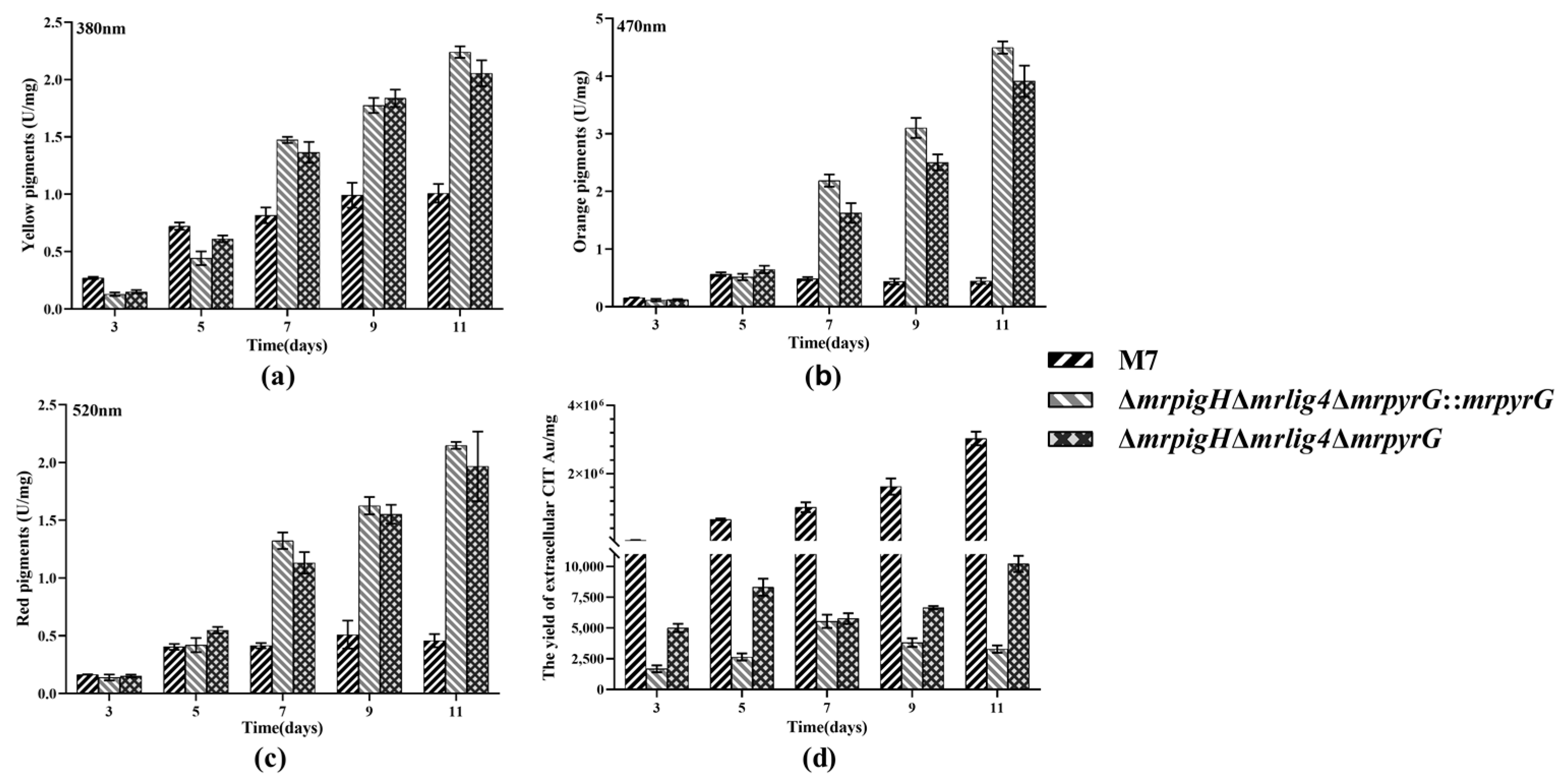
3.4. Analysis of MPs and CIT Production

In order to evaluate the effect of detecting mrpigH on MPs and CITs during fermentation, the samples cultured for 3, 5, 7, 9 and 11 days on PDA were obtained and each test was repeated independently three times. OD values representing yellow, orange and red pigment production were determined using a spectrophotometer at 380 nm, 470 nm and 520 nm, respectively. As shown in Figure 3a–c, from the seventh day to the 11th day, M. ruber M7 produced much fewer MPs (including YPs, OPs and RPs) than 2 mrpigH mutants (ΔmrpigHΔmrlig4ΔmrpyrG::mrpyrG and ΔmrpigHΔmrlig4ΔmrpyrG), whereas the ability of producing MPs among 2 mrpigH mutants showed no obvious difference. After 11 days of cultivation, the YPs, OPs and RPs production in ΔmrpigHΔmrlig4ΔmrpyrG::mrpyrG and ΔmrpigHΔmrlig4ΔmrpyrG were 2.04–2.22, 8.76–10.06, 4.29–4.69 times those of M. ruber M7, respectively.
As to CIT, the UPLC has been performed to detect the production during fermentation. As shown in Figure 3d, CIT produced by M. ruber M7 and all mrpigH mutants showed an obvious difference. At the end of the 11 days of fermentation, CIT production in ΔmrpigHΔmrlig4ΔmrpyrG::mrpyrG and ΔmrpigHΔmrlig4ΔmrpyrG decreased dramatically, and was two to three orders of magnitude less than that of M.ruber M7. Among 2 mrpigH mutants, CIT production in ΔmrpigHΔmrlig4ΔmrpyrG was higher than that of ΔmrpigHΔmrlig4ΔmrpyrG::mrpyrG. The possible cause was that the ΔmrpigHΔmrlig4ΔmrpyrG lacked mrpyrG and had to supply uridine, which might have an effect on the yields of CIT.

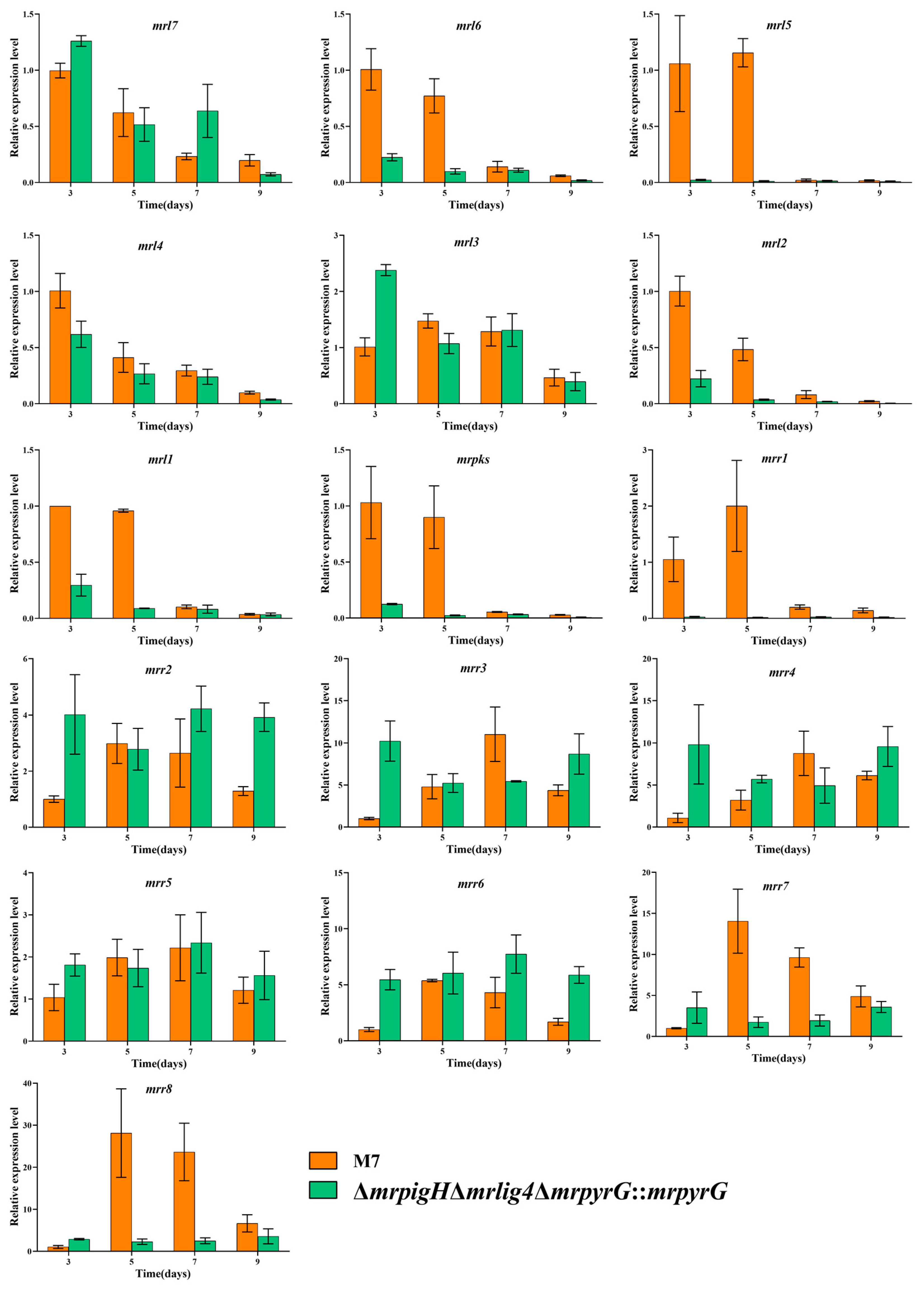
3.5. The Genes’ Expression in MPs and CIT Gene Clusters from mrpigH Mutants

The gene expression in MPs and CIT gene clusters in ΔmrpigHΔmrlig4ΔmrpyrG::mrpyrG and M. ruber M7, were analyzed by RT-qPCR. As shown in Figure 4, the relative expression levels of mrpigA, mrpigB, mrpigC, mrpigD, mrpigE, mrpigF, mrpigG, mrpigJ, mrpigK, mrpigM, mrpigN, mrpigO and mrpigP in ΔmrpigHΔmrlig4ΔmrpyrG::mrpyrG were obviously higher than that of M. ruber M7 on the fifth to ninth days. Therefore, the deletion of mrpigH increased the majority of MPs gene expression level, which might correspond with the enhanced MPs production.
In a previous study, Li et al. [17] considered the CIT biosynthetic gene cluster in M. aurantiacus including 16 genes. He and Cox [18] demonstrated that a minimal set of conserved genes were involving in CIT biosynthesis, including: oxydoreductase (mrl7), dehydrogenase (mrl6), glyoxylase-like domain (mrl5), NAD(P)+ dependent aldehyde dehydrogenase (mrl4), transcriptional regulator (mrl3), Fe(II)-dependent oxygenase (mrl2), serine hydrolase (mrl1), non-reducing polyketide synthase (mrpks),and major facilitator superfamily (MFS) protein (mrr1). As shown in Figure 5, the relative expression levels of mrl6, mrl5, mrl4, mrl2, mrl1, mrpks and mrr1 were dramatically lower than those of M. ruber M7 at 3rd and 5th day. Therefore, the deletion of mrpigH decreased the expression level of core CIT genes, which might correspond with the reduced CIT production.

4. Discussion

Monascus spp. have been widely used in food fermentation for nearly 20 centuries in East and Southeast Asian countries due to their ability of producing vivid MPs [11,19], which are a complex mixture of secondary metabolites (SMs) with a tricyclic azaphilone scaffold, produced via the polyketide pathway by a few filamentous fungi such as Monascus spp. and Penicillium spp. [3,20,21]. MPs are biosynthesized by their gene cluster, which contains 16 genes (mrpigA-mrpigP) in M.ruber M7 [6,7].
In the current study, in order to explore the function of mrpigH, we used the Δmrlig4ΔmrpyrG as the starting strain to generate two kinds of mrpigH disruptants, namely ΔmrpigHΔmrlig4ΔmrpyrG::mrpyrG and ΔmrpigHΔmrlig4ΔmrpyrG. The colonial and microbiological phenotypes and biomasses of the mrpigH mutants showed an obviously difference from those of M. ruber M7 (Figure 2b, d). In particular, the biomasses of mrpigH mutants accumulated more slowly than that of M ruber M7. Our laboratory previously constructed MPs gene knockout strains with the resistance selection markers (hph/neo) in M7, and found that the morphologies and biomasses of mrpigC, mrpigE, mrpigF, mrpigM and mrpigO knockouts were comparable to those of M. ruber M7, whereas the growth rates of mrpigA, mrpigJ, and mrpigK knockouts were increased, and the deletion of mrpigN resulted in a reduction in biomass accumulation compared with M. ruber M7 [22].
Moreover, we have discovered that the deletion of mrpigH in M. ruber M7 can enhance YPs, OPs and RPs (Figure 3a–c), which is mostly consistent with the results obtained in M. purpureus [8]. We also found that deletion of mrpigH dramatically decreased the CIT production (Figure 3d), which is a promising scheme for control of CIT. CIT is a kind of mycotoxin produced via the polyketide pathway by filamentous fungi, mainly by Monascus spp. and Penicillium spp. [23,24,25]. To this end, we investigated the changes in the expression levels of MPs and CIT genes in their gene clusters of mrpigH mutants, and found that the relative gene expression levels almost corresponded with the increase of MPs and decrease of CIT (Figure 4 and Figure 5).
The deep study about the reason for increasing the pigment content while reducing the CIT production by knocking out mrpigH need to be investigated further. In early research, both MPs and CIT were considered to be derived from polyketide pathways [26]. Lately, two hypotheses about the biosynthetic pathways of MPs and CIT were put forward. One of them is based on metabolic pathways; the MPs and CIT shared a common pathway to a certain branch [26]. The other is based on the analysis of the whole genome sequence; their biosynthetic gene clusters had been found separately, thereby, they belonged to two different pathways [6,18].
Previous results suggested that the yields of CIT and MPs in Monascus spp. could affect each other. For example, Xie et al. (2013) and Liu et al. (2014) separately identified that the overexpression of mrpigB and mrpigE in MPs gene cluster of M. ruber M7 resulted in a reduction of CIT production [13,27]. Meanwhile, Liang et al. (2017) obtained a mutant of a putative glyoxalase (orf6) in CIT gene cluster of M. purpureus, and found that the deletion of orf6 could improve the MPs and CIT yields at the same time [28]. A recent study on the biosynthesis of MPs found that acetyl-CoA and malonyl-CoA are catalyzed by a sequence of enzymes to produce MPs precursors. Then the pathway bifurcates into two branches. First, the MPs precursors were reduced by a reductase (MrPigH and/or GME3457, which is encoded outside of the MPs gene cluster in M. ruber M7), yielding the typical YPs monascin and ankaflavin. The other branch of the pathway produces the typical OPs rubropunctatin and monascorubrin by a FAD-dependent oxidoreductase (MrPigF in M. ruber M7). Furthermore, rubropunctatin and monascorubrin were converted into RPs rubropunctamine and monascorubramine through an amination reaction [6,7]. Due to the deletion of mrpigH, the relative expression of mrpigF dramatically increased (Figure 4), which was beneficial for the OPs and RPs production, and the increase in relative yields of OPs and RPs was greater than that of YPs. Recently, Li et al. (2020) reported that MPs biosynthetic gene cluster was a composite supercluster, and the naphthoquinone (monasone) gene cluster was embedded in the MPs gene cluster, and speculated that MrPigH was essential in the biosynthesis of naphthoquinone, but it was a supplemental enzyme in the biosynthesis of MPs [29]. As a result, the mrpigH knockout’s blocking of the naphthoquinone biosynthesis pathway might result in naphthoquinone reduction, and more substrates and intermediates were utilized to synthesize MPs, including YPs. In terms of CIT production, He Yi et al. [18] revealed a minimal set of conserved genes involved in CIT biosynthesis, which included nine genes (mrl7, mrl6, mrl5, mrl4, mrl3, mrl2, mrl1, mrpks and mrr1). When mrpigH was deleted, the majority of the genes in the cluster (mrl6, mrl5, mrl4, mrl2, mrl1, mrpks and mrr1) were obviously down-regulated (Figure 5), which could explain why CIT production has decreased in this study.
In conclusion, the mrpigH gene pertains to the MPs biosynthetic gene cluster of M. ruber M7 and plays a remarkable role in the biosynthesis of MPs and CIT. The disruption of mrpigH had very little effect on the microscopic morphologies, while the mrpigH mutants showed slower biomass accumulation and darker color on PDA. Compared with M. ruber M7, the YPs, OPs and RPs production in the mrpigH mutants (ΔmrpigHΔmrlig4ΔmrpyrG::mrpyrG and ΔmrpigHΔmrlig4ΔmrpyrG) increased dramatically. However, the CIT production of the mrpigH mutants decreased drastically. This work will make some contribution to the regulation of MPs and CIT production in M. ruber M7.

Supplementary Materials

The following are available online at https://www.mdpi.com/article/10.3390/jof7121094/s1, Table S1: Primers used in RT-qPCR.

Author Contributions

L.L. has designed and carried out the present research work, conducted experiments, analyzed the data, and written the present manuscript. N.X. performed the secondary metabolites analysis and phenotypic characterization. F.C. provided place in the laboratory, gave access to the lab facilities for experimentation, and funds for the present work. All authors have read and agreed to the published version of the manuscript.

Funding

This work was supported by the Major Program of the National Natural Science Foundation of China (Nos. 31730068 and 31330059 to F.C.), and the National Key Research and Development Program of China (No. 2018YFD0400404 to F.C.).

Institutional Review Board Statement

Not applicable.

Informed Consent Statement

Not applicable.

Data Availability Statement

The raw data supporting the conclusions of this manuscript will be made available by the authors, without undue reservation, to any qualified researcher.

Conflicts of Interest

The authors declare no conflict of interest. The funders had no role in the design of the study; in the collection, analyses, or interpretation of data; in the writing of the manuscript, or in the decision to publish the results.

References

  1. Chen, W.P.; He, Y.; Zhou, Y.X.; Shao, Y.C.; Feng, Y.L.; Li, M.; Chen, F.S. Edible Filamentous Fungi from the Species Monascus: Early Traditional Fermentations, Modern Molecular Biology, and Future Genomics. Compr. Rev. Food Sci. Food Saf. 2015, 14, 555–567. [Google Scholar] [CrossRef] [Green Version]
  2. Kim, S.C.; Lee, G.D.; Choi, I.H. Breast meat quality of broilers fed fermented red ginseng marc powder mixed with red-koji during storage. Emir. J. Food Agric. 2016, 28, 283–287. [Google Scholar] [CrossRef] [Green Version]
  3. Feng, Y.L.; Shao, Y.C.; Chen, F.S. Monascus pigments. Appl. Microbiol. Biot. 2012, 96, 1421–1440. [Google Scholar] [CrossRef]
  4. Blanc, P.J.; Laussac, J.P.; Lebars, J.; Lebars, P.; Loret, M.O.; Pareilleux, A.; Prome, D.; Prome, J.C.; Santerre, A.L.; Goma, G. Characterization of Monascidin-a from Monascus as Citrinin. Int. J. Food Microbiol. 1995, 27, 201–213. [Google Scholar] [CrossRef]
  5. Shao, Y.C.; Lei, M.; Mao, Z.J.; Zhou, Y.X.; Chen, F.S. Insights into Monascus biology at the genetic level. Appl. Microbiol. Biot. 2014, 98, 3911–3922. [Google Scholar] [CrossRef] [PubMed]
  6. Chen, W.P.; Chen, R.; Liu, Q.P.; He, Y.; He, K.; Ding, X.L.; Kang, L.J.; Guo, X.X.; Xie, N.N.; Zhou, Y.X.; et al. Orange, red, yellow: Biosynthesis of azaphilone pigments in Monascus fungi. Chem. Sci. 2017, 8, 4917–4925. [Google Scholar] [CrossRef] [Green Version]
  7. Chen, W.P.; Feng, Y.L.; Molnar, I.; Chen, F.S. Nature and nurture: Confluence of pathway determinism with metabolic and chemical serendipity diversifies Monascus azaphilone pigments. Nat. Prod. Rep. 2019, 36, 561–572. [Google Scholar] [CrossRef] [PubMed]
  8. Balakrishnan, B.; Park, S.H.; Kwon, H.J. A reductase gene mppE controls yellow component production in azaphilone polyketide pathway of Monascus. Biotechnol. Lett. 2017, 39, 163–169. [Google Scholar] [CrossRef] [PubMed]
  9. Li, L. Establishment of Markerless and Highly Efficient Genetic Modification System in Monascus Ruber M7 and Application of This System in Functional Analysis of Monascus Pigments Genes. Ph.D. Thesis, Huazhong Agricultural University, Wuhan, China, 2021. [Google Scholar]
  10. Chen, F.S.; Hu, X.Q. Study on red fermented rice with high concentration of monacolin K and low concentration of citrinin. Int. J. Food Microbiol. 2005, 103, 331–337. [Google Scholar] [CrossRef]
  11. Jia, L.L.; Yu, J.H.; Chen, F.S.; Chen, W.P. Characterization of the asexual developmental genes brlA and wetA in Monascus ruber M7. Fungal Genet. Biol. 2021, 151, 103564. [Google Scholar] [CrossRef]
  12. Shao, Y.C.; Ding, Y.D.; Zhao, Y.; Yang, S.; Xie, B.J.; Chen, F.S. Characteristic analysis of transformants in T-DNA mutation library of Monascus ruber. World J. Microb. Biot. 2009, 25, 989–995. [Google Scholar] [CrossRef]
  13. Liu, Q.P.; Xie, N.N.; He, Y.; Wang, L.; Shao, Y.C.; Zhao, H.Z.; Chen, F.S. MpigE, a gene involved in pigment biosynthesis in Monascus ruber M7. Appl. Microbiol. Biot. 2014, 98, 285–296. [Google Scholar] [CrossRef] [PubMed]
  14. Li, L.; He, L.; Lai, Y.; Shao, Y.C.; Chen, F.S. Cloning and functional analysis of the G beta gene Mgb1 and the G gamma gene Mgg1 in Monascus ruber. J. Microbiol. 2014, 52, 35–43. [Google Scholar] [CrossRef]
  15. Yuan, X.; Chen, F.S. Cocultivation Study of Monascus spp. and Aspergillus niger Inspired from Black-Skin-Red-Koji by a Double-Sided Petri Dish. Front. Microbiol. 2021, 12, 670684. [Google Scholar] [CrossRef] [PubMed]
  16. Li, L.; Chen, F.S. Effects of mrpigG on Development and Secondary Metabolism of Monascus ruber M7. J. Fungi 2020, 6, 156. [Google Scholar] [CrossRef]
  17. Li, Y.P.; Xu, Y.; Huang, Z.B. Isolation and characterization of the citrinin biosynthetic gene cluster from Monascus aurantiacus. Biotechnol. Lett. 2012, 34, 131–136. [Google Scholar] [CrossRef]
  18. He, Y.; Cox, R.J. The molecular steps of citrinin biosynthesis in fungi. Chem. Sci. 2016, 7, 2119–2127. [Google Scholar] [CrossRef] [PubMed] [Green Version]
  19. Dufosse, L.; Galaup, P.; Yaron, A.; Arad, S.M. Microorganisms and microalgae as sources of pigments for food use: A scientific oddity or an industrial reality? Trends Food Sci. Tech. 2005, 16, 389–406. [Google Scholar] [CrossRef]
  20. Mapari, S.A.S.; Thrane, U.; Meyer, A.S. Fungal polyketide azaphilone pigments as future natural food colorants? Trends Biotechnol. 2010, 28, 300–307. [Google Scholar] [CrossRef]
  21. Liu, Q.P.; Zhong, S.Y.; Wang, X.R.; Gao, S.B.A.; Yang, X.L.; Chen, F.S.; Molnar, I. An Integrated Approach to Determine the Boundaries of the Azaphilone Pigment Biosynthetic Gene Cluster of Monascus ruber M7 Grown on Potato Dextrose Agar. Front. Microbiol. 2021, 12, 1569. [Google Scholar] [CrossRef]
  22. Liu, Q.P. Investigation on the Biosynthetic Pathway of Monascus Pigments and Its Polyketide Synthase Characterization in Monascus Ruber M7. Ph.D. Thesis, Huazhong Agricultural University, Wuhan, China, 2018. [Google Scholar]
  23. Blanc, P.J.; Loret, M.O.; Goma, G. Production of Citrinin by Various Species of Monascus. Biotechnol. Lett. 1995, 17, 291–294. [Google Scholar] [CrossRef]
  24. Li, Y.P.; Pan, Y.F.; Zou, L.H.; Xu, Y.; Huang, Z.B.; He, Q.H. Lower Citrinin Production by Gene Disruption of ctnB Involved in Citrinin Biosynthesis in Monascus aurantiacus Li AS3.4384. J. Agric. Food Chem. 2013, 61, 7397–7402. [Google Scholar] [CrossRef]
  25. Ballester, A.R.; Marcet-Houben, M.; Levin, E.; Sela, N.; Selma-Lazaro, C.; Carmona, L.; Wisniewski, M.; Droby, S.; Gonzalez-Candelas, L.; Gabaldon, T. Genome, Transcriptome, and Functional Analyses of Penicillium expansum Provide New Insights Into Secondary Metabolism and Pathogenicity. Mol. Plant-Microbe Interact. 2015, 28, 232–248. [Google Scholar] [CrossRef] [Green Version]
  26. Hajjaj, H.; Klaebe, A.; Loret, M.O.; Goma, G.; Blanc, P.J.; Francois, J. Biosynthetic pathway of citrinin in the filamentous fungus Monascus ruber as revealed by C-13 nuclear magnetic resonance. Appl. Environ. Microbiol. 1999, 65, 311–314. [Google Scholar] [CrossRef] [PubMed] [Green Version]
  27. Xie, N.N.; Liu, Q.P.; Chen, F.S. Deletion of pigR gene in Monascus ruber leads to loss of pigment production. Biotechnol. Lett. 2013, 35, 1425–1432. [Google Scholar] [CrossRef]
  28. Liang, B.; Du, X.J.; Li, P.; Guo, H.; Sun, C.C.; Gao, J.X.; Wang, S. Orf6 gene encoded glyoxalase involved in mycotoxin citrinin biosynthesis in Monascus purpureus YY-1. Appl. Microbiol. Biot. 2017, 101, 7281–7292. [Google Scholar] [CrossRef]
  29. Li, M.; Kang, L.J.; Ding, X.L.; Liu, J.; Liu, Q.P.; Shao, Y.C.; Molnar, I.; Chen, F.S. Monasone Naphthoquinone Biosynthesis and Resistance in Monascus Fungi. Mbio 2020, 11, e02676-19. [Google Scholar] [CrossRef] [PubMed] [Green Version]
Figure 1. Markerless deletion of mrpigH in Δmrlig4ΔmrpyrG. (a) Schematic representation of the homologous recombination strategy yielding mrpigH markerless deletion strains. (b) Construction of mrpigH disruption construct by Seamless Cloning and assembly method. Lane 1, 5′ flanking region of mrpigH; lane 2, 5′ flanking region of mrpyrG plus mrpyrG ORF regions; lane 3, 5′-1 flanking region of mrpigH; lane 4, 3′ flanking region of mrpigH; lane 5, deletion cassette product. (c) Confirmation of mrpigH homologous recombination events. Three primer pairs were used and PCR amplifications showed distinct bands in different strains. Lane 1, the ΔmrpigHΔmrlig4ΔmrpyrG::mrpyrG strain; lane 2, the Δmrlig4ΔmrpyrG strain. (d) Confirmation of mrpyrG homologous recombination events in ΔmrpigHΔmrlig4ΔmrpyrG::mrpyrG strain. Lane 1, the ΔmrpigHΔmrlig4ΔmrpyrG strain; lane 2, the ΔmrpigHΔmrlig4ΔmrpyrG::mrpyrG strain. (e) Confirmation of mrpigH markerless deletion in ΔmrpigHΔmrlig4ΔmrpyrG strain. Lane 1, the ΔmrpigHΔmrlig4ΔmrpyrG strain; lane 2, the wild-type strain.
Jof 07 01094 g001
Figure 2. Morphologies and biomasses of mrpigH mutants and M. ruber M7. (a) Growth of M. ruber M7 and mrpigH mutants on PDA, PDA with 10 mM uridine, PDA with 10 mM uridine and 0.75 mg/mL 5-FOA for 5 days at 28 °C. A, M. ruber M7; B, ΔmrpigH, mrpigH was disrupted in M. ruber M7 with the hph selection marker [9]; C, ΔmrpigHΔmrlig4ΔmrpyrG::mrpyrG; D, ΔmrpigHΔmrlig4ΔmrpyrG; (b) Colonial morphologies of M. ruber M7 and ΔmrpigHΔmrlig4ΔmrpyrG::mrpyrG on PDA, CYA, MA and G25N plates for 10 days at 28 °C; (c) Cleistothecia and conidia formation of M. ruber M7 and ΔmrpigHΔmrlig4ΔmrpyrG::mrpyrG on different plates (PDA, CYA, G25N, and MA) for 7 days at 28 °C; (d) Biomass (dry cell weight) of M. ruber M7 and mrpigH mutants on PDA plates at 28 °C. The error bar represents the standard deviation between the three repeats.
Jof 07 01094 g002
Figure 3. Production of intracellular MPs and extracellular CIT by M. ruber M7 and mrpigH mutants on the PDA supplied with/without uridine. (a) The yield of yellow pigments; (b) The yield of orange pigments; (c) The yield of red pigments; (d) The yield of CIT. The error bar represents the standard deviation between the three repeats.
Jof 07 01094 g003
Figure 4. RT-qPCR analysis of the mrpigA-mrpigP in M7 and ΔmrpigHΔmrlig4ΔmrpyrG::mrpyrG. mrpigA: Non-reducing polyketide synthase; mrpigB: Transcription factor; mrpigC: C-11-Ketoreductase; mrpigD: 4-O-Acyltransferase; mrpigE: NAD(P)H-dependent oxidoreductase; mrpigF: FAD-dependent oxidoreductase; mrpigG: Serine hydrolase; mrpigH: Enoyl reductase; mrpigI: Transcription factor; mrpigJ: FAS subunit alpha; mrpigK: FAS subunit beta; mrpigL: Ankyrin repeat protein; mrpigM: O-Acetyltransferase; mrpigN: FAD-dependent monooxygenase; mrpigO: Deacetylase; mrpigP: MFS multidrug transporter; M7 and ΔmrpigHΔmrlig4ΔmrpyrG::mrpyrG were incubated in PDA on the 3rd, 5th, 7th, 9th day at 28 °C The beta-actin gene was used as control. The error bars indicate the standard deviations of three independent cultures.
Jof 07 01094 g004
Figure 5. RT-qPCR analysis of the mrl7-mrr8 in M7 and ΔmrpigHΔmrlig4ΔmrpyrG::mrpyrG. mrl7: Oxydoreductase; mrl6: Dehydrogenase; mrl5: Glyoxylase-like domain; mrl4: NAD(P)+ dependent aldehyde dehydrogenase; mrl3: Transcriptional regulator; mrl2: Fe(II)-dependent oxygenase; mrl1: Serine hydrolase; mrpks: Non-reducing PKS; mrr1: Major facilitator superfamily (MFS) protein; mrr2: Histidine phosphatase; mrr3: Unknown protein; mrr4: WD40 protein; mrr5: Carbonic anhydrase; mrr6: Unknown protein; mrr7: Enoyl-(acyl carrier protein) reductase; mrr8: AMP-binding enzyme; M7 and ΔmrpigHΔmrlig4ΔmrpyrG::mrpyrG were incubated in PDA on the 3rd, 5th, 7th, 9th day at 28 °C. The beta-actin gene was used as control. The error bars indicate the standard deviations of three independent cultures.
Jof 07 01094 g005
Table 1. M. ruber strains constructed and used in this study.
StrainParentSource
M. ruber M7M7 [10]Red fermented rice
ΔmrpigHΔmrlig4
ΔmrpyrG::mrpyrG
Δmrlig4ΔmrpyrG [9]This study
ΔmrpigHΔmrlig4
mrpyrG
ΔmrpigHΔmrlig4
ΔmrpyrG::mrpyrG
This study
Table 2. Primers used in this study.
NamesSequences (5′→3′)Descriptions
pigHpyrG 5FGATATCGAATTCCCAATACT
CGTTACCCCGTCCAAGATGG
For amplification of the 993 bp of 5′ flanking regions of the mrpigH
pigHpyrG 5RCGGTGGCAGTCGAAGGGGCA
pigH pyrG pyrGeFTGCCCCTTCGACTGCCACCG
GATTATCGTATAGAGCAATA
For the expression of the1276 bp of the mrpyrG
pigH pyrG pyrGeRTCACTGGTTCTTACAGCCGT
pigHpyrG 5-1FACGGCTGTAAGAACCAGTGA
CGCACACACGTTTCGCACGG
For amplification of the 531 bp of 5′-1 flanking regions of the mrpigH
pigHpyrG 5-1RCGGTGGCAGTCGAAGGGGCA
pigHpyrG 3FTGCCCCTTCGACTGCCACCG
GGCTGGATGCTGCATGTTTT
For amplification of the 775 bp of 3′ flanking regions of the mrpigH
pigHpyrG 3RCTGCAGGAATTCCCAATACT
CGCCGAAGCCCCCTTCCTCT
pigH FGTGCTGGTGCCCGACCTGACFor amplification of the 583 bp of the partial mrpigH
pigH RCGAAGATGAAATTCGACTTGA
pyrG F2GTGCATACTCTACAGATFor amplification of the 498 bp of the partial mrpyrG
pyrG R2CCAAGAAGACGAATGTGA
Labeled with dotted lines letters are nucleotide sequences of pBLUE-T vector; labeled with single underline letters are nucleotide sequences of 5′UTR of mrpigH; labeled with double underline letters are nucleotide sequences of mrpyrG gene; labeled with wavy line letters are nucleotide sequences of 5′-1UTR of mrpigH.
Publisher’s Note: MDPI stays neutral with regard to jurisdictional claims in published maps and institutional affiliations.

