
Malassezia restricta Pneumonia in Solid Organ Transplant Recipients: First Report of Two Cases

 , , ,
, , ,  , ,
, , 
Abstract
:1. Introduction
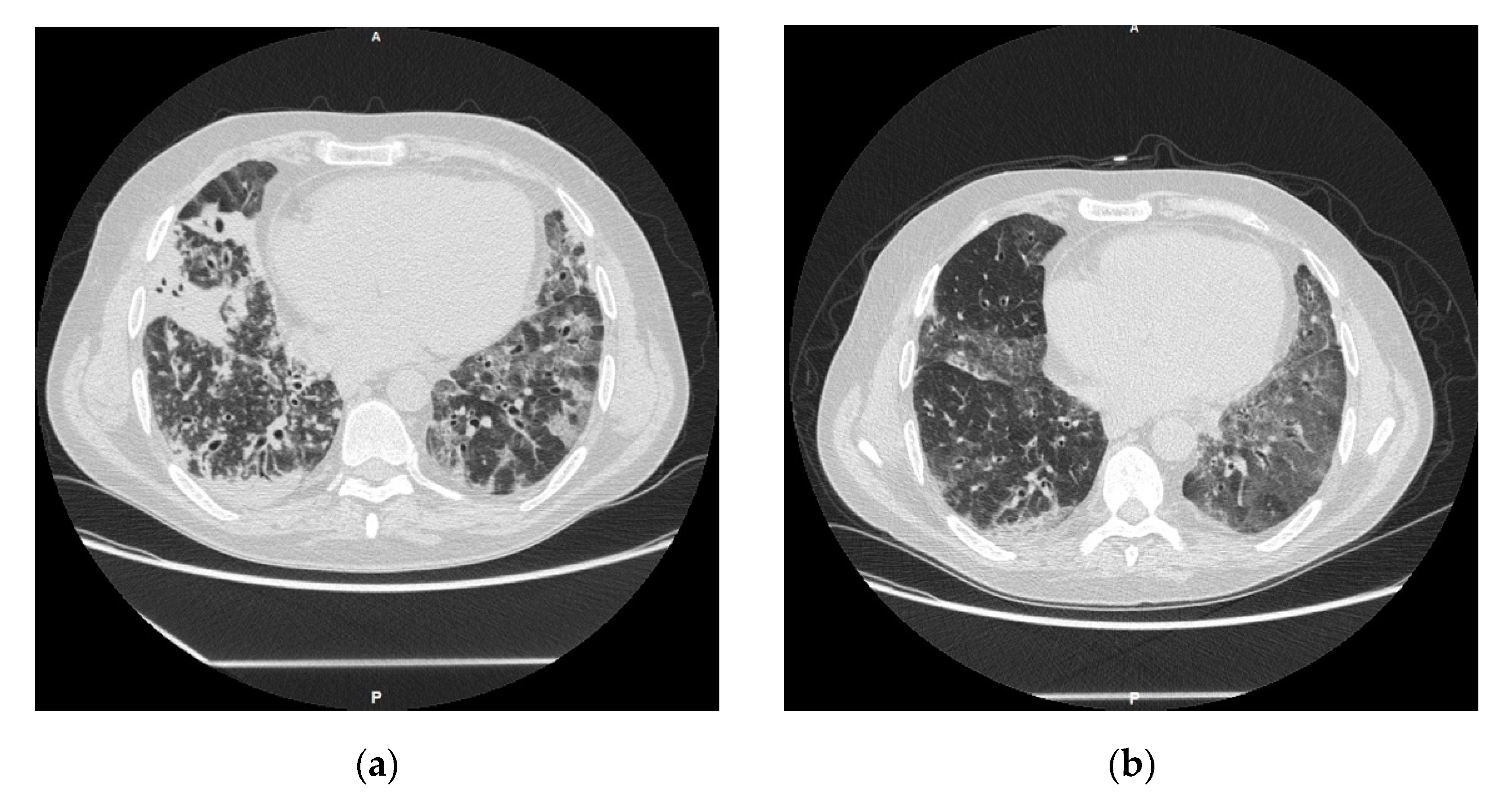
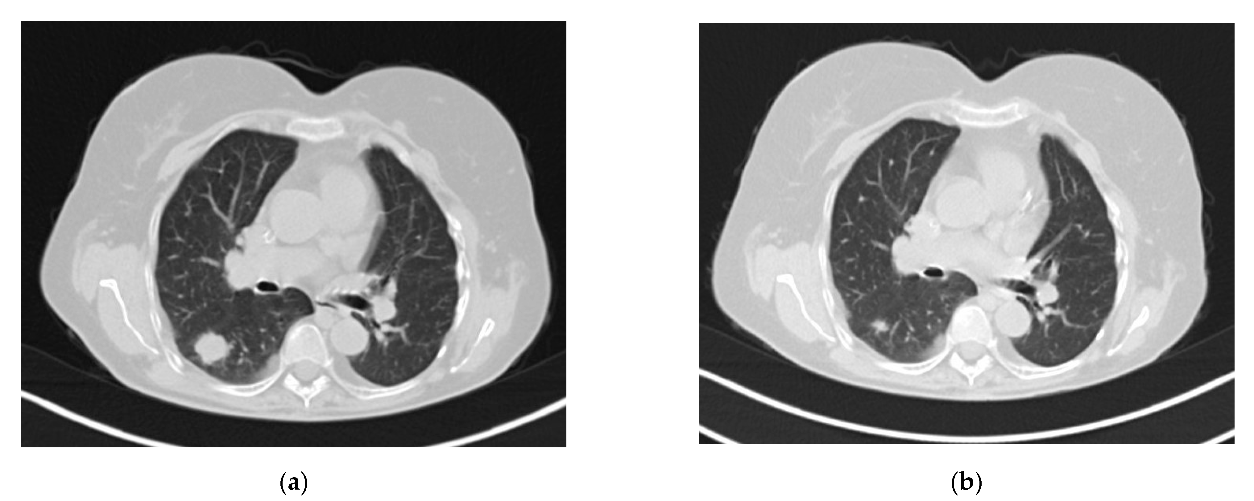
2. Case Reports
2.1. Case 1
2.2. Case 2
3. Comments
4. Conclusions
Author Contributions
Funding
Institutional Review Board Statement
Informed Consent Statement
Data Availability Statement
Conflicts of Interest
References
- Shoham, S.; Dominguez, E.A. The AST Infectious Diseases Community of Practice Emerging fungal infections in solid organ transplant recipients: Guidelines of the American Society of Transplantation Infectious Diseases Community of Practice. Clin. Transplant. 2019, 33, e13525. [Google Scholar] [CrossRef] [PubMed]
- Pappas, P.G.; Alexander, B.D.; Andes, D.; Hadley, S.; Kauffman, C.A.; Freifeld, A.; Anaissie, E.J.; Brumble, L.M.; Herwaldt, L.; Ito, J.; et al. Invasive Fungal Infections among Organ Transplant Recipients: Results of the Transplant-Associated Infection Surveillance Network (TRANSNET). Clin. Infect. Dis. 2010, 50, 1101–1111. [Google Scholar] [CrossRef] [PubMed]
- Pedrosa, A.F.; Lisboa, C.; Rodrigues, A.G. Malassezia infections with systemic involvement: Figures and facts. J. Dermatol. 2018, 45, 1278–1282. [Google Scholar] [CrossRef] [PubMed]
- Houhamdi-Hammou, L.; Benito, Y.; Boibieux, A.; Dupont, D.; Delahaye, F.; Thivolet-Bejui, F.; Wallon, M.; Vandenesch, F.; Bouchiat, C. Malassezia restricta: An Underdiagnosed Causative Agent of Blood Culture-Negative Infective Endocarditis. Clin. Infect. Dis. 2021, 73, 1223–1230. [Google Scholar] [CrossRef] [PubMed]
- Tragiannidis, A.; Bisping, G.; Koehler, G.; Groll, A.H. Minireview: Malasseziainfections in immunocompromised patients. Mycoses 2010, 53, 187–195. [Google Scholar] [CrossRef] [PubMed]
- Weiss, S.J.; Schoch, P.E.; Cunha, B.A. Malassezia furfur fungemia associated with central venous catheter lipid emulsion infusion. Heart Lung 1991, 20, 87–90. [Google Scholar]
- Angiolella, L.; Leone, C.; Rojas, F.; Mussin, J.; Sosa, M.D.L.A.; Giusiano, G. Biofilm, adherence, and hydrophobicity as virulence factors in Malassezia furfur. Med. Mycol. 2017, 56, 110–116. [Google Scholar] [CrossRef] [PubMed]
- Chang, H.J.; Miller, H.L.; Watkins, N.; Arduino, M.; Ashford, D.A.; Midgley, G.; Aguero, S.M.; Pinto-Powell, R.; Von Reyn, C.F.; Edwards, W.; et al. An Epidemic ofMalassezia pachydermatisin an Intensive Care Nursery Associated with Colonization of Health Care Workers’ Pet Dogs. N. Engl. J. Med. 1998, 338, 706–711. [Google Scholar] [CrossRef]
- Blaes, A.; Cavert, W.; Morrison, V. Malassezia: Is it a pulmonary pathogen in the stem cell transplant population? Transpl. Infect. Dis. 2009, 11, 313–317. [Google Scholar] [CrossRef]
- Arendrup, M.C.; Boekhout, T.; Akova, M.; Meis, J.F.; Cornely, O.A.; Lortholary, O.; European Society of Clinical Microbiology and Infectious Diseases Fungal Infection Study Group; European Confederation of Medical Mycology. ESCMID and ECMM joint clinical guidelines for the diagnosis and management of rare invasive yeast infections. Clin. Microbiol. Infect. 2014, 20 (Suppl. 3), 76–98. [Google Scholar] [CrossRef] [Green Version]
- Rojas, F.D.; Córdoba, S.B.; Sosa, M.D.L.; de Los Angeles Sosa, M.; Zalazar, L.C.; Fernández, M.S.; Cattana, M.E.; Alegre, L.R.; Carrillo-Muñoz, A.J.; Giusiano, G.E. Antifungal susceptibility testing of Malasseziayeast: Comparison of two different methodologies. Mycoses 2016, 60, 104–111. [Google Scholar] [CrossRef] [PubMed]
- Morrison, V.A.; Weisdorf, D.J. The spectrum of Malassezia infections in the bone marrow transplant population. Bone Marrow Transplant. 2000, 26, 645–648. [Google Scholar] [CrossRef] [PubMed] [Green Version]
Publisher’s Note: MDPI stays neutral with regard to jurisdictional claims in published maps and institutional affiliations. |
© 2021 by the authors. Licensee MDPI, Basel, Switzerland. This article is an open access article distributed under the terms and conditions of the Creative Commons Attribution (CC BY) license (https://creativecommons.org/licenses/by/4.0/).
Share and Cite
Mularoni, A.; Graziano, E.; Medaglia, A.A.; Buscemi, B.; Eddens, T.; Martino, L.; Di Carlo, D.; Cascio, A.; Conaldi, P.G.; Bertani, A.; et al. Malassezia restricta Pneumonia in Solid Organ Transplant Recipients: First Report of Two Cases. J. Fungi 2021, 7, 1057. https://doi.org/10.3390/jof7121057
Mularoni A, Graziano E, Medaglia AA, Buscemi B, Eddens T, Martino L, Di Carlo D, Cascio A, Conaldi PG, Bertani A, et al. Malassezia restricta Pneumonia in Solid Organ Transplant Recipients: First Report of Two Cases. Journal of Fungi. 2021; 7(12):1057. https://doi.org/10.3390/jof7121057
Chicago/Turabian StyleMularoni, Alessandra, Elena Graziano, Alice Annalisa Medaglia, Barbara Buscemi, Taylor Eddens, Lavinia Martino, Daniele Di Carlo, Antonio Cascio, Pier Giulio Conaldi, Alessandro Bertani, and et al. 2021. "Malassezia restricta Pneumonia in Solid Organ Transplant Recipients: First Report of Two Cases" Journal of Fungi 7, no. 12: 1057. https://doi.org/10.3390/jof7121057
APA StyleMularoni, A., Graziano, E., Medaglia, A. A., Buscemi, B., Eddens, T., Martino, L., Di Carlo, D., Cascio, A., Conaldi, P. G., Bertani, A., & Grossi, P. A. (2021). Malassezia restricta Pneumonia in Solid Organ Transplant Recipients: First Report of Two Cases. Journal of Fungi, 7(12), 1057. https://doi.org/10.3390/jof7121057