Article

Transcription Factor MaMsn2 Regulates Conidiation Pattern Shift under the Control of MaH1 through Homeobox Domain in Metarhizium acridum

1
School of Life Sciences, Chongqing University, Chongqing 401331, China
2
Chongqing Engineering Research Center for Fungal Insecticides, Chongqing 401331, China
3
Key Laboratory of Gene Function and Regulation Technologies under Chongqing Municipal Education Commission, Chongqing 400044, China
*
Authors to whom correspondence should be addressed.
J. Fungi 2021, 7(10), 840; https://doi.org/10.3390/jof7100840
Submission received: 30 August 2021 / Revised: 26 September 2021 / Accepted: 4 October 2021 / Published: 7 October 2021
(This article belongs to the Special Issue Genetic Manipulation of Fungal Model Organisms)

Abstract

:
The growth pattern of filamentous fungi can switch between hyphal radial polar growth and non-polar yeast-like cell growth depending on the environmental conditions. Asexual conidiation after radial polar growth is called normal conidiation (NC), while yeast-like cell growth is called microcycle conidiation (MC). Previous research found that the disruption of MaH1 in Metarhizium acridum led to a conidiation shift from NC to MC. However, the regulation mechanism is not clear. Here, we found MaMsn2, an Msn2 homologous gene in M. acridum, was greatly downregulated when MaH1 was disrupted (ΔMaH1). Loss of MaMsn2 also caused a conidiation shift from NC to MC on a nutrient-rich medium. Yeast one-hybrid (Y1H) and electrophoretic mobility shift assay (EMSA) showed that MaH1 could bind to the promoter region of the MaMsn2 gene. Disrupting the interaction between MaH1 and the promoter region of MaMsn2 significantly downregulated the transcription level of MaMsn2, and the overexpression of MaMsn2 in ΔMaH1 could restore NC from MC of ΔMaH1. Our findings demonstrated that MaMsn2 played a role in maintaining the NC pattern directly under the control of MaH1, which revealed the molecular mechanisms that regulated the conidiation pattern shift in filamentous fungi for the first time.

1. Introduction

Filamentous fungi have dimorphism. The cell can switch between radial polar growth and non-polar yeast-like cell growth depending on the external environmental conditions, including temperature, nutrients, carbon dioxide concentration, pH and other conditions [1,2,3].
Conidia are the life initiation and termination units of filamentous fungi. The stress resistance of conidia is generally higher than that of vegetative cells. Some fungal spores are among the most stress-resistant eukaryotic cells described so far [4]. There are two patterns of asexual conidiation in filamentous fungi: normal conidiation (NC) and microcycle conidiation (MC) [5]. NC occurs after radial polar growth, while non-polar yeast-like cell growth is called MC. In MC, the fungi develop secondary conidia on conidiophores produced from germ tubes or directly from conidial cells. MC usually occurs under unfavorable environmental conditions, such as nutrient deficiency. MC was originally reported in Aspergillus niger and has been found in more than 100 fungi, including entomopathogenic fungi [5,6,7].
Compared with NC, MC has more advantages in mass production and field application, for example, increased conidial yield, improved conidial stress tolerance and more uniform conidia in size [8]. The NC regulation pathway has been thoroughly studied in Aspergillus. Transcription factors BrlA, AbaA and WetA constitute the core regulatory pathway of NC in Aspergillus nidulans [9]. Disrupting any of these three genes can block conidiation [10,11,12]. In addition, upstream regulatory genes, such as FluG and FlbA-E, also regulate the core regulatory pathway of conidiation [13]. Compared with NC, only a few genes involved in MC have been identified. In Fusarium graminearum, the absence of WetA causes longer conidia and the fungi directly produce the second conidia from conidia without mycelium formation, showing MC characteristics [14]. In M. acridum, the downregulation of the mmc gene leads to a conidiation pattern shift from MC to NC with significantly decreased growth and conidia yield [15]. Disruptions of PepdA and MaCreA also lead to a shift in conidiation pattern from MC to NC [16,17].
Homeobox genes, which can bind to DNA by the homeobox domain or homeodomain containing a helix-turn-helix motif, are an important class of transcription factors in eukaryotes [18]. Homeobox genes have been reported to be involved in conidiation processes among different fungi. For instance, PahI plays a negative role in microconidiogenesis in Podospora anserina [19]. The PahI homologous gene Bchox8 is also involved in mycelial development and conidiation in Botrytis cinerea [20]. Mohox4 and Mohox6 can affect conidial size and hyphal development in Magnaporthe oryzae [21]. A previous study showed that the deletion of the homeobox gene MaH1 resulted in a conidiation shift from NC to MC in M. acridum when cultured on a nutrient-rich medium 1/4 SDAY [22]. However, the mechanisms that regulate the shift have not been reported. In this study, we found that the multicopy suppressor of snf 2 (also named Msn2) was downregulated in MaH1-deficient M. acridum, suggesting that MaH1 might regulate Msn2.
Msn2, a C2H2 transcription factor, is regulated by PKA phosphorylation and located in the cytoplasm under normal circumstances. After being stimulated by environmental stresses, such as severe temperature, osmotic and oxidative stresses, Msn2 is rapidly dephosphorylated by PP1 protein phosphatase and translocated into the nucleus [23,24]. Msn2 is also regulated by genes involved in the MAPK pathway [25], Ras–cAMP–PKA pathway [26], Snf1 protein kinase pathway [27], TOR pathway [28] and GSK-3 homologs activity pathway [29]. In the nucleus, Msn2 regulates the transcription of a large number of stress response-related genes by binding to the stress response element in the promoter region [30]. The roles of Msn2 vary in different fungi. Msn2 in B. bassiana (BbMsn2) and M. robertsii (MrMsn2) contribute to conidiation, multiple stress responses and virulence [31]. Bbmsn2 is a pH-dependent negative regulator, which regulates secondary metabolism and produces a red pigment called oosporein [32]. Msn2 in Magnaporthe oryzae (MoMsn2) affects aerial hyphal growth, conidiation and virulence. MoMsn2 is targeted by mitogen-activated protein kinase MoOsm1 and interacts with downstream gene MoCos1 in the osmotic regulation pathway [33]. Msn2 in Verticillium dahliae (VdMsn2) controls mycelial growth, microsclerotia formation and virulence [34].
In this study, we focused on the regulation mechanism of MaH1 and Msn2 in the conidiation pattern shift in M. acridum and found that MaMsn2 played a role in maintaining the NC pattern directly under the control of MaH1.

2. Materials and Methods

2.1. Strains and Culture Conditions

Wild type M. acridum CQMa102 (WT) was deposited in the China General Microbiological Culture Collection Center (CGMCC; No. 0877; GCF_000187405.1). The WT, knockout and complement strains were cultured on nutrient-rich medium 1/4 SDAY (10 g glucose, 2.5 g peptone, 5 g yeast extract, 18 g agar and 1000 mL water) for normal growth and conidiation for 15 days at 28 °C in the dark. Microcycle conidiation was observed on nutrient-limited medium SYA (30 g sucrose, 5 g yeast extract, 3 g NaNO3, 5 g MgSO4, 5 g KCl, 1 g KH2PO4, 0.01 g FeSO4, 0.01 g MnSO4, 18 g agar and 1000 mL water). Agrobacterium tumefaciens AGL-1 and Escherichia coli (E. coli) DH5α were purchased from Bioground (Beijing, China) and cultured on Luria-Bertani medium (LB).

2.2. Bioinformatic Analysis of Genes

The gene and protein sequences were derived from NCBI at https://www.ncbi.nlm.nih.gov/ (accessed on 12 November 2019). The isoelectric point (pI) and molecular weight were predicted at https://web.expasy.org/protparam/ (accessed on 12 November 2019). Conserved domains of target genes were predicted using NCBI Conserved Domain Search at https://www.ncbi.nlm.nih.gov/Structure/cdd/wrpsb.cgi (accessed on 12 November 2019).

2.3. Construction of the Mutant Strains

The target gene knockout strains were generated by homologous recombination. About 1 kb fragments of the 5’ and 3’ flanking regions of the target gene were amplified from WT genomic DNA via PCR, and the two fragments were inserted into PK2-PB vector with a bar gene as selection marker to obtain PK2-5’-bar-3’ knockout vector. The vector was introduced into wild type strain by Agrobacterium-mediated transformation [35], and the target gene was replaced with the bar gene. For the complement strains, the 5’ promoter region and the coding region of the target gene were amplified together from WT genome via PCR and inserted into the PK2-Sur vector to obtain PK2-Sur-CP [36]. The complementary vector was transformed into the knockout strains by the Agrobacterium-mediated method. The target gene was inserted into the genome of the knockout strains to obtain complement strains. Both knockout and complement strains were verified by Southern blot using the DIG High Prime DNA Labeling and Detection Starter Kit I (Cat. No. 11585614910, Roche, Germany). For the MaMsn2:egfp strain and ΔMaH1:MaMsn2OE (MaMsn2 overexpressed with a constitutive glyceraldehyde 3-phosphate dehydrogenase promoter (gpd) in ΔMaH1), the coding region of the MaMsn2 gene was amplified and inserted into PgpdM-bar-egfp and PgpdM-Sur, respectively, then the recombinant plasmids were transformed into wild type strain and ΔMaH1, respectively. All primers used in this study are listed in Supplementary Table S1.

2.4. Conidial Development

Conidia suspension (107 conidia/mL) was prepared with 0.05% Tween 80. Aliquots of 100 μL conidial suspension were spread onto plates evenly. Small pieces of agar media containing fungal colonies were cut at regular intervals. Conidial development was observed under microscope (Leica, Weztlar, Germany) and photographed.

2.5. Conidial Yield and Stress Assay

To measure the conidial yield of the different strains, 1 mL of 1/4 SDAY or SYA medium was added to each well of the 24-cell plates. Two microliters of conidia suspension (106 conidia/mL) of fungal strains was inoculated into each well, and the plates were incubated at 28 °C for 15 days. Conidia of each strain were collected with sterile 0.05% Tween 80 from three wells every 3 days from the 3rd day. The conidia quantity was determined by a hemocytometer. All experiments were performed in triplicate.
The tolerance of conidia to heat shock and UV-B radiation was carried out according to a previous report [8].

2.6. Bioassay

The bioassay with 5th instar nymph of Locusta migratoria was conducted by a previous method [36]. Briefly, for topical inoculation, 107 conidia/mL conidial suspensions of M. acridum were prepared with paraffin oil, and 5 µL was dripped onto the head–thorax junction of insects. The blank control was dripped with paraffin oil. Mortality of locusts was determined every 12 h until all locusts in each treatment died. Thirty locusts were used in each group with three groups for each treatment. The bioassay was performed in triplicate.

2.7. RNA Isolation and Real-Time qPCR (RT-qPCR)

Total RNA extraction was performed using Ultrapure RNA Kit (CWBIO, Beijing, China) according to the manufacturer’s instructions. RNAs were reverse transcribed into cDNAs using PrimeScript™ RT Master Mix (TAKARA, Dalian, China). Quantitative PCR analysis was performed with TB Green qPCR Master Mix (TAKARA, Dalian, China) with paired primers. The reference gene is glyceraldehyde 3 phosphate dehydrogenase (GAPDH). The transcription level of each gene was calculated according to the 2−(ΔΔCt) method [37]. Transcriptions of conidiation-related genes MedA (XM_007809331.1), Som1 (XM_007810626.1), StuA (XM_007811978.1) and AbaA (XM_007814389.1) were analyzed by RT-qPCR. These four genes have been reported to be involved in conidiation in other fungi and M. acridum by our group. The experiments included three replicates.

2.8. Yeast One-Hybrid Assay (Y1H)

Y1H assay was conducted using Matchmaker® One-Hybrid Library Construction & Screening Kit (Cat. No.PT3529-1, TAKARA, Dalian, China) according to the manufacturer’s instructions. Simply put, the MaMsn2 promoter fragment was ligated to the pHis2.1 vector and then the recombinant vector pHis2.1-promoter was transformed into Y187 cells and screened on the SD-His-Trp with the appropriate 3-amino-1,2,4-triazole (3-AT) working concentration. The cDNA of MaH1 was linked to the pGADT7 vector to form pGADT7–MaH1. pHis2.1-promoter and pGADT7–MaH1 vectors were co-transformed into Y187, and then Y187 was cultured under the 3-AT working concentration selected above on SD-His-Trp-Leu medium. p53HIS2 and pGAD-Rec-53 were positive controls, and pHis2.1 and pGADT7–MaH1 served as negative controls.

2.9. EMSA Assay

The sequence of the DNA binding domain of MaH1 was ligated to the pET-32a vector. After sequencing, the recombinant plasmid was introduced into E. coli transetta (DE3). The recombinant MaH1 protein (rMaH1) expression was induced by 0.5 mM IPTG at 18 °C. rMaH1 was purified by Ni2+ affinity chromatography (Cat. No. DP101-02, Transgen, Beijing, China) and preserved at −80 °C. EMSA was conducted using EMSA Probe Biotin Labeling Kit and Chemiluminescent EMSA Kit (Cat. No. GS008, GS009, Beyotime, Shanghai, China). Briefly speaking, the fragments of different MaMsn2 promoter regions were biotin-labeled, respectively, and used as probes. The probes and the protein were incubated at 25 °C for 30 min. The reactants were separated by PAGE electrophoresis at 80 V for 90 min, and then proteins and DNAs were transferred to nylon membrane using a wet transfer unit (Bio-Rad, Hercules, CA, USA) at a constant voltage of 100 V for 50 min. The nylon membrane was then cross-linked by UV radiation. Finally, the Chemiluminescent EMSA Kit (GS009, Beyotime, Shanghai, China) was used to detect the binding of protein and probe.

3. Results

3.1. Bioinformatic Analysis and Deletion of MaMsn2.

The Msn2 homologous gene in M. acridum (MaMsn2, Acc No. MZ556966) has a 1751 bp open reading frame (ORF) and is interrupted by an intron of 152 bp. It encodes a protein of 532 amino acids with a predicted pI of 4.77 and a molecular weight of 56.89 kDa (https://web.expasy.org/protparam/, accessed on 12 November 2019). Multiple sequence alignment showed that MaMsn2 has two C2H2 zinc finger structures in tandem at the C-terminal (Figure 1A,B) with a close relationship to that of Metarhizium rileyi (Figure 1B). In order to analyze the function of MaMsn2, we constructed a MaMsn2 knockout mutant (ΔMaMsn2). The MaMsn2 knockout and complement schematic diagrams are shown in Figure 1C. Southern blot confirmed the correct targeting in ΔMaMsn2 and the insertion of the MaMsn2 complement cassette in complemented strains (CP) (Figure 1D).
In order to explore the localization of MaMsn2 in cells, we fused MaMsn2 with egfp at the C-terminal and analyzed the localization of MaMsn2 by laser scanning confocal microscopy (LSCM). The results showed that the EGFP signal was overlapped with the DAPI nuclear staining in hyphae and conidia (produced from NC or MC pattern), indicating that MaMsn2 was located in the nucleus, likely to be a transcription factor (Figure 1E).

3.2. MaMsn2 Affects Germination and Conidial Yield in M. acridum

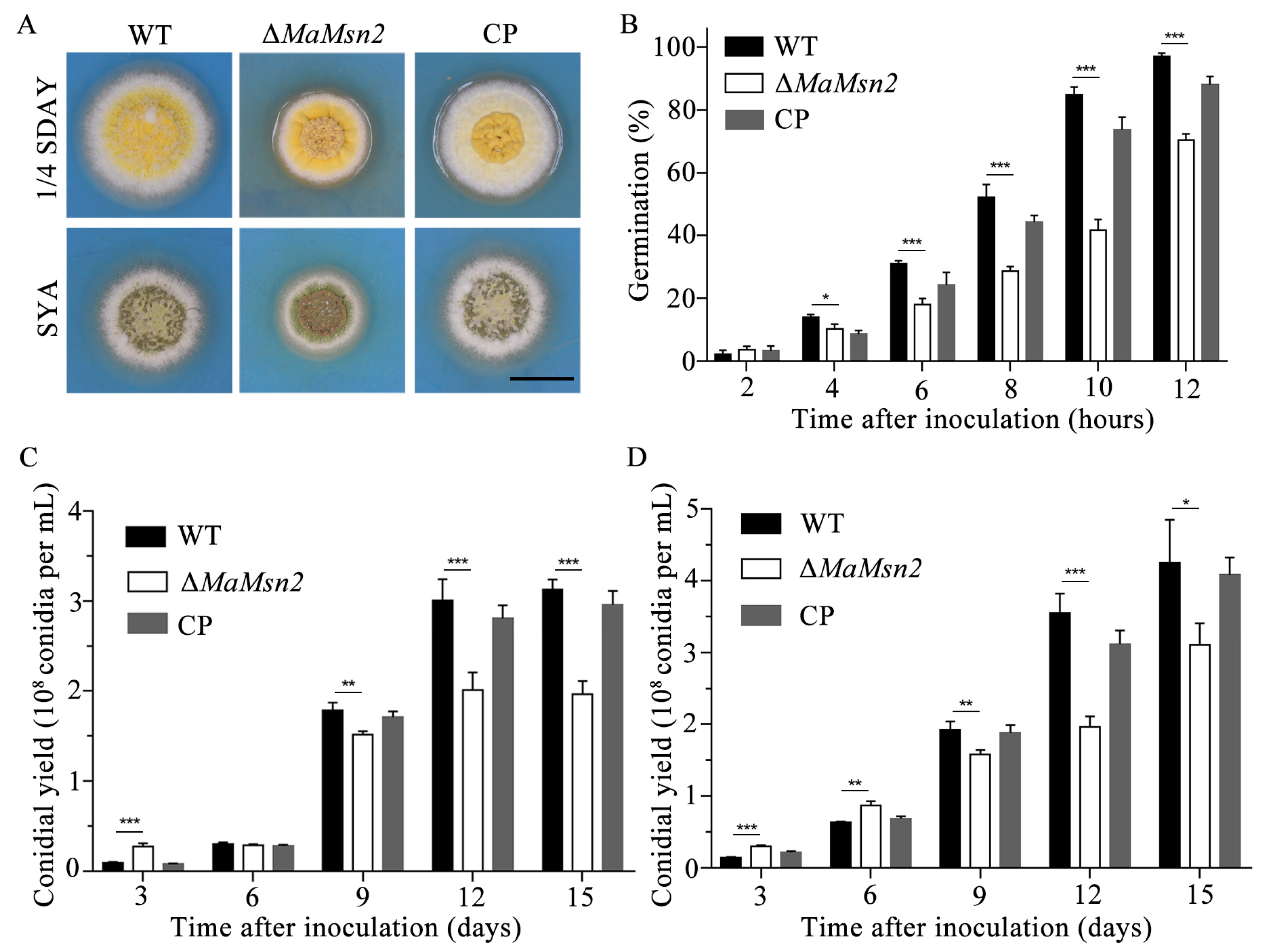
In order to explore the function of MaMsn2 in the germination and conidiation of M. acridum, we measured the germination rate and the conidial yields of ΔMaMsn2 on 1/4 SDAY (nutrient-rich medium) and SYA (nutrient-limited medium). ΔMaMsn2 had a significantly smaller colony and a drastically wrinkled colonial surface compared to the wild type on both 1/4 SDAY and SYA plates (Figure 2A). The germination rate of ΔMaMsn2 was significantly lower than that of the wild type since the 4th hour of culture. At the 12th hour, the germination rate of ΔMaMsn2 was 70% while the WT strain had reached almost 100% (Figure 2B). The conidial yield of ΔMaMsn2 was significantly lower than that of WT and CP strains on 1/4 SDAY (Figure 2C) and SYA media (Figure 2D). Interestingly, at the early stage of conidiation, e.g., on the 3rd day on 1/4 SDAY and 3rd day and 6th day on SYA, the conidial yield of ΔMaMsn2 was significantly higher than that of the WT, but was subsequently exceeded by the WT during the following growth period (Figure 2C,D).

3.3. MaMsn2 Affects Multiple Stress Responses and Virulence in M. acridum

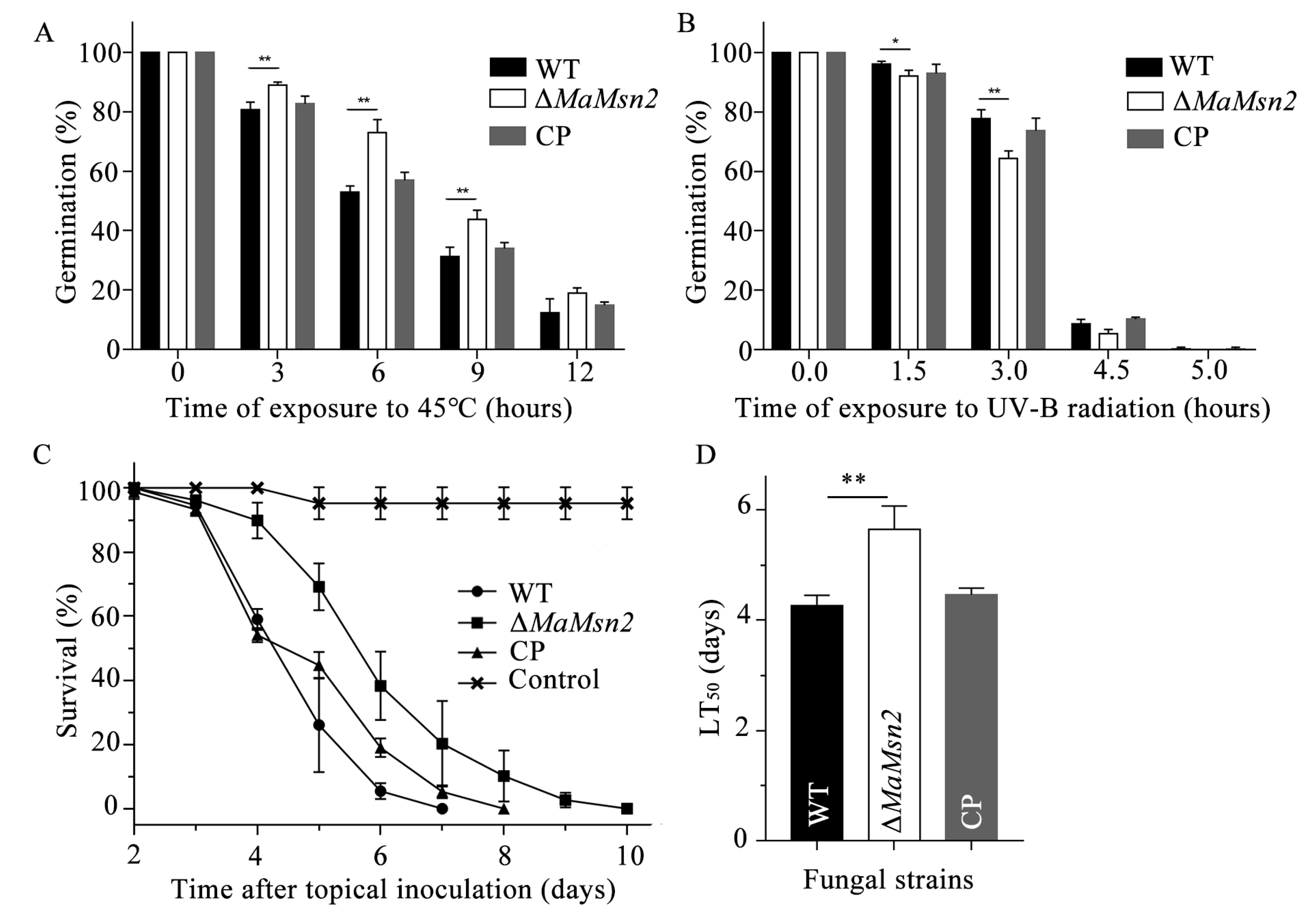
In order to explore the roles of Msn2 in fungal stress tolerance, the germination rate of conidia was determined after heat shock and UV-B stress treatment. The results showed that ΔMaMsn2 had a significantly increased germination rate after 3, 6, 9 and 12 h of treatment at 45 °C (Figure 3A). However, ΔMaMsn2 had a significantly decreased germination rate after exposure to UV-B irradiation (Figure 3B), suggesting a positive role of MaMsn2 in UV-B stress tolerance and a negative role in heat shock stress tolerance.
In order to explore the effect of MaMsn2 on the virulence of M. acridum, a bioassay was conducted using 5th instar nymphs of Locusta by topical inoculation. The results showed that the virulence of ΔMaMsn2 was significantly lower than that of the WT (Figure 3C), and the half-lethal time (LT50) of ΔMaMsn2 was 1.4 days longer than that of the WT strain (Figure 3D).

3.4. MaMsn2 Regulates Conidiation Pattern Shift of M. acridum

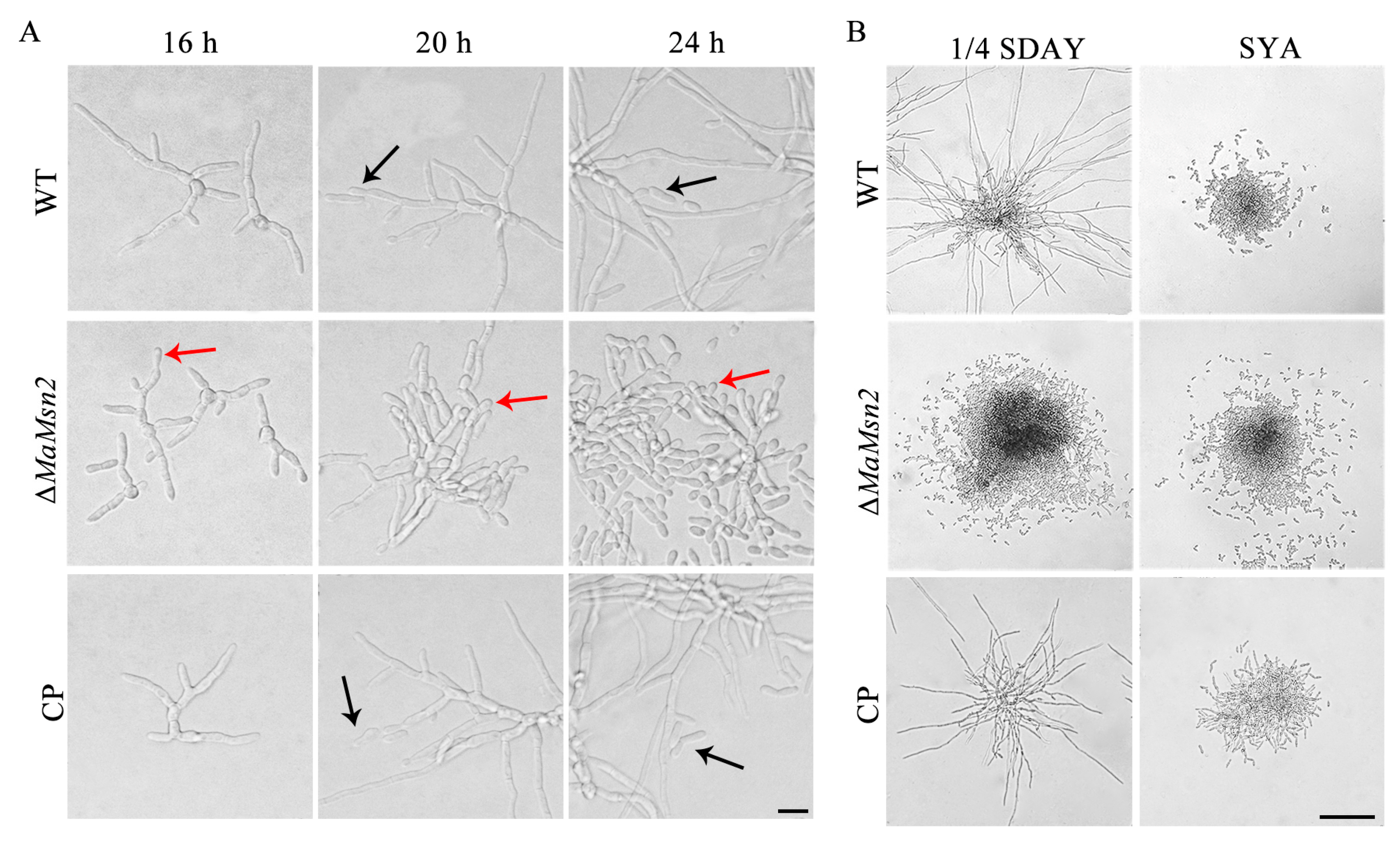
M. acridum conducts normal conidiation on 1/4 SDAY and microcycle conidiation on the SYA medium [8]. In order to analyze the effects of MaMsn2 on the conidiation pattern of M. acridum, conidiation phenotypes of ΔMaMsn2 were observed on 1/4 SDAY and SYA, respectively. On the 1/4 SDAY medium, ΔMaMsn2 produced conidia by microcycle conidiation (Figure 4A), with a drastic suppression of the mycelial growth and extension compared to that of WT and CP strains (Figure 4B). On the SYA medium, ΔMaMsn2 conducted microcycle conidiation with no mycelia as the WT strain (Figure 4B). These results indicated that MaMsn2 was essential for NC and regulated the conidiation pattern shift in M. acridum on 1/4 SDAY.

3.5. MaMsn2 Is Regulated by MaH1

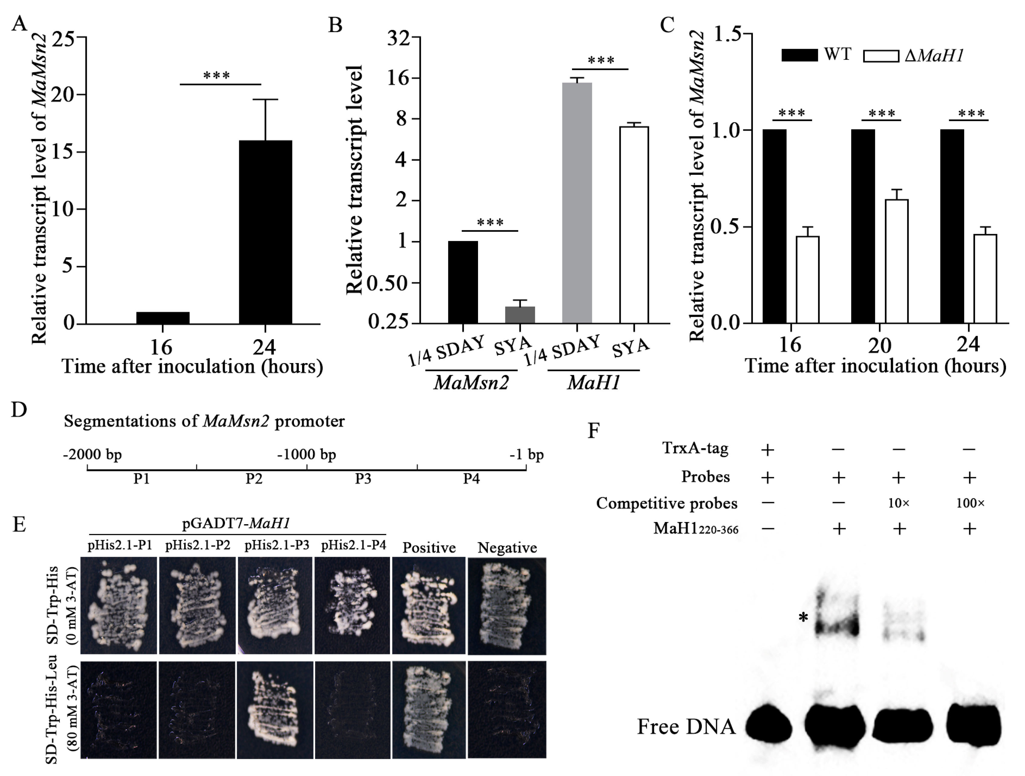
Our previous study showed that MaH1 was involved in the conidiation pattern shift in M. acridum. Similar to ΔMaMsn2, ΔMaH1 also exhibited a microcycle conidiation pattern on nutrient-rich medium 1/4 SDAY [22]. Therefore, we would like to know whether these two genes directly regulate conidiation and whether they have any interaction. First of all, we analyzed the transcription level of MaMsn2 in the WT on 1/4 SDAY at 16 h and 24 h after inoculation, which are the hyphal stage and the beginning of normal conidiation, respectively (Figure 4A). The result showed that the transcription level of MaMsn2 increased by 16 times at 24 h compared to 16 h, suggesting that Msn2 was involved in conidiation (Figure 5A). To compare the transcription levels of MaMsn2 and MaH1 on 1/4 SDAY and SYA, we determined their transcriptions at 24 h after inoculation. At this time point, M. acridum conducts normal conidiation on 1/4 SDAY and microcycle conidiation on SYA. The results showed that the transcription levels of MaMsn2 and MaH1 were significantly higher on 1/4 SDAY than that on SYA (Figure 5B). More importantly, quantitative PCR analysis showed that the transcription level of MaMsn2 was significantly downregulated in ΔMaH1 (Figure 5C). Therefore, MaH1 might regulate conidiation via MaMsn2. In order to confirm this speculation, we performed Y1H and electrophoretic mobility shift assay (EMSA) analysis. Four regions (P1, P2, P3, P4) in the MaMsn2 promoter were assessed, respectively, for a possible interaction with MaH1 (Figure 5D).
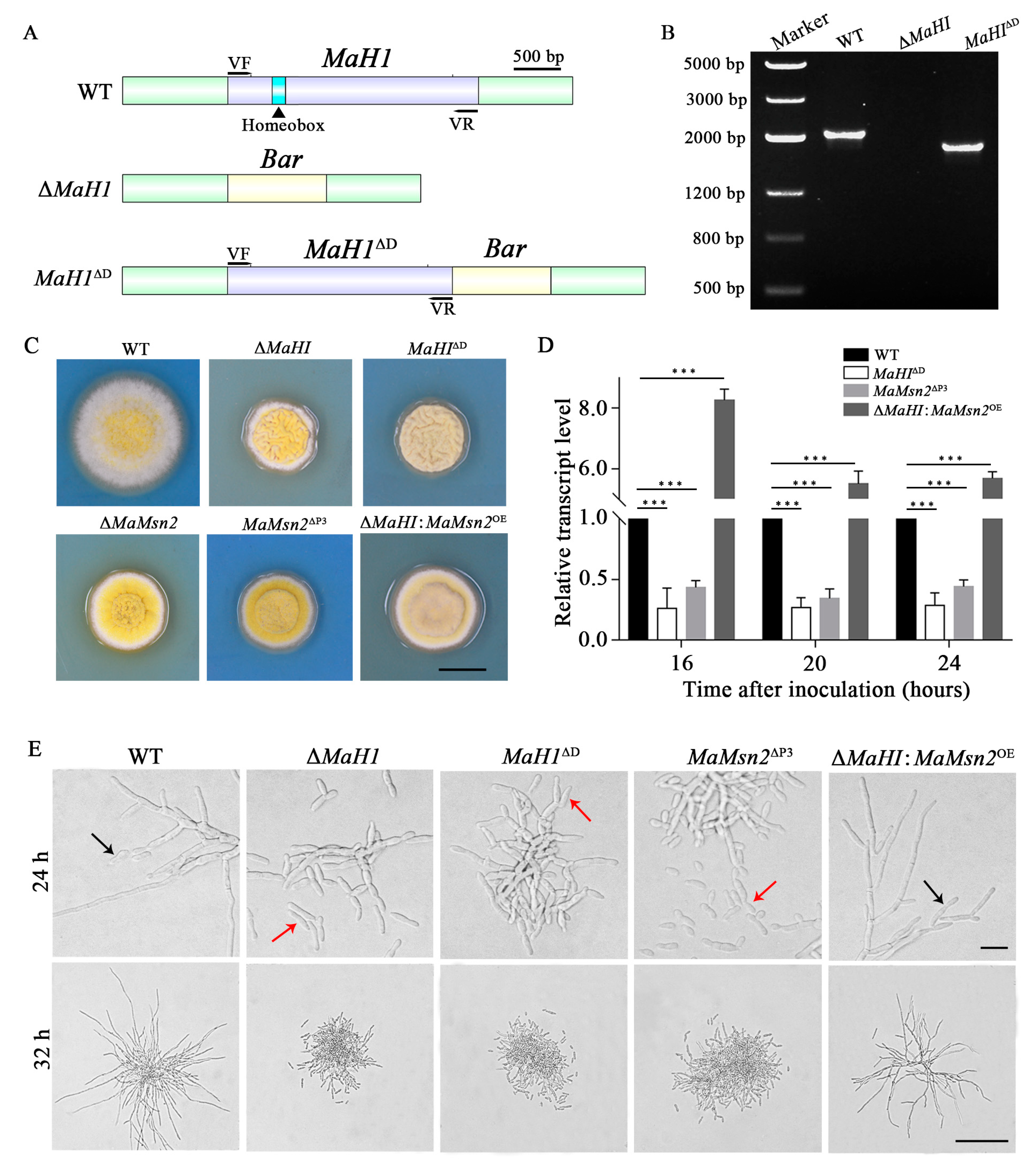
The Y1H result showed that the MaH1 protein directly interacted with the P3 region (−1000 bp to −500 bp), but did not bind to the P1, P2 and P4 regions of the MaMsn2 promoter (Figure 5E). The EMSA result showed that MaH1220–366 containing the DNA binding motif homeobox could bind to a 40 bp fragment of −920 to −880 bp of the MaMsn2 promoter (Figure 5F). These results indicated that MaH1 could directly bind to the MaMsn2 promoter cis-element in M. acridum. In order to further verify the interaction between the binding domain of MaH1 and MaMsn2, we constructed a homeobox domain deletion mutant (MaH1ΔD) (Figure 6A), an engineered strain overexpressing MaMsn2 in ΔMaH1MaH1:MaMsn2OE) and a promoter P3 region deletion mutant (MaMsn2ΔP3). PCR analysis with VF/VR primer pairs confirmed that the homeobox domain region was deleted (Figure 6B). Similar to ΔMaMsn2, the ΔMaH1, MaH1ΔD and MaMsn2ΔP3 colonies all had significantly smaller size with more wrinkles compared to the WT. Meanwhile, the colony size of ΔMaH1:MaMsn2OE was between ΔMaH1 and the WT, and the wrinkles disappeared in ΔMaH1:MaMsn2OE, suggesting a complementation of the defect in ΔMaH1 by overexpressing MaMsn2 (Figure 6C).
RT-qPCR analysis showed that the transcription of MaMsn2 was significantly downregulated in MaH1ΔD and MaMsn2ΔP3, and significantly upregulated in the ΔMaH1:MaMsn2OE strain compared to the WT (Figure 6D). Consistent with ΔMaH1 and ΔMaMsn2, MaH1ΔD and MaMsn2ΔP3 showed microcycle conidiation on 1/4 SDAY, while the conidiation of ΔMaH1:MaMsn2OE shifted to normal conidiation, indicating that MaMsn2 compensated for the conidiation defect of ΔMaH1 (Figure 6E). These results suggested that the homeobox domain in MaH1 was essential for activating the transcription of MaMsn2.

3.6. ΔMaMsn2 and MaH1ΔD Had Similar Phenotypes

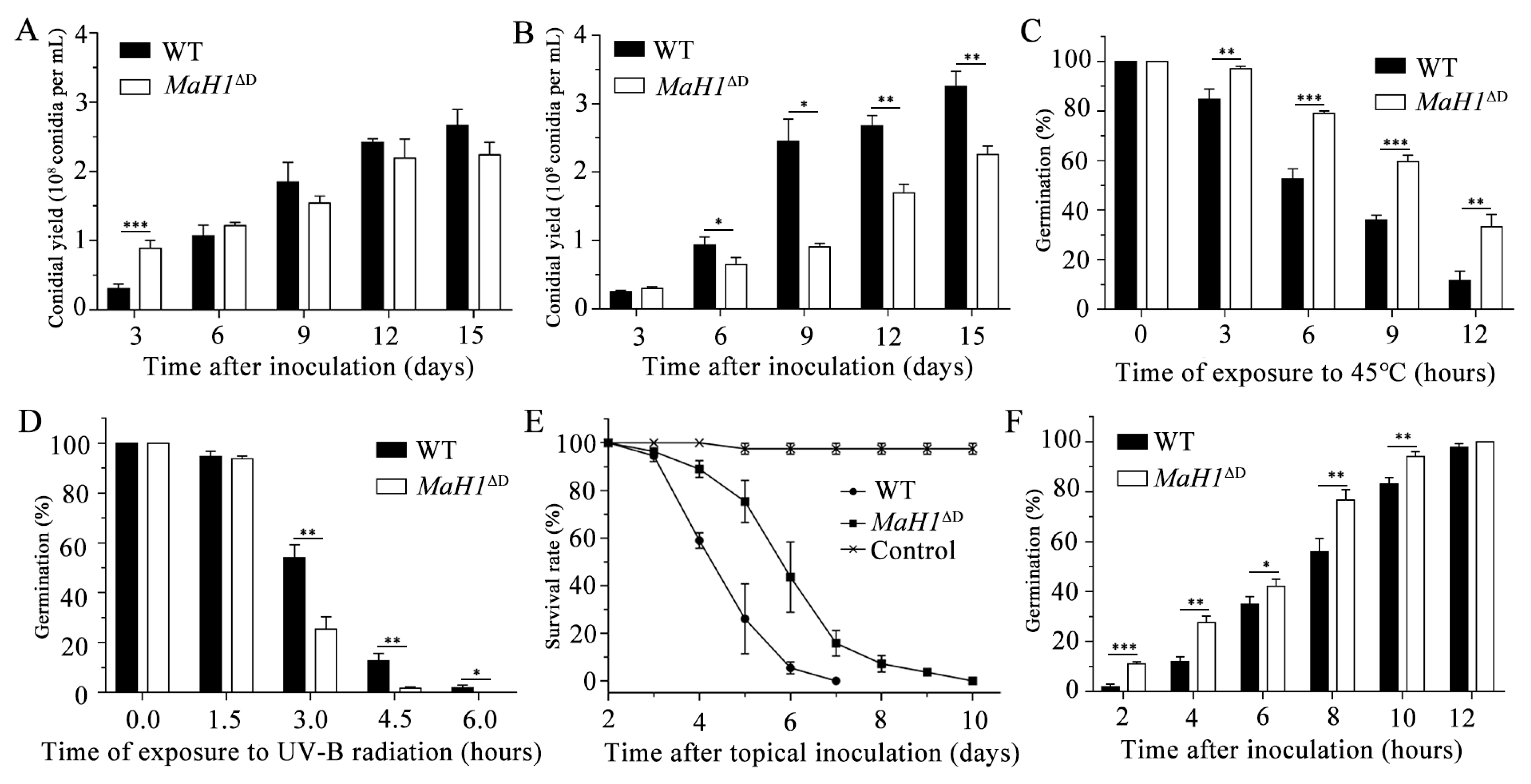
Interaction assay and RT-qPCR analysis demonstrated that MaH1 directly regulated the transcription of MaMsn2. We then measured the conidial yield, tolerance to heat shock and UV-B radiation stresses, virulence and germination rate of MaH1ΔD and compared those results with ΔMaMsn2. The results showed that MaH1ΔD had a similar trend to that of ΔMaMsn2 in determined phenotypes, such as lower conidial yields (Figure 7A,B), stronger resistance to heat shock (Figure 7C), weaker anti-UV-B irradiation ability (Figure 7D) and decreased virulence with topical inoculation (Figure 7E). However, the germination of MaH1ΔD was earlier than that of the WT (Figure 7F), which was opposite to ΔMaMsn2. These similar phenotypes between ΔMaMsn2 and MaH1ΔD also indicated the direct regulation of MaH1 on MaMsn2.

3.7. MaMsn2 Affected the Expression of AbaA and StuA

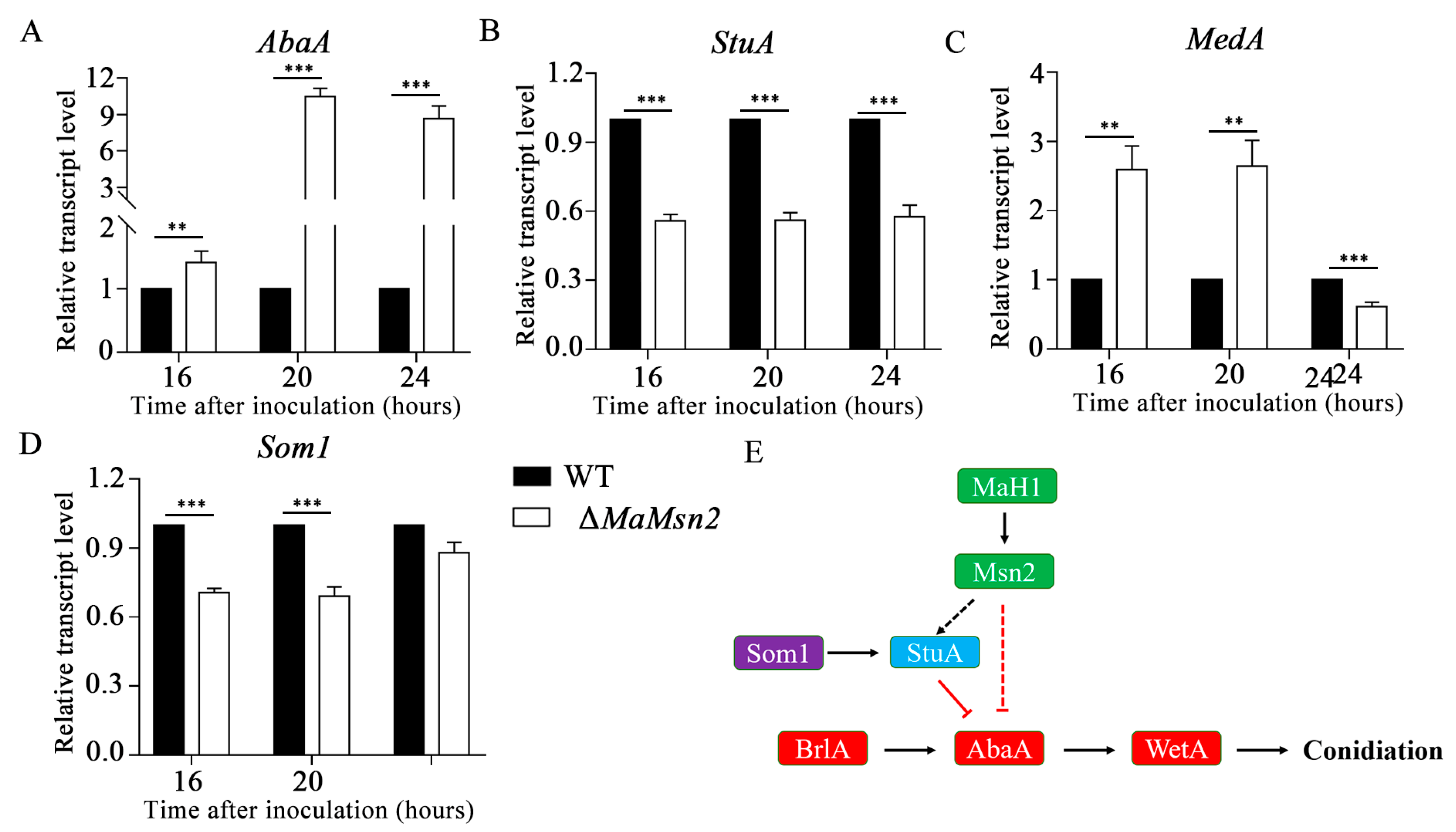
In order to explore the effects of MaMsn2 on conidiation, we measured the time-course transcription level of some conidiation-related genes in ΔMaMsn2. The results showed that AbaA significantly increased in ΔMaMsn2 (Figure 8A), while StuA was down-regulated in ΔMaMsn2 (Figure 8B). The transcriptions of other conidiation-related genes, such as MedA and Som1, did not show consistent changes in ΔMaMsn2 compared to the WT in the conidiation period (Figure 8C,D). These results indicated that MaMsn2 negatively regulated the transcription of AbaA and positively regulated StuA. The prediction of conserved binding sites against the Jaspar 2020 database [38] showed that Msn2 recognition sites were present on AbaA and StuA promoters (data not shown) but not on MedA or Som1 promoters. Combining all the above results, we can infer that the MaH1MaMsn2 pathway affected the central conidiation pathway BrlAAbaAWetA by AbaA and StuA (Figure 8E).

4. Discussion

Msn2 and Msn4 genes widely exist in fungi, and were first reported in yeast as homologous genes. They have a C2H2-type zinc finger structure and participate in different stress responses, such as glucose starvation, heat shock and osmotic and oxidative stress. The transcriptions of stress-related genes CTTI, DDR2 and HSP12 can be regulated through stress response elements by Msn2/4 [39]. However, in filamentous fungi, only Msn2 gene was present while Msn4 has not been found [40]. Consistent with Msn2 homologous genes in other fungi [23,24], MaMsn2 is mainly located in the nucleus in M. acridum.
Msn2 plays an important role in stress response through multiple pathways. In yeast cells, the Ras–cAMP–PKA pathway can regulate the oxidative stress response through Msn2/4 [41], and the high osmolarity glucose (HOG) pathway can also regulate the activity of Msn2/4 [25]. The deletion of the BbMsn2, MrMsn2 or MoMsn2 gene reduced the stress resistance to varying degrees in B. bassiana, M. robertsii and M. oryzae [31,33]. Our study found that the anti-UV-B ability of ΔMaMsn2 slightly decreased, which is consistent with other fungal Msn2. However, different from previous reports, the resistance of ΔMaMsn2 to heat shock increased significantly, indicating that Msn2 functions differently in regulating the response to heat stress in different fungal strains. Our data showed that MaMsn2 was also a virulence factor. Loss of MaMsn2 resulted in decreased virulence. Similar results have been reported in B. bassiana and M. robertsii [31], M. oryzae [33] and V. dahliae [34].
Msn2 is regulated in the process of carbon and nitrogen utilization. TOR1, which is involved in nitrogen source utilization, upregulates the expression level of phosphatase PP2A, which then promotes the nucleus localization of Msn2 through phosphorylation [24,28]. Under low glucose conditions, Msn2 is phosphorylated by the activated protein kinase Snf1 and located in the cytoplasm [23,27]. Microcycle conidiation, as a typical characteristic of filamentous fungi, can be induced by the change in the nutrient composition. On the nutrient-rich 1/4 SDAY medium, the wild type M. acridum firstly grows radial polar hyphae, and then produces conidia on top or both sides of the hyphae. However, ΔMaH1 and ΔMaMsn2 perform microcycle conidiation without hyphal growth on 1/4 SDAY (Figure 6E). Homeobox proteins are widely involved in the hyphal development and conidiation process of filamentous fungi [19,20,21,42,43,44]. In M. oryzae, homeobox protein Htf1 may interact with Acr1 to regulate the conidiation process [45]. Msn2 also regulates the hyphal growth and conidial yield in filamentous fungi such as M. oryzae, V. dahliae and B. bassiana [31,33,34]. Similar to other fungi, our data indicate that MaMsn2 can participate in the nutritional perception process and regulate the process of hyphal development and conidiation in M. acridum.
Our data show that ΔMaH1 and ΔMaMsn2 have a similar conidial phenotype on 1/4 SDAY, suggesting that these two genes are probably in the same regulatory network in the conidial process. The results of qPCR, Y1H and EMSA proved that MaMsn2 is directly regulated by MaH1 in conidiation (Figure 5). Previous studies showed that MaH1 affects conidiation in M. acridum, but does not affect resistances to heat shock, UV-B radiation and virulence [22]. On the contrary, MaMsn2 regulates stress resistances and virulence, indicating that MaMsn2 was not regulated by MaH1 in these two processes. When the DNA binding domain of MaH1 was deleted (MaH1ΔD), the shortened MaH1 could not bind to the promoter region of MaMsn2. Meanwhile, when the promoter region −1000 to −500 bp of MaMsn2 was deleted (MaMsn2ΔP3), MaMsn2 was significantly downregulated. MaH1ΔD exhibited similar phenotypes to ΔMaMsn2 and MaMsn2ΔP3 (Figure 7).
AbaA is a key gene in asexual conidiation in filamentous fungi. The disruption of AbaA leads to an abnormal conidiophore with an “abacus” phenotype [46,47,48]. StuA, an APSES (APSES: Asm1p, Phd1p, Sok2p, Efg1p and StuAp) transcription factor, is necessary for the spatial expression of BrlA and AbaA [49]. The deletion of StuA leads to a very short conidiophore, a lack of metulae and phialides and the formation of conidia directly from vesicles, showing a “stunted” phenotype. The high expression level of StuA inhibits the expressions of BrlA and AbaA [50]. Here, the disruption of MaMsn2 leads to limited hyphal growth and a promoted conidiation process, indicating that MaMsn2 plays a role in maintaining vegetative growth in M. acridum. Based on the qPCR result, MaMsn2 has a strong negative effect on the expression of AbaA, while it has a positive regulatory role on StuA. In addition, conserved binding sites of Msn2 were found on the promoter region of AbaA and StuA genes [38]. Therefore, we infer that MaMsn2 can either directly regulate the expressions of AbaA and StuA or MaMsn2 positively regulated the expression of StuA. (Figure 8E). These results suggest that MaMsn2 might participate in the conidiation process of M. acridum by regulating the transcription of AbaA directly or through StuA. Our analyses expanded the regulatory network of the central conidiation pathway from BrlAAbaAWetA to MaH1MaMsn2AbaA/StuA.
Microcycle conidiation is more applicable in mass production and field application compared with normal conidiation [8]. In this study, we found that MaMsn2 can regulate the conidiation pattern shift in M. acridum, and was directly controlled by another conidiation-related protein MaH1. Our work reports the regulatory mechanism of MC for the first time, which enriches the knowledge of microcycle conidiation and dimorphism in filamentous fungi.

Supplementary Materials

The following are available online at www.mdpi.com/article/10.3390/jof7100840/s1, Table S1: Paired primers used in this paper.

Author Contributions

Conceptualization, Y.X.; Methodology, D.S.; Validation, D.S.; Investigation, D.S.; Resources, Y.C. and Y.X.; Writing—Original Draft Preparation, D.S.; Writing—Review and Editing, Y.C. and Y.X.; Visualization, D.S.; Supervision, Y.C. and Y.X.; Project Administration, Y.C. and Y.X.; Funding Acquisition, Y.X. All authors have read and agreed to the published version of the manuscript.

Funding

This study was supported by the Natural Science Foundation of China (No. 31972330) and Fundamental Research Funds for the Central Universities (2021CDJZYJH-002).

Institutional Review Board Statement

Not applicable.

Informed Consent Statement

Not applicable.

Data Availability Statement

The data presented in this study are available in this article and its Supplementary Materials.

Conflicts of Interest

The authors declare no conflict of interest.

References

  1. Boyce, K.J.; Andrianopoulos, A. Fungal dimorphism: The switch from hyphae to yeast is a specialized morphogenetic adaptation allowing colonization of a host. FEMS Microbiol. Rev. 2015, 39, 797–811. [Google Scholar] [CrossRef] [PubMed] [Green Version]
  2. Pigliucci, M.; Murren, C.J.; Schlichting, C.D. Phenotypic plasticity and evolution by genetic assimilation. J. Exp. Biol. 2006, 209, 2362–2367. [Google Scholar] [CrossRef] [PubMed] [Green Version]
  3. Sudbery, P.E. Growth of Candida albicans hyphae. Nat. Rev. Microbiol. 2011, 9, 737–748. [Google Scholar] [CrossRef]
  4. Wyatt, T.T.; Wosten, H.A.; Dijksterhuis, J. Fungal spores for dispersion in space and time. Adv. Appl. Microbiol. 2013, 85, 43–91. [Google Scholar]
  5. Hanlin, R.T. Microcycle conidiation—A review. Mycoscience 1994, 35, 113–123. [Google Scholar] [CrossRef]
  6. Jung, B.; Kim, S.; Lee, J. Microcyle conidiation in filamentous fungi. Mycobiology 2014, 42, 1–5. [Google Scholar] [CrossRef] [Green Version]
  7. Anderson, J.G.; Smith, J.E. The production of conidiophores and conidia by newly germinated conidia of Aspergillus niger (microcycle conidiation). J. Gen. Microbiol. 1971, 69, 185–197. [Google Scholar] [CrossRef] [Green Version]
  8. Zhang, S.Z.; Peng, G.X.; Xia, Y.X. Microcycle conidiation and the conidial properties in the entomopathogenic fungus Metarhizium acridum on agar medium. Biocontrol. Sci. Technol. 2010, 20, 809–819. [Google Scholar] [CrossRef]
  9. Boylan, M.T.; Mirabito, P.M.; Willett, C.E.; Zimmerman, C.R.; Timberlake, W.E. Isolation and physical characterization of three essential conidiation genes from Aspergillus nidulans. Mol. Cell. Biol. 1987, 7, 3113–3318. [Google Scholar] [CrossRef] [PubMed]
  10. Sewall, T.C.; Mims, C.W.; Timberlake, W.E. Conidium differentiation in Aspergillus nidulans wild-type and wet-white (wetA) mutant strains. Dev. Biol. 1990, 138, 499–508. [Google Scholar] [CrossRef]
  11. Sewall, T.C.; Mims, C.W.; Timberlake, W.E. abaA controls phialide differentiation in Aspergillus nidulans. Plant Cell 1990, 2, 731–739. [Google Scholar] [PubMed] [Green Version]
  12. Adams, T.H.; Boylan, M.T.; Timberlake, W.E. BrlA is necessary and sufficient to direct conidiophore development in Aspergillus nidulans. Cell 1988, 54, 353–362. [Google Scholar] [CrossRef]
  13. Etxebeste, O.; Garzia, A.; Espeso, E.A.; Ugalde, U. Aspergillus nidulans asexual development: Making the most of cellular modules. Trends Microbiol. 2010, 18, 569–576. [Google Scholar] [CrossRef] [PubMed] [Green Version]
  14. Son, H.; Kim, M.G.; Min, K.; Lim, J.Y.; Choi, G.J.; Kim, J.C.; Chae, S.K.; Lee, Y.W. WetA is required for conidiogenesis and conidium maturation in the ascomycete fungus Fusarium graminearum. Eukaryot. Cell 2014, 13, 87–98. [Google Scholar] [CrossRef] [Green Version]
  15. Liu, J.; Cao, Y.; Xia, Y. Mmc, a gene involved in microcycle conidiation of the entomopathogenic fungus Metarhizium anisopliae. J. Invertebr. Pathol. 2010, 105, 132–138. [Google Scholar] [CrossRef] [PubMed]
  16. Song, D.; Shi, Y.; Ji, H.; Xia, Y.; Peng, G. The MaCreA gene regulates normal conidiation and microcycle conidiation in Metarhizium acridum. Front. Microbiol. 2019, 10, 1946. [Google Scholar] [CrossRef]
  17. Li, J.; Su, X.L.; Cao, Y.Q.; Xia, Y.X. Dipeptidase PEPDA is required for the conidiation pattern shift in Metarhizium acridum. Appl. Environ. Microbiol. 2021, 87, e00908-21. [Google Scholar] [CrossRef]
  18. Latchman, D.S. Families of DNA binding transcription factors. In Eukaryotic Transcription Factors, 5th ed.; Latchman, D.S., Ed.; Academic Press: Cambridge, MA, USA, 2008; pp. 96–111. [Google Scholar]
  19. Arnaise, S.; Zickler, D.; Poisier, C.; Debuchy, R. Pah1: A homeobox gene involved in hyphal morphology and microconidiogenesis in the filamentous ascomycete Podospora anserina. Mol. Microbiol. 2001, 39, 54–64. [Google Scholar] [CrossRef]
  20. Antal, Z.; Rascle, C.; Cimerman, A.; Viaud, M.; Billon-Grand, G.; Choquer, M.; Bruel, C. The homeobox BcHOX8 gene in Botrytis cinerea regulates vegetative growth and morphology. PLoS ONE 2012, 7, e48134. [Google Scholar] [CrossRef] [Green Version]
  21. Kim, S.; Park, S.Y.; Kim, K.S.; Rho, H.S.; Chi, M.H.; Choi, J.; Park, J.; Kong, S.; Park, J.; Goh, J.; et al. Homeobox transcription factors are required for conidiation and appressorium development in the rice blast fungus Magnaporthe oryzae. PLoS Genet. 2009, 5, e1000757. [Google Scholar] [CrossRef] [Green Version]
  22. Gao, P.; Li, M.; Jin, K.; Xia, Y. The homeobox gene MaH1 governs microcycle conidiation for increased conidial yield by mediating transcription of conidiation pattern shift-related genes in Metarhizium acridum. Appl. Microbiol. Biotechnol. 2019, 103, 2251–2262. [Google Scholar] [CrossRef] [PubMed]
  23. De Wever, V.; Reiter, W.; Ballarini, A.; Ammerer, G.; Brocard, C. A dual role for PP1 in shaping the Msn2-dependent transcriptional response to glucose starvation. EMBO J. 2005, 24, 4115–4123. [Google Scholar] [CrossRef] [PubMed] [Green Version]
  24. Santhanam, A.; Hartley, A.; Duvel, K.; Broach, J.R.; Garrett, S. PP2A phosphatase activity is required for stress and Tor kinase regulation of yeast stress response factor Msn2p. Eukaryot. Cell 2004, 3, 1261–1271. [Google Scholar] [CrossRef] [PubMed] [Green Version]
  25. Rep, M.; Krantz, M.; Thevelein, J.M.; Hohmann, S. The transcriptional response of Saccharomyces cerevisiae to osmotic shock-Hot1p and Msn2p/Msn4p are required for the induction of subsets of high osmolarity glycerol pathway-dependent genes. J. Biol. Chem. 2000, 275, 8290–8300. [Google Scholar] [CrossRef] [Green Version]
  26. Gorner, W.; Durchschlag, E.; Martinez-Pastor, M.T.; Estruch, F.; Ammerer, G.; Hamilton, B.; Ruis, H.; Schuller, C. Nuclear localization of the C2H2 zinc finger protein Msn2p is regulated by stress and protein kinase A activity. Genes Dev. 1998, 12, 586–597. [Google Scholar] [CrossRef] [PubMed]
  27. Mayordomo, I.; Estruch, F.; Sanz, P. Convergence of the target of rapamycin and the Snf1 protein kinase pathways in the regulation of the subcellular localization of Msn2, a transcriptional activator of STRE (stress response element)-regulated genes. J. Biol. Chem. 2002, 277, 35650–35656. [Google Scholar] [CrossRef] [Green Version]
  28. Beck, T.; Hall, M.N. The TOR signalling pathway controls nuclear localization of nutrient-regulated transcription factors. Nature 1999, 402, 689–692. [Google Scholar] [CrossRef]
  29. Hirata, Y.; Andoh, T.; Asahara, T.; Kikuchi, A. Yeast glycogen synthase kinase-3 activates Msn2p-dependent transcription of stress responsive genes. Mol. Biol. Cell 2003, 14, 302–312. [Google Scholar] [CrossRef] [Green Version]
  30. Schmitt, A.P.; McEntee, K. Msn2p, a zinc finger DNA-binding protein, is the transcriptional activator of the multistress response in Saccharomyces cerevisiae. Proc. Natl. Acad. Sci. USA 1996, 93, 5777–5782. [Google Scholar] [CrossRef] [Green Version]
  31. Liu, Q.; Ying, S.H.; Li, J.G.; Tian, C.G.; Feng, M.G. Insight into the transcriptional regulation of Msn2 required for conidiation, multi-stress responses and virulence of two entomopathogenic fungi. Fungal Genet. Biol. 2013, 54, 42–51. [Google Scholar] [CrossRef] [Green Version]
  32. Luo, Z.B.; Li, Y.J.; Mousa, J.; Bruner, S.; Zhang, Y.J.; Pei, Y.; Keyhani, N.O. Bbmsn2 acts as a pH-dependent negative regulator of secondary metabolite production in the entomopathogenic fungus Beauveria bassiana. Environ. Microbiol. 2015, 17, 1189–1202. [Google Scholar] [CrossRef]
  33. Zhang, H.; Zhao, Q.; Guo, X.; Guo, M.; Qi, Z.; Tang, W.; Dong, Y.; Ye, W.; Zheng, X.; Wang, P.; et al. Pleiotropic function of the putative zinc-finger protein MoMsn2 in Magnaporthe oryzae. Mol. Plant Microbe Interact. 2014, 27, 446–460. [Google Scholar] [CrossRef] [PubMed] [Green Version]
  34. Tian, L.; Yu, J.; Wang, Y.; Tian, C. The C2H2 transcription factor VdMsn2 controls hyphal growth, microsclerotia formation, and virulence of Verticillium dahliae. Fungal Biol. 2017, 121, 1001–1010. [Google Scholar] [CrossRef]
  35. Lazo, G.R.; Stein, P.A.; Ludwig, R.A. A DNA transformation-competent Arabidopsis genomic library in Agrobacterium. Biotechnology 1991, 9, 963–967. [Google Scholar] [CrossRef] [PubMed]
  36. Jin, K.; Ming, Y.; Xia, Y.X. MaHog1, a Hog1-type mitogen-activated protein kinase gene, contributes to stress tolerance and virulence of the entomopathogenic fungus Metarhizium acridum. Microbiology 2012, 158, 2987–2996. [Google Scholar] [CrossRef] [Green Version]
  37. Livak, K.J.; Schmittgen, T.D. Analysis of relative gene expression data using real-time quantitative PCR and the 2-ΔΔCT method. Methods 2001, 25, 402–408. [Google Scholar] [CrossRef] [PubMed]
  38. Fornes, O.; Castro-Mondragon, J.A.; Khan, A.; van der Lee, R.; Zhang, X.; Richmond, P.A.; Modi, B.P.; Correard, S.; Gheorghe, M.; Baranasic, D.; et al. JASPAR 2020: Update of the open-access database of transcription factor binding profiles. Nucleic Acids Res. 2020, 48, D87–D92. [Google Scholar] [CrossRef]
  39. MartinezPastor, M.T.; Marchler, G.; Schuller, C.; MarchlerBauer, A.; Ruis, H.; Estruch, F. The Saccharomyces cerevisiae zinc finger proteins Msn2p and Msn4p are required for transcriptional induction through the stress-response element (STRE). EMBO J. 1996, 15, 2227–2235. [Google Scholar] [CrossRef]
  40. Yang, G.; Liu, G.L.; Wang, S.J.; Chi, Z.M.; Chi, Z. Pullulan biosynthesis in yeast-like fungal cells is regulated by the transcriptional activator Msn2 and cAMP-PKA signaling pathway. Int. J. Biol. Macromol. 2020, 157, 591–603. [Google Scholar] [CrossRef]
  41. Hasan, R.; Leroy, C.; Isnard, A.D.; Labarre, J.; Boy-Marcotte, E.; Toledano, M.B. The control of the yeast H2O2 response by the Msn2/4 transcription factors. Mol. Microbiol. 2002, 45, 233–241. [Google Scholar] [CrossRef]
  42. Cary, J.W.; Harris-Coward, P.; Scharfenstein, L.; Mack, B.M.; Chang, P.K.; Wei, Q.; Lebar, M.; Carter-Wientjes, C.; Majumdar, R.; Mitra, C.; et al. The Aspergillus flavus Homeobox Gene, hbx1, is required for development and aflatoxin production. Toxins 2017, 9, 315. [Google Scholar] [CrossRef] [PubMed] [Green Version]
  43. Zheng, W.; Zhao, X.; Xie, Q.; Huang, Q.; Zhang, C.; Zhai, H.; Xu, L.; Lu, G.; Shim, W.B.; Wang, Z. A conserved homeobox transcription factor Htf1 is required for phialide development and conidiogenesis in Fusarium species. PLoS ONE 2012, 7, e45432. [Google Scholar] [CrossRef] [PubMed]
  44. Colot, H.V.; Park, G.; Turner, G.E.; Ringelberg, C.; Crew, C.M.; Litvinkova, L.; Weiss, R.L.; Borkovich, K.A.; Dunlap, J.C. A high-throughput gene knockout procedure for Neurospora reveals functions for multiple transcription factors. Proc. Natl. Acad. Sci. USA 2006, 103, 10352–10357. [Google Scholar] [CrossRef] [Green Version]
  45. Liu, W.; Xie, S.; Zhao, X.; Chen, X.; Zheng, W.; Lu, G.; Xu, J.R.; Wang, Z. A homeobox gene is essential for conidiogenesis of the rice blast fungus Magnaporthe oryzae. Mol. Plant Microbe Interact. 2010, 23, 366–375. [Google Scholar] [CrossRef] [Green Version]
  46. Andrianopoulos, A.; Timberlake, W.E. The Aspergillus nidulans abaA gene encodes a transcriptional activator that acts as a genetic switch to control development. Mol. Cell. Biol. 1994, 14, 2503–2515. [Google Scholar] [CrossRef] [Green Version]
  47. Clutterbuck, A.J. A mutational analysis of conidial development in Aspergillus nidulans. Genetics 1969, 63, 317–327. [Google Scholar] [CrossRef]
  48. Zhang, A.X.; Mouhoumed, A.Z.; Tong, S.M.; Ying, S.H.; Feng, M.G. BrlA and AbaA govern virulence-required dimorphic switch, conidiation, and pathogenicity in a fungal insect pathogen. mSystems 2019, 4, e00140-19. [Google Scholar] [CrossRef] [Green Version]
  49. Aramayo, R.; Peleg, Y.; Addison, R.; Metzenberg, R. Asm-1+, a Neurospora crassa gene related to transcriptional regulators of fungal development. Genetics 1996, 144, 991–1003. [Google Scholar] [CrossRef] [PubMed]
  50. Busby, T.M.; Miller, K.Y.; Miller, B.L. Suppression and enhancement of the Aspergillus nidulans medusa mutation by altered dosage of the bristle and stunted genes. Genetics 1996, 143, 155–163. [Google Scholar] [CrossRef] [PubMed]
Figure 1. Sequence analysis and intracellular localization of MaMsn2. (A) Msn2 protein sequence alignment. The Msn2 homologous genes are from Aspergillus parasiticus, Aureobasidium melanogenum, Beauveria bassiana ARSEF 2860, Diplodia corticola, Fusarium longipes, M. acridum CQMa 102, M. rileyi, Pichia kudriavzevii, Saccharomyces cerevisiae, Trichoderma harzianum and Zygosaccharomyces parabailii. Asterisks indicate conservative C and H loci. (B) Main domains and phylogenetic relationships of Msn2. ▲: Msn2 in M. acridum. (C) The knockout (upper) and complement (lower) schematic diagram of MaMsn2. (D) The Southern blot verification of ΔMaMsn2 and CP strains. Restriction enzymes HindIII and KpnI were used to digest genomic DNAs. Probe location is shown in the diagram. (E) LSCM images of subcellular localization of MaMsn2:EGFP. Hyphae were collected from 1/4 SDAY grown for 18 h and conidia were collected on 1/4 SDAY and SYA grown for 48 h. NC: Conidia produced on 1/4 SDAY, MC: Conidia produced on SYA. White arrows: DAPI-stained nuclei, red arrows: merged nuclei. Scale bar indicates 5 μm.
Jof 07 00840 g001
Figure 2. Growth and conidiation analysis. (A) The colonies of ΔMaMsn2 grown on 1/4 SDAY and SYA media for 5 days. The scale bar indicates 1 cm. (B) Conidial germination rate of ΔMaMsn2 for 2, 4, 6, 8, 10 and 12 h. (C,D) Conidial yields of ΔMaMsn2 at 3, 6, 9, 12 and 15 days on 1/4 SDAY (C) and SYA (D). (t-test, *: p < 0.05, **: p < 0.01, ***: p < 0.001).
Jof 07 00840 g002
Figure 3. Stress tolerance and insect bioassays. (A) Conidial germination rate of ΔMaMsn2 after heat shock treatment at 45 °C for 0, 3, 6, 9 and 12 h. (B) Conidial germination rate of ΔMaMsn2 after UV-B treatment at 1350 mW/m2 for 0, 1.5, 3.0, 4.5 and 6.0 h. (C) Survival of the locusts following topical inoculation with 5 μL aqueous conidial suspensions of 1×10⁷ conidia/mL of WT, ΔMaMsn2 and CP strains. Control insects were treated with 5 μL paraffin oil. (D) LT50 of different strains in topical inoculation. (t-test, *: p < 0.05, **: p < 0.01).
Jof 07 00840 g003
Figure 4. The conidiation phenotype of ΔMaMsn2. (A) The conidiation of ΔMaMsn2 on 1/4 SDAY medium at 16, 20 and 24 h. Scale bars indicate 5 μm. Black arrow: normal conidiation; Red arrow: microcycle conidiation. (B) The conidiation phenotype of ΔMaMsn2 on 1/4 SDAY and SYA media for 32 h. Scale bars indicate 100 μm.
Jof 07 00840 g004
Figure 5. Interaction analysis between MaH1 and MaMsn2. (A) Transcription level of MaMsn2 in WT on 1/4 SDAY at 16 h and 24 h. The MaMsn2 transcript level at 16 h was set to 1. (t-test, ***: p < 0.001). (B) Transcription levels of MaH1 and MaMsn2 in WT on 1/4 SDAY and SYA at 24 h. The MaMsn2 transcript level on 1/4 SDAY was set to 1. (t-test, ***: p < 0.001) (C) Transcription levels of MaMsn2 in WT and ΔMaH1 on 1/4 SDAY at 16 h, 20 h and 24 h. (t-test, ***: p < 0.001). (D) Different regions of MaMsn2 promoters for binding analysis with MaH1 by Y1H. (E) Y1H assay between MaH1 and different MaMsn2 promoter regions. (F) EMSA assay with purified trxA-MaH1220−366 and probe labeled by biotin. The competitive probe was unlabeled and 10 to 100 fold excess compared to the labeled probe. +: probe or protein added, –: probe or protein not added. Asterisk indicates the position of shifted band.
Jof 07 00840 g005
Figure 6. Complementation of ΔMaH1 by MaMsn2. (A) Schematic diagram of deletion of the MaH1 DNA binding domain. (B) PCR verification of DNA binding domain knockout strain MaH1ΔD. Results show a fragment 135 bp smaller in MaH1ΔD than that of the wild type. (C) Colony morphology of ΔMaH1, DNA binding domain knockout strain of MaH1 (MaH1ΔD), ΔMaMsn2, promoter P3 region knockout strain of MaMsn2 (MaMsn2ΔP3) and ΔMaH1 strain overexpressing MaMsn2MaH1:MaMsn2OE). Scale bars indicate 0.5 cm. (D) The transcript level of MaMsn2 on 1/4 SDAY medium at 16, 20 and 24 h in MaH1ΔD, MaMsn2ΔP3 and ΔMaH1:MaMsn2OE strains (t-test, ***: p < 0.001). (E) The conidiation and hyphal development of different strains. Black arrow: normal conidiation; Red arrow: microcycle conidiation. The scale bars indicate 5 μm at 24 h and 100 μm at 32 h.
Jof 07 00840 g006
Figure 7. Phenotypes of MaH1ΔD. (A,B) Conidial yields of MaH1ΔD at 3, 6, 9, 12 and 15 days on 1/4 SDAY (A) and SYA (B). (C) Conidial germination rate of MaH1ΔD after heat shock treatment at 45 °C for 0, 3, 6, 9 and 12 h. (D) Conidial germination rate of MaH1ΔD after UV-B treatment at 1350 mW/m2 for 0, 1.5, 3.0, 4.5 and 6.0 h. (E) Survival of the locusts following topical inoculation with 5 μL aqueous conidial suspensions of 1 × 10⁷ conidia/mL of WT and MaH1ΔD strains. Control insects were treated with 5 μL paraffin oil. (F) Conidial germination rate of MaH1ΔD for 2, 4, 6, 8, 10 and 12 h (t-test, *: p < 0.05, **: p < 0.01, ***: p < 0.001).
Jof 07 00840 g007
Figure 8. MaMsn2 regulates AbaA and StuA. (AD) The relative expressions of conidiation-related genes in ΔMaMsn2 (t-test, ** : p < 0.01, *** : p < 0.001). (E) Genetic model for MaH1MaMsn2 in conidiation pathway.
Jof 07 00840 g008
Publisher’s Note: MDPI stays neutral with regard to jurisdictional claims in published maps and institutional affiliations.