Article

Cytotoxicity of Aspergillus Section Fumigati Isolated from Health Care Environments

by
Carla Viegas
1,2,3,*,†,
Magdalena Twarużek
4,†,
Beatriz Almeida
1,
Marta Dias
1,3,
Edna Ribeiro
1,
Elisabete Carolino
1,
Ewelina Soszczyńska
4 and
Liliana Aranha Caetano
1,5
1
H&TRC—Health & Technology Research Center, ESTeSL—Escola Superior de Tecnologia da Saúde, Instituto Politécnico de Lisboa, 1990-086 Lisbon, Portugal
2
Public Health Research Centre, NOVA National School of Public Health, Universidade NOVA de Lisboa, 1099-085 Lisbon, Portugal
3
Comprehensive Health Research Center (CHRC), NOVA National School of Public Health, Universidade NOVA de Lisboa, 1099-085 Lisbon, Portugal
4
Department of Physiology and Toxicology, Faculty of Biological Sciences, Kazimierz Wielki University, Chodkiewicza 30, 85-064 Bydgoszcz, Poland
5
Research Institute for Medicines (iMed.Ulisboa), Faculty of Pharmacy, University of Lisbon, 1649-004 Lisbon, Portugal
*
Author to whom correspondence should be addressed.
These authors contributed equally to this work.
J. Fungi 2021, 7(10), 839; https://doi.org/10.3390/jof7100839
Submission received: 25 August 2021 / Revised: 5 October 2021 / Accepted: 5 October 2021 / Published: 7 October 2021
(This article belongs to the Special Issue Fungi in Indoor Environments)

Abstract

:
This study analyzed 57 Aspergillus section Fumigati (AF) isolates collected by active and passive sampling (N = 450) in several health care facilities and from biological sampling of health care workers (N = 25) and controls (N = 22) in Portugal. All isolates were cultured in different media and screened for azole resistance. Cytotoxicity was assessed for 40 isolates in lung epithelial cells and kidney cells using the MTT assay. Aspergillus section Fumigati was prevalent in the health care facilities and in nasal swabs from health care workers and controls. All AF isolates reduced cell viability and presented medium to high cytotoxicity, with cytotoxicity being significantly higher in A549 lung epithelial cells. The cytotoxicity of isolates from air and nasal swab samples suggested the inhalation route as a risk factor. Notably, 42% of AF isolates exhibited a pattern of reduced susceptibility to some of the most used antifungals available for the treatment of patients infected with these fungi. In sum, the epidemiology and clinical relevance of Aspergillus section Fumigati should continue to be addressed. A deeper understanding of the mechanisms underlying Aspergillus-mediated cytotoxicity is necessary.

1. Introduction

Aspergillus section Fumigati is associated with a high mortality rate in at risk patients, such as those with asthma, cystic fibrosis, and chronic obstructive lung disease or with immune suppression, mostly due to invasive pulmonary aspergillosis—a fatal disease [1]. It is predictable that over 30 million patients are at risk of developing invasive aspergillosis worldwide, mainly due to the use of immunosuppressive therapies [2]. Healthcare-associated aspergillosis is most often acquired by inhalation of airborne spores causing pulmonary aspergillosis, which the fungus can disseminate in the bloodstream and reach other organs [3]. Airway colonization by Aspergillus spp. Has also been observed in approximately half of patients in an adult pneumology ward with no symptoms of aspergillosis [4]. Exposure to Aspergillus section Fumigati can be, therefore, considered a risk for both patients and health staff in health care environments (HCE) [5].
The genus Aspergillus is classified into four subgenera (Aspergillus, Circumdati, Fumigati, and Nidulantes) and 20 sections, each including a number of related species [6,7]. The most prevalent in HCE, as in other environments, are Aspergillus sections Fumigati, Flavi, Nigri, and Nidulantes [8,9]. Section Fumigati is the most frequently isolated section both from respiratory samples from patients and from HCE sampling [10]. Section Fumigati is one of the most species-rich sections in the Aspergillus genus, comprising about 50 to 60 potentially pathogenic species for humans [11]. It is the Aspergillus section that has the most reported clinical relevance and is more often associated with respiratory symptoms (mainly A. fumigatus sensu stricto).
The adverse effects resulting from exposure to airborne toxigenic fungi are well known. Long-term exposure to toxigenic fungi interferes with natural killer cell activity, and may cause symptoms such as cough, fever, headache, anxiety, or depression [12]. The induction of immunosuppression and inflammation by exposure to fungi and bioaerosols is also described [13,14]. An additional concern regarding A. fumigatus sensu stricto is the emergence of acquired azole-resistance in clinical practice and in the environment [10,15,16,17,18,19].
Aspergillus section Fumigati’s clinical relevance has been related to the small size of conidia and other virulence factors [9,20,21]. The cytotoxicity effect of Aspergillus section Fumigati toxins, such as gliotoxin, has been described [22,23,24]. While a more recent study described cytotoxic and apoptotic effects of Aspergillus section Fumigati conidia in lung epithelial cells and fibroblasts [25].
In the present study, to evaluate the cytotoxicity of Aspergillus section Fumigati from ten primary health care centers and one central hospital, isolates were obtained by air, passive sampling and workers’ nasal swabbing and were co-cultured with lung epithelial cells and kidney cells. An MTT assay was used to determine IC50 levels, and correlational statistical analysis was performed to explore relations between isolates growth in different media, including azoles, sampling, and the cytotoxicity effect.

2. Materials and Methods

2.1. Health Care Facilities and Sampling Campaign

Ten Primary Health Care Centres (PHCC) and one Central Hospital (CH) were assessed in Lisbon and Oporto, respectively, from June to July 2018, as part of a wider study aiming to propose new procedures to determine exposure to bioburden at HCE [26]. The project protocol was first approved by scientific councils from HCE (ref: 064/CES/INV/2017) and by the Ethical Committee of Escola Superior de Tecnologia da Saúde de Lisboa (ref: CE-ESTeSL-No 45-2018). The protocol was in accordance with the World Medical Association Declaration of Helsinki and the Oviedo Convention, and in agreement with the Portuguese law no 58/2019 of 8 August, regarding data protection [26]. A prior evaluation by a certified exposure assessor was developed on site at each HCE to identify critical control points in workplaces which could involve higher exposure to microbial contamination. A comprehensive sampling campaign was then held, using active and passive sampling methods in both indoor environments (PHCC and CH) [27,28]. Active sampling comprised air sampling by impaction (N = 201). Air sampling by impinger was also performed (N = 56) for molecular detection purposes (not presented). Passive sampling included surface swabs (N = 126), electrostatic dust cloths (EDC, N = 96), settled dust (N = 15), and filters from HVAC system (N = 12) (Table 1).

2.2. Volunteers Enrolment and Biological Sampling

Nasal swabs were collected from volunteer health care workers in the ten PHCC (N = 25) and in the CH (N = 22). A control group of 25 healthy volunteers with no occupational contact with health care facilities was also evaluated in Lisbon. All volunteers signed informed consent prior to enrollment in the study. All inherent ethical principles were duly observed. Biological samples were obtained through a nasopharyngeal swab procedure using transport swabs with Stuart media when necessary. For nasal sampling, a swab was inserted about one centimeter into the nostril and rubbed in a circular way. The same swab was then used to sample the other nostril following the same procedure. For the samples collection, transport swabs with Stuart media were used that were immediately transported to the laboratory after being used.

2.3. Fungal Culture and Screening of Azole Resistance

Malt extract agar (MEA) supplemented with chloramphenicol (0.05%), and dichloran-glycerol agar (DG18) were used to increase selectivity for fungal growth. Sabouraud dextrose agar (SDA) and SDA supplemented with 4 mg/L itraconazole (ITR), 1 mg/L voriconazole (VOR) or 0.5 mg/L posaconazole (POS) were used for screening of azole resistance (adapted from EUCAST 2018) and following the procedures already reported [5,18,19]. The reference strain A. fumigatus ATCC 204,305 was used as a negative control and a pan-azole-resistant strain was used as a positive control (both kindly provided by Reference Unit for Parasitic and Fungal Infections, Department of Infectious Diseases of the National Institute of Health, from Dr. Ricardo Jorge).
After incubation at 27 °C for 5 to 7 days (MEA and DG18) and 27 °C for 4 days (SDA, ITR, VOR, and POS), fungal burden densities found in environmental samples (colony-forming units, CFU/m2) were calculated as previously described [18,27,28]. Fungal species were identified microscopically using tease mount or Scotch tape mount and lactophenol cotton blue mount procedures. Morphological identification was achieved through macro and microscopic characteristics as noted by De Hoog [29].

2.4. Aspergillus Section Fumigati Isolation

After identifying the Fumigati section in any of the media used, an isolate was obtained from each sample in MEA, and the one with the highest possibility of obtaining a pure culture of the Fumigati isolate was selected. Aspergillus section Fumigati isolates in MEA were retested in azole supplemented media. Before the cytotoxicity assay, Aspergillus section Fumigati was inoculated on the yeast extract glucose chloramphenicol (YGC) medium in order to revive the colony. Of note, some Fumigati isolates were unable to grow at this stage and could not be further analyzed. Then the isolates were inoculated on the Czapek’s agar (CZA) medium (final pH = 6.0 ± 0.2, at 25 °C) and grown for 10 days at 25 °C and 10 days at 6 °C. The composition of the CZA medium was as follows: sucrose-30.00 g/L, sodium nitrate-3.00 g/L, dipotassium phosphate-1.00 g/L, potassium chloride-0.50 g/L, magnesium sulphate-0.50 g/L, ferrous sulphate-0.01 g/l, Agar-15.00 g/L.

2.5. Cell Culture

Human A549 lung epithelial cells and swine kidney (SK) cells were maintained in Eagle’s minimum essential medium (MEM) supplemented with 10,000 units of penicillin and 10 mg of streptomycin per mL in 0.9% NaCl (Sigma-Aldrich, Portugal)) and fetal bovine serum (Sigma-Aldrich, USA). Cells were detached from the bottom of the culture vessel with 0.25% (w/v) Trypsin 0.53 mM EDTA, suspended in the culture medium, and the number of cells was counted using Scepter™ 2.0 cell counter (Merck).

2.6. Cytotoxicity Evaluation by the MTT Assay

The cytotoxicity effect was measured by reduction of MTT tetrazolium salt to formazan at 510 nm (Hanelt et al. 1994) in A549 and SK cells, using several dilutions of Aspergillus section Fumigati isolates. Fumigati isolates were exposed to thermal shock by being placed in the temperature of 4 °C for 96 h. From the strains of molds grown in the Petri dishes (Czapek-Dox medium) extracts were prepared to be evaporated later to dryness under a stream of nitrogen. The extracts contained the equivalent of Aspergillus section Fumigati from one Petri dish (62.5 cm2). Next, a series of test dilutions was prepared. The first dilution on assay plate was 31.25 cm2/mL. After the cell count, A549 and SK cells were transferred (100 µL) to a 96-well plate (densities of 2.5 × 10⁵ cells/mL) and exposed to the several dilutions of Aspergillus section Fumigati isolates for 48 h at 5% CO2, 37 °C, and humid atmosphere. The lowest concentration of the isolates causing a drop in absorption to <50% of cell division activity (IC50) was considered the threshold toxicity level.

2.7. Statistical Analysis

Data were analyzed using SPSS V26.0 statistical software for windows. The results were considered significant at the 5% significance level. To characterize the sample, frequency analysis (n, %) was used for qualitative data and graphical representations appropriate to the nature of the data. To test the normality of the data, the Shapiro-Wilk test was used. To study the association between the growth of azoles (no/yes in ITR, VOR, and POS) and the medium (MEA, DG18, and SAB) the Chi-Square test by Monte Carlo simulation was used, since the assumptions of applicability of the Chi-Square test were not verified. To compare the cytotoxicity of IC50 cells (SK and A549) between the MEA and DG18 media (once in the SDA media there were only three observations) and between the growth of azoles (no/yes in ITR, VOR, and POS) the Mann–Whitney test was used. To compare the cytotoxicity of IC50 cells (SK and A459) between the type of environmental samples (air impaction, nasal swabs (PHCC), and nasal swabs (Control)—the others were not considered in the analysis, due to the small number of observations), a Kruskal-Wallis test was used.

3. Results

3.1. Aspergillus Section Fumigati Isolates

A total of 57 Aspergillus section Fumigati isolates were recovered from 450 environmental active and passive samples and 47 samples obtained by nasal swabbing. Aspergillus section Fumigati isolates were recovered with higher prevalence from DG18 (56.1%) and the samples obtained by air impaction, were the environmental samples where it was observed more frequently (8.1%) (Table 2).
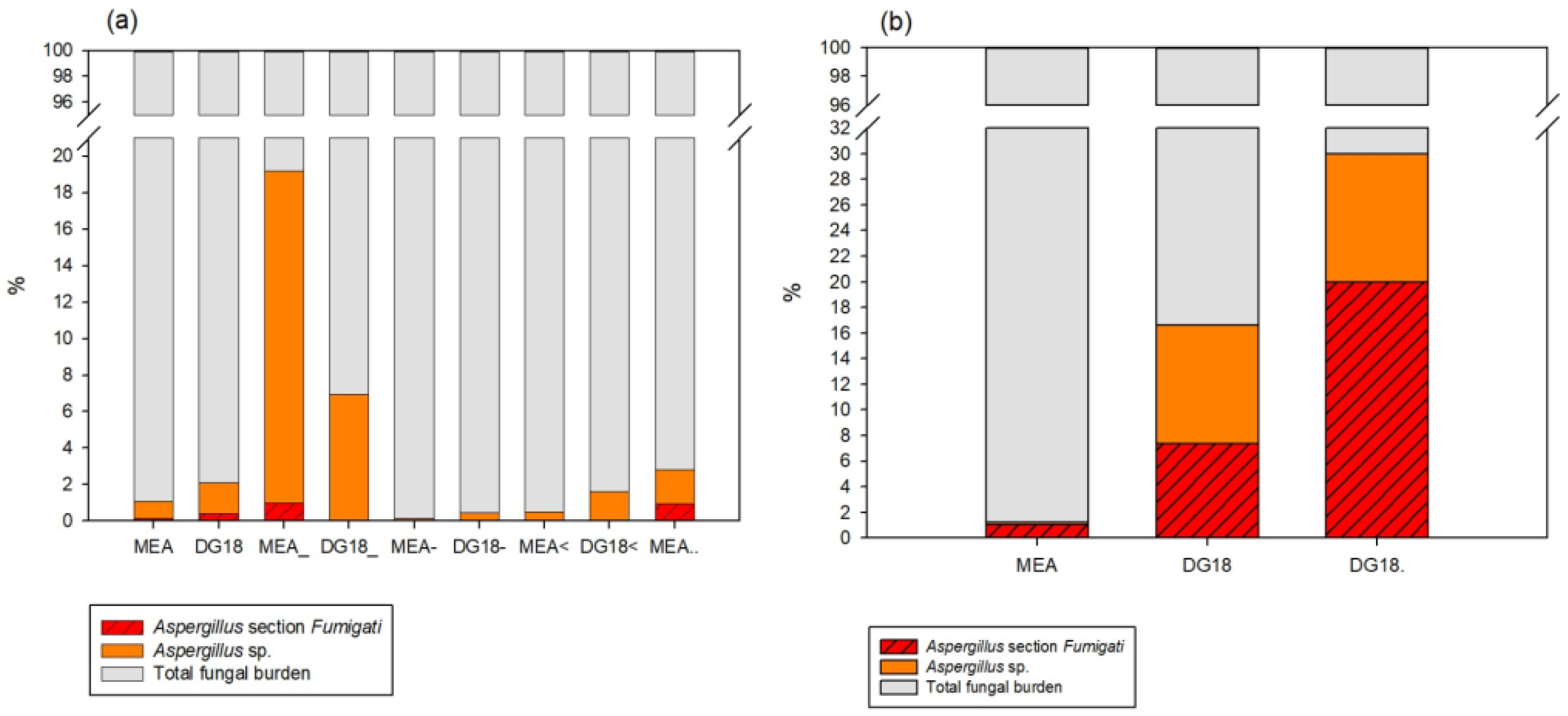
Aspergillus section Fumigati was identified in all but one type of environmental sample (HVAC filters) in the PHCC studied. In the air impaction samples, this section was identified in both MEA and DG18, where it represented 0.13% and 0.41% of the total fungal burden, respectively. In all other types of environmental samples, Aspergillus section Fumigati was only detected in MEA, representing 0.01% of the total fungal burden in the EDCs, 1% in the surface swabs, and 0.94% in the settled dust samples. In the Central Hospital (CH), Aspergillus sp. was only identified in air impaction and settled dust samples. In the air impaction samples, Aspergillus section Fumigati was the most common section both in MEA (1.05%) and in DG18 (7.35%). The same trend was found on settled dust, where Aspergillus section Fumigati was the most common (20% of the total) (Figure 1).

3.2. Aspergillus Section Fumigati Cytotoxicity Effect

The cytotoxicity evaluation was obtained from 40 out of the 57 Aspergillus section Fumigati isolates, using the MTT assay. The lowest concentration of the isolates causing a drop in absorption to <50% of cell division activity was considered the threshold toxicity level. The overview of the results is shown in Table 3. The IC50 ranged from 0.7625 mm2/mL to 0.122 cm2/mL in A549 cells, and from 3.050 mm2/mL to 3.906 cm2/mL in SK cells.
A semi-quantitative scale for cytotoxicity grading was used (adapted from [30]): medium cytotoxic effect for IC50 values ranging from 3.906 cm2/mL to 0.977 cm2/mL; high cytotoxic effect for IC50 values ranging from 0.488 cm2/mL to 0.7625 mm2/mL. The results are depicted in Table 4. Cytotoxicity was confirmed in all Aspergillus section Fumigati isolates, with high cytotoxicity observed in 100% of cases in A549 lung epithelial cells, and in 95% of cases in SK cells. Similar results were obtained with isolates from nasal swabbing of workers and controls.
Of note, 17 Aspergillus section Fumigati isolates (88.2% from environmental sampling) were able to grow in at least one azole (4 mg/L ITR), including 7 isolates from environmental samples (mostly air impaction) that were able to grow in two different azoles (4 mg/L ITR, and 1 mg/L VOR), of which 4 were able to grow in the three tested azoles (4 mg/L ITR, 1 mg/L VOR, and 0.5 mg/L POS) [5,19,27,28]. Regarding nasal samples, two Aspergillus section Fumigati isolates were able to grow in 4 mg/L ITR, one of which was from PHCC staff, and the other from controls.

3.3. Correlation and Comparison Analysis

No significant correlation of IC50 levels was detected between SK and A549 cells (rS = 0.212, p = 0.209). Statistically significant differences in IC50 levels were detected between SK and A549 cells (z = −4.982, p < 0.001), with IC50 being significantly lower in A549 cells. The Wilcoxon test also revealed statistically significant differences of IC50 levels among the two cell types in different media (z = −3.413, p = 0.001 and z = −3.834, p < 0.001, in MEA and in DG18, respectively), with IC50 levels being significantly lower for both media in A549 cells (Table 5). Between MEA and DG18, no statistically significant differences in IC50 levels were detected in SK or A549 cells (U = 158, p = 0.705, and U = 156, p = 0.658, respectively) (Table 6). These results suggest a higher cytotoxicity effect in A549 cells, with no influence of the culture media used.
The Chi-Square test detected one significant association (p = 0.035, 95% C.I. = (0.031, 0.039)) between nongrowth in ITR and growth in MEA and DG18, with Aspergillus section Fumigati presenting a greater predisposition to nongrowth in ITR (Table 7). These results indicate that the three media analyzed (MEA, DG18, and SDA) present a significant variability with respect to fungal growth in ITR.
Comparing the cytotoxicity effect (IC50 levels in SK and A459 cells) with Aspergillus section Fumigati growth (No/Yes) on azole-supplemented media, no statistically significant differences were detected in any of the media (p > 0.05) (Table 8). These results suggest that cytotoxicity had no relation with Aspergillus section Fumigati’s ability to grow on azole-supplemented media. Despite not being significant, it was found that IC50 was lower (meaning higher cytotoxicity) in both cells when isolates were not able to grow in ITR or when they were able to grow in POS. Regarding VOR, IC50 was lower in SK cells for isolates grown in VOR, and in A549 cells when there was no growth in VOR (Table 8).
On the basis of the comparison of IC50 levels for individual isolates of the fungi, there were no statistically significant differences between IC50 levels of the analyzed samples (air impaction, nasal swab (PHCC) and nasal swab (control) for either SK ( χ K W 2 ( 2 ) = 1.454, p = 0.483) or A459 ( χ K W 2 ( 2 ) = 0 .514, p = 0.773)) cells. Therefore, it was not possible to clearly determine which of the compared sample type displays had a more cytotoxicity effect than others.

4. Discussion

In the present study, we exposed human lung epithelial A549 cells and swine kidney (SK) cells to Aspergillus section Fumigati isolates from the HCE and health care workers and found that all Aspergillus section Fumigati isolates tested reduced cell viability, presenting a medium to high cytotoxicity effect in culture. Human lung epithelial cells were used as a model for exposure by inhalation [31], and swine kidney cells as a model for mammal nephrotoxicity [32], considering the reported nephrotoxicity of some Aspergillus section Fumigati toxins [24].
Furthermore, we performed correlational statistical analysis and detected a higher cytotoxic effect in A549 cells, regardless the culture media used. This is particularly concerning regarding the cytotoxicity effect of Aspergillus section Fumigati isolates from air impaction samples and from nasal swabs of PHCC workers and controls, suggesting the inhalation route as a risk factor, especially for individuals suffering from asthma [33] and immunocompromised individuals [9,34,35].
Indeed, cytotoxic toxins of Aspergillus section Fumigati act on different cells to induce cell death. The cytotoxicity and apoptotic effects of gliotoxin, the main secondary metabolite of Aspergillus section Fumigati have been reported in macrophages [22,24]. Trypacidin, another toxin from Aspergillus section Fumigati, was also reported to have a cytotoxicity effect on lung cells [23]. Other studies using lung epithelial cells to address the association between Fumigati conidia and airways colonization revealed contradictory results on the pro-inflammatory effect [36,37].
Very few studies focus on the cytotoxicity of fungi from environments occupied by humans, namely, in HCE. Most available studies focus on the cytotoxicity of Penicillium sp., Aspergillus sp. or Stachybotrys sp. genera, mostly recovered from dwellings with infiltrations and humid environments [38,39,40,41,42], from occupational settings [43,44], or even from protection devices used in high fungal load settings such as the waste sorting industry [45,46,47]. One study evaluating the concentration of airborne fungi in rooms of asthma patients concluded that the home environment was a potential source of exposure to molds and a risk factor for asthma patients [33]. A previous study revealed that 47% of the evaluated airborne fungi, collected from humid apartments in Scotland, displayed cytotoxicity in vivo [38]. Other in vitro studies refer to the cytotoxicity of building materials as related to their contamination by molds and mycotoxins [48].
Regarding the cytotoxicity of the Aspergillus genera, a study by Gniadek et al. reported a low cytotoxicity effect of airborne Aspergillus section Flavi recovered from hospital rooms and tracheostomy tubes [49]. A previous study comparing the cytotoxicity of indoor molds, by means of the MTT assay, concluded that IC50 for Aspergillus section Fumigati spores was higher than for Aspergillus section Nigri spores [40], whereas several other studies describe that Aspergillus section Fumigati present the highest cytotoxicity among Aspergillus species [39,42], including one study in a hospital environment [50].
Aspergillus section Fumigati was more prevalent in DG18 (compared to MEA), thus, supporting the use of more than one culture media in HCE assessments. Indeed, DG18 restricts the size of fungal colonies with higher growth rates that often hinder Aspergillus sp. [51]. Therefore, a more accurate characterization of the contamination by this Aspergillus section should be considered [52].
The high prevalence of section Fumigati obtained by air impaction also highlights the risk of exposure by inhalation of airborne spores [3]. It is known that, when suspended in the air, the 2–3 μm conidia of this Aspergillus section can reach deeply inside the respiratory system (alveoli or the sinuses) after inhalation [53]. Previous work on health care units evaluated the air quality of indoor hospital environments, namely, in adult and new-born intensive care units, as well as surrounding areas such as corridors and hallways, concluding that fungal spores’ contamination was within limits (750 CFU.m−3) according to current norms [54]. Other studies, however, refer to fungal contamination in hospital room’s frequently exceeding limits [5,27,28,55,56,57]. In the enlarged project where the isolates from this study were recovered, the quantification limit complied with the Portuguese legislation in most of the HCE assessed (I/O < 1), which is the cut off to avoid fungal species identification. However, a deeper analysis enabled the identification of harmful fungal species (including section Fumigati among others), which are indicators for corrective measure implementation in the same Portuguese legal framework [27].
Besides fungal quantification suggested in most guidelines and legislation, the identification of toxigenic fungal species is also important for risk assessment [27,28]. Performing regular fungal assessments, targeting for Aspergillus section Fumigati, may help to unveil contamination sources at HCE [5,58]. Moreover, the sampling approach should comprise both active (air) and passive sampling methods and be adjusted to the identified contamination sources, contextual information, and variability of the exposure [52].
The quality guarantee of the HCE is aligned with the Sustainable Development Goals (SDGs), to ensure healthy lives and promote well-being for all at all ages (Goal 3) [59]. Studies held in European hospitals [60] reported that nosocomial infections significantly increase morbidity and mortality rates, with most of these infections being transmitted by airborne pathogens [61]. These specific indoor environments also present a high risk of cross infection between staff and patients. Additionally, fungal contamination in the air and on hospital surfaces has been associated with the number of fungal infections in hospitalized immunocompromised patients [9,34]. Monitoring and control of microbial contamination in HCE is, therefore, mandatory as it is crucial to prevent and control hospital-acquired infections [62,63]. Notably, several Aspergillus section Fumigati isolates from the environmental sampling were also able to grow in at least one azole, including isolates from air samples able to grow in two or more different azoles. This might be particularly critical in the HCE, where patients, visitors, and staff might be exposed, and in particular more susceptible populations, such as immunocompromised individuals [10,15,35]. Azole resistance must be confirmed in future studies, through antifungal susceptibility testing, for a more precise characterization of the relation between cytotoxicity and azole resistance of Aspergillus section Fumigati isolates collected in the environment of healthcare facilities. Unfortunately, the section Fumigati was classified based on macroscopic and microscopic characteristics and because of that it was impossible to identify the species among the section. However, in previous studies held by the same team also with environmental isolates from different indoor environments, results revealed a good correlation between phenotypic and molecular identification [19,64]. Further studies should comprehend molecular identification of Aspergillus isolates and cytotoxicity analyses.
5. Conclusions

In conclusion, Aspergillus section Fumigati was found to be a prevailing species in the assessed health care facilities and in nasal swab samples from health care workers. The presence of cytotoxic and azole resistant Aspergillus section Fumigati isolates in the HCE environment poses an additional risk for patients and health care workers, especially for immunocompromised individuals. The epidemiology and clinical relevance of this species should continue to be addressed, as a reduced susceptibility to azoles—some of the most used antifungals available—may lead to therapeutic failure in the treatment of fungal infections such as invasive aspergillosis. More studies on this topic are necessary to link Aspergillus section Fumigati cytotoxicity with nosocomial aspergillosis.

Author Contributions

Conceptualization, C.V., M.T. and L.A.C.; methodology, C.V., M.T. and L.A.C.; formal analysis, B.A., M.D., E.R., E.C. and E.S.; investigation, C.V., M.T. and L.A.C.; resources, C.V. and M.T.; writing—original draft preparation, C.V., M.T., B.A., M.D., E.R., E.C., E.S and L.A.C.; writing—review and editing, C.V., M.T. and L.A.C.; supervision, C.V., M.T. and L.A.C.; project administration, C.V. and M.T.; funding acquisition, C.V. and M.T. All authors have read and agreed to the published version of the manuscript.

Funding

It was co-supported by FCT—Fundação para Ciência e Tecnologia for funding the project EXPOsE—Establishing protocols to assess occupational exposure to microbiota in clinical settings (02/SAICT/2016—Project no 23222), by the Instituto Politécnico de Lisboa, Lisbon, Portugal for funding the Project “Occupational exposure of ambulance drivers to bioburden” (IPL/2020/BIO-AmbuDrivers_ESTeSL) and by the Polish Minister of Science and Higher Education, under the program “Regional Initiative of Excellence” in 2019–2022 (Grant No. 008/RID/2018/19).

Institutional Review Board Statement

The study was conducted according to the guidelines of the Declaration of Helsinki, and approved by the Ethics Committee of Escola Superior de Tecnologia da Saúde de Lisboa (ref: CE-ESTeSL-No 45-2018).

Informed Consent Statement

Informed consent was obtained from all subjects involved in the study.

Acknowledgments

H&TRC authors gratefully acknowledge the FCT/MCTES national support through the UIDB/05608/2020 and UIDP/05608/2020.

Conflicts of Interest

The authors declare no conflict of interest.

References

  1. Denning, D.W. Invasive aspergillosis. Clin. Infect. Dis. 1998, 26, 781–803. [Google Scholar] [CrossRef]
  2. LIFE. Available online: http://www.life-worldwide.org/ (accessed on 1 September 2021).
  3. Weber, D.J.; Peppercorn, A.; Miller, M.B.; Sickbert-Benett, E.; Rutala, W.A. Preventing healthcare-associated Aspergillus infections: Review of recent CDC/HICPAC recommendations. Med. Mycol. 2009, 47, 199–209. [Google Scholar] [CrossRef] [Green Version]
  4. Tashiro, T.; Izumikawa, K.; Tashiro, M.; Takazono, T.; Morinaga, Y.; Yamamoto, K.; Imamura, Y.; Miyazaki, T.; Seki, M.; Kakeya, H.; et al. Diagnostic significance of Aspergillus species isolated from respiratory samples in an adult pneumology ward. Med. Mycol. 2011, 49, 1–7. [Google Scholar] [CrossRef] [PubMed] [Green Version]
  5. Viegas, C.; Almeida, B.; Gomes, A.Q.; Carolino, E.; Caetano, L.A. Aspergillus spp. prevalence in Primary Health Care Centres: Assessment by a novel multi-approach sampling protocol. Environ. Res. 2019, 175, 133–141. [Google Scholar] [CrossRef] [PubMed]
  6. Houbraken, J.; de Vries, R.P.; Samson, R.A. Modern Taxonomy of Biotechnologically Important Aspergillus and Penicillium Species. Adv. Appl. Microbiol. 2014, 86, 199–249. [Google Scholar]
  7. Hubka, V.; Nováková, A.; Kolařík, M.; Jurjević, Ž.; Peterson, S.W. Revision of Aspergillus section Flavipedes: Seven new species and proposal of section Jani sect. nov. Mycologia 2015, 107, 169–208. [Google Scholar] [CrossRef]
  8. Warris, A.; Voss, A.; Verweij, P.E. Hospital sources of Aspergillus species: New routes of transmission? Rev. Iberoam. Micol. 2001, 18, 156–162. [Google Scholar]
  9. Sabino, R. Exposure to Fungi in Health Care Facilities. In Encyclopedia of Mycology; Elsevier: Amsterdam, The Netherlands, 2021; pp. 1–10. [Google Scholar]
  10. Verweij, P.E.; Chowdhary, A.; Melchers, W.J.G.; Meis, J.F. Azole resistance in Aspergillus fumigatus: Can we retain the clinical use of mold-active antifungal azoles? Clin. Infect. Dis. 2016, 62, 362–368. [Google Scholar] [CrossRef] [PubMed] [Green Version]
  11. Hubka, V.; Dudová, Z.; Kubátová, A.; Frisvad, J.C.; Yaguchi, T.; Horie, Y.; Jurjević, Ž.; Hong, S.-B.; Kolařík, M. Taxonomic novelties in Aspergillus section Fumigati: A. tasmanicus sp. nov., induction of sexual state in A. turcosus and overview of related species. Plant Syst. Evol. 2017, 303, 787–806. [Google Scholar] [CrossRef]
  12. Anyanwu, E.; Campbell, A.W.; Jones, J.; Ehiri, J.E.; Akpan, A.I. The Neurological Significance of Abnormal Natural Killer Cell Activity in Chronic Toxigenic Mold Exposures. Sci. World J. 2003, 3, 1128–1137. [Google Scholar] [CrossRef] [Green Version]
  13. Sutton, P.; Newcombe, N.R.; Waring, P.; Müllbacher, A. In vivo immunosuppressive activity of gliotoxin, a metabolite produced by human pathogenic fungi. Infect. Immun. 1994, 62, 1192–1198. [Google Scholar] [CrossRef] [Green Version]
  14. Cramer, R.A.; Rivera, A.; Hohl, T.M. Immune responses against Aspergillus fumigatus. Curr. Opin. Infect. Dis. 2011, 24, 315–322. [Google Scholar] [CrossRef] [PubMed] [Green Version]
  15. Nature Microbiology Stop neglecting fungi. Nat. Microbiol. 2017, 2, 17120. [CrossRef] [Green Version]
  16. Jeanvoine, A.; Rocchi, S.; Reboux, G.; Crini, N.; Crini, G.; Millon, L. Azole-resistant Aspergillus fumigatus in sawmills of Eastern France. J. Appl. Microbiol. 2017, 123, 172–184. [Google Scholar] [CrossRef] [PubMed]
  17. Vaezi, A.; Fakhim, H.; Javidnia, J.; Khodavaisy, S.; Abtahian, Z.; Vojoodi, M.; Nourbakhsh, F.; Badali, H. Pesticide behavior in paddy fields and development of azole-resistant Aspergillus fumigatus: Should we be concerned? J. Mycol. Med. 2018, 28, 59–64. [Google Scholar] [CrossRef]
  18. Viegas, C.; Almeida, B.; Aranha Caetano, L.; Afanou, A.; Straumfors, A.; Veríssimo, C.; Gonçalves, P.; Sabino, R. Algorithm to assess the presence of Aspergillus fumigatus resistant strains: The case of Norwegian sawmills. Int. J. Environ. Health Res. 2020, 19, 1–9. [Google Scholar] [CrossRef] [PubMed]
  19. Gonçalves, P.; Melo, A.; Dias, M.; Almeida, B.; Caetano, L.A.; Veríssimo, C.; Viegas, C.; Sabino, R. Azole-Resistant Aspergillus fumigatus Harboring the TR34/L98H Mutation: First Report in Portugal in Environmental Samples. Microorganisms 2020, 9, 57. [Google Scholar] [CrossRef]
  20. Watanabe, A.; Kamei, K.; Sekine, T.; Higurashi, H.; Ochiai, E.; Hashimoto, Y.; Nishimura, K. Cytotoxic Substances from Aspergillus fumigatus in Oxygenated or Poorly Oxygenated Environment. Mycopathologia 2004, 158, 1–7. [Google Scholar] [CrossRef]
  21. Varga, J.; Baranyi, N.; Chandrasekaran, M.; Vágvölgyi, C.; Kocsubé, S. Mycotoxin producers in the Aspergillus genus: An update. Acta Biol. Szeged. 2015, 59, 151–167. [Google Scholar]
  22. Malekinejad, H.; Fani, F.; Shafie-Irannejad, V.; Fink-Gremmel, F. Aspergillus fumigatus toxins cause cytotoxic and apoptotic effects on human T lymphocytes (Jurkat cells). World Mycotoxin J. 2013, 6, 65–71. [Google Scholar] [CrossRef]
  23. Gauthier, T.; Wang, X.; Sifuentes Dos Santos, J.; Fysikopoulos, A.; Tadrist, S.; Canlet, C.; Artigot, M.P.; Loiseau, N.; Oswald, I.P.; Puel, O. Trypacidin, Spore-Borne Toxin from Aspergillus fumigatus, Is Cytotoxic to Lung Cells. PLoS ONE 2012, 7, e29906. [Google Scholar] [CrossRef] [PubMed]
  24. Brown, R.; Priest, E.; Naglik, J.R.; Richardson, J.P. Fungal Toxins and Host Immune Responses. Front. Microbiol. 2021, 12. [Google Scholar] [CrossRef] [PubMed]
  25. Kushima, H.; Tsunoda, T.; Matsumoto, T.; Kinoshita, Y.; Izumikawa, K.; Shirasawa, S.; Fujita, M.; Ishii, H. Effects of Aspergillus fumigatus Conidia on Apoptosis and Proliferation in an In Vitro Model of the Lung Microenvironment. Microorganisms 2021, 9, 1435. [Google Scholar] [CrossRef]
  26. Law no 58/2019. Diário da República n.o 151/2019; Série I 2019-08-08; Assembleia da República: Lisbon, Portugal, 2019; pp. 3–40.
  27. Viegas, C.; Almeida, B.; Monteiro, A.; Caetano, L.A.; Carolino, E.; Gomes, A.Q.; Twarużek, M.; Kosicki, R.; Marchand, G.; Viegas, S. Bioburden in health care centers: Is the compliance with Portuguese legislation enough to prevent and control infection? Build. Environ. 2019, 160, 106226. [Google Scholar] [CrossRef]
  28. Viegas, C.; Almeida, B.; Monteiro, A.; Paciência, I.; Rufo, J.; Aguiar, L.; Lage, B.; Diogo Gonçalves, L.M.; Caetano, L.A.; Carolino, E.; et al. Exposure assessment in one central hospital: A multi-approach protocol to achieve an accurate risk characterization. Environ. Res. 2020, 181, 108947. [Google Scholar] [CrossRef]
  29. De Hoog, G.S. Atlas of Clinical Fungi, 2nd ed.; Amer Society for Microbiology: Washington, DC, USA, 2000; ISBN 9789070351434. [Google Scholar]
  30. Gniadek, A.; Krzyściak, P.; Twarużek, M.; Macura, A. Occurrence of fungi and cytotoxicity of the species: Aspergillus ochraceus, Aspergillus niger and Aspergillus flavus isolated from the air of hospital wards. Int. J. Occup. Med. Environ. Health 2017, 30, 231–239. [Google Scholar] [CrossRef]
  31. Swain, R.J.; Kemp, S.J.; Goldstraw, P.; Tetley, T.D.; Stevens, M.M. Assessment of Cell Line Models of Primary Human Cells by Raman Spectral Phenotyping. Biophys. J. 2010, 98, 1703–1711. [Google Scholar] [CrossRef] [Green Version]
  32. Heussner, A.; Bingle, L. Comparative Ochratoxin Toxicity: A Review of the Available Data. Toxins 2015, 7, 4253–4282. [Google Scholar] [CrossRef] [Green Version]
  33. Fairs, A.; Agbetile, J.; Bourne, M.; Hargadon, B.; Monteiro, W.R.; Morley, J.P.; Edwards, R.E.; Wardlaw, A.J.; Pashley, C.H. Isolation of Aspergillus fumigatus from sputum is associated with elevated airborne levels in homes of patients with asthma. Indoor Air 2013, 23, 275–284. [Google Scholar] [CrossRef]
  34. Kanamori, H.; Rutala, W.A.; Sickbert-Bennett, E.E.; Weber, D.J. Review of Fungal Outbreaks and Infection Prevention in Healthcare Settings During Construction and Renovation. Clin. Infect. Dis. 2015, 61, 433–444. [Google Scholar] [CrossRef]
  35. Brambilla, A.; Buffoli, M.; Capolongo, S. Measuring hospital qualities. A preliminary investigation on Health Impact Assessment possibilities for evaluating complex buildings. Acta Biomed. 2019, 90, 54. [Google Scholar]
  36. Berkova, N.; Lair-Fulleringer, S.; Féménia, F.; Huet, D.; Wagner, M.-C.; Gorna, K.; Tournier, F.; Ibrahim-Granet, O.; Guillot, J.; Chermette, R.; et al. Aspergillus fumigatus conidia inhibit tumour necrosis factor-or staurosporine-induced apoptosis in epithelial cells. Int. Immunol. 2006, 18, 139–150. [Google Scholar] [CrossRef] [PubMed] [Green Version]
  37. Beisswenger, C.; Hess, C.; Bals, R. Aspergillus fumigatus conidia induce interferon-signalling in respiratory epithelial cells. Eur. Respir. J. 2012, 39, 411–418. [Google Scholar] [CrossRef] [PubMed] [Green Version]
  38. Smith, J.E.; Anderson, J.G.; Lewis, C.W.; Murad, Y.M. Cytotoxic fungal spores in the indoor atmosphere of the damp domestic environment. FEMS Microbiol. Lett. 1992, 100, 337–343. [Google Scholar] [CrossRef]
  39. Kamei, K.; Watanabe, A.; Nishimura, K.; Miyaji, M. Cytotoxicity of Aspergillus fumigatus Culture Filtrate Against Macrophages. J-STAGE 2002, 43, 37–41. [Google Scholar] [CrossRef] [Green Version]
  40. Schulz, T.; Senkpiel, K.; Ohgke, H. Comparison of the toxicity of reference mycotoxins and spore extracts of common indoor moulds. Int. J. Hyg. Environ. Health 2004, 207, 267–277. [Google Scholar] [CrossRef]
  41. Huttunen, K.; Rintala, H.; Hirvonen, M.-R.; Vepsäläinen, A.; Hyvärinen, A.; Meklin, T.; Toivola, M.; Nevalainen, A. Indoor air particles and bioaerosols before and after renovation of moisture-damaged buildings: The effect on biological activity and microbial flora. Environ. Res. 2008, 107, 291–298. [Google Scholar] [CrossRef]
  42. Slesiona, S.; Gressler, M.; Mihlan, M.; Zaehle, C.; Schaller, M.; Barz, D.; Hube, B.; Jacobsen, I.D.; Brock, M. Persistence versus Escape: Aspergillus terreus and Aspergillus fumigatus Employ Different Strategies during Interactions with Macrophages. PLoS ONE 2012, 7, e31223. [Google Scholar] [CrossRef] [PubMed] [Green Version]
  43. Gutarowska, B.; Skóra, J.; Stępień, L.; Twarużek, M.; Błajet-Kosicka, A.; Otlewska, A.; Grajewski, J. Estimation of fungal contamination and mycotoxin production at workplaces in composting plants, tanneries, archives and libraries. World Mycotoxin J. 2014, 7, 345–355. [Google Scholar] [CrossRef]
  44. Viegas, S.; Caetano, L.; Korkalainen, M.; Faria, T.; Pacífico, C.; Carolino, E.; Quintal Gomes, A.; Viegas, C. Cytotoxic and Inflammatory Potential of Air Samples from Occupational Settings with Exposure to Organic Dust. Toxics 2017, 5, 8. [Google Scholar] [CrossRef] [Green Version]
  45. Viegas, C.; Twarużek, M.; Dias, M.; Almeida, B.; Carolino, E.; Kosicki, R.; Soszczyńska, E.; Grajewski, J.; Caetano, L.A.; Viegas, S. Assessment of the microbial contamination of mechanical protection gloves used on waste sorting industry: A contribution for the risk characterization. Environ. Res. 2020, 189, 109881. [Google Scholar] [CrossRef] [PubMed]
  46. Viegas, C.; Twarużek, M.; Dias, M.; Almeida, B.; Carolino, E.; Soszczyńska, E.; Ałtyn, I.; Viegas, S.; Caetano, L.A. Cytotoxic effect of filtering respiratory protective devices from the waste sorting industry: Is in vitro toxicology useful for risk characterization? Environ. Res. 2020, 191, 110134. [Google Scholar] [CrossRef]
  47. Viegas, C.; Twarużek, M.; Dias, M.; Almeida, B.; Carolino, E.; Soszczyńska, E.; Viegas, S.; Aranha Caetano, L. Cytotoxicity of filtering respiratory protective devices from the waste sorting industry: A comparative study between interior layer and exhalation valve. Environ. Int. 2021, 155, 106603. [Google Scholar] [CrossRef] [PubMed]
  48. Gutarowska, B. Hyphate Fungi in Building Materials. In Increase and Production of Mycotoxins and Allergens; Lodz University of Technology Press: Lodz, Poland, 2010. [Google Scholar]
  49. Gniadek, A.; Krzyściak, P.; Hawryszuk, A.; Macura, A.B.; Brzostek, T.; Składzień, J. Mycobiota of the air in hospital rooms and the fungal colonisation of tracheostomy tubes used by patients diagnosed with larynx cancer-preliminary research. Ann. Parasitol. 2013, 59, 67–71. [Google Scholar]
  50. Gniadek, A.; Macura, A.B.; Górkiewicz, M. Cytotoxicity of Aspergillus fungi isolated from hospital environment. Polish J. Microbiol. 2011, 60, 59–63. [Google Scholar] [CrossRef]
  51. Viegas, C.; Twarużek, M.; Lourenço, R.; Dias, M.; Almeida, B.; Caetano, L.A.; Carolino, E.; Gomes, A.Q.; Kosicki, R.; Soszczyńska, E.; et al. Bioburden Assessment by Passive Methods on a Clinical Pathology Service in One Central Hospital from Lisbon: What Can it Tell Us Regarding Patients and Staff Exposure? Atmosphere 2020, 11, 351. [Google Scholar] [CrossRef] [Green Version]
  52. Viegas, C.; Caetano, L.A.; Viegas, S. Occupational exposure to Aspergillus section Fumigati: Tackling the knowledge gap in Portugal. Environ. Res. 2021, 194, 110674. [Google Scholar] [CrossRef]
  53. Dagenais, T.R.T.; Keller, N.P. Pathogenesis of Aspergillus fumigatus in Invasive Aspergillosis. Clin. Microbiol. Rev. 2009, 22, 447–465. [Google Scholar] [CrossRef] [Green Version]
  54. Macedo, J.I.; Kubota, T.H.; Matsumoto, L.S.; Giordani, A.T.; Takayanagui, A.M.M.; Mendes, A.A.; Bertolini, D.A. Air quality in a hospital environment. WIT Trans. Built. Environ. 2013, 134, 737–747. [Google Scholar]
  55. Falvey, D.G.; Streifel, A.J. Ten-year air sample analysis of Aspergillus prevalence in a university hospital. J. Hosp. Infect. 2007, 67, 35–41. [Google Scholar] [CrossRef]
  56. Krajewska-Kułak, E.; Łukaszuk, C.; Hatzopulu, A.; Bousmoukilia, S.; Terovitou, C.; Amanatidou, A.; Danilidis, D. Indoor air studies of fungi contamination at the Department of Pulmonology and Internal Medicine in Kavala Hospital in Greece. Adv. Med. Sci. 2009, 54. [Google Scholar] [CrossRef] [PubMed] [Green Version]
  57. Kim, K.Y.; Kim, Y.S.; Kim, D. Distribution Characteristics of Airborne Bacteria and Fungi in the General Hospitals of Korea. Ind. Health 2010, 48, 236–243. [Google Scholar] [CrossRef] [PubMed] [Green Version]
  58. Ruiz-Camps, I.; Aguado, J.M.; Almirante, B.; Bouza, E.; Ferrer-Barbera, C.F.; Len, O.; Lopez-Cerero, L.; Rodríguez-Tudela, J.L.; Ruiz, M.; Solé, A.; et al. Guidelines for the prevention of invasive mould diseases caused by filamentous fungi by the Spanish Society of Infectious Diseases and Clinical Microbiology (SEIMC). Clin. Microbiol. Infect. 2011, 17, 1–24. [Google Scholar] [CrossRef] [PubMed] [Green Version]
  59. United Nations, Department of Economic and Social Affairs. The 17 Goals Sustainable Development. Available online: https://sdgs.un.org/goals (accessed on 1 September 2021).
  60. Kaoutar, B.; Joly, C.; L’Hériteau, F.; Barbut, F.; Robert, J.; Denis, M.; Espinasse, F.; Merrer, J.; Doit, C.; Costa, Y.; et al. Nosocomial infections and hospital mortality: A multicentre epidemiological study. J. Hosp. Infect. 2004, 58, 268–275. [Google Scholar] [CrossRef] [PubMed]
  61. Kowalski, W. Hospital Airborne Infection Control; CRC Press: Boca Raton, FL, USA, 2011. [Google Scholar]
  62. Ferdyn-Grygierek, J. Monitoring of indoor air parameters in large museum exhibition halls with and without air-conditioning systems. Build. Environ. 2016, 107, 113–126. [Google Scholar] [CrossRef]
  63. Zahar, J.-R.; Jolivet, S.; Adam, H.; Dananché, C.; Lizon, J.; Alfandari, S.; Boulestreau, H.; Baghdadi, N.; Bay, J.-O.; Bénéteau, A.-M.; et al. Quelles mesures pour maîtriser le risque infectieux chez les patients immunodéprimés? Recommandations formalisées d’experts. J. Mycol. Med. 2017, 27, 449–456. [Google Scholar] [CrossRef] [PubMed]
  64. Simões, D.; Aranha Caetano, L.; Veríssimo, C.; Viegas, C.; Sabino, R. Aspergillus collected in specific indoor settings: Their molecular identification and susceptibility pattern. Int. J. Environ. Health Res. 2021, 31, 248–257. [Google Scholar] [CrossRef]
Figure 1. Aspergillus sp. and Aspergillus section Fumigati distribution in the PHCC samples (a) and CH samples (b) (Adapted from [5,27,28]).
Jof 07 00839 g001
Table 1. Samples collected in the HCE analyzed by culture-based methods (Adopted from [27,28]).
Air ImpactionSurface SwabsEDCSettled DustHVAC Filters
PHCC8181811012
CH120451550
Total201126961512
450
Table 2. Isolates (n) of Aspergillus section Fumigati recovered per sample collected and media applied for first isolation.
MediaSample CollectedTotal
Air ImpactionEDCSurface SwabsSettled DustVacuum BagNasal Swab (PHCC)Nasal Swab (Control)
MEAN611111617
% of total10.5%1.8%1.8%1.8%1.8%1.8%10.5%38.6%
DG18N16001081641
% of total28.1%0.0%0.0%1.8%0.0%14.0%28.1%56.1%
SDAN01011003
% of total0.0%1.8%0.0%1.8%1.8%0.0%0.0%5.3%
Table 3. Distribution of threshold toxicity level (IC50) of Aspergillus section Fumigati isolates.
Dilution StepIC50A549SK
NN
130.7625 mm2/mL40
121.525 mm2/mL90
113.050 mm2/mL106
100.061 cm2/mL1410
90.122 cm2/mL314
80.244 cm2/mL03
70.488 cm2/mL05
60.977 cm2/mL01
43.906 cm2/mL01
N, number of Aspergillus section Fumigati isolates toxic for A549 or SK cells.
Table 4. Level of cytotoxicity of the Aspergillus section Fumigati isolates.
Aspergillus Section Fumigati
Isolates per Sampling
Isolates with Level of Toxicity n (%)
MediumHigh
A549SKA549SK
Air impaction (N = 13) 0013 (100)13 (100)
EDC (N = 1) 001 (100)1 (100)
Settled dust (N = 3) 01 (33.3)3 (100)2 (66.7)
Surface swabs (N = 1) 001 (100)1 (100)
Vacuum bag (N = 2) 002 (100)2 (100)
Nasal swab (control) (N = 11) 01 (9.1)11 (100)10 (90.9)
Nasal swab (PHCC) (N = 9) 009 (100)9 (100)
Table 5. Comparison of IC50 levels between SK and A549 cells from isolates first isolated on MEA and DG18. Wilcoxon test results.
RanksTest Statistics
NMean rankSum of rankszp
GlobalA549-SKNegative ranks35 a18.56649.50−4.982 d0.000 *
Positive ranks1 b16.5016.50
Ties4 c
Total40
MEA mediaA549-SKNegative ranks15 a8.00120.00−3.413 d0.001 *
Positive ranks0 b0.000.00
Ties2 c
Total17
DG18 mediaA549-SKNegative ranks19 a10.00190.00−3.834 d0.000 *
Positive ranks0 b0.000.00
Ties1 c
Total20
a. A549 < SK. b. A549 > SK. c. A549 = SK. d. Based on positive ranks. * Statistically significant differences at the 5% significance level.
Table 6. Comparison of IC50 levels in either SK or A549 cells from isolates first isolated on MEA and DG18. Mann–Whitney test results.
IC50MediaRanksTest Statistics
nMean RankSum of RanksMann–Whitney Up
SKMEA1719.71335.00158.0000.705
DG182018.40368.00
Total37
A549MEA1719.82337.00156.0000.658
DG182018.30366.00
Total37
Table 7. Relation between Aspergillus section Fumigati isolates on azole-supplemented SDA (ITR, VOR, and POS) and first isolation media (MEA, DG18, and SDA). Results of the Chi-Square test by Monte Carlo simulation.
Growth in Azole Supplemented MediaChi-Square Test by Monte Carlo Simulation
ITRTotalp95% Confidence Interval
NoYesLower BoundUpper Bound
MediaMEAn1210220.035 a,*0.0310.039
%54.5%45.5%100.0%
DG18n23932
%71.9%28.1%100,0%
SDAn033
%0.0%100.0%100.0%
Totaln352257
%61.4%38.6%100.0%
VOR
MediaMEAn175220.119 a0.1120.125
%77.3%22.7%100.0%
DG18n30232
%93.8%6.3%100.0%
SDAn213
%66.7%33.3%100.0%
Totaln49857
%86.0%14.0%100.0%
POS
MediaMEAn193220.725 a0.7160.734
%86.4%13.6%100.0%
DG18n30232
%93.8%6.3%100.0%
SDAn303
%100.0%0.0%100.0%
Totaln52557
%91.2%8.8%100.0%
a Based on 10,000 sampled tables with starting seed 2,000,000. * Significant association at the 5% significance level.
Table 8. Comparison of IC50 levels (SK and A459 cells) between growth on azoles (No/Yes) in ITR, VOR, and POS media. Mann–Whitney test results.
Supplemented MediaIC50Growth in the AzolesRanksTest Statistics
nMean RankSum of RanksMann–Whitney Up
ITRSKNo2318.15417.50141.5000.127
Yes1723.68402.50
Total40
A549No2330.68422.50146.5000.164
Yes1738.32397.50
Total40
VORSKNo3320.80686.50105.5000.713
Yes719.07133.50
Total40
A549No3319.98659.5098.5000.530
Yes722.93160.50
Total40
POSSKNo3621.04757.5052.5000.364
Yes415.6362.50
Total40
A549No3621.10759.5050.5000.315
Yes415.1360.50
Total40
Publisher’s Note: MDPI stays neutral with regard to jurisdictional claims in published maps and institutional affiliations.