Article

Four New Species of Hemileccinum (Xerocomoideae, Boletaceae) from Southwestern China

1
CAS Key Laboratory for Plant Diversity and Biogeography of East Asia, Kunming Institute of Botany, Chinese Academy of Sciences, Kunming 650201, China
2
Yunnan Key Laboratory for Fungal Diversity and Green Development, Kunming Institute of Botany, Chinese Academy of Sciences, Kunming 650201, China
3
College of Life Sciences, University of Chinese Academy of Sciences, Beijing 100049, China
*
Author to whom correspondence should be addressed.
J. Fungi 2021, 7(10), 823; https://doi.org/10.3390/jof7100823
Submission received: 27 August 2021 / Revised: 28 September 2021 / Accepted: 28 September 2021 / Published: 30 September 2021

Abstract

:
The genus Hemileccinum belongs to the subfamily Xerocomoideae of the family Boletaceae. In this study, phylogenetic inferences of Hemileccinum based on sequences of a single-locus (ITS) and a multi-locus (nrLSU, tef1-α, rpb1, rpb2) were conducted. Four new species, namely H. abidum, H. brevisporum, H. ferrugineipes and H. parvum were delimited and proposed based on morphological and molecular evidence. Descriptions and line-drawings of them were presented, as well as their comparisons to allied taxa. Our study shed new light on the recognition of the genus. The pileipellis of the species in this genus should mostly be regarded as (sub)epithelium to hyphoepithelium, because the pileipellis of most studied species here is composed of short inflated cells in the inner layer (subpellis) and filamentous hyphae in outer layer (suprapellis). The basidiospores of the studied species, including the type species, H. impolitum, have a warty surface.

1. Introduction

The genus Hemileccinum Šutara was created based on the species H. impolitum (Fr.) Šutara as the type, and H. depilatum (Redeuilh) Šutara [1]. These two species were both originally placed in the genus Boletus L. [2] and later were transferred to the genus Leccinum because of the lateral stipe stratum of the leccinoid type which is predominantly anticlinally arranged, breaking up into characteristic fascicles of hyphae ending in elements of the caulohymenium during growth of the stipe [1,3,4]. However, molecular phylogenetic analyses indicated that these two species are very distant from the species of both Leccinum and Boletus, but similar to the species of Xerocomus; thus they were accordingly transferred to Xerocomus [5,6]. Based on the previous molecular evidence and his own further morphological observations, Šutara established the genus Hemileccinum to accommodate these two species. He emphasized that Hemileccinum was diagnosed by the anatomical structure of the peripheral stipe layers having a lateral stipe stratum of the leccinoid type, which distinguished this genus not only from all the other species belonging to the Xerocomus s.l. but also from those in the genus Boletus [1]. Wu et al. confirmed the monophyly of Hemileccinum and found an additional diagnosed character of this genus, namely the irregularly warty basidiospores under SEM [7,8]. Meanwhile, the genus Corneroboletus N.K. Zeng & Zhu L. Yang was confirmed as a synonym of Hemileccinum due to the similar basidiospore ornamentation and the closely phylogenetic relationship [8].
As ectomycorrhizal fungi, species in the genus Hemileccinum are widely distributed in temperate, subtropical and tropical regions, and play an important role in forest ecology [1,8,9,10,11,12,13,14]. However, the species diversity of Hemileccinum was relatively poorly known in the world until recent studies suggested existence of other potentially new specie of the genus [8,10]. Until now, only 10 species of the genus have been reported in the world according to the database of INDEX FUNGORUM (accessed date: 27 August 2021). Among them, H. impolitum and H. depilatum are from Europe [1], H. subglabripes (Peck) Halling, H. rubropunctum (Peck) Halling and H. hortonii (A.H. Sm. & Thiers) M. Kuo & B. Ortiz are from North America [8,9]. In Asia, H. rugosum G. Wu & Zhu L. Yang is described from China, and H. indecorum (Massee) G. Wu & Zhu L. Yang is from tropical China, Singapore, Thailand [8,15,16].
During past fungal investigations in southwestern China, we encountered four potential new species of Hemileccinum. Our aim in this study is to clarify their molecular phylogenetic positions and to delimit them based on morphological data and molecular evidence.

2. Materials and Methods

2.1. Sample Collection and Morphological Study

In total, seventeen collections were examined in this study, which were collected from the Yunnan Province of southwestern China during the years 2007–2017 (Figure 1). The macroscopic characters of the specimens were described based on fresh basidiomata, and the dried specimens were deposited in the Cryptogamic Herbarium of the Kunming Institute of Botany, Chinese Academy of Sciences (KUN-HKAS). Color codes of the form “5C4” indicate the plate, row, and color block from Kornerup and Wanscher [17]. For microscopic observation, a ZEISS Axiostar Plus microscope (Oberkochen, Germany) was used and the dried specimens were revived in 5% KOH solution or distilled water. Moreover, Melzer’s reagent was applied to test color reactions of the tissue fragments to the solution. Microscopic studies follow Li et al. and Zhou et al. [18,19]. In the descriptions of basidiospores, the abbreviation [n/m/p] means n basidiospores measured from m basidiomata of p collections. The range notation (a)b–c(d) stands for the dimensions of basidiospores in which b–c contains a minimum of 90% of the measured values while a and d in the brackets stand for the extreme values. Q is used to imply “length/width ratio” of a basidiospore in side view; Qm means average Q of all basidiospores ± sample standard deviation. To observe basidiospore ornamentations, a ZEISS Sigma 300 scanning electron microscope (SEM) (Oberkochen, Germany) was used. Genera are abbreviated as follows: H. for Hemileccinum, Ca. for Castanopsis, C. for Castanea, L. for Lithocarpus, P. for Pinus, Q. for Quercus and Rug. for Rugiboletus.

2.2. Molecular Procedures and Phylogenetic Analyses

Total genomic DNA was obtained with the Ezup Column Fungi Genomic DNA Purification Kit (Sangon Biotech, Shanghai, China) according to the manual from material dried with silica gel. A total of five nuclear loci were sequenced, including the internal transcribed spacer (ITS), the large subunit of nuclear ribosomal RNA gene (nrLSU), the polymerase II subunit one (rpb1) gene, the second largest subunit of RNA polymerase II (rpb2), and the translation elongation factor 1-α gene (tef1-α). The primer pairs of ITS1/ITS4 [20,21], LROR/LR5 [22,23] were used for amplifying ITS, nrLSU, respectively. The primer pairs used for amplifying the rpb1, rpb2, tef1-α, followed those in Wu et al. [7]. PCR was performed in a total volume of 25 μL containing 1 μL forward primer, 1 μL reverse primer, 9.5 μL nuclease-free H2O, 12.5 μL BlasTaqTM 2×PCR MasterMix (abm, Richmond, VA, Canada) and 1 μL DNA template. PCR protocol was as follows: pre-denaturation at 95 °C for 5 min, followed by 35 cycles of denaturation at 95 °C for 60 s, 52 °C for 60 s, and 72 °C for 80 s, and then a final elongation at 72 °C for 8 min was included. The PCR products were purified with a Gel Extraction and PCR Purification Combo Kit (Spin-column) (Bioteke, Beijing, China), and then sequenced by ABI-3730-XL DNA Analyzer (Applied Biosystems, Foster City, CA, USA) by using the same primer pairs as in the PCR amplification for sequencing.

2.3. Phylogenetic Analyses

We used BLAST to compare the obtained sequences of the newly collected materials with those in the GenBank database. The BLAST results were used to predict the phylogenetic relationship between the newly collected specimens and known species and indicated that the new materials were genetically similar to the other species of Hemileccinum. In this study, two datasets were produced, the ITS dataset, and the combined nrLSU, tef1-α, rpb1 and rpb2 dataset. The ITS sequences of Hemileccinum species from China were used to infer relationships of Chinese species with those from Europe, North America and East Asia. In the analysis of ITS dataset, Phylloporus rubrosquamosus N.K. Zeng, Zhu L. Yang & L.P. Tang, Phylloporus rubeolus N.K. Zeng, Zhu L. Yang & L.P. Tang, Hourangia cheoi (W.F. Chiu) Xue T. Zhu & Zhu L. Yang and Hourangia pumila (M.A. Neves & Halling) Xue T. Zhu, Halling & Zhu L. Yang were chosen as outgroup [24,25,26]. The combined dataset was mainly used to infer phylogenetic relationships and systematic positions of the Chinese species. In the multigene phylogenetic analysis, including all known genera in the subfamily Xerocomoideae were included. We screened the relevant sequences deposited in GenBank, which were mainly submitted by Wu et al. [7,8], Gelardi et al. [27], Zhu et al. [26], Zeng et al. [24], Neves et al. [28]. We collected a total of 13 ingroup species of 8 known genera within Xerocomoideae and 2 outgroup species outside Xerocomoideae but in the Boletaceae. Detailed information of the voucher specimens is given in Table 1.
The sequences were assembled with SeqMan implemented in Lasergene v7.1 (DNASTAR Inc., Madison, WI, USA), and then aligned by using MAFFT v7.310 [29]. The software Bioedit v7.2.5 [30] was used to check aligned matrices. To assess any potential conflicts in the gene tree topologies for these five loci, single-locus matrices were analyzed using maximum likelihood (ML) in RAxML v8.0.20 [31]. Sequences of the loci without conflicts were then concatenated using Phyutility 2.2 [32,33] for further phylogenetic analyses. The best-fitted substitution model for each gene was determined through MrModeltest v2.4 [34] by using Akaike Information Criterion (AIC). GTR + I + G was inferred as the best-fit model for the nrLSU, tef1-α, rpb1 and ITS selected according to the AIC in MrModeltest v2.4 [34]. SYM + I + G was selected as the best model for rpb2. For the ultimate phylogenetic analyses, Maximum Likelihood (ML) analysis and Bayesian Inference were conducted by RAxML v8.0.20 [31] and MRBAYES v3.2.7 [35], respectively. The parameters of RAxML were set as defaults with 500 bootstrap replicates, except the substitution model which was set as GTRGAMMAI.
BI analyses were conducted with two independent runs of one cold and three heated chains. Runs were performed for 2 million generations, and trees sampled every 100 generations. The convergence was determined with the average standard deviation of split frequencies (<0.01) Chain convergence was determined using Tracer v1.5 to confirm sufficiently large ESS values (>200). The sampled trees were subsequently summarized by using the “sump” and “sumt” commands with a 25% burn-in [31,35]. The Bayesian posterior probabilities (BPP) of internodes were estimated based on the majority rule consensus with the remaining trees.
Table 1. Specimens used in phylogenetic analysis and their GenBank accession numbers. The newly generated sequences are shown in bold.
SpeciesVoucherLocalityGenBank Accession NumberReferences
ITSnrLSUrpb2rpb1tef1-α
Hemileccinum rugosumKUN-HKAS84355China-KT990578KT990413KT990931KT990774[8]
Hemileccinum rugosum KUN-HKAS84970 China-KT990577KT990412-KT990773[8]
Hemileccinum rugosum KUN-HKAS50284 China-KT990576KT990411-KT990772[8]
Hemileccinum subglabripesMICH:KUO-07230802USA-MK601738MK766300- MK721092 [10]
Hemileccinum subglabripes MICH:KUO-07070702 USA-MK601737 MK766299 -MK721091[10]
Hemileccinum subglabripesMICH:KUO-08301402USA-MK601739MK766301-MK721093[10]
Hemileccinum subglabripes72206USA-KF030303-KF030374KF030404[36]
Hemileccinum subglabripes294169USAMN128237 - ---from GenBank
Hemileccinum subglabripes3660-KM248936 - ---from GenBank
Hemileccinum depilatum2137333USAAY127032 - ---from GenBank
Hemileccinum depilatumAF2845Belgium--MG212633-MG212591[37]
Hemileccinum depilatum Bd1 --AF139712---[5]
Hemileccinum impolitumBim 1Germany- AF139715 -KF030375JQ327034[36]
Hemileccinum impolitum47698PortugalAJ419187 - ---[38]
Hemileccinum impolitumBI57407ThailandKM235997 - ---from GenBank
Hemileccinum impolitumBI57408ThailandKM235998 - ---from GenBank
Hemileccinum impolitum17173USAJF907783 - ---[39]
Hemileccinum impolitumKUN-HKAS84869Germany- KT990575 KT990410 KT990930KT990771[8]
Hemileccinum indecorumKUN-HKAS63126China- KF112440 ---[7]
Hemileccinum indecorum OR0863 Thailand--MH614772- MH614726 [16]
Hemileccinum rubropunctumJLF56666USAMH190826 - ---from GenBank
Hemileccinum rubropunctumMES256USAFJ480428 - ---[40]
Hemileccinum rubropunctum NY-792788REH-8501 USA-MK601768MK766327-MK721122[10]
Hemileccinum rubropunctumNY-01193924REH-9597USA- MK601769 MK766328- MK721123 [10]
Hemileccinum sp. KUN-HKAS53421China-KF112432KF112751KF112565KF112235[7]
Hemileccinum hortoniiMICH KUO-07050706USA-MK601821MK766377-MK721175[10]
Hemileccinum albidumKUN-HKAS87225ChinaMZ923777MZ923774MZ936317MZ936334MZ936351This study
Hemileccinum albidumKUN-HKAS83355ChinaMZ923778MZ923775MZ936321MZ936340MZ936357This study
Hemileccinum albidum (T) KUN-HKAS81120ChinaMZ923782MZ923766MZ936320MZ936339MZ936352This study
Hemileccinum albidumKUN-HKAS50503ChinaMZ923781MZ923767MZ936319MZ936335MZ936355This study
Hemileccinum albidumKUN-HKAS50350ChinaMZ923779MZ923768MZ936323MZ936342MZ936359This study
Hemileccinum albidumKUN-HKAS84554ChinaMZ923780-MZ936318MZ936336MZ936358This study
Hemileccinum albidumKUN-HKAS85753ChinaMZ923786-MZ936325MZ936337MZ936353This study
Hemileccinum albidumKUN-HKAS87105China-MZ923769MZ936327MZ936338MZ936356This study
Hemileccinum albidumKUN-HKAS83333ChinaMZ923784-MZ936326MZ936344MZ936361This study
Hemileccinum albidumKUN-HKAS83400ChinaMZ923783MZ923770MZ936324MZ936341MZ936354This study
Hemileccinum albidumKUN-HKAS115749ChinaMZ923785-MZ936322MZ936343MZ936360This study
Hemileccinum brevisporum (T) KUN-HKAS89150ChinaMZ923788MZ923764MZ936328MZ936345MZ936362This study
Hemileccinum brevisporumKUN-HKAS59445China-KT990579KT990414KT990932KT990775[8]
Hemileccinum brevisporumKUN-HKAS67896ChinaMZ923787MZ923765MZ936329MZ936346MZ936363This study
Hemileccinum ferrugineipes (T) KUN-HKAS115554ChinaMZ923792MZ923773MZ936330MZ936350MZ973011This study
Hemileccinum ferrugineipesKUN-HKAS75054China- KF112377 KF112749KF112563KF112234[7]
Hemileccinum ferrugineipesKUN-HKAS93310ChinaMZ923791-MZ936331MZ936347MZ973012This study
Hemileccinum parvumKUN-HKAS99764ChinaMZ923789MZ923771MZ936332MZ936349MZ973009This study
Hemileccinum parvum (T) KUN-HKAS115553ChinaMZ923790MZ923772MZ936333MZ936348MZ973010This study
Heimioporus sp. KUN-HKAS53451 China-KF112345KF112805KF112616KF112226[7]
Heimioporus aff. japonicusKUN-HKAS52236China-KF112346KF112807KF112617KF112227[7]
Heimioporus japonicasKUN-HKAS52237China-KF112347KF112806KF112618KF112228[7]
Aureoboletus tenuisKUN-HKAS75104China-KT990518KT990359KT990897KT990722[8]
Aureoboletus thibetanusKUN-HKAS76655China-KF112420KF112752KF112626KF112236[7]
Pulchroboletus roseoalbidusAMB 12757Italy-NG_060126--KJ729512[27]
Alessioporus ichnusanusAMB 12756Italy-NG_057044--KJ729513[27]
Phylloporus rubrosquamosusKUN-HKAS52552China-KF112391KF112780-KF112289[7]
Phylloporus rubrosquamosusKUN-HKAS54559ChinaNR120124NG_042668--JQ967175[24,25]
Phylloporus rubeolusKUN-HKAS52573ChinaJQ967259JQ967216--JQ967172[24,25]
Xerocomus fraternusKUN-HKAS55328China-KT990681KT990497-KT990869[8]
Xerocomus velutinusKUN-HKAS68135China-KT990673-KT991011KT990861[8]
Hourangia cheoiYang 5153ChinaKP136997KP136947KP136975KP136966KP136924[26]
Hourangia pumilaREH8063IndonesiaJQ003626NG_060636---[28]
Boletellus indistinctusKUN-HKAS77623China-KT990531KT990371 -KT990733[8]
Boletellus indistinctusKUN-HKAS80681China-KT990532KT990368KT990903KT990734[8]
Leccinum variicolorKUN-HKAS57758China-KF112445KF112725KF112591KF112251[7]
Leccinum aff. scabrumKUN-HKAS57266China -KF112442KF112722KF112590KF112248[7]
Leccinum monticolaKUN-HKAS76669China-KF112443KF112723KF112592KF112249[7]
Leccinellum cremeumKUN-HKAS90639China--KT990420KT990936KT990781[8]
Leccinellum sp. KUN-HKAS53410China-KT990585KT990421KT990937-[8]

3. Results

3.1. Molecular Phylogenetic Analysis

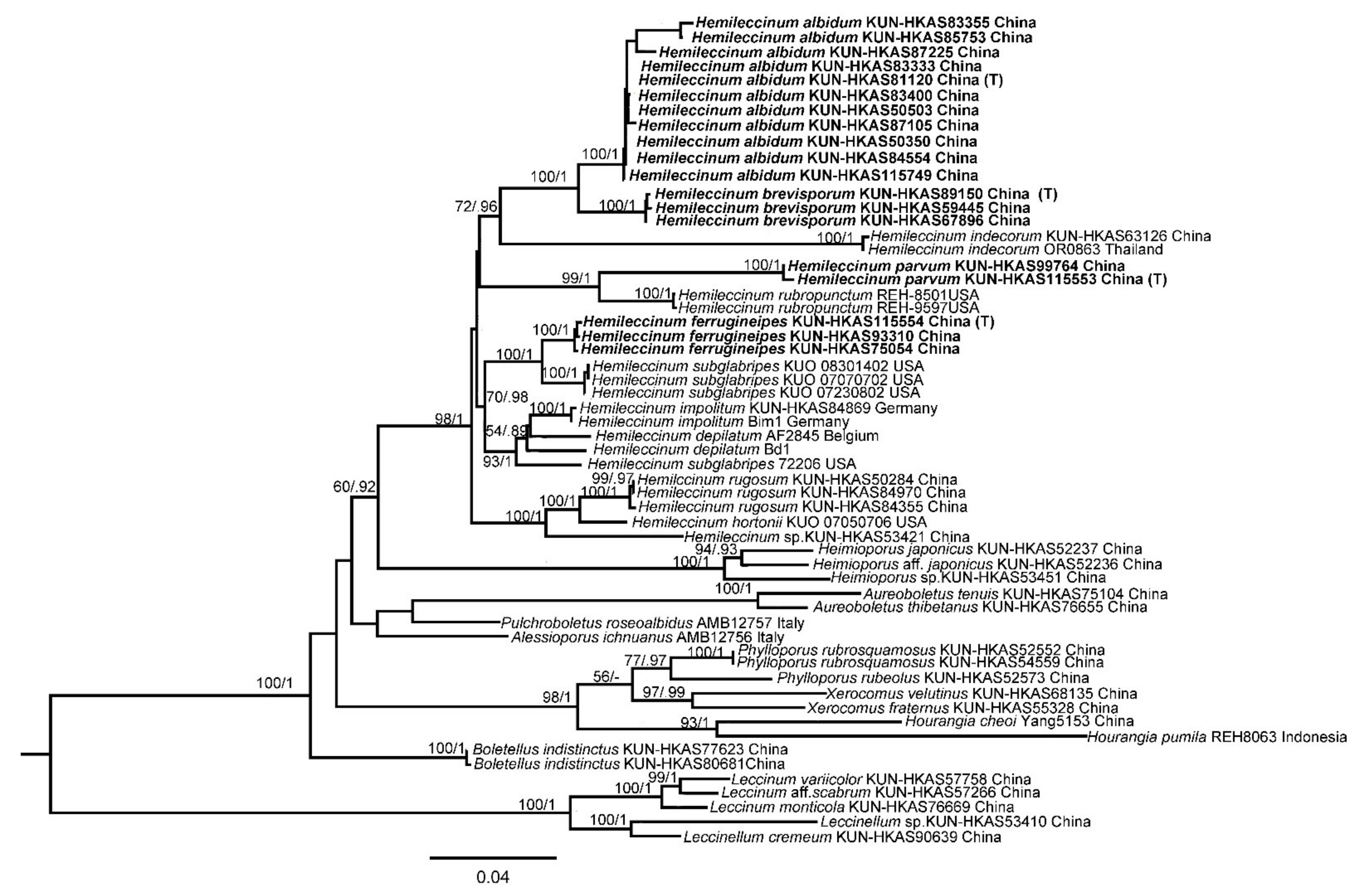
A total of 79 sequences, including 16 for ITS, 12 for nrLSU, 17 for tef1-α, 17 for rpb1, and 17 for rpb2 were newly generated in the present study and aligned with sequences downloaded from GenBank and previous studies. Sequences retrieved from GenBank and obtained in this study were listed in Table 1. ML and BI analyses of the ITS dataset resulted in almost identical topologies and thus only the tree inferred from ML analysis was displayed (Figure 2). Our phylogenetic analyses indicated that Hemileccinum formed a monophyletic group with evident support (MLB/BPP = 100%/1.0). Eight phylogenetic species of the genus Hemileccinum were retrieved, and four of them could be new to science.
According to the four single-locus phylogenetic analyses, no strongly supported (>70% of ML) conflict of topologies was observed. Therefore, sequences of the four DNA loci were concatenated for the final analysis. ML and BI analyses of the concatenated data set resulted in almost identical topologies and thus only the tree inferred from ML analysis was displayed (Figure 3). Our molecular phylogenetic analysis indicated that Hemileccinum is a monophyletic genus with high statistic supports (BP = 98%, PP = 1). Thirteen phylogenetic species of the genus Hemileccinum were retrieved, and four of them could be new to science. By further morphological examinations of the related specimens of those four potential new species, we verified their taxonomic statuses of new species. For detailed information of each species, see below.

3.2. Taxonomy

Hemileccinum albidum Mei-Xiang Li, Zhu L. Yang & G. Wu, sp. nov., Figure 1a–c, Figure 4a–c and Figure 5.
MycoBank no: 840704
Etymology: The epithet ‘albidum’ refers to the somewhat white stipe of this species.
Type: CHINA. Yunnan Province: Jingdong County, Ailao Mt., alt. 2490 m, associated with Castanopsis ceratacantha, Ca. rufescens, Lithocarpus xylocarpus, Quercus griffithii, 22 July 2013, Jiao Qin 682 (KUN-HKAS81120).
Diagnosis: Hemileccinum albidum is distinguished by the combination characters of the even pileus, and the whitish, nearly smooth stipe, with only small, granular scales at the base.
Description: Basidioma stipitate-pileate, small to medium-sized. Pileus 3–9 cm diam, hemispherical to applanate, finely rugose, then smooth, finely subtomentose, dry or slightly viscid when wet; surface of Pileus grey-brown when young, then chrome yellow (5A8), pompeian yellow (5C7) to ochraceous (2D2–5) or golden brown (5D7), somewhat paler along the pileus edge; context white (1A1), yellowish (2A4–5) to brownish (5B5–8, 6C6–8), unchanging on exposure. Hymenophore depressed around the apex of the stipe; hymenophoral surface yellowish (2A4–5) to yellow to sulphur yellow (4A5–4A6) or olivaceous yellow (2A6–7), unchanging when bruised; pores roundish, 0.5–1.0(1.5)/mm; tubes up to 10 mm long, concolorous with the hymenophoral surface, unchanging when bruised. Stipe 5–16 cm long, 1.0–2.5 cm wide, subcylindrical; surface whitish (8A1), cream (1A2) to pale yellow-brown (2A3) or pinkish (13A2) to purplish (11B3–5), fibrillose, sometimes covered with small pale granular scales; context unchanging in color when cut. Basal mycelium white (1A1).
Basidia 25–38 × 10–14 µm, clavate, 4-spored, sterigmata 4–6 µm long. Basidiospores [120/3/3] (10)11–12.5 × (4.0)4.5–5.5 µm, [Q = (2.00)2.18–2.66(2.75), Qm = 2.36 ± 0.12], subfusiform in side view with distinct suprahilar depression, subfusoid in ventral view, brownish yellow, inamyloid, with tiny warts and pinholes on the surface under SEM. Hymenophoral trama nearly phylloporoid with hyphae of the lateral strata touching or almost touching each other with hyphae diverging from the central strand to the subhymenium. Cheilocystidia 41–50 × 8–11 µm, lanceolate to clavate or ventricose, thin-walled, colorless. Pleurocysitidia 46–56 × 8–13 µm, ventricose-subfusiform, with long beak, thin-walled. Pileipellis an hyphoepithelium 170–230 µm thick, composed of moniliform hyphal segments 5–37 µm wide, thin-walled, with narrowly cylindrical to shortly cystidioid terminal cells 10–75 × 3–20 µm. Pileal trama composed of interwoven hyphae 5–34 µm wide. Stipitipellis ca. 130 µm thick, hymeniform, terminal cells broadly clavate, 20–43 × 10–22 µm, sometimes connected with narrow, filamentous hyphae at the outer layer. Caulocystidia abundant, 26–43 × 7–12 µm, thin-walled. Stipe trama composed of parallel hyphae, 3.5–12.0 µm wide. Clamp connections absent in all tissues.
Habitat and distribution: Scattered in subtropical forests dominated by plants of the family Fagaceae (Castanopsis ceratacantha, Ca. rufescens, Ca. calathiformis, Lithocarpus xylocarpus, L. hancei, L. mairei and Quercus griffithii); on acidic, loamy, humid soils; moderately common in southwestern China; fruiting in June to August in southwestern China (Yunnan Province) between 1968 and 2490 m altitude.
Additional specimens examined: CHINA. Yunnan Province: Jingdong County, Ailao Mt., alt. 2490 m, associated with Castanopsis ceratacantha, Ca. rufescens, Lithocarpus xylocarpus and Quercus griffithii, 21 July 2006, Zhu-Liang Yang 4706 (KUN-HKAS50503); same location, 20 July 2006, Yan-Chun Li 596 (KUN-HKAS50350); same location, 23 July 2013, Bang Feng 1359 (KUN-HKAS115749); Longling County, Zhenan Town, alt. 1968 m, associated with Castanopsis calathiformis and Lithocarpus hancei, 11 July 2014, Xiao-Bin Liu 459 (KUN-HKAS87105); same location, 22 July 2014, Xiao-Bin Liu 673 (KUN-HKAS87225); same location, 25 August 2014, Chen Yan 155 (KUN-HKAS85753); Longling County, Xueshan Village, alt. 2000 m, associated with Castanopsis ceratacantha, Lithocarpus mairei and Quercus griffithii, 19 June 2014, Jiao Qin 916 (KUN-HKAS83333); same location, 21 June 2014, Jiao Qin 938 (KUN-HKAS83355); same location, 31 July 2014, Jiao Qin 983 (KUN-HKAS83400); same location, 14 June 2014, Li-Hong Han 258 (KUN-HKAS84554).
Notes: Hemileccinum albidum is distinguished by combination characters of the even pileus and whitish stipe surface covered with concolorous, small granular scales. Phylogenetically, H. albidum is closely related to H. brevisporum. However, the former species differs in its whitish stipe and larger basidiospores (11.0–12.5 × 4.5–5.5 μm). Morphologically, the size, pileus color and shape of H. albidum are similar to those of the European H. depilatum. However, the latter is different from the former by its wrinkled or hammered pileus and the pileipellis composed of hyphae of spherical and shortly cylindrical, terminal cells 16.5–44.0 × 8.5–30.0 μm [4]. Ecologically, H. albidum occurs under trees of Fagaceae in subtropical regions while H. depilatum is distributed in hardwoods, especially with trees of Ulmus and Carpinus in temperate regions [41,42] (Appendix A).
Hemileccinum brevisporum Mei-Xiang Li, Zhu L. Yang & G. Wu, sp. nov., Figure 1d–f, Figure 4d–f and Figure 6.
MycoBank no: 840701
Etymology: The epithet ‘brevisporum’ refers to the short basidiospores.
Type: CHINA. Yunnan Province: Menghai County, alt. 1700 m, associated with Castanopsis calathiformis, Ca. indica and Lithocarpus truncates, 1 July 2014, Kuan Zhao 487 (KUN-HKAS89150).
Diagnosis: Differs from other Hemileccinum species by the combined characters of the dense fine-grained scales on the stipe surface, the shorter basidiospores measuring 9–11 × 4–5 µm and small basidia measuring 18.5–27.0 × 8–11 µm.
Description: Basidioma stipitate-pileate, fleshy, small to medium-sized. Pileus 9 cm diam, glabrous to slightly subtomentose, dry, convex to planate, pale yellow-brown (2A3) to pale red-brown (7A5); context yellowish (3A5–3A6), unchanging when bruised. Hymenophoral surface and tubes concolorous, flash yellow (3A8) to dull yellow (3B3–3B4), unchanging when bruised, pores roundish, 1.0–1.5/mm, tubes 11 mm long, unchanging when injured. Stipe 13–15 cm long, 2.0–2.3 cm wide, subcyclindrical, surface of stipe cream (2A2–3A2) to yellowish (2A4–2A5) at upper part, pale yellow-brown to yellow-brown (6C8) at lower part, covered with small yellowish brown (5D8) dotted scales, context of stipe cream to pale yellow (1A2–1A3), unchanging when bruised. Basal mycelium white to cream (2A2–3A2).
Basidia 18.5–27.0 × 8–11 µm, clavate, hyaline in 5% KOH, 4-spored. Basidiospores [80/2/2], 9–11 × 4–5 µm, [Q = (2.22)2.35–2.50(2.75), Qm = 2.37 ± 0.15], subfusiform and inequilateral in side view with distinct suprahilar depression, subfusoid in ventral view, yellowish to brownish, smooth under light microscopy, but with tiny warts on the surface under SEM. Hymenophoral trama nearly phylloporoid with hyphae of the lateral strata touching or almost touching each other with hyphae diverging from the central strand to the subhymenium; hyphae subcylindrical to cylindrical, 3.5–14.0 µm wide. Cheilocystidia 37–50 × 11–13 µm, ventricose-subfusiform, with long beak, thin-walled. Pleurocystidia 48–67 × 12–16 µm, ventricose subfusiform, with long beak, thin-walled. Pileipellis an hyphoepithelium 150–210 µm thick, composed of moniliform hyphal segments 5–35 µm wide, thin-walled, with narrowly cylindrical to shortly cystidioid terminal cells 6–53 × 4–20 µm. Pileal trama composed of interwoven hyphae 5–37 µm wide. Stipitipellis ca. 100 µm thick, hymeniform, terminal cells broadly clavate, 13–30 × 7.0–12.5 µm, sometimes connected with narrow, filamentous hyphae at the outer layer. Caulobasidia abundant, 18.5–28.0 × 9.0–12.0 µm, thin-walled. Stipe trama composed of parallel hyphae, 4–12 µm wide. Clamp connections absent.
Habitat and distribution: Scattered in subtropical forests dominated by the families Fagaceae (Castanopsis calathiformis, Ca. indica, Ca. orthacantha, Lithocarpus hancei, L. mairei and Quercus griffithii) and Pinaceae (Pinus yunnanensis or P. armandii); on acidic or slightly alkaline, loamy soils; rather rare; fruiting in July to August in southwestern to northwestern Yunnan between 1700 and 2120 m altitude.
Additional specimens examined: CHINA. Yunnan Province: Longling County, alt. 2010 m, associated with Castanopsis calathiformis, Lithocarpus hancei and Pinus yunnanensis, 9 July 2009, Yan-Chun Li 1698 (KUN-HKAS59445); Jianchuan County, Laojunshan town, alt. 2120 m, associated with Castanopsis orthacantha, Lithocarpus mairei, Quercus griffithii and Pinus armandii, 9 August 2010, Qing Cai 334 (KUN-HKAS67896).
Notes: Hemileccinum brevisporum is morphologically similar to H. impolitum because of the ornamentation in the stipe and the slightly subtomentose pileus surface [1,41,42]. However, H. impolitum, originally described from Europe, differs from H. brevisporum, by its much stockier stipe, and larger basidiospores (12–15 × 4–6 μm). Ecologically, H. brevisporum occurs under trees of Fagaceae and Pinaceae in subtropical regions while H. impolitum is distributed in hardwood or floodplain forests, especially with trees of Quercus and Fagus in temperate regions [41,42] (Appendix A).
Hemileccinum ferrugineipes Mei-Xiang Li, Zhu L. Yang & G. Wu, sp. nov., Figure 1g–i, Figure 4g–i and Figure 7.
MycoBank no: 840700
Etymology: The epithet ‘ferrugineipes’ refers to the reddish brown stipe of this species.
Type: CHINA. Yunnan Province: Pu’er City, Simao District, Taiyanghe Nature Reserve, alt. 1200 m, associated with Castanopsis ferox, Ca. calathiformis, Cyclobalanopsis xanthotricha, Quercus fabri, Q. variabilis and Lithocarpus glabra, 24 June 2016, Jian-Wei Liu 584 (KUN-HKAS115554).
Diagnosis: Differs from other Hemileccinum species by the combined characters of rugose pileus, creamy yellow stipe surface when young becoming reddish when mature, and densely scaled surface of the stipe.
Description: Basidioma stipitate-pileate, small to medium-sized. Pileus 3–10 cm diam, clavate to planate, surface rugose, slightly subtomentose, dry, yellowish brown (5E5), olive brown (4E5–6) to dull brown (5E8–5F8), context cream to yellowish (2A4–5), unchanging when bruised. Hymenophoral surface and tubes concolorous, yellow (1A2–1A3) to ochreous (5B7–5C7), unchanging when bruised, pores roundish, 1.5–2.5/mm; tubes 4–6 mm long, unchanging when injured. Stipe 4–10 cm long, 1–2 cm wide, subcylindrical, surface yellowish to yellow at upper part, lower part pale red-brown of stipe pileus; covered with longitudinal striations and densely dotted scales, context cream (1A2) to yellowish, unchanging when bruised. Basal mycelium cream.
Basidia 23–35 × 9–13 µm, clavate, hyaline in 5% KOH, 4-spored. Basidiospores [80/2/2], 11.0–12.5 × 4–5 µm, [Q = (2.30)2.40–2.78(3.00), Qm = 2.63 ± 0.19], subfusiform in side view with distinct suprahilar depression, ellipsoid to somewhat oblong in face view, yellowish to brownish, smooth under light microscopy, but with tiny warts under SEM. Hymenophoral trama nearly phylloporoid with hyphae of the lateral strata touching or almost touching each other with hyphae diverging from the central strand to the subhymenium; hyphae subcylindrical to cylindrical, 4–13 µm wide. Cheilocystidia 36–63 × 7–11 µm, ventricose-subfusiform, with long beak, thin-walled. Pleurocystidia 37–62 × 8–12 µm, ventricose subfusiform, with long beak, thin-walled. Pileipellis an hyphoepithelium 170–270 µm thick, composed of moniliform hyphal segments 5–42 µm wide, thin-walled; always with narrowly cylindrical to shortly cystidioid terminal cells, 20–127 × 4–12 µm. Pileal trama composed of interwoven hyphae 6–38 µm wide. Stipitipellis ca.30–40 µm thick, hymeniform, terminal cells broadly clavate, 15.0–32.0 × 6.5–15.5 µm. Caulobasidia abundant, 28–44 × 9–12 µm, thin-walled. Stipe trama composed of parallel hyphae, 5–13 µm wide. Clamp connections absent.
Habitat and distribution: Scattered in subtropical forests dominated by plants of the family Fagaceae (Castanopsis ferox, Ca. calathiformis, Ca. hystrix, Cyclobalanopsis xanthotricha, Quercus fabri, Q. variabilis and Lithocarpus glabra); on acidic or slightly alkaline, loamy soils; rather rare; fruiting in June to August in southwestern to northwestern Yunnan between 1200 and 1690 m altitude.
Additional specimens examined: CHINA. Yunnan Province: Baoshan City, Longyang District, alt. 1690 m, associated with Castanopsis calathiformis, Quercus fabri and Lithocarpus glabra, 30 July 2017, Pan-Meng Wang 350 (KUN-HKAS93310); Lanping County, alt. 1400 m, associated with Castanopsis hystrix, Quercus fabri and Lithocarpus glabra, 16 August 2011, Gang Wu 759 (KUN-HKAS75054).
Notes: Hemileccinum ferrugineipes is characterized by its rugose pileus and small, dense, dotted scales on the reddish-brown stipe. Phylogenetically, the American species H. subglabripes is close to H. ferrugineipes, but differs from it by its fairly long and slender, nearly smooth stipe [9,10,43,44,45]. Morphologically, H. ferrugineipes is similar to Rugiboletus extremiorientalis (Lj.N.Vassiljeva) G. Wu & Zhu L. Yang and H. hortonii in the rugose pileus and dense scales on the stipe [44,45,46,47]. However, H. ferrugineipes differs from Rug. extremiorientalis, originally described from subtropical Yunnan, China, by its reddish slightly densely scaled surface of the stipe and hyphoepithelium pileipellis. H. ferrugineipes differs from H. hortonii, originally described from Illinois, USA, by its tightly wrinkled pileus and the stockier stipe [9,44,45]. Ecologically, H. ferrugineipes occurs under trees of Fagaceae in subtropical regions; H. hortonii is scattered or in groups on the ground under mixed deciduous woods, occasionally under conifers; H. hortonii is rather rare and might be found in eastern North America, west to Michigan [44,45] (Appendix A).
Hemileccinum parvum Mei-Xiang Li, Zhu L. Yang & G. Wu, sp. nov., Figure 1j–l, Figure 4j–l and Figure 8.
MycoBank no: 840703
Etymology: The epithet ‘parvum’ refers to the small basidioma.
Type: CHINA. Yunnan Province: Wenshan City, Malipo County, alt. 1200 m, associated with Castanea henryi, C. mollissima, Lithocarpus bonnetii and Quercus marlipoensis, 30 July 2017, 532624MF-201-Wu 2299 (KUN-HKAS115553).
Diagnosis: Differs from other Hemileccinum species by the combined characters of the small basidioma, and the rugose surface of pileus, the pale yellow context staining pale blue very slowly when bruised.
Description: Basidioma stipitate-pileate, small. Pileus 3.3–3.6 cm diam, rugose, slightly subtomentose, hemispherical, brownish (5B5–8, 6C6–8) at the central part, becoming paler towards the margin (brownish or yellowish); context pale yellow (1A2–1A3), staining pale blue very slowly when bruised at some spots, 4–5 mm thick. Hymenophoral surface and tubes concolorous, light yellow (3B4–3B5), pores roundish, 1.5–2.0/mm, unchanging when bruised; tubes 4–5 mm long, sinuate near the stipe. Stipe 6.0–9.7 cm long, 0.4–0.9 cm wide, clavate, central, solid, pale yellow (2A2–2A4) at the upper part and becoming paler downwards, surface ornamented with coarsely small squamules; context light yellow (3B4–3B5), unchanging when bruised. Basal mycelium white (1A1).
Basidia 20.5–32.0 × 8.0–10.5 µm, clavate, 4-spored; sterigmata up to 4–5 µm long. Basidiospores [80/2/2], 12–14 × 4.5–5.0 µm, [Q = (2.40)2.50–2.80(2.88), Qm = 2.69 ± 0.11], subfusiform and inequilateral in side view with distinct suprahilar depression, subfusoid in ventral view, yellowish to brownish, inamyloid, smooth under light microscopy, but with tiny warts on the surface under SEM. Hymenophoral trama phylloporoid with hyphae of the lateral strata touching or almost touching each other with hyphae diverging from the central strand to the subhymenium; hyphae subcylindrical to cylindrical, 4–12 µm wide. Cheilocystidia 41–50 × 8–11 µm, lanceolate to clavate or ventricose, thin-walled, colorless. Pleurocysitidia 45–65 × 9–11 µm, ventricose-subfusiform, with long beak, thin-walled. Pileipellis an hyphoepithelium 160–240 µm thick, composed of moniliform hyphal segments 6–30 µm wide, thin-walled, with narrowly cylindrical to shortly cystidioid terminal cells 10–87 × 5–17 µm. Pileal trama composed of interwoven hyphae 6–30 µm wide. Stipitipellis ca. 100 µm thick, hymeniform, terminal cells broadly clavate, 20.0–43.0 × 10.0–21.5 µm, sometimes connected with narrow, filamentous hyphae at the outer layer. Caulocystidia abundant, 24.5–60.0 × 10.5–19.0 µm, thin-walled. Stipe trama composed of parallel hyphae, 3.5–12.0 µm wide. Clamp connections absent in all tissues.
Habitat and distribution: Scattered in subtropical forests dominated by plants of the family Fagaceae (Castanea henryi, C. mollissima, Lithocarpus bonnetii and Quercus marlipoensis); on acidic, wet, fertile soils; rather rare; fruiting in July in southeastern Yunnan between 1200 and 1300 m altitude.
Additional specimens examined: Yunnan Province: Wenshan City, Malipo County, alt. 1300 m, associated with Castanea henryi, C. mollissima, Lithocarpus bonnetii and Quercus marlipoensis, 27 July 2016, Gang Wu 1645 (KUN-HKAS99764).
Notes: Hemileccinum parvum is morphologically similar to H. subglabripes because of the slightly wrinkled pileus and the slender stipe [9,48], However, H. subglabripes, originally described from the USA, differs from H. parvum by the nearly smooth stipe of the latter covered with branny particles on the stem which are pale and easily overlooked, and the larger basidioma. Our data show that H. parvum is phylogenetically close to H. rubropunctum, but the latter differs by its longer stipe and the red scales on it [10,44,45]. Ecologically, H. parvum occurs under trees of Fagaceae in subtropical southeastern Yunnan; H. subglabripes inhabits mixed deciduous trees, sometimes under spruce in eastern and particularly northern North America; and H. rubropunctum grows in mixed woods with oak or chestnut in northeastern North America [44,45] (Appendix A).

4. Discussion

The genus Hemileccinum Šutara is geographically widely distributed, but its species diversity is poorly known. In Asia, only two species have been previously reported with molecular evidence. One is H. indecorum from tropical areas, and the other is H. rugosum from subtropical Yunnan, China [8,15]. In this study, four new species in China were recognized and delimited. They are well-supported by molecular phylogenetic and morphological evidence. The host specificity, altitude and edaphic factors seem to be important for determining the distribution of different species of Hemileccinum. Our newly described species are distributed in the broad-leaved and mixed forests in southwestern China. Hemileccinum albidum and H. brevisporum are distributed on high altitudes: between 1700 and 2500 m a.s.l., while H. ferrugineipes: 1200–1700 m a.s.l., and H. parvum: 1200-1300 m a.s.l. Hemileccinum albidum, H. ferrugineipes and H. parvum are found in subtropical forests and associated with plants of the family Fagaceae (Castanopsis ceratacantha, Ca. rufescens, Ca. ferox, Ca. hystrix, Ca. calathiformis; Castanea henryi, C. mollissima; Cyclobalanopsis xanthotricha; Lithocarpus xylocarpus, L. hancei, L. mairei, L. glabra, L. bonnetii; Quercus griffithii, Q. fabri, Q. variabilis, Q. marlipoensis), growing mostly on acidic soils. However, H. ferrugineipes can also be found in slightly alkaline habitats. Hemileccinum brevisporum is found in subtropical broad-leaved and mixed forests, growing with members of Fagaceae (Castanopsis calathiformis, Ca. indica, Ca. orthacantha; Lithocarpus hancei, L. mairei; Quercus griffithii) and Pinaceae (Pinus yunnanensis or P. armandii) on acidic or slightly alkaline soils. The species we described here are hardly seen in the wild mushroom market, thus their edibility is unknown yet. However, referring to the edibility of the European/American species of Hemileccinum [41,42,43,44,45,49], the newly described species could also be edible, but we need more investigations to confirm this.
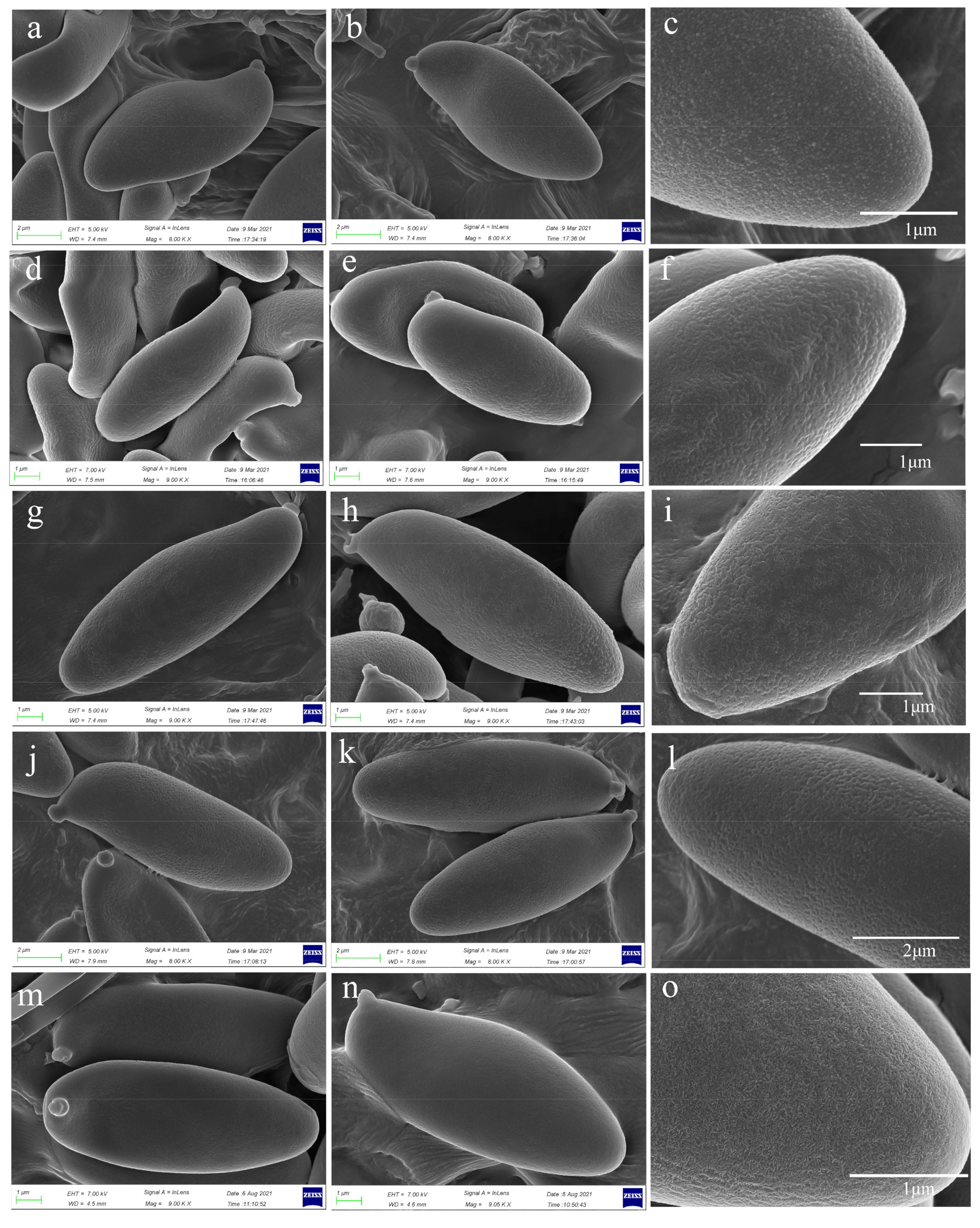
Overall, the proposed new species are significantly different from the Asian species H. indecorum because the viscid pileus and stipe of the latter species are densely covered with whitish to dirty white, small conical to subconical to irregular squamules [15]. They also quite differ from the European species H. impolitum, which has a relatively bald pileal surface and a collapsed trichoderm pileipellis when mature [1]. Šutara reported that the basidiospores of H. impolitum are smooth [1]. Our re-examination of European material of H. impolitum under SEM indicated that there are irregular warts on the surface of basidiospores as with those of other species in Hemileccinum (Figure 4m–o). Accordingly, H. depilatum (Redeuilh) Šutara should also have a warty basidiospore surface.
The description of the new species also sheds new light on the recognition of the genus. The pileipellis of the species in this genus should mostly be regarded as (sub)epithelium to hyphoepithelium, because the pileipellis of most studied species here are composed of short inflated cells in the inner layer (subpellis) and filamentous hyphae in outer layer (suprapellis), with H. indecorum standing at one extremity with whitish to dirty white, small conical to subconical to irregularly shaped squamules on the pileus surface [15] and H. impolitum located at the other extremity with collapsed trichoderm pileipellis when mature [1]. The lateral stipe stratum of H. impolitum in this genus was diagnosed as the leccinoid type, predominantly anticlinally arranged hyphae ending in elements of the caulohymenium [1,3,4]. However, on the basis of the observation on our new species, this feature is not present in all species of Hemileccinum. The structure of the lateral stipe stratum is traceable in our species.
Based on the current study, we increased the species diversity of the genus Hemileccinum from Asia and reconstructed a comprehensive phylogenetic tree which included almost all known species of this genus. However, probably due to the limitations of species sampling or insufficient genetic variation of the DNA loci we used, the deep phylogenetic relationships within the genus remain unresolved. In future work, more species with detailed morphological observations and phylogenomic analysis will provide new evidence for these relationships.

Author Contributions

Conceptualization: Z.L.Y., G.W. and M.-X.L.; molecular experiments and data analysis: M.-X.L.; original draft writing: M.-X.L.; draft review and editing: Z.L.Y. and G.W. All authors have read and agreed to the published version of the manuscript.

Funding

This work was supported by Yunnan Ten-Thousand-Talents Plan-Yunling Scholar Project, Yunnan Ten-Thousand-Talents Plan-Young & Elite Talents Project, and the National Natural Science Foundation of China (No. 31970015).

Institutional Review Board Statement

Not applicable.

Informed Consent Statement

Not applicable.

Data Availability Statement

Publicly available datasets were analyzed in this study. This data can be found here: https://www.ncbi.nlm.nih.gov/; http://www.mycobank.org/; http://purl.org/phylo/treebase/phylows/study/TB2:S28729, accessed on 18 September 2021.

Acknowledgments

The authors are very grateful to their colleagues at Kunming Institute of Botany, Chinese Academy of Sciences, including Kuan Zhao, Jiao Qin, Bang Feng, Yan-Chun Li, Chen Yan, Qing Cai, Li-Hong Han, and Xiao-Bin Liu, and Pan-Meng Wang and Jian-Wei Liu for providing specimens, and Yang-Yang Cui and Geng-Shen Wang for help in morphological observation and phylogenetic analysis, and Zhi-Jia Gu for arranging the scanning electron microscopy.

Conflicts of Interest

The authors declare that there are no conflict of interest.

Appendix A

Key to known species of Hemileccinum in the world
1. Pileus surface pale brown, brown to reddish brown, more or less even2
1′. Pileus surface orange to reddish orange, distinctively wrinkled9
2. Pileus surface slightly subtomentose, without small conical to subconical to irregularly shaped squamules, known in subtropical and temperate regions in the Northern Hemisphere…3
2′. Pileus surface with whitish to dirty white, small conical to subconical to irregularly shaped squamules, known from tropical southeast Asia…H. indecorum
3. Basidioma varied in size, yellowish context without color change when bruised…4
3′. Basidioma usually small in size (≤ 4 cm in daim), the pale yellow context staining pale blue very slowly when bruised, known from subtropical areas…H. parvum
4. Stipe surface covered with obvious ornaments…5
4′. Stipe surface covered with small scales, not obvious6
5. Stipe surface ornamented with distinctly reddish brown longitudinal streaks, known from East AsiaH. ferrugineipes
5′. Stipe surface often covered with red dense fine-grained scales, known from North America…H. rubropunctum
6. Basidiospores longer in length (> 11 µm), and in other parts of the world…7
6′. Basidiospores shorter in length (≤ 11 µm), and distributed in subtropical forests in southwestern China.…H. brevisporum
7. Stipe stout (> 2.5 cm in diam.); pileipellis an trichoderm, collapsed when mature, restricted to EuropeH. impolitum
7′. Stipe slender (≤ 2.5 cm in diam.); pileipellis an hyphoepithelium8
8. Stipe surface whitish, covered with small, granular scales only at the base, distributed in subtropical ChinaH. albidum
8′. Stipe surface yellowish, nearly smooth, covered with indistinctive tiny scales, known from eastern North AmericaH. subglabripes
9. Basidiospores smaller (10–12 × 4–5 µm), distributed in subtropical and tropical ChinaH. rugosum
9′. Basidiospores larger (≥ 12 µm in length), distributed in temperate regions…10
10. Basidiospores narrower (12.0–15.0 × 3.5–4.5 µm), known from North America…H. hortonii
10′. Basidiospores wider (12.0–15.0 × 5–6 µm), known from Europe…H. depilatum

References

  1. Šutara, J. Xerocomus s. l. in the light of the present state of knowledge. Czech Mycol. 2008, 60, 29–62. [Google Scholar] [CrossRef]
  2. Fries, E.M. Epicrisis Systematis Mycologici, seu Synopsis Hymenomycetum; Typographia Academica: Munich, Germany, 1839; p. 421. [Google Scholar]
  3. Bertault, R. Amanites du Maroc (Troisième contribution). Bull. Société Mycol. Fr. 1980, 96, 271–287. [Google Scholar]
  4. Šutara, J. The delimitation of the genus Leccinum. Czech Mycol. 1989, 43, 1–12. [Google Scholar]
  5. Binder, M.; Besl, H. 28S rDNA sequence data and chemotaxonomical analyses on the generic concept of Leccinum (Boletales). Micologia 2000, 71–82. [Google Scholar]
  6. Binder, M.; Hibbett, D.S. Molecular systematics and biological diversification of Boletales. Mycology 2006, 98, 971–981. [Google Scholar] [CrossRef]
  7. Wu, G.; Feng, B.; Xu, J.; Zhu, X.-T.; Li, Y.-C.; Zeng, N.-K.; Hosen, I.; Yang, Z.L. Molecular phylogenetic analyses redefine seven major clades and reveal 22 new generic clades in the fungal family Boletaceae. Fungal Divers. 2014, 69, 93–115. [Google Scholar] [CrossRef]
  8. Wu, G.; Li, Y.-C.; Zhu, X.-T.; Zhao, K.; Han, L.-H.; Cui, Y.-Y.; Li, F.; Xu, J.-P.; Yang, Z.L. One hundred noteworthy boletes from China. Fungal Divers. 2016, 81, 25–188. [Google Scholar] [CrossRef]
  9. Halling, R.E.; Fechner, N.; Nuhn, M.; Osmundson, T.; Soytong, K.; Arora, D.; Binder, M.; Hibbett, D. Evolutionary relationships of Heimioporus and Boletellus (Boletales), with an emphasis on Australian taxa including new species and new combinations in Aureoboletus, Hemileccinum and Xerocomus. Aust. Syst. Bot. 2015, 28, 1–22. [Google Scholar] [CrossRef]
  10. Kuo, M.; Ortiz-Santana, B. Revision of leccinoid fungi, with emphasis on North American taxa, based on molecular and morphological data. Mycology 2020, 112, 197–211. [Google Scholar] [CrossRef]
  11. Singer, R. The Agaricales in Modern Taxonomy, 4th ed.; Koeltz Scientific Books: Koenigstein, Germany, 1986; pp. 1–981. [Google Scholar]
  12. Agerer, R. Fungal relationships and structural identity of their ectomycorrhizae. Mycol. Prog. 2006, 5, 67–107. [Google Scholar] [CrossRef]
  13. Tedersoo, L.; May, T.; Smith, M.E. Ectomycorrhizal lifestyle in fungi: Global diversity, distribution, and evolution of phylogenetic lineages. Mycorrhiza 2009, 20, 217–263. [Google Scholar] [CrossRef] [PubMed]
  14. Ryberg, M.; Matheny, P.B. Asynchronous origins of ectomycorrhizal clades of Agaricales. Proc. R. Soc. B Boil. Sci. 2011, 279, 2003–2011. [Google Scholar] [CrossRef] [Green Version]
  15. Zeng, N.-K.; Cai, Q.; Yang, Z.L. Corneroboletus, a new genus to accommodate the southeastern Asian Boletus indecorus. Mycology 2012, 104, 1420–1432. [Google Scholar] [CrossRef]
  16. Vadthanarat, S.; Lumyong, S.; Raspé, O. Cacaoporus, a new Boletaceae genus, with two new species from Thailand. MycoKeys 2019, 54, 1–29. [Google Scholar] [CrossRef]
  17. Kornerup, A.; Wanscher, J.H. Methuen Handbook of Colour, 3rd ed.; Eyre Methuen: London, UK, 1967; pp. 1–252. [Google Scholar]
  18. Li, Y.C.; Yang, Z.L.; Tolgor, B. Phylogenetic and biogeographic relationships of Chroogomphus species as inferred from molecular and morphological data. Fungal Divers. 2009, 38, 85–104. [Google Scholar]
  19. Zhou, M.; Dai, Y.-C.; Vlasák, J.; Yuan, Y. Molecular Phylogeny and Global Diversity of the Genus Haploporus (Polyporales, Basidiomycota). J. Fungi 2021, 7, 96. [Google Scholar] [CrossRef]
  20. White, T.J.; Bruns, T.; Lee, S.; Taylor, J. Amplification and Direct Sequencing of Fungal Ribosomal RNA Genes for Phylogenetics. In PCR Protocols: A Guide to Methods and Applications; Innis, M., Gelfand, D., Sninsky, J., White, T., Eds.; Academic Press Inc.: New York, NY, USA, 1990; p. 315. [Google Scholar]
  21. Gardes, M.; Bruns, T.D. ITS primers with enhanced specificity for basidiomycetes—Application to the identification of mycorrhizae and rusts. Mol. Ecol. 1993, 2, 113–118. [Google Scholar] [CrossRef] [PubMed]
  22. James, T.Y.; Kauff, F.; Schoch, C.L.; Matheny, P.B.; Hofstetter, V.; Cox, C.; Celio, G.; Gueidan, C.; Fraker, E.; Miadlikowska, J.; et al. Reconstructing the early evolution of Fungi using a six-gene phylogeny. Nature 2006, 443, 818–822. [Google Scholar] [CrossRef] [PubMed]
  23. Vilgalys, R.; Hester, M. Rapid genetic identification and mapping of enzymatically amplified ribosomal DNA from several Cryptococcus species. J. Bacteriol. 1990, 172, 4238–4246. [Google Scholar] [CrossRef] [PubMed] [Green Version]
  24. Zeng, N.-K.; Tang, L.-P.; Li, Y.-C.; Tolgor, B.; Zhu, X.-T.; Zhao, Q.; Yang, Z.L. The genus Phylloporus (Boletaceae, Boletales) from China: Morphological and multilocus DNA sequence analyses. Fungal Divers. 2012, 58, 73–101. [Google Scholar] [CrossRef]
  25. Schoch, C.L.; Robbertse, B.; Robert, V.; Vu, D.; Cardinali, G.; Irinyi, L.; Meyer, W.; Nilsson, R.H.; Hughes, K.; Miller, A.N.; et al. Finding needles in haystacks: Linking scientific names, reference specimens and molecular data for Fungi. Database 2014, 2014, bau061. [Google Scholar] [CrossRef] [PubMed]
  26. Zhu, X.-T.; Wu, G.; Zhao, K.; Halling, R.E.; Yang, Z.L. Hourangia, a new genus of Boletaceae to accommodate Xerocomus cheoi and its allied species. Mycol. Prog. 2015, 14, 1–10. [Google Scholar] [CrossRef]
  27. Gelardi, M.; Simonini, G.; Ercole, E.; Vizzini, A. Alessioporusand Pulchroboletus (Boletaceae, Boletineae), two novel genera for Xerocomus ichnusanus and X. roseoalbidusfrom the European Mediterranean basin: Molecular and morphological evidence. Mycology 2014, 106, 1168–1187. [Google Scholar] [CrossRef] [PubMed] [Green Version]
  28. Neves, M.A.; Binder, M.; Halling, R.; Hibbett, D.; Soytong, K. The phylogeny of selected Phylloporus species, inferred from NUC-LSU and ITS sequences, and descriptions of new species from the Old World. Fungal Divers. 2012, 55, 109–123. [Google Scholar] [CrossRef]
  29. Katoh, K.; Standley, D.M. MAFFT Multiple Sequence Alignment Software Version 7: Improvements in Performance and Usability. Mol. Biol. Evol. 2013, 30, 772–780. [Google Scholar] [CrossRef] [PubMed] [Green Version]
  30. Hall, T.A. BioEdit: A user-friendly biological sequence alignment editor and analyses program for Windows 95/98/NT. Nucleic Acids Symp. Ser. 1999, 41, 95–98. [Google Scholar]
  31. Stamatakis, A. RAxML-VI-HPC: Maximum likelihood-based phylogenetic analyses with thousands of taxa and mixed models. Bioinformatics 2006, 22, 2688–2690. [Google Scholar] [CrossRef]
  32. Stamatakis, A.; Hoover, P.; Rougemont, J. A Rapid Bootstrap Algorithm for the RAxML Web Servers. Syst. Biol. 2008, 57, 758–771. [Google Scholar] [CrossRef]
  33. Smith, S.A.; Dunn, C. Phyutility: A phyloinformatics tool for trees, alignments and molecular data. Bioinformatics 2008, 24, 715–716. [Google Scholar] [CrossRef] [Green Version]
  34. Nylander, J.A.A. MrModeltest v2. Program Distributed by the Author; Evolutionary Biology Centre, Uppsala University: Uppsala, Sweden, 2004. [Google Scholar]
  35. Ronquist, F.; Huelsenbeck, J.P. MrBayes 3: Bayesian phylogenetic inference under mixed models. Bioinformatics 2003, 19, 1572–1574. [Google Scholar] [CrossRef] [Green Version]
  36. Nuhn, M.E.; Binder, M.; Taylor, A.F.; Halling, R.E.; Hibbett, D.S. Phylogenetic overview of the Boletineae. Fungal Biol. 2013, 117, 479–511. [Google Scholar] [CrossRef]
  37. Vadthanarat, S.; Raspé, O.; Lumyong, S. Phylogenetic affinities of the sequestrate genus Rhodactina (Boletaceae), with a new species, R. rostratispora from Thailand. MycoKeys 2018, 29, 63–80. [Google Scholar] [CrossRef] [PubMed]
  38. Martín, M.; Raidl, S. The taxonomic position of Rhizopogon melanogastroides (Boletales). Mycotaxon 2002, 84, 221–228. [Google Scholar]
  39. Osmundson, T.W.; Robert, V.A.; Schoch, C.L.; Baker, L.J.; Smith, A.; Robich, G.; Mizzan, L.; Garbelotto, M.M. Filling Gaps in Biodiversity Knowledge for Macrofungi: Contributions and Assessment of an Herbarium Collection DNA Barcode Sequencing Project. PLoS ONE 2013, 8, e62419. [Google Scholar] [CrossRef] [PubMed] [Green Version]
  40. Smith, M.E.; Pfister, D.H. Tuberculate ectomycorrhizae of angiosperms: The interaction betweenBoletus rubropunctus (Boletaceae) and Quercusspecies (Fagaceae) in the United States and Mexico. Am. J. Bot. 2009, 96, 1665–1675. [Google Scholar] [CrossRef] [Green Version]
  41. Breitenbach, J.; Kränzlin, F. Fungi of Switzerland 3; Boletales and Agaricales Mykologia: Luzern, Switzerland, 1991; pp. 1–361. [Google Scholar]
  42. Courtecuisse, R.; Duhem, B. Mushroom and Toadstools of Britain and Europe; Harper Collins Publishers: New York, NY, USA, 1995; p. 432. [Google Scholar]
  43. Peck, C.H. Boleti of the United States; Bulletin of the New York State Museum: New York, NY, USA, 1889; Volume 2, p. 112. [Google Scholar]
  44. Bessette, A.E.; Bessette, A.R.; Fisher, D.W. Mushroom of Northeastern North America; Syracause University Press: New York, NY, USA, 1997; p. 328. [Google Scholar]
  45. Phillips, R. Mushrooms and Other Fungi of North America; Firefly Books: New York, NY, USA, 2010; p. 263. [Google Scholar]
  46. Wu, G.; Zhao, K.; Li, Y.-C.; Zeng, N.-K.; Feng, B.; Halling, R.E.; Yang, Z.L. Four new genera of the fungal family Boletaceae. Fungal Divers. 2015, 81, 1–24. [Google Scholar] [CrossRef]
  47. Smith, A.H.; Thiers, H.D. The Boletes of Michigan; University of Michigan Press: Ann Arbor, MI, USA, 1971; p. 428. [Google Scholar]
  48. Peck, C.H. Report of the state botanist. Ann. Rep. N. Y. St. Mus. Nat. Hist. 1896, 50, 77–159. [Google Scholar]
  49. Kuo, M. Retrieved from the Mushroom Expert. Com 2020. Available online: http://www.mushroomexpert.com/ (accessed on 30 September 2021).
Figure 1. Fresh basidiomata of Hemileccinum species. (ac) H. albidum ((a,b) Type, KUN-HKAS81120, (c) KUN-HKAS87225); (df) H. brevisporum ((d) KUN-HKAS67896, (e) KUN-HKAS59445, (f) Type, KUN-HKAS89150); (gi) H. ferrugineipes ((g,h) Type, KUN-HKAS115554, (i) KUN-HKAS75054); (jl) H. parvum ((jl) Type, KUN-HKAS115553) Bars = 30 mm.
Jof 07 00823 g001
Figure 2. Maximum-Likelihood phylogenetic tree generated from ITS dataset. Bootstrap values (BP) ≥ 50% from ML analysis and Bayesian posterior probabilities (PP) ≥ 0.90 are shown on the branches. Newly described species are indicated in bold and their type specimens are marked with (T).
Jof 07 00823 g002
Figure 3. Maximum-Likelihood analysis of Hemileccinum with nrLSU, tef1-α, rpb1 and rpb2 sequence data. Bootstrap values (BP) ≥ 50% from ML analysis and Bayesian posterior probabilities (PP) ≥ 0.90 are shown on the branches. Newly described species are indicated in bold and their type specimens are marked with (T).
Jof 07 00823 g003
Figure 4. Basidiospores of Hemileccinum albidum, H. brevisporum, H. ferrugineipes, H. parvum and H. impolitum under SEM. (ac) H. albidum (Type, KUN-HKAS81120); (df) H. brevisporum (Type, KUN-HKAS89150); (gi) H. ferrugineipes (Type, KUN-HKAS115554,); (jl) H. parvum (Type, KUN-HKAS115553); (mo) H. impolitum (KUN-HKAS84869).
Jof 07 00823 g004
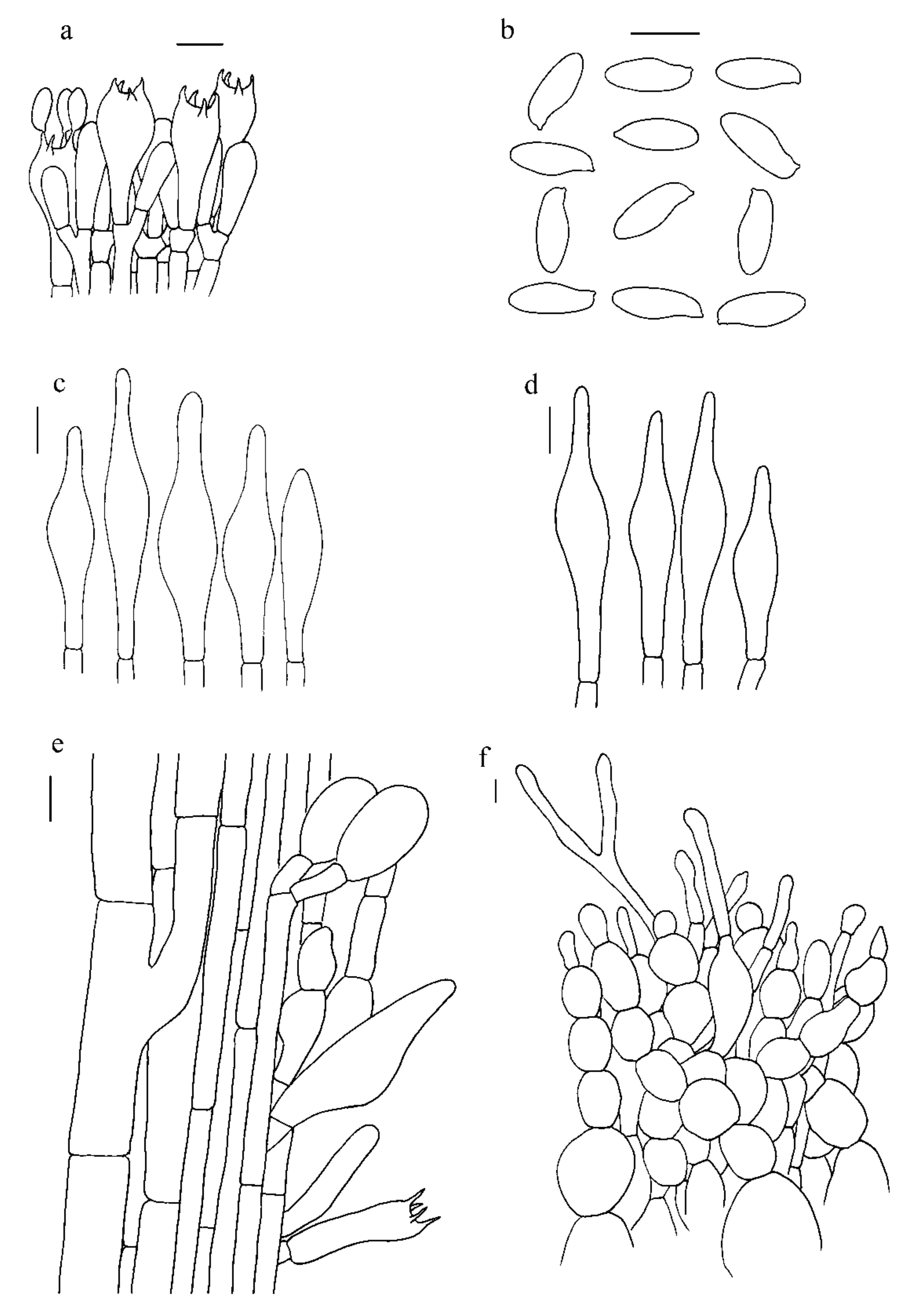
Figure 5. Microscopic features of H. albidum (Type, KUN-HKAS81120). (a). Hymenium and subhymenium; (b). Basidiospores; (c). Cheilocystidia; (d). Pleurocystidia; (e). Stipitipellis; (f). Pileipellis. Bars: a = 20 µm, b = 30 µm, c–e = 20 µm, f = 10 µm.
Jof 07 00823 g005
Figure 6. Microscopic features of H. brevisporum (Type, KUN-HKAS89150). (a). Hymenium and subhymenium; (b). Basidiospores; (c). Pleurocystidia; (d). Cheilocystidia; (e). Stipitipellis; (f). Pileipellis. Bars: a = 20 µm, b = 30 µm, c–e = 20 µm, f = 10 µm.
Jof 07 00823 g006
Figure 7. Microscopic features of H. ferrugineipes (Type, KUN-HKAS115554). (a). Hymenium and subhymenium; (b). Basidiospores; (c). Pleurocystidia; (d). Cheilocystidia; (e). Stipitipellis; (f). Pileipellis. Bars: a = 20 µm, b = 30 µm, c–e = 20 µm, f = 10 µm.
Jof 07 00823 g007
Figure 8. Microscopic features of H. parvum (Type, KUN-HKAS115553) (a). Basidiospores; (b). Hymenium and subhymenium; (c). Pleurocystidia; (d). Cheilocystidia; (e). Stipitipellis; (f). Pileipellis. Bars: a = 30 µm, b–e = 20 µm, f = 10 µm.
Jof 07 00823 g008
Publisher’s Note: MDPI stays neutral with regard to jurisdictional claims in published maps and institutional affiliations.