Article

Unravelling Diaporthe Species Associated with Woody Hosts from Karst Formations (Guizhou) in China

by
Asha J. Dissanayake
1,
Ya-Ya Chen
2,3 and
Jian-Kui (Jack) Liu
1,*
1
Fungal Research Laboratory, School of Life Science and Technology, University of Electronic Science and Technology of China, Chengdu 611731, China
2
Institute of Crop Germplasm Resources, Guizhou Academy of Agricultural Sciences, Guiyang 550006, China
3
Guizhou Key Laboratory of Agricultural Biotechnology, Guizhou Academy of Agricultural Sciences, Guiyang 550006, China
*
Author to whom correspondence should be addressed.
J. Fungi 2020, 6(4), 251; https://doi.org/10.3390/jof6040251
Submission received: 18 August 2020 / Revised: 15 October 2020 / Accepted: 21 October 2020 / Published: 27 October 2020
(This article belongs to the Special Issue Fungal Biodiversity and Ecology)

Abstract

:
Though several Diaporthe species have been reported in China, little is known about the species associated with nature reserves in Guizhou province. During a survey of fungi in six nature reserves in Guizhou province of China, thirty-one Diaporthe isolates were collected from different woody hosts. Based on morphology, culture characteristics and molecular phylogenetic analysis, these isolates were characterized and identified. Phylogenetic analysis of internal transcribed spacer region (ITS), combined with translation elongation factor 1-alpha (tef), β-tubulin (tub), calmodulin (cal) and histone H3 (his) gene regions identified five known Diaporthe species and seven distinct lineages representing novel Diaporthe species. The details of five known species: Diaporthe cercidis, D. cinnamomi, D. conica, D. nobilis and D. sackstonii are given and the seven new species D. constrictospora, D. ellipsospora, D. guttulata, D. irregularis, D. lenispora, D. minima, and D. minusculata are introduced with detailed descriptions and illustrations. This study revealed a high diversity of previously undescribed Diaporthe species associated with woody hosts in various nature reserves of Guizhou province, indicating that there is a potential of Diaporthe species remains to be discovered in this unique landform (Karst formations) in China. Interestingly, the five known Diaporthe species have been reported as pathogens of various hosts, and this could indicate that those newly introduced species in this study could be potentially pathogenic pending further studies to confirm.

1. Introduction

Diaporthe Nitschke (including the Phomopsis asexual morph) belongs to family Diaporthaceae, order Diaporthales and class Sordariomycetes [1,2,3] and its species are found worldwide on a diverse range of host plants as endophytes, pathogens and saprobes [4]. Rossman et al. [5] proposed the name Diaporthe over Phomopsis, as both names are well known amongst plant pathologists and subsequent studies have adopted the latter generic name [4,6,7,8,9,10]. More than 1100 epithets for Diaporthe and 986 for Phomopsis are listed in Index Fungorum (2020) (http://www.indexfungorum.org/, accessed August 2020) with names often based on host association. Many Diaporthe species that are morphologically similar have proven to be genetically distinct [11,12], and several isolates formerly identified based on their hosts were shown to represent different taxa [1]. Diaporthe represents a highly complex genus containing numerous cryptic species. In recent studies, Diaporthe species have been distinguished mainly by their molecular phylogenies, and the best five gene regions to conduct a multi-gene phylogenetic analysis are ITS, tef, tub, cal and his [4,13,14,15,16,17,18,19].
A nature reserve is a protected area of importance for flora, fauna or landscapes of geological or other special interest, which is reserved and managed for purposes of conservation and to provide special opportunities for study or research [20]. The Karst region of Guizhou province is comprised of abundant nature reserves that provide a wide range of ecosystem services such as water supply, soil fertility, ecotourism, recreation, biodiversity conservation and carbon sequestration [20]. However, there are few scientific evaluations made for fungi in national nature reserves and national forest parks in Guizhou province, China [21,22]. During the investigation carried out in 2017 to 2019, several isolates of Diaporthe species were collected from six nature reserves in Guizhou province including Fanjing mountain, Guiyang Huaxi wetland park, Guiyang Xiaochehe wetland park, Maolan nature reserve, Suiyang broad water nature reserve and Xingyi Wanfenglin. Fungi isolated from forest trees in China were recorded in old fungal literature, however, most of them lack living culture and molecular data [23,24]. Although several species of Diaporthe have been previously recorded from Guizhou province with details of culture and molecular data [25,26], little is known to associate these with hosts in nature reserves. Thus, the aim of this study is to describe and illustrate Diaporthe taxa from nature reserves in the Karst region of Guizhou province based on morphological characters and phylogenies derived from combined ITS, tef, tub, cal and his gene sequences.

2. Materials and Methods

2.1. Isolation of Fungal Material, Morphology and Culture Characteristics

From 2017 to 2019, thirty-one Diaporthe specimens were collected in field surveys of decaying saprobic woody hosts in different nature reserves including Fanjing mountain, Guiyang Huaxi wetland park, Guiyang Xiaochehe wetland park, Maolan nature reserve, Suiyang broad water nature reserve and Xingyi Wanfenglin in Karst region of Guizhou province (Table 1). Collected samples were taken to the laboratory for isolation and photographed, documented and then kept at 4 °C for further study.
Species identification was primarily based on morphological observation of the conidiomata or ascomata from host materials and micromorphology supplemented by culture characteristics. Morphological observations were made using a Motic SMZ (Stereoscopic Zoom Microscope) 168 series stereomicroscope and photographed by a Nikon E80i microscope-camera system. Measurements were made with the Tarosoft (R) Image FrameWork [27] and images used for figures were processed with Adobe Photoshop CS v. 5. Single spore isolations were prepared following the method of Chomnunti et al. [28]. Spore germination on 2% water agar (WA) was examined after 24 h and germinating spores were transferred to potato dextrose agar (PDA) media. Cultures were incubated at 25 °C in the dark and colony morphology and conidial characteristics were examined for a total of 31 isolates. Colony color was determined according to Rayner [29] after 5 d to 10 d on PDA at 25 °C in the dark. More than 20 conidiomata/ascomata, 30 asci, and 50 conidia/ascospores were measured to calculate the mean size/length and respective standard deviations (SD). Conidial shape, color and guttulation were also recorded.
Herbarium specimens were deposited at the Herbarium of Cryptogams, Kunming Institute of Botany Academia Sinica (KUN-HKAS), Kunming, China and herbaria of Guizhou Academy of Agricultural Sciences (GZAAS), Guiyang, China. The living cultures were deposited in the China General Microbiological Culture Collection Center (CGMCC) in Beijing, China and Guizhou Culture Collection (GZCC) in Guiyang, China and (Table 1).

2.2. Molecular Based Amplification

Fungal mycelium of 7 d old cultures was scraped for the extraction of genomic DNA using Biospin Fungus Genomic DNA Extraction Kit (BioFlux®) following the manufacturer’s protocol (Hangzhou, China). For the identification of Diaporthe specimens, the internal transcribed spacer region (ITS) was sequenced for all isolates and BLAST search (basic local alignment search tool) at GenBank was used to reveal the closest matching taxa. Besides ITS gene sequence data, translation elongation factor 1-alpha (tef), β-tubulin (tub), calmodulin (cal) and histone H3 (his) gene regions were also employed to support the species identification. The ITS region was amplified using universal primers ITS1 and ITS4 [30]. The target region of the tef gene was amplified using primer pairs EF-728F and EF-986R [31]. A portion of the tub gene was amplified using the primers BT2a and BT2b [32], while the primer pair CAL228F and CAL737R was used to amplify the cal gene region [31]. The primers CYLH3F [33] and H3-1b [32] were used to amplify part of the his gene. The PCR reactions were accomplished in a Bio Rad C1000 thermal cycler. The amplification procedure was performed in a 50 μL reaction volume containing 5–10 ng DNA, 0.8 units Taq polymerase, 1X PCR buffer, 0.2 mM dNTP, 0.3 μm of each primer with 1.5 mM MgCl2. Following the PCR amplification, products were visualized on 1% agarose gel under UV light using a Gel DocTM XR Molecular Imager following ethidium bromide staining. PCR products were purified using minicolumns, purification resin and buffer according to the manufacturer’s protocols (Amersham product code: 27–9602–01). Sequence analysis was carried out by Shanghai Sangon Biological Engineering Technology and Services Co., Ltd. (Shanghai, China).

2.3. Sequence Alignment and Phylogenetic Analyses

To assure the sequence quality, the resulting sequence chromatograms were checked using BioEdit v.5 [34]. An overview phylogenetic tree (provided as a Supplementary Figure S1) for the genus Diaporthe was constructed from ITS sequence data of all type/ex-type/neo-type Diaporthe species from previous studies [10,13,14,15,33,35,36,37,38,39,40,41,42,43,44,45]. Considering this ITS tree, another phylogenetic analysis was conducted including all the isolates obtained in this study (Table 1) together with several closely associated Diaporthe species (Table 2). Diaporthella corylina (CBS 121124) was selected as the outgroup taxon. The sequences were retrieved from GenBank and aligned with the sequences obtained in this study using MAFFT [46] (http://www.ebi.ac.uk/Tools/msa/mafft/) and manually edited with BioEdit [34] for a maximum alignment. Phylogenetic analysis was performed by using PAUP (Phylogenetic Analysis Using Parsimony) v.4.0b10 for maximum parsimony (MP) method [47], RAxML for maximum likelihood (ML) method [48] and MrBayes v.3.1.2 for Bayesian Inference (BI) method [49]. The best model of evolution was determined by MrModeltest v. 2.3 [50]. Maximum likelihood analyses was accomplished using RAxML GUI v. 0.9b2 [51] with 1000 non-parametric bootstrapping iterations, using the general time-reversible model (GTR) with a discrete gamma distribution. The best scoring trees were chosen with final likelihood values.
Ambiguous regions in the MP alignment were excluded, and gaps were treated as missing data. The stability of the trees was evaluated by 1000 bootstrap replications. Branches of zero length were collapsed, and all multiple parsimonious trees saved. Statistics including tree length (TL), consistency index (CI), retention index (RI), relative consistency index (RC) and homoplasy index (HI) were calculated. Differences between the trees inferred under different optimality criteria were evaluated using Kishino–Hasegawa tests (KHT) [52].
Bayesian analyses were performed in MrBayes v.3.0b4 [49] and posterior probabilities (PP) were determined by Markov Chain Monte Carlo sampling (MCMC). MrModeltest v. 2.3 [50] was used for the statistical selection of the best-fit model of nucleotide substitutions and was integrated into the analysis. Six simultaneous Markov chains were run for 106 generations; sampling the trees at every 100th generation. From the 10,000 trees obtained, the first 2000 representing the burn-in phase were discarded. The remaining 8000 trees were used for calculating posterior probabilities in the majority rule consensus tree.
The details of the fungal strains obtained in this study are listed in Table 1 with information of the type cultures and sequence data. Sequences generated in this study were deposited in GenBank (Table 1); alignments and trees were deposited in TreeBASE (www.treebase.org, study ID S27013). Reviewer access URL: http://purl.org/phylo/treebase/phylows/study/TB2:S27013?x-access-code=1369710211c386567d8b43ba36f49adf&format=html. Taxonomic novelties were submitted to the Faces of Fungi database [53], Index Fungorum (Index Fungorum 2020) and MycoBank (www.mycobank.org) [33].

3. Results

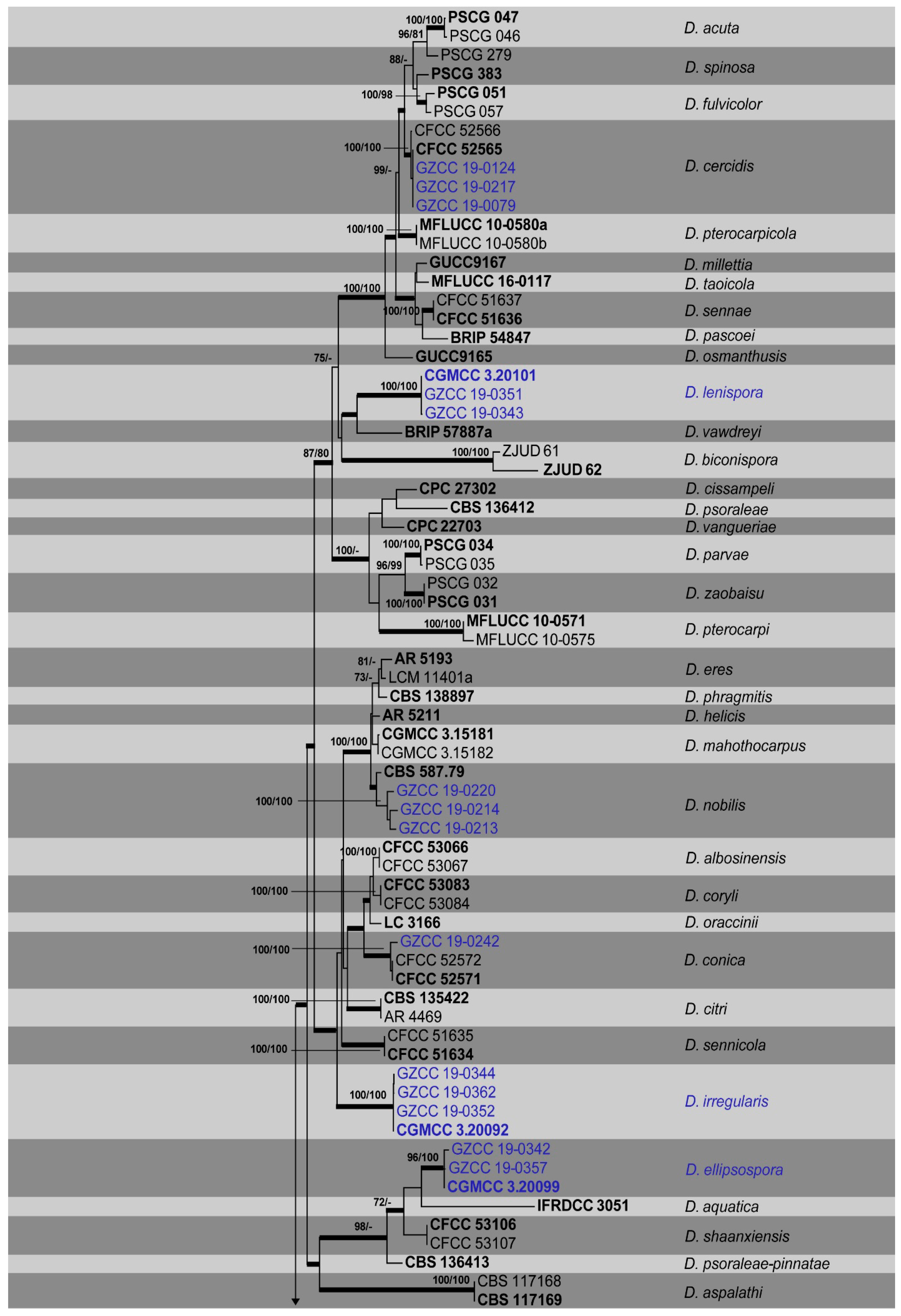
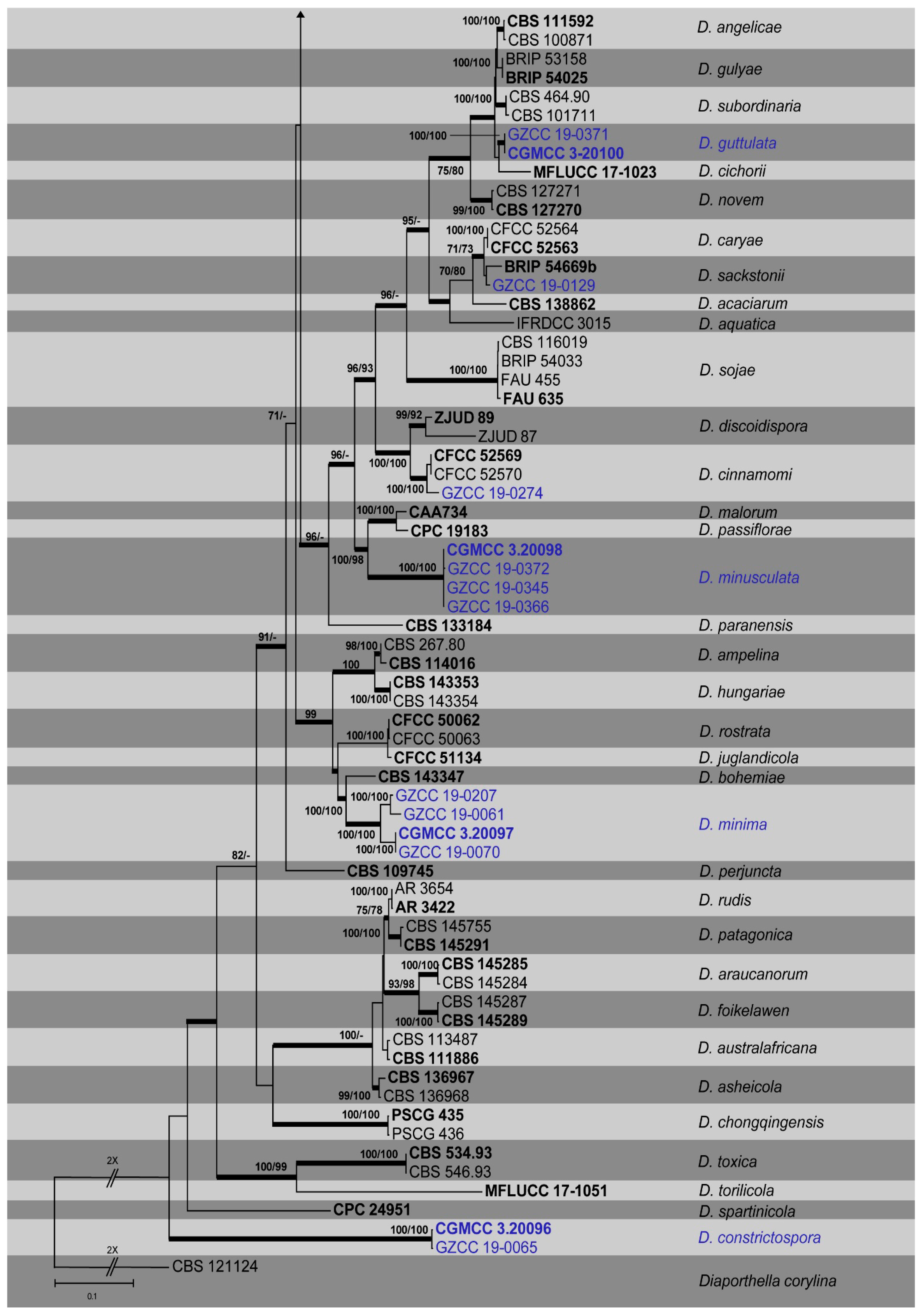
3.1. Phylogenetic Analyses

Saprobic specimens sampled from numerous woody hosts in six nature reserves in the Karst region of Guizhou province, China resulted in the isolation of thirty-one isolates of Diaporthe (Table 1, Figure 1). The ITS gene was employed for the identification of all isolates to the genus level. The ITS, tef, tub, cal and his alignments (including the gaps) were determined to be approximately 570, 470, 450, 610 and 500 bp (base pair) in size, respectively. The combined ITS, tef, tub, cal and his sequences of Diaporthe contained data for 136 isolates, including the outgroup taxon Diaporthella corylina (CBS 121124). The analyses consisted of 31 isolates from this study (Table 1) and 105 sequences (62 type species) originating from GenBank (Table 2). Out of a total of 2594 characters in the MP analyses, 1079 were constant, and 269 were variable and parsimony uninformative. Ten most parsimonious trees resulted from the remaining 1246 parsimony-informative characters (TL = 7439, CI = 0.384, RI = 0.804, RC = 0.309, HI = 0.616). In the ML analyses, the best scoring RAxML tree (Figure 1) with a final likelihood value of −37549.830874 is presented. The matrix had 1675 distinct alignment patterns, with 31.29% of undetermined characters or gaps. Estimated base frequencies were as follows: A = 0.212875, C = 0.328600, G = 0.237362, T = 0.221162; substitution rates AC = 1.075393, AG = 2.704248, AT = 1.155000, CG = 0.851430, CT = 3.774119, GT = 1.000000; gamma distribution shape parameter alpha = 0.464825. The Maximum likelihood (ML) and Bayesian methods (BI) for phylogenetic analyses performed trees with similar topologies.
Table 2. GenBank accession numbers of species included in the phylogenetic analysis (Figure 1). Ex-type/ex-epitype/ex-isotype/ex-neotype isolates are in bold.
Species NameIsolate NumberITStubtefcalhisReference
Diaporthella corylinaCBS 121124KC343004KC343972KC343730KC343246KC343488Vasilyeva et al. [54]
Diaporthe acaciarumCBS 138862KP004460KP004509N/AN/AKP004504Crous et al. [55]
Diaporthe acutaPSCG 047MK626957MK691225MK654802MK691125MK726161Guo et al. [15]
Diaporthe acutaPSCG 046MK626958MK691224MK654803MK691124MK726162Guo et al. [15]
Diaporthe albosinensisCFCC 53066MK432659MK578059MK578133MK442979MK443004Yang et al. [14]
Diaporthe albosinensisCFCC 53067MK432660MK578060MK578134MK442980MK443005Yang et al. [14]
Diaporthe ampelinaCBS 114016AF230751JX275452AY745056AY230751N/AMostert et al. [56]
Diaporthe ampelinaCBS 267.80KC343018KC343986KC343744KC343260KC343502Mostert et al. [56]
Diaporthe angelicaeCBS 111592KC343027KC343995KC343753KC343269KC343511Castlebury et al. [57]
Diaporthe angelicaeCBS 100871KC343025KC343993KC343751KC343267KC343509Castlebury et al. [57]
Diaporthe aquaticaIFRDCC 3015JQ797438N/AN/AN/AN/AHu et al. [58]
Diaporthe aquaticaIFRDCC 3051JQ797437N/AN/AN/AN/AHu et al. [58]
Diaporthe araucanorumCBS 145285MN509711MN509722MN509733N/AN/AZapata et al. [45]
Diaporthe araucanorumCBS 145284MN509710MN509721MN509732N/AN/AZapata et al. [45]
Diaporthe asheicolaCBS 136967KJ160562KJ160518KJ160594KJ160542N/ALombard et al. [59]
Diaporthe asheicolaCBS 136968KJ160563KJ160519KJ160595KJ160543N/ALombard et al. [59]
Diaporthe aspalathiCBS 117168KC343035KC344003KC343761KC343277KC343519van Rensburg et al. [11]
Diaporthe aspalathiCBS 117169KC343036KC344004KC343762KC343278KC343520van Rensburg et al. [11]
Diaporthe australafricanaCBS 111886KC343038KC344006KC343764KC343280KC343522Mostert et al. [56]
Diaporthe australafricanaCBS 113487KC343039KC344007KC343765KC343281KC343523Mostert et al. [56]
Diaporthe biconisporaZJUD61KJ490596KJ490417KJ490475N/AKJ490538Huang et al. [60]
Diaporthe biconisporaZJUD62KJ490597KJ490418KJ490476KJ490539KJ490539Huang et al. [60]
Diaporthe bohemiaeCBS 143347MG281015MG281188MG281536MG281710MG281361Guarnaccia et al. [19]
Diaporthe caryaeCFCC 52563MH121498MH121580MH121540MH121422MH121458Yang et al. [13]
Diaporthe caryaeCFCC 52564MH121499MH121581MH121541MH121423MH121459Yang et al. [13]
Diaporthe cercidisCFCC 52565MH121500MH121582MH121542MH121424MH121460Yang et al. [13]
Diaporthe cercidisCFCC 52566MH121501MH121583MH121543MH121425MH121461Yang et al. [13]
Diaporthe chongqingensisPSCG 435MK626916MK691321MK654866MK691209MK726257Guo et al. [15]
Diaporthe chongqingensisPSCG 436MK626917MK691322MK654867MK691208MK726256Guo et al. [15]
Diaporthe cichoriiMFLUCC 17-1023KY964220KY964104KY964176KY964133N/ADissanayake et al. [9]
Diaporthe cinnamomiCFCC 52569MH121504MH121586MH121546N/AMH121464Yang et al. [13]
Diaporthe cinnamomiCFCC 52570MH121505MH121587MH121547N/AMH121465Yang et al. [13]
Diaporthe cissampeliCPC 27302KX228273KX228384N/AN/AKX228366Crous et al. [61]
Diaporthe citriCBS 135422KC843311KC843187KC843071KC843157MF418281Udayanga et al. [6]
Diaporthe citriAR 4469KC843321KC843197KC843081KC843167N/AUdayanga et al. [6]
Diaporthe conicaCFCC 52571MH121506MH121588MH121548MH121428MH121466Yang et al. [13]
Diaporthe conicaCFCC 52572MH121507MH121589MH121549MH121429MH121467Yang et al. [13]
Diaporthe coryliCFCC 53083MK432661MK578061MK578135MK442981MK443006Yang et al. [14]
Diaporthe coryliCFCC 53084MK432662MK578062MK578136MK442982MK443007Yang et al. [14]
Diaporthe discoidisporaZJUD89KJ490624KJ490445KJ490503N/AKJ490566Huang et al. [60]
Diaporthe discoidisporaZJUD87KJ490622KJ490443KJ490501N/AKJ490564Huang et al. [60]
Diaporthe eresAR 5193KJ210529KJ420799KJ210550KJ434999KJ420850Udayanga et al. [7]
Diaporthe eresCBS 138598KJ210521KJ420787KJ210545KJ435027KJ420837Udayanga et al. [7]
Diaporthe foikelawenCBS 145289MN509714MN509725MN509736N/AN/AZapata et al. [45]
Diaporthe foikelawenCBS 145287MN509713MN509724MN509735N/AN/AZapata et al. [45]
Diaporthe fulvicolorPSCG 051MK626859MK691236MK654806MK691132MK726163Guo et al. [15]
Diaporthe fulvicolorPSCG 057MK626858MK691233MK654810MK691131MK726164Guo et al. [15]
Diaporthe gulyaeBRIP 53158JF431284KJ197271N645799N/AN/AThompson et al. [62]
Diaporthe gulyaeBRIP 54025JF431299KJ197272JN645803N/AN/AThompson et al. [62]
Diaporthe helicisAR 5211KJ210538KJ420828KJ210559KJ435043KJ420875Udayanga et al. [7]
Diaporthe hungariaeCBS 143353MG281126MG281299MG281647MG281823MG281474Guarnaccia et al. [19]
Diaporthe hungariaeCBS 143354MG281127MG281300MG281648MG281824MG281475Guarnaccia et al. [19]
Diaporthe juglandicolaCFCC 51134KU985101KX024634KX024628KX024616KX024622Yang et al. [18]
Diaporthe mahothocarpusCGMCC 3.15181KC153096KF576312KC153087N/AN/AGao et al. [63]
Diaporthe mahothocarpusCGMCC 3.15182KC153097N/AKC153088N/AN/AGao et al. [63]
Diaporthe malorumCAA734KY435638KY435668KY435627KY435658KY435648Santos et al. [16]
Diaporthe millettiaGUCC 9167MK398674MK502089MK480609MK502086N/ALong et al. [26]
Diaporthe nobilisCBS 587.79KC343153KC344121KC343879KC343395KC343637Li et al. [25]
Diaporthe novemCBS 127270KC343155KC344123KC343881KC343397KC343640Santos et al. [64]
Diaporthe novemCBS 127271KC343157KC344125KC343883KC343399KC343641Santos et al. [64]
Diaporthe oracciniiLC 3166KP267863KP293443KP267937N/AKP293517Gao et al. [63]
Diaporthe osmanthusisGUCC 9165MK398675MK502090MK480610MK502087N/ALong et al. [26]
Diaporthe paranensisCBS 133184KC343171KC344139KC343897KC343413KC343655Gomes et al. [4]
Diaporthe parvaePSCG 034MK626919MK691248MK654858N/AMK726210Guo et al. [15]
Diaporthe parvaePSCG 035MK626920MK691249MK654859MK691169MK726211Guo et al. [15]
Diaporthe pascoeiBRIP 54847JX862532KF170924JX862538N/AN/ATan et al. [65]
Diaporthe passifloraeCPC 19183JX069860KY435674KY435633KY435664KY435654Crous et al. [66]
Diaporthe patagonicaCBS 145291MN509717MN509728MN509739N/AN/AZapata et al. [45]
Diaporthe patagonicaCBS 145755MN509718MN509729MN509740N/AN/AZapata et al. [45]
Diaporthe perjunctaCBS 109745KC343172KC344140KC343898KC343414KC343656van Niekerk et al. [67]
Diaporthe phragmitisCBS 138897KP004445KP004507N/AN/AKP004503Crous et al. [55]
Diaporthe psoraleaeCBS 136412KF777158KF777251KF777245N/AN/ACrous et al. [68]
Diaporthe psoraleae-pinnataeCBS 136413KF777159KF777252N/AN/AN/ACrous et al. [68]
Diaporthe pterocarpicolaMFLUCC 10-0580aJQ619887JX275441JX275403JX197433N/AUdayanga et al. [69]
Diaporthe pterocarpicolaMFLUCC 10-0580bJQ619887JX275441JX275403JX197433N/AUdayanga et al. [69]
Diaporthe pterocarpiMFLUCC 10-0571JX197433JX275460JX275416JX197451N/AUdayanga et al. [69]
Diaporthe pterocarpiMFLUCC 10-0575JQ619901JX275462JX275418JX197453N/AUdayanga et al. [69]
Diaporthe rostrataCFCC 50062KP208847KP208855KP208853KP208849KP208851Fan et al. [17]
Diaporthe rostrataCFCC 50063KP208848KP208856KP208854KP208850KP208852Fan et al. [17]
Diaporthe rudisAR 3422KC843331KC843177KC843090KC843146N/AUdayanga et al. [6]
Diaporthe rudisAR 3654KC843338KC843184KC843097KC843153N/AUdayanga et al. [6]
Diaporthe sackstoniiBRIP 54669bKJ197287KJ197267KJ197249N/AN/AThompson et al. [70]
Diaporthe sennaeCFCC 51636KY203724KY228891KY228885KY228875N/AYang et al. [18]
Diaporthe sennaeCFCC 51637KY203725KY228892KY228886KY228876N/AYang et al. [18]
Diaporthe sennicolaCFCC 51634KY203722KY228889KY228883KY228873KY228879Yang et al. [18]
Diaporthe sennicolaCFCC 51635KY203723KY228890KY228884KY228874KY228880Yang et al. [18]
Diaporthe shaanxiensisCFCC 53106MK432654N/AMK578130MK442976MK443001Yang et al. [14]
Diaporthe shaanxiensisCFCC 53107MK432655N/AMK578131MK442977MK443002Yang et al. [14]
Diaporthe sojaeBRIP 54033JF431295N/AKC343901N/AN/AUdayanga et al. [71]
Diaporthe sojaeCBS 116019KC343175KC344143KC343901KC343417KC343659Udayanga et al. [71]
Diaporthe sojaeFAU 455KJ590712KJ610868KJ590755KJ612109KJ659201Udayanga et al. [71]
Diaporthe sojaeFAU 635KJ590719KJ610875KJ590762KJ612116KJ659208Udayanga et al. [71]
Diaporthe spartinicolaCPC 24951KR611879KR857695N/AN/AKR857696Crous et al. [71]
Diaporthe spinosaPSCG 383MK626849MK691234MK654811MK691129MK726156Guo et al. [15]
Diaporthe spinosaPSCG 279MK626925MK691235MK654801MK691126MK726155Guo et al. [15]
Diaporthe subordinariaCBS 464.90KC343214KC344182KC343940KC343456KC343698Gomes et al. [4]
Diaporthe subordinariaCBS 101711KC343213KC344182KC343939KC343455KC343697Gomes et al. [4]
Diaporthe taoicolaMFLUCC 16-0117KU557567KU557591KU557635N/AN/ADissanayake et al. [9]
Diaporthe torilicolaMFLUCC 17-1051KY964212KY964096KY964168KY964127N/ADissanayake et al. [9]
Diaporthe toxicaCBS 534.93KC343220KC344188KC343946KC343462KC343704Williamson et al. [72]
Diaporthe toxicaCBS 546.93KC343222KC344190KC343948KC343464KC343706Williamson et al. [72]
Diaporthe vangueriaeCPC 22703KJ869137KJ869247N/AN/AN/ACrous et al. [55]
Diaporthe vawdreyiBRIP 57887aKR936126KR936128KR936129N/AN/ACrous et al. [73]
Diaporthe zaobaisuPSCG 031MK626922MK691245MK654855N/AMK726207Guo et al. [15]
Diaporthe zaobaisuPSCG 032MK626923MK691246MK654856N/AMK726208Guo et al. [15]
AR: Collection of A.Y. Rossman; BRIP: Queensland Plant Pathology herbarium/culture collection, Australia; CBS: Culture collection of the Centraalbureau voor Schimmelcultures, Fungal Biodiversity Centre, Utrecht, The Netherlands; CFCC: China Forestry Culture Collection Center, China; CGMCC: China General Microbiological Culture Collection; CPC: Collection Pedro Crous, housed at CBS; FAU: Isolates in culture collection of Systematic Mycology and Microbiology Laboratory, USDA-ARS, Beltsville, MD, USA; GUCC: Guizhou culture collection, Guizhou, China; IFRDCC: International Fungal Research and Development Centre Culture Collection, Chinese Academy of Forestry, Kunming, China; LC: Corresponding author’s personal collection (deposited in laboratory State Key Laboratory of Mycology, Institute of Microbiology, Chinese Academy of Sciences); MFLUCC: Mae Fah Luang University Culture Collection, Chiang Rai, Thailand; ZJUD: Zhejiang University. ITS, internal transcribed spacers 1 and 2 together with 5.8S nrDNA; tub, partial beta-tubulin gene; cal, partial calmodulin gene and tef, partial translation elongation factor 1-a gene, his, histone H3 gene.
The isolates obtained in this study were grouped into twelve clades. Three isolates were grouped with the ex-type of Diaporthe cercidis (CFCC 52565) while another three isolates were clustered with the ex-type of D. nobilis (CBS 587.79). In addition one isolate with D. cinnamomi (CFCC 52569), D. conica (CFCC 52571) and D. sackstonii (BRIP 54669b) respectively. Twenty-two isolates did not cluster with any known Diaporthe species; thus, seven novel species, Diaporthe constrictospora (2 isolates, Figure 2), Diaporthe ellipsospora (3 isolates, Figure 3), Diaporthe guttulata (2 isolates, Figure 4), Diaporthe irregularis (4 isolates, Figure 5), Diaporthe lenispora (3 isolates, Figure 6), Diaporthe minima (4 isolates, Figure 7) and Diaporthe minusculata (4 isolates, Figure 8) are determined to be new species based on the morphological and phylogenetic evidence (Figure 1).

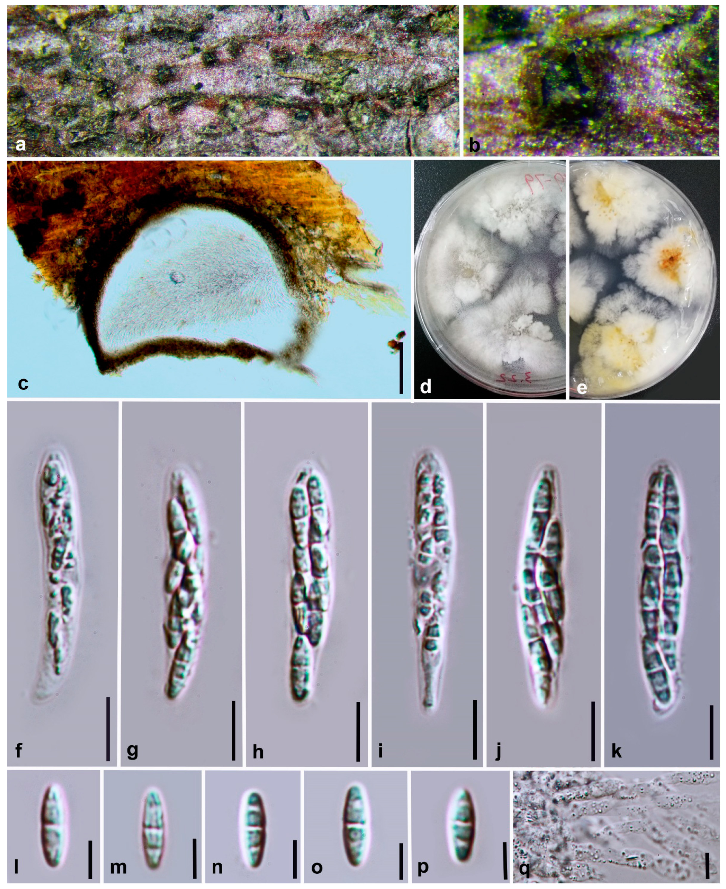
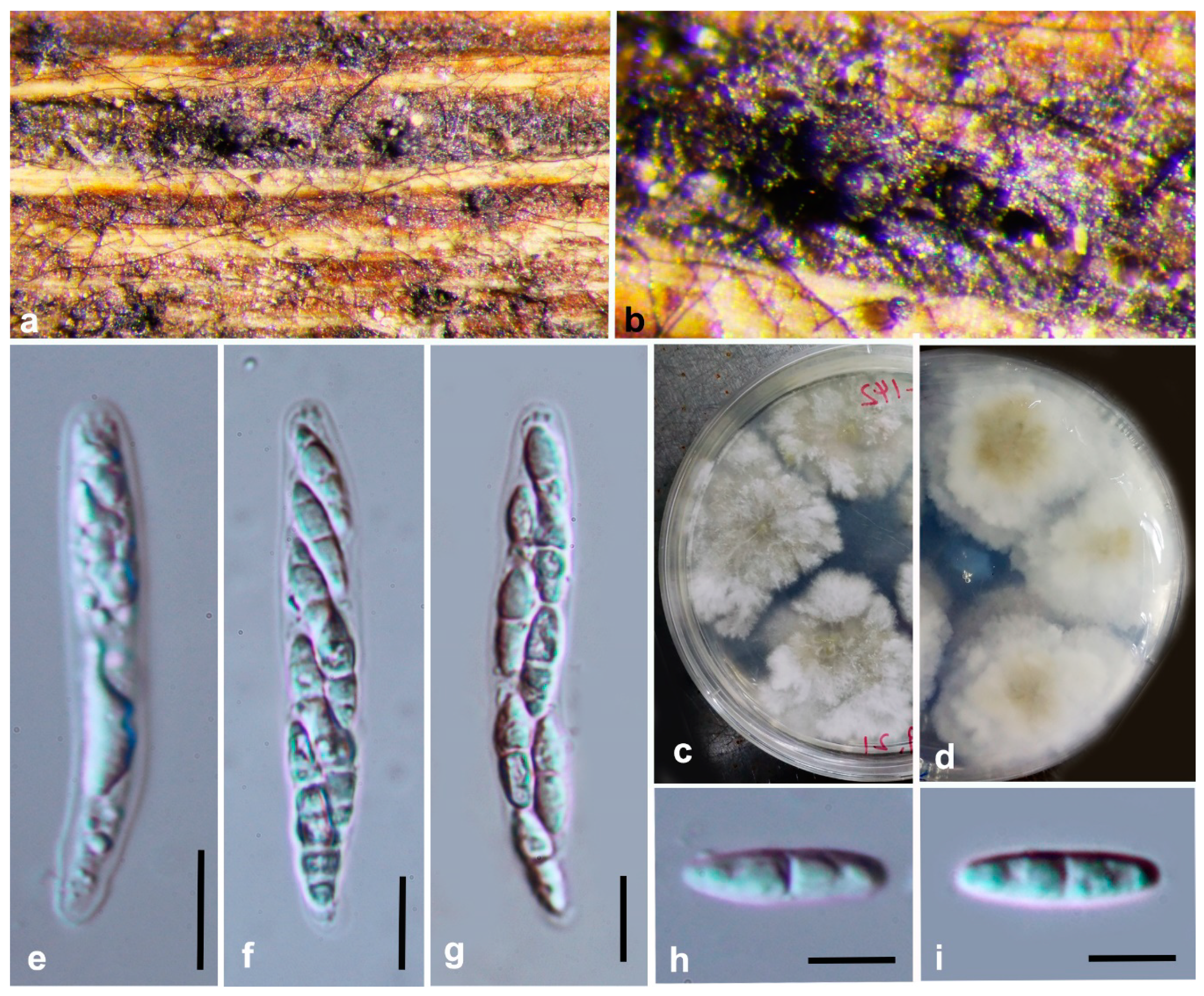
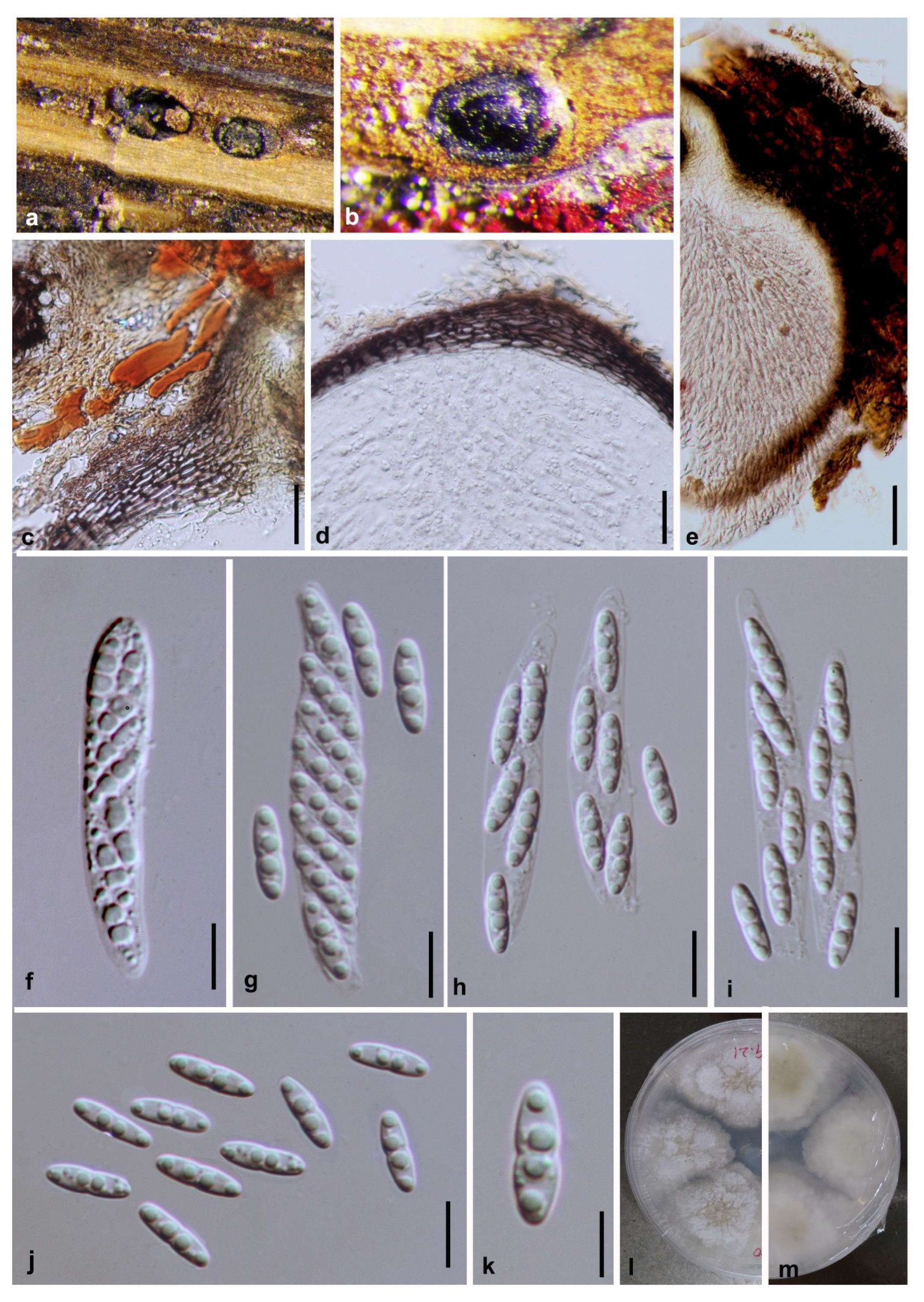
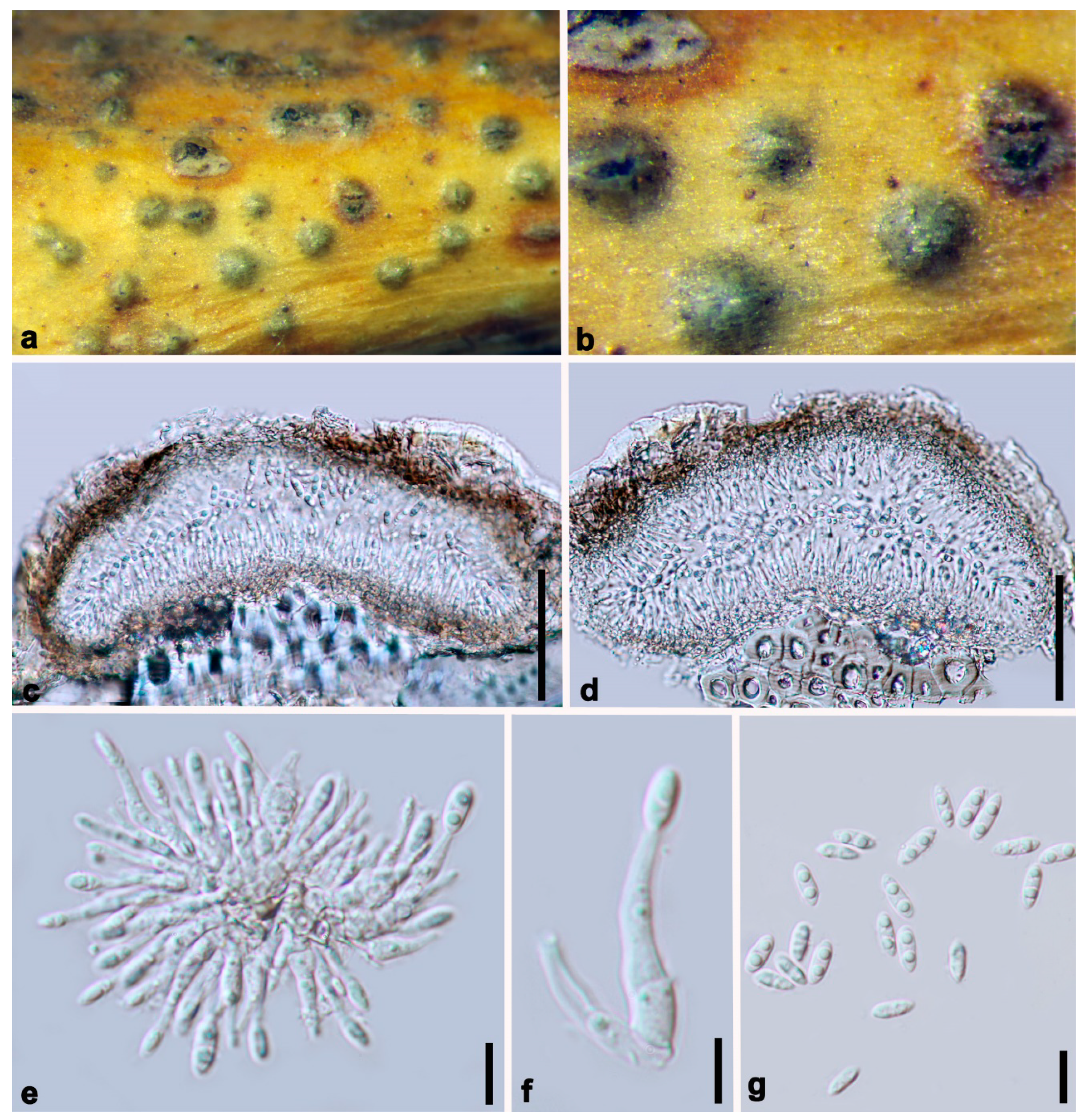
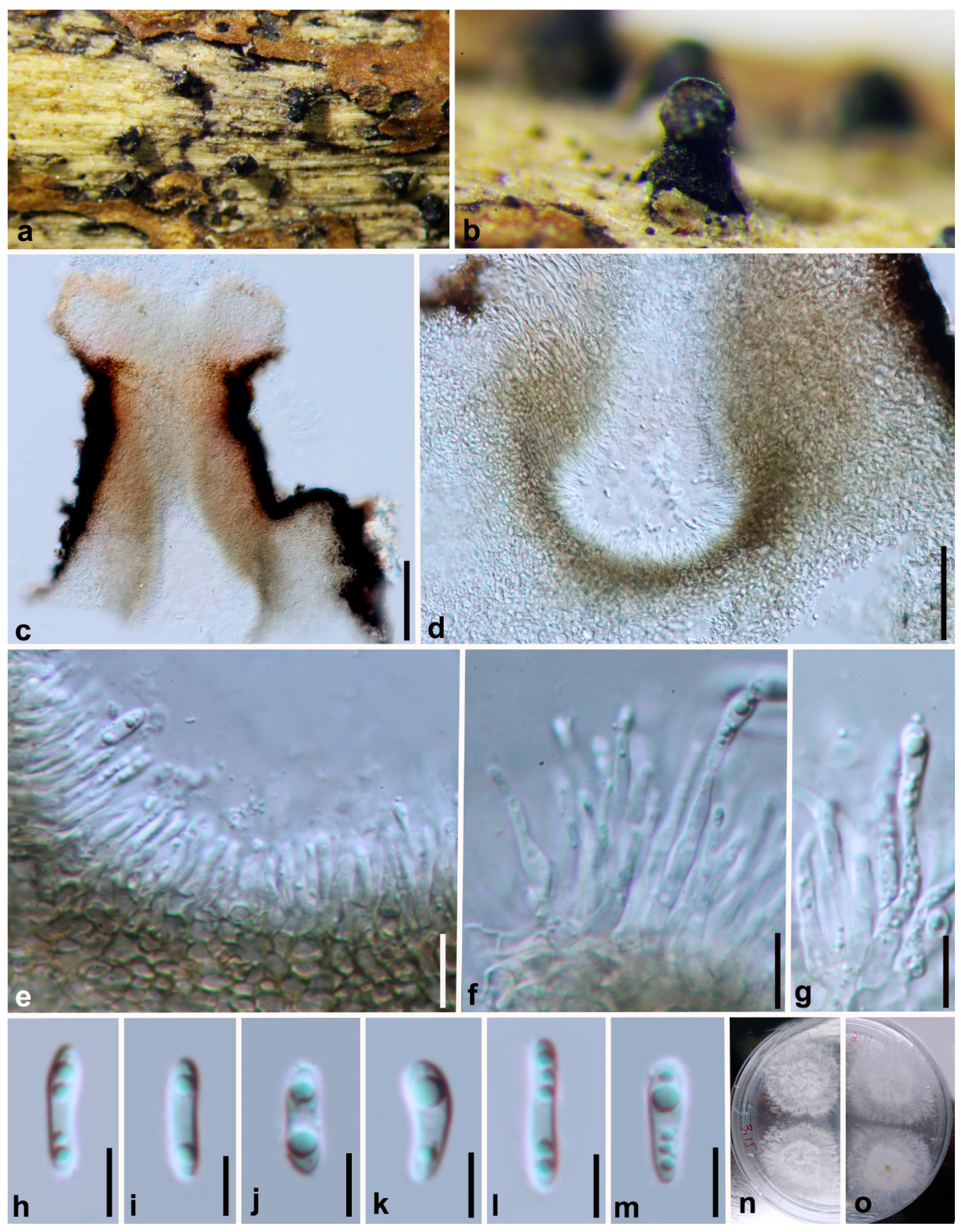
3.2. Morphology and Culture Characteristics

In this study, thirty-one Diaporthe isolates were obtained from decaying woody hosts from six nature reserves in Guizhou province, China (Table 1). The Diaporthe isolates obtained in this study were further categorized based on morphological characteristics. Growth was rapid for all isolates grown on PDA, with mycelia covering the whole surface of the Petri dishes. Aerial mycelium was initially white and turned greyish after incubation in the dark at 25 °C for several days. All species exhibited phenotypic characteristics typical of the genus. The seven new species of Diaporthe described here are phylogenetically distinct from all previously described species for which sequence data are available.

Taxonomy 

Diaporthe constrictospora Y.Y. Chen, A.J. Dissanayake and Jian K. Liu sp. nov. (Figure 2)
Index Fungorum number: IF557388; Facesoffungi Number: FoF07853; MycoBank Number: MB836211
Etymology: The epithet from the Latin words constrictus and spora, refers to the slight central constriction often present in the ascospores.
Holotype: HKAS 107534
Saprobic on decaying wood. Sexual morph: Ascomata 190–240 μm diam, black, globose to subglobose or irregular, clustered or solitary, deeply immersed in host tissue. Asci 40–48 μm × 9–11 μm ( x ¯ = 43 × 8, n = 30), 8-spored, unitunicate, sessile, elongate to clavate. Ascospores 10–12 × 3–4 μm ( x ¯ = 11 × 4, n = 50), hyaline, elongated to elliptical, two-celled, often 4-guttulate, with larger guttules at center and smaller ones at the ends. Asexual morph: Not observed.
Culture characteristics: Colonies covering entire PDA Petri dishes after 10 d at 25 °C producing abundant white aerial mycelium, reverse fuscous black.
Material examined: China, Guizhou Province, Maolan Nature Reserve, saprobic on decaying woody host, April 2017, Y. Y. Chen (HKAS 107534, holotype); ex-type living culture CGMCC 3.20096 = GZCC19-0084; ibid, Guiyang District, Huaxi Wetland Park, saprobic on decaying woody host, July 2017, Y. Y. Chen (GZAAS 19-1784, paratype), living culture GZCC 19-0065.
Notes: Two strains representing Diaporthe constrictospora cluster in a well-supported basal clade (ML/MP/BI = 100/100/1.0) and appear to be distinct from other Diaporthe species, and can be easily recognized by their distinctive phylogenetic placement (Figure 1). Since this species is not closely related to any Diaporthe species and we were unable to compare the nucleotide differences in the alignment. Diaporthe constrictospora is introduced as a phylogenetically distinct species (Figure 1).
Diaporthe ellipsospora Y.Y. Chen, A.J. Dissanayake and Jian K. Liu sp. nov. (Figure 3)
Index Fungorum number: IF557389; Facesoffungi Number: FoF07854; MycoBank Number: MB836175
Etymology: The specific epithet ellipsospora refers to the shape of the ascospores.
Holotype: HKAS 107535
Saprobic on decaying branch. Sexual morph: Ascomata 380–430 μm diam, black, globose to irregular, scattered on dead twigs, immersed in host tissue, protruding through substrata. Paraphyses up to 100 μm long, rarely present, hyaline, smooth, 1–3-septate, cylindrical with obtuse ends, extending above conidiophores. Asci 40–47 ×7–8.5 μm ( x ¯ = 43 × 7.5, n = 30), 8-spored, unitunicate, sessile, elongate to clavate. Ascospores 8.5–13 × 2.5–3.5 μm ( x ¯ = 10.1 × 3.3, n = 50), hyaline, elongated to elliptical, two-celled, often 4-guttulate, with larger guttules at center and smaller ones at ends. Asexual morph: Not observed.
Culture characteristics: Colonies on PDA fast growing, covering entire PDA Petri dish after 8 d at 25 °C. White, aerial mycelium turning grey at edges of plate, reverse yellowish pigmentation developing in centre.
Material examined: China, Guizhou Province, Xingyi Wanfenglin, Saprobic on decaying branch, June 2019, Y.Y. Chen (HKAS 107535, holotype), ex-type living culture CGMCC 3.20099 = GZCC 19-0231; ibid, (GZAAS 19-2061, paratype), living culture GZCC 19-0342; ibid, Maolan Nature Reserve, saprobic on decaying woody host, June 2019, Y. Y. Chen, GZAAS 19-2080, living culture GZCC 19-0357.
Notes:Diaporthe ellipsospora formed an independent clade (Figure 1) and is phylogenetically distinct from D. aquatica in a well-supported clade (ML/MP/BI = 96/100/1.0). Diaporthe ellipsospora can be distinguished from D. aquatica (IFRDCC 3051) only based on ITS locus (17/539) since other gene sequences (tef, tub, cal and his) are unavailable for D. aquatica. Diaporthe ellipsospora can be morphologically differentiated from D. aquatica as the latter has long necks up to 2250 μm [58].
Diaporthe guttulata Y.Y. Chen, A.J. Dissanayake and Jian K. Liu sp. nov. (Figure 4)
Index Fungorum number: IF557390; Facesoffungi Number: FoF07855; MycoBank Number: MB836212
Etymology: Referring to the ascospores with large guttules.
Holotype: HKAS 107536
Saprobic on decaying branch. Sexual morph: Ascomata 560–630 μm diam, black, globose to conical, scattered irregularly, immersed in host tissue with elongated, 300–400 μm long necks protruding through substrata. Asci 45–57 μm × 7–9 μm ( x ¯ = 50 × 8, n = 30), unitunicate, 8-spored, sessile, elongate to clavate. Ascospores 12–15 × 3–4 μm ( x ¯ = 13 × 3.1, n = 50), elongated to elliptical, hyaline, two-celled, often 4-guttulate, with larger guttules at centre and smaller one at ends. Asexual morph: Not observed.
Culture characteristics: Colonies covering entire PDA Petri dishes after 7 d at 25 °C producing abundant white aerial mycelium. Reverse white, turning to grey in centre and no conidia produced.
Material examined: China, Guizhou Province, Maolan Nature Reserve, saprobic on decaying branch, July 2017, Y.Y. Chen (HKAS 107536, holotype), ex-type living culture CGMCC 3.20100 = GZCC 19-0140; ibid, Guiyang District, Suiyang broad water nature reserve, saprobic on decaying woody host, June 2018, Y. Y. Chen (GZAAS 19-2067, paratype), living culture GZCC 19-0371.
Notes:Diaporthe guttulata formed a distinct clade with high support (ML/BI = 88/1.0), and differed with the closely related species D. angelicae, D. cichorii, D. gulyae and D. subordinaria. Diaporthe guttulata can be distinguished from D. angelicae (7/539 in ITS, 8/467 in tef, 7/453 in tub, 9/606 in cal and 10/513 in his); D. cichorii (8/539 in ITS, 13/467 in tef and 7/453 in tub and 21/606 in cal); from D. gulyae (11/539 in ITS, 8/467 in tef and 13/453 in tub) and from D. subordinaria (6/539 in ITS, 5/467 in tef, 15/453 in tub, 13/606 in cal and 11/513 in his). Morphologically, D. guttulata differs from D. cichorii in having larger asci (50–8 vs. 45–6 μm) and ascospores (13–3 vs. 10–3 μm) [57]. The morphological characters of Diaporthe guttulata cannot be compared with D. gulyae and D. subordinaria as these two species have no reported sexual morphs.
Diaporthe irregularis Y.Y. Chen, A.J. Dissanayake and Jian K. Liu sp. nov. (Figure 5)
Index Fungorum number: IF557391; Facesoffungi Number: FoF07856; MycoBank Number: MB836213
Etymology: Refers to the irregular shape of the ascomata.
Holotype: HKAS 107537
Saprobic on decaying woody branch. Sexual morph: Ascomata 390–460 μm diam, black, globose to irregular, scattered evenly on dead branches, immersed in host tissue. Asci 52–66 × 7–9 μm ( x ¯ = 58 × 8, n = 30), 8-spored, unitunicate, sessile, elongate to clavate. Ascospores (10–12 × 3–4 μm ( x ¯ = 11 × 3.5, n = 50), hyaline, two-celled, often 4-guttulate, with larger guttules at center and smaller ones at ends, elongated to elliptical. Asexual morph: Not observed.
Culture characteristics: Colonies covering entire PDA Petri dishes after 10 d at 25 °C producing abundant white aerial mycelium, reverse fuscous black.
Material examined: China, Guizhou Province, Suiyang broad water nature reserve, saprobic on decaying branch, April 2018. Y.Y. Chen (HKAS 107537, holotype), ex-type living culture CGMCC 3.20092 = GZCC 19-0147; ibid., (GZAAS 19-2064, paratype), living culture GZCC 19-0344; ibid., GZAAS 19-2069, living culture GZCC 19-0362; ibid., GZAAS19-2077, living culture GZCC 19-0352.
Notes: Four isolates, representing Diaporthe irregularis, are retrieved in a well-supported clade (ML/MP/BI = 100/100/1.0) and appear to be distinct from other Diaporthe species phylogenetically (Figure 1). Since this species does not closely related to any particular Diaporthe species, we were unable to compare the nucleotide differences in the concatenated alignment. In addition, Diaporthe irregularis can be morphologically distinguished from other Diaporthe species based on the shape and the position of the ascomata.
Diaporthe lenispora Y.Y. Chen, A.J. Dissanayake and Jian K. Liu sp. nov. (Figure 6)
Index Fungorum number: IF IF557392; Facesoffungi Number: FoF07857; MycoBank Number: MB836214
Etymology: Name reflects the ascospores being smooth-walled, from the Latin lenis referring to smooth and spora.
Holotype: HKAS 107538
Saprobic on decaying woody branch. Sexual morph: Ascomata 435–510 μm diam, black, globose to conical, scattered irregularly, immersed in host tissue with elongated, long necks protruding through substrata. Asci 44–53 μm × 9–10 μm ( x ¯ = 48 × 9, n = 30), 8-spored, unitunicate, sessile, elongate to clavate. Ascospores 10–12 × 2–3 μm ( x ¯ = 11 × 2.5, n = 50), hyaline, two-celled, often 4-guttulate, with larger guttules at centre and smaller one at ends, elongated to elliptical. Asexual morph: Not observed.
Culture characteristics: Colonies covering entire PDA Petri dishes after 10 d at 25 °C producing abundant white aerial mycelium, reverse early yellow and turned to fuscous black.
Material examined: China, Guizhou Province, Guizhou Province, Suiyang broad water nature reserve, on decaying branch, April 2018. Y.Y. Chen (HKAS 107538, holotype), ex-type living culture CGMCC 3.20101 = GZCC 19-0145; ibid., Xingyi Wanfenglin, saprobic on decaying woody host, June 2019, Y. Y. Chen (GZAAS 19-2066, paratype), living culture GZCC 19-0343; ibid., saprobic on decaying branch, June 2018. Y.Y. Chen, GZAAS 19-2075, living culture GZCC19-0351.
Notes: In the combined phylogenetic tree, Diaporthe lenispora groups in a distinct clade with maximum support (ML/MP/BI = 100/100/1.0) and it appears to be most closely related to D. vawdreyi (Figure 1). Diaporthe lenispora can be distinguished from D. vawdreyi based on ITS, tef and tub loci (19/539 in ITS, 56/467 in tef and 23/453 in tub), cal and his gene regions are unavailable for D. vawdreyi. We are not able to compare the morphology of D. lenispora and D. vawdreyi as the latter has no reported sexual morph [74].
Diaporthe minima Y.Y. Chen, A.J. Dissanayake and Jian K. Liu sp. nov. (Figure 7)
Index Fungorum number: IF557393; Facesoffungi Number: FoF07858; MycoBank Number: MB836215
Etymology: Named for the small conidia.
Holotype: HKAS 107539
Saprobic on decaying woody branch. Sexual morph: Not observed. Asexual morph: Conidiomata up to 230 μm in diam., immersed, scattered on PDA, dark brown to black, globose, solitary or clustered in groups of 3–5 conidiomata. Conidiophores 9–13 × 1–2 μm ( x ¯ = 11 × 1.5 μm) aseptate, cylindrical, straight or sinuous, densely aggregated, terminal, slightly tapered towards the apex. Alpha conidia 6.5–8.5 × 2–3 μm ( x ¯ = 7 × 2 μm), biguttulate, hyaline, fusiform or oval, both ends obtuse. Beta conidia not observed.
Culture characteristics: Cultures incubated on PDA at 25 °C in darkness. Colony at first flat with white felty mycelium, becoming black in the center and black at the marginal area with 8 d, pycnidia not observed.
Material examined: China, Guizhou Province, Guiyang District, Huaxi Wetland Park, Saprobic on decaying branch, April 2017, Y.Y. Chen (HKAS 107539, holotype), ex-type living culture CGMCC 3.20097 = GZCC 19-0066; ibid., (GZAAS 19-1786, paratype), living culture GZCC19-0070; ibid., GZAAS 19-1787, living culture GZCC19-0061; ibid., GZAAS 19-1788, living culture GZCC19-0207.
Notes: The phylogenetic result showed that isolates of Diaporthe minima clustered closer to D. bohemiae, D. juglandicola and D. rostrata, and formed a distinct lineage (Figure 1) with maximum support (ML/MP/BI = 100/100/1.0). Diaporthe minima can be distinguished from the above closely related species based on ITS, tef, tub, cal and his loci for D. bohemiae (11/539 in ITS, 45/467 in tef, 14/453 in tub, 37/606 in cal, 34/513 in his), D. juglandicola (24/539 in ITS, 19/467 in tef, 12/453 in tub, 27/606 in cal and 47/513 in his) and D. rostrata (19/539 in ITS, 48/467 in tef, 13/453 in tub 17/606 in cal and 49/513 in his). Morphologically, Diaporthe minima differs from D. bohemiae, D. juglandicola and D. rostrata in having smaller alpha conidia (7 × 2 vs. 9 × 3 μm) (7 × 2 vs. 11 × 13 μm) [17,18].
Diaporthe minusculata Y.Y. Chen, A.J. Dissanayake and Jian K. Liu sp. nov. (Figure 8)
Index Fungorum number: IF557394; Facesoffungi Number: FoF07859; MycoBank Number: MB836216
Etymology: Name based on a Latin adjective minusculus, meaning rather small conidiomata.
Holotype: HKAS 107540
Saprobic on decaying branch. Sexual morph: Not observed. Asexual morph: Conidiomata up to 430 μm in diam., superficial, erumpent, scattered on PDA, dark brown to black, globose, solitary or clustered in groups of 3–5 pycnidia, with prominent necks 130–320 μm long. Conidiophores 11–18 × 1.5–2.5 μm ( x ¯ = 14 × 2 μm), aseptate, cylindrical, straight or sinuous, densely aggregated, terminal, slightly tapered towards the apex. Alpha conidia 7–10 × 2–3 μm ( x ¯ = 9 × 2 μm), biguttulate, hyaline, fusiform or oval, both ends obtuse. Beta conidia not observed.
Culture characteristics: Cultures incubated on PDA at 25 °C in darkness showed colony at first white, becoming pale brown with yellowish dots within 10 d, with dense and felted mycelium, visible solitary or aggregated pycnidia at maturity.
Material examined: China, Guizhou Province, Xingyi Wanfenglin, Saprobic on decaying branch, June 2019, Y.Y. Chen (HKAS 107540, holotype), ex-type living culture CGMCC 3.20098 = GZCC 19-0215; ibid., GZAAS 19-2072, living culture GZCC 19-0372; ibid., Suiyang broad water nature reserve, saprobic on decaying woody host, April 2018, Y. Y. Chen (GZAAS19-2062, paratype), living culture GZAAS19-2062; ibid., GZAAS19-2070, living culture GZCC 19-0366.
Notes: The phylogenetic results showe that Diaporthe minusculata clustered close to D. malorum and D. passiflorae, and formed a distinct lineage (Figure 1) with maximum support (ML/MP/BI = 100/100/1.0). Diaporthe minusculata can be distinguished from D. malorum (23/539 in ITS, 41/467 in tef, 29/453 in tub, 52/606 in cal and 35/513 in his) and from D. passiflorae (25/539 in ITS, 13/467 in tef, 11/453 in tub, 22/606 in cal and 17/513 in his). Morphologically, Diaporthe minusculata differs from D. malorum in having conidiomata with a long neck and differs from D. passiflorae in shorter conidiophores (14 × 2 vs. 26 × 4 μm) [16,17,18,66,74].

4. Discussion

Based on the phenotypic characters and the multi-locus phylogeny, the 31 isolates obtained in this study can be recognized as twelve species. Among the five species are previously known and seven species are new to science. These newly discovered species are Diaporthe constrictospora, D. ellipsospora, D. guttulata, D. irregularis, D. lenispora, D. minima and D. minusculata. The other taxa are identified as Diaporthe cercidis [42], D. cinnamomi [42], D. conica [42], D. nobilis [4] and D. sackstonii [70]. Morphological characters of the known species isolated in this study were compared with their original descriptions. Phylogenetically, there were no significant base pair differences between these and their type based combined gene alignments.
A phylogenetic tree derived from an alignment of ITS sequences is beneficial as a guide for the identification of isolates of Diaporthe species [65,75]. ITS sequences offer convincing proof for species demarcation where a limited number of taxa are analyzed, such as species associated with the same host [62,64,76]. However, confusion arises when a large number of species from an extensive range of host species are examined. Santos et al. [77] proposed that tef is a superior phylogenetic marker in Diaporthe than ITS, and has been commonly used as the secondary locus for phylogenetic studies [8,10,64,75]. Gomes et al. [4] studied five loci from 95 species and stated that tef poorly distinguished species, and recommended that his and tub were suitable possibilities as subordinate phylogenetic markers to accompany the authorized fungi barcode: the internal transcribed spacer region (ITS). Dissanayake et al. [10] reviewed the genus Diaporthe and provided a checklist for 171 species with available molecular data (from culture and fruiting body) and a phylogenetic tree using four gene regions (ITS, tef, tub and cal). According to Santos et al. [16], incorporation of a five-loci dataset (ITS, cal, his, tef, tub) was recommended as the best combination for species identification within the genus and recent studies seems to favor the selection of four or five genes [13,14,15,33,38,39,40,41,42,43,44]. Hence, the present study is conducted combining the five gene regions analyses of ITS, tef, tub, cal and his to reveal five known Diaporthe species and to assist in the introduction of seven new Diaporthe species.
Several studies have been conducted to reveal the association of Diaporthe species with various hosts in China. Huang et al. [60] revealed seven apparently undescribed endophytic Diaporthe species (Diaporthe biconispora, D. biguttulata, D. discoidispora, D. multigutullata, D. ovalispora, D. subclavata and D. unshiuensis) on Citrus. Gao et al. [63] identified four novel species (D. apiculata, D. compacta, D. oraccinii, D. penetriteum) and three known species (D. discoidispora, D. hongkongensis, D. ueckerae) associated with Camellia (tea). Gao et al. [78] showed eight new species of Diaporthe (Diaporthe acutispora, D. elaeagni-glabrae, D. incompleta, D. podocarpi-macrophylli, D. undulata, D. velutina, D. xishuangbanica and D. yunnanensis) from leaves of several hosts while Yang et al. [42] introduced twelve new Diaporthe species (Diaporthe acerigena, D. alangii, D. betulina, D. caryae, D. cercidis, D. chensiensis, D. cinnamomi, D. conica, D. fraxinicola, D. kadsurae, D. padina and D. ukurunduensis) from infected forest trees in Beijing, Heilongjiang, Jiangsu, Jiangxi, Shaanxi and Zhejiang Provinces. Three new Diaporthe species: Diaporthe anhuiensis, D. huangshanensis, D. shennongjiaensis and two other known species: D. citrichinensis and D. eres were described as endophytes by Zhou et al. [44]. Yang et al. [14] established three new species: D. albosinensis, D. coryli and D. shaanxiensis isolated from symptomatic twigs and branches at the Huoditang Forest Farm in Shaanxi Province, China. High diversity of Diaporthe species associated with pear shoot canker in China was observed by Guo et al. [15] representing thirteen known species (Diaporthe caryae, D. cercidis, D. citrichinensis, D. eres, D. fusicola, D. ganjae, D. hongkongensis, D. padina, D. pescicola, D. sojae, D. taoicola, D. unshiuensis and D. velutina) and six new species (Diaporthe acuta, D. chongqingensis, D. fulvicolor, D. parvae, D. spinosa and D. zaobaisu). However, the identification of Diaporthe species associated with hosts in nature reserves in China has rarely been studied. Thus, an investigation of Diaporthe species was conducted and this provides the first molecular phylogenetic frame of Diaporthe diversity in six nature reserves in the Karst region of Guizhou province, combined with morphological descriptions.
Among the twelve species identified in this study, four species have been previously isolated from China. Yang et al. [42] introduced Diaporthe cercidis from twigs and branches of Cercis chinensis in Jiangsu Province, D. cinnamomi from symptomatic twigs of Cinnamomum sp. in Zhejiang Province and D. conica from symptomatic branches of Alangium chinense in Zhejiang Province. Diaporthe nobilis has been isolated from Camellia sinensis in Guizhou Province [25]. The other known species: D. sackstonii [70] has been isolated from petioles of sunflower plants (Helianthus annuus) inAustralia. Based on the percentage of occurrence, Diaporthe irregularis sp. nov (13%), D. minima sp. nov (13%), and D. minusculata sp. nov (13%) were categorized as being frequent. Diaporthe cinnamomi, D. conica and D. sackstonii were ranked as infrequent, since only one isolate has been isolated for each species. Interestingly, the type species of the genus, D. eres Nitschke [79] was not observed in our survey. This species is one of the frequent species in most of the studies and appears with 365 Fungus–Host combinations [80].
The discovery of these species of Diaporthe from different nature reserves in Guizhou province as well as worldwide occurrence shows the polyphagous and cosmopolitan behavior of species in this genus. Certainly, it is obvious that performing complementary studies based on sequencing five gene regions of Diaporthe species is essential in order to support reliable species identification. The descriptions and molecular data of Diaporthe species provided in this study would serve as a resource for plant pathologists, plant quarantine officials and taxonomists for better identification of Diaporthe and its species boundaries. Such studies are necessary to investigate this group of fungi in different unexploited biomes, to reveal the degree of diversity and to support more suitable control measures to prevent their dissemination. Importantly, based on the Diaporthe taxa identification in this study coupled with previous studies, it could be concluded that almost all the known species isolated (Diaporthe cercidis, D. cinnamomi, D. conica, D. nobilis and D. sackstonii) as saprobes in this study were pathogenic on various host plants [25,42,70]. This could indicate that the seven newly introducing species could potentially be pathogens even though they were isolated from decaying woody hosts, and their pathogenicity should be evaluated in further studies with more samples (from other kinds of habitats and hosts, as well as the different distributions and substrates). In the meantime, we provided the culture details and deposited them in publicly accessible culture collections for further evaluation or comparison of the life modes of these taxa.

5. Conclusions

We carried out fungal diversity investigations with large-scale sampling in the Karst region of southwestern China and this is the first report of Diaporthe species isolated from nature reserves in Karst region of Guizhou province, China. The identification of twelve Diaporthe species (five known species and seven new species) associated with saprobic woody hosts is documented.

Supplementary Materials

The following are available online at https://www.mdpi.com/2309-608X/6/4/251/s1, Figure S1: Phylogram generated from maximum likelihood analysis of all available type species of Diaporthe, based on ITS sequence data.

Author Contributions

Conceptualization, J.-K.L. and Y.-Y.C.; methodology, Y.-Y.C. and A.J.D.; formal analysis, Y.-Y.C. and A.J.D.; resources, Y.-Y.C. and J.-K.L.; data curation, A.J.D. and J.-K.L.; writing—original draft preparation, A.J.D.; writing—review and editing, A.J.D. and J.-K.L.; supervision, J.-K.L.; project administration, J.-K.L.; funding acquisition, J.-K.L. All authors have read and agreed to the published version of the manuscript.

Funding

This study was supported by the Joint Fund of the National Natural Science Foundation of China and the Karst Science Research Center of Guizhou province (Grant No. U1812401).

Acknowledgments

We are grateful to E.B. Gareth Jones for his valuable suggestions and comments on the manuscript.

Conflicts of Interest

The authors declare no conflict of interest.

References

  1. Hyde, K.D.; Nilsson, R.H.; Alias, S.A.; Ariyawansa, H.A.; Blair, J.E. One stop shop, backbone trees for important phytopathogenic genera, I. Fungal Divers. 2014, 67, 21–125. [Google Scholar] [CrossRef] [Green Version]
  2. Maharachchikumbura, S.S.N.; Hyde, K.D.; Jones, E.B.G.; McKenzie, E.H.C.; Huang, S.K. Towards a natural classification and backbone tree for Sordariomycetes. Fungal Divers. 2015, 72, 199–301. [Google Scholar] [CrossRef]
  3. Maharachchikumbura, S.S.N.; Hyde, K.D.; Jones, E.B.G.; McKenzie, E.H.C.; Bhat, D.J. Families of Sordariomycetes. Fungal Divers. 2016, 79, 1–317. [Google Scholar] [CrossRef]
  4. Gomes, R.R.; Glienke, C.; Videira, S.I.R.; Lombard, L.; Groenewald, J.Z. Diaporthe; a genus of endophytic; saprobic and plant pathogenic fungi. Persoonia 2013, 31, 1–41. [Google Scholar] [CrossRef] [Green Version]
  5. Rossman, A.Y.; Adams, G.C.; Cannon, P.F.; Castlebury, L.A.; Crous, P.W.; Gryzenhout, M.; Jaklitsch, W.M.; Mejia, L.C.; Stoykov, D.; Udayanga, D.; et al. Recommendations of generic names in Diaporthales competing for protection or use. IMA Fungus 2015, 6, 145–154. [Google Scholar] [CrossRef] [Green Version]
  6. Udayanga, D.; Castlebury, L.A.; Rossman, A.Y.; Hyde, K.D. Species limits in Diaporthe; molecular re-assessment of D. citri; D. cytosporella; D. foeniculina and D. rudis. Persoonia 2014, 32, 83–101. [Google Scholar] [CrossRef] [PubMed] [Green Version]
  7. Udayanga, D.; Castlebury, L.A.; Rossman, A.Y.; Chukeatirote, E.; Hyde, K.D. Insights into the genus Diaporthe; phylogenetic species delimitation in the D. eres species complex. Fungal Divers. 2014, 67, 203–229. [Google Scholar] [CrossRef] [Green Version]
  8. Dissanayake, A.J.; Liu, M.; Zhang, W.; Chen, Z.; Udayanga, D. Morphological and molecular characterization of Diaporthe species associated with grapevine trunk disease in China. Fungal Biol. 2015, 11, 283–294. [Google Scholar] [CrossRef]
  9. Dissanayake, A.J.; Zhang, W.; Liu, M.; Hyde, K.D.; Zhao, W.S. Diaporthe species associated with peach tree dieback in Hubei, China. Mycosphere 2017, 8, 533–549. [Google Scholar] [CrossRef]
  10. Dissanayake, A.J.; Phillips, A.J.L.; Yan, J.Y.; Li, X.H.; Hyde, K.D. The current status of species in Diaporthe. Mycosphere 2017, 8, 1106–1156. [Google Scholar] [CrossRef]
  11. van Rensburg, J.C.J.; Lamprecht, S.C.; Groenewald, J.Z.; Castlebury, L.A.; Crous, P.W. Characterization of Phomopsis spp. associated with dieback of rooibos (Aspalathus linearis) in South Africa. Stud. Mycol. 2006, 55, 65–74. [Google Scholar] [CrossRef] [Green Version]
  12. Udayanga, D.; Liu, X.Z.; McKenzie, E.H.C.; Chukeatirote, E.; Bahkali, A.H. The genus Phomopsis: Biology, applications, species concepts and names of common pathogens. Fungal Divers. 2011, 50, 189–225. [Google Scholar] [CrossRef]
  13. Yang, Q.; Du, Z.; Tian, C.M. Phylogeny and morphology reveal two new species of Diaporthe from Traditional Chinese Medicine in Northeast China. Phytotaxa 2018, 336, 159–170. [Google Scholar] [CrossRef] [Green Version]
  14. Yang, Q.; Jiang, N.; Tian, C.M. Three new Diaporthe species from Shaanxi Province; China. MycoKeys 2020, 67, 1–18. [Google Scholar] [CrossRef]
  15. Guo, Y.S.; Crous, P.W.; Bai, Q.; Fu, M.; Yang, M.M.; Wang, X.H.; Du, Y.M.; Hong, N.; Xu, W.X.; Wang, G.P. High diversity of Diaporthe species associated with pear shoot canker in China. Persoonia 2020, 45, 132–162. [Google Scholar] [CrossRef]
  16. Santos, L.; Phillips, A.J.L.; Crous, P.W.; Alves, A. Diaporthe species on Rosaceae with descriptions of D. pyracanthae sp. nov. and D. malorum sp. nov. Mycosphere 2017, 8, 485–511. [Google Scholar] [CrossRef]
  17. Fan, X.L.; Hyde, K.D.; Udayanga, D.; Wu, X.Y. Diaporthe rostrata; a novel ascomycete from Juglans mandshurica associated with walnut dieback. Mycol. Prog. 2016, 14, 82. [Google Scholar] [CrossRef]
  18. Yang, Q.; Fan, X.L.; Du, Z.; Tian, C.M. Diaporthe juglandicola sp. nov. (Diaporthales; Ascomycetes) evidenced by morphological characters and phylogenetic analysis. Mycosphere 2017, 8, 817–826. [Google Scholar] [CrossRef]
  19. Guarnaccia, V.; Groenewald, J.Z.; Woodha, J. Diaporthe diversity and pathogenicity revealed from a broad survey of grapevine diseases in Europe. Persoonia 2018, 40, 135–153. [Google Scholar] [CrossRef] [Green Version]
  20. Wang, C.; Wen, Y. Study on Nature Reserve Management Strategy Based on Externality of Public Goods. Reform Strategy 2011, 3. [Google Scholar]
  21. Xu, Z.L. Dashahe Nature Reserve Assessment and Strategies for Protection and Management. For. Inventory Plan. 2003, 1. [Google Scholar]
  22. Yao, Z.M.; Yu, D.L.; Ran, J.C. Study on the Ecotourism and Community Economic Development of Maolan National Natural Reserve in Guizhou Province. J. Anhui Agric. Sci. 2011, 3, 87–91. [Google Scholar]
  23. Teng, S.C. Fungi of China; Science Press: Beijing, China, 1963; pp. 1–808. (In Chinese) [Google Scholar]
  24. Tai, F.L. Sylloge Fungorum Sinicorum; Science Press: Beijing, China, 1979. [Google Scholar]
  25. Li, Y.; Tan, P.; Zhao, D.G. Diaporthe nobilis; a new record on Camellia sinensis in Guizhou Province; China. Mycosphere 2017, 8, 1–8. [Google Scholar] [CrossRef]
  26. Long, H.; Zhang, Q.; Hao, Y.Y.; Shao, X.Q.; Wei, X.X.; Hyde, K.D.; Wang, Y.; Zhao, D.G. Diaporthe species in south-western China. Mycokeys 2019, 57, 113–127. [Google Scholar] [CrossRef]
  27. Liu, J.K.; Chomnunti, P.; Cai, L.; Phookamsak, R.; Chukeatirote, E.; Jones, E.B.G.; Moslem, M.; Hyde, K.D. Phylogeny and morphology of Neodeightonia palmicola sp. nov. from palms. Sydowia 2010, 62, 261–276. [Google Scholar]
  28. Chomnunti, P.; Hongsanan, S.; Aguirre-Hudson, B.; Tian, Q.; Peršoh, D.; Dhami, M.K.; Alias, A.S.; Xu, J.C.; Hyde, K.D. The sooty moulds. Fungal Divers. 2014, 66, 1–36. [Google Scholar] [CrossRef]
  29. Rayner, R.W. A Mycological Colour Chart; Commonwealth Mycological Institute: Kew, UK, 1970. [Google Scholar]
  30. White, T.J.; Bruns, T.; Lee, S.; Taylor, J. Amplifcation and direct sequencing of fungal ribosomal RNA genes for phylogenetics. PCR Protoc. A Guide Methods Appl. 1990, 18, 315–322. [Google Scholar]
  31. Carbone, I.; Kohn, L.M. A method for designing primer sets for speciation studies in filamentous ascomycetes. Mycologia 1999, 91, 553–556. [Google Scholar] [CrossRef]
  32. Glass, N.L.; Donaldson, G.C. Development of primer sets designed for use with the PCR to amplify conserved genes from filamentous Ascomycetes. Appl. Environ. Microbiol. 1995, 61, 1323–1330. [Google Scholar] [CrossRef] [Green Version]
  33. Crous, P.W.; Gams, W.; Stalpers, J.A. MycoBank: An online initiative to launch mycology into the 21st century. Stud. Mycol. 2004, 50, 19–22. [Google Scholar]
  34. Hall, T. Bioedit. Department of Microbiology. North Carolina State University. Available online: http://wwwmbioncsuedu/BioEdit/Bioedithtml (accessed on 29 March 2020).
  35. Crous, P.W.; Wingfield, M.J.; Burgess, T.I.; Hardy, G.E.J.; Gené, J. Fungal Planet description sheets: 716–784. Persoonia 2018, 40, 240–393. [Google Scholar] [CrossRef] [PubMed]
  36. Crous, P.W.; Luangsa-ard, J.J.; Wingfield, M.J.; Carnegie, A.J.; Hernández-Restrepo, M. Fungal Planet description sheets: 785–867. Persoonia 2018, 41, 238–417. [Google Scholar] [CrossRef] [PubMed]
  37. Crous, P.W.; Carnegie, A.J.; Wingfield, M.J.; Sharma, R.; Mughini, G. Fungal Planet description sheets: 868–950. Persoonia 2019, 42, 291–473. [Google Scholar] [CrossRef] [PubMed]
  38. Guarnaccia, V.; Crous, P.W. Emerging citrus diseases in Europe caused by species of Diaporthe. IMA Fungus 2018, 8, 317–334. [Google Scholar] [CrossRef] [PubMed] [Green Version]
  39. Milagres, C.A.; Belisário, R.; Silva, M.A.; Lisboa, D.O.; Pinho, D.B.; Furtado, G.Q. A novel species of Diaporthe causing leaf spot in Pachira glabra. Trop. Plant. Pathol. 2019, 43, 460–467. [Google Scholar] [CrossRef]
  40. Ozawa, K.; Mochizuki, K.; Takagi, D.; Ishida, K.; Sunada, A.; Ohkusu, K.; Kamei, K.; Hashimoto, A.; Tanaka, K. Identification and antifungal sensitivity of two new species of Diaporthe isolated. J. Infect. Chemother. 2018, 25, 96–103. [Google Scholar] [CrossRef] [PubMed]
  41. Wanasinghe, D.N.; Phukhamsakda, C.; Hyde, K.D.; Jeewon, R.; Lee, H.B. Fungal diversity notes 709–839, taxonomic and phylogenetic contributions to fungal taxa with an emphasis on fungi on Rosaceae. Fungal Divers. 2018, 89, 1–236. [Google Scholar] [CrossRef]
  42. Yang, Q.; Fan, X.L.; Guarnaccia, V.; Tian, C.M. High diversity of Diaporthe species associated with dieback diseases in China; with twelve new species described. Mycokeys 2018, 39, 97–149. [Google Scholar] [CrossRef] [Green Version]
  43. Hyde, K.D.; Tennakoon, D.S.; Jeewon, R.; Bhat, D.J.; Maharachchikumbura, S.S.N. Fungal diversity notes 1036–1150, taxonomic and phylogenetic contributions on genera and species of fungal taxa. Fungal Divers. 2019, 96, 1–242. [Google Scholar] [CrossRef]
  44. Zhou, H.; Hou, C.L. Three new species of Diaporthe from China based on morphological characters and DNA sequence data analyses. Phytotaxa 2019, 422, 157–174. [Google Scholar] [CrossRef]
  45. Zapata, M.; Palma, M.A.; Aninat, M.J.; Piontelli, E. Polyphasic studies of new species of Diaporthe from native forest in Chile; with descriptions of Diaporthe araucanorum sp. nov.; Diaporthe foikelawen sp. nov. and Diaporthe patagonica sp. nov. Int. J. Syst. Evol. Microbiol. 2020, 70, 3379–3390. [Google Scholar] [CrossRef] [PubMed]
  46. Katoh, K.; Toh, H. Recent evelopments in the MAFFT multiple sequence alignment program. Brief. Bioinform. 2010, 9, 286–298. [Google Scholar] [CrossRef] [PubMed] [Green Version]
  47. Swofford, D.L. PAUP*, Phylogenetic Analysis Using Parsimony, * and Other Methods; Version 4.0b10; Sinauer Associates: Sunderland, UK, 2003. [Google Scholar]
  48. Stamatakis, A. RAxML-VI-HPC, maximum likelihood-based phylogenetic analyses with thousands of taxa and mixed models. Bioinformatics 2006, 22, 2688–2690. [Google Scholar] [CrossRef]
  49. Ronquist, F.; Huelsenbeck, J.P. MrBayes 3, Bayesian phylogenetic inference under mixed models. Bioinformatics 2003, 19, 1572–1574. [Google Scholar] [CrossRef] [PubMed] [Green Version]
  50. Nylander, J.A.A. MrModeltest v2. Program Distributed by the Author Evolutionary Biology Centre; Uppsala University: Uppsala, Sweden, 2004. [Google Scholar]
  51. Silvestro, D.; Michalak, I. RaxmlGUI, a Graphical Front-End for RAxML. Available online: http://sourceforgenet/projects/raxmlgui/ (accessed on 29 March 2020).
  52. Kishino, H.; Hasegawa, M. Evaluation of the maximum likelihood estimate of the evolutionary tree topologies from DNA sequence data; and the branching order in Hominoidea. J. Mol. Evol. 1989, 29, 170–179. [Google Scholar] [CrossRef] [PubMed]
  53. Jayasiri, S.C.; Hyde, K.D.; Ariyawansa, H.A.; Bhat, J.; Buyck, B.; Cai, L.; Dai, Y.C.; Abd-Elsalam, K.A.; Ertz, D.; Hidayat, I.; et al. The Faces of Fungi database, fungal names linked with morphology; phylogeny and human impacts. Fungal Divers. 2015, 74, 3–18. [Google Scholar] [CrossRef]
  54. Vasilyeva, L.N.; Rossman, A.Y.; Farr, D.F. New species of the Diaporthales from eastern Asia and eastern North America. Mycologia 2007, 99, 916–923. [Google Scholar] [CrossRef] [PubMed]
  55. Crous, P.W.; Wingfield, M.J.; Schumacher, R.K. Fungal Planet description sheets: 281–319. Persoonia 2014, 33, 212–292. [Google Scholar] [CrossRef]
  56. Mostert, L.; Crous, P.W.; Kang, J.C. Species of Phomopsis and a Libertella sp. occurring on grapevines with specific reference to South Africa; morphological; cultural; molecular and pathological characterization. Mycologia 2001, 93, 146–167. [Google Scholar] [CrossRef]
  57. Castlebury, L.A.; Farr, D.F.; Rossman, A.Y.; Jaklitsch, W.J. Diaporthe angelicae comb. nov.; a modern description and placement of Diaporthopsis in Diaporthe. Mycoscience 2003, 44, 203–208. [Google Scholar] [CrossRef]
  58. Hu, D.M.; Cai, L.; Hyde, K.D. Three new ascomycetes from freshwater in China. Mycologia 2012, 104, 1478–1489. [Google Scholar] [CrossRef]
  59. Lombard, L.; van Leeuwen, G.C.M.; Guarnaccia, V. Diaporthe species associated with Vaccinium; with specific reference to Europe. Phytopathol. Mediterr. 2014, 53, 287–299. [Google Scholar]
  60. Huang, F.; Udayanga, D.; Wang, X.; Hou, X.; Mei, X. Endophytic Diaporthe associated with Citrus, A phylogenetic reassessment with seven new species from China. Fungal Biol. 2015, 119, 331–347. [Google Scholar] [CrossRef]
  61. Crous, P.W.; Wingfield, M.J.; Richardson, D.M. Fungal Planet description sheets: 400–468. Persoonia 2016, 36, 316–458. [Google Scholar] [CrossRef] [Green Version]
  62. Thompson, S.M.; Tan, Y.P.; Young, A.J.; Neate, S.M.; Aitken, E.A. Stem cankers on sunflower (Helianthus annuus) in Australia reveal a complex of pathogenic Diaporthe (Phomopsis) species. Persoonia 2011, 27, 80–89. [Google Scholar] [CrossRef] [Green Version]
  63. Gao, Y.H.; Liu, F.; Cai, L. Unravelling Diaporthe species associated with Camellia. Syst. Biodivers. 2016, 14, 102–117. [Google Scholar] [CrossRef]
  64. Santos, J.M.; Vrandecic, K.; Cosic, J.; Duvnjak, T.; Phillips, A.J.L. Resolving the Diaporthe species occurring on soybean in Croatia. Persoonia 2011, 27, 9–19. [Google Scholar] [CrossRef] [Green Version]
  65. Tan, Y.P.; Edwards, J.; Grice, K.R.E.; Shivas, R.G. Molecular phylogenetic analysis reveals six new Diaporthe species from Australia. Fungal Divers. 2013, 61, 251–260. [Google Scholar] [CrossRef]
  66. Crous, P.W.; Summerell, B.W.; Shivas, R.G.; Burgess, T.I. Fungal Planet description sheets: 107–127. Persoonia 2012, 28, 138–182. [Google Scholar] [CrossRef]
  67. van Niekerk, J.M.; Groenewald, J.Z.; Farr, D.F.; Fourie, P.H.; Halleen, F.; Crous, P.W. Reassessment of Phomopsis species on grapevines. Australas. Plant. Pathol. 2005, 34, 27–39. [Google Scholar] [CrossRef]
  68. Crous, P.W.; Wingfield, M.J.; Guarro, J. Fungal Planet description sheets: 154–213. Persoonia 2013, 31, 188–296. [Google Scholar] [CrossRef]
  69. Udayanga, D.; Liu, X.Z.; MCkenzie, E.H.C.; Chukeatirote, E.; Hyde, K.D. Multi-locus phylogeny reveals three new species of Diaporthe from Thailand. Cryptogam. Mycol. 2012, 33, 295–309. [Google Scholar] [CrossRef]
  70. Thompson, S.M.; Tan, Y.P.; Shivas, R.G.; Neate, S.M. Green and brown bridges between weeds and crops reveal novel Diaporthe species in Australia. Persoonia 2015, 35, 39–49. [Google Scholar] [CrossRef] [PubMed] [Green Version]
  71. Udayanga, D.; Castlebury, L.A.; Rossman, A.Y. The Diaporthe sojae species complex; phylogenetic re-assessment of pathogens associated with soybean; cucurbits and other field crops. Fungal Biol. 2015, 119, 383–407. [Google Scholar] [CrossRef] [PubMed]
  72. Williamson, P.M.; Higher, A.S.; Gams, W. Diaporthe toxica sp. nov. the cause of lupinosis in sheep. Mycol. Res. 1994, 98, 1364–1368. [Google Scholar] [CrossRef]
  73. Crous, P.W.; Wingfield, M.J.; Roux, J.J.; Richardson, D.M. Fungal Planet description sheets; 371–399. Persoonia 2015, 35, 264–327. [Google Scholar] [CrossRef]
  74. Crous, P.W.; Schumacher, R.K.; Wingfield, M.J.; Lombard, L. Fungal Systematics and Evolution; FUSE 1. Sydowia 2015, 67, 81–118. [Google Scholar]
  75. Udayanga, D.; Liu, X.Z.; Crous, P.W.; McKenzie, E.H.C. A multi-locus phylogenetic evaluation of Diaporthe (Phomopsis). Fungal Divers. 2012, 56, 157–171. [Google Scholar] [CrossRef]
  76. Santos, J.M.; Phillips, A.J.L. Resolving the complex of Diaporthe (Phomopsis) species occurring on Foeniculum vulgare in Portugal. Fungal Divers. 2009, 34, 111–125. [Google Scholar]
  77. Santos, J.M.; Correia, V.G.; Phillips, A.J.L. Primers for mating-type diagnosis in Diaporthe and Phomopsis, their use in teleomorph induction in vitro and biological species definition. Fungal Biol. 2010, 114, 255–270. [Google Scholar] [CrossRef]
  78. Gao, Y.; Liu, F.; Duan, W.; Crous, P.W.; Cai, L. Diaporthe is paraphyletic. IMA Fungus 2017, 8, 153–187. [Google Scholar] [CrossRef]
  79. Nitschke, T. Pyrenomycetes Germanici 2; Eduard Trewendt: Breslau, Germany, 1870; pp. 161–320. [Google Scholar]
  80. Farr, D.F.; Rossman, A.Y. Fungal Databases; Systematic Mycology and Microbiology Laboratory; ARS; USDA. (SMML Database). Available online: https://nt.ars-grin.gov/fungaldatabases/ (accessed on 25 March 2020).
Figure 1. Phylogram generated from maximum likelihood analysis of Diaporthe species isolated in this study and their phylogenetically closely related species based on combined internal transcribed spacer region (ITS), tef, tub, cal and his sequence data. Bootstrap support values for ML ≥ 70%, MP ≥ 70%, are indicated above the nodes and the branches are in bold indicate Bayesian posterior probabilities ≥0.9. The tree is rooted with Diaporthella corylina (CBS 121124). Isolate numbers of ex-types and reference strains are in bold. Taxa isolated in this study are in blue.
Jof 06 00251 g001aJof 06 00251 g001b
Figure 2. Diaporthe constrictospora (HKAS 107534, holotype). (a,b) Ascomata on host surface. (c,d) Section ofascomata (e) 10 days old culture on potato dextrose agar (-) from above. (f) 10 days old culture on PDA from reverse. (gj) Asci. (k,l) Ascospores. (m) Germinating ascospore. Scale bars: (c,d) = 100 μm, (gm) = 10 μm.
Jof 06 00251 g002
Figure 3. Diaporthe ellipsospora (HKAS 107535, holotype). (a,b) Ascomata on host surface. (c) Section of an ascoma. (d) 8 days old culture on PDA from above. (e) 8 days old culture on PDA from reverse. (f) Immature ascus. (gk) Mature asci. (lp) Ascospores. (q) Paraphyses. Scale bars: (c) = 100 μm, (fk) = 10 μm, (lq) = 5 μm.
Jof 06 00251 g003
Figure 4. Diaporthe guttulata (HKAS 107536, holotype). (a,b) Ascomata on host surface. (c) Section of an ascoma. (d) 7 days old culture on PDA from above. (e) 7 days old culture on PDA from reverse. (f) Immature ascus. (gj) Mature asci. (k,l) Ascospores. Scale bars: (c) = 100 μm, (fl) = 10 μm.
Jof 06 00251 g004
Figure 5. Diaporthe irregularis (HKAS 107537, holotype). (a,b) Ascomata on host surface. (c) 10 days old culture on PDA from above. (d) 10 days old culture on PDA from reverse. (e) Immature ascus. (f,g) Mature asci. (h,i) Ascospores. Scale bars: (eg) = 10 μm, (h,i) = 5 μm.
Jof 06 00251 g005
Figure 6. Diaporthe lenispora (HKAS 107538, holotype). (a,b) Ascomata on host surface. (c) Ostiole. (d,e) Section of ascomata. (f) Immature ascus. (gi) Mature asci. (j,k) Ascospores. (l) 10 days old culture on PDA from above. (m) 10 days old culture on PDA from reverse. Scale bars: (c) = 100 μm, (d,e) = 50 μm, (fj) = 10 μm, (k) = 5 μm.
Jof 06 00251 g006
Figure 7. Diaporthe minima (HKAS 107539, holotype). (a,b) Conidiomata on host surface. (c,d) Section of conidiomata. (e,f) Alpha conidia attached to conidiogenous cells. (g) Alpha conidia. Scale bars: (c,d) = 50 μm, (eg) = 10 μm.
Jof 06 00251 g007
Figure 8. Diaporthe minusculata (HKAS 107540, holotype). (a,b) Conidiomata on host surface. (ce) Section of conidiomata. (f,g) Alpha conidia attached to conidiogenous cells. (hm) Alpha conidia. (n) 5 days old culture on PDA from above. (o) 5 days old culture on PDA from reverse. Scale bars: (c) = 100 μm, (d) = 50 μm, (eg) = 10 μm, (hm) = 5 μm.
Jof 06 00251 g008
Table 1. Diaporthe species studied in this study (Figure 1). Details of ex-type species introduced in this study are in bold.
SpeciesIsolateLocalityITSteftubcalhis
Diaporthe cercidisGZCC 19-0079Guiyang Xiaochehe Wetland ParkMT385942MT424677MT424698MW022466MW022482
D. cercidisGZCC 19-0124Maolan Nature ReserveMT385943MT424678MT424699MW022467MW022483
D. cercidisGZCC 19-0217Xingyi WanfenglinMT385944MT424679MT424700MW022468MW022484
D. cinnamomiGZCC 19-0274Maolan Nature ReserveMT385945MT424680N/AMT424717MW022485
D. conicaGZCC 19-0242Maolan NatureReserveMT385946MT424681MT424701MW022469MW022486
D. constrictosporaCGMCC 3.20096Maolan Nature ReserveMT385947MT424682MT424702MT424718MW022487
D. constrictosporaGZCC 19-0065Guiyang Huaxi Wetland ParkMT385948MT424683MT424703MT424719N/A
D. ellipsosporaCGMCC 3.20099Xingyi WanfenglinMT385949MT424684MT424704MT424720MW022488
D. ellipsosporaGZCC 19-0342Xingyi WanfenglinMT797176MT793019MT793030MT786247MW022489
D. ellipsosporaGZCC 19-0357Maolan Nature ReserveMT797177MT793020MT793031MT786248MW022490
D. guttulataCGMCC 3.20100Maolan Nature ReserveMT385950MT424685MT424705MW022470MW022491
D. guttulataGZCC 19-0371Suiyang water nature reserveMT797178MT793021MT793032MW022471MW022492
D. irregularisCGMCC 3.20092Suiyang water nature reserveMT385951MT424686MT424706MT424721N/A
D. irregularisGZCC 19-0344Suiyang water nature reserveMT797179MT793022MT793033MT786249N/A
D. irregularisGZCC 19-0362Suiyang water nature reserveMT797180MT793023MT793034MT786250N/A
D. irregularisGZCC 19-0352Suiyang water nature reserveMT797181MT793024MT793035MT786251N/A
D. lenisporaCGMCC 3.20101Suiyang water nature reserveMT385952MT424687MT424707MW022472MW022493
D. lenisporaGZCC 19-0343Xingyi WanfenglinMT797182MT793025MT793036MW022473MW022494
D. lenisporaGZCC 19-0351Xingyi WanfenglinMT797183MT793026MT793037MW022474MW022495
D. minimaCGMCC 3.20097Guiyang Huaxi Wetland ParkMT385953MT424688MT424708MT424722MW022496
D. minimaGZCC 19-0070Guiyang Huaxi Wetland ParkMT385954MT424689MT424709MT424723MW022497
D. minimaGZCC 19-0061Guiyang Huaxi Wetland ParkMT385955MT424690MT424710MT424724MW022498
D. minimaGZCC 19-0207Guiyang Huaxi Wetland ParkMT385956MT424691MT424711MT424725N/A
D. minusculataCGMCC 3.20098Xingyi WanfenglinMT385957MT424692MT424712MW022475MW022499
D. minusculataGZCC 19-0345Suiyang water nature reserveMT797184MT793027MT793038MW022476MW022500
D. minusculataGZCC 19-0366Suiyang water nature reserveMT797185MT793028MT793039MW022477MW022501
D. minusculataGZCC 19-0372Xingyi WanfenglinMT797186MT793029MT793040MW022478MW022502
D. nobilisGZCC 19-0213Fanjing mountainMT385958MT424693MT424713MT424726MW022503
D. nobilisGZCC 19-0220Xingyi WanfenglinMT385959MT424694MT424714MW022479MW022504
D. nobilisGZCC 19-0214Fanjing mountainMT385960MT424695MT424715MW022480MW022505
D. sackstoniiGZCC 19-0129Maolan Nature ReserveMT385962MT424697MT424716MT424727MW022507
Publisher’s Note: MDPI stays neutral with regard to jurisdictional claims in published maps and institutional affiliations.

