Article

Fungus Aspergillus niger Processes Exogenous Zinc Nanoparticles into a Biogenic Oxalate Mineral

1
Institute of Laboratory Research on Geomaterials, Faculty of Natural Sciences, Comenius University in Bratislava, Mlynská dolina, Ilkovičova 6, 842 15 Bratislava, Slovakia
2
Department of Soil Science and Geology, Faculty of Agrobiology and Food Resources, Slovak University of Agriculture in Nitra, Trieda A. Hlinku 2, 949 76 Nitra, Slovakia
3
Nanotechnology Centre, VŠB Technical University of Ostrava, 17. listopadu 15/2172, 708 33 Ostrava, Czech Republic
4
Institute of Electrical Engineering, Slovak Academy of Sciences, Dúbravská cesta 9, 841 04 Bratislava, Slovakia
5
Department of Mineral Resources and Energy Engineering, Jeonbuk National University, 567, Baekje-daero, Deokjin-gu, Jeonju, Jeonbuk 54896, Korea
*
Author to whom correspondence should be addressed.
J. Fungi 2020, 6(4), 210; https://doi.org/10.3390/jof6040210
Submission received: 22 September 2020 / Revised: 5 October 2020 / Accepted: 6 October 2020 / Published: 8 October 2020
(This article belongs to the Special Issue Fungal Nanotechnology)

Abstract

:
Zinc oxide nanoparticles (ZnO NPs) belong to the most widely used nanoparticles in both commercial products and industrial applications. Hence, they are frequently released into the environment. Soil fungi can affect the mobilization of zinc from ZnO NPs in soils, and thus they can heavily influence the mobility and bioavailability of zinc there. Therefore, ubiquitous soil fungus Aspergillus niger was selected as a test organism to evaluate the fungal interaction with ZnO NPs. As anticipated, the A. niger strain significantly affected the stability of particulate forms of ZnO due to the acidification of its environment. The influence of ZnO NPs on fungus was compared to the aqueous Zn cations and to bulk ZnO as well. Bulk ZnO had the least effect on fungal growth, while the response of A. niger to ZnO NPs was comparable with ionic zinc. Our results have shown that soil fungus can efficiently bioaccumulate Zn that was bioextracted from ZnO. Furthermore, it influences Zn bioavailability to plants by ZnO NPs transformation to stable biogenic minerals. Hence, a newly formed biogenic mineral phase of zinc oxalate was identified after the experiment with A. niger strain’s extracellular metabolites highlighting the fungal significance in zinc biogeochemistry.

Graphical Abstract

1. Introduction

Engineered nanoparticles (ENPs) have been increasingly used in diverse applications concerning renewable energy, electronics, material science, medicine, and agriculture [1]. The increase in ENPs’ release to the environment also greatly raises the environmental risks in both frequency and severity [2]. This is especially true for soils and sediments, which represent the major sinks for released ENP. There, the mobility and transformation of ENP are strongly influenced by biogeochemical processes and, thus, they have been increasingly studied in the last two decades [3,4]. This also includes zinc oxide nanoparticles (ZnO NPs) of which, by estimate, up to 8.7 kt end up in soils annually [5].
Zinc oxide, in both bulk and nanosized form, has been extensively used in the industrial and commercial sphere with the estimated global annual production of 1337 and 30 kt in 2014, respectively [6]. Since then, the production of both bulk ZnO and ZnO NPs has shown a rising trend [7,8].
Two main pathways are important to ZnO NP entry to soil environments—(1) unintentional applications with activated sludges that are used as fertilizers, and (2) intentional direct application as a nanofertilizer to supply plants with Zn. Nanotechnology increases the growth and productivity of plants and is used to protect plants from pathogens, and is, thus, increasingly used in agriculture. Therefore, it is expected that the intentional application of ZnO NPs will play a larger role in soil contamination [9,10,11,12,13,14].
The mobility of ZnO NPs and other Zn forms in the soil is influenced by factors such as pH, the content and quality of clay minerals, oxyhydroxides of Al, Fe, Mn, and organic matter [15,16,17,18,19]. Living organisms also have a direct effect on bioavailability and the transformation of ZnO NPs. ZnO NPs have antimicrobial and antifungal properties and, hence, can affect the composition of microbial communities in soils and affect the growth of soil fungi [17,20,21,22].
Fungi may dissolve and transform ENPs, including ZnO NPs, and, under the right conditions, recreate them through biomineralization [23]. They have a big influence on the cycling of elements and the transformation of organic matter, with both processes heavily affecting the bioavailability of the elements in soils. Together with other soil organisms, they mechanically and chemically interact with soil particles and enhance weathering. This results in the translocation of trace elements from the solid phase to soil solutions. Thus, these elements become bioavailable to plants and other soil organisms.
Thanks to their ubiquitous activity, fungi have a considerable role in biogeochemical cycles of different elements, including Zn [24]. Fungal-mediated mineralization is ubiquitous in nature and has been used in industrial, biotechnological, and environmental applications [25]. Biomineralization also affects the ZnO NPs released to soil environments and has not been thoroughly studied.
Many fungi, including Aspergillus niger, were observed to produce extracellular metabolites capable of complexolysis and/or ligand-promoted dissolution [26,27]. The amino acids, carboxylic acids, phenolic compounds, and siderophores are some known metabolites with the aforementioned properties [28]. Among the strong chelating agents, oxalic acid was shown to be produced in large quantities by A. niger and other fungi [29,30]. The extraction efficiency of oxalic acid is pH-dependable and increases with decreasing pH [31]. Thus, it is possible that, in soil microenvironments close to the fungi, oxalic acids and protonation of the environment dissolve Zn, and the Zn is locally redistributed and reprecipitates as Zn oxalate elsewhere [32].
To find out how ZnO NPs interact with filamentous fungus A. niger and how they differ from bulk and ionic Zn forms, A. niger was grown statically for 7 days in growth media enriched by 6.5 mg Zn∙L−1 in form of ZnSO4, ZnO NPs or bulk ZnO. Changes in the pH of the growth media and dry biomass were recorded. The influence of extracellular metabolites of A. niger was observed in a separate experiment, where the metabolites were applied on ZnO NPs and after 5 days, a transformation into biominerals was recorded.

2. Materials and Methods

2.1. Preparation of ZnO Nanoparticle Suspensions and ZnSO4 Solution

For the experiment with fungal growth, three Zn forms were used, ionic Zn in the form of a solution of ZnSO4, suspension of ZnO NPs, and bulk ZnO. Bulk ZnO was acquired in the form of powder (p.a. quality, Chemapol, Prague, Czech Republic). ZnO NP dispersion used in the experiment was purchased from Sigma Aldrich, St. Louis, MO, USA (<100 nm particle size (TEM), ≤40 nm Avg. part. size (APS), 20 wt. % in H2O). ZnO NPs used in this work were also used in the article by Kolenčík et al. [16], and additional characterization of the nanoparticles can be found there. Right before the experiment, the suspension of ZnO NPs with a concentration of 65 mg∙L−1 (1 mmol Zn∙L−1) was prepared by adding 65 µL of ZnO NP dispersion to a 200 mL volumetric flask that was filled to mark with distilled water. The suspension was then sonicated for 15 min in an ultrasonic bath. An ionic zinc solution of 65 mg∙L−1 (1 mmol Zn∙L−1) ZnSO4 was prepared by dissolving 0.2876 g ZnSO4∙7H2O (p.a. quality, CentralChem, Bratislava, Slovakia) in 1 L of distilled water.

2.2. Cultivation of Aspergillus niger

Microscopic filamentous fungus Aspergillus niger (Tiegh.), strain CBS 140837, originally isolated from the mercury-contaminated soil [33], was grown in Sabouraud growth medium (HiMedia, Mumbai, India) in 250 mL Erlenmeyer flasks using a 7-day static cultivation in a growth chamber (dark, 25 °C). Four different types of growth media were created with three forms of Zn at 6.5 mg Zn∙L−1 (0.1 mmol Zn∙L−1), ZnSO4, ZnO NP, and bulk ZnO, and one control without added Zn. The concentration of 6.5 mg Zn∙L−1 was selected in a preliminary experiment with ZnSO4 (Supplementary Material Table S1, Figure S1), where a concentration of 13 mg Zn∙L−1 in the form of ZnSO4 prolonged sporulation with negligible effect on dry biomass weight; a concentration of 26 mg Zn∙L−1 was inhibitory for fungal growth and no compact mycelium was formed after 7 days.
In the case of ZnSO4, and ZnO NPs, 5 mL of either 1 mmol Zn∙L−1 ZnSO4 solution or ZnO NP suspension were added to 45 mL of Sabouraud growth medium in Erlenmeyer flask. Bulk ZnO in the form of 0.0033 g of ZnO powder was added to 45 mL of growth medium and 5 mL of sterilized distilled water put into Erlenmeyer flask. The control experiment was done in Erlenmeyer flasks filled with 45 mL of the growth medium and 5 mL of sterilized distilled water. Each of the Zn forms and control had six replicates. All the Erlenmeyer flasks with the growth media were then put into the ultrasonic bath for 15 min.
After the aforementioned procedure, each of the growth media in Erlenmeyer flasks was inoculated with 50 μL of A. niger spore suspension and grown in the dark in the growth chamber for 7 days. After a 7-day growth period, the weight of dry biomass, the concentration of Zn in dry biomass, pH in the growth media, and the concentration of Zn in growth media in form of ionic Zn and Zn bound in organic or inorganic colloids was measured.
The biomass grown on the top of the growth media was collected and washed several times with distilled water. Afterwards, it was dried out at 60 °C, then weighed, and transferred into PTFE containers and 5 mL of 65% HNO3 was added to digest the biomass. The PTFE containers were put into high-pressure acid digestion vessels, and the vessels were closed afterwards. Then, the vessels were placed into an oven heated to 150 °C for 4 h to digest the biomass.
To discern between Zn bound to colloidal form and ionic Zn, the removed growth medium was centrifuged at 700 g for 1 min, to remove big clusters of residual biomasses bigger than 1000 nm. Then, part of the supernatant was removed and analyzed for Zn concentration (C1000). The concentration of ionic Zn (< 1 nm, C1) was acquired after the ultrafiltration of the supernatant, 6 mL of supernatant was transferred to ultrafiltration centrifugation units (Sartorius Vivaspin® 6 mL, 3 kDa, Goettingen, Germany) which were centrifuged at 3500 g for 20 min. 0.5 mL of filtrate was collected, stabilized with HNO3, and analyzed for Zn concentration. The concentration in filtrates, supernatants, and digested biomass was measured by flame atomic absorption spectrometry (Perkin-Elmer 1100, Perkin-Elmer, Rodgau, Germany). A concentration of Zn bound to colloidal forms (1–1000 nm, C1-1000) was calculated by subtracting the concentration of ionic Zn from the concentration of Zn in colloidal supernatant (C1-1000 = C1000C1).
The dry weight of mycelia and pH was compared for all applications via a two-tail t-test at a significance level α = 0.05. Before the t-test, data were analyzed for differences in variances by F-test, and then a t-test for either equal or unequal variances was used. The statistical evaluation was done with Analysis ToolPak add-in for Microsoft Excel (Redmond, WA, USA).

2.3. Transformation of ZnO Nanoparticles by Extracellular Metabolites of Aspergillus niger

In our experiments with A. niger mycelia, the used concentration was very low, and most, if not all, the ZnO NPs and bulk ZnO were dissolved. Therefore, the experiment with fungal extracellular metabolites, which were also used in other studies of ZnO NPs synthesis [22], was undertaken to find out if these metabolites are able to dissolve ZnO NPs and transform them into Zn biominerals.
To achieve this, the A. niger was statically grown for 7 days on Sabouraud growth media in the growth chamber. It was grown the same way as the control group was grown in the previous experiment.
After the 7-day cultivation period, the mycelium was removed from the Erlenmeyer flask and was put for 3 days into 50 mL of sterilized distilled water. After 3-day cultivation, the mycelium was removed, and the acquired solution was filtered through 0.45 μm membrane filter paper. A small amount of the solution was removed and used for the analysis of oxalic acid produced by the fungal mycelium.
An Erlenmeyer flask was filled with 47.5 mL of a solution of extracellular fungal metabolites, and 500 mg of ZnO NPs was added in the form of 2.5 mL of ZnO NP dispersion. After 5 days of static reaction, the solution was decanted and analyzed for the content of oxalic acid and the white sediment was dried out and sent to X-ray powder diffraction (XRD) analysis and analysis by transmission electron microscopy (TEM). The solutions containing extracellular metabolites were analyzed for the content of oxalic acid by isotachophoresis (ZKI-1, Villa Labeco, Spišská Nová Ves, Slovakia) in itp-itp mode. The acquired isotachopherograms were evaluated by a software suite supplied with the analyzer [34].

2.4. Characterization of ZnO Nanoparticles and Bulk ZnO

Crystalline phases in ZnO NPs, bulk ZnO, and ZnO NPs transformed by the extracellular metabolites of A. niger were identified by XRD [35,36].
Surface morphology and the size of ZnO NPs, and bulk ZnO was examined by TEM. TEM images were collected on instrument JEOL-1200 EX (JEOL Ltd., Tokyo, Japan) operating at accelerating voltages of 120 kV. Samples of ZnO NPs and bulk ZnO were diluted in distilled water and ultrasonicated in order to break up large aggregates. A drop of the suspension was placed onto a carbon-coated grid and then air-dried at room temperature overnight. The ZnO NPs and bulk ZnO used here were also used and characterized in our previous study [9].

3. Results

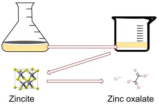
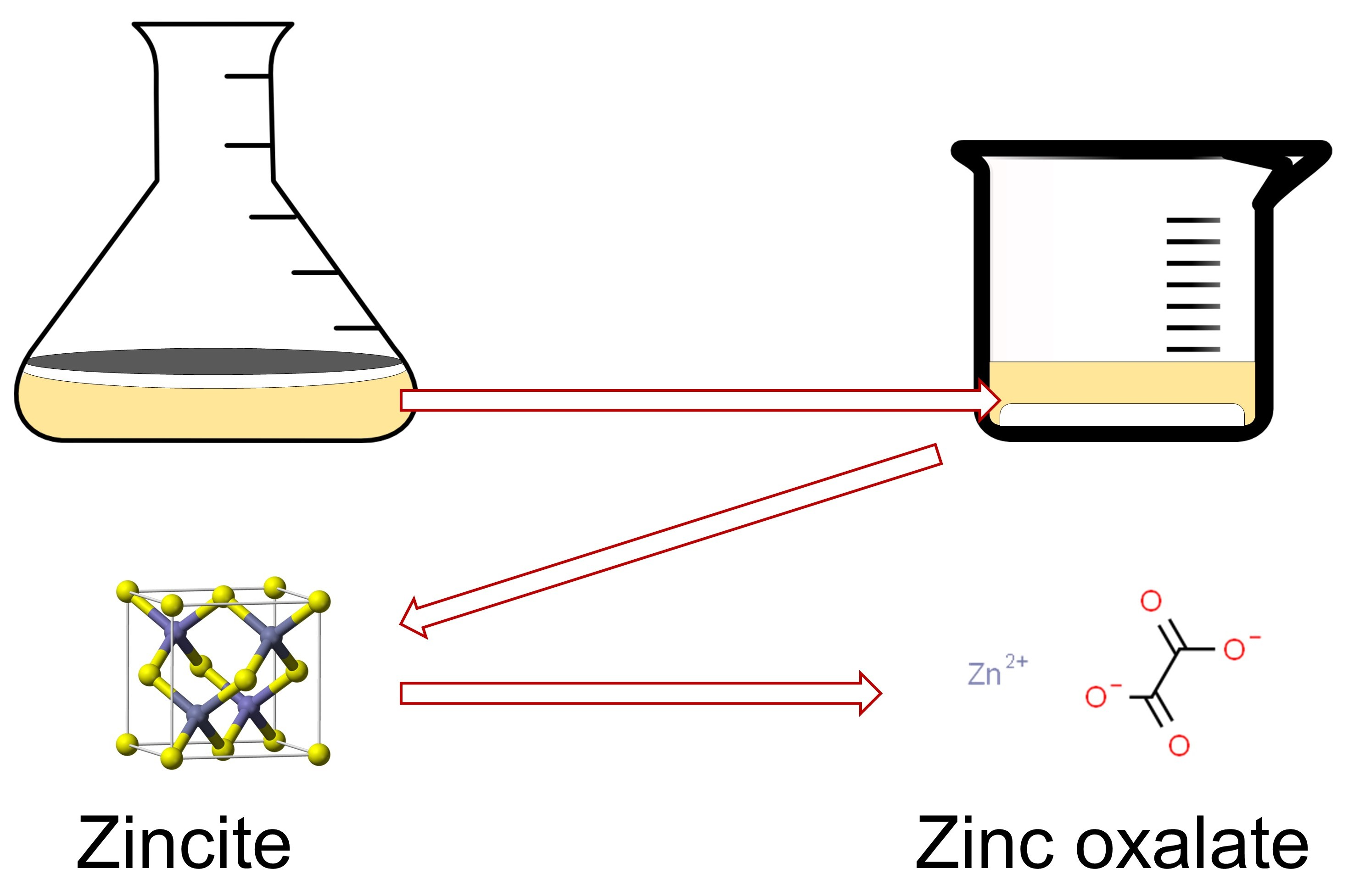
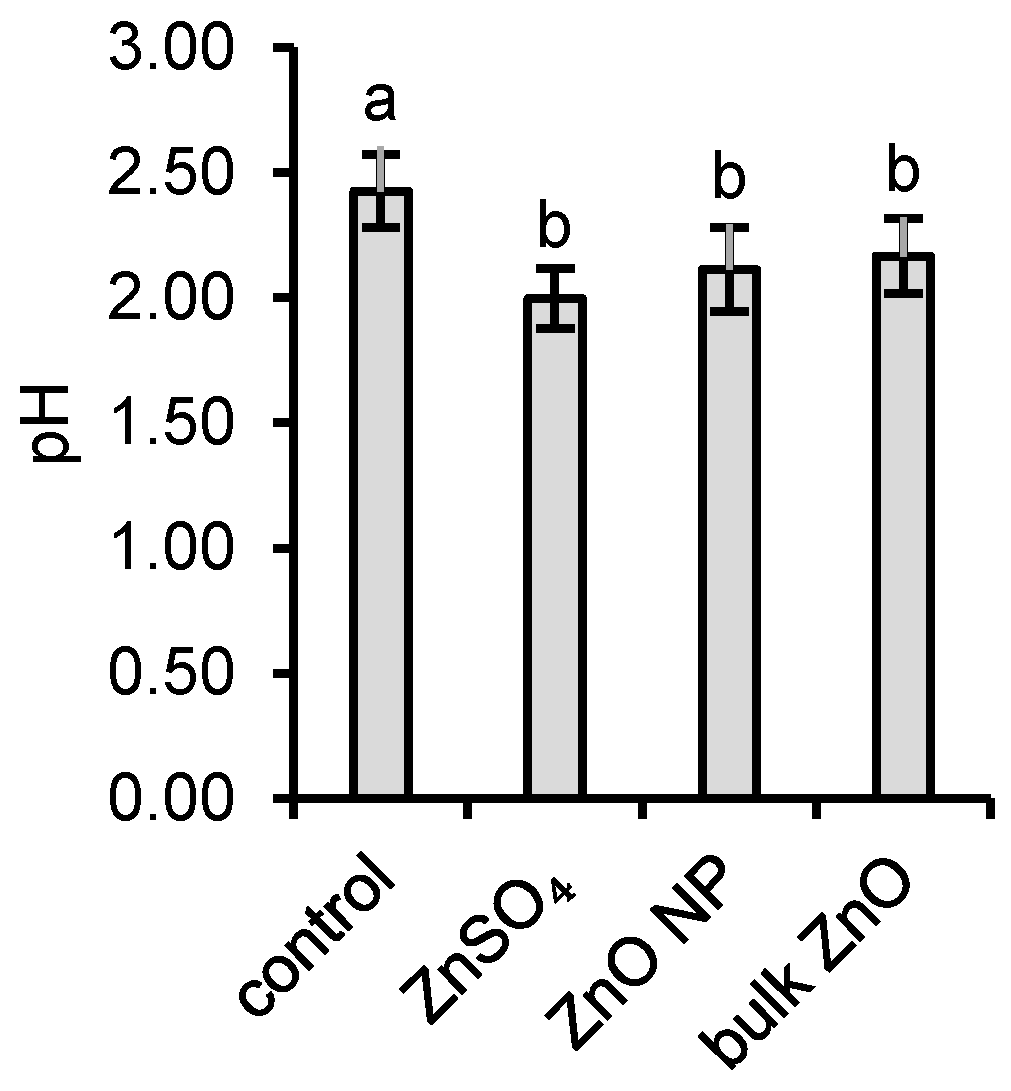
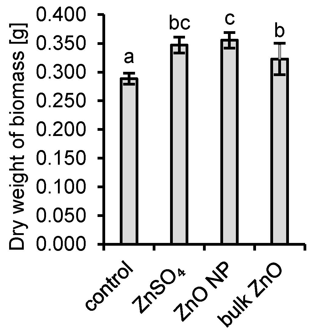
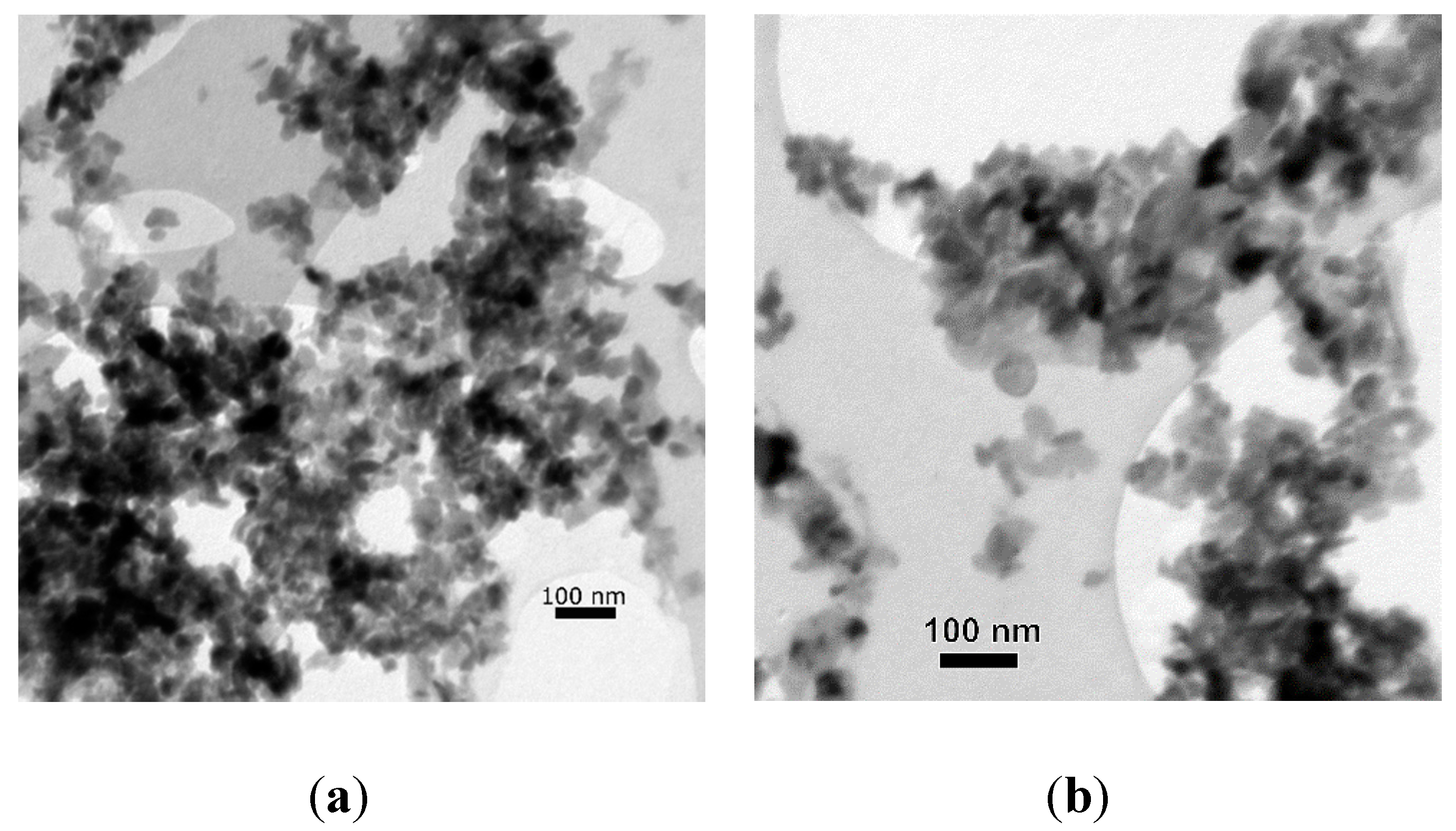
3.1. Interactions of Aspergillus niger with ZnO and Aqueous Zn

Fungal mycelia of Aspergillus niger were grown statically in Erlenmeyer flasks with Sabouraud growth media spiked with either ZnSO4, ZnO NPs, or bulk ZnO and control without added Zn. The overproduction of protons (H+) by microscopic filamentous fungus A. niger during the 7-day cultivation period generated a substantial decrease of pH from 5.6 to 2.4. It was even more intensified in the presence of Zn where the final pH values decreased to 2.0, 2.1, and 2.2 for ZnSO4, ZnO NPs, and bulk ZnO, respectively (Figure 1).
Zn was determined primarily as dissolved in the growth media (Figure 2) regardless of the Zn form applied. Only a negligible fraction was bound to colloidal particles. Thus, the distribution of Zn in colloidal, dissolved and bioaccumulated fractions were similar for ZnSO4, ZnO NPs, and bulk ZnO (Figure 2). The bioaccumulation of Zn by A. niger was very effective for all Zn forms (Figure 2) and represented up to 76% of the total Zn added to the cultivation system.
The applied concentration of 6.5 mg Zn∙L−1 had no adverse effects on fungal growth and a positive effect of Zn on the growth of the fungal biomass was observed (Figure 3). The application of all Zn form resulted in a higher dry weight of mycelia compared to control. The highest mean dry weight of mycelia was observed for ZnO NP application. The weight was significantly higher than both dry weights in bulk ZnO application and in the control experiment without additional Zn.

3.2. Transformation of ZnO Nanoparticles by Fungal Extracellular Metabolites

To experimentally confirm the transformation of ZnO NPs into zinc oxalate, extracellular metabolites of A. niger were applied to ZnO NPs. A partial transformation of ZnO NPs was observed, and a new mineral phase—zinc oxalate dihydrate—was identified (Figure 4).
At the end of the experiment, the oxalic concentration decreased from the initial 11.6 mmol∙L−1 to 1.2 mmol∙L−1. Thus, a 90% decrease in the initial oxalic acid concentration was observed, further confirming the ZnO NPs transformation into the zinc oxalate dihydrate by A. niger. Quantitatively, approximately 43 mg or 8.6% of ZnO NPs was transformed into the zinc oxalate dihydrate in the process. Only a partial transformation was observed. The recrystallization did not result in a significant change in the size of the most particles, and it ranged between 40 and 100 nm (Figure 5).

4. Discussion

4.1. Interactions of Aspergillus niger with ZnO and Aqueous Zn

ZnO NPs are relatively easily dissolvable in soils, especially in more acidic ones [9,37,38]. The dissolution of ZnO NPs is also enhanced by soil organisms, including filamentous fungi, via exudation of various acidic and chelating metabolites. The microbially induced acidification led to the dissolution of ZnO NPs, and bulk ZnO [39] and, therefore, nearly all of the Zn in the solutions of growth media was dissolved regardless of the Zn form.
Since Zn is an essential micronutrient [40,41] and our selected concentration was below the threshold of growth inhibition, all forms of Zn at the applied concentration had a positive effect on the dry weight of biomass after 7 days of cultivation. It was reported that the Zn concentrations as low as 0.065 mg Zn∙L−1 enhanced the fungal growth [42]. The minimal inhibitory concentration of Zn for microscopic filamentous fungi were reported to be as high as 100 mg Zn∙L−1 in the literature [43,44].

4.2. Transformation of ZnO Nanoparticles by Fungal Extracellular Metabolites

Filamentous fungi are capable of releasing extracellular metabolites that chemically deteriorate natural mineral Zn phases in soils and sediments via processes of protonation and chelation. However, under specific conditions (e.g., high biogenic chelate concentrations, alkalic pH), the bioextracted Zn can be reprecipitated from the soil solution to form new mineral phases. It has been reported that fungi react to the presence of ZnO NPs by increasing the production of the extracellular metabolites in order to detoxify and immobilize excessive Zn, and thus the transformation of ZnO NPs occurs in the soil environment [39,41]. Immobilization of Zn dissolved by fungi is facilitated by precipitation with oxalates [39].
We used extracellular metabolites of A. niger to transform ZnO NPs into a zinc oxalate biomineral. A. niger is well known for the high rate of extracellular metabolite production. Oxalic acid is the most produced of organic acids by the fungus when grown on Sabouraud growth media [30].
In our experiment, at the given applied volume and concentration of oxalic acid applied, only a partial transformation of ZnO NPs to zinc oxalates was observed. No significant change in the size of particles was observed. It is most probable that the transformation occurred on the surface of the nanoparticles, creating a surface of Zn oxalate with the core still being ZnO, or the newly formed zinc oxalates did not have time to grow to larger sizes in five days. If a larger volume of the organic acids was applied, both larger amounts of ZnO NPs might have been transformed, and the size of the particles could have seen a bigger change.
A change in Zn minerals into Zn oxalates had been observed before, albeit the minerals were not nanoparticles [32,39,45]. When silicate and sulfide minerals containing Zn were transformed by fungal extracellular metabolites, the resulting crystals of zinc oxalates had various shapes and sizes, with most of the crystals measuring between 50 and 100 µm [39]. When ZnO microparticulate powder was transformed into zinc oxalates by Aspergillus species, the biominerals had a different habitus compared to the minerals formed abiotically. The size of these biominerals was above 50 µm.
The mineral size and shape are also dependent on the place of formation. Minerals of zinc oxalate associated with a fungus have a different shape compared to both biomineral and abiotically formed Zn oxalate. However, the biominerals formed farther away from the fungal mycelium had a shape that was more similar to the abiotically formed Zn oxalate [32]. Since our experiment was only done with extracellular metabolites, the smaller change in the size and shape upon partial transformation may also result from the same processes that led to the increase in similarities between the biomineral formed further away from the mycelium and the abiotically formed mineral.
Aspergillus spp. and other fungi can biomineralize inorganic nanoparticles [46] and transform these nanoparticles into more stable oxalate compounds [25,47]. They play a large role in the cycling of elements in the soil environment, and their abilities can also be used for the bioremediation of contaminated areas [48].

5. Conclusions

Fungi, through their ability to acidify their environment, producing strong chelating agents, and the ability to bioaccumulate trace elements in their mycelia, can considerably affect the mobility of elements in soils and locally increase their bioavailability. Our observations affirm that the transformation of ZnO NPs into biogenic mineral phases occurs under the influence of ubiquitous soil fungus A. niger and that these processes may happen, under the right circumstances, in the soil environment. The role of soil fungi is seminal in the distribution of ionic and nanoparticulate forms of Zn. Therefore, fungi should be taken into consideration when models of bioavailability and mobility are constructed for soil environments. Furthermore, these fungal abilities can help us develop new methods of bioremediation in the future.

Supplementary Materials

The following are available online at https://www.mdpi.com/2309-608X/6/4/210/s1, Figure S1: Photographs of Aspergillus niger mycelia after 7-day cultivation in the preliminary test, Table S1: Weight of dry biomass after 7-day static cultivation in the preliminary test.

Author Contributions

Conceptualization, M.Š. and M.U.; Data curation, M.Š.; Formal analysis, M.Š.; Funding acquisition, M.U.and P.M.; Investigation, M.Š.; Methodology, M.Š., M.U. and P.M.; Project administration, P.M.; Resources, M.K., I.V., E.D., H.K. and M.B.; Supervision, M.U., M.K. and P.M.; Validation, M.B.; Visualization, M.Š.; Writing—original draft, M.Š.; Writing—review and editing, M.U. All authors have read and agreed to the published version of the manuscript.

Funding

This research was funded by the Scientific Grant Agency of the Ministry of Education, Science, Research and Sport of the Slovak Republic and the Slovak Academy of Sciences (Vedecká grantová agentúra MŠVVaŠ SR a SAV) under the contracts Nos. VEGA 1/0153/17, VEGA 1/0146/18, and by Cultural and Educational agency of the Ministry of Education, Science, Research and Sport of the Slovak Republic (Kultúrna a edukačná grantová agentúra MŠVVaŠ SR) under the contract No. KEGA 013SPU-4/2019.

Conflicts of Interest

The authors declare no conflict of interest.

References

  1. Hochella, M.F.; Mogk, D.W.; Ranville, J.; Allen, I.C.; Luther, G.W.; Marr, L.C.; McGrail, B.P.; Murayama, M.; Qafoku, N.P.; Rosso, K.M.; et al. Natural, incidental, and engineered nanomaterials and their impacts on the Earth system. Science 2019, 363, eaau8299. [Google Scholar] [CrossRef] [PubMed] [Green Version]
  2. Sun, T.Y.; Mitrano, D.M.; Bornhöft, N.A.; Scheringer, M.; Hungerbühler, K.; Nowack, B. Envisioning Nano Release Dynamics in a Changing World: Using Dynamic Probabilistic Modeling to Assess Future Environmental Emissions of Engineered Nanomaterials. Environ. Sci. Technol. 2017, 51, 2854–2863. [Google Scholar] [CrossRef] [PubMed]
  3. Peijnenburg, W.; Praetorius, A.; Scott-Fordsmand, J.; Cornelis, G. Fate assessment of engineered nanoparticles in solids dominated media—Current insights and the way forward. Environ. Pollut. 2016, 218, 1365–1369. [Google Scholar] [CrossRef] [PubMed]
  4. Šebesta, M.; Matúš, P. Separation, determination, and characterization of inorganic engineered nanoparticles in complex environmental samples. Chem. List. 2018, 112, 583–589. [Google Scholar]
  5. Keller, A.A.; McFerran, S.; Lazareva, A.; Suh, S. Global life cycle releases of engineered nanomaterials. J. Nanopart. Res. 2013, 15, 1–17. [Google Scholar] [CrossRef]
  6. MarketsandMarkets. Zinc Oxide Market by Application & by Region—Global Trends and Forecasts to 2020; MarketsandMarkets™ Research Private Ltd.: Hadapsar, India, 2015. [Google Scholar]
  7. Future Markets Inc. The Global Market for Zinc Oxide Nanoparticles; Future Markets Inc.: Edinburgh, UK, 2020. [Google Scholar]
  8. MarketsandMarkets. Zinc Oxide Market by Process (French Process, Wet Process, American Process), Grade (Standard, Treated, USP, FCC), Application (Rubber, Ceramics, Chemicals, Agriculture, Cosmetics & Personal Care, Pharmaceuticals), Region—Global Forecast to 2024; MarketsandMarkets™ Research Private Ltd.: Hadapsar, India, 2019. [Google Scholar]
  9. Šebesta, M.; Nemček, L.; Urík, M.; Kolenčík, M.; Bujdoš, M.; Vávra, I.; Dobročka, E.; Matúš, P. Partitioning and stability of ionic, nano- and microsized zinc in natural soil suspensions. Sci. Total Environ. 2020, 700, 134445. [Google Scholar] [CrossRef]
  10. Raliya, R.; Saharan, V.; Dimkpa, C.; Biswas, P. Nanofertilizer for Precision and Sustainable Agriculture: Current State and Future Perspectives. J. Agric. Food Chem. 2018, 66, 6487–6503. [Google Scholar] [CrossRef]
  11. Prasad, R.; Bhattacharyya, A.; Nguyen, Q.D. Nanotechnology in Sustainable Agriculture: Recent Developments, Challenges, and Perspectives. Front. Microbiol. 2017, 8, 1014. [Google Scholar] [CrossRef] [Green Version]
  12. Medina-Velo, I.A.; Barrios, A.C.; Zuverza-Mena, N.; Hernandez-Viezcas, J.A.; Chang, C.H.; Ji, Z.; Zink, J.I.; Peralta-Videa, J.R.; Gardea-Torresdey, J.L. Comparison of the effects of commercial coated and uncoated ZnO nanomaterials and Zn compounds in kidney bean (Phaseolus vulgaris) plants. J. Hazard. Mater. 2017, 332, 214–222. [Google Scholar] [CrossRef] [Green Version]
  13. Raliya, R.; Tarafdar, J.C.; Biswas, P. Enhancing the Mobilization of Native Phosphorus in the Mung Bean Rhizosphere Using ZnO Nanoparticles Synthesized by Soil Fungi. J. Agric. Food Chem. 2016, 64, 3111–3118. [Google Scholar] [CrossRef]
  14. Liu, R.; Lal, R. Potentials of engineered nanoparticles as fertilizers for increasing agronomic productions. Sci. Total Environ. 2015, 514, 131–139. [Google Scholar] [CrossRef] [PubMed]
  15. Kabata-Pendias, A.; Szteke, B. Trace Elements in Abiotic and Biotic Environments; CRC Press: Boca Raton, FL, USA, 2015; pp. 355–366. [Google Scholar]
  16. Kolenčík, M.; Ernst, D.; Komár, M.; Urík, M.; Šebesta, M.; Dobročka, E.; Černý, I.; Illa, R.; Kanike, R.; Qian, Y. Effect of foliar spray application of zinc oxide nanoparticles on quantitative, nutritional, and physiological parameters of foxtail millet (Setaria italica l.) under field conditions. Nanomaterials 2019, 9, 1559. [Google Scholar] [CrossRef] [PubMed] [Green Version]
  17. García-Gómez, C.; Fernández, M.D.; García, S.; Obrador, A.F.; Letón, M.; Babín, M. Soil pH effects on the toxicity of zinc oxide nanoparticles to soil microbial community. Environ. Sci. Pollut. Res. 2018, 25, 28140–28152. [Google Scholar] [CrossRef] [PubMed]
  18. Šebesta, M.; Kolenčík, M.; Matúš, P.; Kořenková, L. Transport and distribution of engineered nanoparticles in soils and sediments. Chem. List. 2017, 111, 322–328. [Google Scholar]
  19. Šebesta, M.; Kolenčík, M.; Urík, M.; Bujdoš, M.; Vávra, I.; Dobročka, E.; Smilek, J.; Kalina, M.; Diviš, P.; Pavúk, M.; et al. Increased Colloidal Stability and Decreased Solubility—Sol-Gel Synthesis of Zinc Oxide Nanoparticles with Humic Acids. J. Nanosci. Nanotechnol. 2019, 19, 3024–3030. [Google Scholar] [CrossRef]
  20. Erazo, A.; Mosquera, S.A.; Rodríguez-Paéz, J.E. Synthesis of ZnO nanoparticles with different morphology: Study of their antifungal effect on strains of Aspergillus niger and Botrytis cinerea. Mater. Chem. Phys. 2019, 234, 172–184. [Google Scholar] [CrossRef]
  21. Rajput, V.D.; Minkina, T.M.; Behal, A.; Sushkova, S.N.; Mandzhieva, S.; Singh, R.; Gorovtsov, A.; Tsitsuashvili, V.S.; Purvis, W.O.; Ghazaryan, K.A.; et al. Effects of zinc-oxide nanoparticles on soil, plants, animals and soil organisms: A review. Environ. Nanotechnol. Monit. Manag. 2018, 9, 76–84. [Google Scholar] [CrossRef]
  22. Nisar, P.; Ali, N.; Rahman, L.; Ali, M.; Shinwari, Z.K. Antimicrobial activities of biologically synthesized metal nanoparticles: An insight into the mechanism of action. JBIC J. Biol. Inorg. Chem. 2019, 24, 929–941. [Google Scholar] [CrossRef]
  23. Mohamed, A.A.; Fouda, A.; Abdel-Rahman, M.A.; Hassan, S.E.-D.; El-Gamal, M.S.; Salem, S.S.; Shaheen, T.I. Fungal strain impacts the shape, bioactivity and multifunctional properties of green synthesized zinc oxide nanoparticles. Biocatal. Agric. Biotechnol. 2019, 19, 101103. [Google Scholar] [CrossRef]
  24. Gadd, G.M. Metals, minerals and microbes: Geomicrobiology and bioremediation. Microbiology 2010, 156, 609–643. [Google Scholar] [CrossRef]
  25. Qin, W.; Wang, C.; Ma, Y.; Shen, M.; Li, J.; Jiao, K.; Tay, F.R.; Niu, L. Microbe-Mediated Extracellular and Intracellular Mineralization: Environmental, Industrial, and Biotechnological Applications. Adv. Mater. 2020, 32, 1907833. [Google Scholar] [CrossRef] [PubMed]
  26. Burford, E.P.; Fomina, M.; Gadd, G.M. Fungal involvement in bioweathering and biotransformation of rocks and minerals. Mineral. Mag. 2003, 67, 1127–1155. [Google Scholar] [CrossRef]
  27. Polák, F.; Urík, M.; Bujdoš, M.; Matúš, P. Aspergillus niger enhances oxalate production as a response to phosphate deficiency induced by aluminium(III). J. Inorg. Biochem. 2020, 204, 110961. [Google Scholar] [CrossRef] [PubMed]
  28. Fomina, M.; Burford, E.P.; Gadd, G.M. Fungal dissolution and transformation of minerals: Significance for nutrient and metal mobility. In Fungi in Biogeochemical Cycles; Gadd, G.M., Ed.; Cambridge University Press: Cambridge, UK, 2006; pp. 236–266. ISBN 9780511550522. [Google Scholar]
  29. Polák, F.; Urík, M.; Matúš, P. Low molecular weight organic acids in soil environment. Chem. List. 2019, 113, 307–314. [Google Scholar]
  30. Boriová, K.; Urík, M.; Bujdoš, M.; Pifková, I.; Matúš, P. Chemical mimicking of bio-assisted aluminium extraction by Aspergillus niger’s exometabolites. Environ. Pollut. 2016, 218, 281–288. [Google Scholar] [CrossRef]
  31. Ström, L.; Owen, A.G.; Godbold, D.L.; Jones, D.L. Organic acid behaviour in a calcareous soil implications for rhizosphere nutrient cycling. Soil Biol. Biochem. 2005, 37, 2046–2054. [Google Scholar] [CrossRef]
  32. Sutjaritvorakul, T.; Gadd, G.M.; Whalley, A.J.S.; Suntornvongsagul, K.; Sihanonth, P. Zinc Oxalate Crystal Formation by Aspergillus nomius. Geomicrobiol. J. 2016, 33, 289–293. [Google Scholar] [CrossRef]
  33. Urík, M.; Hlodák, M.; Mikušová, P.; Matúš, P. Potential of Microscopic Fungi Isolated from Mercury Contaminated Soils to Accumulate and Volatilize Mercury(II). Water Air Soil Pollut. 2014, 225, 2219. [Google Scholar] [CrossRef]
  34. Sádecká, J.; Polonský, J. Determination of organic acids in tobacco by capillary isotachophoresis. J. Chromatogr. A 2003, 988, 161–165. [Google Scholar] [CrossRef]
  35. Bačík, P.; Ozdín, D.; Miglierini, M.; Kardošová, P.; Pentrák, M.; Haloda, J. Crystallochemical effects of heat treatment on Fe-dominant tourmalines from Dolní Bory (Czech Republic) and Vlachovo (Slovakia). Phys. Chem. Miner. 2011, 38, 599–611. [Google Scholar] [CrossRef]
  36. Ondruška, J.; Trnovcová, V.; Štubňa, I.; Bačík, P. Depolarization currents in illite. J. Therm. Anal. Calorim. 2018, 131, 2285–2289. [Google Scholar] [CrossRef]
  37. Du, W.; Sun, Y.; Ji, R.; Zhu, J.; Wu, J.; Guo, H. TiO2 and ZnO nanoparticles negatively affect wheat growth and soil enzyme activities in agricultural soil. J. Environ. Monit. 2011, 13, 822–828. [Google Scholar] [CrossRef] [PubMed]
  38. Kool, P.L.; Ortiz, M.D.; van Gestel, C.A.M. Chronic toxicity of ZnO nanoparticles, non-nano ZnO and ZnCl2 to Folsomia candida (Collembola) in relation to bioavailability in soil. Environ. Pollut. 2011, 159, 2713–2719. [Google Scholar] [CrossRef] [PubMed]
  39. Wei, Z.; Liang, X.; Pendlowski, H.; Hillier, S.; Suntornvongsagul, K.; Sihanonth, P.; Gadd, G.M. Fungal biotransformation of zinc silicate and sulfide mineral ores. Environ. Microbiol. 2013, 15, 2173–2186. [Google Scholar] [CrossRef] [PubMed]
  40. Couri, S.; Pinto, G.A.S.; de Senna, L.F.; Martelli, H.L. Influence of metal ions on pellet morphology and polygalacturonase synthesis by Aspergillus niger 3T5B8. Braz. J. Microbiol. 2003, 34, 16–21. [Google Scholar] [CrossRef] [Green Version]
  41. Tarafdar, J.C.; Agrawal, A.; Raliya, R.; Kumar, P.; Burman, U.; Kaul, R.K. ZnO nanoparticles induced synthesis of polysaccharides and phosphatases by Aspergillus fungi. Adv. Sci. Eng. Med. 2012, 4, 324–328. [Google Scholar] [CrossRef]
  42. Wold, W.S.M.; Suzuki, I. The citric acid fermentation by Aspergillus niger: Regulation by zinc of growth and acidogenesis. Can. J. Microbiol. 1976, 22, 1083–1092. [Google Scholar] [CrossRef]
  43. Sardella, D.; Gatt, R.; Valdramidis, V.P. Assessing the efficacy of zinc oxide nanoparticles against Penicillium expansum by automated turbidimetric analysis. Mycology 2018, 9, 43–48. [Google Scholar] [CrossRef] [Green Version]
  44. Sawai, J.; Yoshikawa, T. Quantitative evaluation of antifungal activity of metallic oxide powders (MgO, CaO and ZnO) by an indirect conductimetric assay. J. Appl. Microbiol. 2004, 96, 803–809. [Google Scholar] [CrossRef]
  45. Sayer, J.A.; Gadd, G.M. Solubilization and transformation of insoluble inorganic metal compounds to insoluble metal oxalates by Aspergillus niger. Mycol. Res. 1997, 101, 653–661. [Google Scholar] [CrossRef]
  46. Hulkoti, N.I.; Taranath, T.C. Biosynthesis of nanoparticles using microbes—A review. Colloids Surf. B Biointerfaces 2014, 121, 474–483. [Google Scholar] [CrossRef] [PubMed]
  47. Gadd, G.M. Fungi, Rocks, and Minerals. Elements 2017, 13, 171–176. [Google Scholar] [CrossRef]
  48. Akhtar, N.; Mannan, M.A. Mycoremediation: Expunging environmental pollutants. Biotechnol. Rep. 2020, 26, e00452. [Google Scholar] [CrossRef] [PubMed]
Figure 1. pH of the growth media after the experiment; a and b represent the statistically similar means between different groups at α = 0.05 (two-tail t-test).
Jof 06 00210 g001
Figure 2. Distribution of Zn in the experimental system (a) and concentration of Zn in (b) particles sized 1 to 1000 nm, (c) dissolved fraction, and (d) dry biomass.
Jof 06 00210 g002
Figure 3. The dry weight of biomass of Aspergillus niger after the experiment; a, b and c represent the statistically similar means between different groups at α = 0.05 (two-tail t-test).
Jof 06 00210 g003
Figure 4. Diffractogram of the partially transformed ZnO NP to Zn oxalate dihydrate.
Jof 06 00210 g004
Figure 5. Transmission electron microscopy image of (a) Pristine ZnO nanoparticles (b) Partially transformed ZnO nanoparticles to Zn oxalate dihydrate.
Jof 06 00210 g005

