Article

Ecology and Fungicolous Lifestyle of Hypomyces aurantius Associated with the Newly Recorded Host Lyophyllum littoralis


1
Department of Food Processing, Arda Vocational School, Trakya University, Edirne 22100, Türkiye
2
Department of Organic Agriculture, Başkale Vocational School, Van Yüzüncü Yıl University, Van 65080, Türkiye
*
Author to whom correspondence should be addressed.
J. Fungi 2026, 12(9), 632; https://doi.org/10.3390/jof12090632
Submission received: 12 July 2026 / Revised: 8 August 2026 / Accepted: 18 August 2026 / Published: 24 August 2026
(This article belongs to the Section Environmental and Ecological Interactions of Fungi)

Abstract

Despite developing on other fungi and forming complex ecological interactions, fungicolous fungi are among the understudied groups. This study examined the fungicolous fungi Hypomyces aurantius and its host species, Lyophyllum littoralis, in detail using morphological and molecular data. Both species are new records for the Turkish mycobiota. The macroscopic and microscopic characteristics of both species were evaluated in depth, and morphological descriptions were corroborated by molecular analyses based on the nuclear ITS region. It was observed that H. aurantius heavily colonised L. littoralis, a previously unreported host, causing significant softening and degradation of the host tissue. We investigated extracellular enzyme activities to determine the mycoparasitic activity of H. aurantius on the host fungus and its environmental survival strategies. We detected positive enzyme activities. These results suggest that H. aurantius is a potential mycoparasite. This study contributes to our understanding of fungal biodiversity in Türkiye and provides a foundation for understanding the ecology of supra-fungal fungi and fungicolous/mycoparasitic interactions. conclusions.

1. Introduction

Fungicolous fungi are specialised organisms that spend all or part of their life cycle on the mycelium, sporocarp or reproductive structures of other fungi. They can develop saprotrophic, commensal, necrotrophic or mycoparasitic life strategies, playing important roles in the structure of fungal communities, interspecies competition, sporocarp decomposition and nutrient cycling in natural ecosystems [1,2,3,4]. Macrofungal sporocarps, in particular, provide suitable, albeit short-lived, microhabitats for fungicolous fungi due to their high water and organic matter content. In recent years, the widespread use of molecular methods has revealed that macrofungal sporocarps harbour richer and more diverse host-associated fungal communities than previously thought. However, the taxonomic composition, host preferences, and ecological functions of these communities remain poorly understood [2,5,6].
Mycoparasitism, one of the life forms of fungi, is considered one of the most specialized examples of fungus–fungus interactions. Mycoparasitic fungi can recognize a suitable host through chemical signals, direct themselves toward the host hyphae to establish contact, and colonize host tissues with the help of various biochemical mechanisms. This process involves not only physical contact but also the combined action of hydrolytic enzymes capable of breaking down the cell wall, oxidative enzyme systems, and various secondary metabolites [7,8]. The degradation of the fungal cell wall’s major components and structural proteins is considered one of the key steps in successful colonization. Therefore, extracellular enzyme production serves as an important indirect indicator in the evaluation of the physiological characteristics of fungicolous fungi and their potential mycoparasitic life strategies [7].
Hypomyces (Fr.) Tul. & C. Tul. is one of the most important fungicolous genera in the Hypocreaceae family and includes species that develop on numerous macrofungal hosts worldwide. Demonstrating fungicolous and mycoparasitic characteristics across various macrofungus groups, particularly Boletales, Helotiales, Agaricales, Pezizales and Polyporales, Hypomyces (Fr.) Tul. & C. Tul. is an ascomycete genus comprising over 150 recognised species that spread as powdery textures and puffy crusts on fungal surfaces [9,10,11]. Members of the genus can cause discolouration, the formation of a superficial mycelial covering, deformation and, in later stages, tissue degradation in host sporocarps. These characteristics mean they are considered one of the most important biotic factors affecting the dynamics of natural fungal populations [1,12]. Taxa belonging to the genus have been reported to be associated with environments other than fungi, such as various plant substrates (e.g., leaves, wood, and bark), mosses, lichens, rocks and humus [13].
Hypomyces species exhibit remarkable diversity in their host preferences. While some species show a high degree of dependence on specific genera or families, others can grow on macrofungi belonging to different taxonomic groups. Therefore, new host records provide important information not only from a mycobiotic perspective but also in understanding the host adaptation, ecological resilience, and evolutionary relationships of the species [1]. Recent phylogenetic studies have revealed that the host spectrum and infection strategies of Hypomyces (anamorphic stage Cladobotryum) are more complex than previously thought [12,14]. Therefore, documenting new fungicolous relationships with morphological and molecular data contributes not only to updating species distributions but also to a better understanding of the ecology and evolution of fungicolous fungi [2]. Among these species, Hypomyces aurantius (anamorph: Cladobotryum varium) has been reported from a wide range of macrofungal hosts, including species of Panellus, Flammulina, Morchella, poroid fungi and several other agaricoid basidiomycetes. Although the species is regarded as having a relatively broad host range, host associations remain incompletely documented because many historical records lack molecular confirmation. Consequently, every well-documented host association contributes to a more accurate understanding of its ecological amplitude and host specificity [1,15,16,17].
H. aurantius, which has generally been recorded on fungi such as Panellus, the winter mushroom (Flammulina velutipes (Curtis) Singer), morels (Morchella) and poroid species [11,17,18], has not been reported on Lyophyllum littoralis to date. This study aimed to confirm, using morphological and molecular methods, the previously unreported fungal association between H. aurantius and L. littoralis. In this context, the macro- and micromorphological characteristics of both taxa were examined in detail; their molecular identities were confirmed using ITS sequences, and their phylogenetic positions were assessed. Furthermore, the cultural characteristics of H. aurantius and its extracellular enzyme production profile were determined, and the species’ fungicolous lifestyle and potential mycoparasitic strategies were evaluated from a physiological perspective. Thus, the study not only presents the first records of H. aurantius and L. littoralis for the Turkish mycobiota, but also contributes to the existing body of knowledge on the host relationships and ecological characteristics of Hypomyces species by revealing a previously unreported host relationship between these two species in the light of morphological, molecular and physiological data.

2. Materials and Methods

2.1. Sample Collection

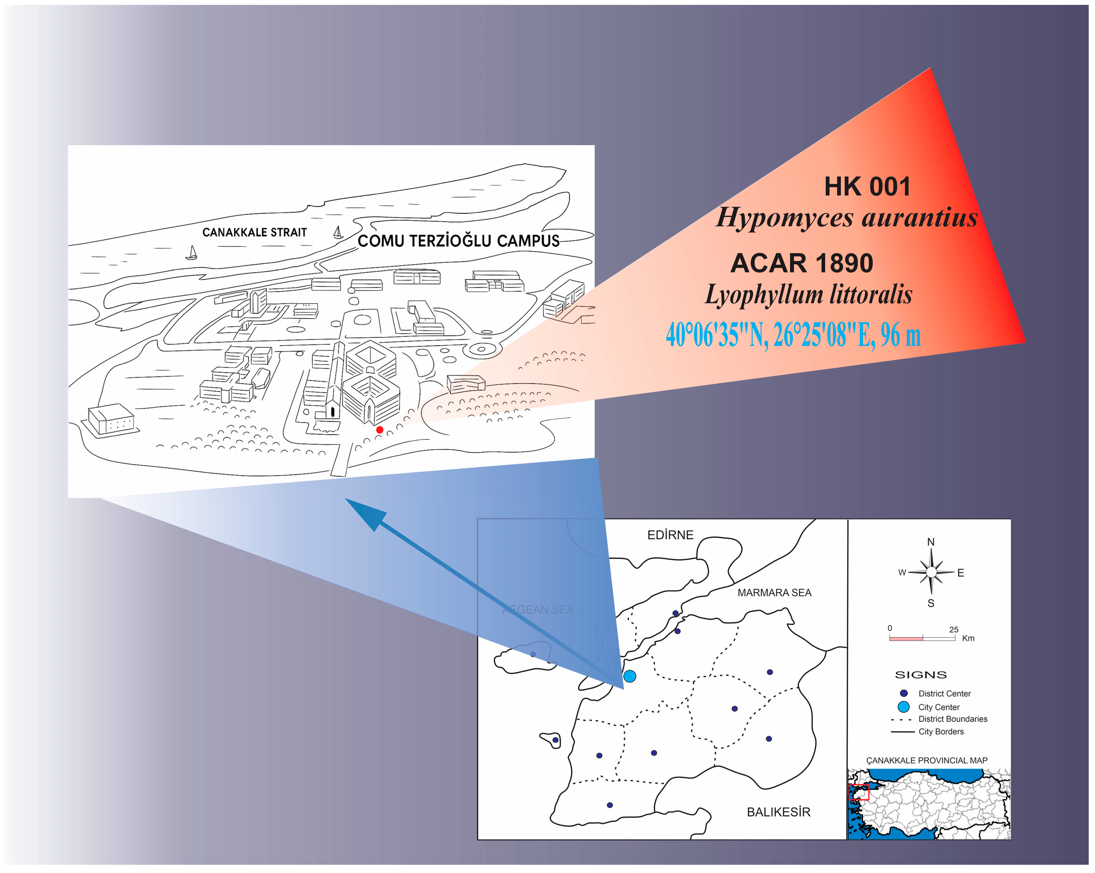
The materials for this study, healthy macrofungus specimens bearing fungicolous microfungi, were collected in January 2025 from the vicinity of the Faculty of Science, Terzioğlu Campus, Çanakkale Onsekiz Mart University (Figure 1). The collected specimens were photographed in their natural environment, and environmental conditions and macromorphological characteristics (pileus, lamella, stipe, etc.) were examined in detail, with all details recorded. Specimens showing fungicolous growth were carefully placed in sterile bags, while healthy specimens were placed in separate boxes and quickly transported to the laboratory.

2.2. Isolation of Fungicolous Microfungi

The isolation of the fungicolous microfungi was performed by examining the macrofungi, which were brought to the laboratory in sterile bags, under a stereomicroscope. To this end, structures such as the microfungus’s mycelium and spore masses observed on the host fungus were transferred to PDA and MEA plates using sterile swabs. The inoculated plates were then incubated at 25 °C for seven days under a daylight cycle. After incubation, the growing fungicolous microfungi was obtained as a pure culture by isolating single spores. Stock cultures were obtained from this pure culture by transferring it to tubes containing slanted PDA agar medium, which were then stored in the microbiology laboratory at Trakya University Arda Vocational School.

2.3. Fungal Identification

Traditional taxonomic methods, combined with advanced molecular techniques, were employed to identify and classify host fungi and their associated fungicolous fungi. In this study, the macroscopic and microscopic characteristics of the samples were examined in detail, and the data obtained were evaluated by comparing them with ITS rDNA (nuclear internal transcribed spacer) gene sequences and phylogenetic analyses.

2.3.1. Morphological Identification

Mycelium discs (5 mm) were extracted from the active growth tips of the Fungikol mushroom culture and inoculated onto plates of Potato Dextrose Agar (PDA, Merck, Darmstadt, Germany) and Malt Extract Agar (1.5% malt extract and 1.5% agar, Merck, Darmstadt, Germany). After incubation, the colonies were examined for their macroscopic and microscopic characteristics, and measurements were taken. Healthy macrofungus samples brought to the laboratory were also subjected to detailed micromorphological examination.
Specimens of both the host and the fungicolous fungi were examined under a stereomicroscope (Motic SMZ-171 Stereo Zoom Microscope, Motic Hong Kong Limited, Hong Kong, China) and a light microscope (Leica DM500, Leica Microsystems GmbH, Wetzlar, Germany). To reliably evaluate the microscopic characteristics of each structure (e.g., basidiospore, basidium, conidiospore and conidiogenic cell), measurements were taken at least 30 times. In addition, electron microscopy (SEM) images were used to identify the macrofungus specimen. To accurately and precisely present the obtained micromorphological findings, scientific drawings were prepared using the 64-bit CorelDRAW Graphics Suite 2022 (64-bit; Corel Corporation, Ottawa, ON, Canada), along with microphotographs.
The macro- and micro-morphological characteristics of the Fungicolous species were assessed based on the descriptions provided by Tulasne & Tulasne (1860) [19], Rogerson & Samuels (1993) [20], and Arnold & Yurchenko (2007) [21]. The host species, meanwhile, were assessed by comparison with the descriptions provided by Ballero & Contu (1990) [22], Contu (1998) [23], *Archivio Micologico* (2016) [24], and iNaturalist (2026) [25].
Following identification, H. aurantius was prepared as a fungarium specimen and preserved as a stock culture (code HK001) in the Microbiology Laboratory of Arda Vocational School, Trakya University. The dried basidiomata of L. littoralis were deposited in the fungarium of the Department of Biology, Faculty of Science, Van Yüzüncü Yıl University, under the personal fungarium number Acar1890. The collection, isolation, and micromorphological examination of the fungi were performed according to the procedures described by [26].

2.3.2. Molecular Identification

Genomic DNA was extracted from the macrofungal samples used in this study following the CTAB protocol [27]. The nuclear ribosomal internal transcribed spacer (nrITS) region was amplified by polymerase chain reaction (PCR) from DNA extracted from samples HK001 and ACAR 1890. DNA isolation and PCR procedures were carried out in accordance with the protocols described in previous studies [28,29,30,31], thereby ensuring methodological consistency and reproducibility. For DNA isolation, the EurX GeneMATRIX Plant & Fungi DNA Isolation Kit (EURx Ltd., Gdańsk, Poland) was used, and the manufacturer’s recommended protocol was followed. The ITS1 (5′-TCCGTAGGTGAACCTGCGG-3′) and ITS4 (5′-TCCTCCGCTTATTGATATGC-3′) primer pair was selected for the amplification of the ITS region [32,33]. The PCR was carried out using Taq DNA polymerase; the reaction mixture consisted of 10× PCR buffer, MgCl2 (25 mM), dNTP mix (20 mM), primers, Taq DNA polymerase (2 U) and DNA template. The PCR cycle consisted of the following steps: an initial denaturation at 94 °C for 5 min; followed by denaturation at 94 °C for 30 s, annealing at the optimum temperature for 30 s, extension at 72 °C for 1 min, and finally a final extension at 72 °C for 10 min. The reaction was terminated indefinitely at 4 °C. The resulting PCR products were purified using the MAGBIO HighPrep PCR Clean-up System (MagBio Genomics, Gaithersburg, MD, USA); sequencing was performed bidirectionally using an ABI 3730XL (Applied Biosystems, Foster City, CA, USA) sequencer and the BigDye Terminator v3.1 Cycle Sequencing Kit. The resulting sequences were edited using BioEdit Sequence Alignment Editor version 7.2.5 [34] and assembled using the CAP contig assembly method. For species identification, the obtained sequences were compared with reference sequences in the GenBank database [33]. Type-derived ITS sequences were searched for both target species in GenBank and the UNITE database. As no type-derived ITS sequences were available for Hypomyces aurantius or Lyophyllum littoralis, representative reference sequences retrieved from GenBank were selected for phylogenetic analyses.

2.4. Extracellular Enzyme Production and Screening

An extracellular enzyme screen was carried out to evaluate the mycoparasitic activity of H. aurantius on its host fungus and its environmental survival strategies. To determine protease activity, media containing two different substrates (casein and gelatine) were used: Modified Casein agar (0.5% yeast extract, 0.03% calcium hydroxide, 0.02% calcium chloride, 1.5% agar, 1% casein, pH 5.8) [35] and Glucose Yeast Extract Peptone Agar with gelatine (1% peptone, 0.5% glucose, 2% glucose, 1.5% agar, 0.4% gelatine) [36]. To determine lipase activity, a medium supplemented with 1% tributyrin (0.5% peptone, 0.3% yeast extract, 2% agar, 1% tributyrin and 0.05% Tween 80) was used [37]. In the investigation of laccase activity, PDA media were used, both containing 0.15 mM CuSO4, with the addition of 2 mM ABTS and 4 mM guaiacol, respectively [38]. For the determination of amylase activity, a culture medium containing 0.1% glucose, 0.01% yeast extract, 0.05% peptone, 1.6% agar, and 0.2% soluble starch was prepared [36].
Mycelial discs, obtained from fresh H. aurantius cultures using a sterile mushroom corer (5 mm in diameter) were inoculated into the centre of the prepared culture media. All experiments were carried out in triplicate, and the Petri dishes were incubated at 25 ± 2 °C and observed daily. Protease, lipase and amylase activities were assessed as positive based on the presence of the formation of a halo around the colony. Laccase activity was determined by the characteristic green colour formation around the colonies resulting from the oxidation of ABTS and by the reddish-brown colour formation resulting from the oxidation of guaiacol. Following incubation, the casein agar plates were treated with a 1% HCl solution, whilst the gelatin-containing plates were treated with a saturated ammonium sulphate solution to cause opacification and render the hydrolysis zones around the colonies visible. In contrast, starch-containing media were flooded with an iodine solution, which stained the agar blue-black. Clear zones surrounding the colonies, where starch hydrolysis had occurred, were considered indicative of amylolytic activity [35,36,37,38].
The capacity for producing extracellular enzymes is expressed as the enzyme index (EI), which is calculated by dividing the total diameter of the reaction zone (i.e., the hydrolysis or colour change zone) by the diameter of the fungal colony. The formula is as follows [39]:
EI = Colony diameter/Diameter of reaction region including colony.
The diameters of both the colony and the hydrolysis zone were measured in millimetres using a digital calliper. Measurements were obtained from three independent replicates, and the results are presented as the mean ± standard deviation (SD).

3. Results

3.1. Morphological Results

Based on the taxonomic keys cited in the scientific references, the phylogenetic and morphological identification of H. aurantius and its host L. littoralis, which were collected and isolated from their natural habitat, along with brief descriptions of the specimens, macro- and micromorphological photographs, micromorphological drawings, and the collection site and date, ITS rDNA (nuclear internal transcribed spacer) gene sequences and phylogenetic analyses are presented below.
Ascomycota Caval.-Sm.
Sordariomycetes O.E. Erikss. & Winka
Hypocreaceae De Not.
Hypomyces Fr.
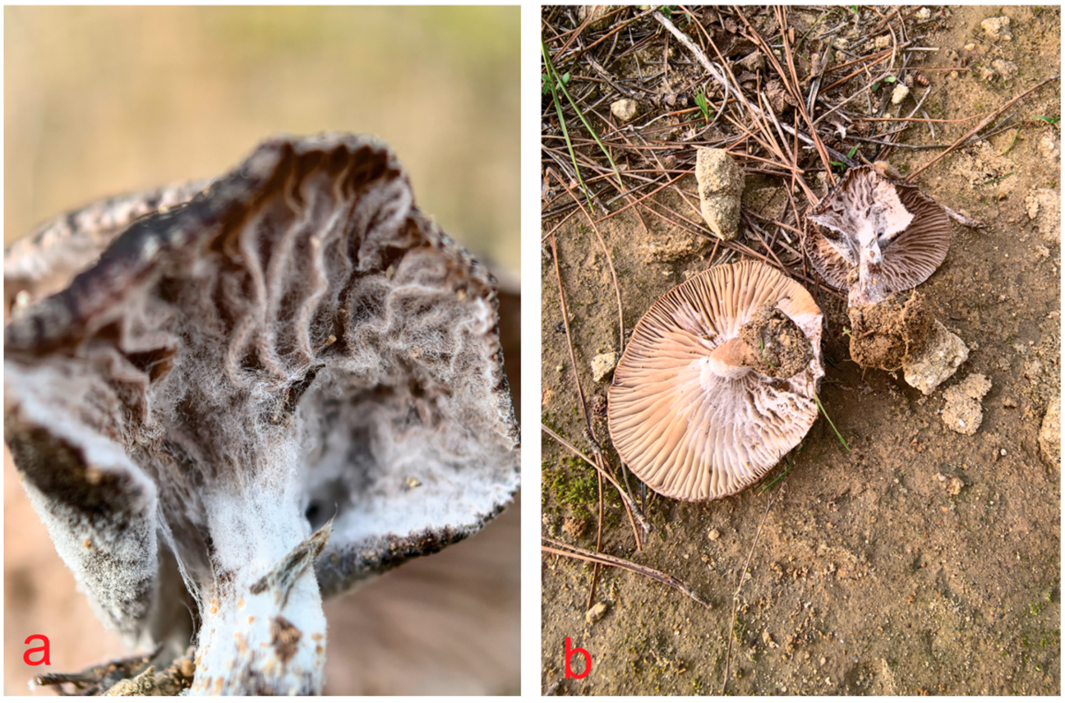
Hypomyces aurantius (Pers.) Fuckel, Jb. nassau. Ver. Naturk. 23–24: 183 (1870) [1869–70] (Figure 2)
GenBank No: PV715959
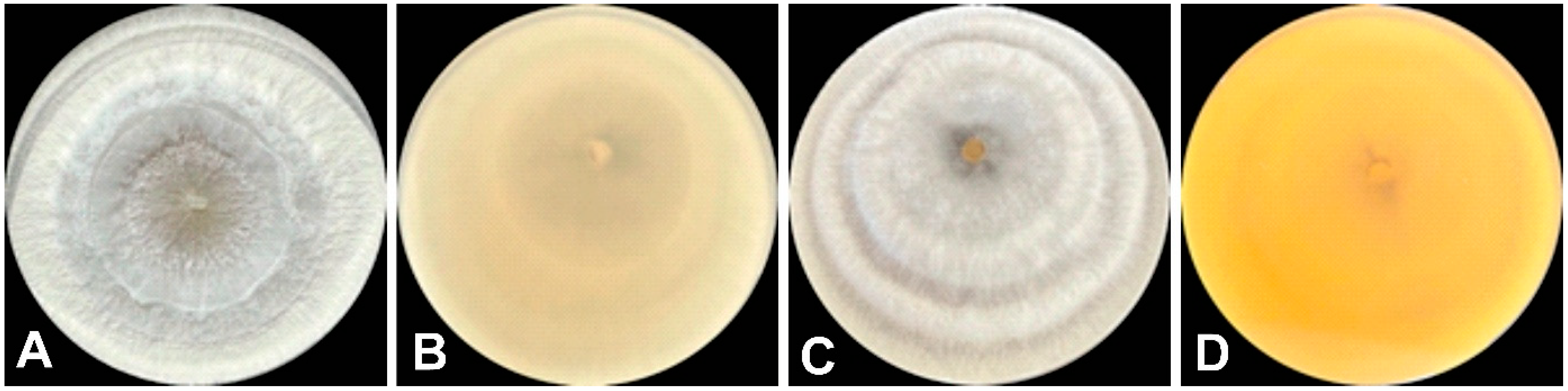
Colonial morph: Colony on PDA reaching 70–80 mm in diameter after 5 days at 25 °C, diffuse, cottony to floccose, with a well-developed aerial mycelium. The central region is compact and slightly raised (umbonate); abundant conidia imparting a powdery texture to the colony surface. Initially white to cream, becoming pale yellowish-brown with age. Reverse light beige to ochre, with distinct zonation (Figure 3).
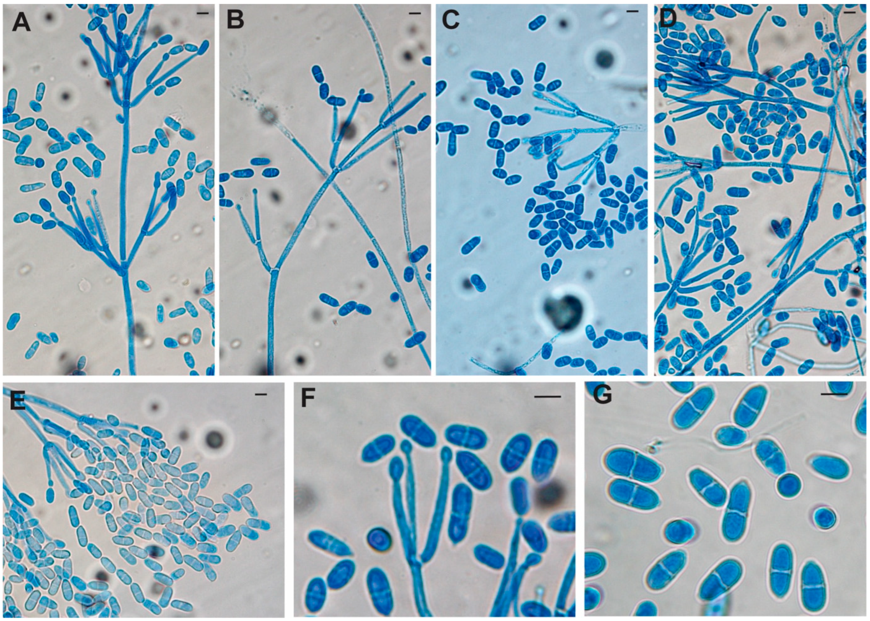
L. littoralis was found growing on the fresh fruiting body. Asexual morph: White mycelium on the host fruiting body. Hyphomycetous growth was observed on PDA medium; the mycelium consisted of hyaline, septate and distinctly branched hyphae with straight walls. Conidiophores up to 90 μm in length, hyaline, with a structure that is either unbranched or branched vertically once or more, the branches tapering slightly towards the tip and terminating in conidiogenous cells (20–45 μm in length). Conidia 12.5–21.5 × 5.5–10.0 μm, initially spherical to hemispherical, becoming ellipsoidal to obovoid at maturity; smooth-walled, aseptate or rarely 1-septate, with a truncate basal hilum. Conidia formed singly and arranged in dry, basipetal chains joined end-to-end. Chlamydospores not observed. Sexual morph not observed (Figure 4).
Specimens examined: Türkiye, Çanakkale, Terzioğlu Campus, Near the Faculty of Science, 40°06′35″ N, 26°25′08″ E, 96 m, on fresh basidiomata of L. littoralis, 3 January 2025, HK001.
Distribution and habitat: Hypomyces aurantius have been reported from different geographical regions and is associated with a wide range of macrofungal hosts. It grows on the basidiomata of various fungi, including decaying polypores, Panellus spp., cultivated mushrooms, and other macrofungi reported in the literature. During colonization, it forms a characteristic orange-coloured mould layer on the host surface, frequently accompanied by discoloration, deformation, and progressive deterioration of host tissues [1,17,18].
Basidiomycota Whittaker ex R.T. Moore
Agaricomycetes Doweld
Agaricales Underw
Lyophyllaceae Jülich
Lyophyllum P. Karst.
Lyophyllum littoralis (Ballero & Contu) Contu, Boll. Gruppo Micol. G. Bresadola 41 (3): 193 (1998) (Figure 5, Figure 6 and Figure 7)
Basionym: Calocybe littoralis Ballero & Contu
GenBank No: PV715960
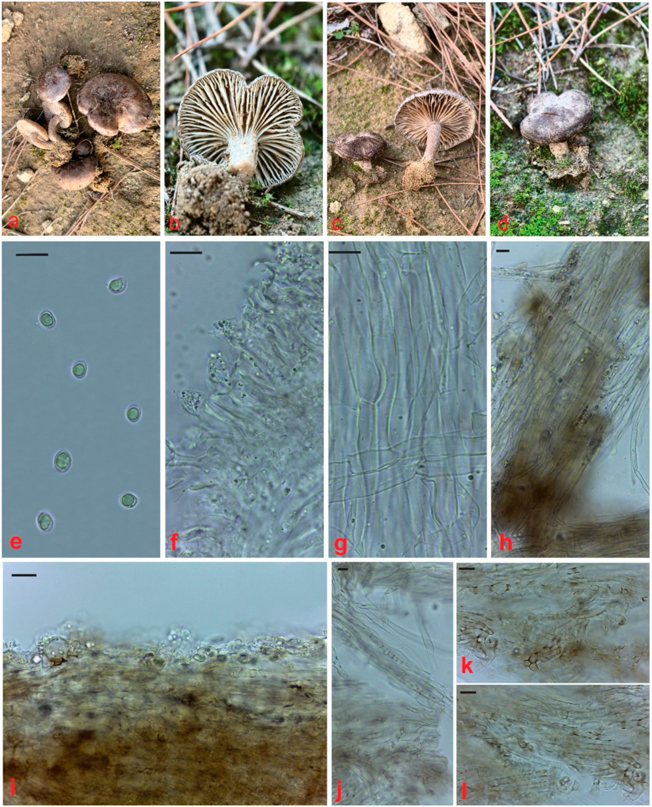
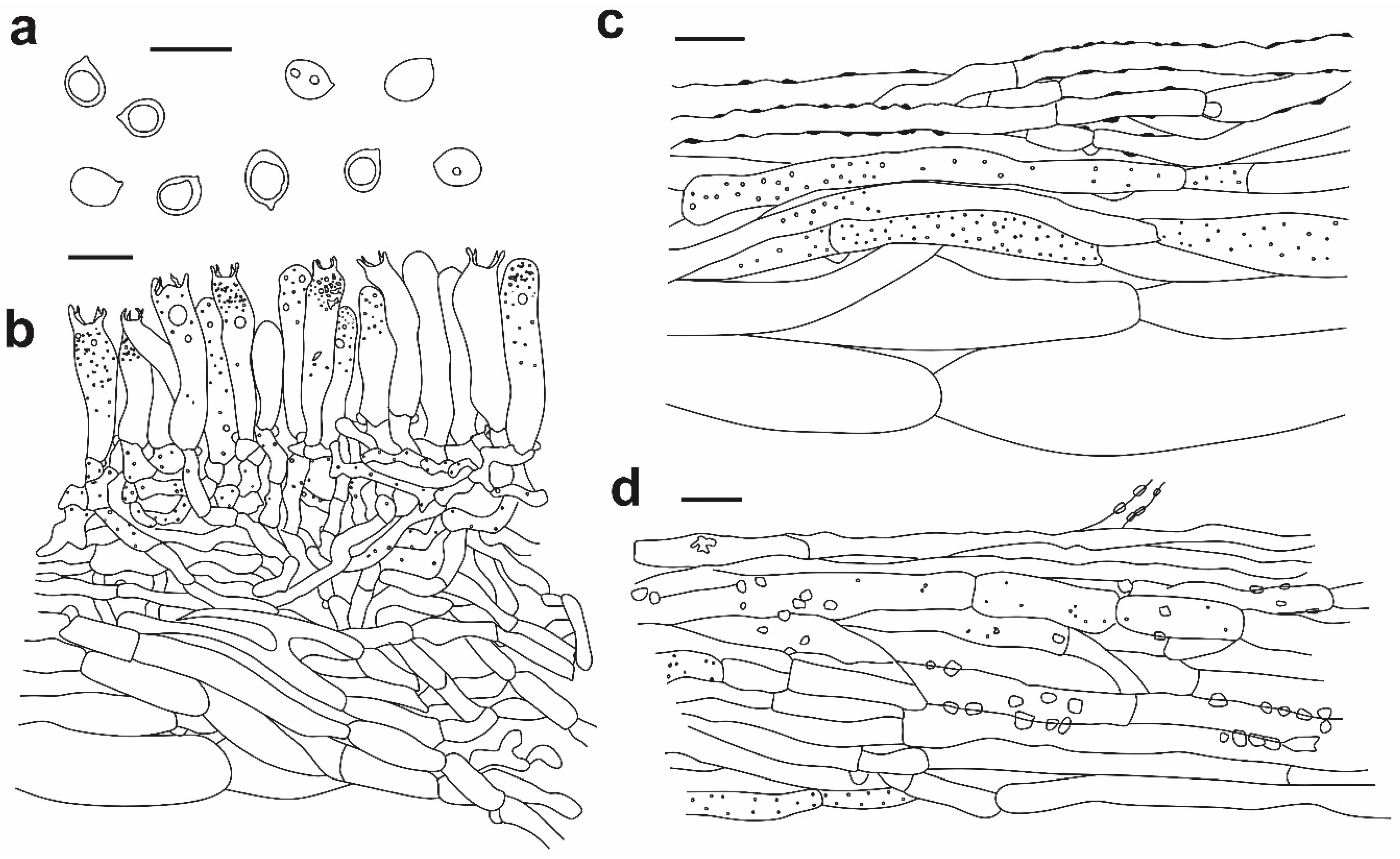
Pileus 30–100 mm, shiny to matte, spotted, brownish grey, sometimes slightly fibrillate from center to margins, pileus usually covered with a white powdery layer, margins darker, sometimes with brown transverse stripes on margins, pileus shape in young specimens usually rounded, curved when mature, margin curved downwards. Fleshy, thin, and rubbery, the stipe is not quickly broken; young specimens are generally odorless, while mature specimens emit a nutmeg odor. Stipe 15–35 × 4–10 mm, hollow, white or whitish fibrous near pileus, brown towards the base, grayish brown; in mature specimens, usually grey. Lamellae adnexed or adnate, white when young, pale yellowish gray with age, margin typically straight, sometimes slightly wavy. The short lamellae are of variable length and irregularly arranged between the main ones. Basidia (21.4–)24–32(–35) × 4.5–6.2(–7) µm, (n = 25), cylindrical to clavate, hyaline, with large or small drops, sometimes siderophilous granulation near the apex, with clamp connections. Marginal cells similar to basidia. Spores 4.8–6.2(–7.5) × 4.5–6 µm, (n = 30), av. 5.8 µm, globose to subglobose, smooth, hyaline, usually with a significant drop in the center, sometimes without a drop or with several tiny drops. Hyphae of lamellae up to 14 µm, hyaline, cylindrical, with basal clamp. Pileipellis, the uppermost layer, brown to yellowish brown incrusted, 3.5–6.5 µm wide, with clamp, middle layer siderophilous granulation, cylindrical, brown to yellowish brown, up to 15 µm, with clamp, lowest layer cylindrical parallel hyphae, smooth, hyaline, without granules, up to 18 µm wide, with clamp. Stipitipellis a cutis of elongate and crystalline hyphae, hyaline to yellowish brown, 2–6 µm wide. Clamp connection present at some septa.
Specimens examined: Türkiye, Çanakkale, Terzioğlu Campus, near the Faculty of Science, 40°06′35″ N, 26°25′08″ E, 96 m, under Pinus brutia Ten trees, 3 January 2025, Acar 1890.
Distribution and habitat: L. littoralis fruits in Mediterranean forests, especially under pine trees, between November and January. Although it usually develops in clusters attached to a common base, it can sometimes be seen as single individuals [22,24].

3.2. Phylogenetic Results

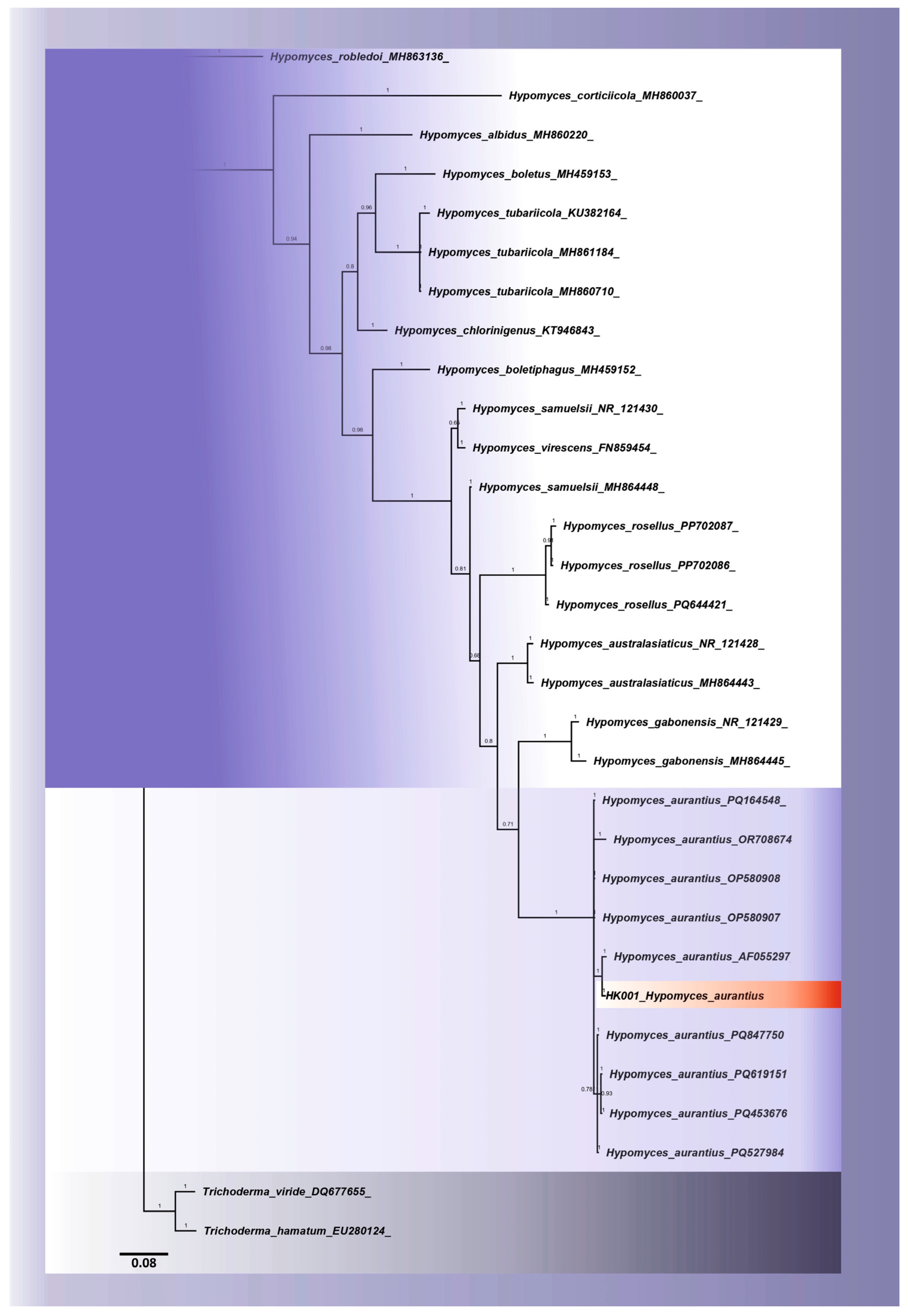
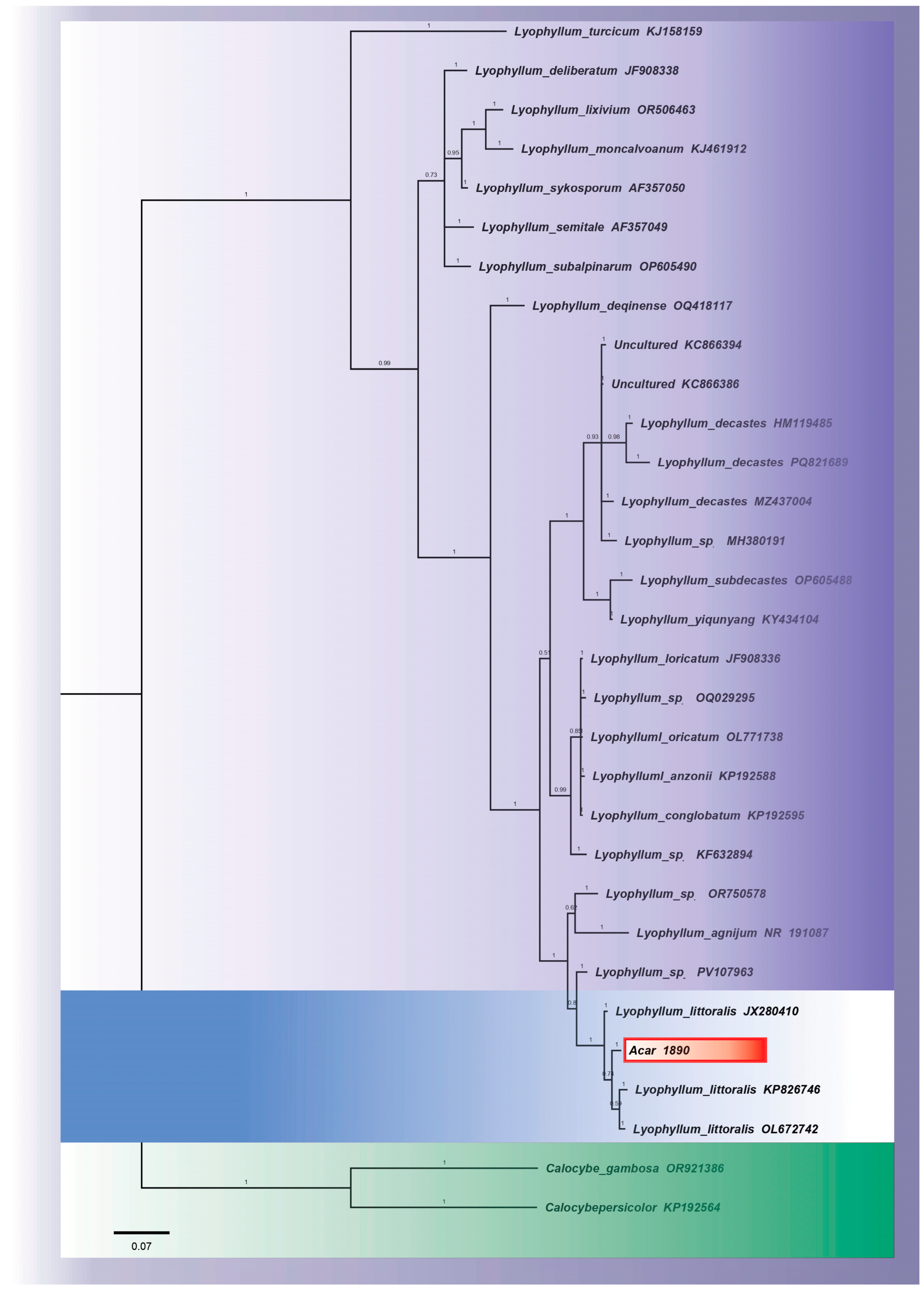
The nrITS sequences obtained in this study were used to identify the two taxa under investigation at the molecular level. The newly generated sequences (PV715959 for H. aurantius and PV715960 for L. littoralis) were aligned with reference sequences from the GenBank database and included in Bayesian phylogenetic analyses. The H. aurantius (PV715959) sequence was positioned within the same clade as the reference H. aurantius sequences in the resulting phylogenetic tree, within a group supported by high posterior probability values. No topological deviation was observed within this clade, and the results clearly demonstrate that the species has been correctly identified at the species level. Similarly, the L. littoralis (PV715960) sequence was also placed within a well-supported clade alongside the reference L. littoralis sequences. In particular, the fact that the Acar 1890 specimen shows a direct sister-group relationship with the L. littoralis sequences reported in the literature strongly supports the species identification. Furthermore, the fact that this clade is distinct from other taxa within the L. decastes complex demonstrates that the species is molecularly distinguishable. The phylogenetic trees generated from both analyses consistently recovered the examined specimens within their respective species, with stable topological placement (Figure 8 and Figure 9). When evaluated in conjunction with morphological data, these findings demonstrate that the identification of both taxa has been carried out with a high degree of reliability.

3.3. Results for Extracellular Enzyme Production and Screening

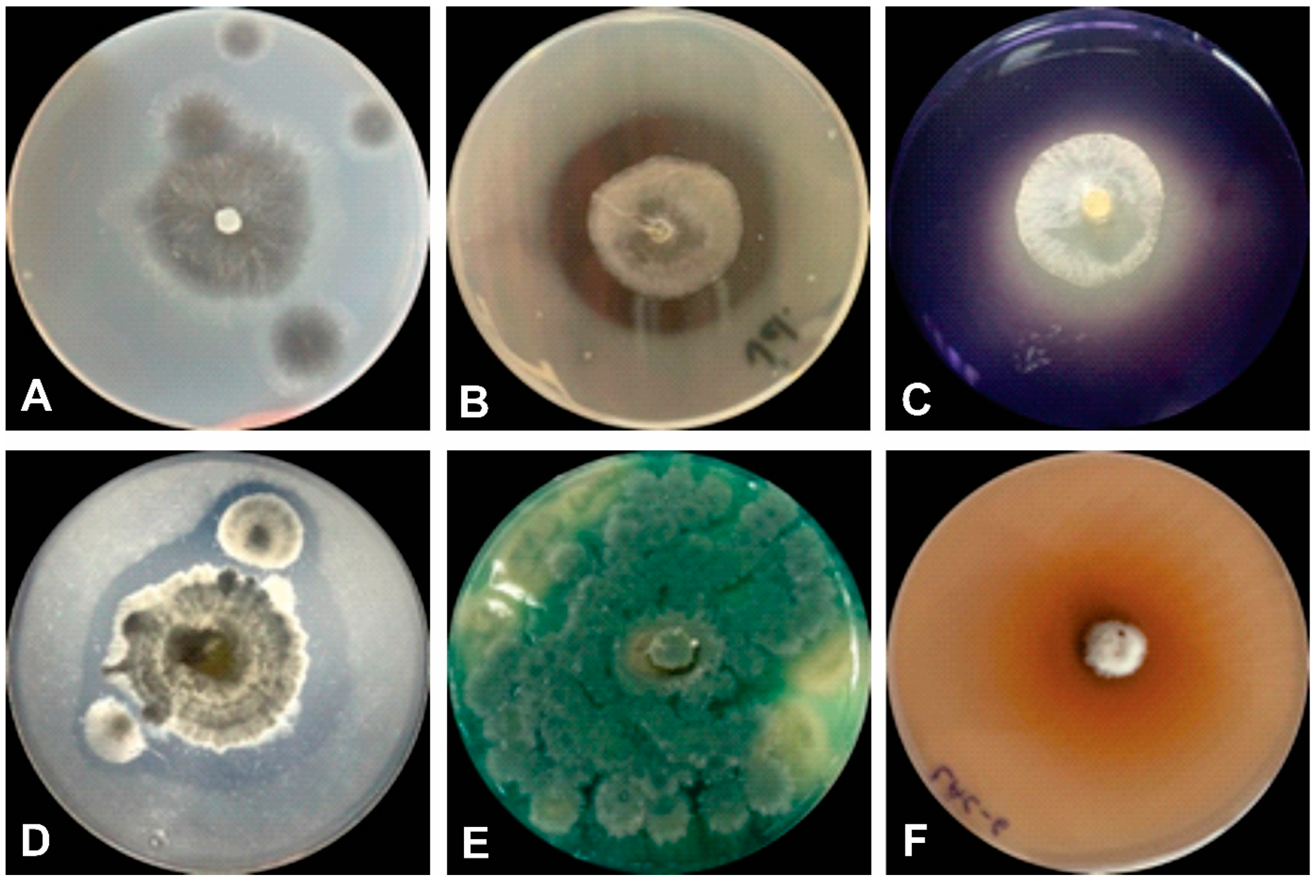
H. aurantius showed positive reactions for all tested extracellular enzymes (protease, lipase, amylase, and laccase) (Figure 10).
The extracellular enzymatic profile of H. aurantius was evaluated using qualitative plaque assays, and enzyme production was expressed as the enzyme index (EI) (see Table 1 for details). The isolate exhibited positive activity for all tested enzymes; however, the level of enzyme production varied according to the substrate. The results of the qualitative analysis demonstrated that the isolate was capable of producing both hydrolytic enzymes, including gelatinase, amylase and lipase, and oxidative enzymes, such as laccase.

4. Discussion

This study presents the first morphological and molecular characterization of Hypomyces aurantius and its host, Lyophyllum littoralis, two species newly recorded for the Turkish mycobiota. Hypomyces aurantius is a widely distributed fungicolous species that has been reported from numerous geographical regions worldwide. Despite its broad distribution, its host associations remain incompletely documented, and recent studies have demonstrated that the species is capable of colonizing a wider range of basidiomycete hosts than previously recognized. In the present study, H. aurantius was found on L. littoralis for the first time. This newly documented host association expands the known host range of the species and contributes to a better understanding of its ecological interactions and host adaptation.
The genus Lyophyllum Karsten belongs to the family Lyophyllaceae and comprises saprophytic agaric fungi found worldwide, characterised by white cyanophilic spores and siderophilic granules in their basidia [38]. The section Difformia (Fr.) Kühner, to which L. littoralis (Ballero & Contu) Contu belongs, is characterised by clustered fruiting bodies, does not turn brown, and is defined by its distinctive growth habits. It is currently known that Lyophyllum sect. Difformia comprises 15 caespitose and/or non-browning Lyophyllum species worldwide [40,41,42]. Some species of this genus, which plays an important role in natural ecosystems and possesses significant economic value, have been cultivated in Asia, the Americas, and Europe [43]. Lyophyllum littoralis is a Mediterranean species that typically grows in caespitose clusters in coniferous forests, particularly beneath Pinus spp. Morphologically, it closely resembles L. decastes but differs in its ecological preferences and geographical distribution, with L. littoralis occurring predominantly in warmer Mediterranean habitats, whereas L. decastes is associated with cooler temperate regions. The morphological characteristics and ITS-based phylogenetic placement of the Turkish specimen are fully consistent with these diagnostic features, supporting its identification as L. littoralis.
The genus Hypomyces was first described by Fries (1825) [44] as a subgenus of Hypocrea Fr., and was subsequently reclassified as an independent genus by Tulasne & Tulasne (1860) [19]. A comprehensive taxonomic study of this group was conducted by Arnold (1971) [45], who highlighted the distinguishing characteristics of Hypomyces and related genera. The life cycle of Hypomyces comprises anamorphic and teleomorphic reproductive stages. Many Hypomyces species in the asexual stage were previously assigned different scientific names and placed in different genera (Cladobotryum, Mycogone, Sepedonium, Verticillium, and others) [12,46]. Some Hypomyces species are rarely observed at certain stages of their life cycle. For example, whilst the sexual reproductive stage of some species that attack boletes is very rarely encountered, the asexual stage is rarely observed in other species. Furthermore, the anamorphic reproductive stage of many Hypomyces species remains unknown. This complicates the identification of species, as there are distinct morphological differences between the asexual and sexual stages. Consequently, the identification of Hypomyces species cannot always be based on consistent morphological characteristics. Nevertheless, they can be identified using molecular techniques and by the brightly coloured perithecia of species within the genus, which cause lesions and growth abnormalities on the host fungus [47,48].
Although the phylogenetic analyses of both species were based solely on the nrITS region, the obtained sequences showed high similarity to reference sequences and were consistently recovered within well-supported species-level clades in the Bayesian phylogenetic analyses. However, it has been reported that ITS data alone may not always be sufficient for a more reliable assessment of species boundaries within Hypomyces and the related Cladobotryum complex; therefore, multi-gene phylogenetic approaches incorporating additional loci such as LSU, tef1 and rpb2 may enhance taxonomic resolution [1,49].
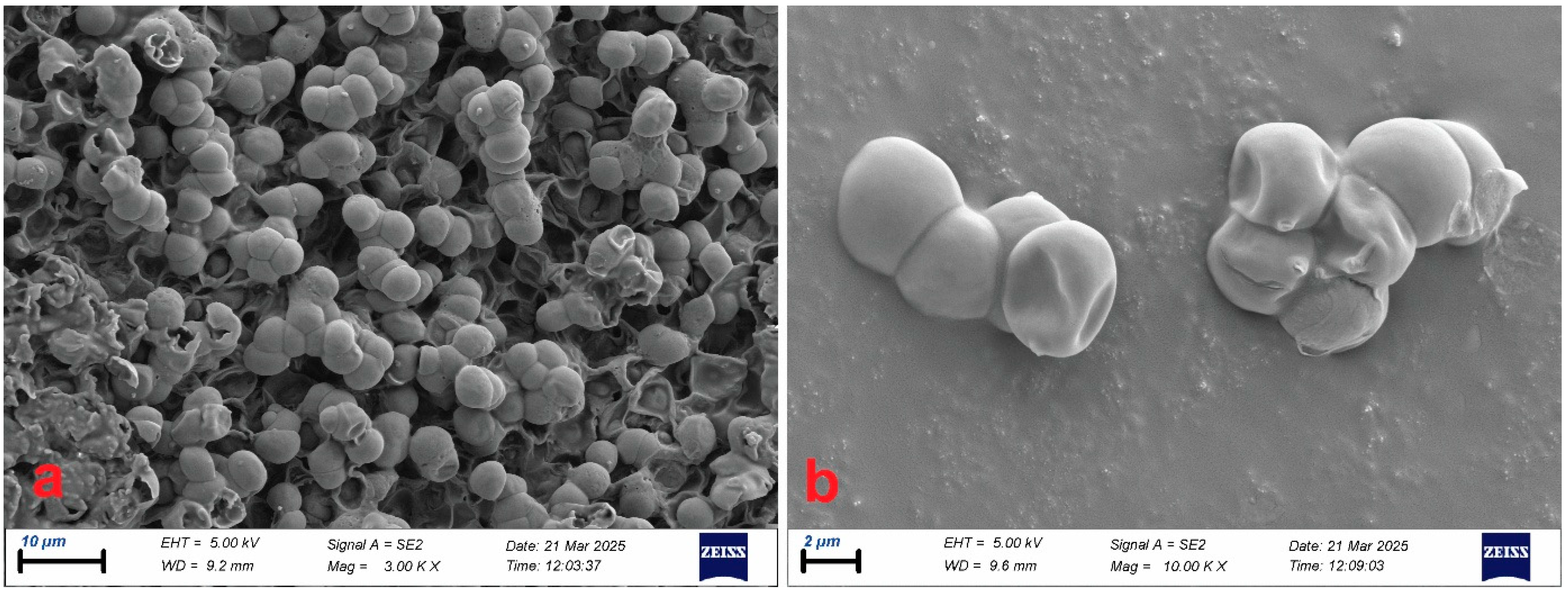
In this study, mycoparasitic structures could not be directly visualised at the electron microscope level. One of the main reasons for this is the short duration of the active phase of the parasitism in the field and the difficulty in preserving suitable specimens in a manner appropriate for SEM analysis. However, SEM images of L. littoralis spores provided a clearer visualization of the species’ diagnostic characteristics.
H. aurantius is known in the literature as a species that exhibits mycoparasitic and pathogenic effects on various macrofungi. In particular, it has been reported to cause cobweb disease in morel species and to have a broad host range [50]. Cobweb disease is a serious fungal disease caused by Hypomyces/Cladobotryum species and characterised by a white, hairy mycelial network that spreads rapidly across the surface of the growing medium. The disease begins with the invasion of primordia, leading to discolouration and progressive rot in the fruit bodies [17,51]. It has been reported that H. aurantius parasitises Panellus species in China. In particular, it is known that such mycoparasites also cause infection in economically important species, such as the previously cultured Flammulina velutipes [18,52]. H. aurantius has also been reported on Lenzites warnieri in the Balkan Mountains [53,54]. It has been reported that H. aurantius on Xylobolus subpileatus constitutes the sole record on Xylobolus, a genus thought to be closely related to Stereum, as cited by [55,56].
The successful establishment of mycoparasitic fungi that colonise their fungal hosts is a complex biological process dependent on the coordinated secretion of extracellular hydrolytic and oxidative enzymes capable of breaking down the structural components of the host cell wall [57,58]. In this study, the protease, lipase, amylase and laccase activities identified in H. aurantius indicate that the species possesses an extracellular enzyme repertoire capable of supporting its fungicolous lifestyle. Similarly, studies conducted on various fungicolous fungi have shown that these organisms can commonly produce proteases, amylases, cellulases and various oxidoreductases, and that these enzymes may play a role in both nutrient acquisition and host colonisation [39]. The protease activities identified in our study suggest a biochemical capacity for facilitating hyphal penetration and nutrient acquisition through the hydrolysis of proteins in the host fungal cell wall and intercellular matrix. Similarly, lipase activity may support the colonisation process by contributing to the breakdown of lipid components in the cell membrane [57]. Laccase activity, on the other hand, demonstrates that H. aurantius possesses the capacity to produce not only hydrolytic but also oxidative enzymes. Although laccase is not an enzyme that directly breaks down the fungal cell wall, it may indirectly support host colonisation by contributing to processes such as the oxidation of phenolic and aromatic compounds involved in the host’s chemical defence, the conversion of organic matter, and the reduction in oxidative stress [59,60]. Amylase activity may contribute to the utilisation of carbon sources by facilitating the hydrolysis of storage polysaccharides found in host tissues or in surrounding organic material. Furthermore, this amylolytic capacity may indicate a metabolic flexibility that could support the organism’s survival outside the host on carbon-rich substrates such as soil organic matter or decaying plant residues [61,62].
However, chitinases and β-1,3-glucanases, which hydrolyze chitin and β-1,3-glucan, the major polysaccharides of the host fungal cell wall, have long been recognized as key enzymes in mycoparasitic fungi. In particular, it has been reported that enzymes that degrade β-1,3-glucan show a strong correlation with mycoparasitic success in fungicolous fungi, whereas cellulase production is not a determining factor for successful mycoparasitism [63]. Recent genomic studies also demonstrate that fungicolous fungi possess a rich repertoire of CAZyme enzymes and that these enzymes play a fundamental role in nutrient acquisition through the degradation of the host cell wall [4]. On the other hand, it has been shown that a fungicolous lifestyle does not always imply mycoparasitism; in some species, commensal relationships with the host fungus can develop despite chitinase and β-1,3-glucanase activities being quite low [64]. Consequently, the fact that chitinase and β-1,3-glucanase activities were not assessed in our study limits the ability to draw direct conclusions regarding the enzymatic mechanisms by which H. aurantius degrades the host cell wall. Nevertheless, the identified enzyme activities suggest that the species may possess the biochemical capacity necessary for host colonisation and nutrient acquisition. In future, investigating chitinase and β-1,3-glucanase activities in conjunction with biochemical assays, transcriptome analyses and CAZyme-based genomic approaches will contribute to a much more detailed understanding of mycoparasitic strategy of H. aurantius and its interaction mechanisms with L. littoralis [65,66].
The intense mycelial growth and marked softening and decomposition of the host tissue observed in this study are consistent with the symptoms reported in the literature. However, L. littoralis is a new host for this species, previously unreported, and is documented here for the first time. However, as experimental validation of pathogenicity could not be carried out, the interaction in question has been assessed as a ‘possible mycoparasitic relationship’. Consequently, this study not only contributes to Türkiye’s fungal biodiversity but also represents a step towards filling gaps in the literature regarding the documentation of fungicolous/possible mycoparasitic fungi in nature. Future multi-locus phylogenetic studies incorporating additional loci (e.g., LSU, tef1-α, and rpb2) together with type-derived reference sequences, where available, as well as culture-based isolations and controlled mycoparasitism experiments, will provide improved phylogenetic resolution and enable a more detailed understanding of the biology, species delimitation, and ecological roles of these species.

Author Contributions

Conceptualization, H.K. and İ.A.; methodology, H.K. and İ.A.; validation, H.K. and İ.A.; formal analysis, H.K. and İ.A.; investigation, H.K. and İ.A.; resources, H.K. and İ.A.; data curation, H.K. and İ.A.; writing—original draft preparation, H.K. and İ.A.; writing—review and editing, H.K. and İ.A. All authors have read and agreed to the published version of the manuscript.

Funding

This research received no external funding.

Institutional Review Board Statement

Not applicable.

Informed Consent Statement

Not applicable.

Data Availability Statement

The data supporting the findings of this study are available within the article. The newly generated nrITS sequences have been deposited in GenBank under accession numbers PV715959–PV715960. Additional data are available from the corresponding author upon reasonable request.

Conflicts of Interest

The authors declare no conflicts of interest.

References

  1. Põldmaa, K. Tropical species of Cladobotryum and Hypomyces producing red pigments. Stud. Mycol. 2011, 68, 1–34. [Google Scholar] [CrossRef] [Scilit] [PubMed]
  2. Maurice, S.; Arnault, G.; Nordén, J.; Botnen, S.S.; Miettinen, O.; Kauserud, H. Fungal sporocarps house diverse and host-specific communities of fungicolous fungi. ISME J. 2021, 15, 1445–1457. [Google Scholar] [CrossRef] [Scilit] [PubMed]
  3. Sucharzewska, E.; Ejdys, E.; Kubiak, D. Fungicolous fungi on microscopic fungi parasitic to the vegetation of the urban environment. Acta Mycol. 2023, 58, 176036. [Google Scholar] [CrossRef] [Scilit]
  4. Huo, W.; Cui, L.; Yan, P.; He, X.; Zhang, L.; Liu, Y.; Dai, L.; Qi, P.; Hu, S.; Qiao, T.; et al. Diversity and composition of fungicolous fungi residing in macrofungi from the Qinling mountains. J. Fungi. 2024, 10, 601. [Google Scholar] [CrossRef] [Scilit] [PubMed]
  5. Wang, Z.; Jiang, Y.; Deane, D.C.; He, F.; Shu, W.; Liu, Y. Effects of host phylogeny, habitat and spatial proximity on host specificity and diversity of pathogenic and mycorrhizal fungi in a subtropical forest. New Phytol. 2019, 223, 462–474. [Google Scholar] [CrossRef] [Scilit] [PubMed]
  6. Bennett, A.E.; Groten, K. The costs and benefits of plant–arbuscular mycorrhizal fungal interactions. Annu. Rev. Plant Biol. 2022, 73, 649–672. [Google Scholar] [CrossRef] [Scilit] [PubMed]
  7. Gruber, S.; Seidl-Seiboth, V. Self versus non-self: Fungal cell wall degradation in Trichoderma. Microbiology 2012, 158, 26–34. [Google Scholar] [CrossRef] [Scilit] [PubMed]
  8. Kowalski, T.; Bilański, P. Fungicolous fungi on Pseudosclerotial plates and apothecia of Hymenoscyphus fraxineus and their biocontrol potential. Microorganisms 2022, 10, 2250. [Google Scholar] [CrossRef] [Scilit] [PubMed]
  9. Mihál, I.; Cicák, A.; Bucinova, K. Distribution, ecology, and taxonomical notes to species of genera Hypomyces and Nectria (Hypocreales) in Slovakia. Mikol. I Fitopatol. 2007, 41, 242. [Google Scholar]
  10. Wijayawardene, N.N.; Hyde, K.D.; Rajeshkumar, K.C.; Hawksworth, D.L.; Madrid, H.; Kirk, P.M.; Braun, U.; Singh, R.V.; Crous, P.W.; Kukwa, M.; et al. Notes for genera: Ascomycota. Fungal Divers. 2017, 86, 1–594. [Google Scholar] [CrossRef] [Scilit]
  11. Yu, F.M.; Jayawardena, R.S.; Luangharn, T.; Zeng, X.Y.; Li, C.J.Y.; Bao, S.X.; Ba, H.; Zhou, D.Q.; Tang, S.M.; Hyde, K.D.; et al. Species diversity of fungal pathogens on cultivated mushrooms: A case study on morels (Morchella, Pezizales). Fungal Divers. 2024, 125, 157–220. [Google Scholar] [CrossRef] [Scilit]
  12. Rossman, A.Y.; Seifert, K.A.; Samuels, G.J.; Minnis, A.M.; Schroers, H.J.; Lombard, L.; Crous, P.; Põldmaa, K.; Cannon, P.F.; Summerbel, R.C.; et al. Genera in Bionectriaceae, Hypocreaceae, and Nectriaceae (Hypocreales) proposed for acceptance or rejection. IMA Fungus 2013, 4, 41–51. [Google Scholar] [CrossRef] [Scilit] [PubMed]
  13. Gajanayake, A.J.; Jayawardena, R.S.; Perera, R.H.; Luangharn, T.; Hyde, K.D. Additions to fungicolous Hypomyces from northern Thailand: A novel species, combinations and new records. Fungal Biol. 2025, 129, 101535. [Google Scholar] [CrossRef] [Scilit] [PubMed]
  14. Lakkireddy, K.; Khonsuntia, W.; Kües, U. Mycoparasite Hypomyces odoratus infests Agaricus xanthodermus fruiting bodies in nature. AMB Express 2020, 10, 141. [Google Scholar] [CrossRef] [Scilit] [PubMed]
  15. Tamm, H.; Põldmaa, K. Diversity, host associations, and phylogeography of temperate aurofusarin-producing Hypomyces/Cladobotryum including causal agents of cobweb disease of cultivated mushrooms. Fungal Biol. 2013, 117, 348–367. [Google Scholar] [CrossRef] [Scilit] [PubMed]
  16. Kellock, L.; Dix, N.J. Antagonism by Hypomyces aurantius: I. Toxins and hyphal interactions. Trans. Br. Mycol. Soc. 1984, 82, 327–334. [Google Scholar] [CrossRef] [Scilit]
  17. Sun, J.Z.; Liu, X.Z.; Jeewon, R.; Li, Y.L.; Lin, C.G.; Tian, Q.; Zhao, Q.; Xiao, X.P.; Hyde, K.; Nilthong, S. Fifteen fungicolous Ascomycetes on edible and medicinal mushrooms in China and Thailand. Asian J. Mycol. 2019, 2, 129–169. [Google Scholar] [CrossRef] [Scilit]
  18. Deng, Y.; Zhang, Q.; Ming, R.; Lin, L.; Lin, X.; Lin, Y.; Li, X.; Xie, B.; Wen, Z. Analysis of the mitochondrial genome in Hypomyces aurantius reveals a novel twintron complex in fungi. Int. J. Mol. Sci. 2016, 17, 1049. [Google Scholar] [CrossRef] [Scilit] [PubMed]
  19. Tulasne, L.R.; Tulasne, C. De quelques Sphéries fongicoles, a propos d’un mémoire de M. Antoine de Bary sur les Nyctalis. Ann. Des. Sci. Nat. Botanique. Sér. 1860, 4, 5–19. [Google Scholar]
  20. Rogerson, C.T.; Samuels, G.J. Polyporicolous species of Hypomyces. Mycologia 1993, 85, 213–272. [Google Scholar] [CrossRef] [Scilit]
  21. Arnold, G.R.; Yurchenko, E.O. The first contribution on mycophilous fungi from Belarus. Mycena 2007, 7, 4–19. [Google Scholar]
  22. Ballero, M.; Contu, M. A new species of Calocybe (Agaricales, Lyophylleae) from litoral pine woods of Sardinia (Italy). Mycotaxon 1990, 39, 473–476. [Google Scholar] [CrossRef] [Scilit]
  23. Contu, M. Studies on Sardinian Lyophyllaceae—III. New taxa and new records for the Sardinian mycoflora. Boll. Gruppo Micol. G. Bresadola 1998, 41, 189–195. [Google Scholar]
  24. Archivio Micologico. Lyophyllum littoralis (Ballero & Contu) Contu 1998. 2016. Available online: https://www.funghiitaliani.it/topic/57527-lyophyllum-littoralis-ballero-contu-contu-1998/ (accessed on 23 April 2026).
  25. iNaturalist. Lyophyllum littoralis. 2026. Available online: https://www.inaturalist.org/taxa/1032030-Lyophyllum-littoralis (accessed on 23 April 2026).
  26. Senanayake, I.C.; Rathnayaka, A.R.; Marasinghe, D.S.; Calabon, M.S.; Gentekaki, E.; Lee, H.B.; Hurdeal, V.G.; Pem, D.; Dissanayake, L.S.; Wijesinghe, S.N.; et al. Morphological approaches in studying fungi: Collection, examination, isolation, sporulation and preservation. Mycosphere 2020, 11, 2678–2754. [Google Scholar] [CrossRef] [Scilit]
  27. Rogers, S.O.; Bendich, A.J. Extraction of total cellular DNA from plants, algae and fungi. In Plant Molecular Biology Manual; Springer: Dordrecht, Netherlands, 1994; pp. 183–190. [Google Scholar]
  28. Acar, I. New records from Turkey: Cortinarius barbatus, C. osmophorus, & C. saturninus. Mycotaxon 2022, 136, 819–830. [Google Scholar] [CrossRef] [Scilit]
  29. Akata, I.; Ediş, G.; Kumru, E.; Acar, İ.; Şahin, E. Taxonomic Studies on Rhodocybe asyae Specimens Discovered in a New Location. Kahramanmaraş Sütçü İmam Univ. J. Agric. Nat. 2024, 27, 124–132. [Google Scholar] [CrossRef] [Scilit]
  30. Akata, I.; Kumru, E.; Şahin, E.; Acar, İ.; Kaya, E. Amanita vidua: A new record for Turkish Amanita Section Phalloideae based on morphological and molecular data. Trak. Univ. J. Nat. Sci. 2024, 25, 97–110. [Google Scholar] [CrossRef] [Scilit]
  31. Akata, I.; Ediş, G.; Kumru, E.; Acar, İ.; Şahin, E. Initial Report of Inocybe costinitii in Türkiye with Morphological and Molecular Data. Kahramanmaraş Sütçü İmam Univ. J. Agric. Nat. 2024, 27, 1304–1312. [Google Scholar] [CrossRef] [Scilit]
  32. White, T.J.; Bruns, T.; Lee, S.J.W.T.; Taylor, J. Amplification and direct sequencing of fungal ribosomal RNA genes for phylogenetics. PCR Protoc. A Guide Methods Appl. 1990, 18, 315–322. [Google Scholar] [CrossRef] [Scilit]
  33. Schoch, C.L.; Seifert, K.A.; Huhndorf, S.; Robert, V.; Spouge, J.L.; Levesque, C.A.; Chen, W.; Fungal Barcodig Consortium. Nuclear ribosomal internal transcribed spacer (ITS) region as a universal DNA barcode marker for Fungi. Proc. Natl. Acad. Sci. USA 2012, 109, 6241–6246. [Google Scholar] [CrossRef] [Scilit] [PubMed]
  34. Hall, T.A. BioEdit: A user-friendly biological sequence alignment editor and analysis program for Windows 95/98/NT. Nucleic Acids Symp. Ser. 1999, 41, 95–98. [Google Scholar]
  35. Larsen, M.D.; Kristiansen, K.R.; Hansen, T.K. Characterization of the proteolytic activity of starter cultures of Penicillium roqueforti for production of blue veined cheeses. Int. J. Food Microbiol. 1998, 43, 215–221. [Google Scholar] [CrossRef] [Scilit] [PubMed]
  36. Sunitha, V.H.; Devi, D.N.; Srinivas, C. Extracellular enzymatic activity of endophytic fungal strains isolated from medicinal plants. World J. Agric. Sci. 2013, 9, 1–9. Available online: https://www.idosi.org/wjas/wjas9(1)13/1.pdf (accessed on 17 August 2026).
  37. Özcan, K. Yıldız Gölü Sedimentinden İzole Edilen Aktinobakterilerin Antimikrobiyal ve Enzim Üretim Kapasitelerinin Araştırılması. Karadeniz Fen Bilim. Derg. 2019, 9, 144–151. [Google Scholar] [CrossRef] [Scilit]
  38. Fernández-Remacha, D.; González-Riancho, C.; Lastra Osua, M.; González Arce, A.; Montánchez, I.; García-Lobo, J.M.; Estrada-Tejedor, R.; Kaberdin, V.R. Analysis of laccase-like enzymes secreted by fungi isolated from a cave in northern Spain. MicrobiologyOpen 2022, 11, e1279. [Google Scholar] [CrossRef] [Scilit] [PubMed]
  39. Couceiro, D.D.M.; Santiago, S.R.S.D.S.; Souza, E.M.M.D.; Silva, G.F.D.; Souza, A.D.L.D.; Astolfi Filho, S.; Souza, A.Q.L.D. Investigation of the Enzymatic Activity of Fungicolous Fungi in Basidiomes of Hymenochaetaceae. Braz. Arch. Biol. Technol. 2025, 68, e25250100. [Google Scholar] [CrossRef] [Scilit]
  40. Moncalvo, J.M.; Rehner, S.A.; Vilgalys, R. Systematics of Lyophyllum section Difformia based on evidence from culture studies and ribosomal DNA sequences. Mycologia 1993, 85, 788–794. [Google Scholar] [CrossRef] [Scilit]
  41. Consiglio, G.; Contu, M. Il genere Lyophyllum P. Karst. emend. Kühner, in Italia. Riv. Micol. 2002, 45, 99–181. [Google Scholar]
  42. Sesli, E.; Vizzini, A.; Contu, M. Lyophyllum turcicum (Agaricomycetes: Lyophyllaceae), a new species from Turkey. Turk. J. Bot. 2015, 39, 512–519. [Google Scholar] [CrossRef] [Scilit]
  43. Tang, S.M.; Yu, F.M.; Karunarathna, S.C.; Luo, Z.L.; Niu, K.Y.; Li, R.Y.; Li, L.; Su, X.J.; Li, S.H. Morpho-phylogenetic analyses of two novel edible mushrooms from China and a mini review of Lyophyllum (Agaricales, Lyophyllaceae) cultivation and bioactivities. MycoKeys 2025, 112, 307. [Google Scholar] [CrossRef] [Scilit] [PubMed]
  44. Fries, E. Systema Orbis Vegetabilis: Primas Lineas Novae Constructionis Periclitatur Elias Fries; e Typographia academica; University of Zurich: Zurich, Switzerland, 1825; Volume 1. [Google Scholar]
  45. Arnold, G.R. Zur systematik der Hypomycetaceae. Nova Hedwig. 1971, 21, 529–535. [Google Scholar]
  46. Douhan, G.W.; Rizzo, D.M. Host-parasite relationships among bolete infecting Hypomyces species. Mycol. Res. 2003, 107, 1342–1349. [Google Scholar] [CrossRef] [Scilit] [PubMed]
  47. Rossman, A.Y.; Samuels, G.J.; Rogerson, C.T.; Lowen, R. Genera of Bionectriaceae, Hypocreaceae and Nectriaceae (Hypocreales, Ascomycetes). Stud. Mycol. 1999, 42, 1–248. [Google Scholar]
  48. Valdez, G.U.; Douhan, G.W. Geographic structure of a bolete-infecting cryptic species within the Hypomyces microspermus species complex in California. Mycologia 2012, 104, 14–21. [Google Scholar] [CrossRef] [Scilit] [PubMed]
  49. Karimi, O.; Rathnayaka, A.R.; Gajanayake, A.J.; Farias, A.R.G.; Mamarabadi, M.; Chethana, K.W.T. Taxonomy and phylogenetic appraisal of Hypomyces iranica sp. nov. (Hypocreaceae, Hypocreales). Asian J. Mycol. 2022, 5, 187–201. [Google Scholar] [CrossRef] [Scilit]
  50. Wang, Y.; Li, S.; Wang, L.; Zhao, Q.; Bai, M.; Naicker, O.; Li, Q.; Zhang, C. Cobweb disease on Morchella sextelata caused by Hypomyces spp. in Sichuan province, China. Crop Prot. 2024, 180, 106656. [Google Scholar] [CrossRef] [Scilit]
  51. Gajanayake, A.J.; Jayawardena, R.S.; Hyde, K.D.; Luangharn, T.; Liyanage, W.K.K.; Caige, L.; Zhao, Q. Fungal threats to global mushroom cultivation: Diseases, competitor molds, and management strategies—A review. Mycosphere 2025, 16, 3130–3176. [Google Scholar] [CrossRef] [Scilit]
  52. Lan, Y.; Cong, Q.; Yu, Q.; Liu, L.; Cui, X.; Li, X.; Wang, Q.; Yang, S.; Yu, H.; Kong, Y. Genome sequencing of three pathogenic fungi provides insights into the evolution and pathogenic mechanisms of the cobweb disease on cultivated mushrooms. Foods 2024, 13, 2779. [Google Scholar] [CrossRef] [Scilit] [PubMed]
  53. Stoykov, D.Y.; Gyosheva, M.M. New data on fungicolous ascomycetes in Bulgaria. Phytol. Balc. 2017, 23, 341–344. [Google Scholar]
  54. Stoykov, D.; Stoyanova, Z.; Alvarado, P. Report and first molecular data of Alternaria infectoria, Lecanicillium fungicola, Mycogone rosea, Sarocladium strictum and Sepedonum chrysospermum, growing on wild mushrooms in Bulgaria. Bulg. J. Agric. Sci. 2024, 30, 38–46. [Google Scholar]
  55. Chamuris, G.P. The Nonstipitate Stereoid Fungi in The Northeastern United States and Adjacent Canada; Mycologia Memoir 14; J. Cramer: Berlin, Germany; Stuttgart, Germany, 1988; p. 247p. [Google Scholar]
  56. Poldmaa, K. Three species of Hypomyces growing on basidiomata of Stereaceae. Mycologia 2003, 95, 921–933. [Google Scholar] [CrossRef] [Scilit] [PubMed]
  57. Mukherjee, P.K.; Mendoza-Mendoza, A.; Zeilinger, S.; Horwitz, B.A. Mycoparasitism as a mechanism of Trichoderma-mediated suppression of plant diseases. Fungal Biol. Rev. 2022, 39, 15–33. [Google Scholar] [CrossRef] [Scilit]
  58. Bradley, E.L.; Ökmen, B.; Doehlemann, G.; Henrissat, B.; Bradshaw, R.E.; Mesarich, C.H. Secreted glycoside hydrolase proteins as effectors and invasion patterns of plant-associated fungi and oomycetes. Front. Plant Sci. 2022, 13, 853106. [Google Scholar] [CrossRef] [Scilit] [PubMed]
  59. Baldrian, P. Fungal laccases–occurrence and properties. FEMS Microbiol. Rev. 2006, 30, 215–242. [Google Scholar] [CrossRef] [Scilit] [PubMed]
  60. Janusz, G.; Pawlik, A.; Świderska-Burek, U.; Polak, J.; Sulej, J.; Jarosz-Wilkołazka, A.; Paszczyński, A. Laccase properties, physiological functions, and evolution. Int. J. Mol. Sci. 2020, 21, 966. [Google Scholar] [CrossRef] [Scilit] [PubMed]
  61. El-Gendi, H.; Saleh, A.K.; Badierah, R.; Redwan, E.M.; El-Maradny, Y.A.; El-Fakharany, E.M. A comprehensive insight into fungal enzymes: Structure, classification, and their role in mankind’s challenges. J. Fungi 2021, 8, 23. [Google Scholar] [CrossRef] [Scilit] [PubMed]
  62. Jin, F.J.; Hu, S.; Wang, B.T.; Jin, L. Advances in genetic engineering technology and its application in the industrial fungus Aspergillus oryzae. Front. Microbiol. 2021, 12, 644404. [Google Scholar] [CrossRef] [Scilit] [PubMed]
  63. Hijwegen, T. Glycolytic activities in some fungicolous fungi. Neth. J. Plant Pathol. 1992, 98, 77–80. [Google Scholar] [CrossRef] [Scilit]
  64. He, P.; He, X.; Zhang, C. Interactions between Psilocybe fasciata and its companion fungus Acremonium strictum. Ecol. Res. 2006, 21, 387–395. [Google Scholar] [CrossRef] [Scilit]
  65. Tzelepis, G.; Karlsson, M. The fungal chitinases. In Encyclopedia of Mycology; Elsevier: Amsterdam, The Netherlands, 2021; pp. 23–31. [Google Scholar] [CrossRef] [Scilit]
  66. Kumari, R.; Koul, B.; Kumar, V.; Kumar, A.; Somal, M.K.; Sachan, R.S.K. Protease and chitinase activity of Trichoderma isolates and their synergy with biochar in enhancing chickpea defense related enzymes. Front. Microbiol. 2025, 16, 1699251. [Google Scholar] [CrossRef] [Scilit] [PubMed]
Figure 1. Map of the research area (Çanakkale, Türkiye).
Jof 12 00632 g001
Figure 2. H. aurantius (a,b). On fresh basidiomata of L. littoralis.
Jof 12 00632 g002
Figure 3. Colony morphology of H. aurantius showing concentric mycelial zones. (A,B) Top and bottom views of colonies on PDA medium after 5 days at 25 °C; (C,D) Top and bottom views of colonies on MEA medium after 5 days at 25 °C.
Jof 12 00632 g003
Figure 4. Micromorphology of H. aurantius. (AF) Conidiophores with conidiogenous cells formed in verticils and conidia; (G) One-septate conidia. Scale bars: 10 μm.
Jof 12 00632 g004
Figure 5. Lyophyllum littoralis (ad). Fresh basidiomata in natural habitat, (e) spores, (f) basidia, (g) hyphae of lamellae, (h) stipipellis, (il) pileipellis. Scale bar: 10 µm.
Jof 12 00632 g005
Figure 6. L. littoralis (a) basidiospores, (b) hymenophoral trama, (c) pileipellis, (d) stipipellis. Scale bar: 10 µm.
Jof 12 00632 g006
Figure 7. Scanning electron micrographs (SEM) of the basidiospores of Lyophyllum littoralis: (a) general view of basidiospores at 3.00 K× magnification; (b) detailed view of individual basidiospores at 10.00 K× magnification, showing their surface morphology. Scale bars: (a) 10 µm; (b) 2 µm.
Jof 12 00632 g007
Figure 8. Bayesian inference (BI) phylogenetic tree of Hypomyces based on nrITS sequences. Bayesian posterior probability values (BPP ≥ 0.50) are shown at the nodes. The newly generated sequence HK001 (H. aurantius), highlighted in red, clusters within the H. aurantius clade. Background colors distinguish the major groups represented in the tree: the H. aurantius clade is shown in light purple, the remaining Hypomyces taxa in purple, and the outgroup taxa in gray. Trichoderma viride and T. hamatum were used as outgroups. The scale bar represents substitutions per site.
Jof 12 00632 g008
Figure 9. Bayesian inference (BI) phylogenetic tree of Lyophyllum based on nrITS sequences. Bayesian posterior probability values (BPP ≥ 0.50) are indicated at the nodes. The newly generated sequence Acar 1890 (L. littoralis), highlighted in red, clusters within the L. littoralis clade. Background colors distinguish the major groups represented in the tree: the L. littoralis clade is shown in blue, the remaining Lyophyllum taxa in purple, and the outgroup taxa in green. Calocybe gambosa and C. persicolor were used as outgroups. The scale bar represents substitutions per site.
Jof 12 00632 g009
Figure 10. Representative Petri plate assays demonstrating extracellular enzymatic activities of H. aurantius. (A) caseinase, (B) gelatinase, (C) amylase, (D) lipase, (E) laccase activity on ABTS medium, and (F) laccase activity on guaiacol medium.
Jof 12 00632 g010
Table 1. Qualitative extracellular enzyme activities of H. aurantius expressed as enzyme index (EI, mean ± SD, n = 3).
EnzymeSubstrateEI (Mean ± SD)
Proteasegelatine1.521 ± 0.04
Proteasecasein1.021 ± 0.01
Amylasesoluble starch1.489 ± 0.03
Lipasetrybutirin1.382 ± 0.05
Laccaseguaiacol3.063 ± 0.06
LaccaseABTS1.401 ± 0.05
Disclaimer/Publisher’s Note: The statements, opinions and data contained in all publications are solely those of the individual author(s) and contributor(s) and not of MDPI and/or the editor(s). MDPI and/or the editor(s) disclaim responsibility for any injury to people or property resulting from any ideas, methods, instructions or products referred to in the content.