Article

Taxonomic Revision of Ampelomyces Strains (Dothideomycetes, Pleosporales) from All-Russian Collection of Microorganisms


by
Nataliya Ivanushkina
,
Anastasia Danilogorskaya
and
Galina Kochkina
*
All-Russian Collection of Microorganisms (VKM), G.K. Skryabin Institute of Biochemistry and Physiology of Microorganisms (IBPM RAS), Pushchino Scientific Center for Biological Research of the Russian Academy of Sciences (PSCBR RAS), 142290 Pushchino, Russia
*
Author to whom correspondence should be addressed.
J. Fungi 2026, 12(8), 558; https://doi.org/10.3390/jof12080558
Submission received: 26 May 2026 / Revised: 23 July 2026 / Accepted: 28 July 2026 / Published: 30 July 2026
(This article belongs to the Special Issue Ascomycota: Diversity, Taxonomy and Phylogeny, 4th Edition)

Abstract

Fungi of the genus Ampelomyces are among the most significant hyperparasites used for biological control of phytopathogenic fungi, particularly those causing powdery mildew on various plants. Most species of this genus were described by the Soviet scientist O. Rudakov (1979), 13 original strains of which, including several type cultures, are deposited in the All-Russian Collection of Microorganisms (VKM). These strains were studied for their morphological characteristics and multilocus phylogenetic analysis (ITS, LSU, tub2, and rpb2). However, none of them are Ampelomyces. Based on the results obtained, it was established that all studied strains belong to other taxa of the order Pleosporales: Didymella glomerata, D. pomorum, Nothophoma brennandiae, N. quercina, N. spiraeae. The species Ampelomyces artemisiae, A. heraclei, A. polygoni, A. ulicis, and A. uncinulae, for which the material type was studied, should be the heterotypic (taxonomic) synonyms of the taxon Didymella glomerata. Historical experimental data confirming hyperparasitism are provided for nine of the studied strains.

1. Introduction

Fungi of the genus Ampelomyces (Pezizomycotina, Dothideomycetes, Pleosporales, Phaeosphaeriaceae) are the oldest biological antagonists of powdery mildew fungi (fungi of the family Erysiphaceae), which infect agricultural plants across the globe. Fungi of the genus Ampelomyces were among the first mycophilic fungi to be thoroughly studied [1]. This genus, with its enormous practical significance, is currently the subject of intense study by mycologists. The fungi described as Ampelomyces have been studied by many scientists, who have established that the early stage of mycoparasitism by these fungi appears to be biotrophic; however, subsequently, the infected parasite cytoplasm begins to die off, and the fungal interaction progresses to a necrotrophic stage. The hyperparasitic fungus does not produce toxins; however, it does suppress sporulation [2]. Thus, they were among the first micromycetes used for biological control of plant-parasitic fungi. Various biofungicides like them have been developed and patented based on their environmentally friendly and cost-effective nature [3,4]. Overall, Ampelomyces strains have been reported in association with more than 65 fungal species from eight genera of the Erysiphaceae family worldwide [5].
According to Mycobank (https://www.mycobank.org/) on 01 April 2026, the genus Ampelomyces currently contains 18 legitimate species, 16 of which were described by the renowned Soviet mycologist O. Rudakov (1979) in the journal “Mycology and Phytopathology” in Russian [6] but are not widely accessible to specialists. Some strains were deposited in the All-Russian Collection of Microorganisms (VKM), where they have been stored since the 1980s. However, species of this genus have been described solely based on phenotypic characteristics, considering the specialization of hyperparasites to the fungal host species and, often, the host plants. Our thorough analysis of the macro- and micromorphological characteristics of species revealed that the distinctions among them are unclear, and the association of species with both parasitic fungi and host plants is quite broad. At one time, this even served as the basis for combining all powdery mildew hyperparasites into a single species—Ampelomyces quisqualis. However, most specialists did not accept this, and various species of this genus exist. The primary criteria for defining species boundaries were the shape and size of conidia, specialization of the host fungi, and the cultural–morphological characteristics of colonies on nutrient media [6].
In Mycobank, all species described by Rudakov are currently transferred to the genus Cicinnobolus, except for Ampelomyces quercinus and Ampelomyces parasiticus, which are now named Nothophoma quercina and Phyllosticta parasitica, respectively. This is likely because until the mid-1970s, de Bary’s proposed name Cicinnobolus was widely used for powdery mildew hyperparasites, and the description of the genus Ampelomyces was incomplete. Although some studies still classify powdery mildew hyperparasites as members of the genus Ampelomyces based solely on morphological characteristics [7], studies have emerged showing that formally described species do not match the phylogenetic clades derived based on DNA sequence analysis. Therefore, a taxonomic revision of the genus is necessary [5]. Numerous sequences of fungi of the genus Ampelomyces are publicly available in the NCBI genome database, but it is unknown whether they actually belong to this genus [8]. Importantly, there are assumptions that some Didymella or Phoma strains were misidentified and deposited under the name Ampelomyces in the world’s largest mycological collections. This poses a serious challenge to further research based on information from open databases [9]. The VKM collection is no exception.
The high practical value of Ampelomyces genus members and the need to study their phylogenetic relationships served as the basis for this taxonomic review of 13 strains from this genus in the VKM. Within the framework of a study based on polyphasic taxonomic methods, the type and syntype strains of species described by O. L. Rudakov in 1979 were studied [6].

2. Materials and Methods

2.1. Morphological Studies

Thirteen Ampelomyces strains from the VKM fungi collection were studied (Table 1). Cultures were cultivated on malt extract agar (MEA), oatmeal agar (OA), and potato dextrose agar (PDA) at 25 °C. Colony diameters were measured after 7 days of incubation, and colony characteristics were noted. The color of colonies was determined on OA according to alphanumeric codes of Kornerup and Wanscher (1978) [10]. Morphological observations of reproductive structures were made from cultures grown on OA. Pycnidia and conidia (50 for each strain) were measured under a microscope at 400× magnification.

2.2. DNA Extraction, PCR Amplification, and Sequencing

The internal transcribed spacer (ITS), large subunit (LSU) of ribosomal DNA, partial DNA-directed RNA polymerase II subunit (rpb2), and β-tubulin (tub2) genes were amplified and sequenced for the studied strains. The primers ITS5/ITS4 [11] and LR0R [12]/LR5 [13] were used to amplify ITS and LSU regions, while the primers Btub2Fd/Btub4Rd [14] and RPB2-5F/RPB2-7CR [15] were used to amplify the partial tub2 and rpb2 genes, respectively. The amplification reactions were performed in a total volume of 25 μL, including a 2.5× reaction mixture for PCR from Syntol (https://www.syntol.ru (accessed on 12 January 2026); cat. N M-428) at 10 μL, MgCl2 (25 μM) at 1.5 μL, forward primer (10 μM) at 1 μL and reverse primer (10 μM) at 1 μL, dd H2O at 10.5 μL and a resulting template DNA solution of 1 μL. PCR was carried out according to the [16] protocol with annealing temperatures for the primer pairs LR0R/LR5 and Btub2Fd/Btub4Rd selected experimentally. The final PCR conditions for the amplification were as follows: 95 °C for 3 min; 34 cycles at 95 °C for 15 s; 53 °C for 15 s (ITS5/ITS4) or 55 °C for 15 s (LR0R/LR5) or 58 °C (RPB2-5F/RPB2-7CR) or 62 °C for 15 s (Btub2Fd/Btub4Rd), and 72 °C for 1 min; and a final elongation for 5 min at 72 °C. PCR amplification products were stained with SYBR Green I and checked using electrophoresis in 1% agarose gel.
Enzymatic purification of amplicons and their subsequent sequencing using Sanger’s method were performed at Eurogen (https://www.evrogen.ru/ (accessed on 1 April 2026)). The results were compared with sequences in the NCBI BLAST database (see Supplementary Material). The nucleotide sequences of the ITS, LSU, rpb2, and tub2 genes obtained were deposited in the GenBank database with corresponding accession numbers.

2.3. Phylogenetic Analysis

Sequences were inspected using the Bioedit sequence alignment editor v. 7.0.0 (https://bioedit.software.informer.com/7.0/ (accessed on 1 April 2026)) and aligned with MEGA 6.06. The alignments were concatenated using the Microsoft Excel 2010 text formula “Concatenate”. A multilocus phylogenetic analysis was based on the combined ITS, LSU, rpb2, and tub2 sequences for all strains studied, all species currently accepted in the genus Nothophoma (Qian Chen & L. Cai) [17], and those studied that were closest to the genus Didymella Saccardo [17,18] for which nucleotide sequences were available. The tree was rooted using the ex-type Phoma herbarum (CBS 615.75). Sequences of representative Didymellaceae strains and type species were obtained from GenBank (Table S1—Supplementary Materials).
Phylogenetic analysis of combined aligned data was performed using both maximum likelihood (ML) and Bayesian tree inference. Maximum likelihood analysis was conducted in IQ-TREE [19] with default parameters. Modeltest in IQ-TREE was used to determine the most suitable nucleotide substitution model for nucleotide datasets according to the Bayesian information criterion (BIC), TNe + I + G4. The combined dataset was analyzed as a single unpartitioned matrix; ModelFinder identified TNe + I + G4 as the optimal overall model. Bootstrap values with 1000 replicates were calculated for tree branches. Bayesian inference was performed in MrBayes 3.2.1 using a Markov chain Monte Carlo (MCMC) sampling method. The analysis was performed using two independent runs, each utilizing four chains (one cold and three heated), under default software settings unless otherwise specified. Sampling was performed every 100 generations, and the chains were run continuously until the average standard deviation of split frequencies dropped below the strict convergence threshold of 0.01. A standard 25% burn-in fraction was applied to discard trees sampled prior to stationarity. Maximum likelihood phylograms were used to represent data with both bootstrap values ≥80% and/or posterior probabilities ≥95%. Trees were visualized on the interactive tree of life (iTOL) v. 3 [20].

3. Results

3.1. Morphological Characteristics

Table 2 shows the colony sizes on different media and individual phenotypic characteristics of cultures on OA medium.
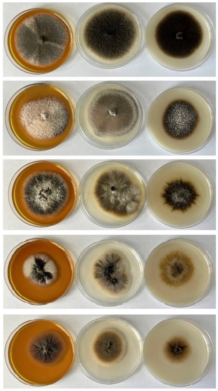
All strains exhibited significant similarity in growth rate and the morphological characteristics of colonies for different media. Colonies of all strains, in the case of abundant aerial mycelium formation, were elevated, with a woolly to flaky surface. In the absence of aerial mycelium, colonies were flat and only slightly woolly, with a large number of superficial pycnidia. Three strains were sterile on all media tested (VKM F-2782, VKM F-2799, and VKM F-5158). The remaining cultures showed pycnidial sporulation. Pycnidia are light-to-dark brown, thin-walled, and consist of polygonal cells. Conidiogenous cells are enteroblastic phialides; conidia are unicellular, hyaline, oval-to-elliptical, and smooth-walled. Cultures of VKM F-2800 and VKM F-5157, in addition to conidia, formed chains of Alternaria-like chlamydospores measuring 22–45 × 12–18 µm.
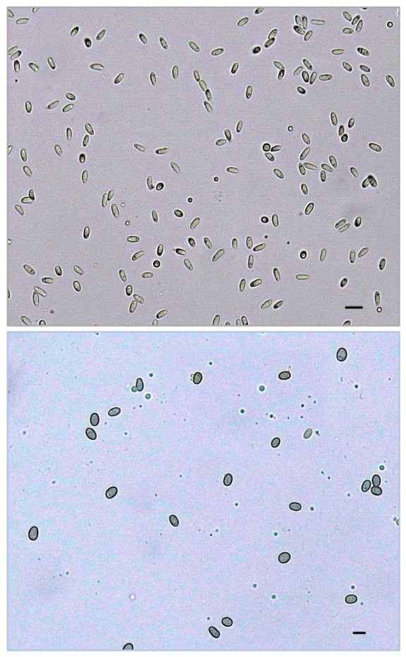
All cultures with sporulation were divided into two groups. The first group had eight cultures—VKM F-2758, VKM F-2768, VKM F-2794, VKM F-2797, VKM F-2800, VKM F-2839, VKM F-5157, VKM F-5160—with similar-sized pycnidia (aver. 71–162 × 60–120 µm) and differently sized biguttulate thin-walled conidia (aver. 3.8–6.1 × 1.9–2.7 μm). Two cultures—VKM F-5159 and VKM F-5161—had significantly larger (2.5 times) pycnidia, wider conidia than those of the first group of strains, thick walls, and no guttulate (aver. 4.3–6.5 × 3.1–4.1 μm).
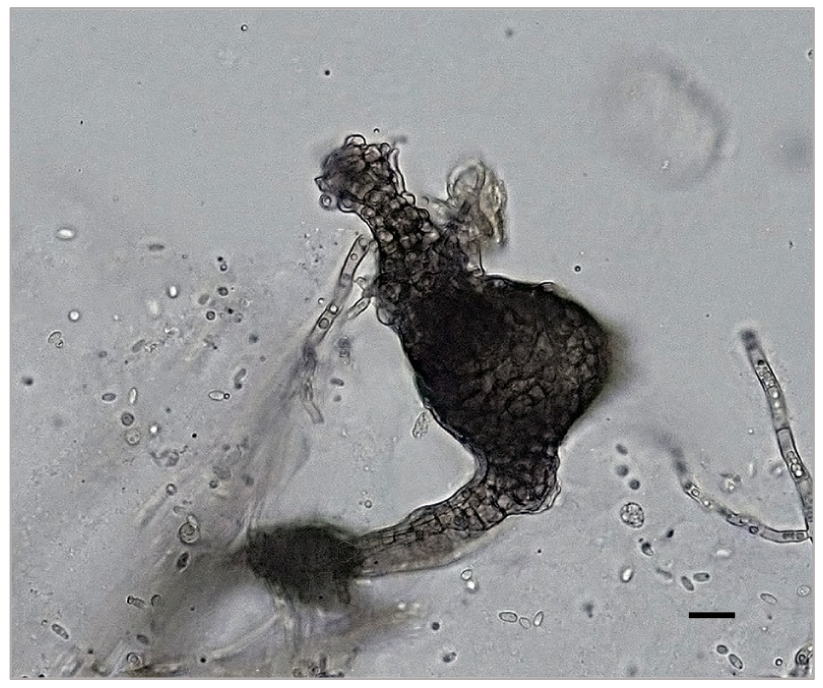
It should be noted that the morphological characteristics of the studied cultures are very similar to those of the Phoma sensu lato species complex. In particular, when dividing the genera Ampelomyces and Phoma by morphology, one of the main criteria is the immersion of pycnidia in the mycelium (Phoma) or their superficial location (Ampelomyces) [21]. However, most often this relates to the morphology of cultures on natural substrates, whether plant leaves or fungi. In vitro, especially after several passages, morphology can change, including the number and arrangement of pycnidia. Figure 1 shows a pycnidial conidiomata with a long stalk of the culture VKM F-2800, later identified as Didymella glomerata.

3.2. Multilocus Phylogenetic Analysis

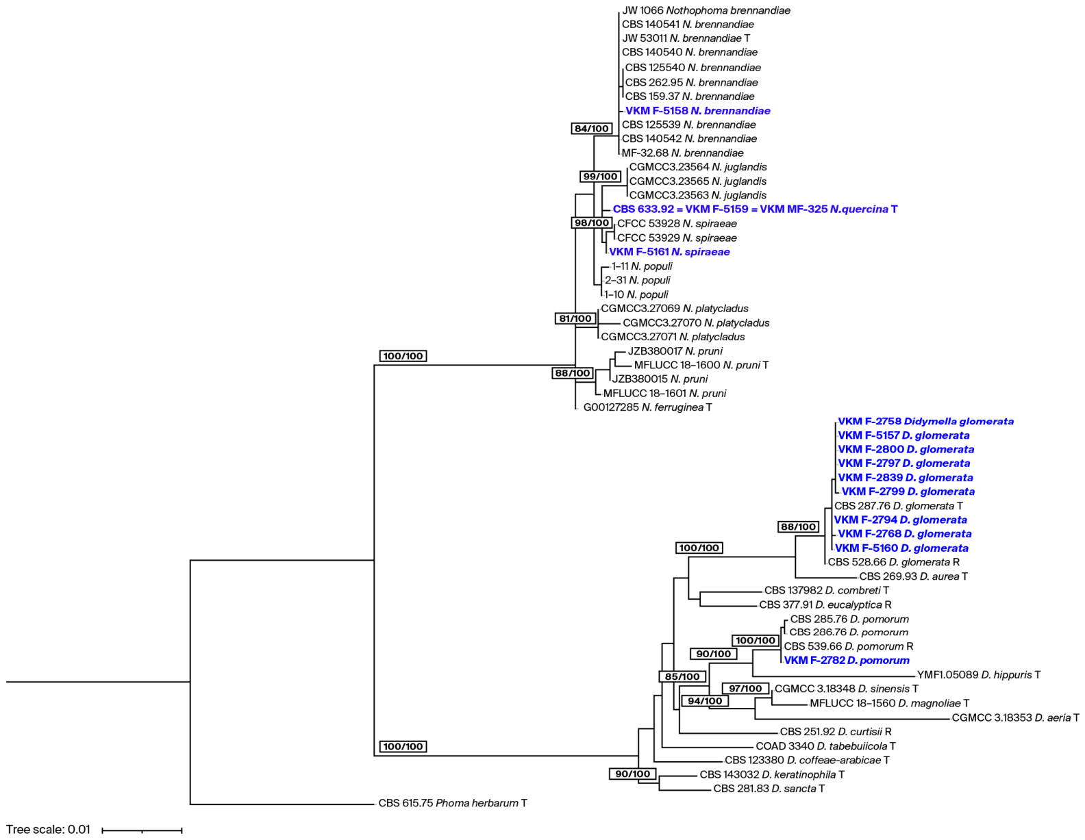
The results of multilocus phylogenetic analysis based on the ITS, LSU, rpb2, and tub2 sequences are presented in Table 3 and Figure 2.
All strains were closely related to representatives of phoma-like fungi of the family Didymellaceae and were also in the order Pleosporales. It was clear that none of the studied strains belonged to the genus Ampelomyces (order Pleosporales, family Phaeosphaeriaceae). We also compared the studied strains with the representative cultures of the genus Ampelomyces, and phylogenomic differences were confirmed (Table S2).

4. Discussion

Several decades ago, when studying hyperparasites on powdery mildew fungi, the presence of intracellular pycnidia automatically indicated the presence of fungi of the genus Ampelomyces, and studies mainly focused on determining the natural occurrence of hyperparasites and the intensity of mycoparasitism [22]. Their separation from representatives of phoma-like fungi based on cultural–morphological characteristics is difficult.
Many fungi have distinct phenotypic characteristics that allow for a fairly accurate identification of their taxon, at least at the genus level. This is not the case with Ampelomyces. The description of species in the genus Ampelomyces by Rudakov (1979) [6] was carried out based on phenotypic characteristics on nutrient media immediately after isolation. Since the pycnidia of each strain were found to be highly variable, the main criteria for distinguishing species were colony growth characteristics and conidia morphology. A detailed study of the morphology of the same strains after half a century of storage in the VKM revealed that differences in morphology exist; however, in some cases, they are within the range of intraspecific variability (Table 2).
Phoma is the most widespread genus in the order Pleosporales, comprising over 3000 species, and over 100 species are known as phytopathogens. This genus is polyphyletic and taxonomically controversial, with unclear boundaries. Phoma fungi are commonly associated with devastating diseases in economically important agricultural crops. These fungi can produce various phytotoxins that impair photosynthesis and cause electrolyte leakage from plant cells [23]. However, hyperparasitism has been infrequently reported regarding its representativeness in this genus. For example, P. glomerata (tm. Didymella glomerata) has been shown to initiate mycoparasitism in relation to Microsphaera penicillata, which causes leaf disease in sycamore trees [24]. Strains of Phoma sp. have been found to be hyperparasitic in relation to the basidiomycete Puccinia melanocephala, the causal agent of sugarcane rust in Cuba [25]. However, all these studies were conducted before the genomic era, when fungi were described solely based on morphological characteristics, so there is uncertainty about the correct taxonomic classification of mycophilic fungi.
A recent phylogenetic study of a wide range of fungi in the family Didymellaceae, order Pleosporales, conducted multilocus phylogenetic analysis based on ITS, LSU, rpb2, and tub2 sequences, taking into account morphological differences. This analysis not only clarified the phylogenetic relationships and established boundaries [26] of these species, but also determined their differences from fungi in the family Phaeosphaeriaceae, in which genus Ampelomyces is included. Species morphologically similar to fungi of the genus Phoma were found in at least six families of the order Pleosporales. Furthermore, there is evidence that phoma-like fungi are distributed across 20 families of six orders of ascomycetous fungi [18].
About a thousand sequences of fungi of the genus Ampelomyces have been deposited in the NCBI database. However, when compared with VKM fungi sequences, they were not closely related. Since there is no certainty that all deposited sequences belong specifically to fungi of the genus Ampelomyces, for a detailed comparison, we selected sequences from strains in the largest global collections: CBS (Netherlands), ATCC (USA), and DSM (Germany). It turned out that 34 sequences could be compared at the ITS and LSU loci. None of them showed close similarity to VKM strains (Table S2). The similarity percentages for ITS and LSU were in the range 82.88–95.76% and 92.52–95.57%, respectively. Thus, none of the strains studied belonged to the genus Ampelomyces.
The results of phylogenetic analysis for most VKM strains exhibited similarity to fungi of the genus Didymella. The majority of strains were assigned to Didymella glomerata (VKM F-2758, VKM F-2768, VKM F-2794, VKM F-2797, VKM F-2799, VKM F-2800, VKM F-2839, VKM F-5157, and VKM F-5160) (Table 2). As shown in the table, spore-bearing strains of this group have similar micromorphology in their conidiogenous apparatus. Their cultural–morphological characteristics are also similar and correspond to the description of Didymella glomerata based on a study, among other cultures, of the representative culture CBS 528.66 in vitro [27], as shown in Table 4.
Representatives of D. glomerata are considered widespread soil saprotrophs. There are several reports of these fungi on inorganic substrates, including asbestos, cement, and paint [26]. Their geographic distribution is large: in Russia, strains of this species have been detected on Apiaceae, Liliaceae, Rosaceae, and Salicaceae plants [18]. The strains studied in our work—most of which belong to the species D. glomerata—were subjected to experimental testing to determine their parasitic activity against powdery mildew pathogens after isolation from the natural environment by Rudakov. Cucumber (Cucumis sativus) leaves were infected with the phytopathogenic fungi Botrytis cinerea and Erysiphe cichoracearum (current name: Golovinomyces cichoracearum). Afterward, conidial suspensions of hyperparasites were applied dropwise to the phytopathogenic fungi, and the inhibitory effect was observed. Spores germinated, and hyphae entwined with the host fungal mycelium and penetrated deep into the cells [1]. The strains listed in Table 1 were found to be parasitic based on Rudakov’s data.
The strain of VKM F-2782, defined as Ampelomyces quisqualis, was classified as Didymella pomorum based on phylogenetic analysis. Unfortunately, this strain was sterile and did not form conidiogenic structures on any of the media we used. However, the cultural–morphological properties of this strain [31] do not contradict its assignment to the species via molecular genetic analysis (Table 4). It was found that similarity to the representative culture of Didymella pomorum CBS 539.66 is 100% (LSU and rpb2) and 99.79% (ITS) (Table 4). Representatives of this species are opportunistic pathogens that prefer a temperate climate and are often found in soil and plant seeds [27], as well as plants of various families, including Russia. Strain VKM F-2782 also exhibited parasitic properties in laboratory experiments by O. Rudakov (Table 1).
The division of strains into two groups based on morphological characteristics (Table 2) was also reflected in the results of multilocus testing. Two strains, VKM F-5159 and VKM F-5161, exhibited significant differences in micromorphology compared to other strains studied (Figure 3 and Figure 4). The larger pycnidia and ovoid, thick-walled conidia with a length-to-width ratio of 1.4 distinguished them from strains classified as belonging to the genus Didymella. These had a length-to-width ratio of 2.2 and thin-walled, ellipsoidal conidia. These strains, as well as a sterile strain of VKM F-5158, were assigned to the genus Nothophoma.
This genus was described in 2015 [32] based on a multilocus phylogenetic analysis of a large number of strains, attempting to divide the genera into an Ascochyta–Didymella–Phoma complex. As a result, nine new genera were described with a high level of accuracy, including the genus Nothophoma. The experimental data classified this genus as a member of the family Didymellaceae. The strain VKM F-5159, originally identified as Ampelomyces quisqualis and later transferred to the CBS collection (CBS 633.92), has been included in several phylogenetic studies and was initially considered Phoma fructicola [29]. However, according to phylogenetic analysis, it was then classified in the Nothophoma cluster as a type strain of the species Nothophoma quercina [32]. The replica of this strain stored in VKM was assigned to this species.
The strain VKM F-5158, defined as Ampelomyces quercinus, was found to be a representative of the species Nothophoma brennandiae, first described in strains isolated from soil samples in the Netherlands [28]. Subsequently, these fungi were found in Europe associated with plants of the families Betulaceae, Oleaceae, Rosaceae and Ulmaceae, and in Canada in domestic dust [17]. The similarity of the strain VKM F-5158 to the holotype culture of Nothophoma brennandiae JW53011 (CBS 145912) is 100% (ITS, LSU and rpb2) and 99.69% (tub2). A strain stored in VKM was extracted from the powdery mildew of an oak tree.
Strain VKM F-5161 was assigned to the species Nothophoma spiraeae. This is confirmed by the results of molecular genetic analysis and is not contradicted by the morphology of this strain or its comparison with the morphology of this species’ type strain CFCC 53928 [30] (Table 4).
Thus, studying the type of material of species Ampelomyces artemisiae, A. heraclei, A. polygoni, A. ulicis, and A. uncinulae, as described by O. Rudakov [6], showed that they are heterotypic (taxonomic) synonyms of the taxon Didymella glomerata.
Taxonomy.
Didymella glomerata (Corda) Q. Chen & L. Cai 2015
= Ampelomyces artemisiae (Voglino 1905) Rudakov 1979
= Ampelomyces heraclei (Dejeva 1967) Rudakov 1979
= Ampelomyces polygoni (Potebnia 1907) Rudakov 1979
= Ampelomyces ulicis (J.F. Adams 1907) Rudakov 1979
= Ampelomyces uncinulae (Fautrey 1893) Rudakov 1979
Ampelomyces artemisiae (Voglino) Rudakov 1979
Typus: Republic of Moldova, fungus, Golovinomyces cichoracearum (syn. Erysiphe cichoracearum) on Acacia sp., MF-332 (VKM F-2794).
The type strain corresponds morphologically (Table 2) and phylogenetically (Figure 2) to Phoma glomerata, which justifies the recognition of this species as a heterotypic synonym of Phoma glomerata.
Ampelomyces heraclei (Dejeva) Rudakov 1979
Typus: Republic of Moldova, fungus, Plasmopara viticola, MF-245 (VKM F-2768).
The type strain corresponds morphologically (Table 2) and phylogenetically (Figure 2) to Phoma glomerata, which justifies the recognition of this species as a heterotypic synonym of Phoma glomerata.
Ampelomyces polygoni (Potebnia) Rudakov 1979
Syntypus: Krasnodar, Russia, fungus, Erysiphe cruciferarum on Polygonum sp., MF-197 (VKM F-2758).
Syntypus: Krasnodar, Russia, fungus, Erysiphe cruciferarum on Polygonum sp., MF-368 (VKM F-2799).
The syntypes strains correspond morphologically (Table 2) and phylogenetically (Figure 2) to Phoma glomerata, which justifies the recognition of this species as a heterotypic synonym of Phoma glomerata.
Ampelomyces ulicis (J. Adams) Rudakov 1979
Typus: Moscow Region, Russia, fungus, Erysiphe cruciferarum on Convolvulus arvensis, MF-342 (VKM F-2797).
The type strain corresponds morphologically (Table 2) and phylogenetically (Figure 2) to Phoma glomerata, which justifies the recognition of this species as a heterotypic synonym of Phoma glomerata.
Ampelomyces uncinulae (Fautrey) Rudakov 1979
Typus: Republic of Moldova, fungus, Podosphaera clandestina (syn. Erysiphe clandestina), MF-484 (VKM F-2839).
The type strain corresponds morphologically (Table 2) and phylogenetically (Figure 2) to Phoma glomerata, which justifies the recognition of this species as a heterotypic synonym of Phoma glomerata.
For all other species described by Rudakov, the type of material was not preserved, or the species were described based on the study of only herbarium material that has not survived to this day.
Since some strains re-identified here as Didymella glomerata exhibited hyperparasitic behavior under the conditions studied by Rudakov, our research suggests that hyperparasitism is possible for a wider range of taxa of the order Pleosporales, in addition to Ampelomyces.
Hyperparasitic fungi are of great ecological importance, mediating interactions between host plants and primary parasites and playing a significant role in regulating the population sizes of both partners. Certainly, the nature of hyperparasitic relationships needs to be further investigated, and all strains of the genus Ampelomyces stored in mycological culture collections should be carefully checked and, if necessary, redescribed.

Supplementary Materials

The following supporting information can be downloaded at https://www.mdpi.com/article/10.3390/jof12080558/s1. Table S1: GenBank accession numbers of reference strains; Table S2: Identity (%) of studied strains with reference sequences of Ampelomyces spp. in NCBI.

Author Contributions

N.I.—conceptualization, methodology, supervision, writing—review & editing. A.D.—formal analysis, investigation. G.K.—investigation, validation, writing—original draft. All authors have read and agreed to the published version of the manuscript.

Funding

This work was financially supported by the Ministry of Science and Higher Education of the Russian Federation (State Assignment No. 075-15-2025-485).

Institutional Review Board Statement

Not applicable.

Informed Consent Statement

Not applicable.

Data Availability Statement

The DNA sequences obtained in this study have been submitted to GenBank.

Conflicts of Interest

The authors declare no conflicts of interest.

References

  1. Rudakov, O.L. Mycophilic Fungi, Their Biology and Practical Significance; Nauka: Moscow, Russia, 1981. (In Russian) [Google Scholar]
  2. Manjunatha, L.; Singh, S.; Ravikumara, B.M.; Reddy, G.N.; Senthilkumar, M.  Ampelomyces. In Beneficial Microbes in Agro-Ecology; Amaresa, N., Kumar, M.S., Annapurna, K., Kumar, K., Sankaranarayanan, A., Eds.; Academic Press: Amsterdam, The Netherlands, 2020; Chapter 44; pp. 833–860. [Google Scholar] [CrossRef]
  3. Sztejnberg, A. Ampelomyces quisqualis AQ10 (CNCM I-807) for Biological Control of Powdery Mildew. U.S. Patent 5,190,754, 2 March 1993. [Google Scholar]
  4. Carbó, A.; Torres, R.; Usall, J.; Ballesta, J.; Teixidó, N. Biocontrol potential of Ampelomyces quisqualis strain CPA–9 against powdery mildew: Conidia production in liquid medium and efficacy on zucchini leaves. Sci. Hortic. 2020, 267, 109337. [Google Scholar] [CrossRef]
  5. Németh, M.Z.; Seress, D.; Nonomura, T. Fungi parasitizing powdery mildew fungi: Ampelomyces strains as biocontrol agents against powdery mildews. Agronomy 2023, 13, 1991. [Google Scholar] [CrossRef]
  6. Rudakov, O.L. Fungi of the genus Ampelomyces Ces. ex Schlecht. Mycol. Phytopathol. [Mikol. I Fitopatol.] 1979, 13, 104–110. (In Russian) [Google Scholar]
  7. Athira, K.; Ragupathi, N.; Raguchander, T. Morphological characterization of Ampelomyces spp., a hyperparasite of Bhendi (Abelmoschus esculentus (L.) Moench) powdery mildew. J. Appl. Nat. Sci. 2017, 9, 1954–1957. [Google Scholar] [CrossRef]
  8. Prahl, R.E.; Shahjahan, K.; Ravinesh, C.D. Ampelomyces mycoparasites of powdery mildews—A review. Can. J. Plant Pathol. 2023, 45, 391–404. [Google Scholar] [CrossRef]
  9. Prahl, R.E. Nuclear Ribosomal DNA Secondary Structures and Statistical Approach for the Phylogeny of Ampelomyces. Ph.D. Thesis, University of Southern Queensland, Toowoomba, QLD, Australia, 2022. [Google Scholar] [CrossRef]
  10. Kornerup, A.; Wanscher, J.H. Methuen Handbook of Colour, 3rd ed.; Eyre Methuen: London, UK, 1978. [Google Scholar]
  11. White, T.J.; Bruns, T.; Lee, S.; Taylor, J. Amplification and direct sequencing of fungal ribosomal RNA genes for phylogenetics. In PCR Protocols: A Guide to Methods and Applications; Innis, M.A., Gelfand, D.H., Sninsky, J.J., White, T.J., Eds.; Academic Press: San Diego, CA, USA, 1990; pp. 315–322. [Google Scholar]
  12. Rehner, S.A.; Samuels, G.J. Taxonomy and phylogeny of Gliocladium analysed from nuclear large subunit ribosomal DNA sequences. Mycol. Res. 1994, 98, 625–634. [Google Scholar] [CrossRef]
  13. Vilgalys, R.; Hester, M. Rapid genetic identification and mapping of enzymatically amplified ribosomal DNA from several Cryptococcus species. J. Bacteriol. 1990, 172, 4238–4246. [Google Scholar] [CrossRef] [PubMed]
  14. Woudenberg, J.H.; Aveskamp, M.M.; de Gruyter, J.; Spiers, A.G.; Crous, P.W. Multiple Didymella teleomorphs are linked to the Phoma clematidina morphotype. Persoonia 2009, 22, 56–62. [Google Scholar] [CrossRef] [PubMed]
  15. Liu, Y.J.; Whelen, S.; Hall, B.D. Phylogenetic relationships among ascomycetes: Evidence from an RNA polymerse II subunit. Mol. Biol. Evol. 1999, 16, 1799–1808. [Google Scholar] [CrossRef] [PubMed]
  16. Cai, F.; Druzhinina, I.S. In honor of John Bissett: Authoritative guidelines on molecular identification of Trichoderma. Fungal Divers. 2021, 107, 1–69. [Google Scholar] [CrossRef]
  17. Hou, L.; Groenewald, J.; Pfenning, L.; Yarden, O.; Crous, P.; Cai, L. The Phoma–like dilemma. Stud. Mycol. 2020, 96, 309–396. [Google Scholar] [CrossRef] [PubMed]
  18. Gomzhina, M.M.; Gasich, E.L. Didymellaceae species associated with Convolvulaceae plants, with description of three new species. Mycologia 2025, 117, 765–787. [Google Scholar] [CrossRef] [PubMed]
  19. Minh, B.Q.; Schmidt, H.A.; Chernomor, O.; Schrempf, D.; Woodhams, M.D.; von Haeseler, A.; Lanfear, R. IQ-TREE 2: New models and efficient methods for phylogenetic inference in the genomic era. Mol. Biol. Evol. 2020, 35, 1530–1534. [Google Scholar] [CrossRef] [PubMed]
  20. Letunic, I.; Bork, P. Interactive tree of life (iTOL) v3: An online tool for the display and annotation of phylogenetic and other trees. Nucleic Acids Res. 2016, 44, W242–W245. [Google Scholar] [CrossRef] [PubMed]
  21. Sutton, B.C. The Coelomycetes. Fungi Imperfecti with Pycnidia, Acervuli and Stromata; Commonwealth Mycological Institute: London, UK, 1980. [Google Scholar]
  22. Kiss, L. Natural occurrence of Ampelomyces intracellular mycoparasites in mycelia of powdery mildew fungi. New Phytol. 1998, 140, 709–714. [Google Scholar] [CrossRef] [PubMed]
  23. Deb, D.; Khan, A.; Dey, N. Phoma diseases: Epidemiology and control. Plant Pathol. 2020, 69, 1203–1217. [Google Scholar] [CrossRef]
  24. Sullivan, R.F.; White, J.F. Phoma glomerata as a mycoparasite of powdery mildew. Appl. Environ. Microbiol. 2000, 66, 425–427. [Google Scholar] [CrossRef] [PubMed][Green Version]
  25. Sandoval, I.; López, M.O. Nuevos hiperparásitos de la roya de la caña de azúcar Puccinia melanocephala H. y P. Syd. Rev. ATAC 1990, 49, 20–26. [Google Scholar]
  26. Chen, Q.; Hou, L.W.; Duan, W.J.; Crous, P.W.; Cai, L. Didymellaceae revisited. Stud. Mycol. 2017, 87, 105–159. [Google Scholar] [CrossRef] [PubMed]
  27. Boerema, G.H. Contributions towards a monograph of Phoma (Coelomycetes)—II. Section Peyronellaea. Persoonia-Mol. Phylogeny Evol. Fungi 1993, 15, 197–221. [Google Scholar]
  28. Hou, L.; Hernandez–Restrepo, M.; Groenewald, J.Z.; Cai, L.; Crous, P.W. Citizen science project reveals high diversity in Didymellaceae (Pleosporales, Dothideomycetes). MycoKeys 2020, 65, 49–99. [Google Scholar] [CrossRef] [PubMed]
  29. Aveskamp, M.M.; de Gruyter, J.; Woudenberg, J.H.C.; Verkley, G.J.M.; Crous, P.W. Highlights of the Didymellaceae: A polyphasic approach to characterise Phoma and related pleosporalean genera. Stud. Mycol. 2010, 65, 1–60. [Google Scholar] [CrossRef] [PubMed]
  30. Zhang, L.X.; Yin, T.; Pan, M.; Tian, C.M.; Fan, X.L. Occurrence and identification of Nothophoma spiraeae sp. nov. in China. Phytotaxa 2020, 430, 147–156. [Google Scholar] [CrossRef]
  31. Chen, Q.; Jiang, J.R.; Zhang, G.Z.; Cai, L.; Crous, P.W. Resolving the Phoma enigma. Stud. Mycol. 2015, 82, 137–217. [Google Scholar] [CrossRef] [PubMed]
  32. Boerema, G.H.; de Gruyter, J.; Noordeloos, M.E.; Hamers, M.E.C. Phoma Identification Manual. Differentiation of Specific and Infra–Specific Taxa in Culture; CABI Publishing: Wallingford, UK, 2004. [Google Scholar] [CrossRef]
Figure 1. Pycnidium on the stalk of Didymella glomerata VKM F-2800. Scale bar 10 μm.
Jof 12 00558 g001
Figure 2. Phylogenetic tree of Didymella and Nothophoma species and strains constructed based on maximum likelihood analysis using the combined ITS, LSU, rpb2, and tub2 datasets. Maximum likelihood bootstrap support values (MLBS ≥ 80%) and Bayesian posterior probabilities (BPP ≥ 95%) are indicated at nodes (MLBS/BPP). Studied strains are shown in blue, and type and representative strains are marked with T or R, respectively, and are presented in Table S1.
Jof 12 00558 g002
Figure 3. Colony morphology in 7 days on MEA, PDA and OA (from left to right) of strains (from top to bottom): VKM F-2800 Didymella glomerata (previous name A. humuli); VKM F-2782 Didymella pomorum (previous name A. quisqualis); VKM F-5158 Nothophoma brennandiae (previous name A. quercinus); VKM F-5159 Nothophoma quercina (previous name A. quercinus); VKM F-5161 Nothophoma spiraeae (previous name Ampelomyces sp.).
Jof 12 00558 g003
Figure 4. Conidia of Didymella glomerata VKM F-2800 (top) and Nothophoma spiraeae VKM F-5161 (bottom). Scale bar 5 μm.
Jof 12 00558 g004
Table 1. List of strains studied in the current work.
Number in CollectionsTaxon Name
(Received as)
Identification Number 1Status of the Strain 2Substrate (Including Host)
VKM F-2758 3
(ATCC 38608)
Ampelomyces polygoniMF-197Syntypefungus, Erysiphe cruciferarum on Polygonum sp., Krasnodar, Russia
VKM F-2768 4 (ATCC 36804)Ampelomyces heracleiMF-245Typefungus, Plasmopara viticola, Republic of Moldova
VKM F-2782 3
(ATCC 36856)
Ampelomyces quisqualisMF-283-fungus, Golovinomyces cichoracearum (syn. Erysiphe cichoracearum), Caucasus area, Russia
VKM F-2794 3 (ATCC 38609)Ampelomyces artemisiaeMF-332Typefungus, Golovinomyces cichoracearum (syn. Erysiphe cichoracearum) on Acacia sp., Republic of Moldova
VKM F-2797 3Ampelomyces ulicisMF-342Typefungus, Erysiphe cruciferarum on Convolvulus arvensis, Republic of Moldova
VKM F-2799 3Ampelomyces polygoniMF-368Syntypefungus, Erysiphe cruciferarum on Polygonum sp., Krasnodar, Russia
VKM F-2800 3 (ATCC 38616)Ampelomyces humuliMF-369-fungus, Podosphaera macularis (syn. Sphaerotheca macularis) on Potentilla sp., Moscow Region, Russia
VKM F-2839 3
(ATCC 36853)
Ampelomyces uncinulaeMF-484Typefungus, Podosphaera clandestina (syn. Erysiphe clandestina), Republic of Moldova
VKM F-5157Ampelomyces sp.MF-350-fungus, powdery mildew on Polygonum sp., Astrakhan, Russia
VKM F-5158 3
(ATCC 38614)
Ampelomyces quercinusMF-349-fungus, Erysiphe alphitoides (syn. Microsphaera alphitoides) on Quercus sp., Russia
VKM F-5159
(CBS 633.92,
ATCC 36786)
Ampelomyces quercinusMF-325-fungus, Golovinomyces cichoracearum (syn. Erysiphe cichoracearum) on Quercus sp., Russia
VKM F-5160Ampelomyces ulicisMF-348-fungus, Erysiphe communis on Medicago sp., Moscow Region, Russia
VKM F-5161Ampelomyces sp.MF-343-fungus, Golovinomyces cichoracearum (syn. Erysiphe cichoracearum) on Alnus sp., Khanka Lake, Far East, Russia
1 The number given to the strain when it was isolated from the natural substrates. 2 Type strain (or nomenclatural type) is a reference specimen of a fungus to which the name of a particular species is officially associated. Syntype is one of several strains simultaneously designated in the protologue. 3 Experimentally studied parasitism on the host Erysiphe cichoracearum (current name—Golovinomyces cichoracearum) [1]. 4 Experimentally studied parasitism on the host Botrytis cinerea [1].
Table 2. Phenotypic characteristics of the studied strains.
Strain NumberColony Diameter (cm) After 7 Days on MediaColony Colours on OASize (μm)
OAPDAMEAMyceliumReversePycnidiaConidia
VKM F-27587.07.58.0whitegreyish-brown56–133 × 52–963.7–6.0 × 2.0–2.6
VKM F-27687.07.57.5brownish-greybrownish-grey66–173 × 65–1494.1–7.0 × 1.8–3.2
VKM F-27825.07.07.0white in center, olive-brown in peripherygreyish-brownnono
VKM F-27947.08.07.5brownish-greybrownish-grey69–156 × 65–1393.9–5.8 × 1.9–2.9
VKM F-27976.07.38.0greyish-brownbrownish-grey103–250 × 83–1784.1–5.8 × 1.9–2,6
VKM F-27996.05.86.5brown in center, light in peripherybrownnono
VKM F-28006.57.57.0dark-brownbrownish-grey59–101 × 35–613.7–6.1 × 1.8–2.6
VKM F-28397.08.07.5light-brownbrown60–131 × 40–823.5–6.0 × 1.8–2.7
VKM F-51577.08.07.0light-brownbrown45–100 × 42–783.7–6.4 × 1.8–2.5
VKM F-51605.56.57.0white in center, orange-gray in peripherywhite71–165 × 68–1523.5–6.3 × 2.0–2.8
VKM F-51585.56.56.5brownbrownnono
VKM F-51595.05.34.8brownish-orangewhite235–474 × 149–3294.0–6.1 × 3.2–4.2
VKM F-51614.85.35.0yellowish-browndark-blond155–433 × 144–2984.6–6.8 × 3.0–4.0
Table 3. Results of multilocus phylogenetic analysis of studied strains based on the ITS, 28S, rpb2, and tub2 sequences.
Strain NumberDeposited in VKM asGenBank Accession NumbersIdentified as
ITStub2rpb2LSU
VKM F-2758Ampelomyces polygoni (Potebnia 1907) Rudakov 1979-PX283766PX283753-Didymella glomerata (Corda 1840) Qian Chen et L. Cai 2015
VKM F-2768A. heraclei (Dejeva 1967) Rudakov 1979PX289730PX283767PX283754PX289754D. glomerata (Corda 1840) Qian Chen et L. Cai 2015
VKM F-2794A. artemisiae (Voglino 1905) Rudakov 1979PX289732PX283768PX283756PX289756D. glomerata (Corda 1840) Qian Chen et L. Cai 2015
VKM F-2797A. ulicis (J.F. Adams 1907) Rudakov 1979PX289733PX283769PX283757PX289757D. glomerata (Corda 1840) Qian Chen et L. Cai 2015
VKM F-2799A. polygoni (Potebnia 1907) Rudakov 1979PX289734PX283770PX283758PX289758D. glomerata (Corda 1840) Qian Chen et L. Cai 2015
VKM F-2800A. humuli (Fautrey 1890) Rudakov 1979PX289735PX283771PX283759PX289759D. glomerata (Corda 1840) Qian Chen et L. Cai 2015
VKM F-2839A. uncinulae (Fautrey 1893) Rudakov 1979PX289736PX283772PX283760PX289760D. glomerata (Corda 1840) Qian Chen et L. Cai 2015
VKM F-5157Ampelomyces sp.PX275617PX283773PX283761PX275713D. glomerata (Corda 1840) Qian Chen et L. Cai 2015
VKM F-5160A. ulicis (J.F. Adams 1907) Rudakov 1979PX275620PX283776PX283764PX275716D. glomerata (Corda 1840) Qian Chen et L. Cai 2015
VKM F-2782A. quisqualis Cesati 1852PX289731-PX283755PX289755D. pomorum (Thümen 1879) Qian Chen et L. Cai 2015
VKM F-5158Ampelomyces quercinus (Sydow 1915) Rudakov 2015PX275618PX283774PX283762PX275714Nothophoma brennandiae Hernández-Restrepo, L.W. Hou, L. Cai et P.W. Crous 2020
VKM F-5159Ampelomyces quercinus (Sydow 1915) Rudakov 2015PX275619PX283775PX283763PX275715N. quercina (Sydow et P. Sydow 1915) Qian Chen et L. Cai 2015
VKM F-5161Ampelomyces sp.PX275621PX283777PX283765PX275717N. spiraeae L.X. Zhang et X.L. Fan 2020
Table 4. Phenotypic characteristics of the reference strains.
Strain
Number
(T or R Culture)
SpeciesColony Diameter (cm) After 7 Days on MediaColony Colours on OASize (μm)Reference
OAPDAMEAMyceliumReversePycnidiaConidia
CBS 528.66Didymella
glomerata
3.5–7.0-6.5–7.5olivaceousolivaceous to blackish100–300(3.5)4–8.5(10) × 1.5–3(3.5)[27]
CBS 539.66D. pomorum4.5–6.0-5.5–7.5olivaceousbrownish to blackish100–200(4)5–7(8) × 1.5–2.5(3)[27]
CBS 145912Nothophoma
brennandiae
5.0–5.55.0–5.54.7–5.0dark brick to sepia, cinnamon to the edge-155–350 × 100–3003–8.5 × 1.5–3[28]
CBS 633.92N. quercina5.5–6.8-5.5–7.5olivaceous near the colony centreconcolourous(50–)65–130(–150) × (65–)95–200(–220)(5–)5.5–7.5(–8.5) × 3–4.5(–5)[29]
CFCC 53928N. spiraeae---hazel-(145–)155–280(300) × (120–)140–230(–250)5–6.5(–7) × (3)3.5–4[30]
Disclaimer/Publisher’s Note: The statements, opinions and data contained in all publications are solely those of the individual author(s) and contributor(s) and not of MDPI and/or the editor(s). MDPI and/or the editor(s) disclaim responsibility for any injury to people or property resulting from any ideas, methods, instructions or products referred to in the content.