Article

Members of the Fusarium fujikuroi Species Complex Isolated from Asymptomatic Wetland Grasses in Argentina Include Previously Described Species Pathogenic on Cereal Crops and a Novel Species


1
Instituto de Investigaciones en Micología y Micotoxicología (IMICO), CONICET-Universidad Nacional de Rio Cuarto, Ruta 36 Km 601, Río Cuarto 5800, Córdoba, Argentina
2
USDA, Agricultural Research Service, National Center for Agricultural Utilization Research, (UNIT), 1815 N University St., Peoria, IL 61604, USA
*
Author to whom correspondence should be addressed.
J. Fungi 2026, 12(6), 444; https://doi.org/10.3390/jof12060444
Submission received: 17 April 2026 / Revised: 11 June 2026 / Accepted: 14 June 2026 / Published: 17 June 2026
(This article belongs to the Special Issue Morphology, Phylogeny and Pathogenicity of Fusarium—2nd Edition)

Abstract

The floodplains of the Paraná and Paraguay rivers form the Chaco wetland, one of the most species-rich plant ecosystems in Argentina. Because wild grasses can serve as reservoirs of fungal species that cause disease and mycotoxin contamination of cereal crops, we examined asymptomatic, wild grasses from the Chaco wetlands for the presence of the genus Fusarium, which includes multiple species that cause agriculturally important diseases and/or mycotoxin contamination of crops. We focused our efforts on the identification and characterization of the multispecies lineage known as the Fusarium fujikuroi species complex (FFSC). Using morphological traits and partial DNA sequences of the TEF1 gene, we determined that 58 isolates recovered from the grasses were members of FFSC. Fifty of the isolates were identified as one of six FFSC species, including the economically important plant pathogenic species F. proliferatum, F. subglutinans, and F. verticillioides. To our knowledge, two of the species, F. anthophilum and F. pseudocircinatum, have not been reported previously in Argentina. Our analyses also indicated that eight of the FFSC isolates were a novel species, herein described as Fusarium varsavskyanum. A polymerase chain reaction (PCR) assay and genome sequence data indicate that each isolate of F. varsavskyanum isolate had only one mating type idiomorph (MAT1-1 or MAT1-2), which suggests that the fungus is heterothallic. Genome sequence analysis indicated that F. varsavskyanum has the genetic potential to produce, (i) the emerging mycotoxins fusaric acid and beauvericin (or enniatins); (ii) the pigments bikaverin, carotenoids, and fusarubin; and (iii) the plant hormones auxins, cytokinins, and gibberellins. Thus, asymptomatic grasses from the Chaco wetland can harbor Fusarium species that in some agroecosystems can cause economically important diseases and/or mycotoxin contamination of crops. It remains to be determined whether the genotypes of Fusarium species that occur on the wetland grasses, including F. varsavskyanum genotypes, can negatively impact agriculture.

1. Introduction

The grass family, Poaceae, is one of the most species-rich families within the kingdom Plantae. Poaceae species are both ecologically and economically important because they are a dominant component of multiple ecosystems, and multiple species are used as cereal crops and in pastures [1]. Approximately 70% of the land area of Argentina is natural grassland [2]. This includes grasslands in the Chaco Wetlands located in the east of Chaco Province. The wetlands are formed by the Paraná and Paraguay river floodplain and are one of the three most biodiverse ecosystems in Argentina. The wetland landscape is a complex of open water, aquatic vegetation, gallery forests as well as grasslands. A diversity of grasses palatable to livestock and a temperate climate make the grassland of the Chaco Wetlands suitable for grazing cattle year-round [3]. Among the most common grass species in the Chaco Wetlands are Leersia hexandra, Luziola peruviana, Sorghastrum setosum, Spartina argentinensis and Cynodon dactylon. In a previous study, examination of 175 asymptomatic grass plants representing 12 genera revealed that all the plants were contaminated with Fusarium mycotoxins. The predominant mycotoxins were zearalenone (ZEA), T-2 toxin, and HT-2 toxin. Other mycotoxins/metabolites that occurred less frequently and/or at lower levels were beauvericin, fumonisin B1, and equisetin [4]. All the metabolites noted above are reported to be produced by one or more species of Fusarium [5,6].
A mycological analysis of the Chaco Wetland grasses revealed that Fusarium was recovered from 60 to 100% of plants per sample site [4]. Analysis of the Fusarium isolates recovered from asymptomatic grasses revealed that some were T-2 and HT-2 toxin-producing species, including F. chaquense, a recently described member of the multispecies lineage known as the Fusarium sambucinum species complex [7]. Another species recovered was a member of the F. incarnatum-equiseti species complex and was likely responsible for ZEA contamination observed in the wetland grasses [8]. Morphology-based analyses revealed that other isolates recovered from the Chaco Wetland grasses were members of the F. fujikuroi species complex (FFSC).
FFSC is one of the most species-rich lineages within the genus Fusarium. Comparative morphological and molecular phylogenetic studies indicate that the FFSC comprises over 60 phylogenetically distinct species that can be further resolved into three clades—the African, American and Asian clades—which were proposed to have diverged from one another on different continents after fragmentation of the supercontinent Gondwana [9,10,11,12,13,14,15]. FFSC is a global threat to food/feed security and safety because many of its species cause economically important crop diseases and/or produce mycotoxins that are health hazards to humans and livestock [6,14,16,17]. Crop diseases caused by fungi in the FFSC include ear rot, stalk rot and seedling blight of maize, pitch canker of pine trees, bakanae disease of rice, pokkah boeng disease of sugarcane, and mango malformation [17].
Multiple members of the FFSC can occur as both pathogens and endophytes of plants from natural and agricultural environments [18]. For example, F. fujikuroi causes bakanae of rice but it also forms endophytic associations with aquatic plants of the genus Echinochloa [19]; F. circinatum causes pitch canker of pine trees but it is also an endophyte of wild grasses [20,21]; and F. giganteum is a pathogen of maize but also an endophyte of the forage grass Panicum maximum [22]. There are also FFSC members that are endophytes of grasses in natural environments but not reported to be plant pathogens. These FFSC members include the Andropogon and Sorghastrum endophyte F. konzum [23] the Coix gasteenii endophyte F. coicis, the Sorghum interjectum endophyte F. tjaebata [24] and the Brachiaria spp. endophytes F. caapi and F. brachiariae [25]. The fact that some crop pathogens are endophytes in other plants has important agricultural implications. For example, endophyte-infected asymptomatic plants are potential reservoirs of inoculum that could initiate disease epidemics in crops and/or potential sources of genetic diversity of plant pathogens [26].
The objective of the current study was to determine species diversity among 58 Fusarium isolates previously recovered from asymptomatic wild grasses from the Chaco Wetlands. In the previous study, the isolates were identified by morphological characters as members of the FFSC. In the current study, we used DNA-based analyses to determine the species identities of the fungal isolates and to formally describe and characterize the biology and chemistry of a novel species discovered among the isolates.

2. Materials and Methods

2.1. Fusarium Isolates

The Fusarium isolates (n = 58) analyzed in the current study were previously isolated from the aerial parts asymptomatic grasses (Poaceae). The grasses were collected in July 2011 and September and February 2014 from the Chaco Wetlands (Chaco Province), Argentina. All the isolates were morphologically characterized according to Leslie and Summerell [27] as members of the FFSC. The strains are deposited at the Research Institute on Mycology and Mycotoxicology (IMICO, UNRC-CONICET), Universidad Nacional de Rio Cuarto culture collection (RC). Cultures are maintained in 15% glycerol at −80 °C. Two isolates (RC-J82 and RC-J132 = type) of the novel species described herein were also deposited at the United States Department of Agriculture’s ARS Culture Collection (NRRL) as accessions NRRL 64882 and NRRL 64883 (Table 1).

2.2. Phylogenetic Analysis and Whole-Genome Sequencing

Isolates were grown in complete medium [27] and incubated on an orbital shaker (150 rpm, New Brunswick Scientific CO., INC, Edison, NJ, USA) for at least three days at 25 ± 1 °C. The resulting mycelia were harvested by filtration through non-gauze milk filters (Ken AG, Ashland, OH, USA). Excess water was removed by blotting mycelia between clean paper towels, and dried mycelia were stored frozen at −20 °C. DNA extraction was performed with the acetyl-trimethyl ammonium bromide method [26]. DNA concentration was estimated using a NanoDrop 2000 instrument (Thermo Fisher Scientific Inc., Waltham, MA, USA).
Partial sequence of the translation elongation factor-1α gene (TEF1) was analyzed following PCR amplification with the primers and conditions described by O’Donnell et al. [10]. All PCR products generated were separated by electrophoresis through 1.5% (w/w) agarose gels stained with ethidium bromide (5 μg/mL), visualized and photographed using a MiniBIS Pro gel documentation system (DNR Bio-Imaging Systems, Jerusalem, Israel) to confirm that a ~700 bp fragment was amplified. Fragments purification and sequencing of both strands was performed commercially (Macrogen, Inc., Seoul, South Korea) using the same primers used for the PCR amplifications. As both DNA strands were sequenced, they were aligned with ClustalW [28], as implemented in the program BioEdit version 7.0.9.0 [29] in order to detect and correct sequencing errors. The TEF1 sequences of selected strains from each species were deposited in GenBank under the accession numbers indicated in Table 1. Nucleotide sequence comparisons were performed using the Basic Local Alignment Search Tool (BLAST, https://blast.ncbi.nlm.nih.gov, accessed on 30 July 2025) at the National Centre for Biotechnology Information (NCBI). Multiple sequence alignment of the TEF1 gene was performed using the Web-based program MAFFT (http://mafft.cbrc.jp/aligment/software, accessed on 5 August 2025.) [30]. TEF1 sequences from reference FFSC strains and other Fusarium species obtained from GenBank were included in the analysis (Table 2). Based on this alignment, phylogenetic analyses were performed on selected strains by the maximum parsimony (MP) method using TNT 1.1 (Tree Analysis Using New Technology) [31], maximum likelihood (ML) using PhyML 3.1 [32] and Bayesian inference (BI) using MrBayes 3.2.6 [33]. The MP analysis used the heuristic search option with 1000 random addition sequences with tree bisection reconnection (TBR) branch swapping, saving 10 trees per replicate. Clade stability was assessed by 1000 bootstrap replications. Gaps were treated as “fifth state”. For ML and BI analyses, the best substitution model for the TEF1 alignment (i.e., TrN + G model) was determined using the Akaike information criterion (AIC) as implemented in jModelTest 2.1.10 [34]. For ML analysis, the robustness of the best tree was evaluated by 1000 bootstrap replications. For BI analysis, two runs with four chains each were run for 10 million generations with a sampling frequency of every 100 generations and the first 25% of trees from each run were discarded as burn-in. Fusarium oxysporum NRRL 22902 (AF160312) was used as the outgroup.
For those isolates that could not be identified to species using TEF1 sequence, partial sequences of the second largest subunit of RNA polymerase (RPB2), beta-tubulin (TUB2), and calmodulin (CDM1) genes were amplified using previously described primers and conditions [35,36,37,38,39] and PCR products were evaluated as described above for TEF1 amplicons. Phylogenetic analyses of each gene partition and the combined dataset were performed by BI as described above. The TIM3ef + G model was used for RPB2, TIM1 + G for TUB2, and TrNef + I for CDM. For the combined dataset, each gene was treated as a separate partition with independent parameter estimations. The resulting partial sequences of RPB2, TUB2 and CDM were deposited in GenBank under accession numbers indicated in Table S1.
Whole genome sequence data of strains NRRL 64882 and NRRL 64883 were generated using a MiSeq platform (Illumina, San Diego, CA, USA) at USDA ARS NCAUR. Strains were grown in liquid GYP medium (2% glucose, 1% peptone, and 0.3% yeast extract) for two days, and the resulting mycelia were harvested by filtration, lyophilized, and ground to a powder. Genomic DNA was extracted from ground mycelia using the Genomic-Tip 20/G protocol (Qiagen, Aarhus, Denmark) and then used to prepare sequencing libraries with the Nextera XT DNA Library Preparation Kit (New England BioLabs, Ipswich, MA, USA). Using CLC Genomics Workbench (CLC Bio-Qiagen, Aarhus, Denmark), MiSeq-generated sequence reads were screened against genome sequences of 84 bacterial species to remove potential contaminating sequence reads, trimmed to remove low-quality data at the ends of reads, and then subjected to de novo assembly using the following parameter settings: word size = 20; bubble size = 50; minimum contig length = 500; auto-detect paired distances = checked; and perform scaffolding = checked. The resulting assemblies for strains NRRL 64882 and NRRL 64883 have been deposited at DDBJ/ENA/GenBank as accessions JBNHW000000000 and JBNHUV000000000, respectively.
To determine which Fusarium secondary metabolite biosynthetic genes are present in the genome sequences of strains NRRL 64882 and NRRL 64883, we downloaded nucleotide sequences of genes required for synthesis of 31 Fusarium mycotoxins, pigments, plant hormones and other secondary metabolites from the GenBank/National Center for Biotechnology Information (NCBI) or Joint Genome Institute MycoCosm databases. For a given gene cluster, we generally selected sequences from a species in which the cluster is well characterized. The sequences of the genes and species from which they were retrieved are shown in Supplementary File S1. The gene sequences were then used as queries in BLASTn analysis against the assembled NRRL 64882 and NRRL 64883 genome sequences in an in-house database maintained in CLC Genomics Workbench 12.0 (CLC Bio–Qiagen, Aarhus, Denmark), as previously described [40,41].
Full-length sequences of 20 housekeeping genes from the genome sequences of NRRL 64882, NRRL 64883, and strains of 21 other Fusarium species in the FFSC were also subjected to phylogenetic analysis (Table S2). The 20 housekeeping genes were selected based on their use in previous investigations of phylogenetic relationships of Fusarium species [42,43,44,45]. The 20 genes were retrieved using the BLASTn function [46] in the CLC Genomics Workbench against an inhouse database of genome sequences retrieved from GenBank. Sequences for individual genes, with intron sequences, were aligned using the MUSCLE option in the program MEGA 7.0 [47]. Each alignment was subjected to maximum likelihood analysis using the program IQ-TREE version 1.6.12 with the ultrafast bootstrapping method [48,49]. Alignments for individual genes were concatenated using SequenceMatrix [50], and the resulting concatenated alignment was subjected to partitioned maximum likelihood analysis in IQ-TREE with ultrafast bootstrapping and to gene concordance factor analysis (Supplementary File S2).

2.3. Chemical Analysis

To assess the potential of F. varsavskyanum to produce mycotoxins and other secondary metabolite, strains NRRL 64882 and NRRL 64883 were grown on V8 Juice agar (20% V8 juice, 0.3% CaCO3, 2% agar) at 28 °C. After 7 days, two 5 mm plugs of the resulting cultures were placed in 6-dram vials containing autoclaved maize kernel medium (2.5 g cracked maize kernels and 1.2 mL distilled water). After incubation in the dark for 7 days at 25 °C, the kernel cultures were extracted with 10 mL 86:14 acetonitrile/water for 30 min with shaking. HPLC-MS analysis was performed using a Dionex UltiMate U3000 liquid chromatography system coupled to a QExactive high resolution mass spectrometer equipped with an electrospray ionization (ESI) source (ThermoFisher Scientific, Waltham, MA, USA). Metabolites were separated using a Phenomenex Kinetex 2 mm × 50 mm XB-C18 100A column (2.6 μm particle size, 100 Å pore size, Phenomenex, Torrance, CA)). For analysis of enniatins, beauvericin, bikaverin, bostrycoidin, and fusarubin, elution of metabolites was accomplished in a binary gradient flow of mobile phase A [water/acetic acid (99.7: 0.3 v/v)] and mobile phase B [methanol/acetic acid (99.7: 0.3 v/v)], in which the injection volume was 10 μL. The gradient of 20–95% mobile phase B over 5 min was delivered at a flow rate of 0.6 mL/min. The HPLC flow was coupled to the mass spectrometer operated in positive mode utilizing the following parameters: 320 °C capillary temperature, 310 °C heater temperature, and spray voltage of 4.00 kV for positive ESI. For analysis of fusaric acid and moniliformin, the HPLC utilized a gradient of mobile phase A [water/formic acid (99.1: 0.1 v/v)] and mobile phase B [methanol/formic acid (99.1: 0.1 v/v)]; injection volume was 10 μL, and the HPLC column was a Waters XBridge 4.6 mm × 150 mm BEH-C18 column (5 μm particle size, 130 nm pore size, Waters Corporation, Milford, MA, USA)). The gradient of 5–95% mobile phase B over 5 min was delivered at a flow rate of 0.8 mL/min. The HPLC flow was coupled to the mass spectrometer operated in negative mode utilizing the following parameters: 320 °C capillary temperature, 310 °C heater temperature, and spray voltage of −4.00 kV for negative ESI. For both positive and negative mode experiments, the mass spectrometer was operated in full MS mode (m/z range 150/2000 and 70,000 resolution). Quantifications and identifications of each metabolite were performed by comparison to purified standards. Instrument operation and data processing were done using Xcalibur data acquisition and interpretation software (ThermoFisher Scientific, Waltham, MA, USA). Limits of quantitation for fusaric acid, beauvericin, bikaverin, bostrycoidin, fusarubin, moniliformin as well as enniatins A, A1, B, and B1 were 1 ng/μL.

2.4. Morphological Characterization of the Novel Species

Morphology of Fusarium isolates were examined after 15 days’ growth of cultures initiated from a single conidium. Cultures were grown on carnation leaf agar (CLA) medium at 25 °C under a 12 h light/12 h dark photoperiod with cold white and black, fluorescent lamps. Morphological characters examined included the shape and size of macroconidia produced in sporodochia, the shape and mode of formation of microconidia, including the type of conidiogenous cells, and production of chlamydospores. Measurements and photomicrographs were recorded from a minimum of 20 elements for each structure, using sterile water as mounting medium and a Motic® Panthera L Life Sciences microscope with a built-in Smart CAM digital head and ImageOnDevice System and Images Plus 3.0 software (Motic Electric Group, Xiamen, China). Dimensions are given as the range of measurements with extremes in parentheses followed by mean ± standard deviation (SD). Pigmentation of colonies was determined by growing isolates on potato dextrose agar (PDA) and Spezieller Nährstoffarmer agar (SNA). Color of pigmentation was determined using a standardized color atlas [51].
Mean growth rates at 5, 15, 20, 25, 30 and 35 °C were obtained from diameters of colonies initiated from single spore on PDA (90 mm Petri dishes with 20 mL agar medium) measured after 72 h of incubation in the dark. Three replicate plates for each isolate were used at each temperature [27].

2.5. Mating Type Idiomorph and Sexual Crosses

Mating-type idiomorphs (MAT-1 and MAT-2) of eight isolates of the novel species were determined using the PCR protocol described by Montoya-Martínez et al. [52]. Genome sequence data were used to confirm the mating types of two strains, NRRL 64882 and NRRL 64883. Crosses of isolates with opposite mating types were done as described by Klittich and Leslie [53]. Female parents were incubated on carrot agar, while male parents were grown on SNA slants. Petri plates with crosses were incubated at 20 °C under a 12 h white light photoperiod and evaluated weekly over five weeks for the production of perithecia and ascospore exudation. Each isolate was tested as a female and as a male parent in separate heterothallic crosses. All crosses were repeated at least twice. Female-fertile testers E-3693 (MAT-E1) and E-3696 (MAT-E2) of F. subglutinans were also used. Tester strains were provided by the Department of Plant Pathology, Kansas State University, Manhattan, KS, USA.

3. Results

3.1. Determination of Species Identity

Fifty-eight Fusarium isolates recovered from asymptomatic grasses growing in the Chaco Wetlands were identified as members of the FFSC based on the following morphological characters: (i) isolates produced both microconidia and macroconidia; (ii) microconidia were borne in chains or false heads and were oval or obovoid; (iii) macroconidia were straight or slightly curved; (iv) chlamydospores were not produced; and (v) colony pigmentation. We did an initial determination of species identity of the isolates using sequences of partial fragments of TEF1. The TEF1 sequence from each isolate was used as a query in BLASTn analysis against the GenBank database with only known species of FFSC selected as references. This analysis provided strong evidence for the species identity of 50 isolates based on high levels (99–100%) of sequence identity. That is, the sequences from 2, 4, 6 and 33 isolates were 100% identical to the FFSC species F. anthophilum, F. subglutinans, F. verticillioides, and F. proliferatum, respectively. The sequences from 1 and 4 other isolates were 99 and 99–100% identical to the FFSC species F. temperatum and F. pseudocircinatum, respectively.
The BLASTn results did not provide clear evidence for the species identity of the eight remaining isolates identified by morphology as members of the FFSC. In the BLASTn analysis, the highest level of identity (97%) of the TEF1 sequences of the eight isolates was to reference sequences for F. anthophilum, F. bactridioides, F. bulbicola, F. circinatum, F. guttiforme, and F. subglutinans, all of which were members of the American clade of FFSC. The TEF1 sequences of the eight isolates were 99–100% identical to one another. Furthermore, in the morphological analysis described above, the morphology of the eight isolates was the same. Thus, the results of both the BLASTn and morphological analyses provide evidence that these isolates were all members of a novel species in the American clade of FFSC.
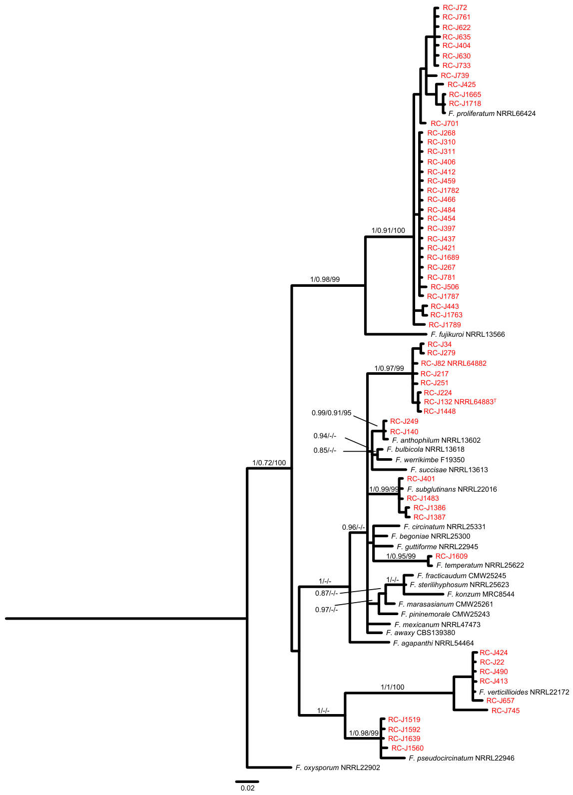
To further assess species identities, the TEF1 sequences of the grass isolates were aligned to FFSC reference sequences retrieved from the Fusarium ID 3.0 and GenBank databases (Table 2), and the resulting alignment was subjected to MP, ML and BI phylogenetic-tree-building analyses. The resulting trees confirmed the species identities determined by BLASTn analysis. That is, the 50 grass isolates identified to species in BLASTn analysis were resolved into exclusive and well supported clades with sequences from references strains of F. proliferatum, F. verticillioides, F. subglutinans, F. pseudocircinatum, F. anthophilum or F. temperatum (Figure 1). The trees also included data for the eight isolates for which the species identity was not determined in the BLASTn analysis. In the tree, the eight isolates formed an exclusive clade that did not include sequences from any of the reference strains. This result provides further evidence that the eight isolates are the same species and phylogenetically distinct from all the reference strains (Figure 1).
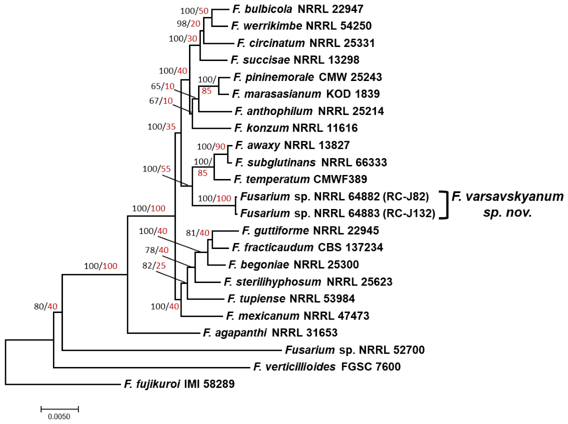
To further assess whether the unidentified isolates are a phylogenetically distinct species of FFSC, we inferred a species tree from concatenated alginments of 20 full-length housekeeping genes retrieved from genome sequences of two of the isolates (NRRL 64882 and NRRL 64883) and reference strains of 21 other species of FFSC, including 18 species from the American clade. In the resulting species tree, NRRL 64882 and NRRL 64883 formed an exclusive and well-supported clade that was sister to a clade consisting of F. awaxy, F. subglutinans, and F. temperatum (Figure 2). Thus, trees inferred from the partial TEF1 sequence, the combined dataset of TEF1, RPB2, TUB2 and CDM1 (Figure S1), and the 20 full-length housekeeping genes were consistent with NRRL 64882 and NRRL 64883 being strains of a novel species, hereinafter described as Fusarium varsavskyanum sp. nov., within the American clade of FFSC.

3.2. Distribution of Fusarium Species on Grasses

All F. verticillioides and F. anthophilum isolates, two F. subglutinans isolates, and most F. proliferatum isolates were recovered from grasses collected during the winter when inflorescences were not present. Because identification of these grasses to genus and species required inflorescences, we identified the host plants of these isolates only to the taxonomic level of the family Poaceae (Table 1). The remaining F. proliferatum isolates were recovered from Leersia luziola, Paspalum notatum, Eragrostis sp., Hymenachme sp. or Diplacha sp. Each of the four F. pseudocircinatum isolates was recovered from a different grass genus: Spartina sp., Eriochloa sp., Chloris sp., and Cynodon dactilon. One F. subglutinans isolate was obtained from Elionurus sp., while two others and the one F. temperatum isolate were recovered from Panicum sp. (Table 1).

3.3. Secondary Metabolite Biosynthetic Genes

BLAST analysis using sequences of known secondary metabolite biosynthetic genes to query genome sequences of F. varsavskyanum strains NRRL 64882 and NRRL 64883 revealed the distribution (i.e., presence and absence) of multiple biosynthetic genes or gene clusters required for production of mycotoxins, plant hormones, or other secondary metabolites that are produced by other Fusarium species. The distribution was identical in both F. varsavskyanum strains (Table 3). With respect to mycotoxins, the fusaric acid and beauvericin/enniatin biosynthetic gene clusters were detected in the two F. varsavskyanum genome sequences, but the fumonisin, fusarin, trichothecene, and zearalenone clusters were not detected. Biosynthetic gene clusters for the plant hormones auxins, cytokinins, and gibberellins were also detected in the F. varsavskyanum genome sequences. Additional gene clusters or individual genes that were detected were those that confer production of the polyketide-derived metabolites fusarubin and bikaverin, the nonribosomal peptides ferricrocin and fusarinine, and the terpenes eremophilene, koraiol, carotenoids, guai-6-10(14)-diene, and α-acorenol (Table 3). With three exceptions, the distribution of the genes/gene clusters in the F. varsavskyanum strains was the same as the distribution in the closely related species F. subglutinans and F. temperatum (Table 3). The exceptions were: (1) the six-gene depudecin cluster—F. subglutinans and F. temperatum had intact orthologs of the cluster, but both F. varsavskyanum strains had only a partial cluster that lacked the polyketide synthase gene (DEP1), which is critical for depudecin production; (2) the two-gene beauvericin/enniatin biosynthetic gene cluster—the F. varsavskyanum strains and F. temperatum had intact orthologs of this cluster, but as previously reported [54], the F. subglutinans ortholog of the nonribosomal peptide synthase gene (NRPS22) had multiple mutations that most likely rendered the gene nonfunctional; and (3) the six-gene fujikurin cluster—F. temperatum had an intact ortholog of this cluster, but the two F. varsavskyanum strains and F. subglutinans had none of the cluster genes.

3.4. Mycotoxin and Pigment Analysis

Extracts of maize kernel cultures of F. varsavskyanum strains NRRL 64882 and NRRL 64883 were subjected to a targeted HPLC-MS analysis of six secondary metabolites for which the corresponding biosynthetic gene clusters were identified in the genome sequences of the two strains. The six metabolites comprised three mycotoxins—beauvericin, enniatins, and fusaric acid—and three pigments—bikaverin, bostrycoidin, and fusarubin (Table 3). Despite the presence of the biosynthetic gene clusters in the strains, the only known secondary metabolite detected in culture extracts was fusaric acid. This mycotoxin was detected in extracts of the three replicate cultures of both strains at an average level of 1700 μg/g cracked maize kernel (range 1390–2190 μg/g). We also examined the culture extracts for the presence of moniliformin, a metabolite for which the biosynthetic genes have not been identified. Moniliformin was not detected in the culture extracts of either F. varsavskyanum strain.

3.5. Mating Type Identification and Sexual Stage Induction

Mating type idiomorphs (MAT-1/MAT-2) were identified for all F. varsavskyanum strains based on the results of a standard PCR assay. One strain (RC-J82 = NRRL 64882) carried the MAT-1 idiomorph, and the other seven carried the MAT-2 idiomorph. Analysis of genome sequences revealed that NRRL 64882 had the MAT-1 idiomorph with the MAT1-1-1 and MAT1-1-2 genes, and strain NRRL 64883 had the MAT-2 idiomorph with the MAT1-2-1 and MAT1-2-3 genes. None of the attempted mating crosses between isolates of F. varsavskyanum or between isolates of F. varsavskyanum and mating type tester strains of F. subglutinans resulted in the formation of perithecia after 10 weeks of incubation.

3.6. Taxonomy

Fusarium varsavskyanum E. Cendoya, M. J. Nichea, C. Romero, R.H. Proctor & M.L Ramirez sp. nov. (Figure 3).
MycoBank: MB 846905
Types: ARGENTINA, CHACO PROVINCE Chaco Wetlands, Ramsar site no. 1366 (S 27°30′56.8″ W 59°05′22.9″), originally isolated from Poaceae plants, July 2011, Maria L. Ramirez RC-J132 (holotype RCVC 9963, a dry culture of RC-J132, NRRL 64883 Herbarium of the Natural Sciences Department, National University of Río Cuarto, Córdoba, Argentina). Ex-type culture NRRL 64883 = RC-J132. GenBank: TEF1 = OQ134082; RPB2 = OQ134078; TUB2 = OQ134093; CDM1 = OQ134088. COMPLETE GENOME = JBNHUV000000000.
Diagnosis: F. varsavskyanum resembles F. subglutinans. Colony, false head morphology, polyphialides and rate of growth can overlap and so are not reliable criteria. Thus, the presence of abundant microconidia with piriform shape is the main marker to use to distinguish F. varsavskyanum from F. subglutinans.
Etymology: Named in honor of the late Dr. Edith Varsavsky, pioneer in the study of mycotoxicogenic fungi and mycotoxins in Argentina.
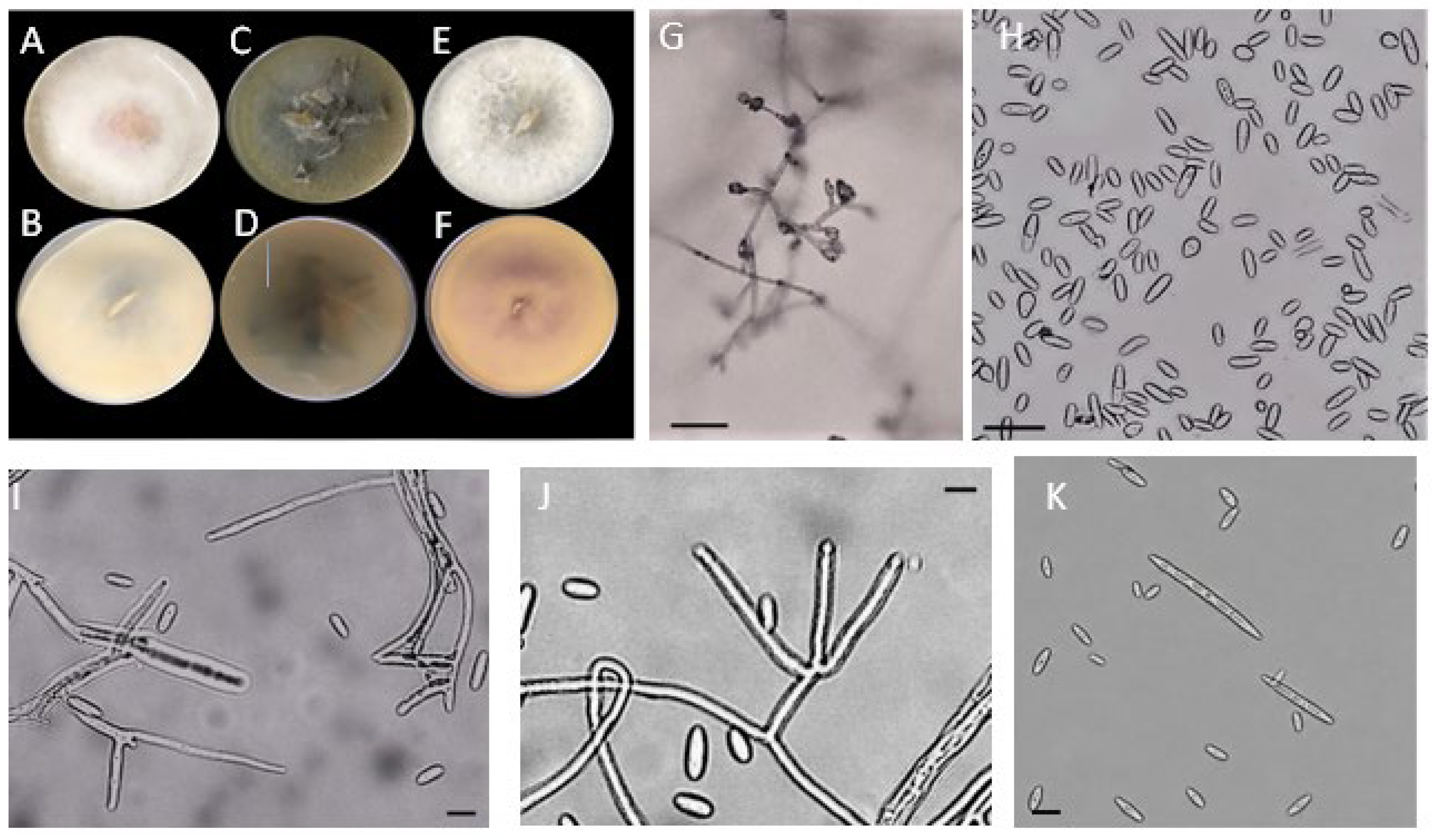
Description: Colonies on PDA under 12/12 h photoperiod cold white and black-fluorescent lamps at 25 °C growing rapidly, reaching 3.4 cm at 25 °C in 4 days, pink or vinaceous to violet; aerial mycelium abundant (Figure 3).
Microscopic characters: On CLA and SNA sporodochia can be present, when present they are tan to orange. Aerial mycelium floccose. Conidiophores usually erect and branched. Macroconidia abundant, falcate to rather straight, 3-5-septate, with a distinct foot-cell, (54.1–)54.3–67.4(–85.5) × (5.3–)5.4–6.3(–7.4) µm in total range, 65.3 ± 14.8 × 6.6 ± 1.0 on average ± SD (n = 20). Mesoconidia are present: (29.9–)31.5–41.5(–44.9) × (5.7–)5.9–8.1(–8.3) µm in total range, 36.3 ± 4.4 × 6.7 ± 0.9 on average ± SD (n = 21). Microconidia produced on mono- and polyphialides and aggregated in false heads and palisade, usually unicellular, ovoid 0 to 1 septate, mostly 0 septate: (10.5–)11.2–19.5(–20.2) × (5.0–)5.1–7.5(–7.7) µm in total range, 15.1 ± 2.3 × 6.4 ± 0.6 on average ± SD (n = 51)., and piriform: (10.2–)10.6–18.6(–18.7) × (7.1–)7.9–11.5(–11.7) µm in total range, 14.4 ± 2.1 × 9.6 ± 1.1 on average ± SD (n = 58). Chlamydospores absent.
Distribution: Argentina
Additional strains examined: RC-J251, RC-J224, RC-J1448 and RC-J82 = NRRL 64882.
Most F. varsavskyanum isolates were isolated from grasses collected during winter, when inflorescences were not present. As a result, identification of the grasses to genus and species was not possible. For one isolate, however, the identity of the grass host was determined to be Elionorus sp. (Table 1).

4. Discussion

The results of the current study indicate that isolates of FFSC recovered from asymptomatic wild grasses collected from the Chaco Wetlands of Argentina included both previously described species and one novel species, F. varsavskyanum. As far as we are aware, our results are the first report that wild grasses in Argentina can harbor previously described FFSC species that cause disease and mycotoxin contamination of agricultural crops. The mycotoxin-producing pathogenic species were F. proliferatum, F. pseudocircinatum, F. subglutinans, F. temperatum and F. verticillioides. The occurrence of these species as endophytes in wild grasses from the Chaco Wetlands is consistent with a previous study in which multiple mycotoxins produced by members of the FFSC were detected in the same grass samples from which the Fusarium isolates were recovered [4].
Our results are also consistent with previous studies showing that asymptomatic grasses in other parts of the world can be infected with pathogenic Fusarium species, such as F. circinatum [21], F. graminearum and F. verticillioides [55]. Our results also add to information that natural ecosystems can harbor novel Fusarium species and are potential inoculum reservoirs of plant pathogenic and mycotoxigenic Fusarium species [25]. To our knowledge, F. varsavskyanum is the second novel species of Fusarium recovered from wild grasses in the Chaco Wetlands.
Phylogenetic analysis of partial sequence of TEF1, RPB2, TUB2 and CDM1 from eight isolates as well as 20 housekeeping genes retrieved from genome sequence data of NRRL 64882 and NRRL 64883 provided support for the identification of F. varsavskyanum as a novel species within the American clade of FFSC. The Americas are the geographic origin of Elionorus species, the only grass sample contaminated with F. varsavskyanum that could be identified to the level of genus. This is consistent with the phylogeographic hypothesis that divided FFSC species into three major clades (African, American and Asian) that corresponded to the geographic origin of each clade [10]. Fusarium varsavskyanum is phylogenetically distinct from previously described species in the American clade. It differs from F. subglutinans in morphology of microconidia; both species produce ovoid microconidia, but F. varsavskyanum also produces abundant piriform microconidia. This difference is a key morphological marker to distinguish F. varsavskyanum from F. subglutinans. Colony characters and macroconidia shape are not sufficiently different to distinguish the two species, and neither species produced chlamydospores.
Results of the MAT PCR assay revealed that each F. varsavskyanum strain contains a MAT-1 or MAT-2 idiomorph, which suggests that this fungus has the genetic potential for a heterothallic sexual reproductive mode. Therefore, the failure of F. varsavskyanum strains to cross on carrot agar might be because the experimental conditions employed were suboptimal for the novel species or the strains that we isolated were not female-fertile. Low percentages of female-fertile isolates have been reported in several species within the FFSC [56,57]. The failure of F. varsavskyanum isolates to cross as a male parent with a female fertile tester strain of F. subglutinans provides additional evidence for reproductive isolation of these two species.
The analysis of distribution of biosynthetic gene clusters in the genome sequences revealed that the gene cluster content of F. varsavskyanum was similar to two of its closest relatives, F. subglutinans and F. temperatum (Table 3). The analysis of gene clusters indicated that F. varsavskyanum has the genetic potential to produce the plant hormones auxins, cytokinins and gibberellins. Production and/or potential to produce these plant hormones has been reported previously in some other members of FFSC, including F. fujikuroi and F. proliferatum, and could contribute to endophytic growth by enhancing growth and development of the plant hosts [58,59]. The presence in the F. varsavskyanum genomes of the gene clusters that confer production of the pigments bikaverin, carotenoids and fusarubins was expected given that the bikaverin cluster occurs widely among members of FFSC, and the carotenoid and fusarubin clusters occur widely in the genus Fusarium [58,60,61,62]. These pigments have potential to provide protection from solar radiation, but bikaverin and fusarubin also exhibit anti-bacterial and anti-fungal activity [63]. The genome sequence analysis indicated that F. varsavskyanum has the genetic potential to produce only three mycotoxins, beauvericin, enniatins and fusaric acid, which are considered emerging mycotoxins because they are not regulated and their impact on health of humans, pets and livestock is poorly understood [64]. It is notable the F. varsavskyanum genome sequences lacked genes that confer production of the mycotoxins fumonisins, trichothecenes, and zearalenone, which are among the mycotoxins of most concern to food and feed safety. Based on these findings, F. varsavskyanum does not have potential to cause contamination of wild grasses or crops with mycotoxins of major concern. The presence of beauvericin/enniatin biosynthetic genes in the F. varsavskyanum genome sequences indicates that it is one of the potential contributors to the previously observed beauvericin contamination in grasses from the Chaco Wetland [4].
Although the genome of F. varsavskyanum included gene clusters that confer production of six secondary metabolites for which we had standards, the HPLC-MS analysis indicated that strains NRRL 64882 and NRRL 64883 produced only fusaric acid under the culture conditions used in the current study. The finding that F. varsavskyanum strains do not produce the metabolic products of biosynthetic gene clusters present in their genome is not surprising. Indeed, lack of production of secondary metabolite products of gene clusters in fungi is common under laboratory conditions [65]. Different substrates on which Fusarium strains are grown can affect whether or not the strains produce secondary metabolites [66]. Thus, F. varsavskyanum strains might produce secondary metabolites other than fusaric acid if they were grown on other substrates. A possible cause for lack of production of a metabolite even though the corresponding gene cluster is present is that the cluster genes are not expressed under some laboratory conditions, but natural habitat(s) of fungi include conditions that induce expression of the genes.
Although recovery of members of FFSC from asymptomatic grasses suggests that the fungi can exist as endophytes in the grasses, most of the previously described FFSC species that were recovered are plant pathogens of one or more agricultural crops. But previous studies also indicate that some members of the FFSC can exist as either endophytes or pathogens of crops depending on environmental conditions [67,68]. Thus, it is possible that the grass isolates examined in this study, or at least some of them, are pathogenic on crops and/or on wild grasses at some point during the grass lifecycles or under environmental conditions that did not exist when the grasses were sampled [4]. Because we did not assess the potential pathogenicity of the grass isolates in this study, it remains to be determined whether they are pathogenic on crops and/or wild grasses. At least four of the FFSC species recovered from the grasses are maize pathogens: F. proliferatum, F. subglutinans, F. temperatum and F. verticillioides. This is potentially significant given the importance of maize to agriculture in Argentina. Although no commercial maize fields are located in or near the Chaco wetlands, wild Poaceae plants located closer to agricultural fields have potential to serve as a source of inoculum for these pathogenic species. F. proliferatum and F. verticillioides are the two most important contributors to fumonisin contamination in maize [6]. Fusarium subglutinans is also a pathogen of maize, but it cannot produce FBs. The host range of F. subglutinans is not clear, but it includes teosinte and native North American grasses, as well as a range of other monocots and dicots. Fusarium temperatum was first described in 2011 as a species closely related to F. subglutinans and causes diseases in maize, including seedling blight, stalk rot, and ear rot [69]. Fusarium temperatum has also been isolated from sorghum [70], wheat [71] and recently from cruciferous plants [72]. This fungus produces beauvericin, moniliformin and fusaproliferin [73].
To our knowledge, this study is the first report of the occurrence of F. pseudocircinatum in Argentina. This species is broadly distributed across ecologically diverse habitats, and it is an important plant pathogen that infects several wild and cultivated plants [27]. Fusarium pseudocircinatum has been associated with mango malformation in Mexico and the Dominican Republic [74,75] and with stunting and malformation of sunflower plants in Brazil [76]. In Argentina, sunflowers are an important crop, whereas mango production occurs on a small scale. Isolates of F. pseudocircinatum are reported to produce moniliformin, fusaric acid, and beauvericin [77].
To our knowledge, this study is also the first report of the occurrence of F. anthophilum in Argentina. This species is also widely distributed and has been recovered from several plant species (orchids, millet, wheat and rice) in temperate regions of the world [27,78]. Although, F. anthophilum has not been associated with plant disease, recently it was reported to cause drying on South American jelly palm (Butia odorata) in Brazil [79]. In Australia, F. anthophilum has been recovered from the native grasses Austrostripa aristiglumis [80] and Sorghum leiocladium [81]. The fungus is not associated with any human or animal diseases. However, some strains have been reported to produce the mycotoxins moniliformin, fumonisins, beauvericin and fusaproliferin [5,17]. It is noteworthy that F. anthophilum predominantly produces the C series of fumonisins rather than the B series [6]. Therefore, although this species occurs in Chaco Wetland grasses and produces fumonisin, it is unlikely to contribute to the previously reported fumonisin B1 contamination in the grasses [4].
Because of their ability to produce fumonisins, F. proliferatum and F. verticillioides are likely causes of the previously reported fumonisin contamination in Chaco Wetland grasses [4]. In addition, F. proliferatum, F. temperatum, F. anthophilum and F. pseudocircinatum could be the main source of beauvericin in those grasses.
There is a growing body of evidence indicating that natural ecosystems can serve as reservoirs of inoculum of both previously recognized and novel pathogens of agricultural crops [21,26,81,82,83,84]. Burges [26] suggested that weeds and wild plants near and in agricultural fields could serve as alternative hosts for fungal pathogens of crops and, therefore, impact epidemics of crop diseases. In Australia, natural ecosystems are a rich source of genetic diversity of Fusarium species and a reservoir of known and potential pathogens. Burgess [26] stated that studies of Fusarium in these ecosystems can contribute to a better understanding of the origin and ecology of novel pathogens at the species or subspecies level. He emphasized that extensive systemic surveys of Fusarium species are needed in natural ecosystems of Africa, Asia and South America. Thus, the current and previous [7,8] studies on Fusarium species in Chaco Wetland grasses contribute to understanding the potential impact of natural ecosystems on agriculture in South America. Similarly, Mourelos et al. [83] found that Fusarium graminearum, the predominant cause of Fusarium head blight of wheat in Argentina, occurs frequently in gramineous and non-gramineous weeds near fields of cereal crops in the Argentinian province of Buenos Aires.
Together, our results add to data on potential causes of mycotoxin contamination and fungal communities in natural grassland ecosystems. Moreover, the recovery of F. chaquense [7] and F. varsavskyanum from Chaco Wetlands grasses suggests that this important natural ecosystem in Argentina could harbor additional novel microbial species.

Supplementary Materials

The following supporting information can be downloaded at: https://www.mdpi.com/article/10.3390/jof12060444/s1, Figure S1: Bayesian phylogenetic tree inferred from the combined dataset TEF1, RPB2, TUB2 and CDM1 sequences showing NRRL 64882 and NRRL 64883 being members of the American clade of the FFSC and a novel species. Bayesian posterior probability scores ≥ 0.8 are shown at the internodes. F. oxysporum NRRL 22902 was used as the outgroup.; Table S1: Source information for TEF1, RPB2, CMD1 and TUB reference sequences used in the phylogenetic analyses; Table S2: Summary of phylogenetic data for the 20 housekeeping genes used to infer the species phylogeny in this study. File S1: Fasta_formatted sequences of Fusarium secondary metabolite biosynthetic genes. File S2: Nexus-formatted file with concatenated and partitioned alignment of exon and intron sequences of 20 housekeeping genes from F. varsavskyanum strains RC-J82 (NRRL 64882) and RC-J132 (NRRL 64883) as well as reference strains of 21 other members of the Fusarium fujikuroi species complex.

Author Contributions

Conceptualization, M.L.R. and E.C.; methodology, E.C., C.J.R.D., M.J.N., M.B. and S.A.P.; formal analysis, R.H.P., M.L.R.; investigation, E.C., C.J.R.D. and S.A.P.; resources, M.L.R.; data curation, R.H.P.; writing—original draft preparation, E.C., S.A.P. and M.L.R.; writing—review and editing, M.L.R., E.C., S.A.P. and R.H.P. All authors have read and agreed to the published version of the manuscript.

Funding

This work was supported by grants from Agencia Nacional de Promoción Científica y Tecnológica (MINCyT) through PICT-2019-2949-Prestamo BID and by Consejo Nacional de Investigaciones Científicas y Técnicas: PIP 11220200100021CO and PUE 22920200100004. This work was supported in part by the U.S. Department of Agriculture, Agricultural Research Service.

Institutional Review Board Statement

Not applicable.

Informed Consent Statement

Not applicable.

Data Availability Statement

All sequences generated in this study were deposited in GenBank, with the accession numbers provided in Table 1 and Table S1. The original contributions presented in this study are included in the article and Supplementary Materials. Further inquiries can be directed to the corresponding author.

Acknowledgments

We are grateful to Amy E. McGovern and Crystal E. Probyn for generation and assembly of genome sequence data. Mention of trade names or commercial products in this publication is solely for the purpose of providing specific information and does not imply recommendation or endorsement by the U.S. Department of Agriculture. USDA is an equal opportunity provider and employer.

Conflicts of Interest

The authors declare no conflicts of interest.

References

  1. GPWG I—Grass Phylogeny Working Group. Phylogeny and subfamilial classification of the grasses (Poaceae). Ann. Mo. Bot. Gard. 2001, 88, 373–457. [CrossRef]
  2. Arelovich, H.M.; Bravo, R.D.; Martinez, M.F. Development, characteristic, and trends for beef cattle production in Argentina. Anim. Front. 2011, 1, 37–45. [Google Scholar] [CrossRef]
  3. Neiff, J.J. Diversity in some tropical wetland systems of South America. In Biodiversity in Wetlands: Assessment, Function and Conservation; Gopal, B., Junk, W., Davis, J., Eds.; Backhuys Publishers: Leiden, The Netherlands, 2001; pp. 157–186. Available online: https://www.researchgate.net/publication/272418968_Diversity_in_some_tropical_wetland_systems_of_South_America (accessed on 11 June 2026).
  4. Nichea, M.J.; Palacios, S.A.; Chiacchiera, S.M.; Sulyok, M.; Krska, R.; Chulze, S.N.; Torres, A.M.; Ramirez, M.L. Presence of multiple mycotoxins and other fungal metabolites in native grasses from a wetland ecosystem in Argentina intended for grazing cattle. Toxins 2015, 7, 3309–3329. [Google Scholar] [CrossRef] [PubMed]
  5. Desjardins, A.E. Fusarium Mycotoxins Chemistry, Genetics and Biology; American Phytopathology Society Press: St. Paul, MN, USA, 2006; 260p. [Google Scholar]
  6. Munkvold, G.P.; Proctor, R.H.; Moretti, A. Mycotoxin production in Fusarium according to contemporary species concepts. Annu. Rev. Phytopathol. 2021, 59, 373–402. [Google Scholar] [CrossRef] [PubMed]
  7. Nichea, M.J.; Proctor, R.H.; Probyn, C.E.; Palacios, S.A.; Cendoya, E.; Sulyok, M.; Chulze, S.N.; Torres, A.M.; Ramirez, M.L. Fusarium chaquense, sp. nov, a novel type A trichothecene–producing species from native grasses in a wetland ecosystem in Argentina. Mycologia 2022, 114, 46–62. [Google Scholar] [CrossRef] [PubMed]
  8. Nichea, M.J.; Cendoya, E.; Zachetti, V.G.; Demonte, L.D.; Repetti, M.R.; Palacios, S.A.; Ramirez, M.L. Characterization of members of the Fusarium incarnatum–equiseti species complex from natural and cultivated grasses intended for grazing cattle in Argentina. J. Fungi 2026, 12, 26. [Google Scholar] [CrossRef]
  9. Aoki, T.; O’Donnell, K.; Geiser, D.M. Systematics of key phytopathogenic Fusarium species: Current status and future challenges. J. Gen. Plant Pathol. 2014, 80, 189–201. [Google Scholar] [CrossRef]
  10. O’Donnell, K.; Cigelnik, E.; Nirenberg, H.I. Molecular systematics and phylogeography of the Gibberella fujikuroi species complex. Mycologia 1998, 90, 465–493. [Google Scholar] [CrossRef]
  11. O’Donnell, K.; Ward, T.J.; Robert, V.A.; Crous, P.W.; Geiser, D.M.; Kang, S. DNA sequence-based identification of Fusarium: Current status and future directions. Phytoparasitica 2015, 43, 583–595. [Google Scholar] [CrossRef]
  12. Sandoval-Denis, M.; Guarnaccia, V.; Polizzi, G.; Crous, P.W. Symptomatic Citrus trees reveal a new pathogenic lineage in Fusarium and two new Neocosmospora species. Persoonia 2018, 40, 1–25. [Google Scholar] [CrossRef] [PubMed]
  13. Sandoval-Denis, M.; Swart, W.J.; Crous, P.W. New Fusarium species from the Kruger National Park, South Africa. Mycokeys 2018, 34, 63–92. [Google Scholar] [CrossRef] [PubMed]
  14. Al-Hatmi, A.M.S.; Sandoval-Denis, M.; Nabet, C.; Ahmed, S.A.; Demar, M.; Normand, A.C.; De Hoog, G.S. Fusarium volatile, a new potential pathogen from a human respiratory sample. Fungal Syst. Evol. 2019, 4, 71–181. [Google Scholar] [CrossRef] [PubMed]
  15. Yilmaz, M.; Sandoval-Denis, M.; Lombard, L.; Visagie, C.M.; Wingfield, B.D.; Crous, P.W. Redefining species limits in the Fusarium fujikuroi species complex. Persoonia 2021, 46, 129–162. [Google Scholar] [CrossRef] [PubMed]
  16. Nirenberg, H.I.; O’Donnell, K. New Fusarium species and combinations within the Gibberella fujikuroi species complex. Mycologia 1998, 90, 434–458. [Google Scholar] [CrossRef]
  17. Munkvold, G. Fusarium species and their associated mycotoxins. In Mycotoxigenic Fungi: Methods and Protocols, Methods in Molecular Biology; Moretti, A., Susca, A., Eds.; Humana Press: New York, NY, USA, 2017; pp. 51–106. [Google Scholar]
  18. Leslie, J.F.; Zeller, K.A.; Wohler, M.; Summerell, B.A. Interfertility of two mating populations in the Gibberella fujikuroi species complex. Eur. J. Plant Pathol. 2004, 110, 611–618. [Google Scholar] [CrossRef]
  19. Carter, L.L.A.; Leslie, J.F.; Webster, R.K. Population structure of Fusarium fujikuroi from California rice and water grass. Phytopathology 2008, 98, 992–998. [Google Scholar] [CrossRef] [PubMed]
  20. Swett, C.L.; Gordon, T.R. First report of grass species (Poaceae) as naturally occurring hosts of the pine pathogen Gibberella circinata. Plant Dis. 2012, 96, 908. [Google Scholar] [CrossRef] [PubMed]
  21. Swett, C.L.; Porter, B.; Fourie, G.; Steenkamp, E.T.; Gordon, T.R.; Wingfield, M.J. Association of the pitch canker pathogen Fusarium circinatum with grass hosts in commercial pine production areas of South Africa. South. For. 2014, 76, 161–166. [Google Scholar] [CrossRef]
  22. Maia, N.C.; Melo, M.P.; Guimarães, S.S.C.; Matos, K.S.; Moreira, S.I.; Lana, U.D.P.; Cardoso, P.G. Panicum maximum as a reservoir of a potential maize pathogen. Eur. J. Plant Pathol. 2021, 159, 95–104. [Google Scholar] [CrossRef]
  23. Zeller, K.A.; Summerell, B.A.; Bullock, S.; Leslie, J.F. Gibberella konza (Fusarium konzum) sp. nov. from prairie grasses, a new species in the Gibberella fujikuroi species complex. Mycologia 2003, 95, 943–954. [Google Scholar] [CrossRef]
  24. Laurence, M.H.; Walsh, J.L.; Shuttleworth, L.A.; Robinson, D.M.; Johansen, R.M.; Petrovic, T.; Vu, T.T.H.; Burgess, L.W.; Summerell, B.A.; Liew, E.C.Y. Six novel species of Fusarium from natural ecosystems in Australia. Fungal Divers. 2015, 77, 349–366. [Google Scholar] [CrossRef]
  25. Costa, M.M.; Melo, M.P.; Carmo, F.S.; Moreira, G.M.; Guimarães, E.A.; Rocha, F.S.; Costa, S.S.; Abreu, L.M.; Pfenning, L.H. Fusarium species from tropical grasses in Brazil and description of two new taxa. Mycol. Prog. 2021, 20, 61–72. [Google Scholar] [CrossRef]
  26. Burgess, L.W. 2011 McAlpine Memorial Lecture—A Love Affair with Fusarium. Australas. Plant Pathol. 2014, 43, 359. [Google Scholar] [CrossRef]
  27. Leslie, J.F.; Summerell, B.A. The Fusarium Laboratory Manual; Blackwell Professional Publishing: Ames, IA, USA, 2006; 388p. [Google Scholar]
  28. Thompson, J.D.; Higgins, D.G.; Gibson, T.J. CLUSTAL W: Improving the sensitivity of progressive multiple sequence alignment through sequence weighting, position-specific gap penalties and weight matrix choice. Nucleic Acids Res. 1994, 22, 4673–4680. [Google Scholar] [CrossRef] [PubMed]
  29. Hall, T.A. BioEdit: A user-friendly biological sequence alignment editor and analysis program for Windows 95/98/NT. Nucleic Acids Symp. Ser. 1999, 41, 95–98. Available online: https://www.semanticscholar.org/paper/BIOEDIT:-A-USER-FRIENDLY-BIOLOGICAL-SEQUENCE-EDITOR-Hall/0ae262d9cf78536754bc064e07113ab5e978f208 (accessed on 11 June 2026).
  30. Katoh, K.; Standley, D.M. MAFFT multiple sequence alignment software version 7: Improvements in performance and usability. Mol. Biol. Evol. 2013, 30, 772–780. [Google Scholar] [CrossRef] [PubMed]
  31. Goloboff, P.A.; Farris, J.S.; Nixon, K.C. TNT, a free program for phylogenetic analysis. Cladistics 2008, 24, 774–786. [Google Scholar] [CrossRef]
  32. Guindon, S.; Dufayard, J.-F.; Lefort, V.; Anisimova, M.; Hordijk, W.; Gascuel, O. New Algorithms and Methods to Estimate Maximum-Likelihood Phylogenies: Assessing the Performance of PhyML 3.0. Syst. Biol. 2010, 59, 307–321. [Google Scholar] [CrossRef] [PubMed]
  33. Ronquist, F.; Teslenko, M.; Van Der Mark, P.; Ayres, D.L.; Darling, A.; Höhna, S.; Larget, B.; Liu, L.; Suchard, M.A.; Huelsenbeck, J.P. MrBayes 3.2: Efficient Bayesian phylogenetic inference and model choice across a large model space. Syst. Biol. 2012, 61, 539–542. [Google Scholar] [CrossRef] [PubMed]
  34. Posada, D. jModelTest: Phylogenetic model averaging. Mol. Biol. Evol. 2008, 25, 1253–1256. [Google Scholar] [CrossRef] [PubMed]
  35. Sung, G.H.; Sung, J.M.; Hywel-Jones, N.L.; Spatafora, J.W. A multi-gene phylogeny of Clavicipitaceae (Ascomycota, Fungi): Identification of localized incongruence using a combinational bootstrap approach. Mol. Phylogenet. Evol. 2007, 44, 1204–1223. [Google Scholar] [CrossRef] [PubMed]
  36. Liu, Y.J.; Whelen, S.; Hall, B.D. Phylogenetic relationships among ascomycetes: Evidence from an RNA polymerse II subunit. Mol. Biol. Evol. 1999, 16, 1799–1808. [Google Scholar] [CrossRef] [PubMed]
  37. O’Donnell, K.; Nirenberg, H.I.; Aoki, T.; Cigelnik, E. A multigene phylogeny of the Gibberella fujikuroi species complex: Detection of additional phylogenetically distinct species. Mycoscience 2000, 41, 61–78. [Google Scholar] [CrossRef]
  38. O’Donnell, K.; Sarver, B.A.; Brandt, M.; Chang, D.C.; Noble-Wang, J.; Park, B.J.; Sutton, D.A.; Benjamin, L.; Lindsley, M.; Padhye, A.; et al. Phylogenetic diversity and microsphere array-based genotyping of human pathogenic Fusaria, including isolates from the multistate contact lens-associated U.S. keratitis outbreaks of 2005 and 2006. J. Clin. Microbial. 2007, 45, 2235–2248. [Google Scholar] [CrossRef]
  39. O’Donnell, K.; Cigelnik, E. Two divergent intragenomic rDNA ITS2 types within a monophyletic lineage of the fungus Fusarium are non orthologous. Mol. Phylogenet. Evol. 1997, 7, 103–116. [Google Scholar] [CrossRef] [PubMed]
  40. Edwards, J.; Auer, D.; De Alwis, S.K.; Summerell, B.; Aoki, T.; Proctor, R.H.; Busman, M.; O’Donnell, K. Fusarium agapanthi sp. nov., a novel bikaverin and fusarubin-producing leaf and stem spot pathogen of Agapanthus praecox (African lily) from Australia and Italy. Mycologia 2016, 108, 981–992. [Google Scholar] [CrossRef] [PubMed]
  41. Gräfenhan, T.; Johnston, P.R.; Vaughan, M.M.; McCormick, S.P.; Proctor, R.H.; Busman, M.; Ward, T.J.; O’Donnell, K. Fusarium praegraminearum sp. nov., a novel nivalenol mycotoxin-producing pathogen from New Zealand can induce head blight on wheat. Mycologia 2016, 108, 1229–1239. [Google Scholar] [CrossRef] [PubMed]
  42. Sarver, B.A.; Ward, T.J.; Gale, L.R.; Broz, K.; Kistler, H.C.; Aoki, T.; Nicholson, P.; Carter, J.; O’Donnell, K. Novel Fusarium head blight pathogens from Nepal and Louisiana revealed by multilocus genealogical concordance. Fungal Genet. Biol. 2011, 48, 1096–1107. [Google Scholar] [CrossRef] [PubMed]
  43. Brown, D.W.; Proctor, R.H. Insights into natural products biosynthesis from analysis of 490 polyketide synthases from Fusarium. Fungal Genet. Biol. 2016, 89, 37–51. [Google Scholar] [CrossRef] [PubMed]
  44. Villani, A.; Proctor, R.H.; Kim, H.S.; Brown, D.W.; Logrieco, A.F.; Amatulli, M.T.; Moretti, A.; Susca, A. Variation in secondary metabolite production potential in the Fusarium incarnatum-equiseti species complex revealed by comparative analysis of 13 genomes. BMC Genom. 2019, 20, 314. [Google Scholar] [CrossRef] [PubMed]
  45. Geiser, D.M.; Al-Hatmi, A.; Aoki, T.; Arie, T.; Balmas, V.; Barnes, I.; Bergstrom, G.C.; Bhattacharyya, M.K.K.; Blomquist, C.L.; Bowden, R.; et al. Phylogenomic analysis of a 55.1 kb 19-gene dataset resolves a monophyletic Fusarium that includes the Fusarium solani species complex. Phytopathology® 2021, 111, 1064–1079. [Google Scholar] [CrossRef] [PubMed]
  46. Altschul, S.F.; Madden, T.L.; Schaffer, A.A.; Zhang, J.; Zhang, Z.; Millwer, W.; Lipman, D.J. Gapped BLAST and PSIBLAST: A new generation of protein database search programs. Nucleic Acids Res. 1997, 25, 3389–3402. [Google Scholar] [CrossRef] [PubMed]
  47. Kumar, S.; Stecher, G.; Tamura, K. MEGA7: Molecular evolutionary genetics analysis version 7.0 for bigger datasets. Mol. Biol. Evol. 2016, 33, 1870–1874. [Google Scholar] [CrossRef] [PubMed]
  48. Minh, B.Q.; Nguyen, M.A.; von Haeseler, A. Ultrafast approximation for phylogenetic bootstrap. Mol. Biol. Evol. 2013, 30, 1188–1195. [Google Scholar] [CrossRef] [PubMed]
  49. Nguyen, L.-T.; Schmidt, H.A.; von Haeseler, A.; Minh, B.Q. IQ-TREE: A fast and effective stochastic algorithm for estimating maximum likelihood phylogenies. Mol. Biol. Evol. 2014, 32, 268–274. [Google Scholar] [CrossRef] [PubMed]
  50. Vaidya, G.; Lohman, D.J.; Meier, R. SequenceMatrix: Concatenation software for the fast assembly of multi-gene datasets with character set and codon information. Cladistics 2011, 27, 171–180. [Google Scholar] [CrossRef] [PubMed]
  51. Kornerup, A.; Wanscher, J.H. Methuen Handbook of Colour; Eyre Methuen: London, UK, 1984; 252p. [Google Scholar]
  52. Montoya-Martínez, A.C.; Rodríguez-Alvarado, G.; Fernández-Pavía, S.P.; Proctor, R.H.; Kim, H.S.; O’Donnell, K. Design and validation of a robust multiplex polymerase chain reaction assay for MAT idiomorph within the Fusarium fujikuroi species complex. Mycologia 2019, 111, 772–781. [Google Scholar] [CrossRef] [PubMed]
  53. Klittich, C.J.R.; Leslie, J.F. Identification of a second mating population within the Fusarium moniliforme anamorph of Gibberella fujikuroi. Mycologia 1992, 84, 541–547. [Google Scholar] [CrossRef]
  54. Susca, A.; Villani, A.; Haidukowski, M.; Epifani, F.; Logrieco, A.; Moretti, A. Molecular biodiversity in Fusarium subglutinans and F. temperatum: A valuable tool to distinguish the two sister species and determine the beauvericin chemotype. J. Fungi 2024, 10, 785. [Google Scholar] [CrossRef] [PubMed]
  55. Szécsi, Á.; Magyar, D.; Tóth, S.; Szőke, C. Poaceae: A rich source of endophytic fusaria. Acta Phytopathol. Entomol. Hung. 2013, 48, 19–32. [Google Scholar] [CrossRef]
  56. Steenkamp, E.T.; Wingfield, B.D.; Coutinho, T.A.; Zeller, K.A.; Wingfield, M.J.; Marasas, W.F.O.; Leslie, J.F. PCR-based identification of MAT-1 and MAT-2 in the Gibberella fujikuroi species complex. Appl. Environ. Microbiol. 2000, 66, 4378–4382. [Google Scholar] [CrossRef] [PubMed]
  57. Laraba, I.; Kim, H.S.; Proctor, R.H.; Busman, M.; O’Donnell, K.; Felker, F.C.; Aime, M.C.; Koch, R.A.; Wurdack, K.J. Fusarium xyrophilum, sp. nov., a member of the Fusarium fujikuroi species complex recovered from pseudoflowers on yellow-eyed grass (Xyris spp.) from Guyana. Mycologia 2020, 112, 39–51. [Google Scholar] [CrossRef] [PubMed]
  58. Niehaus, E.M.; Munsterkotter, M.; Proctor, R.H.; Brown, D.W.; Sharon, A.; Idan, Y.; Oren-Young, L.; Sieber, C.M.; Novak, O.; Pencik, A.; et al. Comparative “Omics” of the Fusarium fujikuroi species complex highlights differences in genetic potential and metabolite synthesis. Genome Biol. Evol. 2016, 8, 3574–3599. [Google Scholar] [CrossRef] [PubMed]
  59. Malonek, S.; Bömke, C.; Bornberg-Bauer, E.; Rojas, M.C.; Hedden, P.; Hopkins, P.; Tudzynski, B. Distribution of gibberellin biosynthetic genes and gibberellin production in the Gibberella fujikuroi species complex. Phytochemistry 2005, 66, 1296–1311. [Google Scholar] [CrossRef] [PubMed]
  60. Brown, D.W.; Kim, H.S.; McGovern, A.E.; Probyn, C.E.; Proctor, R.H. Genus-wide analysis of Fusarium polyketide synthases reveals broad chemical potential. Fungal Genet. Biol. 2022, 160, 103696. [Google Scholar] [CrossRef] [PubMed]
  61. Hansen, F.T.; Gardiner, D.M.; Lysoe, E.; Fuertes, P.R.; Tudzynski, B.; Wiemann, P.; Sondergaard, T.E.; Giese, H.; Brodersen, D.E.; Sorensen, J.L. An update to polyketide synthase and non-ribosomal synthetase genes and nomenclature in Fusarium. Fungal Genet. Biol. 2015, 75, 20–29. [Google Scholar] [CrossRef] [PubMed]
  62. Hoogendoorn, K.; Barra, L.; Waalwijk, C.; Dickschat, J.S.; van der Lee, T.A.J.; Medema, M.H. Evolution and diversity of biosynthetic gene clusters in Fusarium. Front. Microbiol. 2018, 9, 1158. [Google Scholar] [CrossRef] [PubMed]
  63. Son, S.W.; Kim, H.Y.; Choi, G.J.; Lim, H.K.; Jang, K.S.; Lee, S.O.; Lee, S.; Sung, N.D.; Kim, J.C. Bikaverin and fusaric acid from Fusarium oxysporum show antioomycete activity against Phytophthora infestans. J. Appl. Microbiol. 2008, 104, 692–698. [Google Scholar] [CrossRef] [PubMed]
  64. Gruber-Dorninger, C.; Novak, B.; Nagl, V.; Berthiller, F. Emerging mycotoxins: Beyond traditionally determined food contaminants. J. Agric. Food Chem. 2017, 65, 7052–7070. [Google Scholar] [CrossRef] [PubMed]
  65. Harvey, C.J.B.; Tang, M.; Schlecht, U.; Horecka, J.; Fischer, C.R.; Lin, H.C.; Li, J.; Naughton, B.; Cherry, J.; Miranda, M.; et al. HEx: A heterologous expression platform for the discovery of fungal natural products. Sci. Adv. 2018, 4, eaar5459. [Google Scholar] [CrossRef] [PubMed]
  66. Shi, W.; Tan, Y.; Wang, S.; Gardiner, D.M.; De Saeger, S.; Liao, Y.; Wang, C.; Fan, Y.; Wang, Z.; Wu, A. Mycotoxigenic potentials of Fusarium species in various culture matrices revealed by mycotoxin profiling. Toxins 2016, 9, 6. [Google Scholar] [CrossRef] [PubMed]
  67. Blacutt, A.A.; Gold, S.E.; Voss, K.A.; Gao, M.; Glenn, A.E. Fusarium verticillioides: Advancements in understanding the Toxicity, virulence, and niche adaptations of a model mycotoxigenic pathogen of maize. Phytopathology 2018, 108, 312–326. [Google Scholar] [CrossRef] [PubMed]
  68. Munkvold, G.P.; Desjardins, A.E. Fumonisins in maize: Can we reduce their occurrence? Plant Dis. 1997, 81, 556–565. [Google Scholar] [CrossRef] [PubMed]
  69. Scauflaire, J.; Gourgue, M.; Munaut, F. Fusarium temperatum sp. nov. from maize, an emergent species closely related to Fusarium subglutinans. Mycologia 2011, 103, 586–597. [Google Scholar] [CrossRef] [PubMed]
  70. Levic, J.; Munaut, F.; Scauflaire, J.; Stankovic, S.; Ivanovic, D.; Krnjaja, V. Polyphasic approach used for distinguishing Fusarium temperatum from Fusarium subglutinans. J. Agric. Sci. Technol. 2019, 21, 221–232. Available online: https://ror.circle-u.eu/items/d77d3977-5fac-42b3-9c28-f4dec86b2db5 (accessed on 11 June 2026).
  71. Van Coller, G.J.; Rose, L.J.; Boutigny, A.-L.; Ward, T.J.; Lamprecht, S.C.; Viljoen, A. The distribution and type B trichothecene chemotype of Fusarium species associated with head blight of wheat in South Africa during 2008 and 2009. PLoS ONE 2022, 17, e0275084. [Google Scholar] [CrossRef] [PubMed]
  72. Orina, A.S.; Gagkaeva, T.Y.; Gavrilova, O.P. Novel cruciferous plant host of Fusarium temperatum, a species mainly associated with maize. Australas. Plant Dis. Notes 2023, 18, 37. [Google Scholar] [CrossRef]
  73. Fumero, M.V.; Villani, A.; Susca, A.; Haidukowski, M.; Cimmarusti, M.T.; Toomajian, C.; Leslie, J.F.; Chulze, S.N.; Moretti, A. Fumonisin and beauvericin chemotypes and genotypes of the sister species Fusarium subglutinans and Fusarium temperatum. Appl. Environ. Microbiol. 2020, 86, e00133-20. [Google Scholar] [CrossRef] [PubMed]
  74. García-López, E.; Mora-Aguilera, J.A.; Villegas-Monter, A.; Tovar-Pedraza, J.M.; Serra, C.; Batista-Marte, C.M. First report of Fusarium pseudocircinatum causing mango malformation disease in Dominican Republic. Plant Dis. 2016, 100, 1501. [Google Scholar] [CrossRef]
  75. Freeman, S.; Otero-Colina, G.; Rodríguez-Alvarado, G.; Fernández-Pavía, S.; Maymon, M.; Ploetz, R.C.; Aoki, T.; O’Donnell, K. First Report of mango malformation disease caused by Fusarium pseudocircinatum in Mexico. Plant Dis. 2014, 98, 1583. [Google Scholar] [CrossRef] [PubMed]
  76. De Farias, O.R.; Cruz, J.M.F.L.; Silva Veloso, J.; Souza, J.T.; Duarte, I.G.; Barbosa, P.R.R.; Varanda, C.M.R.; Materatski, P.; Félix, M.D.R.F.; Oliveira, M.D.M.; et al. Fusarium pseudocircinatum causing stunting and malformation of sunflower plants in Brazil. Plant Dis. 2022. Epub ahead of print. [Google Scholar] [CrossRef] [PubMed]
  77. Montoya-Martínez, A.C.; O’Donnell, K.; Busman, M.; Vaughan, M.M.; McCormick, S.P.; Santillán-Mendoza, R.; Pineda-Vaca, D.; Clapes-Garduño, L.; Fernández-Pavía, S.P.; Ploetz, R.C.; et al. Weeds harbor Fusarium species that cause malformation disease of economically important trees in western Mexico. Plant Dis. 2022, 106, 612–622. [Google Scholar] [CrossRef] [PubMed]
  78. Summerell, B.A.; Leslie, J.F. Fifty years of Fusarium: How could nine species have ever been enough? Fungal Divers. 2011, 50, 135–144. [Google Scholar] [CrossRef]
  79. Victoria-Arellano, A.D.; Chaves, F.C.; Marquez-Licona, G.; Solano-Báez, A.R.; Francisco, C.M.; Dallagnol, L.J. First report of Fusarium anthophilum causing drying on Butia odorata in Rio Grande do Sul, Brazil. J. Plant Dis. Prot. 2023, 131, 627–630. [Google Scholar] [CrossRef]
  80. Bentley, A.R.; Petrovic, T.; Griffiths, S.P.; Burgess, L.W.; Summerell, B.A. Crop pathogens and other Fusarium species associated with Austrostipa aristiglumis. Australas. Plant Pathol. 2007, 36, 434–438. [Google Scholar] [CrossRef]
  81. Walsh, J.L. Fusarium Species Associated with Savanna Ecosystems in Australia: Taxonomy, Ecology and Pathogenicity. Ph.D. Dissertation, University of Sydney, Sydney, Australia, 2007. [Google Scholar]
  82. Herron, D.A.; Wingfield, M.J.; Fru, F.; Wingfield, B.D.; Steenkamp, E.T. Grasses as a refuge for Fusarium circinatum L.—Evidence from South Africa. South. J. For. Sci. 2020, 82, 253–262. [Google Scholar] [CrossRef]
  83. Mourelos, C.A.; Malbrán, I.; Balatti, P.A.; Ghiringhelli, P.D.G.; Lori, A. Gramineous and non-gramineous weed species as alternative hosts of Fusarium graminearum, causal agent of Fusarium head blight of wheat, in Argentina. Crop Prot. 2014, 65, 100–104. [Google Scholar] [CrossRef]
  84. Lofgren, L.A.; LeBlanc, N.R.; Certano, A.K.; Nachtigall, J.; LaBine, K.M.; Riddle, J.; Broz, K.; Dong, Y.; Bethan, B.; Kafer, C.W.; et al. Fusarium graminearum: Pathogen or endophyte of North American grasses? New Phytol. 2018, 217, 1203–1212. [Google Scholar] [CrossRef] [PubMed]
Figure 1. Bayesian inference phylogenetic tree inferred from partial TEF1 sequences showing the phylogenetic relatedness of Fusarium species associated with Poaceae plants with other species of the Fusarium fujikuroi species complex. Bayesian posterior probability scores ≥  0.7, followed by ML bootstrap values ≥  0.70, and maximum parsimony bootstrap support values > 70%, are shown at the internodes. The studied strains are in red. F. oxysporum NRRL 22902 was used as the outgroup.
Jof 12 00444 g001
Figure 2. Species tree showing the relationships of strains NRRL 64882 (RC-J82) and NRRL 64883 (RC-J132), herein described as Fusarium varsavskyanum sp. nov., to reference strains of other members of the Fusarium fujikuroi species complex. The tree was inferred by maximum likelihood analysis of concatenated alignments of coding regions of 20 housekeeping genes. Numbers near branches are bootstrap values based on 1000 replicates (black type), or gene concordance values expressed as number of individual gene trees (out of 20) that included the branch (red type). The housekeeping genes and a summary of phylogenetic information derived from them are listed in Supplementary Table S2. A nexus formatted alignment file of the data is provided in Supplementary File S2.
Jof 12 00444 g002
Figure 3. Macroscopic characteristics of Fusarium varsavskyanum NRRL 64882. (A,B). Colony on Spezieller Nährstoffarmer agar. (A). Colony surface. (B). Colony reverse. (C,D). Colony on carnation leaf agar. (C). Colony surface. (D). Colony reverse. (E,F). Colony color on potato dextrose agar. (E). Colony surface. (F). Colony reverse. (G). Microconidia produced on mono- and polyphialides and aggregated in false heads and palisade. (H). Microconidia, usually unicellular, ovoid 0 to 1 septate, mostly 0 septate, and piriform. (I). Polyphialides (J). Branched monophialides. (K). Mesoconidia. Bars: (G,H) = 50 μm; (IK) = 10 μm.
Jof 12 00444 g003
Table 1. Information on isolates of the Fusarium fujikuroi species complex recovered from Chaco Wetland grasses.
Fusarium
Species
StrainMonth
Year
HostGPS CoordinatesGenBank Accession No.
TEF1RPB2CMD1TUB2
F. anthophilumRC-J249July 2011PoaceaeS 27°31′08.4″
W 59°04′37.6″
MT123767
RC-J140July 2011PoaceaeS 27°31′10.7″
W 59°04′36.3″
MT123777
F. proliferatumRC-J72July 2011PoaceaeS 27°31′09.2″
W 59°04′36.8″
PZ453710
RC-J268July 2011PoaceaeS 27°31′10.8″
W 59°04′34.9″
PZ453711
RC-J310July 2011PoaceaeS 27°30′58.1″
W 59°05′23.7″
PZ453712
RC-J311July 2011PoaceaeS 27°30′58.1″
W 59°05′23.7″
PZ453713
RC-J397July 2011PoaceaeS 27°31′41.7″
W 59°04′33.1″
PZ453714
RC-J406July 2011PoaceaeS 27°31′41.8″
W 59°04′32.4″
PZ453715
RC-J412July 2011PoaceaeS 27°31′41.7″
W 59°04′31.6″
PZ453716
RC-J437July 2011PoaceaeS 27°31′41.7″
W 59°04′32.3″
PZ453717
RC-J459July 2011PoaceaeS 27°31′26.2″
W 59°05′00.4″
PZ453718
RC-J484July 2011PoaceaeS 27°31′25.8″
W 59°04′59.9″
PZ453719
RC-J506July 2011PoaceaeS 27°34′16.4″
W 60°23′42.4″
PZ453720
RC-J701July 2011PoaceaeS 27°33′34.5″
W 60°25′02.1″
PZ453721
RC-J739July 2011PoaceaeS 27°33′52.7″
W 60°24′39.6″
PZ453722
RC-J761July 2011PoaceaeS 27°34′53.1″
W 60°24′43.0″
PZ453723
RC-J404July 2011PoaceaeS 27°31′41.7″
W 59°04′31.6″
PZ453724
RC-J1689February 2014PoaceaeS 27°31′04.1″
W 59°05′05.6″
PZ453725
RC-J630July 2011PoaceaeS 27°33′28.3″
W 60°25′01.5″
PZ453726
RC-J781July 2011PoaceaeS 27°33′28.8″
W 60°25′00.3″
PZ453727
RC-J267July 2011PoaceaeS 27°31′10.8″
W 59°04′34.9″
PZ453728
RC-J1718September 2014Leersia luziolaS 27°31′06,9″
W 59°05′01.0″
MT123773
RC-J1782September 2014Paspalum notatumS 27°30′39.0″
W 59°04′58.7″
PZ453729
RC-J443July 2011PoaceaeS 27°31′41.2″
W 59°04′32.7″
PZ453730
RC-J421July 2011PoaceaeS 27°31′42.9″
W 59°04′31.5″
PZ453731
RC-J425July 2011PoaceaeS 27°31′42.9″
W 59°04′31.5″
PZ453732
RC-J454July 2011PoaceaeS 27°31′26.8″
W 59°05′00.4″
PZ453733
RC-J466July 2011PoaceaeS 27°31′25.6″
W 59°05′00.5″
PZ453734
RC-J622July 2011PoaceaeS 27°33′28.2″
W 60°25′05.3″
PZ453735
RC-J635July 2011PoaceaeS 27°33′28.8″
W 60°25′00.3″
PZ453736
RC-J733July 2011PoaceaeS 27°33′51.7″
W 60°24′41.0″
PZ453737
RC-J1665September 2014Diplacha sp.S 27°31′07.0″
W 59°05′00.9″
MT123775
RC-J1763September 2014Hymenachme sp.S 27°30′39.0″
W 59°04′58.7″
PZ453738
RC-J1787September 2014Dichantium sp.S 27°30′34.5″
W 59°05′02.7″
PZ453739
RC-J1789September 2014Dichantium sp.S 27°30′34.5″
W 59°05′02.7″
PZ453740
F. pseudocircinatumRC-J1560September 2014Shcloris sp.S 27°30′49.5″
W 59°05′06.1″
MT123782
RC-J1592September 2014Spartina sp.S 27°30′49.5″
W 59°05′06.1″
MT123774
RC-J1519September 2014Eriochloa sp.S 27°30′49.5″
W 59°05′06.1″
MT123771
RC-J1639September 2014Cynodon dactylonS 27°30′49.5″
W 59°05′06.1″
MT123783
F. subglutinansRC-J401July 2011PoaceaeS 27°31′ 41.7″
W 59°04′33.1″
MT123768
RC-J1483September 2014Elionurus sp.S 27°30′49.7″
W 59°05′06.3″
MT123770
RC-J1386February 2014Panicum sp.S 27°30′50.3″
W 59°05′06.4″
MT123780
RC-J1387February 2014Panicum sp.S 27°30′50.3″
W 59°05′06.4″
MT123781
F. temperatumRC-J1609September 2014Panicum sp.S 27°30′50.3″
W 59°05′06.4″
MT123772
F. varsavskyanum sp. nov.RC-J34July 2011PoaceaeS 27°34′ 16.4″
W 60°23′42.4″
PZ453703
RC-J82, NRRL 64882July 2011PoaceaeS 27°34′ 14.0″
W 60°23′47.4″
OQ134079OQ134074OQ134084OQ134089
RC-J217July 2011PoaceaeS 27°34′ 02.3″
W 60°23′45.6″
PZ453704
RC-J224July 2011PoaceaeS 27°34′ 02.3″
W 60°23′45.6″
OQ134080OQ134075OQ134085OQ134090
RC-J251July 2011PoaceaeS 27°34′ 01.8″
W 60°23′43.2
OQ134081OQ134076OQ134086OQ134091
RC-J132T, NRRL 64883July 2011PoaceaeS 27°34′ 05.7″
W 60°23′48.6″
OQ134082OQ134078OQ134088OQ134093
RC-J1448February 2014Elionurus sp.S 27°30′49.7″
W 59°05′06.3″
OQ134083OQ134077OQ134087OQ134092
RC-J279July 2011PoaceaeS 27°31′ 08.2″
W 59°04′38.2″
PZ453707
F. verticillioidesRC-J424July 2011PoaceaeS 27°31′42.9″
W 59°04′31.5″
MT123769
RC-J22July 2011PoaceaeS 27°31′10.6″
W 59°04′38.4″
MT123776
RC-J413July 2011PoaceaeS 27°31′41.7″
W 59°04′31.6″
MT123778
RC-J490July 2011PoaceaeS 27°31′25.9″
W 59°05′00.5″
MT123779
RC-J657July 2011PoaceaeS 27°33′ 34.2″
W 60°24′59.7″
PZ453708
RC-J745July 2011PoaceaeS 27°33′ 53.5″
W 60°24′36.2″
PZ453709
Table 2. Source information for TEF1 reference sequences used in phylogenetic analyses.
Fusarium SpeciesStrain NumberHostOriginGenBank Accession Number
TEF1
F. agapanthiNRRL 54464Agapanthus sp.AustraliaMN193856
F. anthophilumNRRL 13602Hippeastrum sp.GermanyAF160292
F. awaxyCBS 139380Corn stalkUSAMN534058
F. begoniaeNRRL 25300Begonia elatiorGermanyAF160293
F. bulbicolaNRRL 13618Nerine bowdeniiGermany AF160294
F. circinatumNRRL 25331Pinus radiataUSAAF160295
F. fracticaudumCMW 25245Pinus maximinoiiColombiaKJ541059
F. fujikuroiNRRL 13566Oryza sativaTaiwanAF160279
F. guttiformeNRRL 22945Ananas comosusEnglandAF160297
F. konzumMRC 8544Sorghastrum nuttansUSAEU220235
F. marasasianumCMW 25261Pinus patulaColombiaKJ541063
F. mexicanumNRRL 47473Mangifera indica inflorescenceMexicoGU737416
F. oxysporumNRRL 22902Pseudotsuga menziesiiUSAAF160312
F. pininemoraleCMW 25243Pinus tecunumaniiColombiaKJ541064
F. proliferatumNRRL 66424Tropical rain forest soilPapua New GuineaMN534059
F. pseudocircinatumNRRL 22946Solanum sp.GhanaAF160271
F. subglutinansNRRL 22016Zea maysUSAHM057336.1
F. sterilihyphosumNRRL 25623MangoSouth AfricaAF160300
F. succisaeNRRL 13613Succisa pratensisGermanyAF160291
F. temperatumNRRL 25622Zea maysSouth AfricaAF160301
F. tupienseNRRL 53984Mangifera indicaBrazilGU737404
F. verticillioidesNRRL 22172Zea maysGermanyAF160262
F. werrikimbeF19350 = CBS 125535Sorghum leiocladumAustraliaEF107131
NRRL: ARS Culture Collection, United States Department of Agriculture; CBS: Culture Collection of the Westerdijk Fungal Biodiversity Institute, Utrecht, the Netherlands; CMW: Culture Collection of the Forestry and Agricultural Biotechnology Institute (FABI), University of Pretoria, Pretoria, South Africa; MRC: Medical Research Council, Tygerberg, South Africa.
Table 3. Distribution of secondary metabolite biosynthetic genes/gene clusters in F. varsavskyanum sp. nov. strains NRRL 64882 and NRRL 64883 and one representative strain each of the related species F. subglutinans and F. temperatum.
F. varsavskyanum sp. nov.F. subglutinansF. temperatum
Metabolite Gene/Gene Cluster aNRRL 64882NRRL 64883NRRL 66333CFWF389
Polyketides
2-AOD-3-oNoNoNoNo
AurofusarinNoNoNoNo
BikaverinYesYesYesYes
DepudecinPartialPartialYesYes
EquisetinPartialPartialPartialPartial
FujikurinNoNoNoYes
FumonisinNoNoNoNo
Fusaric AcidYesYesYesYes
FusaridioneNoNoNoNo
FusarielinNoNoNoNo
FusarinNoNoNoNo
FusarubinYesYesYesYes
GibepyroneYesYesYesYes
ZearalenoneNoNoNoNo
Non-ribosomal peptides
ApicidinNoNoNoNo
Beauvericin/EnniatinYesYesNoYes
FerricrocinYesYesYesYes
FusarinineYesYesYesYes
GramilinNoNoNoNo
MalonichromeNoNoNoNo
Polyketide-Non-ribosomal peptides
FusaristatinNoNoNoNo
W493-BNoNoNoNo
Terpenes
a-AcorenolYesYesYesYes
EremophileneYesYesYesYes
Guai-6,10(14)-dieneYesYesYesYes
KoraiolYesYesYesYes
CarotenoidsYesYesYesYes
CulmorinNoNoNoNo
TrichotheceneNoNoNoNo
Pant Hormones
AuxinYesYesYesNo
Cytokinin_Cluster 1YesYesYesYes
Cytokinin_Cluster 2YesYesYesYes
Gibberellin (=terpene)YesYesYesYes
Other metabolites
ButenolideNoNoNoNo
a For each metabolite, “No” indicates the biosynthetic gene or gene cluster was not detected; “Yes” indicates detected; and “Partial” indicates part of the cluster was detected. The genome sequences of F. subglutinans and F. temperatum used in this analysis were previously reported and had GenBank/NCBI accessions JAAOAV01 and LJGR01, respectively.
Disclaimer/Publisher’s Note: The statements, opinions and data contained in all publications are solely those of the individual author(s) and contributor(s) and not of MDPI and/or the editor(s). MDPI and/or the editor(s) disclaim responsibility for any injury to people or property resulting from any ideas, methods, instructions or products referred to in the content.