Article

Antifungal Activity of Native Trichoderma afroharzianum and Arcopilus cupreus Against Neopestalotiopsis rosae Causing Strawberry Root and Crown Rot in Mexico


by
Karla Jenifer Juárez-Cruz
1,
Alfredo Jiménez-Pérez
1,
Federico Castrejón-Ayala
1,
Gabriela Trejo-Tapia
1,
Lauro Soto-Rojas
2,
Alma Rosa Solano-Báez
1,* and
Guillermo Márquez-Licona
1,*
1
Centro de Desarrollo de Productos Bióticos, Instituto Politécnico Nacional, Yautepec 62731, Morelos, Mexico
2
Posgrado en Fitosanidad-Entomología y Acarología, Colegio de Postgraduados, Campus Montecillo, Montecillo, Texcoco 56264, Estado de Mexico, Mexico
*
Authors to whom correspondence should be addressed.
J. Fungi 2026, 12(6), 388; https://doi.org/10.3390/jof12060388
Submission received: 26 April 2026 / Revised: 23 May 2026 / Accepted: 26 May 2026 / Published: 28 May 2026
(This article belongs to the Special Issue Plant Pathogenic Fungal Infections, Biocontrol and Novel Fungicides)

Abstract

Root and crown rot of strawberry is an emerging disease of concern in Mexico. Like other soil-borne diseases, it has spread widely due to the vegetative propagation of this crop. This study aimed to evaluate the in vitro antagonistic activity of four native Trichoderma strains and one strain of Arcopilus cupreus against a population of Neopestalotiopsis rosae, as a potential biological control alternative. Thirty-five commercial strawberry fields across the three main strawberry-producing states were sampled, yielding 103 fungal isolates. Nearly 70% of the recovered isolates belonged to the Neopestalotiopsis genus. Sixteen representative isolates were identified as N. rosae based on morphology and multilocus sequence phylogeny (ITS, β-tub, tef1-α) and confirmed as pathogenic through pathogenicity tests. Four native T. afroharzianum isolates recovered from the rhizosphere of healthy strawberry plants were identified by multilocus sequence analysis (rpb2, tef1-α). All T. afroharzianum isolates exhibited antagonistic activity in dual-culture assays, inhibiting mycelial growth by 71–73%, surpassing the effectiveness of the commercial fungicide cyprodinil + fludioxonil (average inhibition 50%). In contrast, the A. cupreus isolate recovered from a native medicinal plant showed an average inhibition of 38%. These results highlight native T. afroharzianum as a promising sustainable alternative for integrated management of strawberry root and crown rot in Mexico.

1. Introduction

The strawberry (Fragaria × ananassa; Rosaceae) is among the most widely cultivated and consumed berry species [1]. Mexico is the fifth-largest producer of strawberries worldwide, with an average annual production of 641,552 tons [2]. Michoacán (411,927 tons), Baja California (137,424 tons), and Guanajuato (107,920 tons) are the three main producing states [3]. This crop faces numerous phytosanitary challenges, with fungal diseases being the predominant factor deterring productivity. This problem affects production from the seedling stage onward and reduces fruit quality. In Mexico, strawberry seedlings are produced through vegetative propagation, which complicates the acquisition of certified, pathogen-free plants, primarily because nurseries lack effective means of producing healthy plants and implementing phytosanitary practices to reduce pathogen inoculum. To reduce production costs, standard methods include reusing trays, ineffective disinfection practices, and the use of non-sterile substrates (personal communication with producers and affected growers).
During the strawberry production stage, primary soil-borne diseases include anthracnose caused by Colletotrichum acutatum, charcoal rot caused by Macrophomina sp., crown and root rot caused by Phytophthora cactorum, P. citricola, and Pythium, leaf and crown rot caused by Neopestalotiopsis spp., Fusarium wilt caused by Fusarium oxysporum, Verticillium wilt caused by Verticillium dahliae, and root rot caused by Rhizoctonia spp. [4,5]. However, the most significant in recent years is root and crown rot caused by Neopestalotiopsis rosae, first reported in Mexico in 2020 [6]. This pathogen, originally reported as a phytopathogen on Rosa sp. plants in New Zealand in 1998 [7], was first detected in Egypt [8], affecting strawberry crops. From there, it has spread to other countries where strawberries are cultivated, including China [9], Mexico [6], Turkey [10], USA [11], Paraguay [12], India [13], Germany [14], and Italy [15]. In Mexico, the disease has been exacerbated by the lack of certified, disease-free plant material, which has led to its spread across most strawberry-growing regions, and by unknown levels of soil inoculum. Additionally, the lack of cultural practices that reduce inoculum sources, such as harvesting crop residues, contributes to the problem.
Infection by N. rosae primarily causes root and crown rot in strawberries; however, it can also infect other parts of the plant, with specific symptoms in each tissue type. On leaves, the disease starts with small brown spots that spread across the leaf blade, leading to leaf blight, during which black pycnidia and conidia form. Infected petioles develop sunken, dark-brown lesions, causing the leaves to wilt. Likewise, the pathogen can infect the fruit, creating slightly sunken, light-brown spots where black pycnidia develop. The interior of infected crowns displays areas of intense red surrounded by dark-brown margins, which ultimately leads to plant death. On the roots, rot symptoms begin with the development of large, dark-brown areas [6,16].
The main strategy for managing the disease is the use of synthetic fungicides; thus, multiple studies have evaluated the efficacy of various fungicidal compounds [5,6,8,17,18]. However, there is growing concern about environmental issues, public health risks, and the development of resistance to fungicides used in agriculture [19]. Biological control offers an alternative management strategy to synthetic fungicides [20]. Among the organisms used in this approach, Trichoderma is one of the most extensively studied fungal genera due to its diverse mechanisms of action against other fungi [21]. For example, T. asperellum has demonstrated a significant in vitro inhibitory effect against isolates of N. clavispora, the causal agent of strawberry root rot in China [20]. Additionally, T. reesei and T. harzianum have demonstrated significant inhibition of N. chrysea mycelial growth, which causes leaf spots on strawberries in Bangladesh [22]. In field experiments, T. asperellum and T. koningiopsis have proven effective against N. rosae [5]. In contrast, the use of Arcopilus cupreus as a biological control agent against plant pathogenic fungi has not been extensively studied. However, this species shows potential as a biocontrol agent due to its production of antifungal metabolites, mycoparasitic activity, competition for space and nutrients, or a combination of these mechanisms [23]. A. cupreus inhibits the growth of Phytophthora spp. [24] and has demonstrated antifungal activity against Botrytis cinerea, Pythium ultimum, and Rhizoctonia solani [25].
Although evaluations of biological products and synthetic fungicides for disease management have been conducted in Mexico [5], this disease continues to be a serious limiting factor in strawberry production, as no integrated management program has yet been developed that is both accessible and adaptable to the diverse management practices and microclimatic conditions present in production areas. In addition, the continuous evolution and adaptation of phytopathogenic fungi in response to human-driven crop management practices [26,27] underscore the ongoing need to discover new, more effective, and sustainable management strategies. Therefore, the present study aimed to evaluate the in vitro antagonistic activity of four native Trichoderma strains and one of A. cupreus against a population of N. rosae, the causal agent of root and crown rot in strawberries in Mexico, as a potential biological control alternative. We hypothesized that native antagonistic fungi isolated from Mexican strawberry agroecosystems could effectively suppress different N. rosae genotypes.

2. Materials and Methods

2.1. Recollection of Diseased Strawberry Plants

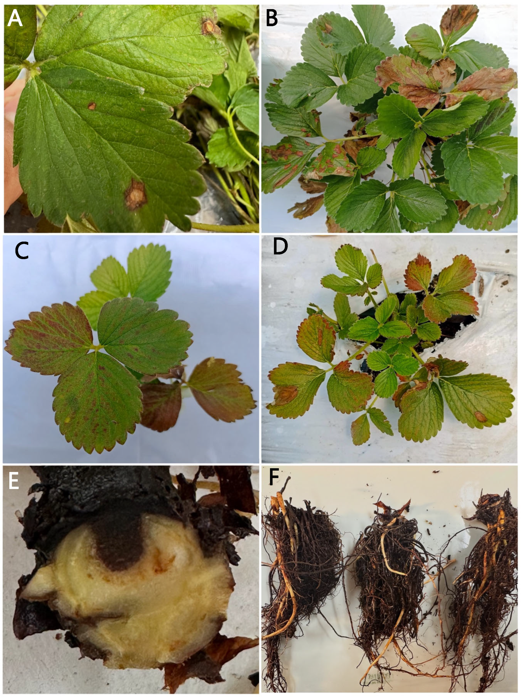
In October 2023, diseased strawberry plant samples were collected in Michoacán (Central-Western Region), Guanajuato (Central-Western Region), and Baja California (Northwest Region), guided by observations of characteristic foliar symptoms induced by Neopestalotiopsis (Figure 1A,B). Only plants showing symptoms of root and crown rot were included. Sampling was conducted across diverse production systems and levels of technification to capture the broadest possible range of pathogen diversity associated with strawberry root and crown rot. Plants were initially selected based on reddish-purple leaf discoloration (Figure 1C,D), a symptom commonly associated with the early stages of root and crown rot. Upon excavation, plants exhibited extensive root necrosis with dark-brown discoloration (Figure 1E), and their crowns showed intense reddish internal coloration, surrounded by necrotic margins (Figure 1F). A total of 35 samples were collected, each comprising 10 plants exhibiting initial disease symptoms. The collected samples were placed in airtight plastic bags and transported in a cooler (10 ± 2 °C) to the Phytopathology Laboratory at the Centro de Desarrollo de Productos Bióticos, Instituto Politécnico Nacional (CEPROBI-IPN), located in Yautepec, Morelos. During the next two production cycles (2024–2025), strawberry plants with symptoms of root and crown rot were collected, and only the frequency of isolation at the genus level was determined.

2.2. Isolation, Purification, and Preservation of the Fungi Associated with the Disease

The samples were washed under running water until most of the soil and organic matter were removed. To isolate the fungi associated with the disease, 5-mm tissue fragments from the transition zone between healthy and diseased tissue were collected from each sample with a scalpel. The recovered tissue was surface-disinfected with 1.2% sodium hypochlorite for 2 min, then rinsed three times with sterile distilled water. After disinfection, the tissue was placed on sterile paper towels and allowed to dry for 2 h. Four tissue fragments were placed in each Petri dish containing potato dextrose agar (PDA; BD Bioxon®, Cuautitlán Izcalli, Estado de Mexico, Mexico), and the plates were incubated at 25 °C for 48 h. Colonies were then transferred to fresh PDA and incubated at 25 ± 2 °C for 10 days [28]. To obtain axenic cultures, isolates were purified by the single-spore technique. Conidia from the fungal colonies were suspended in 1 mL of sterile distilled water in a microtube and vigorously agitated. From this conidial suspension, 200 μL were plated onto water agar (AA; BD Bioxon®, Cuautitlán Izcalli, Estado de Mexico, Mexico), and the plates were incubated at 25 ± 2 °C for 24 h. From each isolate, one germinating conidium was selected and transferred to PDA. Plates were incubated at 25 °C under a 12-h light/dark photoperiod for 7 days [28]. Of the isolates recovered from strawberry plants, 16 representative isolates were selected based on collection sites and morphotypes. Each isolate was preserved in triplicate in 10% glycerol and stored at 4 °C. The isolates were deposited under IPN 10.0227—IPN 10.0242 in the Culture Collection of Phytopathogenic Fungi of the CEPROBI-IPN.

2.3. Cultural Characterization

Descriptions of colony morphology and reproductive structures were based on colonies grown on PDA and incubated at 22 ± 2 °C under a 12-h light/dark photoperiod for 7 days [7]. The growth kinetics of each isolate were determined on PDA at 22 ± 2 °C by marking the colony margin every 24 h until the fungus completely covered the plate. Daily growth was measured with a vernier Traceable (Fisherbrand, Pittsburgh, PA, USA), and the mycelial growth rate was calculated using the formula of Zervakis et al. (2001) [29]. Semi-permanent slides of the reproductive structures were prepared with lactic acid as the mounting medium and sealed with a wax ring. Observations were made using an Axio Imager A2 compound microscope (Zeiss®, Oberkochen, Ostalb, Germany). Images were captured with an Axiocam ICc5 camera (Zeiss®, Oberkochen, Ostalb, Germany), and measurements were made using the ZEN software lite version (Zeiss®, Oberkochen, Ostalb, Germany). For morphometric characterization, only three representative isolates (IPN 10.0227, IPN 10.0239, and IPN 10.0242) were selected. The length and width of conidia (n = 100) and basal cells (n = 50) were measured, along with the length of apical and basal appendages and the number of appendages.

2.4. Molecular Identification

Genomic DNA was extracted from the sixteen selected isolates using colonies grown on PDA at 22 ± 2 °C under a 12 h light/dark photoperiod for 7 days, following the protocol for the “DNeasy Plant Mini Kit” (Qiagen, Hilden, Nordrhein-Westfalen, Germany). The quality and quantity of the extracted DNA were assessed using a spectrophotometer, NanoDrop 2000 (Thermo Scientific, Wilmington, DE, USA). The recovered DNA was stored at −20 °C. The ITS region and partial sequences of the β-tubulin (β-tub) and translation elongation factor 1-alpha (tef1-α) genes were amplified using the primer pairs ITS5 + ITS4 [30], T1 + Bt2b [31,32], and EF1-728f + EF-2 [33,34], respectively. The PCR reactions were conducted in a total volume of 50 μL, including 25 μL of PCR Master Mix 2X (Promega Corporation, Madison, WI, USA), 2.5 μL of each primer (10 μM), 16 μL of nuclease-free water, and 4 μL of the DNA template. The PCR cycling parameters were as follows: ITS region: initial denaturation at 95 °C for 3 min; followed by 40 cycles of 95 °C for 30 s, 55 °C for 50 s, and 72 °C for 60 s; with a final extension at 72 °C for 7 min. β-tub: initial denaturation at 95 °C for 3 min; followed by 40 cycles of 94 °C for 30 s, 55 °C for 50 s, and 72 °C for 60 s; with a final extension at 72 °C for 7 min. Tef1-α: initial denaturation at 95 °C for 5 min; followed by 40 cycles of 94 °C for 30 s, 52 °C for 30 s, and 72 °C for 30 s; with a final extension at 72 °C for 7 min. PCR products were visualized by electrophoresis on a 1% agarose gel and purified using the MiniElute Gel Extraction Kit (Qiagen®, Hilden, Nordrhein-Westfalen, Germany) according to the manufacturer’s instructions. The PCR products were sequenced in both directions by Macrogen® (Seoul, Republic of Korea). Sequence quality was verified by inspecting the electropherograms using the 4Peaks software v1.8 (https://nucleobytes.com/4peaks/index.html, accessed on 2 March 2025). Consensus sequences were assembled using UGene v52.1 [35] and compared against the GenBank database using the BLASTn tool (https://blast.ncbi.nlm.nih.gov/Blast.cgi, accessed on 5 March 2025) to determine isolate identity at the genus level. The consensus sequences were deposited in the GenBank database of the National Center for Biotechnology Information (NCBI) (Table 1). A reference phylogeny [7] was used to construct the phylogenetic matrix.
Sequences from species included in the phylogeny were downloaded from the NCBI nucleotide database and aligned with our sequences using the MAFFT v7 online service [36]. The alignments were trimmed in MEGA 12 [37] and then concatenated in Mesquite 4.01 [38] for the multigene analysis. For phylogenetic reconstruction, a Bayesian Inference (BI) analysis was performed using MrBayes 3.2.7 [39], applying the nucleotide substitution models GTR + I (ITS), GTR + G (β-tub), and HKY + I (tef1-α), all identified by the Akaike Information Criterion (AIC) in jModelTest 2.1.10 [40]. The Markov Chain Monte Carlo (MCMC) process was run for 1 × 10⁷ generations to estimate posterior probabilities, with trees sampled every 1000 generations and 25% discarded during the burn-in phase. Additionally, a Maximum Likelihood (ML) phylogenetic analysis was performed using RAxML 8.2.12 via the raxmlGUI 2.0 interface [41], under the GTR + I + G substitution model. Node support was assessed using 1000 rapid bootstrap replicates. Phylogenies were visualized and edited using FigTree (http://tree.bio.ed.ac.uk/software/figtree/, accessed on 14 March 2025).

2.5. Pathogenicity Tests

Healthy strawberry plantlets (var. Sayualita), 60 days old, were transplanted from trays into 1 L plastic pots containing sterile substrate (peat moss mixed with perlite at a 5:1 v/v ratio). The plants were acclimatized for 15 days in a greenhouse at 25 ± 5 °C. Agronomic management involved daily irrigation and weekly fertilization with a mixed solution containing Raizal 400 (Arysta LifeSciences, Saltillo, Coahuila, Mexico) and Gro-Green (Gro-Green Campbell, Gomez Palacio, Durango, Mexico), each at 1 g × L−1. Each plant was irrigated with 250 mL of this solution.
The inoculum for the sixteen isolates was increased by culturing on PDA plates and incubating in the dark at 25 ± 2 °C for 15 days. Inoculum recovery was performed by scraping the pionotal masses with a sterile spatula and transferring them into a 1% Tween 60 solution. The inoculum was homogenized using a handheld blender, applying 10-s pulses for a total of one minute. The inoculum concentration was adjusted to 1 × 10⁶ conidia/mL using a Neubauer chamber (Marienfeld, Meno-Tauber, Baden-Wurtemberg, Germany). Inoculation was performed by injecting 5 mL of the conidial suspension into the crown of six strawberry plants for each isolate [42]; the plants were then covered with a transparent plastic bag and incubated at 25 ± 2 °C for 7 days. As a control, six plants were treated the same way, with the inoculum replaced by sterile distilled water. The experimental design followed was a completely randomized design. For the evaluation of disease severity in pathogenicity tests, a scale was developed based on crown inoculation symptoms previously reported by other authors [6,43,44], consisting of the following levels: 0 = 0% healthy plant, 1 = 20% yellowing of the oldest leaves, 2 = 40% stunted growth, 3 = 60% wilting of young leaves and leaf necrosis, 4 = 80% death of the oldest leaves while central leaves remain green, 5 = 100% plant death. The disease severity percentage was calculated using the following formula [45].
P = i = 0 5 n i × V i 5 × N × 100
where:
P = severity percentage.
n i = number of plants in category i.
V i = numerical value of category i.
N = sample size.
Re-isolations were made from the inoculated plants. The recovered fungi from artificially inoculated symptomatic plants were identified based on morphological characteristics.
Data from the pathogenicity tests were analyzed using a one-way analysis of variance (ANOVA), with the N. rosae isolate as the main source of variation. When significant differences were detected, treatment means were compared using Bonferroni-corrected pairwise comparisons at a significance level of p < 0.05. Prior to analysis, data were examined to verify compliance with the assumptions of normality and homogeneity of variance. All statistical analyses were conducted in R version 4.5.3, with a significance threshold of 5%. The experiment was repeated twice with similar results.

2.6. Collection, Isolation, and Identification of Native Biocontrol Agents

Native Trichoderma isolates were obtained from four localities in the state of Michoacán with no history of strawberry cultivation. At each locality, a 1-kg rhizosphere soil sample from healthy strawberry plants was collected. For Trichoderma isolation, soil samples were dried to constant weight, homogenized, and 1 g of soil was transferred into a test tube containing 9 mL of sterile distilled water. From this suspension, three additional serial dilutions were prepared. From the final dilution, 200 µL were spread onto Trichoderma Specific Medium (TSM) [46,47]. Plates were incubated at 25 ± 2 °C for 96 h, after which colonies growing on the medium were recovered. Purification was performed using the single-spore culture technique, and isolates were preserved in triplicate in 10% glycerol. In total, 25 Trichoderma isolates were obtained; only the four with the highest antifungal activity in preliminary tests were selected, identified, and evaluated in this study. The selected isolates were deposited under the accession numbers MBP1–MBP-4 in the Collection of Native Microorganisms with Biotechnological Potential (MBP) of the CEPROBI-IPN.
Descriptions of colonies and morphology of the Trichoderma isolates were based on isolates grown on PDA and SNA incubated at 25 ± 2 °C with a 12 h light/dark photoperiod for 3 weeks [48]. The growth kinetics of each isolate were determined on PDA by measuring mycelial growth every 24 h [29]. Measurements of the reproductive structures were made using the same microscopes used for the strawberry fungi.
For molecular identification of the Trichoderma isolates, DNA extraction from pure cultures was performed using the same kit employed for the isolates recovered from the strawberry. The ITS region and partial sequences of the RNA polymerase II second-largest subunit (rpb2) and translation elongation factor 1-alpha (tef1-α) genes were amplified using the primer pairs ITS5 + ITS4 [30], RPB2-5F2 [49]/fRPB2-7CR [50], and EF1/EF2 [34]. Amplicon visualization and purification, as well as sequencing and sequence processing, were performed as previously described. The multigene phylogenetic analysis used to determine the phylogenetic species of Trichoderma was conducted with a reference phylogeny (Table 2) [48], following the previously described methodology for alignment, trimming, and concatenation. For phylogenetic reconstruction, Bayesian Inference (BI) was conducted using the nucleotide substitution models GTR+I (rpb2) and HKY+I (tef1-α). The ITS region was used to corroborate the isolate’s genus-level identity, but it was not included in the phylogenetic analysis. The Markov Chain Monte Carlo (MCMC) procedure was run for 1 × 10⁶ generations to estimate posterior probabilities, sampling trees every 1000 generations and discarding the first 25% as burn-in. In addition, Maximum Likelihood (ML) analysis was performed under the GTR+I substitution model, with node support evaluated using 1000 rapid bootstrap replicates. The resulting phylogenies were visualized and edited with FigTree.
The Arcopilus cupreus isolate (IPN 10.0154) was previously obtained from Baccharis conferta Kunth [51], a native medicinal plant commonly found in the Iztaccihuatl–Popocatepetl National Park. Although this fungus was reported to cause mild leaf spot symptoms in its host, it was serendipitously found to inhibit the growth of phytopathogenic fungi through antibiosis, prompting its inclusion in the present study to assess its potential as a biocontrol agent against N. rosae.

2.7. Antagonism Tests

In dual-culture assays, four Trichoderma sp. isolates (MBP1–MBP4) and one Arcopilus cupreus isolate (IPN 10.0154) were confronted with sixteen Neopestalotiopsis isolates. Each Neopestalotiopsis isolate was inoculated along one edge of the Petri dish, while the biological control agent was placed along the opposite edge, with approximately 6 cm between the colonies. Three replicates were established per treatment, and the inoculated plates were incubated at 25 ± 2 °C until the non-confronted fungus (control) completely covered the plate. In addition, a commercial fungicide, cyprodinil + fludioxonil (375 + 250 g kg−1), formulated as Switch® 62.5WG (Syngenta S.A. de C.V., Ciudad de Mexico, Mexico), was evaluated using the poisoned food technique on PDA at a concentration of 1 μg mL−1 [52]. The experiment was established under a completely randomized factorial design, with antagonist treatment and Neopestalotiopsis isolate as factors, using three replicates per treatment. Colony growth was recorded every 24 h, and daily radial growth was measured with a digital caliper. Mean values of the percentage of radial growth inhibition (PRGI) were calculated using the formula described by Dennis and Webster (1971) [53]:
P R G I = R 1 R 2 R 1 × 100
where:
R1 = diameter of the control colony.
R2 = diameter of the colony in confrontation with the antagonistic organism.
Prior to analysis, data were examined to verify compliance with the assumptions of normality and homogeneity of variance. The inhibition percentages were subjected to a two-way analysis of variance (ANOVA), and means were compared using the Holm–Sidak test in R version 4.5.3 (p < 0.05). The antagonistic capacity of the Trichoderma isolates was classified according to the criteria described by Bell et al. (1982) [54], based on observations from dual culture assays against N. rosae isolates.

3. Results

3.1. Isolation of Strawberry Pathogens

Sampling was conducted in 35 commercial strawberry fields across the three principal strawberry-producing states of Mexico (Michoacán, Baja California, and Guanajuato). From the processed samples, 103 fungal isolates were recovered, of which 73 (68%) belonged to the genus Neopestalotiopsis, 30 (28%) to Macrophomina, and four (4%) to Fusarium. This data showed that Neopestalotiopsis was the most frequently recovered genus from symptomatic plants across the surveyed states. However, in 2024 and 2025, a consistent increase in the recovery rate of Macrophomina isolates was observed, with Macrophomina exceeding Neopestalotiopsis in some localities of Michoacán and Guanajuato during the 2024 season. In 2023 samples, mixed infections were detected in 25% of processed samples, with fungi from two genera recovered from the same plant, indicating the frequent coexistence of multiple pathogens in affected fields. Of the 73 Neopestalotiopsis isolates obtained, 16 representative isolates were randomly selected for further studies, including 12 from Michoacán, two from Baja California, and two from Guanajuato. These isolates were subjected to morphological identification, multilocus phylogenetic analysis, pathogenicity tests, and subsequent in vitro antifungal assays using native biological control agents.

3.2. Cultural Characterization

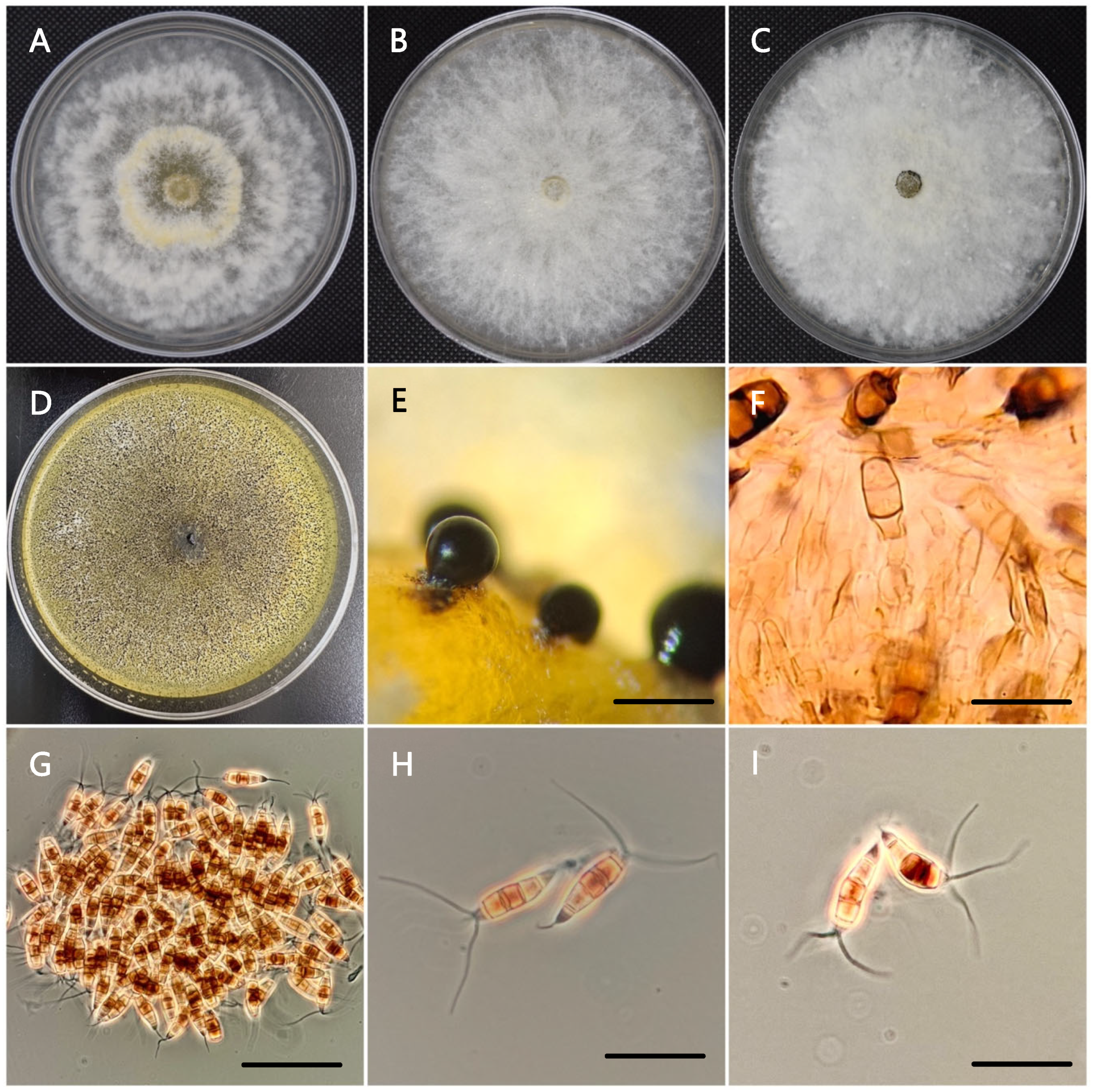
Colonies of all isolates grown on PDA produced abundant white, cottony mycelium with globose, solitary, semi-immersed, dark-brown to shiny pionnotal masses of the acervular type (Figure 2A–C). The reverse of the colonies exhibited a pale-yellow pigmentation. Colony margins and growth patterns varied among isolates, allowing classification into three morphotypes based on macroscopic characteristics. Pionnotal masses developed 14 days after inoculation (Figure 2D,E). Conidiogenous cells were hyaline, cylindrical, and smooth-walled, measuring 6–18 × 3–6 µm (Figure 2F). Conidia were fusiform and 5-celled, with three dark-brown median cells and separated by four dark septa. The basal cell was conical and hyaline, bearing a single appendage. The apical cell was cylindrical, hyaline, thin-walled, and smooth, with two to four tubular, filiform apical appendages. These appendages were not inserted at a single apical crest but rather individually attached at different points along the upper half of the apical cell and were unbranched. (Figure 2G–I).
Although isolates were overall morphologically similar, statistical analysis of conidial length and width separated them into two groups based on geographic origin. Isolates from Michoacán and Guanajuato produced larger conidia (22.0–37.0 × 7.5–9.5 µm) compared with isolates from Baja California (21.0–23.0 × 6.0–6.5 µm). Similarly, pionnotal masses from Michoacán (105 µm) and Guanajuato (103 µm) were substantially larger than those from Baja California (22 µm). Mycelial growth rates also differed significantly (p ≤ 0.05), with isolates from Michoacán and Guanajuato growing faster (6.3 mm day−1) than those from Baja California (4.1 mm day−1). Based on the observed morphological characteristics, all isolates were consistent with descriptions of the genus Neopestalotiopsis [7,55].

3.3. Molecular Identification

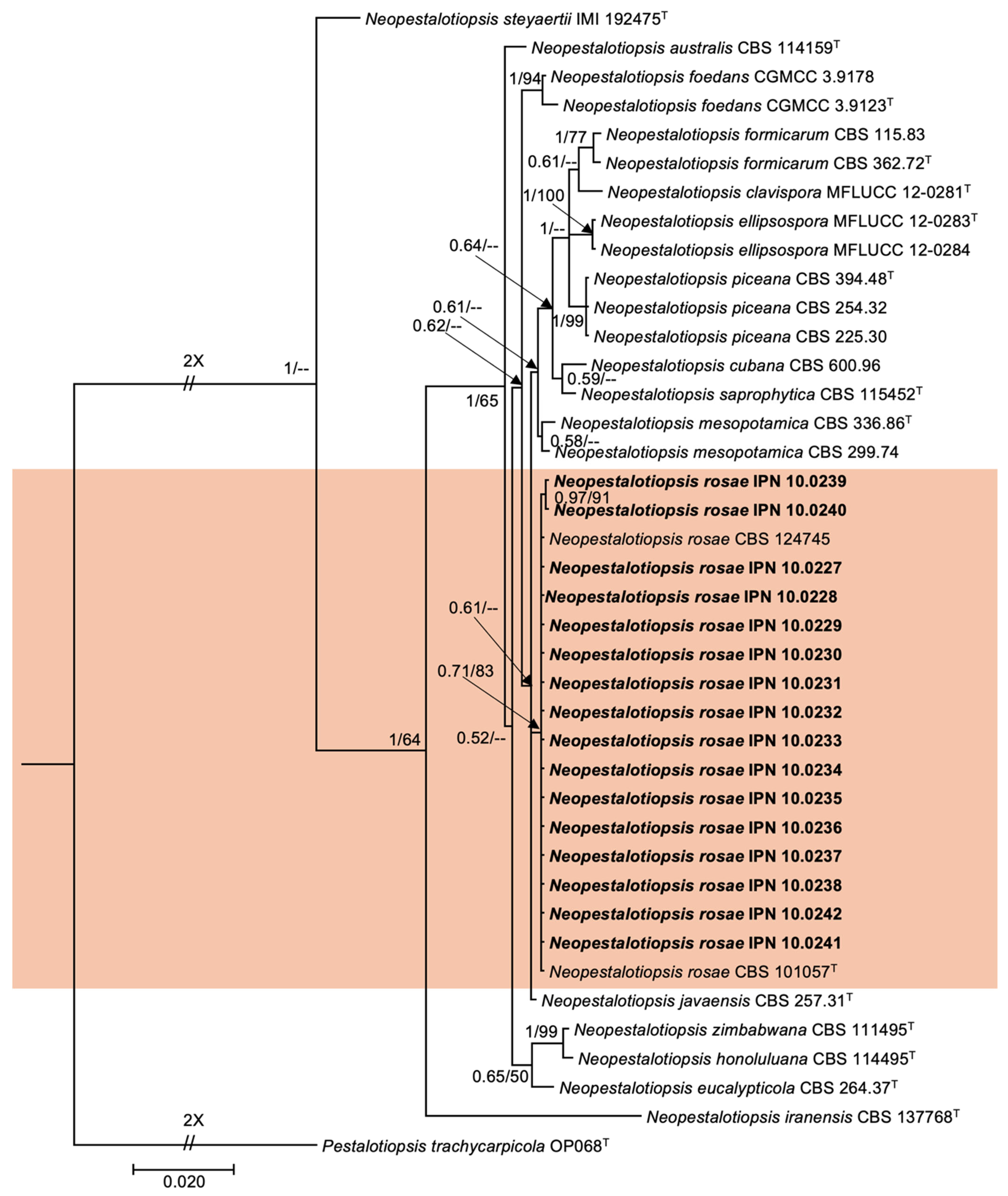
BLASTn searches against the GenBank database revealed that all obtained sequences showed 99–100% similarity to species within the genus Neopestalotiopsis. Multilocus phylogenetic analysis based on combined ITS, tef1-α, and β-tub sequences placed all 16 isolates within a well-supported clade corresponding to N. rosae. The isolates clustered with the ex-type strain CBS101057, with support of 0.71/83 (Posterior Probability/Bootstrap) (Figure 3). These results confirmed that the isolates recovered from strawberry plants exhibiting root and crown rot symptoms were N. rosae.

3.4. Pathogenicity Tests

All 16 N. rosae isolates were pathogenic. Inoculated strawberry plants developed characteristic symptoms starting 7 days after inoculation. Initial symptoms included foliar chlorosis, followed by progressive wilting and eventual plant death. Internally, affected crowns exhibited reddish-brown discoloration consistent with root and crown rot. Control plants remained asymptomatic throughout the experiment. The experiment was conducted twice, with similar results. Statistical analysis showed significant differences in disease severity among the N. rosae isolates (p < 0.05). Based on multiple comparisons of means, nine severity percentage groups were identified (Table 3). Isolates collected from Michoacán caused the highest severity, followed by those from Guanajuato, whereas isolates from Baja California resulted in significantly lower severity. Thus, the most aggressive isolate was IPN 10.0228 from Michoacán (90.05% severity), whereas the least aggressive was IPN 10.0240 from Baja California (26.70%). These results indicate substantial variability in aggressiveness among geographically distinct N. rosae isolates.

3.5. Identification of Native Biocontrol Agents

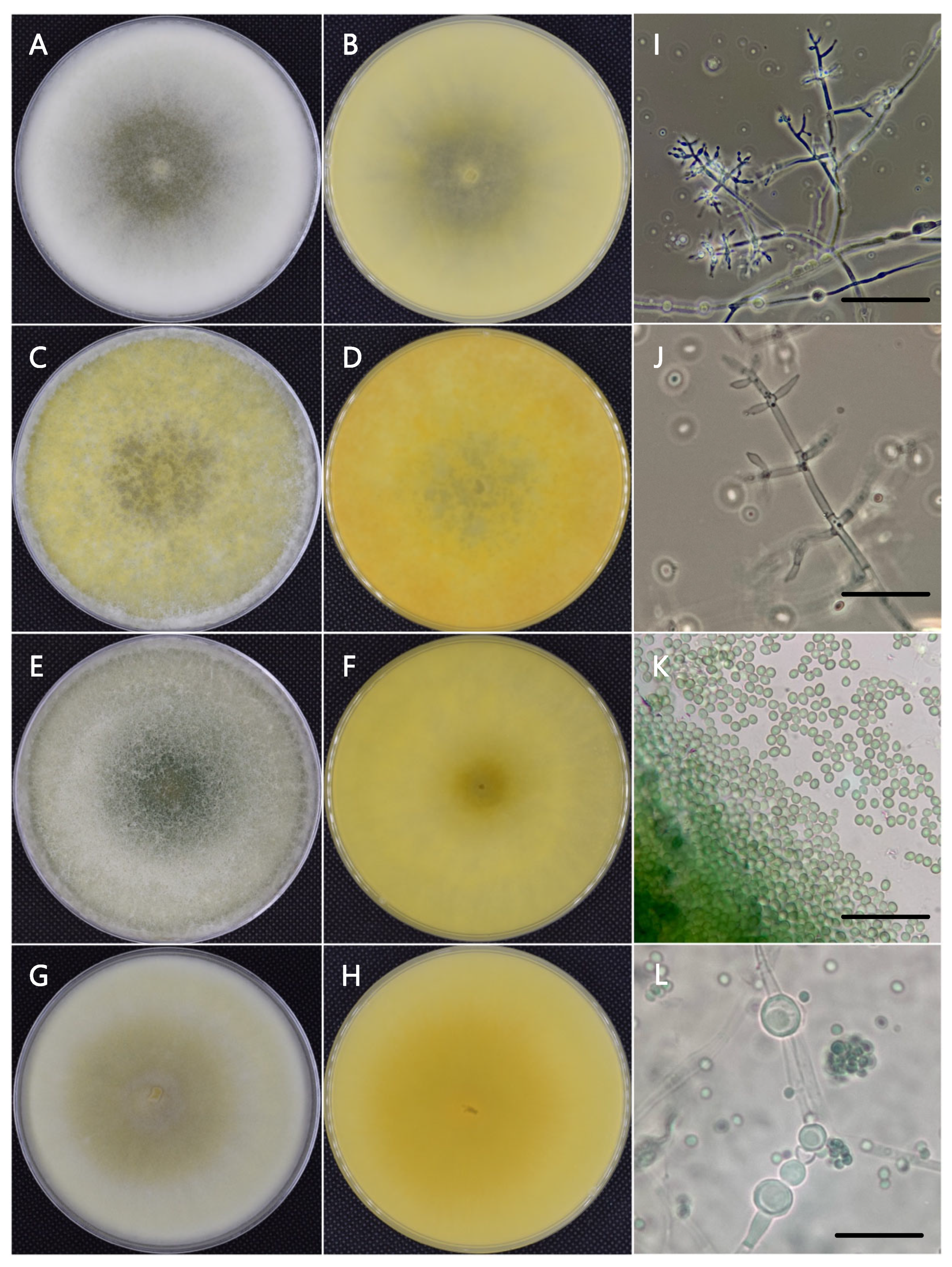
After 72 h of incubation at 25 °C on PDA, Trichoderma colonies reached an average diameter of 58 mm, whereas on SNA medium, they reached 55 mm. At 35 °C, colony diameters averaged 40 mm on PDA and 47 mm on SNA. After 96 h of incubation at 25 °C on PDA, colonies developed abundant cottony mycelium and produced yellow pigment that occasionally diffused into the medium, whereas isolates grown on SNA produced sparse aerial mycelium. Conidiophores were pyramidal with opposite branching; the main axis and each branch terminated in a cruciform verticil bearing up to five phialides. Phialides were lageniform to ampulliform, measuring 7.0 × 3.0 µm, with a basal width of 2.0 µm and a supporting cell width of 2.7 µm. The length/width ratio of phialides was 2.6; the ratio between phialide length and supporting cell width was 3.0; and the ratio between phialide width and supporting cell width was 1.2. Conidia were ovoid to globose, smooth-walled, green, and measured 3.0 × 2.5 µm. Chlamydospore formation was frequent after 7 days of incubation in three of the four isolates (Figure 4).
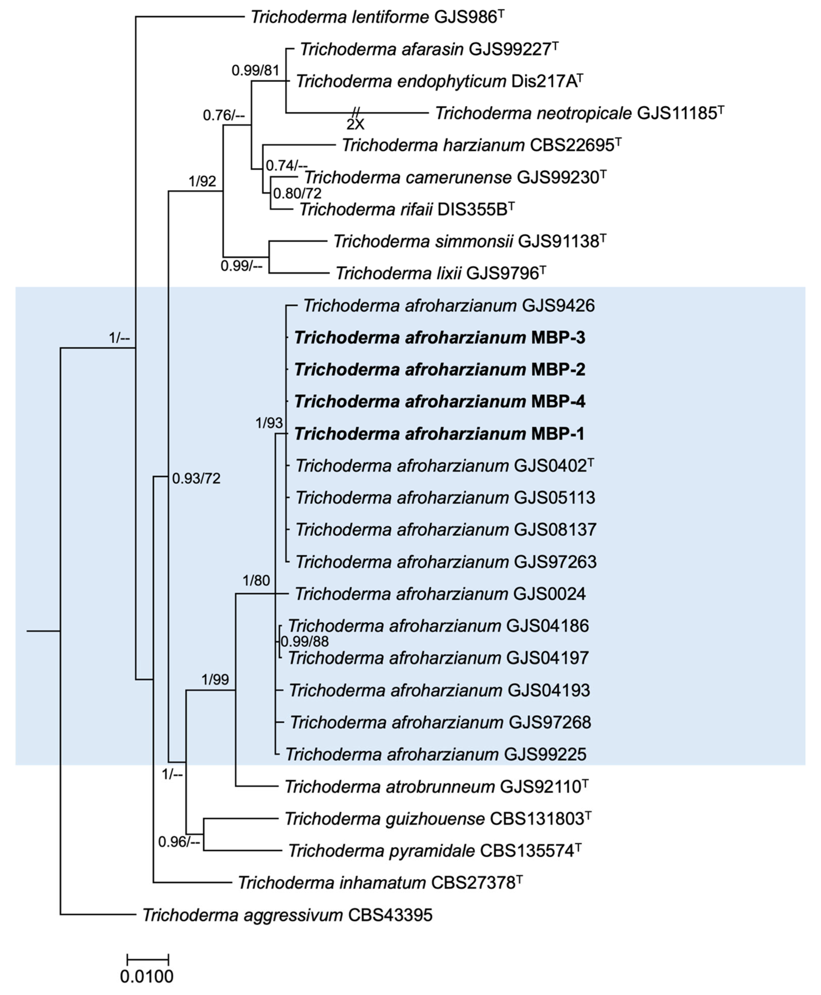
BLASTn searches against the GenBank database showed 99–100% sequence similarity with species of the genus Trichoderma. Multilocus phylogenetic analysis based on combined rpb2 and tef1-α sequences placed all four isolates within the clade corresponding to Trichoderma afroharzianum, with strong Bayesian posterior probability and Maximum Likelihood bootstrap support (1.00/93) (Figure 5). Therefore, these isolates were confidently identified as Trichoderma afroharzianum.

3.6. Biocontrol Tests

All Trichoderma afroharzianum isolates (MBP-1-MBP-4) significantly inhibited the mycelial growth of the 16 Neopestalotiopsis rosae isolates, with mean percentages of radial growth inhibition (PRGI) values ranging from 71.0% to 73.0% compared to the untreated control. Statistical analysis showed significant differences among T. afroharzianum isolates. Multiple mean comparisons divided them into two groups: isolates MBP-1 (73%) and MBP-2 (72%) formed the first group, exhibiting the highest antagonistic activity, while isolates MBP-3 and MBP-4 (71%) made up the second group. In contrast, the commercial fungicide fludioxonil + cyprodinil (Switch® 62.5WG) showed a mean PRGI of 50% against the same pathogen isolates, forming a third group.
When evaluated individually against each N. rosae isolate, 13 of the 16 T. afroharzianum isolates showed no significant differences in PRGI values against most pathogen isolates. However, significant differences were observed for isolates IPN 10.0232, IPN 10.0237, and IPN 10.0240. Isolate MBP-1 exhibited higher inhibition against IPN 10.0232 (78.3%) and IPN 10.0237 (77.5%) compared with isolate MBP-3 (71.2% and 70.6%, respectively). Against isolate IPN 10.0240, MBP-1 achieved 50.9% inhibition, significantly greater than isolates MBP-2, MBP-3, and MBP-4 (40.2–44.2%) (Table 4). Variation in pathogen susceptibility to T. afroharzianum was also evident. For example, the isolate MBP-1 inhibited isolates IPN 10.0242 and IPN 10.0241 by 81.8–82.2%, whereas inhibition of isolates IPN 10.0240 and IPN 10.0238 ranged from 50.9–51.1%. Similar patterns were observed for isolates MBP-1, MBP-3, and MBP-4, which consistently showed higher inhibition against isolates IPN 10.0241 and IPN 10.0233 but reduced activity against IPN 10.0240 and IPN 10.0238. These results indicate considerable variability in sensitivity among N. rosae isolates.
According to the antagonism classification by Bell et al. (1982) [54], isolates MBP-1 and MBP-2 were classified as Class I, characterized by complete overgrowth of Trichoderma, covering the pathogen colony. Isolates MBP-3 and MBP-4 were classified as Class II, showing overgrowth of at least two-thirds of the pathogen colony surface.
In contrast, Arcopilus cupreus exhibited significantly lower antagonistic activity, with a mean PRGI of 38.1% across all N. rosae isolates. The commercial fungicide showed a higher overall mean inhibition (70.8%) than A. cupreus, but displayed marked variability among pathogen isolates, with PRGI values ranging from 11.8% to 92.8%. Significant differences in fungicide sensitivity were detected for isolates IPN 10.0227, IPN 10.0228, IPN 10.0229, IPN 10.0230, IPN 10.0232, IPN 10.0233, IPN 10.0234, IPN 10.0235, and IPN 10.0238, whereas the remaining isolates did not differ significantly (Table 5). Overall, both biological and chemical treatments showed differential efficacy across N. rosae isolates, highlighting pronounced intra-population variability in response to control measures.

4. Discussion

Due to the rapid increase in global strawberry demand over recent years, the strawberry production industry has expanded, and new cultivation areas have developed [3,56]. As a leading producer, Mexico has experienced increased production as cultivation has expanded into new agricultural regions [3]. However, the movement of plant material increases the risk of pathogen dissemination. Current agricultural practices, including vegetative propagation, introduce pathogens into new production areas through infected plant material from nurseries [57,58,59,60,61]. The lack of commercial cultivars with full resistance to crown and root rot caused by N. rosae further exacerbates the epidemiological risk [62,63,64]. The symptoms observed in strawberry plants were similar across collection regions and consistent with previous reports describing strawberry root and crown rot symptomatology [6,16].
The cultural and morphological characteristics observed in isolates from strawberry plants showing root and crown rot symptoms, along with multigene phylogenetic analysis, enabled us to identify the causal agent as N. rosae. Although some nodes in the Neopestalotiopsis phylogeny show moderately supported values, the most comprehensive study of the genus Neopestalotiopsis [7] indicates that species within it are distinguishable through multigene phylogenetic reconstruction using the internal transcribed spacer (ITS), partial sequences of the genes β-tub, and tef1-α. However, these markers do not fully resolve the phylogeny of all 30 clades; thus, some clades remain poorly supported, and one clade is still classified as Neopestalotiopsis spp. Unfortunately, to date, higher-resolution markers are lacking to distinguish between species. The supports reported in this study align with those documented in the first report of the pathogen in Mexico [6]. Otherwise, the Trichoderma phylogeny, based on Chaverri et al. (2015) [48], was resolved using the amplified genes, yielding high support values.
Neopestalotiopsis rosae has been reported as the causal agent of strawberry crown rot in Egypt [8], Mexico [6], Taiwan [44], and China [9]. Since its first report in Mexico, this disease has rapidly spread across all strawberry-producing regions of the country, mainly due to the distribution of infected propagative material, including other pathogens, such as Macrophomina. This situation is largely attributable to prevailing artisanal propagation practices in Mexico, including the reuse of trays or pots previously contaminated with infested soil without proper disinfection (personal communication from technicians and affected growers).
As previously reported in Mexico [6], only the species N. rosae was found in the samples analyzed in the present study. The proportions of fungi isolated from strawberry plants with root and crown rot symptoms represent a first approximation to the diversity of species involved in strawberry plant mortality in the field. In the present study, nearly one-third of the plants with these symptoms belonged to the genus Macrophomina, indicating that this fungus is an emerging pathogen in strawberry plantations in Mexico. This highlights the epidemiological importance of Macrophomina as an emerging strawberry pathogen. Macrophomina phaseolina has been reported as the causal agent of crown rot in strawberry in Argentina [65], Chile [66], and the United States [67]. However, in Mexico, the phylogenetic species of Macrophomina affecting strawberry plants has not yet been confirmed through molecular methods. This fungus could become increasingly important, and research should focus on the timely development of management strategies to minimize losses caused by this pathogen.
Artificially inoculated strawberry plants showed characteristic disease symptoms seven days after inoculation. Affected plants exhibited growth retardation, yellowing of outer leaves, dark-brown lesions, and crown rot with reddish-brown discoloration, leading to plant death, symptoms previously reported for this disease [6,18]. Therefore, selection of disease-tolerant germplasm can be conducted 15 days after inoculation, when symptoms are visible. The inoculum concentration and inoculation method used in this study were appropriate, as they induced characteristic disease symptoms in a shorter time than reported by Dardani et al. (2025) [15], who documented initial symptoms appearing 10–12 days after inoculation, and even faster than reported by Sigillo et al. (2020) [42], who observed symptoms up to two months after inoculation.
Cultural and morphological characterization of the native biocontrol agents placed the four isolates within the genus Trichoderma, while phylogenetic species identification was conducted using multigene phylogenetic analysis of concatenated ITS sequences and partial rpb2 and tef1-α sequences. This analysis confidently confirmed that the four isolates belong to the species Trichoderma afroharzianum, based on reference phylogeny [48].
All isolates of T. afroharzianum exhibited strong antagonistic activity against the 16 N. rosae isolates, with mean inhibition ranging from 71 to 73%. This range exceeded that reported by Olivares-Rodríguez et al. (2024) [68], who evaluated 15 Trichoderma spp. isolates against Neopestalotiopsis isolates from strawberry; only the T2 strain reached 70% inhibition, while the others showed lower levels. In this study, the interaction between strain MBP-1 of T. afroharzianum and isolate IPN 10.0242 resulted in a PIMG of 82.2%. The average PIMG of 71–73% is consistent with previous reports [17,68], which describe a 66.6–76% range as a significant in vitro inhibition range for N. rosae and N. clavispora recovered from strawberry by Trichoderma spp. The variation in the in vitro biocontrol activity of T. afroharzianum isolates against N. rosae is expected, as pathogen suppression levels can depend on the strain used and its adaptability to specific biotic and abiotic conditions [53]. T. afroharzianum has proven effective as a biocontrol agent against phytopathogenic fungi in wheat (Fusarium culmorum) [69], tomato (Alternaria alternata, Botrytis cinerea, Fusarium oxysporum) [70,71,72], onion (Stromatinia cepivora) [73], garlic (Sclerotium cepivorum) [74], and mustard (Sclerotinia sclerotiorum) [75]. Moreover, strain T22, reclassified as T. afroharzianum, has been used in commercial biocontrol products [48,76]. However, to the best of our knowledge, this is the first study to explore the biocontrol potential of native Mexicans T. afroharzianum isolates against N. rosae.
Microscopic observation of interactions between T. afroharzianum and N. rosae isolates revealed that mycoparasitism was the primary mechanism underlying the biocontrol effect. Because the isolates were able to grow rapidly, coil around and parasitize the pathogen’s hyphae, lyse them, and occupy space and resources, thereby limiting the growth of N. rosae, we can conclude that the T. afroharzianum isolates exhibit microparasitism and competence. Furthermore, the yellow coloration observed in the culture media in which T. afroharzianum grew indicates that these isolates produce compounds that inhibit the growth of N. rosae isolates. Therefore, the isolates likely produce compounds with antibiotic, toxic, and lytic capacities. These mechanisms not evaluated in the present study cannot be ruled out, including the production of hydrolytic enzymes, antibiosis (antifungal and volatile compounds), competition for space and nutrients, and the induction of host plant defense mechanisms previously reported for T. afroharzianum [70,72,73,75]. These findings demonstrate the potential of native T. afroharzianum isolates as biological control agents. In dual-culture assays, inhibition exceeding 70% is commonly used as a benchmark for selecting promising Trichoderma isolates for development as biocontrol agents [77,78,79].
Arcopilus cupreus is characterized by the abundant production of yellow, orange, or red exudates (secreted products) [80], a feature observed in both dual cultures and controls in the present study. The production of metabolites was evidenced by the presence of inhibition halos in encounters with N. rosae. Although A. cupreus showed moderate antagonistic activity, its inhibition levels were lower than those of T. afroharzianum. Acting through mechanisms such as production of antifungal metabolites, mycoparasitism, and competition for space and nutrients, enabling control of a broad range of phytopathogenic fungi [23]. Previous reports indicate antifungal activity of A. cupreus against Phytophthora spp. recovered from mandarin roots, achieving PIMG values of 62.5–73.2% [24]. Additionally, other species of this genus have demonstrated antifungal activity, such as isolate YZXR of A. aureus, which inhibited up to 80% of the growth of Fusarium fujikuroi, the causal agent of leaf spot in Polygonatum odoratum. Nevertheless, further research is needed to fully assess the potential of this genus as a biological control agent.
The commercial fungicide Switch® 62.5WG (Fludioxonil + Cyprodinil) showed an average PIMG of 50%, with isolate IPN 10.0230 being the most sensitive (92.8% PIMG) and isolate IPN 10.0240 the least sensitive (11.8%). In contrast, Rebollar-Alviter et al. (2020) [6] reported 100% PIMG when evaluating the same fungicide against two N. rosae isolates obtained from strawberry crowns. These results highlight pathogen variability and the importance of conducting population studies that capture this diversity and relate isolate responses with geographic origin, to avoid generalized management recommendations, given the broad variability observed among isolates in the present study. Overall, the marked variability observed in both biological and chemical control responses emphasizes the need for population-based management strategies in Mexican strawberry production systems. Integrated disease management programs incorporating locally adapted biological agents may provide a more reliable alternative to confront the adaptive capacity of phytopathogenic fungi to intensive agricultural practices and stop relying on synthetic fungicides. Laboratory evaluation of biocontrol agents using dual-culture assays is a standard method for assessing the mechanisms by which these agents act against pathogens. These tests allow rapid comparison of many isolates and selection of the best ones. However, they capture only some mechanisms in the controlled interaction, not the full range of interactions that occur in the rhizosphere or within the plant. Therefore, because the present study was mainly focused on in vitro antagonistic assays, and although the results are promising, future greenhouse or field validation experiments are needed.
Finally, native or endemic biocontrol agents, such as our isolates of T. afroharzianum, offer competitive advantages over non-endemic organisms marketed as biological pesticides. For example, native biocontrol agents are adapted to local biotic and abiotic conditions, making them more effective than commercial products [81]. Furthermore, using native biocontrol agents carries fewer ecological risks, since introducing non-native species that could become invasive can have significant detrimental impacts on the environment and lead to serious consequences not only in the geographic area where they are released but also beyond their initial introduction [82,83]. However, it is essential to optimize formulations to maintain the viability and efficacy of these native biocontrol agents, ensuring they are competitive in quality and practicality and attractive to producers in their region of origin.

5. Conclusions

The causal agent of strawberry root and crown rot in the states of Michoacán, Guanajuato, and Baja California, Mexico, is N. rosae. According to the PRGI results, all evaluated T. afroharzianum strains and A. cupreus showed potential as biological control agents against N. rosae. Although biological control generally acts more slowly than chemical control, the native isolates exhibited a more consistent inhibition response across all N. rosae isolates than the fungicide did. Therefore, the application of biological control agents is recommended as a preventive strategy for disease management. Given the limited exploration of the antifungal activity of the isolates tested in this study (MBP1–MBP4 and IPN 10.0154), further research is needed to evaluate their control efficacy against N. rosae in greenhouse or field assays, as well as characterize the mechanisms of action of the evaluated T. afroharzianum isolates, including the identification of antifungal metabolites and their potential plant growth-promoting effects on strawberry cultivars. Due to the high variability in the response of N. rosae isolates to fungicide treatment, there is a clear need to understand the differential sensitivity of pathogen genotypes to different fungicidal active ingredients used in each strawberry-producing region. Finally, because chemical control remains the main disease management strategy for Mexican strawberry growers, continued research should evaluate additional fungicidal molecules and increase the number of N. rosae isolates tested, thereby updating the available information on the efficacy of current fungicides for managing this disease.

Author Contributions

Conceptualization, G.M.-L. and A.R.S.-B.; Methodology, G.M.-L. and A.R.S.-B.; Investigation, K.J.J.-C., A.R.S.-B., A.J.-P., F.C.-A., G.T.-T., G.M.-L. and L.S.-R.; writing—original draft preparation, K.J.J.-C., A.R.S.-B., A.J.-P., F.C.-A., G.T.-T., G.M.-L. and L.S.-R.; review and editing, K.J.J.-C., A.R.S.-B., A.J.-P., F.C.-A., G.T.-T., G.M.-L. and L.S.-R.; supervision, A.R.S.-B.; funding acquisition, G.T.-T., G.M.-L. and A.R.S.-B. All authors have read and agreed to the published version of the manuscript.

Funding

This research was financially supported by the Secretary of Research and Graduate Studies of the National Polytechnic Institute (Instituto Politécnico Nacional), grant number 20260096.

Institutional Review Board Statement

Not applicable.

Informed Consent Statement

Not applicable.

Data Availability Statement

The original contributions presented in this study are included in the article. Further inquiries can be directed to the corresponding authors.

Acknowledgments

During the preparation of this work, the authors used ChatGPT (model 5.5) for the purposes of improving language clarity and grammar of the manuscript. The authors have reviewed and edited the output and take full responsibility for the content of this publication.

Conflicts of Interest

The authors declare no conflicts of interest.

References

  1. Morales-Quintana, L.; Moya, M.; Santelices-Moya, R.; Cabrera-Ariza, A.; Rabert, C.; Pollmann, S.; Ramos, P. Improvement in the physiological and biochemical performance of strawberries under drought stress through symbiosis with Antarctic fungal endophytes. Front. Microbiol. 2022, 13, 939955. [Google Scholar] [CrossRef]
  2. FAO. Food and Agriculture Data; FAO: Rome, Italy, 2026. [Google Scholar]
  3. SIAP. Panorama Agroalimentario, 2025 ed.; Servicio de Información Agroalimentaria y Pesquera, Ed.; Secretaria de Agricultura y Desarrollo Rural: Ciudad de México, Mexico, 2025.
  4. Avilés, M.; Pastrana, A.M.; Borrero, C. Emerging Diseases in Spain Strawberry Crops: Neopestalotiopsis Leaf and Crown Rot and Fusarium Wilt. Plants 2024, 13, 3441. [Google Scholar] [CrossRef]
  5. Acosta-González, U.; Leyva-Mir, S.G.; Silva-Rojas, H.V.; Rebollar-Alviter, A. Preventive and Curative Effects of Treatments to Manage Strawberry Root and Crown Rot Caused by Neopestalotiopsis rosae. Plant Dis. 2023, 108, 1278–1288. [Google Scholar] [CrossRef] [PubMed]
  6. Rebollar-Alviter, A.; Silva-Rojas, H.V.; Fuentes-Aragón, D.; Acosta-González, U.; Martínez-Ruiz, M.; Parra-Robles, B.E. An Emerging Strawberry Fungal Disease Associated with Root Rot, Crown Rot and Leaf Spot Caused by Neopestalotiopsis rosae in Mexico. Plant Dis. 2020, 104, 2054–2059. [Google Scholar] [CrossRef]
  7. Maharachchikumbura, S.S.N.; Hyde, K.D.; Groenewald, J.Z.; Xu, J.; Crous, P.W. Pestalotiopsis revisited. Stud. Mycol. 2014, 79, 121–186. [Google Scholar] [CrossRef]
  8. Essa, T.A.; Kamel, S.M.; Ismail, A.M.; El-Ganainy, S. Characterization and chemical control of Neopestalotiopsis rosae the causal agent of strawberry root and crown rot in Egypt. Egypt. J. Phytopathol. 2018, 46, 1–19. [Google Scholar] [CrossRef]
  9. Sun, Q.; Harishchandra, D.; Jia, J.; Zuo, Q.; Zhang, G.; Wang, Q.; Yan, J.; Zhang, W.; Li, X. Role of Neopestalotiopsis rosae in causing root rot of strawberry in Beijing, China. Crop Prot. 2021, 147, 105710. [Google Scholar] [CrossRef]
  10. Erdurmuş, D.; Palacıoğlu, G.; Erdurmuş, G.; Bayraktar, H. First report of Neopestalotiopsis rosae causing leaf spot and crown rot of strawberry in Turkey. J. Plant Pathol. 2023, 105, 315. [Google Scholar] [CrossRef]
  11. Lawrence, D.P.; Brittain, G.D.; Aglave, B.; Sances, F.V. First Report of Neopestalotiopsis rosae Causing Crown and Root Rot of Strawberry in California. Plant Dis. 2023, 107, 566. [Google Scholar] [CrossRef]
  12. Fernández-Ozuna, Y.A.; Gini Álvarez, A.R.; Lopez-Nicora, H.D.; Arrúa Alvarenga, A.A.; Colmán, A.A. First report of Neopestalotiopsis rosae causing leaf spot and crown rot on strawberry (Fragaria × ananassa) in Paraguay. New Dis. Rep. 2023, 48, e12239. [Google Scholar] [CrossRef]
  13. Chandana, R.; Poonacha, T.T.; Chethan, D.; Karan, R.; Kruthika, R.; Khan, F.; Ashwini, K.S.; Bevanur, A.; Vani, Y.; Ramesh, G.V.; et al. Neopestalotiopsis rosae, a novel pathogen causing leaf blight and crown rot of strawberries in India. Physiol. Mol. Plant Pathol. 2024, 133, 102377. [Google Scholar] [CrossRef]
  14. Schierling, T.E.; Voegele, R.T.; El-Hasan, A. First Report on the Emergence of Neopestalotiopsis rosae as a Severe Economic Threat to Strawberry Production in Germany. Microorganisms 2025, 13, 6. [Google Scholar] [CrossRef]
  15. Dardani, G.; Martino, I.; Aloi, F.; Carli, C.; Giordano, R.; Spadaro, D.; Guarnaccia, V. Characterization of Neopestalotiopsis Species Associated with Strawberry Crown Rot in Italy. Agronomy 2025, 15, 422. [Google Scholar] [CrossRef]
  16. Ávila-Hernández, J.G.; León-Ramírez, C.G.; Abraham-Juárez, M.d.R.; Tlapal-Bolaños, B.; Olalde-Portugal, V.; Délano-Frier, J.P.; Martínez-Antonio, A.; Aguilar-Zárate, P. Neopestalotiopsis spp.: A Threat to Strawberry Production and Management. Horticulturae 2025, 11, 288. [Google Scholar] [CrossRef]
  17. Amrutha, P.; Vijayaraghavan, R. Evaluation of Fungicides and Biocontrol Agents against Neopestalotiopsis clavispora Causing Leaf Blight of Strawberry (Fragaria × ananassa Duch.). Int. J. Curr. Microbiol. Appl. Sci. 2018, 7, 622–628. [Google Scholar] [CrossRef]
  18. Baggio, J.S.; Rebello, C.S.; de Morais, M.B.; Marin, M.V.; Gama, A.B.; Forcelini, B.B.; Mertely, J.C.; Peres, N.A. Efficacy of Single- and Multi-Site Fungicides Against Neopestalotiopsis spp. of Strawberry. Plant Dis. 2023, 107, 2177–2184. [Google Scholar] [CrossRef] [PubMed]
  19. Islam, T.; Danishuddin; Tamanna, N.T.; Matin, M.N.; Barai, H.R.; Haque, M.A. Resistance Mechanisms of Plant Pathogenic Fungi to Fungicide, Environmental Impacts of Fungicides, and Sustainable Solutions. Plants 2024, 13, 2737. [Google Scholar] [CrossRef] [PubMed]
  20. Liu, P.; Yang, R.; Wang, Z.; Ma, Y.; Ren, W.; Wei, D.; Ye, W. Biocontrol Potential of Trichoderma asperellum CMT10 against Strawberry Root Rot Disease. Horticulturae 2024, 10, 246. [Google Scholar] [CrossRef]
  21. Howell, C.R. Mechanisms Employed by Trichoderma Species in the Biological Control of Plant Diseases: The History and Evolution of Current Concepts. Plant Dis. 2003, 87, 4–10. [Google Scholar] [CrossRef]
  22. Sultana, S.; Sikder, M.M.; Ahmmed, M.S.; Alam, N. Neopestalotiopsis chrysea Causing Leaf Spot Disease of Strawberry Plants in Bangladesh. J. Plant Sci. 2022, 17, 66–74. [Google Scholar] [CrossRef]
  23. Zhang, H.; Yang, Q. Expressed sequence tags-based identification of genes in the biocontrol agent Chaetomium cupreum. Appl. Microbiol. Biotechnol. 2007, 74, 650–658. [Google Scholar] [CrossRef]
  24. Noireung, P. Efficacy of Arcopilus cupreus as biological agent to control Phytophthora spp. causing root rot of mandarin citrus. Curr. Res. Environ. Appl. Mycol. 2020, 10, 494–501. [Google Scholar] [CrossRef]
  25. Mao, B.-Z.; Huang, C.-w.; Yang, G.; Chen, Y.-Z.; Chen, S. Separation and determination of the bioactivity of oosporein from Chaetomium cupreum. Afr. J. Biotechnol. 2010, 9, 5955–5961. [Google Scholar]
  26. Heitman, J.; Howlett, B.J.; Crous, P.W.; Stukenbrock, E.H.; James, T.Y.; Gow, N.A.R. The Fungal Kingdom; American Society for Microbiology Press: Washington, DC, USA, 2018. [Google Scholar]
  27. Madhushan, A.; Weerasingha, D.B.; Ilyukhin, E.; Taylor, P.W.J.; Ratnayake, A.S.; Liu, J.-K.; Maharachchikumbura, S.S.N. From Natural Hosts to Agricultural Threats: The Evolutionary Journey of Phytopathogenic Fungi. J. Fungi 2025, 11, 25. [Google Scholar] [CrossRef]
  28. Crous, P.W.; Verkley, G.J.M.; Groenewald, J.Z.; Houbraken, J. Fungal Biodiversity; Westerdijk Fungal Biodiversity Institute: Utrecht, The Netherlands, 2019. [Google Scholar]
  29. Zervakis, G.; Philippoussis, A.; Ioannidou, S.; Diamantopoulou, P. Mycelium growth kinetics and optimal temperature conditions for the cultivation of edible mushroom species on lignocellulosic substrates. Folia Microbiol. 2001, 46, 231–234. [Google Scholar] [CrossRef] [PubMed]
  30. White, T.; Bruns, T.; Lee, S.; Taylor, J. Amplification and direct sequencing of fungal ribosomal RNA Genes for phylogenetics. In PCR Protocols, a Guide to Methods and Applications; Academic Press: Cambridge, MA, USA, 1990; pp. 315–322. [Google Scholar]
  31. Glass, N.L.; Donaldson, G.C. Development of primer sets designed for use with the PCR to amplify conserved genes from filamentous ascomycetes. Appl. Environ. Microbiol. 1995, 61, 1323–1330. [Google Scholar] [CrossRef] [PubMed]
  32. O’Donnell, K.; Cigelnik, E. Two divergent intragenomic rDNA ITS2 types within a monophyletic lineage of the fungus Fusarium are nonorthologous. Mol. Phylogenet. Evol. 1997, 7, 103–116. [Google Scholar] [CrossRef]
  33. Carbone, I.; Kohn, L.M. A method for designing primer sets for speciation studies in filamentous ascomycetes. Mycologia 1999, 91, 553–556. [Google Scholar] [CrossRef]
  34. O’Donnell, K.; Kistler, H.C.; Cigelnik, E.; Ploetz, R.C. Multiple evolutionary origins of the fungus causing Panama disease of banana: Concordant evidence from nuclear and mitochondrial gene genealogies. Proc. Natl. Acad. Sci. USA 1998, 95, 2044–2049. [Google Scholar] [CrossRef]
  35. Okonechnikov, K.; Golosova, O.; Fursov, M. Unipro UGENE: A unified bioinformatics toolkit. Bioinformatics 2012, 28, 1166–1167. [Google Scholar] [CrossRef] [PubMed]
  36. Katoh, K.; Rozewicki, J.; Yamada, K.D. MAFFT online service: Multiple sequence alignment, interactive sequence choice and visualization. Brief. Bioinform. 2019, 20, 1160–1166. [Google Scholar] [CrossRef]
  37. Kumar, S.; Stecher, G.; Suleski, M.; Sanderford, M.; Sharma, S.; Tamura, K. MEGA12: Molecular Evolutionary Genetic Analysis Version 12 for Adaptive and Green Computing. Mol. Biol. Evol. 2024, 41, msae263. [Google Scholar] [CrossRef]
  38. Maddison, W.P.; Maddison, D.R. Mesquite: A Modular System for Evolutionary Analysis; Version 4.03; 2026; Available online: https://www.mesquiteproject.org (accessed on 2 March 2025).
  39. Ronquist, F.; Huelsenbeck, J.P. MrBayes 3: Bayesian phylogenetic inference under mixed models. Bioinformatics 2003, 19, 1572–1574. [Google Scholar] [CrossRef]
  40. Darriba, D.; Taboada, G.L.; Doallo, R.; Posada, D. jModelTest 2: More models, new heuristics and parallel computing. Nat. Methods 2012, 9, 772. [Google Scholar] [CrossRef] [PubMed]
  41. Edler, D.; Klein, J.; Antonelli, A.; Silvestro, D. raxmlGUI 2.0: A graphical interface and toolkit for phylogenetic analyses using RAxML. Methods Ecol. Evol. 2021, 12, 373–377. [Google Scholar] [CrossRef]
  42. Sigillo, L.; Ruocco, M.; Gualtieri, L.; Pane, C.; Zaccardelli, M. First report of Neopestalotiopsis clavispora causing crown rot in strawberry in Italy. J. Plant Pathol. 2020, 102, 281. [Google Scholar] [CrossRef]
  43. Baggio, J.S.; Forcelini, B.B.; Wang, N.-Y.; Ruschel, R.G.; Mertely, J.C.; Peres, N.A. Outbreak of Leaf Spot and Fruit Rot in Florida Strawberry Caused by Neopestalotiopsis spp. Plant Dis. 2021, 105, 305–315. [Google Scholar] [CrossRef] [PubMed]
  44. Wu, H.-Y.; Tsai, C.-Y.; Wu, Y.-M.; Ariyawansa, H.-A.; Chung, C.-L.; Chung, P.-C. First Report of Neopestalotiopsis rosae Causing Leaf Blight and Crown Rot on Strawberry in Taiwan. Plant Dis. 2021, 105, 487. [Google Scholar] [CrossRef]
  45. Townsend, G.R.; Heuberger, J.W. Methods for estimating losses caused by diseases in fungicide experiments. Plant Dis. Report. 1943, 27, 340–343. [Google Scholar]
  46. Elad, Y.; Chet, I.; Henis, Y. Degradation of plant pathogenic fungi by Trichoderma harzianum. Can. J. Microbiol. 1982, 28, 719–725. [Google Scholar] [CrossRef]
  47. Okon, Y.; Chet, I.; Henis, Y. Effects of Lactose, Ethanol and Cycloheximide on the Translocation Pattern of Radioactive Compounds and on Sclerotium Formation in Sclerotium rolfsii. Microbiology 1973, 74, 251–258. [Google Scholar] [CrossRef]
  48. Chaverri, P.; Branco-Rocha, F.; Jaklitsch, W.; Gazis, R.; Degenkolb, T.; Samuels, G.J. Systematics of the Trichoderma harzianum species complex and the re-identification of commercial biocontrol strains. Mycologia 2015, 107, 558–590. [Google Scholar] [CrossRef] [PubMed]
  49. Sung, G.-H.; Sung, J.-M.; Hywel-Jones, N.L.; Spatafora, J.W. A multi-gene phylogeny of Clavicipitaceae (Ascomycota, Fungi): Identification of localized incongruence using a combinational bootstrap approach. Mol. Phylogenetics Evol. 2007, 44, 1204–1223. [Google Scholar] [CrossRef]
  50. Liu, Y.J.; Whelen, S.; Hall, B.D. Phylogenetic relationships among ascomycetes: Evidence from an RNA polymerse II subunit. Mol. Biol. Evol. 1999, 16, 1799–1808. [Google Scholar] [CrossRef]
  51. Solano-Báez, A.R.; Victoria-Arellano, A.D.; Trejo-Espino, J.L.; Trejo-Tapia, G.; Flores-Moctezuma, H.E.; Márquez-Licona, G. Etiology of leaf blight of Chinese brush (Baccharis conferta Kunth) in Mexico. Botany 2025, 103, 1–9. [Google Scholar] [CrossRef]
  52. Sinclair, J.B.; Dhingra, O.D. Basic Plant Pathology Methods, 2nd ed.; CRC Press: Boca Raton, FL, USA, 1995. [Google Scholar]
  53. Dennis, C.; Webster, J. Antagonistic properties of species-groups of Trichoderma: III. Hyphal interaction. Trans. Br. Mycol. Soc. 1971, 57, 363–369, IN1–IN2. [Google Scholar] [CrossRef]
  54. Bell, D.K.; Wells, H.D.; Markham, C.R. In vitro antagonism of Trichoderma species against six fungal plant pathogens. Phytopathology 1982, 72, 379–382. [Google Scholar] [CrossRef]
  55. Baggio, J.S.; Marin, M.V.; Peres, N.A. Phytophthora Crown Rot of Florida Strawberry: Inoculum Sources and Thermotherapy of Transplants for Disease Management. Plant Dis. 2021, 105, 3496–3502. [Google Scholar] [CrossRef]
  56. Krüger, E.; Josuttis, M.; Nestby, R.; Toldam-Andersen, T.B.; Carlen, C.; Mezzetti, B. Influence of growing conditions at different latitudes of Europe on strawberry growth performance, yield and quality. J. Berry Res. 2012, 2, 143–157. [Google Scholar] [CrossRef]
  57. Debode, J.; Hemelrijck, W.; Xu, X.-M.; Maes, M.; Creemers, P.; Heungens, K. Latent entry and spread of Colletotrichum acutatum (species complex) in strawberry fields. Plant Pathol. 2015, 64, 385–395. [Google Scholar] [CrossRef]
  58. Holmes, G.; Mansouripour, S.; Hewavitharana, S. Strawberries at the Crossroads: Management of Soilborne Diseases in California without Methyl Bromide. Phytopathology 2020, 110, 956–968. [Google Scholar] [CrossRef]
  59. Morresi, V.; Capocasa, F.; Balducci, F.; Diamanti, J.; Mezzetti, B. Nursery Propagation Systems for High-Quality Strawberry (Fragaria × ananassa Duch.) Plug Plant Production from Micropropagated, Soilless-Grown Mother Plants. Horticulturae 2025, 11, 888. [Google Scholar] [CrossRef]
  60. Pastrana, A.; Basallote-Ureba, M.; Aguado, A.; Capote, N. Potential Inoculum Sources and Incidence of Strawberry Soilborne Pathogens in Spain. Plant Dis. 2017, 101, 751–760. [Google Scholar] [CrossRef] [PubMed]
  61. Pastrana, A.; Watson, D.; Gordon, T. Transmission of Fusarium oxysporum f. sp. fragariae Through Stolons in Strawberry Plants. Plant Dis. 2019, 103, 1249–1251. [Google Scholar] [CrossRef]
  62. Alam, E.; Moyer, C.; Verma, S.; Peres, N.A.; Whitaker, V.M. Exploring the genetic basis of resistance to Neopestalotiopsis species in strawberry. Plant Genome 2024, 17, e20477. [Google Scholar] [CrossRef]
  63. Guan, W.; Bonkowski, J.; Creswell, T.; Egel, D.S. Strawberry Cultivar Susceptibility to Neopestalotiopsis Leaf Spot in Indiana. Plant Health Prog. 2023, 24, 135–139. [Google Scholar] [CrossRef]
  64. Gunacti, H. Emergence of Neopestalotiopsis rosae causing leaf spot and fruit rot on strawberry and assessment of cultivar response to infection. Euphytica 2026, 222, 31. [Google Scholar] [CrossRef]
  65. Baino, O.M.; Salazar, S.M.; Ramallo, A.C.; Kirschbaum, D.S. First Report of Macrophomina phaseolina Causing Strawberry Crown and Root Rot in Northwestern Argentina. Plant Dis. 2011, 95, 1477. [Google Scholar] [CrossRef]
  66. Sánchez, S.; Gambardella, M.; Henríquez, J.L.; Díaz, I. First Report of Crown Rot of Strawberry Caused by Macrophomina phaseolina in Chile. Plant Dis. 2013, 97, 996. [Google Scholar] [CrossRef] [PubMed]
  67. Pedroncelli, L.; Henry, P.M.; Putman, A.I. Colonization of Strawberry Plants by Macrophomina phaseolina After Root or Crown Inoculations. PhytoFrontiers™ 2025, 5, 345–354. [Google Scholar] [CrossRef]
  68. Olivares-Rodriguez, G.; Angeles-Núñez, J.G.; Mondragón-Rojas, F.; Rivas-Valencia, P.; Zárate-Castrejón, J.L.; Mariscal-Amaro, L.A.; Díaz-Espino, L.F.; Martínez-Martínez, T.O. Control in vitro de Neopestalotiopsis sp. aislada de fresa empleando Trichoderma y fungicidas comerciales. Rev. Mex. Fitopatol. 2024, 42, 1–14. [Google Scholar] [CrossRef]
  69. Bouanaka, H.; Bellil, I.; Harrat, W.; Boussaha, S.; Benbelkacem, A.; Khelifi, D. On the biocontrol by Trichoderma afroharzianum against Fusarium culmorum responsible of fusarium head blight and crown rot of wheat in Algeria. Egypt. J. Biol. Pest Control 2021, 31, 68. [Google Scholar] [CrossRef]
  70. Philip, B.; Behiry, S.; Salem, M.; Amer, M.; El-Samra, I.; Abdelkhalek, A.; Heflish, A. Trichoderma afroharzianum TRI07 metabolites inhibit Alternaria alternata growth and induce tomato defense-related enzymes. Sci. Rep. 2024, 14, 1874. [Google Scholar] [CrossRef]
  71. Shi, Z.; Zhang, M.; Chen, G.; Cao, A.; Wang, Q.; Yan, D.; Fang, W.; Li, Y. Evaluation of newly combination of Trichoderma with dimethyl disulfide fumigant to control Fusarium oxysporum, optimize soil microbial diversity and improve tomato yield. Ecotoxicol. Environ. Saf. 2025, 292, 117903. [Google Scholar] [CrossRef] [PubMed]
  72. Zhao, J.; Liu, T.; Zhang, D.; Wu, H.-Q.; Zhang, T.-T.; Dong, D. Biocontrol Potential of Trichoderma afroharzianum TM24 Against Grey Mould on Tomato Plants. Curr. Microbiol. 2021, 78, 4115–4126. [Google Scholar] [CrossRef] [PubMed]
  73. Natey, B.; Kasem, A.M.M.A.; Rashad, Y.; Abo-Dahab, N.F. Trichoderma afroharzianum B3R12: A potent biocontrol agent against Stromatinia cepivora, the causal agent of onion white rot. Nov. Res. Microbiol. J. 2024, 8, 2712–2733. [Google Scholar] [CrossRef]
  74. Ofodile, L.; Ani, E.; Ayodeji, A. Trichoderma tomentosum and T. afroharzianum as potential biocontrol agents for white rot disease Sclerotium cepivorum berk of garlic in Ambo, Ethiopia. Niger. J. Bot. 2024, 37, 97–109. [Google Scholar] [CrossRef]
  75. Baruah, P.; Tewari, A.K.; Tripathi, R.; Purohit, R. Unraveling the Antagonistic Potential of Trichoderma for Combating Sclerotinia Rot of Mustard. J. Basic Microbiol. 2025, 65, e70040. [Google Scholar] [CrossRef]
  76. Degenkolb, T.; Fog Nielsen, K.; Dieckmann, R.; Branco-Rocha, F.; Chaverri, P.; Samuels, G.; Thrane, U.; von Döhren, H.; Vilcinskas, A.; Brückner, H. Peptaibol, Secondary-Metabolite, and Hydrophobin Pattern of Commercial Biocontrol Agents Formulated with Species of the Trichoderma harzianum Complex. Chem. Biodivers. 2015, 12, 662–684. [Google Scholar] [CrossRef]
  77. Atlagić, K.; Cvetić Antić, T.; Lukičić, J.; Kruščić, K.; Živić, M.; Unković, N.; Pajić, T.; Stevanović, K.; Todorović, N.V. Biocontrol Potential of Native Trichoderma Strains Toward Soil-Borne Phytopathogenic and Saprotrophic Fungi. J. Fungi 2025, 11, 535. [Google Scholar] [CrossRef]
  78. Hlaiem, S.; Yangui, I.; Ezzine, O.; Jamâa, M.B. In vitro evaluation of antagonistic potentiality of Trichoderma harzianum against Diplodia spp. phytopathogenics fungi. Egypt. J. Biol. Pest Control 2023, 33, 1–7. [Google Scholar] [CrossRef]
  79. Morais, E.M.; Silva, Á.; De Sousa, F.W.A.; De Azevedo, I.M.C.; Silva, H.; Santos, A.; Júnior, J.B.; Carvalho, C.; Eberlin, M.; Porcari, A.; et al. Endophytic Trichoderma strains isolated from forest species of the Cerrado-Caatinga ecotone are potential biocontrol agents against crop pathogenic fungi. PLoS ONE 2022, 17, e0265824. [Google Scholar] [CrossRef] [PubMed]
  80. Wang, X.W.; Houbraken, J.; Groenewald, J.Z.; Meijer, M.; Andersen, B.; Nielsen, K.F.; Crous, P.W.; Samson, R.A. Diversity and taxonomy of Chaetomium and chaetomium-like fungi from indoor environments. Stud. Mycol. 2016, 84, 145–224. [Google Scholar] [CrossRef]
  81. Ayaz, M.; Li, C.-H.; Ali, Q.; Zhao, W.; Chi, Y.-K.; Shafiq, M.; Ali, F.; Yu, X.-Y.; Yu, Q.; Zhao, J.-T.; et al. Bacterial and Fungal Biocontrol Agents for Plant Disease Protection: Journey from Lab to Field, Current Status, Challenges, and Global Perspectives. Molecules 2023, 28, 6735. [Google Scholar] [CrossRef]
  82. Lahlali, R.; Ezrari, S.; Radouane, N.; Kenfaoui, J.; Esmaeel, Q.; El Hamss, H.; Belabess, Z.; Barka, E.A. Biological Control of Plant Pathogens: A Global Perspective. Microorganisms 2022, 10, 596. [Google Scholar] [CrossRef] [PubMed]
  83. Ramudingana, P.; Makhado, N.; Kamutando, C.N.; Thantsha, M.S.; Mamphogoro, T.P. Fungal Biocontrol Agents in the Management of Postharvest Losses of Fresh Produce—A Comprehensive Review. J. Fungi 2025, 11, 82. [Google Scholar] [CrossRef] [PubMed]
Figure 1. Symptoms caused by Neopestalotiopsis rosae on strawberry plants. (A) Leaf spots. (B) Leaf blight. (C) Reddish-purple coloration on leaves. (D) Necrosis on the edges of the leaves. (E) Crown rot. (F) Root rot.
Jof 12 00388 g001
Figure 2. Cultural and morphological characteristics of Neopestalotiopsis rosea isolates recovered from crown rot symptoms. (A). Morphotype 1: isolate IPN 10.0227 (Michoacán). (B). Morphotype 2: isolate IPN 10.0242 (Guanajuato). (C). Morphotype 3: IPN 10.0239 (Baja California). (D). Pionotal masses in PDA 21 days after plate inoculation. (E). Close-upside view of pionotal masses in culture medium. (F). Conidiogenic cells. (G). Pionotal mass under a stereoscopic microscope. (H,I). Conidia with appendages. Scale Bars: (E) = 100 µm; (F) = 20 µm; (G) = 50 µm. (H,I) = 30 µm.
Jof 12 00388 g002
Figure 3. Concatenated phylogenetic tree based on Bayesian Inference and Maximum Likelihood using partial tef1-α, β-tub, and ITS sequence data from species of the genus Neopestalotiopsis. Posterior probabilities (>0.5) and bootstrap support values (>50%) are shown at the nodes. Type isolates are indicated with a T. Isolates from this study are highlighted in bold. The tree was rooted with Pestalotiopsis trachycarpicola OP068.
Jof 12 00388 g003
Figure 4. Morphology of isolates of Trichoderma afroharzianum recovered from the rhizosphere of strawberry plants. (AH) Colonies on PDA after 8 days of incubation in complete darkness at 25 °C. (A,B) Isolate MBP-3. (C,D) Isolate MBP-2. (E,F) Isolate MBP-4. (G,H) Isolate MBP-1. (A,C,E,G) Front view of the colonies. (B,D,F,H) Underside of the colonies. (I,J) Conidiophores. (K) Conidia. (L) Chlamydospores. Scale Bars: (I) =100 µm. (JL) = 30 µm.
Jof 12 00388 g004
Figure 5. Concatenated phylogenetic tree based on Bayesian Inference and Maximum Likelihood using partial tef1-α and rpb2 sequence data from species of the genus Trichoderma. Posterior probabilities (>0.7) and bootstrap support values (>70%) are shown at the nodes. Type isolates are indicated with a T. Isolates from this study are highlighted in bold. The tree was rooted with Trichoderma aggressivum CBS43395.
Jof 12 00388 g005
Table 1. Neopestalotiopsis isolates included in the phylogenetic analyses. GenBank accession numbers in bold were generated in this study.
SpeciesIsolateCollection SiteHostGenBank Accession Numbers
ITStub2tef1
N. australisCBS 114159 *AustraliaTelopea sp.KM199348KM199432KM199537
N. clavisporaMFLUCC 12-0281 *ChinaMagnolia sp.JX398979JX399014JX399045
N. cubanaCBS 600.96CubaLeaf litterKM199347KM199438KM199521
N. ellipsosporaMFLUCC 12-0282 *ChinaDead plant materialsJX398980JX399016JX399047
 MFLUCC 12-0284ThailandDead plant materialsJX398981JX399015JX399046
N. eucalypticolaCBS 264.37 *Eucalyptus globulusKM199376KM199431KM199551
N. foedansCGMCC 3.9123 *ChinaMangrove plantJX398987JX399022JX399053
 CGMCC 3.9178ChinaNeodypsis decaryiJX398989JX399024JX399055
N. formicarumCBS 362.72 *GhanaDead Formicidae (ant)KM199358KM199455KM199517
 CBS 115.83CubaPlant debrisKM199344KM199444KM199519
N. honoluluanaCBS 114495 *U.S.A.Telopea sp.KM199364KM199457KM199548
N. iranensisCBS 137768 *IranFragaria × ananassaKM074048KM074057KM074051
N. javaensisCBS 257.31 *IndonesiaCocos nuciferaKM199357KM199437KM199543
N. mesopotamicaCBS 336.86 *IraqPinus brutiaKM199362KM199441KM199555
 CBS 299.74TurkeyEucalyptus sp.KM199361KM199435KM199541
N. piceanaCBS 394.48 *UKPicea sp.KM199368KM199453KM199527
 CBS 254.32IndonesiaCocos nuciferaKM199372KM199452KM199529
 CBS 225.30Mangifera indicaKM199371KM199451KM199535
N. rosaeCBS 101057 *New ZealandRosa sp.KM199359KM199429KM199523
 CBS 124745USAPaeonia suffruticosaKM199360KM199430KM199524
 IPN 10.0227Michoacán, Mexico Fragaria × ananassaPX912482PX912159PX912143
 IPN 10.0228Michoacán, Mexico Fragaria × ananassaPX912483PX912160PX912144
 IPN 10.0229Michoacán, Mexico Fragaria × ananassaPX912484PX912161PX912145
 IPN 10.0230Michoacán, Mexico Fragaria × ananassaPX912485PX912162PX912146
 IPN 10.0231Michoacán, Mexico Fragaria × ananassaPX912486PX912163PX912147
 IPN 10.0232Michoacán, Mexico Fragaria × ananassaPX912487PX912164PX912148
 IPN 10.0233Michoacán, MexicoFragaria × ananassaPX912488PX912165PX912149
 IPN 10.0234Michoacán, MexicoFragaria × ananassaPX912489PX912166PX912150
 IPN 10.0235Michoacán, MexicoFragaria × ananassaPX912490PX912167PX912151
 IPN 10.0236Michoacán, MexicoFragaria × ananassaPX912491PX912168PX912152
 IPN 10.0237Michoacán, MexicoFragaria × ananassaPX912492PX912169PX912153
 IPN 10.0238Michoacán, MexicoFragaria × ananassaPX912493PX912170PX912154
 IPN 10.0239B. California, Mexico Fragaria × ananassaPX912494PX912171PX912155
 IPN 10.0240B. California, MexicoFragaria × ananassaPX912495PX912172PX912156
 IPN 10.0241Guanajuato, Mexico Fragaria × ananassaPX912496PX912173PX912157
 IPN 10.0242Guanajuato, MexicoFragaria × ananassaPX912497PX912174PX912158
N. saprophyticaCBS 115452 *ChinaLitsea rotundifoliaKM199345KM199433KM199538
N. steyaertiiIMI 192475 *AustraliaEucalyptus viminalisKF582796KF582794KF582792
N. zimbabwanaCBS 111495 *ZimbabweLeucospermum cuneiformeJX556231KM199456KM199545
P. trachicarpicolaIFRDCC 2403ChinaPodocarpus macrophyllusKC537809KC537823KC537816
* = ex-holotype or ex-epitype culture.
Table 2. Trichoderma isolates included in the phylogenetic analyses. GenBank accession numbers in bold were generated in this study.
SpeciesVoucher/Culture Nos.Geographic OriginSubstrateGenbank Accession Numbers
rpb2tef1
T. afarasin *CBS 130755
= IMI 393967
= G.J.S. 99-227
CameroonSoilAF348093
T. afroharzianumG. Harman 1295-22
= ATCC 29847
= T22
=G.J.S 94-26
Colombia and New YorkG. Harman, patented biocontrol strainAF469194
G.J.S. 00-24MexicoSoilFJ442726AF443940
CBS 130439
= G.J.S. 04-02
Montana, USASugar beetFJ463401
CBS 124620 *
= G.J.S. 04-186
PeruOn basidioma of Moniliophthora roreri on fruit of TheobromaFJ442691FJ463301
CBS 124614
= G.J.S. 04-193
PeruOn basidioma of Moniliophthora roreri on fruit of TheobromaFJ442709FJ463298
CBS 130443
= G.J.S. 04-197
PeruOn basidioma of Moniliophthora roreri on fruit of TheobromaFJ442740FJ463302
G.J.S. 05-113ItalyWheat seedFJ463378
CBS 134709
= IBT 41409
= G.J.S. 08-137
Ingredient in CannaKP115273
G.J.S. 97-263JapanSoilAF348091
G.J.S. 97-268JapanSoilAF348105
IMI 393972
= G.J.S. 99-22
CameroonSoilAF348106
MBP-1   Mexico SoilPX922578PX922574
MBP-2  MexicoSoilPX922576PX922572
MBP-3MexicoSoilPX922575PX922571
MBP-4MexicoSoilPX922577PX922573
T. aggressivumCBS 433.95Northern IrelandMushroom compostFJ442704AF348097
T. atrobrunneum *CBS 548.92
= G.J.S. 92-110
FranceDecorticated wood of Fagus sp.AF443942
T. camerunense *CBS 137272
= G.J.S. 99-230
CameroonSoilAF348107
T. endophyticum *CBS 130729
= IMI 395208
= Dis 217a
EcuadorTheobroma gileri trunk endophyteFJ463319
T. guizhouense *CBS 131803ChinaSoilJQ901400JN215484
T. harzianum *CBS 226.95U.K.SoilAF545549AF348101
T. inhamatum *CBS 273.78
= IMI 287526
= G.J.S. 95-39
ColombiaSoilFJ442725AF348099
T. lentiforme *CBS 100542
= IMI 393968
= G.J.S. 98-6
French GuianaDecorticated woodAF469195
T. lixii *CBS 110080
= ATCC MYA-2478
= G.J.S. 97-96
= BPI 745654
ThailandDecayed Ganoderma basidiocarpAF443938
T. neotropicale *G.J.S. 11-185
= LA11
PeruHevea guianensis trunk endophyteHQ022771
T. pyramidale *CBS 135574
= S73
ItalyOlea europaeaKJ665334KJ665699
T. rifaii *CBS 130746
= Dis 355b
EcuadorTheobroma gileri trunk endophyteFJ463324
T. simmonsii *CBS 130431
= G.J.S. 91-138
USA, MarylandDecaying barkFJ442757AF443935
* = ex-holotype or ex-epitype culture.
Table 3. Disease severity (DS) of Neopestalotiopsis isolates from inoculated strawberry plants.
IsolateOrigenDS
IPN 10.0228Michoacán90.05 a
IPN 10.0227Michoacán86.71 ab
IPN 10.0237Michoacán85.05 ab
IPN 10.0231Michoacán82.05 ab
IPN 10.0233Michoacán80.05 abc
IPN 10.0238Michoacán80.05 abc
IPN 10.0232Michoacán74.05 abc
IPN 10.0235Michoacán70.05 abcd
IPN 10.0236Michoacán70.05 abcd
IPN 10.0230Michoacán66.71 abcd
IPN 10.0234Michoacán60.05 abcde
IPN 10.0241Guanajuato58.05 abcde
IPN 10.0229Michoacán50.05 bcde
IPN 10.0242Guanajuato42.05 cde
IPN 10.0239B. California34.05 de
IPN 10.0240B. California26.70 e
Control 0.0
DS ranked from highest to lowest. n = 6, F = 3.428, df = 15, 48 p < 0.001. Means followed by the same letter do not differ significantly.
Table 4. Percentage of inhibition of mycelial growth of Trichoderma afroharzianum isolates against Neopestalotiopsis rosae isolates.
Neopestalotiopsis rosae Isolates
T. afroharzianumIPN 10.0227IPN 10.0228IPN 10.0229IPN 10.0230IPN 10.0231IPN 10.0232IPN 10.0233IPN 10.0234IPN 10.0235IPN 10.0236IPN 10.0237IPN 10.0238IPN 10.0239IPN 10.0240IPN 10.0241IPN 10.0242
MBP-176.8 a79.7 a75.8 a73.5 a77.2 a78.3 a74.5 a75.3 a75.3 a73.4 a77.5 a51.1 a70.0 a50.9 a82.2 a81.8 a
MBP-275.2 a76.9 a75.7 a73.6 a75.8 a75.3 a76.7 a75.3 a75.3 a76.8 a73.4 ab52.3 a73.4 a44.2 b75.8 a84.5 a
MBP-376.3 a77.1 a71.8 a68.3 a71.8 a71.2 a78.0 a74.3 a74.3 a77.1 a70.6 b50.5 a71.8 a42.3 b74.2 a80.9 a
MBP-476.0 a73.3 a74.7 a70.0 a75.7 a73.5 a76.7 a79.7 a79.7 a72.7 a73.6 ab48.8 a72.1 a40.2 b72.9 a83.1 a
Fungicide27.2 b87.2 b92.6 b92.8 b38.9 b71.2 b48.4 b53.3 b53.3 b41.2 b43.1 c16.6 b31.5 b11.8 c60.4 b60.3 b
Fungicide = Switch® S62.5WG. Mean ± SE (1.698), n = 3, F = 554.070, df = 60, 160 p < 0.001. Isolates sharing the same letter in the same column did not show significant differences. Two-way ANOVA and mean separation according to the Holm–Sidak test with a significance level of p > 0.05.
Table 5. Percentage of inhibition of mycelial growth of Arcopilus cupreus on isolates of Neopestalotiopsis rosae.
Neopestalotiopsis rosae Isolates
Isolate IPN 10.0227IPN 10.0228IPN 10.0229IPN 10.0230IPN 10.0231IPN 10.0232IPN 10.0233IPN 10.0234IPN 10.0235IPN 10.0236IPN 10.0237IPN 10.0238IPN 10.0239IPN 10.0240IPN 10.0241IPN 10.0242
A. cupreus36.7 a33.3 a43.3a30.0 a40.0 a40.0 a50.0 a20.0 a56.7 a40.0 a43.3 a16.7 a30.0 a10.0 a60.0 a60.0 a
Fungicide27.2 b87.2 b92.6 b92.8 b38.9 a71.2 b48.4 b53.3 b53.3 b41.2 a43.1 a16.6 b31.5 a11.8 a60.4 a60.3 a
Fungicide = Switch® S62.5WG. Mean ± 0.04, (F = 24.43, df = 15, p < 0.001). Isolated items that do not share the same letter within the same column do not show significant differences, according to the LSD test of the Holm–Sidak test with a significance level of p > 0.05.
Disclaimer/Publisher’s Note: The statements, opinions and data contained in all publications are solely those of the individual author(s) and contributor(s) and not of MDPI and/or the editor(s). MDPI and/or the editor(s) disclaim responsibility for any injury to people or property resulting from any ideas, methods, instructions or products referred to in the content.