Article

Three New Species and One New Record of Termitomyces from China

1
College of Biological Science and Food Engineering, Southwest Forestry University, Kunming 650201, China
2
State Key Laboratory of Phytochemistry and Natural Medicines, Kunming Institute of Botany, Chinese Academy of Sciences, Kunming 650201, China
3
Yunnan Key Laboratory for Fungal Diversity and Green Development, Kunming Institute of Botany, Chinese Academy of Sciences, Kunming 650201, China
4
Southwest Survey and Planning Institute of National Forestry and Grassland Administration, Kunming 650031, China
5
Administrative Bureau of Damingshan Nature Reserve of Guangxi, Nanning 530114, China
*
Author to whom correspondence should be addressed.
These authors contributed equally to this work.
J. Fungi 2026, 12(6), 385; https://doi.org/10.3390/jof12060385
Submission received: 4 March 2026 / Revised: 5 May 2026 / Accepted: 7 May 2026 / Published: 27 May 2026
(This article belongs to the Section Fungal Evolution, Biodiversity and Systematics)

Abstract

Termitomyces is an economically and ecologically important genus mainly distributed in the southwestern and southern regions of China. We performed phylogenetic analyses of this genus using Maximum Likelihood and Bayesian analyses based on the nuclear ribosomal internal transcribed spacer (ITS), the small subunit of mitochondrial DNA (mtSSU), and the large subunit of nuclear ribosomal DNA (nrLSU) in this study. Three new species, namely T. albus, T. apicoannulatus, and T. pseudointermedius, and one new record to China, T. pakistanensis, were described and illustrated based on morphological and molecular evidence. Detailed morphological descriptions, colour photographs, line drawings of microstructures, and comparisons with phylogenetically and morphologically related species are provided.

1. Introduction

The genus Termitomyces R. Heim was originally established based on the species T. striatus (Beeli) R. Heim [1]. Members of this genus are distinguished by several consistent morphological features: a glabrous pileal surface, typically with a pointed perforatorium; white and free lamellae; a white to pale pink spore deposit; a slender or sometimes obtuse stipe that invariably bears a long, root-like pseudorhiza; and typically clavate, obovoid to pyriform, sometimes utriform cheilo- and pleurocystidia [2,3,4,5,6]. As a monophyletic group within the family Lyophyllaceae (Agaricales, Basidiomycota), Termitomyces forms a natural evolutionary lineage united by a unique suite of anatomical characteristics and obligate symbiotic habits. All species of this genus form obligate symbiotic associations with fungus-growing termites of the subfamily Macrotermitinae [1,3,7,8,9,10]. This mutualistic association, in which the fungus forms the essential fungal crop in termite-cultivated fungus combs, underpins the ecological success and broad geographic distribution of the genus. Species of this genus are largely confined to tropical and subtropical regions, with major diversity hotspots in Africa and Asia [7,8]. Beyond their ecological role, many species are highly valued edible mushrooms and constitute a significant economic component of local markets and subsistence economies across their distribution range [11,12,13,14,15,16,17,18,19,20,21,22,23,24,25,26].
Early efforts to classify Chinese Termitomyces led to the proposal of a separate genus, Sinotermitomyces M. Zang [27]. However, subsequent molecular phylogenetic works [3,19,20,21,22,23,24,25,26], notably by Frøslev et al. [3], indicated that this taxon is best synonymized under Termitomyces. Following this unification, Wei et al. [28,29] described a new species, T. bulborhizus T.Z. Wei, Y.J. Yao, Bo Wang & Pegler, and recognized eleven species in China based on morphological analyses. Further contributions included the report of T. intermedius as a new record for China [20]. In recent years, the application of integrated morphological and molecular phylogenetic approaches has yielded several new species and new records from China [4,6,16,26,30,31]. Worldwide, 71 species have been reported in the genus Termitomyces (Index Fungorum, https://www.indexfungorum.org/Names/Names.asp, accessed on 10 April 2026).
Despite increased taxonomic efforts both globally and regionally, the diversity and systematics of Termitomyces in China remain inadequately documented. Many putative new taxa still lack formal description, and comprehensive phylogenetic frameworks that include recent collections are needed. The present study documents three new species and one new record of Termitomyces from China based on morphological characteristics and molecular phylogenetic evidence using the internal transcribed spacer (ITS), the mitochondrial small subunit (mtSSU), and the nuclear ribosomal large subunit (nrLSU).

2. Materials and Methods

2.1. Specimen Collections

Specimens of Termitomyces were gathered across China’s Yunnan and Guangxi Provinces. Fresh basidiomata were photographed in the field, and macroscopic features were documented for each specimen, accompanied by field notes including the collection date, geographic location, and habitat details. Specimens were dried at 45 °C using a food dehydrator, then stored in sealed plastic bags and deposited in the fungal herbarium (HKAS) of the Herbarium KUN (Kunming Institute of Botany, Chinese Academy of Sciences) for further taxonomic research. Additionally, small samples from each specimen were preserved in silica gel for subsequent molecular analyses.

2.2. Morphological Studies

Macroscopic features were obtained via specimen data sheets and field photographs, following the colour codes defined by Kornerup and Wanscher [32]. For microscopic features, the structures of the pileipellis, context hyphae, subhymenium hyphae, basidia, cystidia, and basidiospores were examined using a ZEISS Axiostar Plus microscope (Carl Zeiss AG, Oberkochen, Germany). Tissues were sectioned and mounted in 10% KOH for rehydration. The sections were then stained with Cotton Blue to test for cyanophily and Melzer’s reagent to test for amyloidity and dextrinoidity [23,33,34]. Microscopic illustrations of the basidiospores, pileipellis, and hymenium were drawn freehand under 1000× magnification. At least 20 basidiospores from each basidioma were measured, with the notation “basidiospores (n/m/p)” indicating n basidiospores measured from m basidiomata of p specimens. Spore length (L) and width (W) represent the arithmetic averages of all measured spores, while Q refers to the variation in the length/width ratios of basidiospores in side view. Qm indicates the average Q ± sample standard deviation [35,36,37]. Since the shape of basidiospores is largely determined by their length/width ratio, the following categories are defined: globose (L/W ratio = 1.01–1.05), subglobose to broadly ellipsoid (L/W ratio = 1.05–1.3), ellipsoid (L/W ratio = 1.3–1.6), and elongated (L/W ratio = 1.6–2). The term “masl” refers to the height above sea level [38].

2.3. DNA Extraction, PCR, and DNA Sequencing

Genomic DNA was extracted from dried specimens using the CTAB method [39]. The PCR mixture consisted of 1 μL of DNA solution (approximately 20 ng), 1 μL of each primer, and 15 μL of 2 × Taq PCR Master Mix, containing Taq DNA Polymerase, MgCl2, and dNTPs (Beijing Biomed Gene Technology Co., Ltd., Beijing, China). The final volume was adjusted to 30 μL with distilled sterile water. The PCR conditions were as follows: denaturation at 95 °C for 4 min, followed by 35 cycles of 30 s at 94 °C, 40 s at 53 °C, and 1 min at 72 °C, with a final extension at 72 °C for 8 min and a cooling step at 14 °C [40,41]. In this study, the primers used for nrLSU amplification were LR0R and LR5 [42]; the mtSSU region was amplified with Termitomyces-specific primer pairs, viz., SSUFW105 and SSUREV475 [43]; and the internal transcribed spacer (ITS) regions were amplified using primers ITS1F/ITS4 [34,44,45,46]. When full ITS region amplification failed, the sequence was divided into two overlapping fragments and successfully amplified using primer pairs ITS1F/5.8S and 5.8SR/ITS4 [43]; the sequences from both fragments were then concatenated to generate the complete ITS sequence. Newly generated sequences were deposited in GenBank (Table 1).

2.4. Sequence Alignment and Phylogenetic Analyses

DNA sequences were assembled using SeqMan (DNASTAR Lasergene v.9). Sequences of Termitomyces generated in this study and selected sequences retrieved from GenBank (Table 1) were aligned using Mafft V7.490 [53] and further refined manually with PhyDE version 0.9971 [54] where necessary.
Phylogenies and node support values were first inferred by Maximum Likelihood (ML) from the three single-locus datasets separately, using RAxMLGUI 2.0.10 [55] with the GTRGAMMAI model and 1000 bootstrap replicates, in combination with an ML search. Since no conflicts (BS ≥ 70%) were detected among the topologies, the three single-locus datasets were concatenated using Sequence Matrix [56]. Partitioned ML analysis was performed on the concatenated dataset, as described above. The Bayesian inference (BI) analysis was performed with MrBayes 3.2 [57]. The best-fit model for the ITS-mtSSU-nrLSU dataset was GTR + F + G4+ I, determined by the Akaike Information Criterion (AIC) and ModelFinder (PhyloSuite version 1.2.3) [58,59,60]. Two runs of six chains each and sampled every 1000 generations were stopped after 9,065,000 generations, when the average standard deviation of split frequencies went below 0.01. The first 25% of the generations were discarded as burn-in, and Bayesian PPs were then calculated from the posterior distribution of the retained Bayesian trees. A clade was considered to be strongly supported if it showed a bootstrap support value (BS) ≥ 70% and a posterior probability (PP) ≥ 0.90. Phylogenetic trees were displayed in FigTree v. 1.4.0 (https://tree.bio.ed.ac.uk/software/figtree/, accessed on 6 February 2026). Genetic distances and sequence alignment statistics for each gene fragment and the concatenated dataset were calculated using the p-distance model in MEGA 12 [61].

3. Results

3.1. Phylogenetic Analyses

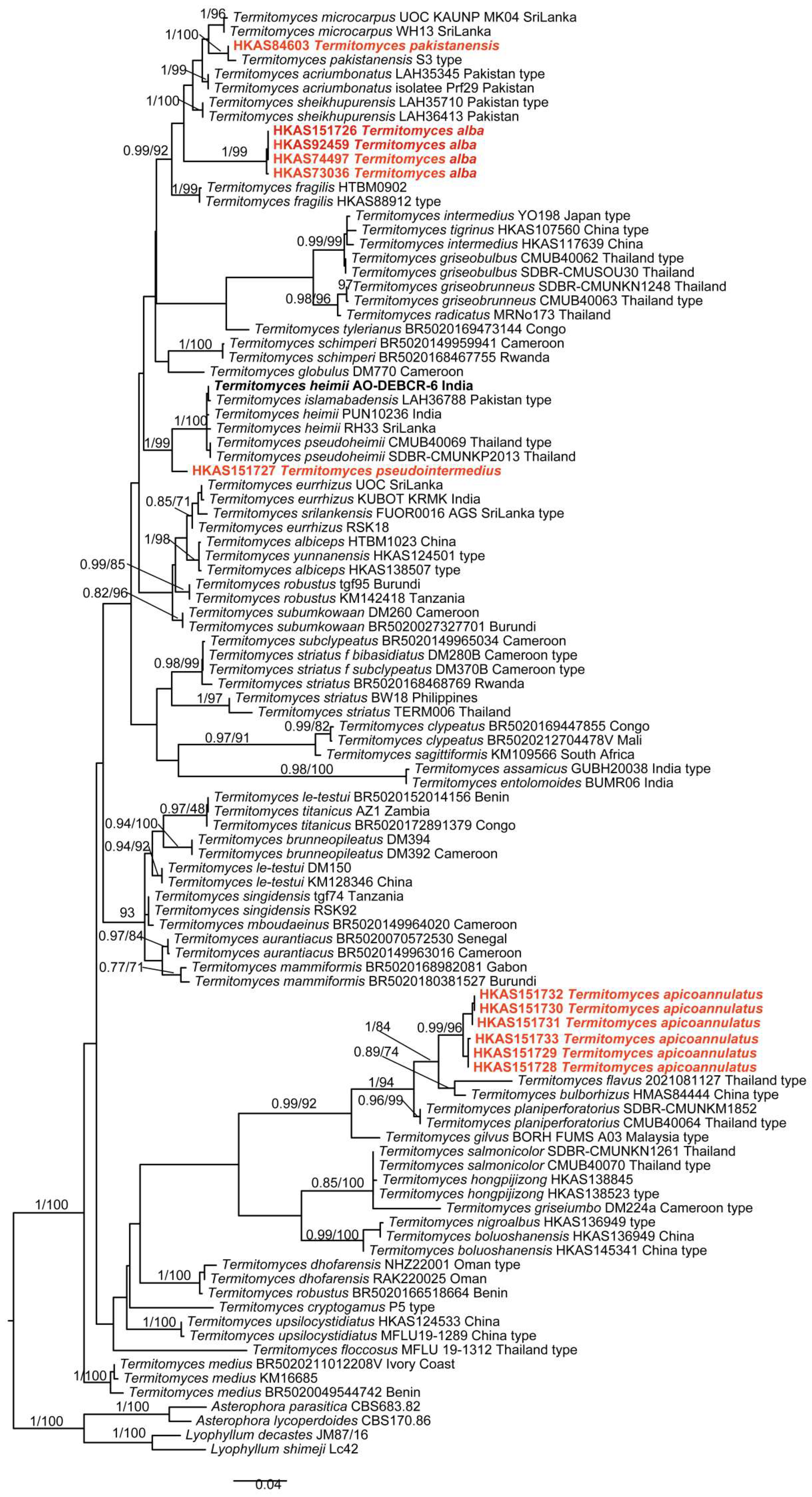
Phylogenetic analyses were conducted based on 80 ITS sequences, 62 nrLSU sequences and 47 mtSSU sequences, of which 12 ITS sequences, nine nrLSU sequences and nine mtSSU sequences were newly generated in this study (Table 1). Lyophyllum shimeji (Kawam.) Hongo, L. decastes (Fr.) Singer, Asterophora lycoperdoides (Bull.) Ditmar, and A. parasitica (Bull.) Singer were used as the outgroup taxa [6]. The concatenated dataset consisted of 2657 bp, including gaps, and contained 704 parsimony-informative sites. The breakdown by individual marker was as follows: the ITS fragment was 1457 bp with 403 parsimony-informative sites; the nrLSU fragment measured 821 bp with 236 parsimony-informative sites; and the mtSSU fragment was 379 bp with 82 parsimony-informative sites. The combined alignment was submitted to TreeBASE (S30371). ML and BI approaches showed minimal differences in their evaluation results; thus, only the ML tree was used for display (Figure 1). Our phylogenetic analyses indicated that sequences of the new species T. albus form a distinct lineage with high support values (1/100) and cluster together with T. acriumbonatus Usman & Khalid, T. sheikhupurensis Izhar, Khalid & H. Bashir, T. microcarpus (Berk. & Broome) R. Heim and T. pakistanensis A. Razaq. The sequence of the new species T. pseudointermedius clusters together with those of T. heimii Natarajan T. islamabadensis S. Ashraf, Usman & Khalid, and T. pseudoheimii Paloi, Suwannar. & Kuml with high support values (1/99). The new species T. apicoannulatus is closely related and clusters together with T. flavus S.M. Tang & S.H. Li and T. bulborhizus T.Z. Wei, Y.J. Yao, Bo Wang & Pegler with high support values (1/84).

3.2. Taxonomy

Termitomyces albus Yan C. Li, Wen J. Wu, sp. nov. (Figure 2 and Figure 3)
MycoBank: 862706.
Etymology: “Albus” refers to the white basidiomata of this species.
Type: China, Yunnan Province: Pu’er, Sun-River National Park, 22.59°N, 101.11°E, altitude 1430 masl, 20 September 2025, Wen-Jing Wu 018 (KUN-HKAS151726, GenBank Acc. No. ITS: PX924552, nrLSU: PX916413, mtSSU: PX924563).
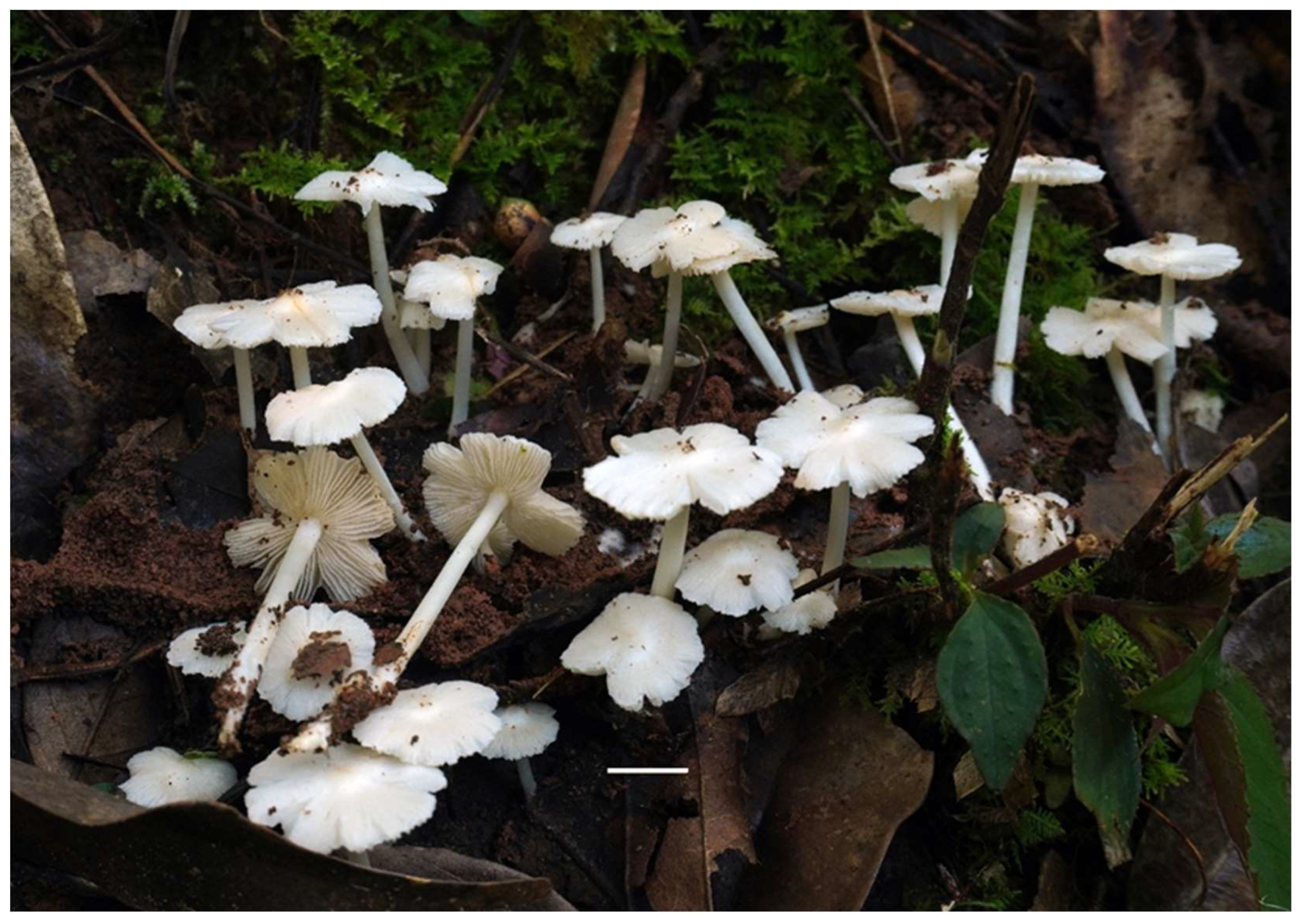
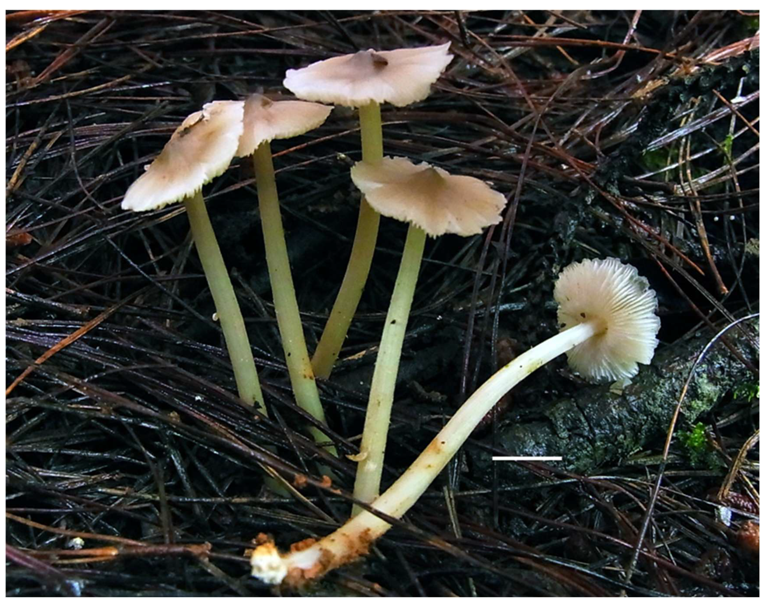
Figure 2. Fresh basidiomata of Termitomyces albus. Bar = 10 mm (KUN-HKAS151726, holotype).
Jof 12 00385 g002
Diagnosis: Termitomyces albus is distinguished from other species of this genus by its small, entirely white basidiomata; applanate pileus with a blunt perforatorium; fistulose, frangible stipe; elongated basidiospores; and thick-walled cheilo- and pleurocystidia.
Description: Basidiomata very small to small. Pileus 0.8–2.5 cm in diam., applanate, with a blunt perforatorium at centre; surface white (1A1), margin more or less radially splitting; context white (1A1), unchanging in colour when bruised. Lamellae free, dense, white (1A1), with small lamellulae, margin serrate. Stipe 2–4.3 × 0.1–0.25 cm, central, cylindrical, white (1A1), surface smooth, fistulose and frangible; context white, fibrous. Annulus absent. Pseudorhiza absent or present; when present, with a ceramic white (1A1) surface, connected to subterranean termite nests. Odour indistinct.
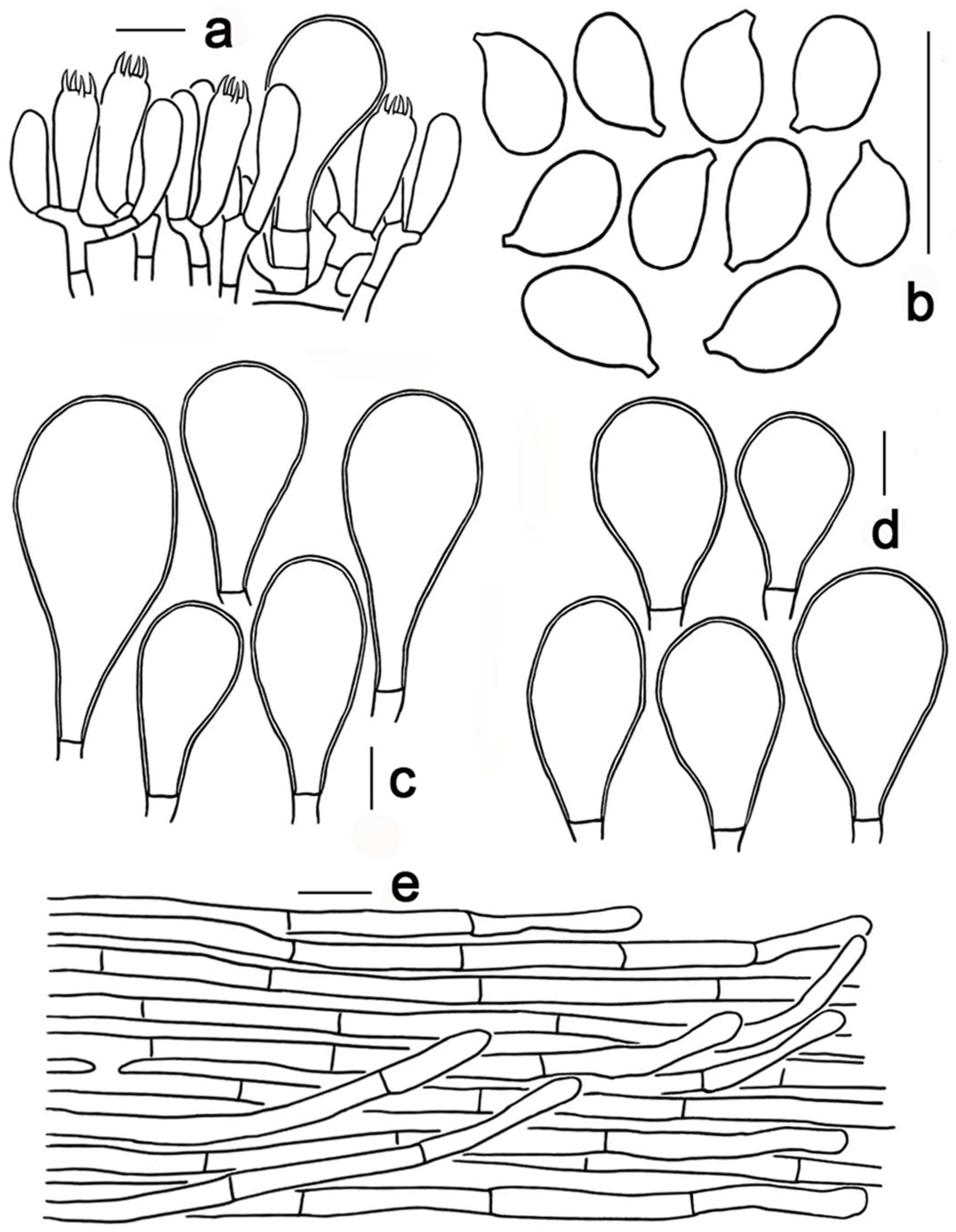
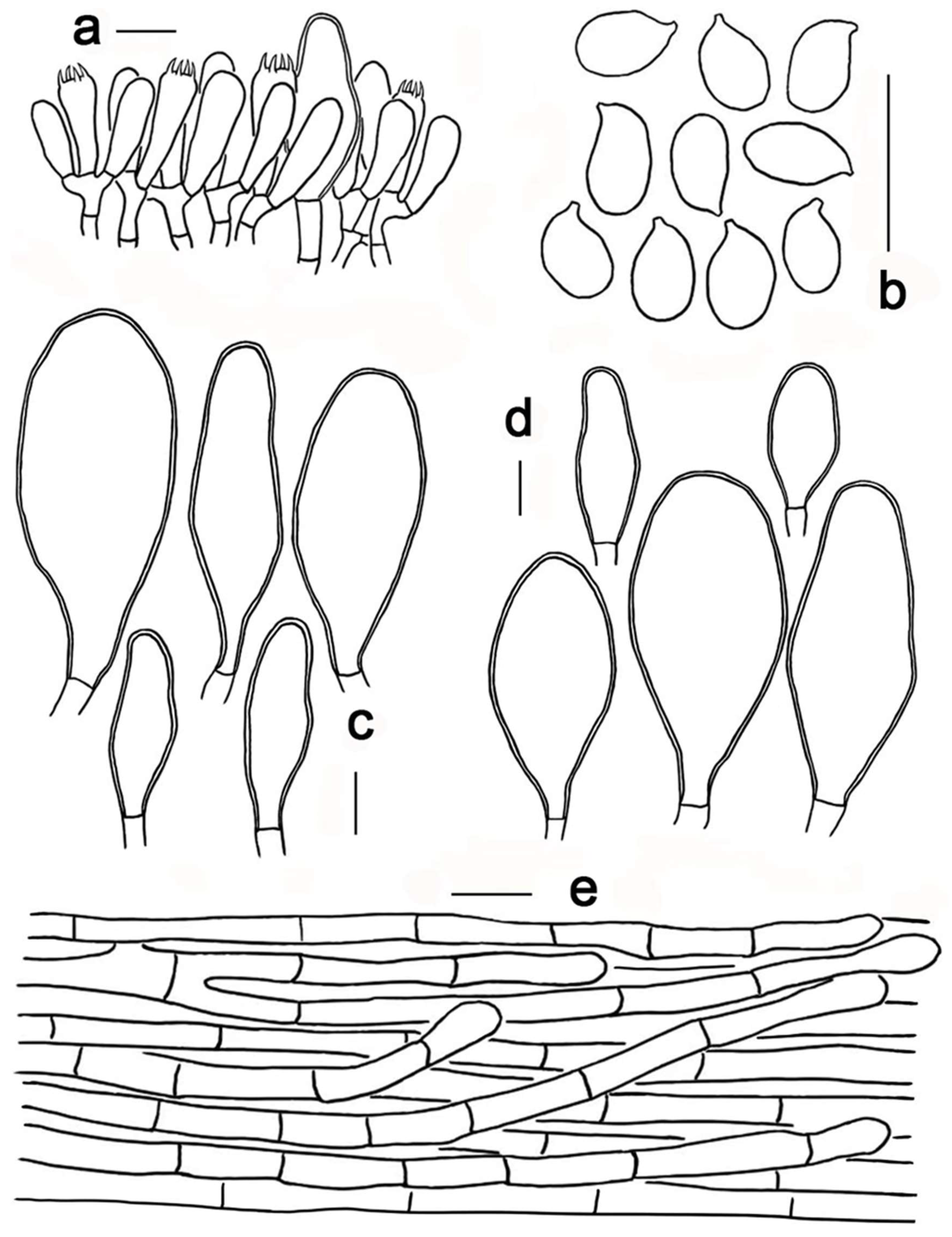
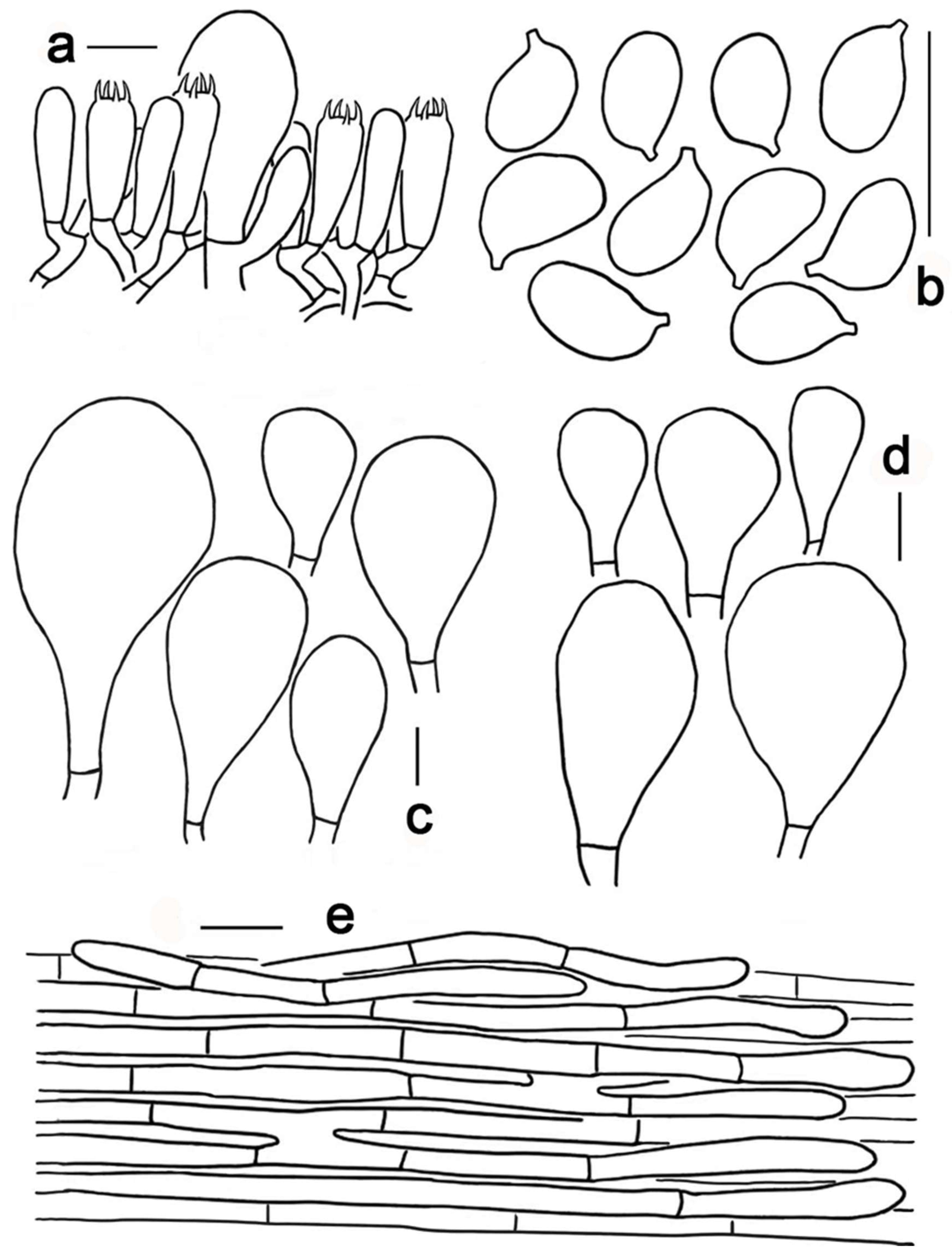
Figure 3. Termitomyces albus (KUN-HKAS151726, holotype). (a) Basidia, basidioles and pleurocystidium; (b) basidiospores; (c) cheilocystidia; (d) pleurocystidia; (e) pileipellis. Scale bars: (ae) = 10 µm.
Jof 12 00385 g003
Basidia 18–26 × 7–10 μm, clavate, mostly four-spored, rarely two-spored, thin-walled, nearly hyaline in 10% KOH. Basidiospores [200/10/4] 6–7.5 (–8) × 3–4.5 (–5) μm, sterigmata 2–4 μm long, Lm × Wm = 6.8 ± 0.5 × 3.9 ± 0.4 μm, Q = 1.6–1.8, Qm = 1.74 ± 0.22, elongated, thin-walled, smooth, nearly hyaline in 10% KOH, non-amyloid, non-dextrinoid. Cheilocystidia abundant, 28–60 × 17–28 μm, thick-walled (1–2 μm thick), clavate, utriform or pyriform, nearly hyaline in KOH, scattered. Pleurocystidia abundant, 20–42 × 17–26 μm, similar to cheilocystidia in shape, thick-walled (1–2 μm thick), nearly hyaline in KOH. Pileipellis a cutis, composed of radially arranged interwoven to subparallel hyphae; terminal cells 18–47 × 2–5 μm, clavate to cylindrical. Clamp connections absent in all tissues.
Habitat: Gregarious on the ground above the subterranean termitarium.
Distribution: Currently known from Yunnan Province, China.
Additional specimens examined: China, Yunnan Province: Wenshan Prefecture, Qiubei County, Zhongxiang Village, 8 August 2015, Zhao Kuan 829 (KUN-HKAS92459, GenBank Acc. No. ITS: PX924555, nrLSU: PX916414, mtSSU: PX924564); Nujiang Lisu Autonomous Prefecture, Lushui City, Laowo Town, Chongren Village, 7 August 2011, Yuan Mingsheng 1017 (KUN-HKAS74497, GenBank Acc. No. ITS: PX924554); Dali Bai Autonomous Prefecture, Binchuan County, Jizushan Town, Nanshan, 1 September 2011, Yuan Mingsheng BC-74 (KUN-HKAS73036, GenBank Acc. No. ITS: PX924553, mtSSU: PX924565).
Notes: Termitomyces albus is characterized by its small, entirely white basidiomata; applanate pileus with a blunt central perforatorium; white, cylindrical, fistulose, frangible stipe; absent pseudorhiza; elongated basidiospores, and thick-walled cheilo- and pleurocystidia.
The differences between T. albus and several related small-sized Termitomyces species, i.e., T. fragilis L. Ye, Karun., J.C. Xu, K.D. Hyde & P.E. Mortimer, T. acriumbonatus, T. sheikhupurensis, T. microcarpus and T. pakistanensis, are as follows: T. fragilis has a brownish grey to greyish white pileus which typically exceeds 3 cm in diam., surface mixed with tiny white filamentous striations, and obtusely pointed perforatorium [4]. Termitomyces acriumbonatus has a creamy white pileus with slightly greyish striations, a greyish brown pointed perforatorium, subglobose to ellipsoid basidiospores measuring 6.1–8.7 × 4.5–6.5 μm, and distinct pseudorhiza [25]. Termitomyces microcarpus has a relatively large pileus (up to 5 cm in diam.) which is pale grey or with a faint ochraceous tinge at the centre and white or cream-coloured towards the margin, a papilla perforatorium, a solid stipe, a white pseudorhiza, ovoid to ellipsoid basidiospores measuring 5.6–6.9 × 3.7–4.8 μm, and thin-walled pleuro- and cheilocystidia [2]. Termitomyces pakistanensis has a cream pileus with camel brown fibrillose, olive grey to brown stipe, and ellipsoid to ovoid basidiospores measuring 4.1–8.8 × 3.7–6.1 μm [50]. Termitomyces sheikhupurensis has a light brownish grey to dull orange pileus with a conspicuous nipple-shaped perforatorium, surface radially mixed with white sulcate striations; a pale yellow to light brownish grey solid stipe; subelliptic to ellipsoid basidiospores measuring 5.5–8.07 × 4.4–6.13 μm; and thin-walled, polymorphic cheilo- and pleurocystidia [5]. The interspecific genetic distances between T. albus and the above species are as follows: T. acriumbonatus (0.116), T. sheikhupurensis (0.109), T. microcarpus (0.099), T. pakistanensis (0.138), and T. fragilis (0.129).
Termitomyces apicoannulatus Yan C. Li, Wen J. Wu, sp. nov. (Figure 4 and Figure 5)
MycoBank: 862708.
Etymology: “Apicoannulatus” highlights the key diagnostic feature: the apical annulus.
Type: China, Guangxi Province: Nanning City, Shanglin County, Xiangxian Town, Liulian Village, Guangxi Daming Mountain National Nature Reserve, 23.29 N, 108.60 E, altitude 230 masl, 7 August 2025, Yan-Chun Li 8384 (KUN-HKAS151733, GenBank Acc. No. ITS: PX924559, nrLSU: PX916418, mtSSU: PX924571).
Diagnosis: Termitomyces apicoannulatus is distinguished from other species of this genus by its brownish yellow to orange-white pileus always with a blunt perforatorium, the glabrous pileal surface always covered with detachable white tomentum when young; a pale orange stipe covered with white fibrillose scales; the presence of an apical, often evanescent annulus; a brown pseudorhiza; elongated basidiospores; and thick-walled cheilo- and pleurocystidia.
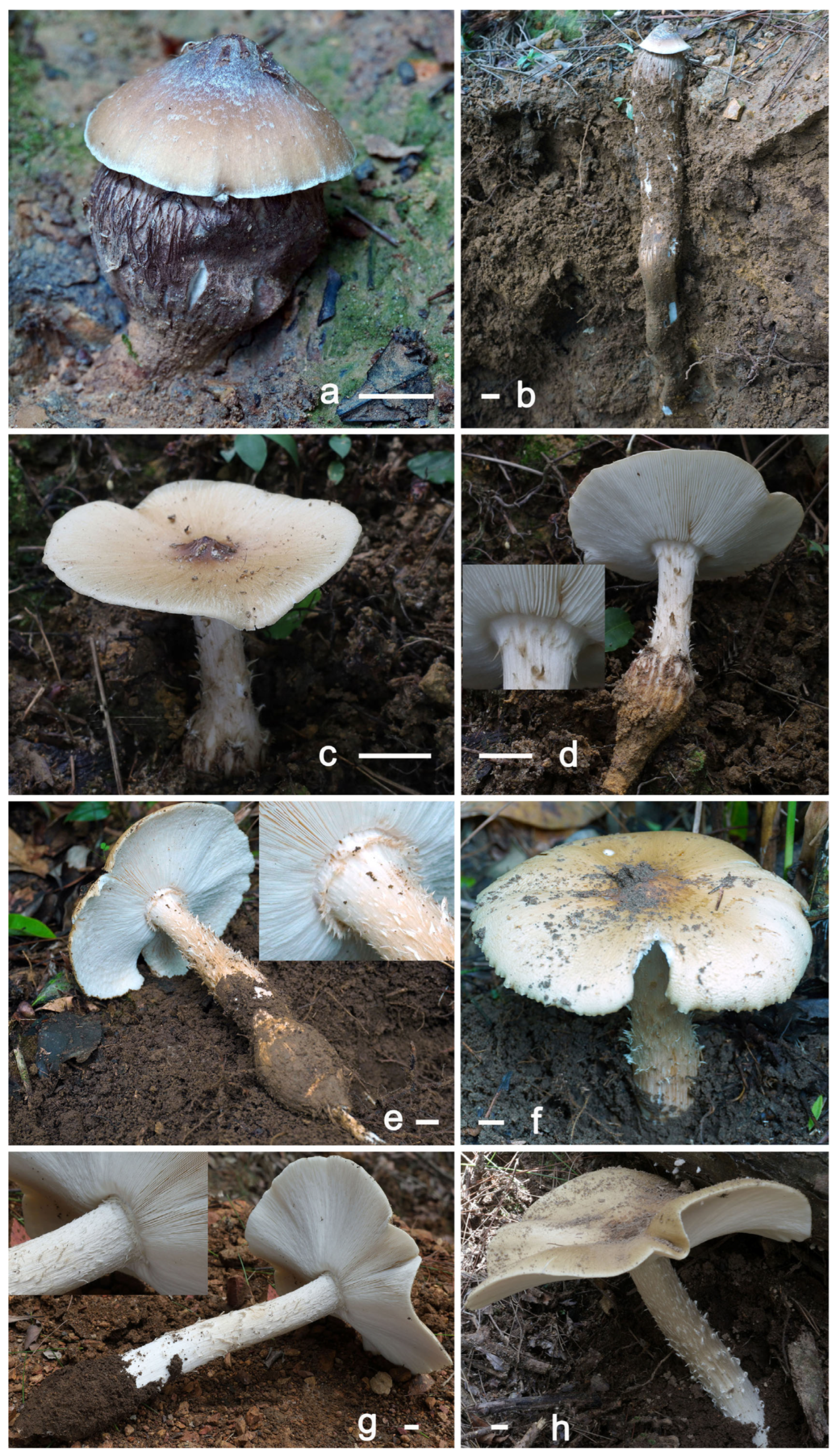
Description: Basidiomata small to medium-sized. Pileus 3.2–23.4 cm in diam., conical, covered with easily detachable white tomentum when young; becoming applanate at maturity with a blunt perforatorium at centre; surface brownish orange or brownish yellow (5C6–5C8) to light brown or dark brown (6D8–6F8) at centre, remaining parts greyish orange, light brown, orange-white to yellowish white (6B4–6D6, 5A2–5B4, 2A2–4A2), gradually paler towards margin; margin involute; context white (1A1), unchanging in colour when bruised. Lamellae free, dense, white (1A1), with small lamellulae, margin serrate. Stipe 3.3–20.4 × 0.6–2.5 cm, cylindrical, orange-white to pale orange (5A2–5A3), subcylindrical, solid, fibrous, surface densely to sparsely covered with white fibrillose scales, nearly uniform in width or noticeably swollen at base, surface brown (6E8); context white, fibrous. Annulus apical, often fragmented and disappearing when mature. Pseudorhiza present, connected to subterranean termite nests; surface brown (6E8); context solid, fibrous. Odour slightly fragrant.
Basidia 17–22 × 5–7 μm, clavate, mostly four-spored, rarely one-, two- or three-spored, thin-walled, nearly hyaline in 10% KOH. Basidiospores [60/3/3] 6–7 (–8) × 3–4 (–5) μm, sterigmata 2–4 μm, Lm × Wm = 6.8 ± 0.6 × 3.6± 0.6 μm, Q = 1.6–2.0, Qm = 1.89 ± 0.36, elongated, thin-walled, smooth, nearly hyaline in 10% KOH, non-amyloid, non-dextrinoid. Cheilocystidia abundant, 28–65 × 9–25 μm, thick-walled (1–2 μm thick), clavate to utriform, nearly hyaline in KOH, scattered. Pleurocystidia abundant, 25–57 × 10–26 μm, similar to cheilocystidia in shape, thick-walled (1–2 μm thick), nearly hyaline in KOH. Pileipellis a cutis, composed of radially arranged interwoven to subparallel hyphae; terminal cells 13–36 × 3–6 μm, clavate to subcylindrical. Clamp connections absent in all tissues.
Figure 4. Fresh basidiomata of Termitomyces apicoannulatus. Bars = 10 mm. ((a,b): HKAS151728; (c,d): KUN-HKAS151729; (e,f): KUN-HKAS151733, holotype; (g,h): KUN-HKAS151730).
Jof 12 00385 g004
Figure 5. Termitomyces apicoannulatus (KUN-HKAS151733, holotype). (a) Basidia, basidioles and pleurocystidium; (b) basidiospores; (c) cheilocystidia; (d) pleurocystidia; (e) pileipellis. Scale bars: (ae) = 10 µm.
Jof 12 00385 g005
Habitat: Solitary on the ground above the subterranean termitarium.
Distribution: Currently known from Guangxi Province, China.
Additional specimens examined: China, Guangxi Province: Nanning City, Shanglin County, Xiangxian Town, Liulian Village, Guangxi Daming Mountain National Nature Reserve, 23.29 N 108.60 E, altitude 230 masl, 7 August 2025, Yan-Chun Li 8381 (KUN-HKAS151731, GenBank Acc. No. ITS: PX924561, nrLSU: PX916420, mtSSU: PX924569); nearby this location, altitude 465 masl, 9 August 2025, Yan-Chun Li 8383 (KUN-HKAS151732, GenBank Acc. No. ITS: PX924562, nrLSU: PX916421, mtSSU: PX924570); Shanglin County, Dafeng Town, Dongchun Village, Guangxi Daming Mountain National Nature Reserve, 23.29 N 108.60 E, altitude 230 masl, 9 August 2025, Yan-Chun Li 8229 (KUN-HKAS151728, GenBank Acc. No. ITS: PX924557, nrLSU: PX916416); nearby this location, the same date, altitude 498 masl, Yan-Chun Li 8296 (KUN-HKAS151729, GenBank Acc. No. ITS: PX924558, nrLSU: PX916417, mtSSU: PX924567); Shanglin County, Mingliang Town, Luozhen Village, Guangxi Daming Mountain National Nature Reserve, 23.29 N 108.60 E, altitude 400 masl, 10 August 2025, Yan-Chun Li 8322 (KUN-HKAS151730, GenBank Acc. No. ITS: PX924560, nrLSU: PX916419, mtSSU: PX924568).
Notes: Termitomyces apicoannulatus is characterized by a brownish yellow to orange-white pileus with a blunt perforatorium; a pale orange stipe covered with white fibrillose scales; an apical, often evanescent annulus; a brown pseudorhiza; elongated basidiospores; and thick-walled cheilo- and pleurocystidia.
In our phylogenetic analysis, T. apicoannulatus clustered together with T. bulborhizus (interspecific genetic distance 0.085), T. flavus (interspecific genetic distance 0.310), T. gilvus C.S. Yee & Seelan (interspecific genetic distance 0.079) and T. planiperforatorius Paloi & Suwannar (interspecific genetic distance 0.064). However, T. bulborhizus has a brown to pale brown pileus, whitish or fulvous brown stipe, prominent globose bulbous stipe base, absent annulus, and ovoid to ellipsoid basidiospores measuring 6.0–9.0 × 4.0–6.0 μm [28]. Termitomyces flavus has a large pileus that can reach 40 cm wide, a brownish orange to brownish yellow pileal surface, a whitish or fulvous brown stipe, absent annulus, and ellipsoid basidiospores measuring 6.0–8.8 × 4.1–6.2 μm [26]. Termitomyces gilvus has a brownish orange pileus, clavate and relatively large basidia (21.8–29.2 × 6.1–8.3 μm), and thin-walled cheilo- and pleurocystidia [24]. Termitomyces planiperforatorius has a greyish orange to light brown pileus with a slightly round to flat perforatorium, a cracked pileal surface with filamentous squamules when mature, and thin-walled cheilo- and pleurocystidia [51].
Termitomyces pakistanensis A. Razaq, in Razaq, Ishaq, Ilyas & Sadia, Microsc. Res. Tech. 86(1): 117 (2023). (Figure 6 and Figure 7)
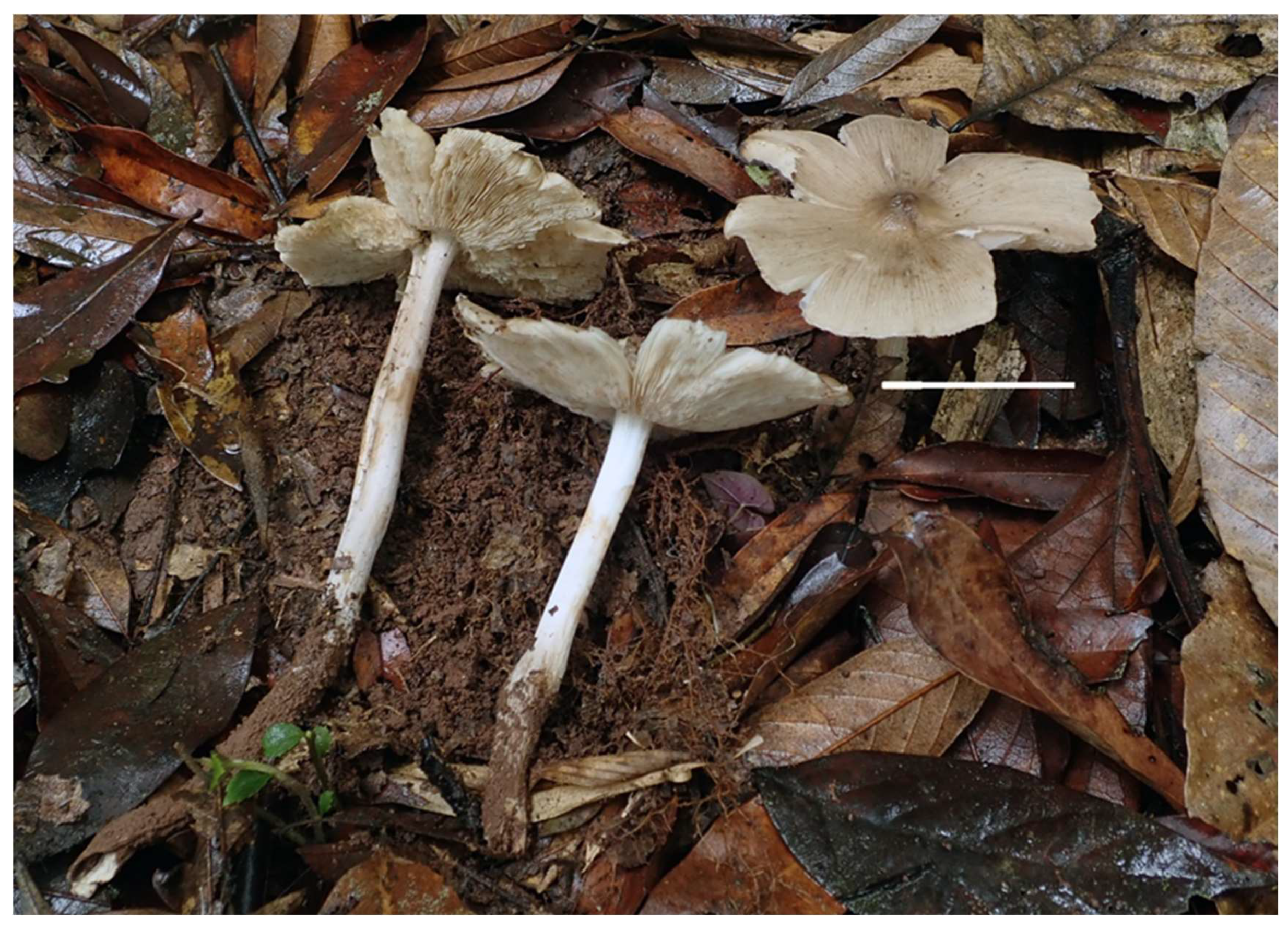
Basidiomata small. Pileus 1–2.4 cm in diam., initially conical to parabolic, becoming applanate at maturity, with a blunt perforatorium at centre; surface olive brown to yellowish brown (4E8, 5E7–5E8), brownish orange to orange-white to white (6C5–6C4, 6A2–6A1) elsewhere, gradually lightening from the centre toward the margin; margin more or less radially splitting; context white (1A1), unchanging in colour when bruised. Lamellae free, moderately spaced, white (1A1), with small lamellulae, margin smooth to serrate. Stipe 5–7.5 × 0.2–0.3 cm, central, cylindrical, white (1A1), surface smooth, fistulose and frangible. Annulus absent. Pseudorhiza absent, connected to subterranean termite nests. Odour indistinct.
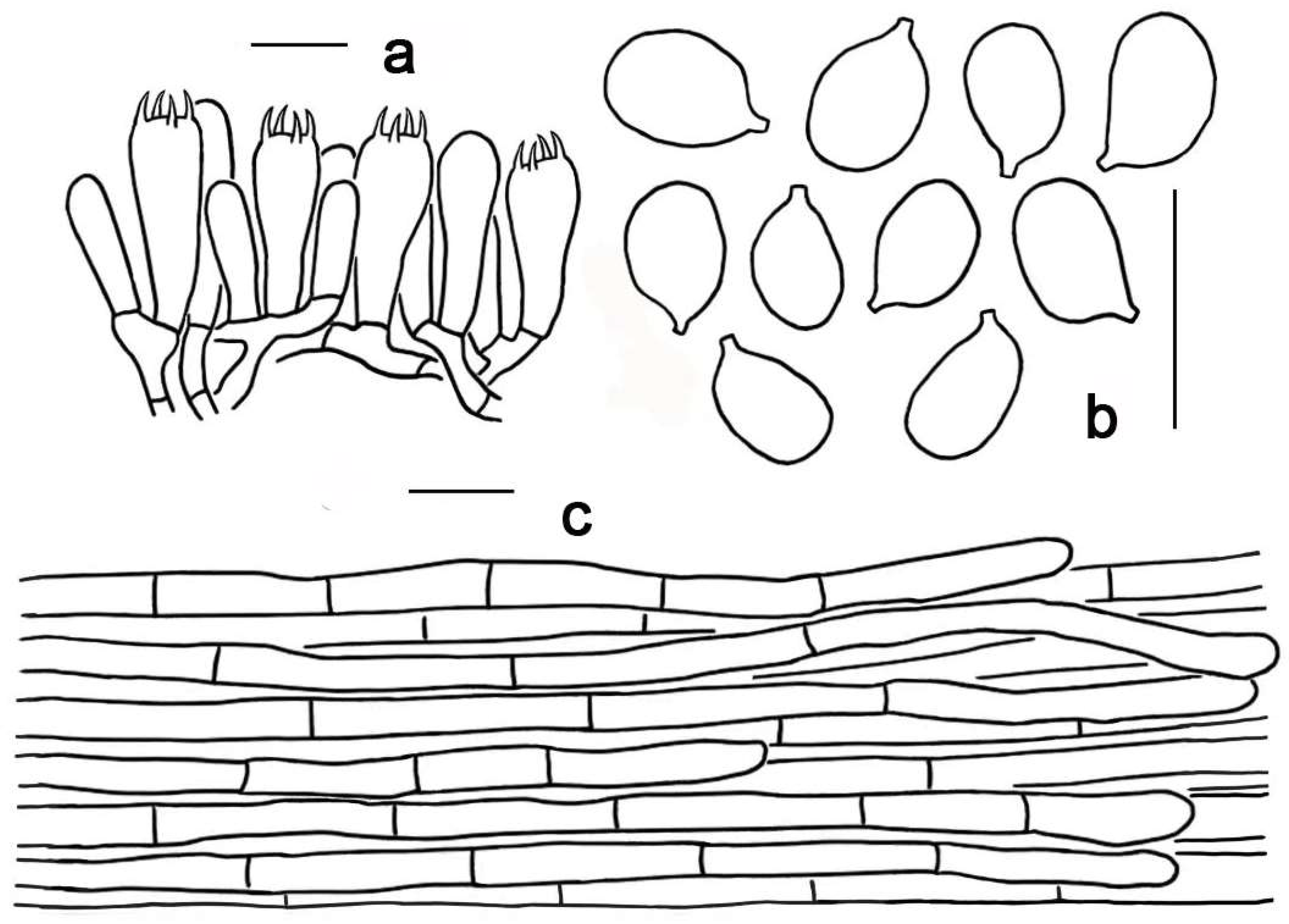
Basidia 20–24 × 5–7 μm, clavate, mostly four-spored, rarely one-, two- or three-spored, thin-walled, nearly hyaline in 10% KOH. Basidiospores [120/6/1] 6–7 (–7.5) × 4–5 μm, sterigmata 2–4 μm, Lm × Wm = 6.0 ± 0.5 × 4.3 ± 0.5 μm, Q = 1.3–1.7, Qm = 1.40 ± 0.20, ellipsoid to elongated, thin-walled, smooth, nearly hyaline in 10% KOH, non-amyloid, non-dextrinoid. Cheilo- and pleurocystidia are not observed. Pileipellis a cutis, composed of radially arranged interwoven to subparallel hyphae; terminal cells 18–50 × 4–7 μm, clavate to subcylindrical. Clamp connections absent in all tissues.
Figure 6. Fresh basidiomata of Termitomyces pakistanensis. Bar = 10 mm (KUN-HKAS84603).
Jof 12 00385 g006
Figure 7. Termitomyces pakistanensis (KUN-HKAS84603). (a) Basidia, basidioles; (b) basidiospores; (c) pileipellis. Scale bars: (ac) = 10 µm.
Jof 12 00385 g007
Habitat: Gregarious on the ground above the subterranean termitarium.
Distribution: Currently known from Pakistan and China.
Specimen examined: China, Yunnan Province: Baoshan City, Longyang District, Lujiang Town, Bawan Village, Gaoligong Mountain near the Nankang Management Station, 17 June 2014, Li-Hong Han 307 (KUN-HKAS84603, GenBank Acc. No.ITS PX953039).
Notes: The ITS sequence from the Chinese collection is almost identical to that of T. pakistanensis from the type specimen (genetic distance 0.007), suggesting that they are conspecific. Termitomyces pakistanensis is originally characterized by its cream pileus with camel brown fibrillose squamules; a long, slender, hollow stipe; absent annulus and pseudorhiza; moderately small, ellipsoid to ovoid basidiospores; and clavate cheilo- and pleurocystidia [50]. However, the Chinese specimen has an olive brown to brownish orange pileus, a smooth pileal surface, and an absence of cheilo- and pleurocystidia. These traits supplement the characteristics of this species.
Termitomyces pseudointermedius Yan C. Li, Wen J. Wu, sp. nov. (Figure 8 and Figure 9)
MycoBank: 862707.
Etymology: “Pseudointermedius” refers to this species being morphologically similar to T. intermedius.
Type: China, Yunnan Province: Pu’er City, Jiangcheng County, Menglie Town, 22.31° N, 101.53° E, altitude 1047 masl, 30 June 2019, Si-Peng Jian 633 (KUN-HKAS151727, GenBank Acc. No. ITS: PX924556, nrLSU: PX916415, mtSSU: PX924566).
Figure 8. Fresh basidiomata of Termitomyces pseudointermedius. Bar = 50 mm (KUN-HKAS151727, holotype).
Jof 12 00385 g008
Diagnosis: Termitomyces pseudointermedius is distinguished from other species of this genus by a grey to greyish beige pileus, with a blunt and greyish brown to brown perforatorium at the centre, and concolorous sulcate striations towards the margin; a white, solid, smooth stipe; greyish white pseudorhiza; ellipsoid to elongated basidiospores; and thin-walled cheilo- and pleurocystidia.
Description: Basidiomata medium-sized. Pileus 9.5–10.5 cm in diam., applanate, with a blunt perforatorium at centre; surface greyish brown to brown (5E3–5F4) at centre, grey to greyish beige (4B1–4C2) elsewhere, gradually paler towards margin; radially mixed with concolorous sulcate striations, margin more or less radially splitting; context white (1A1), unchanging in colour when bruised. Lamellae free, dense, white (1A1), with small lamellulae, margin smooth to serrate. Stipe 13–19 × 0.8–1 cm, cylindrical, white (1A1), solid, surface nearly glabrous; context white, fibrous. Annulus absent. Pseudorhiza present, connected to subterranean termite nests; greyish white (1B1); context solid, fibrous. Odour slightly fragrant.
Basidia 17–22 × 6–8 μm, clavate, mostly four-spored, rarely one-, two- or three-spored, thin-walled, nearly hyaline in 10% KOH. Basidiospores [60/3/1] (6–) 7–8 × 4–5 μm, sterigmata 2–4 μm, Lm × Wm = 7.5 ± 0.4 × 4.4 ± 0.5 μm, Q = 1.5–1.8, Qm = 1.70 ± 0.21, ellipsoid to elongated, thin-walled, smooth, nearly hyaline in 10% KOH, non-amyloid, non-dextrinoid. Cheilocystidia seldom, 26–68 × 17–32 μm, thick-walled (1–2 μm thick), clavate, utriform or pyriform, nearly hyaline in KOH, scattered. Pleurocystidia abundant, 22–41 × 10–25 μm, similar to cheilocystidia in shape, thick-walled (1–2 μm), nearly hyaline in KOH. Pileipellis a cutis, composed of radially arranged interwoven to subparallel hyphae; terminal cells 18–35 × 4–5 μm, subclavate to subcylindrical. Clamp connections absent in all tissues.
Figure 9. Termitomyces pseudointermedius (KUN-HKAS151727, holotype). (a) Basidia, basidioles and pleurocystidium; (b) basidiospores; (c) cheilocystidia; (d) pleurocystidia; (e) pileipellis. Scale bars: (ae) = 10 µm.
Jof 12 00385 g009
Habitat: Scattered on the ground above the subterranean termitarium.
Distribution: Currently known from Yunnan Province, China.
Notes: Termitomyces pseudointermedius is characterized by a greyish beige pileus with a blunt perforatorium at centre and concolorous radially arranged sulcate striations at the margin, a white stipe, a greyish white pseudorhiza, ellipsoid to elongated basidiospores, and thin-walled cheilo- and pleurocystidia.
In our phylogenetic analyses, this species clustered with another clade consisting of three taxa: T. heimii, T. islamabadensis, and T. pseudoheimii. However, T. islamabadensis and T. pseudoheimii showed no significant morphological differences from T. heimii [21,51,62]. Moreover, the clade contained sequences of T. heimii from its type location: India. Therefore, this clade represents only a single species, i.e., T. heimii. There are significant morphological differences between T. pseudointermedius and T. heimii. Termitomyces heimii has a white pileus with a blunt and white perforatorium, a thick annulus, and ellipsoid basidiospores measuring 7–8.4 × 5.5–7 μm [62]. Morphologically, T. pseudointermedius resembles T. intermedius Har. Takah. & Taneyama in sharing a greyish beige, similarly sized and coloured pileus and stipe. However, T. intermedius has a white pseudorhiza; sparse to abundant, thin-walled cheilocystidia which are polymorphic and occasionally bicellular; and abundant, thin-walled, clavate pleurocystidia. Moreover, they are highly divergent in their ITS and mtSSU sequences [2,6].

4. Discussion

To date, approximately 71 species of Termitomyces have been reported from Asia and Africa [19,23,29,63]. Their notable nutritional and medicinal properties make them a sought-after food commodity [15,30]. In China, a total of 20 species of Termitomyces have been previously reported. However, T. cylindricus S.C. He and T. albiceps S.C. He (originally described from Guizhou Province by He in 1986) were initially synonymized with other Chinese species due to morphological similarities but later were revalidated as distinct species [30]. Meanwhile, another two recently described species [6]—T. tigrinus S.M. Tang & Raspé and T. yunnanensis S.M. Tang & Raspé—were treated as synonyms of T. intermedius and T. cylindricus, respectively [30]. Additionally, six species—T. le-testui (R. Heim) R. Heim, T. entolomoides R. Heim, T. eurrhizus (Berk.) R. Heim, T. mammiformis R.Heim, T. microcarpus (Berk & Broome) R. Heim, and T. tylerianus Otieno—lacking voucher specimens and molecular data [12,27,29,64,65,,66], have been temporarily excluded from the list of Chinese species. Thus, only 14 species have been confirmed to distribute in China [4,6,16,17,19,30,31,67] (Table 2).
In this study, three newly described species, T. albus, T. apicoannulatus, and T. pseudointermedius, and a species new to China, i.e., T. pakistanensis, are reported from China. These species are mainly distributed in the southwestern (e.g., Yunnan Province) and southern (e.g., Guangxi Province) regions of China. Southwestern and southern China have a subtropical to tropical monsoon climate, characterized by year-round warm and humid conditions with abundant rainfall. These provide long-term stable natural conditions for the survival and activity of termites. Simultaneously, the rich variety of vegetation types and complex ecosystems may further promote the differentiation of termite species, thereby driving the co-evolution and diversity accumulation of their symbiotic Termitomyces species. Thus, these areas are likely to harbour a high species diversity of Termitomyces, suggesting that they may represent important distribution and evolutionary centres for this group of fungi.

Author Contributions

Conceptualization, Y.-C.L. and B.X.; Field Sampling, Y.-C.L., W.-J.W., J.-C.Y., H.-L.Z., Q.X., and G.-R.Z.; Molecular Experiments and Data Analysis, W.-J.W. and J.-C.Y.; Morphological Observation, Y.-C.L., W.-J.W., and J.-C.Y.; Writing—Original Draft Preparation, W.-J.W.; Writing—Review and Editing, Y.-C.L. and B.X. All authors have read and agreed to the published version of the manuscript.

Funding

This research was funded by the 2025 National Nature Reserve Project under the Central Financial Forestry and Grassland Ecological Protection and Restoration Fund (NNZC2025-G3-990471-ZCZB) and the Yunnan Xingdian Talents Support Plan—Science and Technology Leading Talents Program (202305AB350004).

Institutional Review Board Statement

Not applicable.

Informed Consent Statement

Not applicable.

Data Availability Statement

The original contributions presented in this study are included in the article. Further inquiries can be directed to the corresponding author.

Acknowledgments

The authors appreciate the anonymous reviewers for their valuable comments and suggestions. We express our gratitude to Si-Peng Jian (Lanzhou University) and Li-Hong Han (Kunming Institute of Botany, CAS) for providing related specimens. We also extend our sincere gratitude to Rong-Jin Hunag, Guang-Dao Wei, Wei-Zhong Wu and Tan-Zhuang Wei (Administrative Bureau of Damingshan Nature Reserve of Guangxi) for providing invaluable help during the fieldwork and specimen collection.

Conflicts of Interest

The authors declare no conflicts of interest.

References

  1. Heim, R. Les champignons des termitières: Nouveaux aspects d’un problème de biologieet de systématique générales. Rev. Sci. 1942, 80, 69–86. [Google Scholar]
  2. Heim, R. Termites et champignons. In Les Champignons Termitophiles d’Afrique Noire et d’Asie Méridionale; Ed Boubée: Paris, France, 1977; pp. 1–205. [Google Scholar]
  3. Frøslev, T.G.; Aanen, D.K.; Laessøe, T.; Rosendahl, S. Phylogenetic relationships of Termitomyces and related taxa. Mycol. Res. 2003, 107, 1277–1286. [Google Scholar] [CrossRef]
  4. Ye, L.; Karunarathna, S.C.; Li, H.; Xu, J.; Hyde, K.D.; Mortimer, P.E. A survey of Termitomyces (Lyophyllaceae, Agaricales), including a new species, from a subtropical forest in Xishuangbanna, China. Mycobiology 2019, 47, 391–400. [Google Scholar] [CrossRef]
  5. Izhar, A.; Khalid, A.N.; Bashir, H. Termitomyces sheikhupurensis sp. nov. (Lyophyllaceae, Agaricales) from Pakistan, evidence from morphology and DNA sequences data. Turk. J. Bot. 2020, 44, 694–704. [Google Scholar] [CrossRef]
  6. Tang, S.M.; Vadthanarat, S.; He, J.; Raghoonundon, B.; Yu, F.M.; Karunarathna, S.C.; Li, S.H.; Raspé, O. Morphological and molecular analyses reveal two new species of Termitomyces (Agaricales, Lyophyllaceae) and morphological variability of T. intermedius. MycoKeys 2023, 95, 61–82. [Google Scholar] [CrossRef]
  7. Pegler, D.N.; Vanhaecke, M. Termitomyces of Southeast Asia. Kew Bull. 1994, 49, 717. [Google Scholar] [CrossRef]
  8. Wei, T.Z. Systematics of the genus Termitomyces in China. Ph.D. Thesis, Institute of Microbiology, Chinese Academy of Sciences, Beijing, China, 2004. [Google Scholar]
  9. Aanen, D.K.; Eggleton, P. Fungus-growing termites originated in African rain forest. Curr. Biol. 2005, 15, 851–855. [Google Scholar] [CrossRef] [PubMed]
  10. van de Peppel, L.J.J.; Nieuwenhuis, M.; Auxier, B.; Grum-Grzhimaylo, A.A.; Cárdenas, M.E.; de Beer, Z.W.; Lodge, D.J.; Smith, M.E.; Kuyper, T.W.; Franco-Molano, A.E.; et al. Ancestral predisposition toward a domesticated lifestyle in the termite-cultivated fungus Termitomyces. Curr. Biol. 2021, 31, 4413–4421. [Google Scholar] [CrossRef] [PubMed]
  11. Parent, G.; Thoen, D. Food Value of Edible Mushrooms from Upper-Shaba Region. Econ. Bot. 1977, 31, 436–445. [Google Scholar] [CrossRef]
  12. Yang, Z.L.; Shuai, J.G. Termitomyces in Xishuangbana of Yunnan, China. Edible Fungi 1990, 12, 2. [Google Scholar]
  13. Yang, Z.L. A delicious tropical mushroom–Termitomyces heimii occurring in Yunnan, China. Edible Fungi 1990, 9, 28. [Google Scholar]
  14. Chandra, K.; Ghosh, K.; Roy, S.K.; Mondal, S.; Maiti, D.; Ojha, A.K.; Das, D.; Mondal, S.; Islam, S.S. A water-soluble glucan isolated from an edible mushroom Termitomyces microcarpus. Carbohydr. Res. 2007, 342, 2484–2489. [Google Scholar] [CrossRef]
  15. Maity, P.; Nandi, A.K.; Pattanayak, M.; Manna, D.K.; Sen, I.K.; Chakraborty, I.; Bhanja, S.K.; Sahoo, A.K.; Gupta, N.; Islam, S.S. Structural characterization of a heteroglycan from an edible mushroom Termitomyces heimii. Int. J. Biol. Macromol. 2020, 151, 305–311. [Google Scholar] [CrossRef] [PubMed]
  16. Tang, S.M.; He, M.Q.; Raspé, O.; Luo, X.; Zhang, X.L.; Li, Y.J.; Su, K.M.; Li, S.H.; Thongklang, N.; Hyde, K.D. Two new species of Termitomyces (Agaricales, Lyophyllaceae) from China and Thailand. Phytotaxa 2020, 439, 231–242. [Google Scholar] [CrossRef]
  17. Paloi, S.; Kumla, J.; Paloi, B.P.; Srinuanpan, S.; Hoijang, S.; Karunarathna, S.C.; Acharya, K.; Suwannarach, N.; Lumyong, S. Termite Mushrooms (Termitomyces), a Potential Source of Nutrients and Bioactive Compounds Exhibiting Human Health Benefits: A Review. J. Fungi 2023, 9, 112. [Google Scholar] [CrossRef] [PubMed]
  18. Hussain, S.; Al-Kharousi, M.; Al-Owaisi, A.A.; Al-Maqbali, D.; Al-Muharabi, M.A.; Al-Shabibi, Z.; Al-Balushi, A.H.; Saady, N.A.; Velazhahan, R.; Rashan, L.; et al. The genus Termitomyces: Outline, phylogeny, and divergence times estimation with description of a new edible species from Arabian Peninsula. Sydowia 2023, 76, 185–198. [Google Scholar] [CrossRef]
  19. Mossebo, D.C.; Essouman, E.P.F.; Machouart, M.C.; Gueidan, C. Phylogenetic relationships, taxonomic revision and new taxa of Termitomyces (Lyophyllaceae, Basidiomycota) inferred from combined nLSU- and mtSSU-rDNA sequences. Phytotaxa 2017, 321, 71. [Google Scholar] [CrossRef]
  20. Huang, Q.J.; Bau, T.; Zhang, M.; Zhou, W.; Wang, C.Q.; Li, T.H. Termitomyces intermedius: A mush-room species distributed in south subtropics and southern edge of temperate zone. Acta Edulis Fungi 2017, 24, 74–78. [Google Scholar]
  21. Ashraf, S.; Usman, M.; Khalid, A.N. Termitomyces islamabadensis sp. nov. (Lyophyllaceae, Agaricales) from the Foothills of the Pakistani Himalayas. Biol. Bull. 2022, 49, S66–S76. [Google Scholar] [CrossRef]
  22. Das, L.R.; Chattopadhyay, P.; Dutta, A.K.; Narzary, D.; Rana, T.S. Termitomyces assamicus (Lyophyllaceae)—A new species of Termitomyces from India. Phytotaxa 2023, 599, 126–136. [Google Scholar] [CrossRef]
  23. Jannual, N.; Nipitwattanaphon, M.; Hasin, S.; Kaewgrajang, T. Morphological and molecular characterization of Termitomyces (Lyophyllaceae, Agaricales) in Thailand. Biodiversitas 2020, 21, 2481–2491. [Google Scholar] [CrossRef]
  24. Sathiya Seelan, J.S.; Shu Yee, C.; She Fui, F.; Dawood, M.; Tan, Y.S.; Kim, M.J.; Park, M.S.; Lim, Y.W. New Species of Termitomyces (Lyophyllaceae, Basidiomycota) from Sabah (Northern Borneo), Malaysia. Mycobiology 2020, 48, 95–103. [Google Scholar] [CrossRef]
  25. Usman, M.; Khalid, A.N. Termitomyces acriumbonatus sp. nov. (Lyophyllaceae, Agaricales) from Pakistan. Phytotaxa 2020, 477, 217–228. [Google Scholar] [CrossRef]
  26. Tang, S.M.; Li, E.X.; Luo, H.M.; Ao, C.C.; Lv, T.; Li, S.H. Termitomyces flavus sp. nov. (Lyophyllaceae, Agaricales), a new species from northern Thailand. Chiang Mai J. Sci. 2024, 51, e2024060. [Google Scholar] [CrossRef]
  27. Zang, M. Sinotermitomyces, a new genus of Amanitaceae from Yunnan, China. Mycotaxon 1981, 13, 171–174. [Google Scholar] [CrossRef]
  28. Wei, T.Z.; Yao, Y.J.; Wang, B.; Pegler, D.N. Termitomyces bulborhizus sp. nov. from China, with a key to allied species. Mycol. Res. 2004, 108, 1458–1462. [Google Scholar] [CrossRef]
  29. Wei, T.Z.; Tang, B.H.; Yao, Y.J. Revision of Termitomyces in China. Mycotaxon 2009, 108, 257–285. [Google Scholar] [CrossRef]
  30. Yang, K.L.; Lin, J.Y.; Li, G.M.; Li, X.Y.; Liu, Z.C. Taxonomic Revision of Termitomyces Species in China. J. Fungal Res. 2025. (In Chinese) [Google Scholar] [CrossRef]
  31. Ao, C.C.; Mu, L.D.; Luo, H.M.; Tang, S.M.; Zhao, L.; Ye, Y.; Luo, Z.L.; Li, S.H. New species of Termitomyces (Agaricales, Lyophyllaceae) from Yunnan Province, China. Phytotaxa 2025, 694, 235–246. [Google Scholar] [CrossRef]
  32. Kornerup, A.; Wanscher, J.H. Taschenlexikon der Farben, 3rd ed.; Muster-Schmidt Verlag: Göttingen, Germany, 1981; pp. 1–241. [Google Scholar]
  33. Niemel, T.; Dai, Y.C.; Kinnunen, J.; Schigel, D.S. New and in north Europe rare polypore species (Basidiomycota) with annual, monomitic basidiocarps. Karstenia 2004, 44, 67–77. [Google Scholar] [CrossRef][Green Version]
  34. Cao, T.; Hu, Y.P.; Yu, J.R.; Wei, T.Z.; Yuan, H.S. A phylogenetic overview of the Hydnaceae (Cantharellales, Basidiomycota) with newtaxa from China. Stud. Mycol. 2021, 99, 100121. [Google Scholar] [CrossRef]
  35. Cortés-Pérez, A.; Guzmán-Dávalos, L.; Ramírez-Cruz, V.; Villalobos-Arámbula, A.R.; Ruiz-Sanchez, E.; Ramírez-Guillén, F. New species of bioluminescent Mycena sect. Calodontes (Agaricales, Mycenaceae) from Mexico. J. Fungi 2023, 9, 902. [Google Scholar] [CrossRef] [PubMed]
  36. Song, H.B.; Bau, T. Conocybe Section Pilosellae in China: Reconciliation of Taxonomy and Phylogeny Reveals Seven New Species and a New Record. J. Fungi 2023, 9, 924. [Google Scholar] [CrossRef]
  37. Zhou, H.M.; Liu, H.G.; Gates, G.M.; Wu, F.; Dai, Y.-C.; Cooper, J.A. Phylogeny and Diversity of the Genus Pseudohydnum (Auriculariales, Basidiomycota). J. Fungi 2022, 8, 658. [Google Scholar] [CrossRef]
  38. Largent, J.D.; Watling, R. How to Identify Mushrooms to Genus III: Microscopic Features; Mad River Press: Eureka, CA, USA, 1977; pp. 1–148. [Google Scholar]
  39. Gardes, M.; Bruns, T.D. ITS primers with enhanced specificity for basidiomycetes-application to the identification of mycorrhizae and rusts. Mol. Ecol. 1993, 2, 113–118. [Google Scholar] [CrossRef]
  40. Li, Y.C.; Yang, Z.L.; Tolgor, B. Phylogenetic and biogeographic relationships of chroogomphus species as inferred from molecular and morphological data. Fungal Divers. 2009, 38, 85–104. [Google Scholar]
  41. Li, Y.C.; Feng, B.; Yang, Z.L. Zangia, a new genus of Boletaceae supported by molecular and morphological evidence. Fungal Divers. 2011, 49, 125–143. [Google Scholar] [CrossRef]
  42. Vilgalys, R.; Hester, M. Rapid genetic identification and mapping of enzymatically amplified ribosomal DNA from several Cryptococcus species. J. Bacteriol. 1990, 172, 4238–4246. [Google Scholar] [CrossRef]
  43. Aanen, D.K.; Eggleton, P.; Rouland-Lefèvre, C.; Guldberg-Frøslev, T.; Rosendahl, S.; Boomsma, J.J. The evolution of fungus-growing termites and their mutualistic fungal symbionts. Proc. Natl. Acad. Sci. USA 2002, 99, 14887–14892. [Google Scholar] [CrossRef]
  44. Tian, X.; Karunarathna, S.C.; Xu, R.; Lu, Y.; Suwannarach, N.; Mapook, A.; Bao, D.; Xu, J.; Tibpromma, S. Three new species, two new records and four new collections of Tubeufiaceae from Thailand and China. J. Fungi 2022, 8, 206. [Google Scholar] [CrossRef] [PubMed]
  45. Zeng, Z.Q.; Zhuang, W.Y. Three new species of Fusicolla (Hypocreales) from China. J. Fungi 2023, 9, 572. [Google Scholar] [CrossRef]
  46. Xu, Y.D.; Jiang, L.; Yang, Z.L.; Liu, J.-W.; Cai, Q.; Xiao, Y.-Q.; Chen, Z.-H.; He, Z.-M. Lepista cremeoinvoluta and L. multiformis (Clitocybaceae, Agaricales), a new species and a newly recorded species from Southwestern China. Mycol. Prog. 2024, 23, 47. [Google Scholar] [CrossRef]
  47. Hofstetter, V.; Clémençon, H.; Vilgalys, R.; Moncalvo, J.M. Phylogenetic analyses of the Lyophylleae (Agaricales, Basidiomycota) based on nuclear and mitochondrial rDNA sequences. Mycol. Res. 2002, 106, 1043–1059. [Google Scholar] [CrossRef]
  48. van de Peppel, L.J.J.; De Beer, Z.W.; Aanen, D.K.; Auxier, B. Termitomyces cryptogamus sp. nov. associated with Macrotermes natalensis in Africa. Mycotaxon 2022, 137, 41–50. [Google Scholar] [CrossRef]
  49. Dasanthi, N.; Thambugala, K.M.; Karunarathna, S.C. Morphological and molecular insights into the hidden edible mushroom diversity in Sri Lanka. Phytotaxa 2024, 645, 192–213. [Google Scholar] [CrossRef]
  50. Razaq, A.; Ishaq, A.; Ilyas, S.; Niaz, S.; Sadia, S. Termitomyces pakistanensis, a new mushroom species from Pakistan based on scanning electron microscopy and ITS-rDNA barcoding. Micros. Res. Tech. 2023, 86, 115–121. [Google Scholar] [CrossRef] [PubMed]
  51. Paloi, S.; Kumla, J.; Phonrob, W.; Paloi, B.P.; Suwannarach, N. Unveiling the Hidden Diversity of Termitomyces (Lyophyllaceae, Agaricales) in Northern Thailand: Identification of Five New Species and the First Report of Termitomyces acriumbonatus. J. Fungi 2025, 11, 830. [Google Scholar] [CrossRef] [PubMed]
  52. Ediriweera, A.N.; Voto, P.; Karunarathna, S.C.; Dilshan, B.C. Termitomyces srilankensis sp. nov. (Lyophyllaceae, Agaricales), a new species from Sri Lanka. Mycol. Obs. 2023, 6, 47–53. [Google Scholar]
  53. Katoh, K.; Standley, D. MAFFT multiple sequence alignment software version 7: Improvements in performance and usability. Mol. Biol. Evol. 2013, 30, 772–780. [Google Scholar] [CrossRef]
  54. Muller, J.; Muller, K.; Neinhuis, C.; Quandt, D. PhyDE—Phylogenetic Data Editor 2010, Version 0.9971. 2010. Available online: http://www.phyde.de/contact.html (accessed on 6 February 2026).
  55. Edler, J.K.; Alexandre, A. raxmlGUI 2.0: A graphical interface and toolkit for phylogenetic analyses using RAxML. Methods Ecol. Evol. 2020, 12, 373–377. [Google Scholar] [CrossRef]
  56. Vaidya, G.; Lohman, D.J.; Meier, R. SequenceMatrix: Concatenation software for the fast assembly of multi-gene datasets with character set and codon information. Cladistics 2011, 27, 171–180. [Google Scholar] [CrossRef]
  57. Ronquist, F.; Teslenko, M.; Mark, P.V.D.; Ayres, D.L.; Darling, A.; Höhna, S.; Larget, B.; Liu, L.; Suchard, M.A.; Huelsenbeck, J.P. MrBayes 3.2: Efficient Bayesian phylogenetic inference and model choice across a large model space. Syst. Biol. 2012, 61, 539–542. [Google Scholar] [CrossRef] [PubMed]
  58. Tamura, K.; Nei, M.; Kumar, S. Prospects for inferring very large phylogenies by using the neighbor-joining method. Proc. Natl. Acad. Sci. USA 2004, 101, 11030–11035. [Google Scholar] [CrossRef]
  59. Kalyaanamoorthy, M.B.Q.; Wong, T.K.F. ModelFinder: Fast model selection for accurate phylogenetic estimates. Nat. Methods 2017, 14, 587–589. [Google Scholar] [CrossRef]
  60. Zhang, D.; Gao, F.; Jakovli, I.; Zou, H.; Zhang, J.; Wen, X.L.; Wang, G.T. PhyloSuite: An integrated and scalable desktop platform for streamlined molecular sequence data management and evolutionary phylogenetics studies. Mol. Ecol. Resour. 2020, 20, 348–355. [Google Scholar] [CrossRef]
  61. Kumar, S.; Stecher, G.; Suleski, M.; Sanderford, M.; Sharma, S.; Tamura, K. MEGA12: Molecular Evolutionary Genetic Analysis Version 12 for Adaptive and Green Computing. Mol. Biol. Evol. 2024, 41, msae263. [Google Scholar] [CrossRef]
  62. Natarajan, K. South Indian Agaricales V: Termitomyces heimii. Mycologia 1979, 71, 853–855. [Google Scholar] [CrossRef]
  63. Aryal, H.P.; Budhathoki, U. New Report of Termitophilic Fungi from Nepal. J. Bas. Appl. Pl. Sci. 2017, 1, 104. [Google Scholar]
  64. Bi, Z.S. Revision of Termitomyces albuminosus (Berk.) Heim. Edible Fungi 1986, 5, 8. [Google Scholar]
  65. Bi, Z.S.; Zheng, G.Y.; Li, T.H.; Wang, Y.Z. Macrofungus Flora of the Mountainous District of North Guangdong; Guangdong Science and Technology Press: Guangzhou, China, 1990; pp. 1–450. [Google Scholar]
  66. Mao, X.L. The Macrofungi in China; Henan Science and Technology Press: Zhengzhou, China, 2000; pp. 1–719. [Google Scholar]
  67. Zhang, D.C.; Li, Z.P. Termitomyces in Liangshan Region. Edible Fungi 1988, 7, 28–29. (In Chinese) [Google Scholar]
  68. He, S.C. Taxonomic study of Termitomyces in Guizhou. Acta Mycol. Sin. 1985, 4, 103–108. (In Chinese) [Google Scholar]
Figure 1. Phylogenetic relationships among species of Termitomyces inferred from combined ITS, mtSSU and nrLSU sequences, using the Maximum Likelihood and Bayesian inference approaches (ML topology is shown). The ML bootstrap support (≥50) and Bayesian posterior probability (≥0.95) are shown at the branches (PP/BS). Sequences newly generated in this study are in red. The bold sequence marks the representative species of this clade. Vouchers are indicated after the species names. Lyophyllum shimeji, L. decastes, Asterophora lycoperdoides and A. parasitica were used as outgroup taxa.
Jof 12 00385 g001
Table 1. Voucher specimens and GenBank accession numbers for sequences used in phylogenetic analyses. Sequences newly generated in this study are shown in bold.
TaxonVoucherCountryGenBank AccessionsReference
ITSmtSSUnrLSU
Asterophora lycoperdoidesCBS683.82/AF357038AF357110 AF223191[47]
A. parasiticaCBS170.86/AF357037AF357109 AF223190[47]
Lyophyllum decastesJM87/16/AF357059AF357136AF042583[47]
L. shimejiLc42/AF357060AF357137AF357078[47]
Termitomyces acriumbonatusLAH36362PakistanMT179687/MT179690[25]
T. acriumbonatusLAH36345 (holotype)PakistanMT179688/MT179689[25]
T. albicepsHKAS138507 (epitype)ChinaPQ807321//[30]
T. albicepsHTBM1023ChinaPQ807302/PQ796798[30]
T. assamicusGUBH20038 (holotype)IndiaOQ346313//[22]
T. assamicusGUBH20039IndiaOQ976999//[22]
T. aurantiacusBR5020070572530SenegalOQ275438OQ275504OQ275322GenBank
T. aurantiacusBR5020149963016CameroonOQ275437OQ275503OQ275321GenBank
T. boluoshanensisHKAS145341 (holotype)China//PQ796817[30]
T. boluoshanensisHTBM1049China//PQ796820[30]
T. bulborhizusHMAS84444 (holotype)ChinaOQ275467OQ275528OQ275379[28]
T. clypeatusBR5020169447855CongoOQ275461OQ275490OQ275382GenBank
T. clypeatusBR5020212704478VMaliOQ275460OQ275489OQ275381GenBank
T. cryptogamusP5 (holotype)South AfricaMW251838/MW567773[48]
T. dhofarensisRAK-22-0025OmanOR297696/OR338597[18]
T. dhofarensisNHZ-22-001 (holotype)OmanOR297694//[18]
T. entolomoidesBUMR06IndiaMK743955//GenBank
T. eurrhizusKUBOT-KRMKIndiaMW479420//GenBank
T. eurrhizusUOC MAT MT03Sri LankaKP943505//GenBank
T. flavusYAAS2021081127 (holotype)ThailandPP264695PP264701PP264704[26]
T. floccosusMFLU19-1312 (holotype)ThailandMT683161MN701029MN633305[16]
T. fragilisHKAS88912 (holotype)ChinaKY214475//[4]
T. fragilisHTBM0902ChinaPQ807352/PQ796856[30]
T. heimiiRH33Sri LankaOR139836//[49]
T. heimiiPUN10236IndiaMK920156//GenBank
T. heimiiAO-DEBCR-6IndiaKT459337 KT459338GenBank
T. hongpijizongHKAS138523 (holotype)China//PQ796830[30]
T. hongpijizongHKAS138845China//PQ796831[30]
T. intermediusGDGM46569ChinaMF488971//[20]
T. intermediusHKAS 117639ChinaON557370ON557368ON556485[6]
T. intermediusYO198 (paratype)JapanAB968241//GenBank
T. islamabadensisLAH36788 (holotype)PakistanMW520178/OM100949[21]
T. le-testuiBR5020152014156BeninOQ275442OQ275507OQ275344GenBank
T. mammiformisBR5020168982081GabonOQ275440//GenBank
T. mammiformisBR5020180381527BurundiOQ275439OQ275506OQ275323GenBank
T. mediusBR5020211012208VCôte d’IvoireOQ275435OQ275515OQ275330GenBank
T. mediusBR5020049544742BeninOP179299  [6]
T. microcarpusWH13Sri LankaOR139835//[49]
T. microcarpusUOC KAUNP MK04Sri LankaKP780436//GenBank
T. pakistanensisS3 (paratype)PakistanOP688123//[50]
T. pakistanensisHKAS84603ChinaPX953039//This study
T. pseudoheimiiCMUB40069ThailandPQ897224PV020679PQ897223[51]
T. pseudoheimiiSDBR–CMUNKP2013ThailandPQ897225PV020680PV020680[51]
T. radicatusMRNo173ThailandLC068787//GenBank
T. robustusBR5020166518664BeninOQ275445OQ275547OQ275335GenBank
T. schimperiBR5020149959941CameroonOQ275414OQ275524OQ275371GenBank
T. schimperiBR5020168467755RwandaOQ275413OQ275523OQ275370GenBank
T. sheikhupurensisLAH35710 (holotype)PakistanMT192217/MT192228[5]
T. sheikhupurensisLAH36413PakistanMT192218//[5]
T. srilankensisFUOR0016 AGS (holotype)Sri LankaON685313//[52]
T. srilankensisCUHAM957IndiaPP915962//GenBank
T. striatusTERM006ThailandMN160260/MN160260[23]
T. striatusBW18PhilippinesOP179298//[6]
T. striatusBR5020168468769RwandaOP179297OP179294OP168081[6]
T. striatus f. bibasidiatusDM280BCameroon/KY809193KY809241[19]
T. striatus f. subclypeatusDM370BCameroon/KY809220KY809268[19]
T. subclypeatusBR5020149965034CameroonOQ275427OQ275513OQ275365GenBank
T. subumkowaanDM260BCameroon/KY809227KY809275[19]
T. subumkowaanBR5020027327701Burundi/OQ785347OQ753824GenBank
T. tigrinusHKAS107560 (holotype)ChinaMT683156MT683152MT679729[6]
T. titanicusBR5020172891379CongoOQ275443OQ275508OQ275336GenBank
T. titanicusAZ1ZambiaOQ645458 OQ644496GenBank
T. tylerianusBR5020169473144CongoOQ275417OQ275522OQ275353GenBank
T. upsilocystidiatusMFLU19-1289 (holotype)ChinaMT683160MN636642MN636637[16]
T. upsilocystidiatusHKAS124533ChinaOQ275446OQ275544OQ275318GenBank
T. yunnanensisHKAS124501 (holotype)ChinaOP179295OP179290OP168083[6]
T. pseudointermediusHKAS151727ChinaPX924556PX924566PX916415This study
T. albusHKAS151726ChinaPX924552PX924563PX916413This study
T. albusHKAS92459ChinaPX924555PX924564PX916414This study
T. albusHKAS74497ChinaPX924554//This study
T. albusHKAS73036ChinaPX924553PX924565/This study
T. apicoannulatusHKAS151733ChinaPX924559PX924571PX916418This study
T. apicoannulatusHKAS151728ChinaPX924557/PX916416This study
T. apicoannulatusHKAS151729ChinaPX924558PX924567PX916417This study
T. apicoannulatusHKAS151732ChinaPX924562PX924570PX916421This study
T. apicoannulatusHKAS151730ChinaPX924560PX924568PX916419This study
T. apicoannulatusHKAS151731ChinaPX924561PX924569PX916420This study
Table 2. Information on 14 known species of Termitomyces in China.
TaxonVoucherReferences
T. albicepsHKAS138507 (epitype)[30,68]
T. albusHKAS151726 (holotype)This study
T. apicoannulatusHKAS151733This study
T. boluoshanensisHKAS145341 (holotype)[30]
T. bulborhizusHMAS84444 (holotype)[28]
T. cylindricusS37[30,68]
T. flavusYAAS2021081127 (holotype)[30]
T. fragilisHKAS88912 (holotype)[4]
T. heimiiAO-DEBCR-6[13]
T. hongpijizongHKAS138523[30]
T. intermediusGDGM46569[20]
T. pakistanensisHKAS84603This study
T. pseudointermediusHKAS151727 (holotype)This study
T. upsilocystidiatusMFLU19-1289 (holotype)[16]
Disclaimer/Publisher’s Note: The statements, opinions and data contained in all publications are solely those of the individual author(s) and contributor(s) and not of MDPI and/or the editor(s). MDPI and/or the editor(s) disclaim responsibility for any injury to people or property resulting from any ideas, methods, instructions or products referred to in the content.