Review

Into the Wild: A Look at Candida albicans Outside the Clinical Setting

by
Aline Dias Valério
,
Graciéle Cunha Alves de Menezes
,
Carlos Augusto Rosa
and
Susana Johann
*
Departamento de Microbiologia, Instituto de Ciências Biológicas, Universidade Federal de Minas Gerais, Av. Antônio Carlos, 6627, Belo Horizonte P.O. Box 486, 31270-901, MG, Brazil
*
Author to whom correspondence should be addressed.
J. Fungi 2025, 11(9), 622; https://doi.org/10.3390/jof11090622
Submission received: 15 July 2025 / Revised: 6 August 2025 / Accepted: 12 August 2025 / Published: 26 August 2025

Abstract

Candida albicans is a yeast extensively studied for its role in the human microbiota and as a crucial opportunistic pathogen. Despite a vast body of information about this organism, its ecology in natural habitats remains poorly understood. Most studies suggest that C. albicans is typically isolated only from endothermic hosts or human-associated substrates. However, several reports have demonstrated the presence of C. albicans in environmental samples such as rivers, soils, and plant materials. In this mini-review, we present studies that have isolated C. albicans from natural environments and discuss the importance of expanding research efforts to gain a more comprehensive understanding of the ecology of this yeast.

Graphical Abstract

1. Introduction

Candida albicans is a fungal species that was first described in 1839 by Langenbeck [1]. It belongs to the phylum Ascomycota, subphylum Saccharomycotina, class Pichiomycetes, order Serinales, family Debaryomycetaceae, and genus Candida [2]. Since its first isolation, C. albicans has been extensively studied, primarily owing to its role in the human microbiota and as an opportunistic pathogen. As an etiological agent of infections, this microorganism, together with other opportunistic species of the genus Candida, accounts for approximately 20% of invasive infections worldwide [3]. However, this number could be significantly higher, given that the diagnosis and identification of fungal infections are generally neglected [3]. Due to the great clinical impact of C. albicans, scientific research is focused mainly on its role in the microbiota and as a pathogen. For a long time, this area of study led researchers to believe that this fungus was exclusively present in clinical and endothermic environments [4]. Unlike other Candida species, the isolation of C. albicans from environmental samples was long considered unlikely. However, some authors have reported that C. albicans can occasionally be recovered from environmental substrates, although such occurrences are rare [5,6]. In cases where C. albicans was detected in nature, it has often been suggested that these findings may result from contamination during sample collection. However, subsequent research contradicted this, and C. albicans was found in several natural environments outside the clinical setting (Figure 1, Table 1). The earliest reports of C. albicans isolated from natural environments date back to the mid-20th century, with detections on furze (Ulex sp.) flowers and Myrtus communis leaves from a hillside grazed by sheep and goats in Portugal [7], as well as on grass in a pasture in New Zealand [8]. Decades later, C. albicans was recovered from a broader range of ecological niches, including the flower of an African tulip tree in the Cook Islands and the fruit of Stenocereus hystrix (Cactaceae) in Jamaica [6], as well as from oak trees in ancient wood pastures in the United Kingdom [9]. This yeast was later isolated from diverse habitats, including forest soils, decaying wood, rivers, swamps, beaches, and even Antarctic permafrost [4,10,11,12,13,14]. Consequently, there is still a lack of knowledge concerning the phenotypic characteristics expressed by this fungus in natural environments. Furthermore, the behavior of these isolates in the clinical setting and whether the sites of isolation in nature serve as sources of contamination for humans are still uncertain. Given the significance of the subject matter and the scarcity of research on the presence of C. albicans in environmental samples, the present study aims to compile available data from the literature and expand our understanding the ecology of this microorganism.

2. Candida albicans in Soil

Since the 1950s, studies have reported the isolation of C. albicans on different types of soil from various parts of the world (Table 1). However, researchers remained uncertain whether these fungi were a result of saprophytic feeding habits or if they originated from human contamination during the collection process. Ajello [15] isolated and identified species of pathogenic fungi, including C. albicans, in soil samples collected from regions of North America, Central America, South America and Africa. However, only a single C. albicans isolate was obtained from a soil sample collected in Shelby County, Tennessee, USA. Rogers and Beneke [16] identified 21 isolates of C. albicans in soil samples collected from various locations across Brazil. Notably, 17 of these isolates were obtained from soils containing cow and hog dung, as well as from chicken house soils on farms near Belo Horizonte (Minas Gerais) and Piracicaba (São Paulo). Two isolates were recovered from cave soil in the Itatiaia Park region (Minas Gerais). One isolate was found in sand near the steps of Santos beach in São Paulo, and another was isolated from flower bed soil in a park in São Paulo. This study was the first to report the isolation of C. albicans from soil samples in southern Brazil. However, the authors acknowledged the potential influence of anthropogenic and/or zoonotic factors at the sampling sites, which may have contributed to the presence of C. albicans in these environments [16]. Subsequently, another study investigated various samples of Brazilian Amazonian soils as a natural reservoir for pathogenic fungi [12]. C. albicans was identified among the species, albeit at low frequency and with random distribution; only a single isolate was found. The authors noted that the material was collected from areas previously studied in their bat research. Interestingly, they reported that the C. albicans isolate recovered from the soil was identical to the one previously isolated from internal bat organs. Mok et al. [17] observed that Amazonian soils harbor a rich diversity of yeasts, although most species were found at low frequencies, with random geographical distribution and an apparent lack of species clustering. Interestingly, some of the yeast species recovered from the soil had also been previously isolated from bats in an earlier study by the same research group. The authors noted that while the species compositions of the environmental and animal yeast reservoirs were somewhat similar, they were not identical. Maciel et al. [13] investigated the presence of C. albicans and other opportunistic yeasts in sand from different recreational beaches in Brazil and characterized their pathogenic potential. These authors found two isolates of this species during the winter season, one in Rio de Janeiro and another in Paraná. Virulence tests carried out with C. albicans isolates revealed their morphogenic capacity, high adhesion rate in oral epithelial cells, biofilm-forming ability, and significant virulence in a murine model. Consequently, the authors suggested a potential risk of contamination by C. albicans for beachgoers. The beaches where Maciel et al. [13] conducted their collections were characterized by a high degree of anthropic interference, and it cannot be excluded that human factors may contribute to the presence of C. albicans in the collected samples.
Candida albicans isolates have also been found at low frequency in soils adjacent to peach trees (Prunus persica) in southwestern Slovakia (two isolates) [19], as well as in soils from Iowa and Wisconsin, United States [4]. Based on their findings, Opulente et al. [4] suggest that opportunistic pathogens like C. albicans can persist in non-clinical environments, potentially using these habitats on a temporary basis. This implies that the ecology of these yeasts may be more complex than is currently assumed. Using independent cultivation techniques, Sautour et al. [28] developed a nested PCR approach to detect C. albicans DNA in soils from different locations in France. Out of the 460 soil samples analyzed in the study, only 7 (1.5%) showed C. albicans DNA signatures. In order to understand the parameters that affect yeast survival in French soils, the authors analyzed soil samples and, using Spearman’s correlation test, observed that C. albicans was able to survive for up to 30 days in 80% of the soils tested. Sautour [28] demonstrated that the short-term survival of C. albicans in soils was associated with certain chemical factors such cation exchange capacity (CEC) and clay content. To analyze this, the authors conducted an experiment using 20 different soil samples and the yeast Debaryomyces occidentalis, an environmental yeast employed as a control, which demonstrated prolonged survival in the tested soils. For the C. albicans strain, survival depended on soil type: in 11 of the soils, the population even increased after 7 days, particularly in the soil with the most acidic pH (4.9), while the poorest survival occurred in the soil with the highest pH (8.0). A significant effect of lower pH values and exchangeable minerals such as aluminum, manganese, and sodium was observed in enhancing the survival of C. albicans in soil after 7 days. Over 30 days, higher cation exchange capacity (CEC) and clay content were identified as the main factors contributing to the long-term survival of C. albicans in soils. These findings suggest the existence of potential environmental niches that can support the persistence of this yeast.

3. Candida albicans in Plants

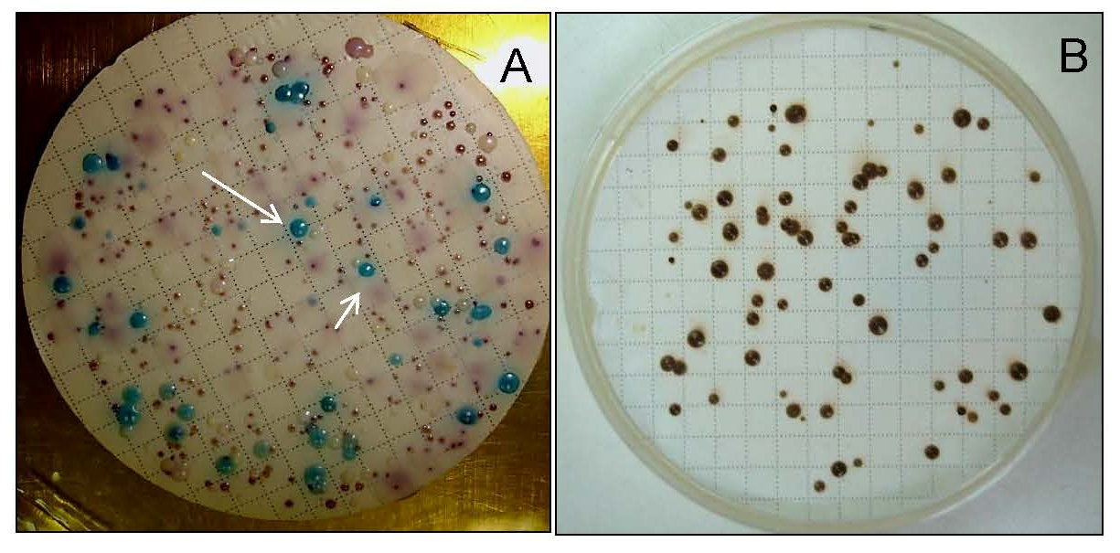
Studies suggest evidence that C. albicans can exhibit a wide ecological range when isolated from natural sources like plants (Table 1). Van Uden et al. [7] reported the successful isolation of C. albicans from floral specimens of Ulex sp. (Fabaceae) and Myrtus communis (Myrtaceae) in Portugal. In the same study, in vivo tests revealed the high virulence potential of these isolates, causing the death of the rabbits 4 days after infection. Despite these findings, the authors approached, with caution, the saprophytic characteristics exhibited by these yeasts. Although the authors acknowledged the difficulty of definitively confirming plants as a natural habitat, they effectively demonstrated the adaptability of C. albicans to survive outside its traditional endothermic hosts. Khan et al. [21] reported the isolation of C. albicans from the stem of Calotropis procera, a medicinal plant widely used in India. Additionally, the yeast was found associated with decaying wood in the Galápagos Archipelago, Ecuador [22]. However, in both studies, the authors did not delve into the implications of detecting this opportunistic pathogen in these habitats. C. albicans isolates were recovered from oak forests in different regions of Europe [9,10]. Robinson et al. [9] isolated C. albicans (three strains) from ancient oak trees in northern Europe and suggested that the existence of wild populations of this yeast on northern European trees could potentially explain the previously puzzling maintenance of aquaporin genes associated with freeze tolerance in C. albicans [29]. Bensasson et al. [10] compared the genetic characteristics of these oak isolates with yeasts obtained from clinical settings. Their findings revealed that oak isolates predominantly exhibited a diploid nature, similar to clinical isolates. However, C. albicans oak isolates showed a higher degree of heterozygosity compared to clinical isolates. Based on these observations, the authors propose that C. albicans inhabits the environments of these trees for a prolonged period, showing higher levels of genome-wide heterozygosity. This suggests that they were subjected to different selection or mutation pressures. This study provides insights into the adaptation and long-term evolution of C. albicans in distinct ecological niches [10]. Barros et al. [23] explored the diversity of yeasts present in samples isolated from rotting wood in the Amazon forest biomes. C. albicans was among the species present in the material collected in the forest. The authors showed, in this study, that the choice of different culture media directly significantly influences the ability to isolate a greater richness of yeast species. This insight leads us to believe that with more comprehensive monitoring, a greater quantity and variety of yeasts can be discovered in the environment. Lopes et al. [12] also found the presence of C. albicans associated with rotting wood samples obtained from Brazilian ecosystems, from the areas of Atlantic rainforest (three isolates), Cerrado (one isolate), and Amazonian Forest (one isolate). The number of C. albicans isolates found by the authors was not high in each region; however, the authors were using selective media for isolation of cellobiose-fermenting yeast species. Perhaps if more specific media for C. albicans isolation were used, such as MCa medium or CHROMagar-Candida (Figure 2), more isolates of this species could be found in these biomes [8,30].
Figure 2. Yeast colonies grown on nitrocellulose membranes using Candida CHROMagar-Candida (arrow: Candida albicans forming green colonies) (A) and MCa media (brown colonies: C. albicans) (B). The figures are the property of the authors.
Jof 11 00622 g002

4. Candida albicans in Freshwater and Sea

Isolation of C. albicans in water bodies has been considered by many researchers as indicative of a contamination site by human wastes (Table 1). In studies where the diversity of microorganisms in seawater from Brazilian beaches was verified, C. albicans was frequently isolated at low densities [8,31]. However, in the evaluation of virulence factors expressed by these environmental isolates, Maciel et al. [13] discovered the ability of morphogenesis and biofilm formation through both in vitro and in vivo experiments (Figure 3). The authors highlighted that these results demonstrated the potential of yeasts isolated from environmental samples to maintain their virulence and induce disease.
Striving to identify the sources of microbiological pollution that may harm the health of bathers, Prigitano et al. [18] investigated the fungal populations in both the sands and water of the beaches of two largest Italian lakes and in sands of the Mediterranean coast in Southern Italy. The survey revealed the existence of opportunistic pathogenic fungi in both locations, with C. albicans being exclusively detected in the coastal regions at low density. As part of an effort to collect data on fungi in beach sand and water, both in coastal bathing areas and inland waters, Brandão et al. [20] isolated several fungi known as opportunistic human pathogens, such as species of the genus Aspergillus, Cryptococcus and Candida. Notably, among the findings of this study, C. albicans was also isolated from samples of sand and seawater from beaches in the Mediterranean, Black Sea and Northwest Europe regions.
C. albicans isolates were also obtained from water samples from different rivers used for human consumption across parts of the world [24,25,26]. Kulesza et al. [25], in a 20-year retrospective study, found that the presence of this opportunistic pathogen is more prominent in areas in Poland where the river flows close to cities, indicating anthropic influence. Monopathi et al. [26] observed the genetic similarity between efflux pump genes (associated with resistance traits) between environmental and clinical isolates, indicating that the environmental isolates already possess resistance to antifungals such as azoles. In both studies, the authors emphasize that these findings represent crucial epidemiological factors with potential implications for clinical environments. The presence of this opportunistic pathogen in these rivers can pose significant risks to people and animals that come into contact with and/or consume this water, especially concerning immunosuppressed individuals. C. albicans isolates were recovered from groundwater in residential wells located in regions of the state of Mato Grosso do Sul, Brazil [27]. This study observed that the C. albicans isolates produced hemolysin, a recognized key virulence factor, along with phospholipase, facilitating the host cell invasion process (Figure 3). Cupozak-Pinheiro et al. [27] highlight the importance of verifying the presence of potentially pathogenic/virulent yeasts in residential wells, which could lead to opportunistic infections in individuals consuming this water. In addition to running water environments, swampy environments can act as reservoirs for C. albicans. Water samples were collected from various depths in a swampy river in South Africa, and the presence of C. albicans was detected using quantitative real-time PCR (qRT-PCR) [14]. The river under investigation was contaminated by sewage, indicating clear anthropogenic influence on the findings. The study suggested that oxygen-limited, reducing zones of wetlands act as a niche for C. albicans outside its human host and a potential external reservoir for this human commensal. The authors emphasize the epidemiological importance of identifying these reservoirs of opportunistic pathogens outside the hospital environment for disease management.

5. Clinical Importance of the Isolation of Candida albicans in Natural Environments

Studies involving environmental yeasts, including C. albicans, are invaluable for advancing knowledge in ecology, taxonomy, and biotechnological applications. However, specifically regarding the study of environmental isolates of C. albicans, it is essential to consider the potential implications for clinical settings. Studies have demonstrated that investigating environmental isolates can significantly enhance our comprehension of the evolutionary events that underlie fungal virulence (Figure 3) [32,33,34]. Notably, studies conducted by Steenbergen et al. [32], Casadevall et al. [33], and Lemos Tavares et al. [34] have unveiled the crucial role of interaction between soil-dwelling amoebae and various fungal species, including C. albicans, in shaping the exhibited virulence factors of these fungi. These studies suggest that some virulence factors present in these microorganisms may have emerged through selective pressure during interaction with amoebas in natural settings, highlighting the intricate evolutionary processes at play. For instance, filamentation, which is believed to have evolved as a defense mechanism against amoebae predation, has now been recognized as a crucial factor enabling fungal evasion of the human immune system and tissue invasion. In addition to being a possible tool that would assist in the study of the evolution of virulence factors in fungi, research focused on environmental isolates of opportunistic pathogens can be useful in assessing the behavior of these microorganisms in the face of climate change. Studies on the subject show that climate change can influence host–pathogen interactions, disease spread, antimicrobial resistance, and virulence of these microorganisms [35,36,37,38,39]. Leach et al. [38] found a relationship between the transcription factors responsible for inducing gene expression of genes linked to heat shock proteins and the induction of the transcriptional program associated with the virulence factors of C. albicans.
This study reinforces the idea that exposure to stress conditions, such as rising temperature, can influence the expression of genes associated with virulence factors in this fungus. Resistance to antifungal treatment may be another factor that can be assessed in environmental isolates. Research has shown that opportunistic species of the genus Candida (Loderomyces/Candida albicans clade) obtained in nature may be resistant to treatment with antifungal agents, even without previous contact with these drugs [8,40,41,42,43,44,45]. Understanding the mechanisms involved in this characteristic can help researchers anticipate the emergence of multidrug-resistant isolates and the advancement of novel antifungal drugs.

6. Conclusions

There exists a knowledge gap concerning the ecological aspects of opportunistic pathogens, particularly C. albicans. The pursuit of knowledge in the clinical domain tends to overlook the significance of comprehending the characteristics of these microorganisms present in non-endothermic host environments. The impact of isolating C. albicans from natural habitats on the epidemiology of opportunistic infections remains unclear. Limited data exist regarding the expression of virulence factors by these microorganisms in the wild. It is unknown whether selective pressures in these environments influence traits that serve as secondary sources of contact, leading to contamination and human infections. Essentially, our understanding of C. albicans in the non-clinical setting is insufficient. Consequently, it is imperative to invest in research within this field to provide data on its unknown ecology, which may be linked to its clinical pathogenicity. Expanding our knowledge of the natural reservoirs of these yeasts is crucial to gaining a better understanding of the pathophysiology of exogenous infections.

Author Contributions

A.D.V.: data curation; writing—original draft, editing; G.C.A.d.M.: writing and review; C.A.R.: writing and review; S.J.: writing, supervision, review and editing. All authors have read and agreed to the published version of the manuscript.

Funding

This work is part of the project “INCT Yeasts: Biodiversity, preservation and biotechnological innovation”, funded by Conselho Nacional de Desenvolvimento Científico e Tecnológico (CNPq), Brazil, grant #406564/2022-1. Funding was also provided by Conselho Nacional de Desenvolvimento Científico e Tecnológico (CNPq), grant 314952/2021-7, Coordenação de Aperfeiçoamento de Pessoal de Nível Superior (CAPES) and Pró-Reitoria de Pesquisa da UFMG. We extend our appreciation to Dr. Ana Raquel O. Santos and Natália Maciel for supplying images from their personal collection to compile the figures in this manuscript.

Conflicts of Interest

The authors declare no conflicts of interest.

References

  1. Gow, N.; Yadav, B. Microbe Profile: Candida albicans: A shape-changing, opportunistic pathogenic fungus of humans: This article is part of the Microbe Profiles collection. Microbiol. 2017, 163(8), 1145–1147. [Google Scholar]
  2. Groenewald, M.; Hittinger, C.T.; Bensch, K.; Opulente, D.; Shen, X.-X.; Li, Y.; Liu, C.; LaBella, A.; Zhou, X.; Limtong, S.; et al. A genome-informed higher rank classification of the biotechnologically important fungal subphylum Saccharomycotina. Stud. Mycol. 2023, 105, 1–22. [Google Scholar] [CrossRef]
  3. Lamoth, F.; Lockhart, S.R.; Berkow, E.L. Changes in the epidemiological landscape of invasive candidiasis. J. Antimicrob. Chemother. 2018, 73 (suppl1), i4–i13. [Google Scholar] [CrossRef]
  4. Opulente, D.A.; Langdon, Q.K.; Buh, K.V.; Haase, M.A.B.; Sylvester, K.; Moriarty, R.V.; Jarzyna, M.; Considine, S.L.; Schneider, R.M.; Hittinger, C.T. Pathogenic budding yeasts isolated outside of clinical settings. FEMS Yeast Res. 2019, 19, foz032. [Google Scholar] [CrossRef]
  5. Barnett, J.A. A history of research on yeasts 12: Medical yeasts part 1, Candida albicans. Yeast 2008, 25, 385–417. [Google Scholar] [CrossRef] [PubMed]
  6. Lachance, M.-A.; Boekhout, T.; Scorzetti, G.; Fell, J.W.; Kurtzman, C.P. 2011 Chapter 90 Candida Berkhout. In The Yeasts, 5th ed.; Kutrzman, C., Fell, J.W., Boekhout, T., Eds.; Elsevier: London, UK, 1923; pp. 987–1278. [Google Scholar]
  7. Van Uden, N.; De Matos Faia, M.; Assis-Lopes, L. Isolation of Candida albicans from vegetable sources. J. Gen. Microbiol. 1956, 15, 151–153. [Google Scholar] [CrossRef] [PubMed]
  8. Di Menna, M. Candida albicans from Grass Leaves. Nature 1958, 181, 1287–1288. [Google Scholar] [CrossRef]
  9. Robinson, H.A.; Pinharanda, A.; Bensasson, D. Summer temperature can predict the distribution of wild yeast populations. Ecol. Evol. 2016, 6, 1236–1250. [Google Scholar] [CrossRef] [PubMed]
  10. Bensasson, D.; Dicks, J.; Ludwig, J.M. Diverse lineages of Candida albicans live on old oaks. Genetics 2019, 211, 277–288. [Google Scholar] [CrossRef]
  11. Kochkina, G.; Ivanushkina, N.; Ozerskaya, S. Ancient fungi in Antarctic permafrost environments. FEMS Microbiol. Ecol. 2012, 82, 501–509. [Google Scholar] [CrossRef]
  12. Lopes, M.R.; Lara, C.A.; Moura, M.E.F.; Uetanabaro, A.P.T.; Morais, P.B.; Vital, M.J.; Rosa, C.A. Characterization of the diversity and physiology of cellobiose-fermenting yeasts isolated from rotting wood in Brazilian ecosystems. Fungal Biol. 2018, 122, 668–676. [Google Scholar] [CrossRef]
  13. Maciel, N.O.; Johann, S.; Brandão, L.R.; Kucharíková, S.; Morais, C.G.; Oliveira, A.P.; Freitas, G.J.; Borelli, B.M.; Pellizzari, F.M.; A Santos, D.; et al. Occurrence, antifungal susceptibility, and virulence factors of opportunistic yeasts isolated from Brazilian beaches. Mem. Inst. Oswaldo Cruz. 2019, 114, e180566. [Google Scholar] [CrossRef]
  14. Stone, W.; Jones, B.L.; Wilsenach, J.; Botha, A. External ecological niche for Candida albicans within reducing, oxygen-limited zones of wetlands. Appl. Environ. Microbiol. 2012, 78, 2443–2445. [Google Scholar] [CrossRef]
  15. Ajello, L. Soil as natural reservoir for human pathogenic fungi. Science 1956, 123, 876–879. [Google Scholar] [CrossRef] [PubMed]
  16. Rogers, A.L.; Beneke, E.S. Human pathogenic fungi recovered from Brasilian soil. Mycopathol. Mycol. Appl. 1964, 22, 15–20. [Google Scholar] [CrossRef] [PubMed]
  17. Mok, W.Y.; Luizão, R.C.; Do Socorro Barreto Da Silva, M.; Teixeira, M.F.; Muniz, E.G. Ecology of pathogenic yeasts in Amazonian soil. Appl. Environ. Microbiol. 1984, 47, 390–394. [Google Scholar] [CrossRef]
  18. Prigitano, A.; Trovato, L.; Esposto, M.C.; Brandão, J.; Cogliati, M.; Gatta, G.D.; Grancini, A.; Migliorisi, G.; Oliveri, S.; Romanò, L.; et al. Fungal diversity in lake and sea beaches of Italy: Relevance to human health. Sci. Total Environ. 2023, 859, 160417. [Google Scholar] [CrossRef]
  19. Vadkertiová, R.; Dudášová, H.; Stratilová, E.; Balaščáková, M. Diversity of yeasts in the soil adjacent to fruit trees of the Rosaceae family. Yeast 2019, 36, 617–631. [Google Scholar] [CrossRef]
  20. Brandão, J.; Gangneux, J.P.; Arikan-Akdagli, S.; Esposto, M.C.; Brandão, J.; Cogliati, M.; Gatta, G.D.; Grancini, A.; Migliorisi, G.; Oliveri, S.; et al. Mycosands: Fungal diversity and abundance in beach sand and recreational waters—Relevance to human health. Sci. Total Environ. 2021, 781, 146598. [Google Scholar] [CrossRef] [PubMed]
  21. Khan, R.; Shahzad, S.; Choudhary, M.I.; Khan, S.A.; Ahmad, A. Biodiversity of the endophytic fungi isolated from Calotropis procera (AIT.) R. BR. Pak. J. Bot. 2007, 39, 2233–2239. [Google Scholar]
  22. Guamán-Burneo, M.C.; Dussán, K.J.; Cadete, R.M.; Cheab, M.A.; Portero, P.; Carvajal-Barriga, E.J.; da Silva, S.S.; Rosa, C.A. Xylitol production by yeasts isolated from rotting wood in the Galápagos Islands, Ecuador, and description of Cyberlindnera galapagoensis f.a., sp. nov. Antonie Van Leeuwenhoek 2015, 108, 919–931. [Google Scholar] [CrossRef]
  23. Barros, K.O.; Alvarenga, F.B.M.; Magni, G.; Souza, G.F.L.; Abegg, M.A.; Palladino, F.; da Silva, S.S.; Rodrigues, R.C.L.B.; Sato, T.K.; Hittinger, C.T.; et al. The Brazilian Amazonian rainforest harbors a high diversity of yeasts associated with rotting wood, including many candidates for new yeast species. Yeast 2023, 40, 84–101. [Google Scholar] [CrossRef]
  24. Bakhiet, S.; Ahmed, W.; Mohammed, W. Significance of fungal species isolated from blue nile river and tuti island on drinking water quality. J. Appl. Life Sci. Int. 2016, 7, 1–6. [Google Scholar] [CrossRef] [PubMed]
  25. Kulesza, K.; Biedunkiewicz, A.; Nowacka, K.; Glinka, P. Potentially pathogenic fungi of the Candida genus isolated from the Łyna River—A 20-year study. Ann. Parasitol. 2018, 64, 217–223. [Google Scholar]
  26. Monapathi, M.E.; Bezuidenhout, C.C.; Rhode, O.H.J. Efflux pumps genes of clinical origin are related to those from fluconazole-resistant Candida albicans isolates from environmental water. Water Sci. Technol. 2018, 77, 899–908. [Google Scholar] [CrossRef] [PubMed]
  27. Cupozak-Pinheiro, W.J.; Araújo De Almeida-Apolonio, A.; Sasaki, M.H.; Maran, N.H. Candida species contamination in drinking groundwater from residence wells in three municipalities of midwestern Brazil and the potential human health risks. Microb. Pathog. 2022, 169, 105660. [Google Scholar] [CrossRef]
  28. Sautour, M.; Lemaître, J.; Ranjard, L.; Truntzer, C.; Basmaciyan, L.; Depret, G.; Hartmann, A.; Dalle, F. Detection and survival of Candida albicans in soils. Environ. DNA 2021, 3, 1093–1101. [Google Scholar] [CrossRef]
  29. Tanghe, A.; Carbrey, J.M.; Agre, P.; Thevelein, J.M.; Van Dijck, P. Aquaporin expression and freeze tolerance in Candida albicans. Appl. Environ. Microbiol. 2005, 71, 6434–6437. [Google Scholar] [CrossRef]
  30. Buck, J.D.; Bubucis, P.M. Membrane filter procedure for enumeration of Candida albicans in natural waters. Appl. Environ. Microbiol. 1978, 35, 237–242. [Google Scholar] [CrossRef]
  31. Pinto, K.C.; Hachich, E.M.; Sato, M.I.Z.; Di Bari, M.; Coelho, M.C.L.S.; Matté, M.H.; Lamparelli, C.C.; Razzolini, M.T.P. Microbiological quality assessment of sand and water from three selected beaches of South Coast, São Paulo State, Brazil. Water Sci. Technol. 2012, 66, 2475–2482. [Google Scholar] [CrossRef]
  32. Steenbergen, J.N.; Nosanchuk, J.D.; Malliaris, S.D.; Casadevall, A. Interaction of Blastomyces dermatitidis, Sporothrix schenckii, and Histoplasma capsulatum with Acanthamoeba castellanii. Infect. Immun. 2004, 72, 3478–3488. [Google Scholar] [CrossRef] [PubMed]
  33. Casadevall, A.; Fu, M.; Guimaraes, A.; Albuquerque, P. The ‘amoeboid predator-fungal animal virulence’ hypothesis. J. Fungi 2019, 5, 10. [Google Scholar] [CrossRef] [PubMed]
  34. Lemos Tavares, P.; Carvalho Ribeiro, A.; Kercher Berte, F. The interaction between Sporothrix schenckii sensu stricto and Sporothrix brasiliensis with Acanthamoeba castellanii. Mycoses 2020, 63, 302–307. [Google Scholar] [CrossRef]
  35. Altizer, S.; Ostfeld, R.S.; Johnson, P.T.J.; Kutz, S.; Harvell, C.D. Climate change and infectious diseases: From evidence to a predictive framework. Science 2013, 341, 514–519. [Google Scholar] [CrossRef]
  36. Raffel, T.R.; Romansic, J.M.; Halstead, N.T.; McMahon, T.A.; Venesky, M.D.; Rohr, J.R. Disease and thermal acclimation in a more variable and unpredictable climate. Nat. Clim. Change 2013, 3, 146–151. [Google Scholar] [CrossRef]
  37. Maynard, J.; Van Hooidonk, R.; Eakin, C.M.; Puotinen, M.; Garren, M.; Williams, G.; Heron, S.F.; Lamb, J.; Weil, E.; Willis, B.; et al. Projections of climate conditions that increase coral disease susceptibility and pathogen abundance and virulence. Nat. Clim. Change 2015, 5, 688–694. [Google Scholar] [CrossRef]
  38. Leach, M.D.; Farrer, R.A.; Tan, K.; Miao, Z.; Walker, L.A.; Cuomo, C.A.; Wheeler, R.T.; Brown, A.J.P.; Wong, K.H.; Cowen, L.E. Hsf1 and Hsp90 orchestrate temperature-dependent global transcriptional remodelling and chromatin architecture in Candida albicans. Nat. Commun. 2016, 7, 11704. [Google Scholar] [CrossRef]
  39. Cavicchioli, R.; Ripple, W.J.; Timmis, K.N.; Azam, F.; Bakken, L.R.; Baylis, M.; Behrenfeld, M.J.; Boetius, A.; Boyd, P.W.; Classen, A.T.; et al. Scientists’ warning to humanity: Microorganisms and climate change. Nat. Rev. Microbiol. 2019, 17, 569–586. [Google Scholar] [CrossRef]
  40. Medeiros, A.O.; Kohler, L.M.; Hamdan, J.S.; Missagia, B.S.; Barbosa, F.A.; Rosa, C.A. Diversity and antifungal susceptibility of yeasts from tropical freshwater environments in Southeastern Brazil. Water Res. 2008, 42, 3921–3929. [Google Scholar] [CrossRef]
  41. Brandão, L.R.; Medeiros, A.O.; Duarte, M.C.; Barbosa, A.C.; Rosa, C.A. Diversity and antifungal susceptibility of yeasts isolated by multiple-tube fermentation from three freshwater lakes in Brazil. J. Wather Health 2010, 8, 279–289. [Google Scholar] [CrossRef]
  42. Brilhante, R.S.N.; Castelo Branco, D.S.C.M.; Duarte, G.P.S.; Paiva, M.A.; Teixeira, C.E.; Zeferino, J.P.; Monteiro, A.J.; Cordeiro, R.A.; Sidrim, J.J.; Rocha, M.F. Yeast microbiota of raptors: A possible tool for environmental monitoring: Use of Candida spp. for environmental monitoring. Environ. Microb. Rep. 2012, 4, 189–193. [Google Scholar] [CrossRef]
  43. Castelo-Branco, D.S.C.M.; Brilhante, R.S.N.; Paiva, M.A.N.; Teixeira, C.E.C.; Caetano, E.P.; Ribeiro, J.F.; Cordeiro, R.A.; Sidrim, J.J.C.; Monteiro, A.J.; Rocha, M.F.G. Azole-resistant Candida albicans from a wild Brazilian porcupine (Coendou prehensilis): A sign of an environmental imbalance? Med. Mycol. 2013, 51, 555–560. [Google Scholar] [CrossRef] [PubMed]
  44. Brilhante, R.S.N.; Paiva, M.A.N.; Sampaio, C.M.S.; Castelo-Branco, D.S.C.M.; Alencar, L.P.; Bandeira, T.J.P.G.; Cordeiro, R.A.; Neto, W.d.A.P.; Moreira, J.L.B.; Sidrim, J.J.C.; et al. Surveillance of azole resistance among Candida spp. As a strategy for the indirect monitoring of freshwater environments. Water Air Soil. Pollut. 2015, 226, 52. [Google Scholar] [CrossRef]
  45. Rocha, M.F.G.; Alencar, L.P.; Paiva, M.A.N.; Melo, L.M.; Bandeira, S.P.; Ponte, Y.B.; Sales, J.A.; Guedes, G.M.M.; Castelo-Branco, D.S.C.M.; Bandeira, T.J.; et al. Cross-resistance to fluconazole induced by exposure to the agricultural azole tetraconazole: An environmental resistance school? Mycoses 2016, 59, 281–290. [Google Scholar] [CrossRef] [PubMed]
Figure 1. Representation of the ecological habitats where C. albicans was found in natural environments. In order from left to right, the pictures include flowers found in the Brazilian Amazon Rainforest (A,B), trees from the Brazilian Amazon Rainforest (C), decomposing wood (D), rivers present in the Amazon Rainforest (E,F), and two beaches from the coastline in Brazil (G,H). The figures are the property of the authors.
Jof 11 00622 g001
Figure 3. Candida albicans egg culture medium exhibited the presence of a phospholipase halo (arrow) (A). C. albicans adhered to oral epithelial cells in the presence of blastoconidia, hyphae, and pseudohyphae (B). Colonies of C. albicans grown in Spider culture medium exhibited filamentation ability (C). The figures are the property of the authors.
Jof 11 00622 g003
Table 1. Isolates of Candida albicans from different habitats.
Number of IsolatesSubstract/HabitatSiteCity/CountryReference
1SoilJeffersonIowa, USAOpulente et al. [4]
1SoilNortheasternWisconsin, USAOpulente et al. [4]
1Plant Matter
Soil
Mesa Canyon San AntonioTexas, USAOpulente et al. [4]
1Plant Matter
Soil/Duff
Mesa Cyn San Antonio, TXTexas, USAOpulente et al. [4]
1SoilShelby CountyTenesesse, USAAjello et al. [15]
8Soils containing cow and hog dungFarms near Belo HorizonteMinas Gerais, BrazilRogers and Beneke [16]
8Chicken house soilsFarms near Belo HorizonteMinas Gerais, BrazilRogers and Beneke [16]
2Cave soilItatiaia Park areaMinas Gerais, BrasilRogers and Beneke [16]
1Chicken yard soilPiracicaba and
Vicinity
Piracicaba, São PauloRogers and Beneke [16]
1Sand near stepsSantos beach areaSantos, BrasilRogers and Beneke [16]
1Park flower bed soilSão PauloSão Paulo, BrazilRogers and Beneke [16]
1Amazonian soilsBrazilian Amazon BasinBrazilian Amazon Basin, BrazilMok et al. [17]
3SandBeachParaná and Rio de Janeiro, BrazilMaciel et al. [13]
28.3 CFU/gSandsSicilian Costal beachItalyPrigitano et al. [18]
2Soil near peach treeSouthwest of SlovakiaSlovakiaVadkertiová et al. [19]
1.7 CFU/gSandsIrlandaIrlandaBrandao et al. [20]
5.0 CFU/gSandsBlack seaRomaniaBrandao et al. [20]
27.1 CFU/gSandsMediterraneanSerbiaBrandao et al. [20]
3.3 CFU/gSandsMediterraneanTurkeyBrandao et al. [20]
1Bark treeWyalusing State ParkWisconsin, USAOpulente et al. [4]
1Plant Matter—Fruit;
Ericaceae
Berry
West Sand IslandOregon, USAOpulente et al. [4]
1Plant Matter—Fruit;
Ericaceae
Berry
Hungarian FallsMichigan, USAOpulente et al. [4]
1Plant Matter—Fruit;
Ericaceae
Berry
Hungarian FallsMichigan, USAOpulente et al. [4]
1Flower of African tulip tree (Spathodea campanulate, Bignoniaceae)RarontohgaCook IslandsLachance et al. [6]
1Fruit of Stenocereus hystrix (Cactaceae)-JamaicaLachance et al. [6]
1Leaves of Myrtus communisNear the top of
a hill (300 m. high) near Vermoil
Estremadura, PortugualVan Uden et al. [7]
2Flowers of furze (Ulex sp.)Near the top of
a hill (300 m. high) near Vermoil
Estremadura, PortugualVan Uden et al. [7]
3OakNew Forest, North EuropeUKRobinson et al. [9]
1Steam of Calotropis procera (Ait.) R. Br.Karachi University campusPakistanKhan et al. [21]
2Rotting wood samplesGalápagos ArchipelagoEcuadorGuama’n-Burneo et al. [22]
1Rotting woodBrazilian Amazonian rainforestsBrazilBarros et al. [23]
3Rotting woodAtlantic RainforestBrazilLopes et al. [12]
1Rotting woodCerradoBrazilLopes et al. [12]
1Rotting woodAmazonian rainforestsBrazilLopes et al. [12]
2SeawaterRecreational beachesParaná, BrazilMaciel et al. [13]
5 CFU/mlWaterSicilian Costal beachItalyPrigitano et al. [18]
3.8 CFU/mlWaterNorthwest EuropeIrlandBrandao et al. [20]
3.8 CFU/mLWaterMediterraneanSerbiaBrandao et al. [20]
25 and 10% *WaterBlue Nile
River
SudanBakhiet et al. [24]
**WaterŁyna RiverOlsztyn, PolandKulesza et al. [25]
37WaterNorth West Province RiversSouth AfricaMonopathi et al. [26]
1WaterGroundwater for human consumption from wellsMato Grosso do Sul, BrazilCupozak-Pinheiro et al. [27]
* In this study, the authors reported that Candida albicans was found in 25% and 10% of the samples using two different culture media. ** In this study, the authors did not specify how many Candida albicans isolates were found.
Disclaimer/Publisher’s Note: The statements, opinions and data contained in all publications are solely those of the individual author(s) and contributor(s) and not of MDPI and/or the editor(s). MDPI and/or the editor(s) disclaim responsibility for any injury to people or property resulting from any ideas, methods, instructions or products referred to in the content.