Article

Identification of Macadamia integrifolia Leaf Blight Disease Caused by Pestalotiopsis colombiensis in China

1
Yunnan Provincial Key Laboratory for Conservation and Utilization of In-forest Resource, Southwest Forestry University, Kunming 650224, China
2
Key Laboratory of Forest Disaster Warning and Control of Yunnan Province, Southwest Forestry University, Kunming 650224, China
3
School of Forestry, Northeast Forestry University, Harbin 150040, China
4
Key Laboratory of National Forestry and Grassland Administration on Forest and Grassland Pest Monitoring and Warning, Center for Biological Disaster Prevention and Control, National Forestry and Grassland Administration, Shenyang 110034, China
*
Authors to whom correspondence should be addressed.
J. Fungi 2025, 11(9), 613; https://doi.org/10.3390/jof11090613
Submission received: 15 July 2025 / Revised: 8 August 2025 / Accepted: 18 August 2025 / Published: 22 August 2025

Abstract

Macadamia integrifolia, a tropical and subtropical fruit tree with significant economic and nutritional value, faces serious fungal disease problems during cultivation that severely affect yield and quality. In November 2024, leaf blight symptoms of M. integrifolia were observed in Menglian, Pu’er, Yunnan, China, with a disease incidence of 23% in the field. Initial symptoms included small spots that enlarged into circular to irregular lesions with red-brown centers and brown to black margins. Finally, the leaves turned yellow and became scorched, eventually leading to massive leaf shedding. Infected leaf samples were collected, and fungal strains were isolated, purified, and inoculated via spore suspension, followed by re-isolation. The strains were conclusively identified as Pestalotiopsis colombiensis (SWFUCB2, SWFUCB1) through an integrated approach combining DNA extraction, polymerase chain reaction (PCR), sequencing, phylogenetic reconstruction, and morphological characterization. This is the first report of P. colombiensis causing M. integrifolia leaf blight disease in China, filling a gap in research on this disease. This study provided important information for epidemiological research on this disease and the development of comprehensive leaf blight disease control strategies.

1. Introduction

Macadamia integrifolia, a member of the Proteaceae family, is a tall-canopy evergreen tree characterized by smooth leaves with serrated margins. Native to the coastal subtropical rainforests of Australia, this species is now primarily distributed across Oceania, Southeast Asia, and China. Its kernels are nutrient-rich, containing high levels of unsaturated fatty acids, protein, minerals, and vitamins. These kernels also exhibit antioxidant, lipid-lowering, and anti-inflammatory properties, contributing to their significant economic value and earning them the title of the “King of nuts”. Leaf diseases impair photosynthesis, thereby reducing the synthesis and accumulation of photosynthetic products and ultimately affecting both fruit yield and quality [1]. With the expansion of cultivation areas, various fungal diseases have increasingly emerged in crops. Currently, known diseases of M. integrifolia include canker disease [2], leaf spot disease [3], dieback disease [4], anthracnose [5], and black rot and flower blights [6], all of which result in substantial economic losses. Therefore, timely detection, accurate identification, and effective management of these diseases are essential. Such research enhances macadamia yield and quality and also plays a critical role in the sustainable development of the macadamia industry.
In 1949, Steyaert classified the genus Pestalotia spp. into three distinct genera—Pestalotia spp., Pestalotiopsis spp., and Truncatella spp.—based on the morphology of their conidia [7]. Species within Pestalotia spp. are necrotrophic plant pathogens capable of infecting a wide range of host plants, causing diseases such as leaf spot, gray mold, and leaf blight. Studies have demonstrated that fungi in this genus can produce various enzymes and toxins that degrade plant cell walls, facilitate nutrient acquisition, and induce plant tissue necrosis [8]. Pestalotiopsis colombiensis is a member of the broader Pestalotia complex and belongs to the genus Pestalotiopsis. Pestalotiopsis spp. are typical phytopathogens with a broad host range, responsible for numerous plant diseases including canker [9], leaf blotch [10], trunk disease [11], rot disease [12], dieback [13], and various post-harvest diseases. Morphologically, Pestalotiopsis spp. produce fusiform conidia with four septa (five cells), where the basal and terminal cells are hyaline, and the median cells are brown. The apical cell is cylindrical to subcylindrical, hyaline, thin-walled, and smooth, with tubular apical appendages [14]. These conidia are formed through budding, representing the asexual reproductive stage of the fungus. Due to its diverse dispersal mechanisms, Pestalotiopsis is widely distributed across various ecological environments.
In recent years, Pestalotiopsis and Neopestalotiopsis have emerged as significant pathogens of M. integrifolia, causing leaf and flower diseases across all major production regions worldwide. Initial symptoms are irregular light-brown leaf spots that expand and coalesce [15,16]; flower infections appear as necrotic blight and rachis dieback, collectively termed “dry flower disease” [17]. These pathogens have been recorded in diverse regions: N. clavispora caused severe leaf spot epidemics in Brazil [16] and both leaf spots and flower blight in China [15]. In Australia, five novel Neopestalotiopsis species—N. drenthii, N. maddoxii, N. olumideae, N. vheenae and N. zakeelii—were isolated from diseased macadamia inflorescences [6], alongside the previously described P. macadamiae and N. macadamiae as primary agents of dry flower disease [17]. Collectively, Pestalotiopsis and Neopestalotiopsis form a widespread, genetically diverse pathogen complex affecting both vegetative and reproductive tissues of macadamia, with major implications for global disease management.
Yunnan is the main Macadamia-producing region in China. Yunnan’s sub-tropical plateau monsoon climate aligns well with the plant’s environmental requirements, creating a natural haven for macadamia nut growth. Macadamia trees grow well in humid, relatively low-fertility soil and areas with annual rainfall exceeding 1000 mm, summer temperatures under 39 °C, and frost-free winters [18]. In November 2024, leaf blight symptoms of M. integrifolia were observed in Menglian, Pu’er, Yunnan, China (22°19′43.6″ N, 99°35′30.2″ E), with a disease incidence of 23% in the field. This is the first report of P. colombiensis causing leaf blight on M. integrifolia in China. It represents a significant advancement in understanding the etiology of this economically important disease. The findings provide a critical foundation for developing targeted and sustainable disease management strategies, which are essential for safeguarding Yunnan’s macadamia industry and ensuring its long-term productivity and economic viability.

2. Materials and Methods

2.1. Sample Collection and Isolation

In November 2024, diseased leaves of M. integrifolia (Disi 1) were collected from Menglian, Pu’er, Yunnan, China (22°19′43.6″ N, 99°35′30.2″ E). A five-point sampling method was employed during the field survey, covering a total of 50 trees. From each tree, 20 leaves were collected from four cardinal directions—east, south, west, and north—to investigate the occurrence of foliar diseases. Subsequently, 20 diseased leaves were randomly selected and examined under a microscope (Olympus CX33, Nagano, Japan). To obtain a pure fungal isolate, diseased leaves exhibiting typical spot symptoms were surface-sterilized with 75% ethanol for 10 s followed by 1% sodium hypochlorite (NaClO) for 40 s. The leaves were then rinsed three times with sterile distilled water and air-dried on sterilized filter paper. Using insect needles under microscopic guidance, individual conidia were isolated and transferred to potato dextrose agar (PDA; 200 g potato, 20 g glucose, and 17 g agar per liter) (Beijing Land Bridge Technology Co., Ltd., Beijing, China). Once single colonies developed, they were subcultured onto fresh PDA slants for preservation and incubated at 25 °C [19]. Pure cultures were obtained through single-spore isolation, resulting in six fungal strains. The representative specimens were deposited at Southwest Forestry University.

2.2. Morphological Identification

Morphological features were observed under an optical light microscope (Olympus CX33). The length, width, and morphological characteristics of conidia and conidiophore were measured (n = 50).

2.3. DNA Extraction, Polymerase Chain Reaction and Sequencing

Mycelial plugs (0.5 cm in diameter) were cultured on potato dextrose agar (PDA) for 5 days. Three plugs were used to inoculate 100 mL of liquid potato dextrose (PD) medium (200 g/L potato and 20 g/L dextrose). The cultures were incubated at 25 °C in the dark with 120 rpm for 8 days. Then, 100 mg of mycelium was collected, and total DNA was extracted using a Plant Genomic DNA Kit (Tiangen Biotech, DP305, Beijing, China).
The integrity of the extracted DNA was assessed using 0.8% agarose gel electrophoresis. The internal transcribed spacer (ITS) regions, large subunit ribosomal RNA gene (LSU), and translation elongation factor 1-alpha gene (TEF1-α) were amplified via PCR, respectively (Table 1). The reaction mixture for PCR contained 12.5 μL of 2 × Taq PCR Mix (Tiangen Biotech, T201), 1 μL of DNA sample, 1 μL each of the forward and reverse primers, and double-distilled water at a volume up to 25 μL. The PCR cycling conditions were as follows: 94 °C for 3 min; 32 cycles of 94 °C for 30 s, 58 °C for 30 s, and 72 °C for 2 min; and a final extension at 72 °C for 10 min. All primers were synthesized by Sangon Biotech Co., Ltd. (Shanghai, China). The amplified products were analyzed by means of agarose gel electrophoresis and sequenced using the Sanger method by Sangon Biotech Co., Ltd. (Shanghai, China).

2.4. DNA Sequence Analysis

The genetic sequences were analyzed and compared to existing sequences in GenBank using BLAST(2.17.0). These sequences, along with reference sequences retrieved from GenBank, were subsequently manually edited and aligned using MEGA (11) [25]. Phylogenetic relationships were inferred through the maximum likelihood method based on a heuristic search strategy. Bootstrap support values were estimated with 1000 replicates to assess the reliability of tree branches [26]. Sequence identity (%) was calculated using the MegAlign program (17.6) (DNASTAR Inc., Madison, WI, USA).

2.5. Pathogenicity Test

Twenty attached leaves (Disi 1) were inoculated via needle puncture with the CB1 conidial suspension (1 × 10⁶ spores/mL), and another twenty control leaves were inoculated with sterile water at 26 °C and 70% relative humidity. After 15 days, the onset of disease was observed in both treated and control groups. When symptoms developed, the pathogen was reisolated from the infected tissues and compared to the original inoculated strain based on morphological and molecular characteristics.

3. Results

3.1. Sample Collection

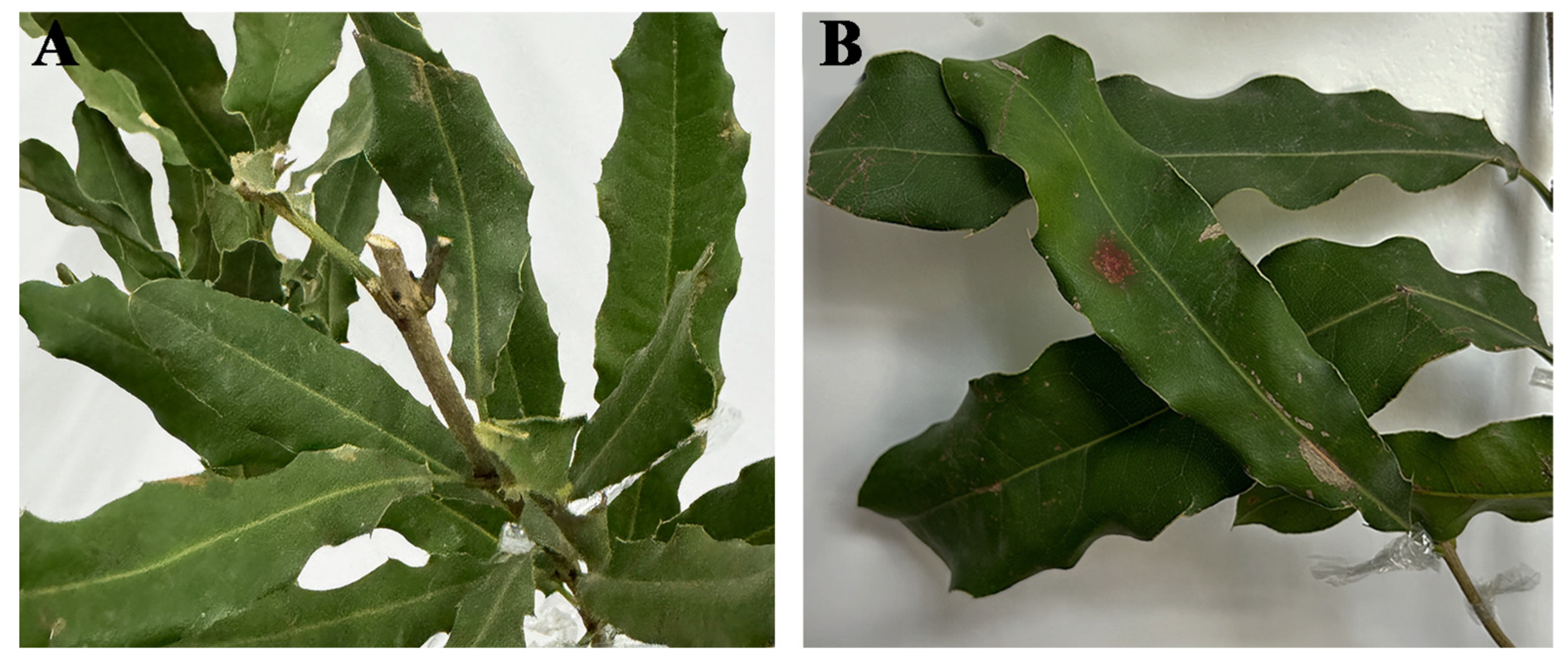
In November 2024, a total of 80 leaf blight cases were observed in Macadamia integrifolia, with diseased leaf incidence of 23% in the field (Figure 1A). Initial symptoms appeared as small spots that expanded into circular to irregular lesions, characterized by red-brown centers and brown to black margins. As the disease progressed, leaves turned yellow and exhibited a scorched appearance, ultimately resulting in extensive defoliation (Figure 1B).

3.2. Morphological Data

Six strains were obtained through single-spore isolation. The CB1 colonies exhibited a thick and villous morphology, with white aerial surfaces and yellowish undersides. Acervuli were spherical or club-shaped, scattered or semi-emergent to emergent, dark brown to black in color, and reached diameters of up to 650 μm on PDA medium after 15 days (Figure 2C). Conidia were fusiform and four-septate, with dimensions measuring 18.79–28.25 × 5.54–9.00 μm. The basal and terminal cells were hyaline, while the three median cells were brown, with a combined length of 13.00–17.05 μm. Individual measurements for each median cell were as follows: second cell: 4.44–6.12 μm; third cell: 4.14–5.59 μm; fourth cell: 4.14–6.00 μm. The apical cell was cylindrical to subcylindrical, hyaline, and thin-walled, bearing two to three unbranched, filiform, tubular appendages 12.74–28.89 μm in length, extending from the apical crest. A single, central, tubular basal appendage 3.26–5.00 μm in length was observed (n = 50). These morphological features were consistent with those for P. colombiensis (Figure 2A,B).

3.3. Phylogenetic Analyses

The internal transcribed spacer (ITS), translation elongation factor 1-alpha (TEF1-α), and large subunit ribosomal RNA gene (LSU) partial genes were amplified. BLAST analysis of the sequence showed a homology of 99.13% (573/578), 100.00% (868/868), and 100.00% (475/475) with P. colombiensis (KU715149, NG069213 and KM199488).
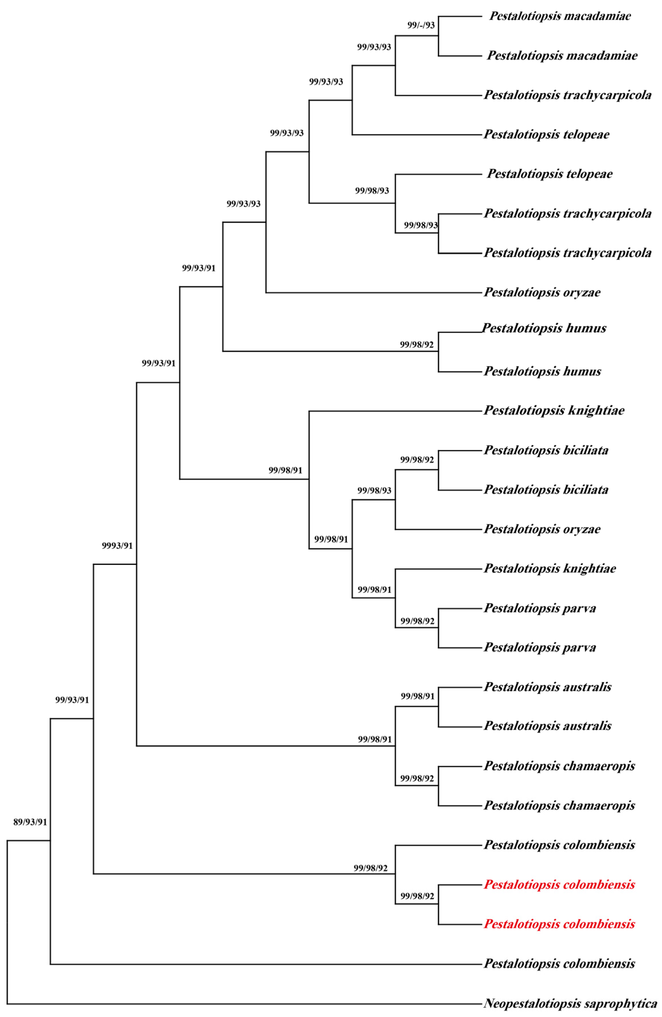
All sequences were deposited in GenBank (ITS: PQ895603 (CB1), PQ895604 (CB2); LSU: PQ895622 (CB1), PQ895623 (CB2); TEFl-α: PQ997934 (CB1), PQ997935 (CB2)). Phylogenetic analysis using the neighbor-joining analysis with concatenated sequences was performed using MEGA 10. The sequences were aligned with 76 sequences from 12 taxa retrieved from GenBank (Table 2). The resulting phylogenetic tree shows strong bootstrap support for P. colombiensis (Figure 3).

3.4. Pathogenicity Test Using M. Integrifolia Leaves

To confirm the pathogenicity of the isolate, twenty attached leaves were inoculated with a conidial suspension (1 × 10⁶ conidia/mL), while the control group was inoculated with ddH2O (Figure 4A). After 15 days, the inoculated leaves displayed typical blight symptoms resembling those seen under natural field conditions, whereas the control leaves remained asymptomatic. The strain CB2 was reisolated from the re-inoculated leaves, and the morphological and molecular characteristics were consistent with those of P. colombiensis CB1 (Figure 4B).

4. Discussion

Pestalotiopsis spp. is known to cause a wide range of plant diseases, including canker disease, leaf blotch disease, trunk disease, rot disease, dieback disease, and various post-harvest disorders. Although Pestalotiopsis species are recognized as pathogens of numerous host plants, there have been no prior reports regarding the pathogenicity of P. colombiensis. Macadamia integrifolia is among the susceptible hosts, as Pestalotiopsis spp. have been implicated in several diseases affecting this crop. For instance, P. macadamiae causes dry flower disease in Australia [17], and Pestalotiopsis-induced macadamia flower blight has also been documented. Additionally, P. clavispora is associated with husk rot in macadamia [27]. However, until now, Pestalotiopsis spp. has not been reported to cause leaf blight on M. integrifolia, and P. colombiensis has not previously been linked to any plant disease. Therefore, this study provides the first evidence and foundational insights into the pathogenic potential of P. colombiensis.
A spectrum of foliar fungal disorders have been documented on macadamia leaves, such as yellow halo leaf blight [3], brown leaf blight [28], leaf anthracnose [16], black leaf blight [29], and leaf spot [30], as detailed in several recent reports. This study provides important insights into the distribution and etiology of macadamia leaf blight in China. The initial symptoms manifested as small spots that expanded into circular or irregular lesions, characterized by red-brown centers and brown to black margins. To date, leaf spot disease caused by Neofusicoccum parvum has been reported on Macadamia integrifolia in Lincang, China. These lesions, measuring 3 to 5 mm in diameter, appeared as small, round brown spots with yellowish edges [31], represented another foliar disease affecting macadamia leaves. Additionally, leaf blight caused by P. trachicarpicola has been documented on Panax notoginseng in China. Its early symptoms included water-soaked discoloration, which later developed into dry yellow areas accompanied by slight leaf shrinkage [32]. As the disease progressed, leaf shrinkage and discoloration intensified, ultimately leading to leaf necrosis and defoliation. Based on the distinct symptomatology, this study differentiates the current disease from previously reported cases and identifies it as M. integrifolia leaf blight. Morphological and molecular analyses both clearly indicate that the causal agent belongs to the species P. colombiensis. This represents the first confirmed report of P. colombiensis causing leaf blight on M. integrifolia in China. Previous records of P. colombiensis on M. integrifolia lacked sufficient documentation, but this study now confirms its pathogenicity with confidence. Therefore, M. integrifolia should be formally recognized as a host species of P. colombiensis. These findings contribute valuable data for future epidemiological investigations and the development of targeted management strategies for the macadamia industry.
Currently, Pestalotiopsis has attracted significant research attention due to its ability to secrete a wide range of biologically active metabolites [33]. In addition to beneficial compounds, some species within this genus are known to produce phytotoxic secondary metabolites [34]. P. longiseta, P. theae [35], and P. oenotherae [36] have been reported to synthesize toxic substances. However, it remains unknown whether P. colombiensis possesses similar capabilities. This study did not investigate the pathogenic mechanisms of P. colombiensis; therefore, future research will focus on elucidating its pathogenicity in relation to M. integrifolia. Such investigations will provide a scientific basis for developing effective prevention and control strategies.
Several biological control agents have demonstrated efficacy in managing leaf blight caused by Pestalotiopsis spp. Bacillus spp. exhibit strong inhibitory effects against these pathogens [37,38]. Similarly, Trichoderma harzianum, Dickeya spp., and Streptomyces spp. have shown promising preventive and therapeutic potential [39,40,41]. Chemical fungicides such as Nerol and Flumax also display high efficacy in controlling Pestalotiopsis infections [42,43]. With increasing consumer demand for food safety and quality, along with stricter international trade standards for agricultural products, high-quality macadamia nuts have gained growing market popularity. Effective control of leaf blight is essential for maintaining healthy plant growth, improving fruit quality, and enhancing overall yield. Research into biological control strategies targeting pathogens responsible for macadamia diseases is emerging as a key trend in sustainable agriculture. Our team will continue to explore methods for preventing and managing M. integrifolia leaf disease caused by P. colombiensis, contributing to the refinement of theoretical frameworks in plant pathology while offering valuable insights for the management of other crop diseases.
M. integrifolia is widely consumed and highly valued globally as a nutritious and health-promoting food. In the early stages of macadamia cultivation in Yunnan, China, numerous Australian nut varieties were introduced and grown. The main production areas of macadamia nuts in Yunnan are concentrated in Lincang and Pu’er. Through adaptive breeding programs, several high-quality cultivars have been selected, including A4, A16, HAES660, and HAES788. Among these, seven varieties—A16, 816, Guang 11, HAES344, HAES660, Gui Re 1, and Changning 1—have been widely adopted in Yunnan [44]. The suitable M. integrifolia cultivars for Yunnan are comparable to those cultivated in Guangxi, Guizhou, the Hawaiian Islands, and Queensland, Australia. No cases of leaf blight have been reported in these regions, suggesting that the disease may become a significant constraint on M. integrifolia production, thus requiring further investigation. However, this study did not explore the relationship between cultivar type and disease incidence or assess the potential for disease-resistant varieties.
This research highlights that leaf blight could emerge as a major threat to macadamia production, underscoring the importance of developing effective disease management strategies. These findings provide a theoretical foundation for the precise prevention and control of M. integrifolia diseases, contributing to the optimization and localization of disease management practices. These efforts will support the healthy and sustainable development of the M. integrifolia industry in China. Furthermore, this study offers novel insights into the field of plant pathology and fills a critical research gap regarding pathogenic fungi affecting M. integrifolia in China, thereby holding significant academic value for future studies.

Author Contributions

Writing—review and editing, H.Y. and J.C.; conceptualization, J.C. and X.H. (Xin Hao); methodology, X.H. (Xiahong He); software and validation, Y.L.; investigation, L.M. and W.D.; resources and data curation, X.H. (Xin Hao); visualization, H.Y. and Y.L.; funding acquisition, X.H. (Xiahong He). All authors have read and agreed to the published version of the manuscript.

Funding

The study was supported by the Southwest Forestry University Forestry major in Yunnan Province First-Class Construction Discipline (LXXK-2024Z04), Students Innovation and Entrepreneurship Training Program (S202510677008), Yunnan Fundamental Research Projects (202501AU070070), the Collaborative Innovation Achievement Project of Heilongjiang Double First-class Disciplines (LJGXCG2023-001), the China Agriculture Research System (CARS-21).

Institutional Review Board Statement

Not applicable.

Informed Consent Statement

Not applicable.

Data Availability Statement

The original contributions presented in this study are included in the article. Further inquiries can be directed to the corresponding authors.

Acknowledgments

We gratefully acknowledge the macadamia growers of Menglian for their collaboration in leaf sampling and disease surveys.

Conflicts of Interest

The authors declare no conflicts of interest.

References

  1. Aquino-Bolaños, E.N.; Mapel-Velazco, L.; Martín-del-Campo, S.T.; Chávez-Servia, J.L.; Martínez, A.J.; Verdalet-Guzmán, I. Fatty acids profile of oil from nine varieties of Macadamia nut. Int. J. Food Prop. 2017, 20, 1262–1269. [Google Scholar] [CrossRef]
  2. Jeff-Ego, O.S.; Drenth, A.; Topp, B.; Henderson, J.; Akinsanmi, O.A. Prevalence of Phytophthora species in macadamia orchards in Australia and their ability to cause stem canker. Plant Pathol. 2020, 69, 1270–1280. [Google Scholar] [CrossRef]
  3. Prasannath, K.; Galea, V.J.; Akinsanmi, O.A. Characterisation of leaf spots caused by Neopestalotiopsis clavispora and Colletotrichum siamense in macadamia in Australia. Eur. J. Plant Pathol. 2020, 156, 1219–1225. [Google Scholar] [CrossRef]
  4. Jeff-Ego, O.S.; Akinsanmi, O.A. Botryosphaeriaceae causing branch dieback and tree death of macadamia in Australia. Australas. Plant Pathol. 2019, 48, 59–64. [Google Scholar] [CrossRef]
  5. Khan, J.; Drenth, A.; Akinsanmi, O.A. Prevalence, identity and seasonal variation of leaf diseases in Australian macadamia nurseries. Eur. J. Plant Pathol. 2025, 172, 391–410. [Google Scholar] [CrossRef]
  6. Prasannath, K.; Shivas, R.G.; Galea, V.J.; Akinsanmi, O.A. Neopestalotiopsis Species Associated with Flower Diseases of Macadamia integrifolia in Australia. J. Fungi 2021, 7, 771. [Google Scholar] [CrossRef]
  7. Steyaert, R.L. Contribution à l’étude monographique de Pestalotia de Not. et Monochaetia Sacc. (Truncatella gen. nov. et Pestalotiopsis gen. nov.). Bull. Jard. Bot. État Brux. 1949, 19, 285–354. [Google Scholar] [CrossRef]
  8. Yan, Y.; Yang, C.; Zeng, Q.; Wang, F.; Sun, Q.; Liu, L.; Xu, X.; Liu, Y. Diversity and pathogenicity of pestalotioid fungi infecting Camellia oleifera in China. Sci. Rep. 2025, 15, 13634. [Google Scholar] [CrossRef]
  9. Patel, J.S.; Norman, D.; Brennan, M.; Ali, G.S. First Report of Elm Canker Caused by Pestalotiopsis mangiferae in the United States. Plant Dis. 2013, 97, 426. [Google Scholar] [CrossRef] [PubMed]
  10. Keith, L.M.; Matsumoto, T.K. First Report of Pestalotiopsis Leaf Blotch on Mangosteen in Hawaii. Plant Dis. 2013, 97, 146. [Google Scholar] [CrossRef]
  11. Liu, Y.J.; Huang, B.; Wu, Z.H. First Report of Pestalotiopsis vismiae Causing Trunk Disease of Chinese Hickory (Carya cathayensis) in China. Plant Dis. 2014, 98, 1588. [Google Scholar] [CrossRef]
  12. Valencia, A.L.; Torres, R.; Latorre, B.A. First Report of Pestalotiopsis clavispora and Pestalotiopsis spp. Causing Postharvest Stem End Rot of Avocado in Chile. Plant Dis. 2011, 95, 492. [Google Scholar] [CrossRef] [PubMed]
  13. González, P.; Alaniz, S.; Montelongo, M.J.; Rauduviniche, L.; Rebellato, J.; Silvera-Pérez, E.; Mondino, P. First Report of Pestalotiopsis clavispora Causing Dieback on Blueberry in Uruguay. Plant Dis. 2012, 96, 914. [Google Scholar] [CrossRef] [PubMed]
  14. Maharachchikumbura, S.S.N.; Hyde, K.D.; Groenewald, J.Z.; Xu, J.; Crous, P.W. Pestalotiopsis revisited. Stud. Mycol. 2014, 79, 121–186. [Google Scholar] [CrossRef]
  15. Qiu, F.; Xu, G.; Zheng, F.Q.; Zhou, J.; Zheng, L.; Miao, W.G.; Xie, C.P. First Report of Neopestalotiopsis clavispora Causing Leaf Spot on Macadamia (Macadamia integrifolia) in China. Plant Dis. 2019, 104, 288. [Google Scholar] [CrossRef]
  16. Santos, C.C.; Domingues, J.L.; Santos, R.F.; Spósito, M.B.; Santos, A.; Novaes, Q.S. First Report of Neopestalotiopsis clavispora Causing Leaf Spot on Macadamia in Brazil. Plant Dis. 2019, 103, 1790. [Google Scholar] [CrossRef]
  17. Akinsanmi, O.A.; Nisa, S.; Jeff-Ego, O.S.; Shivas, R.G.; Drenth, A. Dry Flower Disease of Macadamia in Australia Caused by Neopestalotiopsis macadamiae sp. nov. and Pestalotiopsis macadamiae sp. nov. Plant Dis. 2016, 101, 45–53. [Google Scholar] [CrossRef]
  18. Niu, M.; Yang, G.; Yan, H.; Liu, C.; Ding, Y.; Deng, S.; Niu, Q.; Li, S.; Tao, L. Current Status and Policy Recommendations for the Development of the Macadamia Nut Industry in Yunnan Province. China Agric. Technol. Ext. 2025, 41, 25–28. [Google Scholar]
  19. Chen, J.; Hao, X.; Liu, X.; Liu, Z.; Ma, W.; Gao, F. Identification of Caragana arborescens shoot blight disease caused by Phaeobotryon caraganae sp. nov. (Botryosphaeriales) in China. Eur. J. Plant Pathol. 2019, 155, 537–544. [Google Scholar] [CrossRef]
  20. Carbone, I.; Kohn, L.M. A Method for Designing Primer Sets for Speciation Studies in Filamentous Ascomycetes. Mycologia 1999, 91, 553–556. [Google Scholar] [CrossRef]
  21. O’Donnell, K.; Kistler, H.C.; Cigelnik, E.; Ploetz, R.C. Multiple evolutionary origins of the fungus causing Panama disease of banana: Concordant evidence from nuclear and mitochondrial gene genealogies. Proc. Natl. Acad. Sci. USA 1998, 95, 2044–2049. [Google Scholar] [CrossRef] [PubMed]
  22. Vilgalys, R.; Hester, M. Rapid genetic identification and mapping of enzymatically amplified ribosomal DNA from several Cryptococcus species. J. Bacteriol. 1990, 172, 4238–4246. [Google Scholar] [CrossRef]
  23. Rehner, S.A.; Samuels, G.J. Taxonomy and phylogeny of Gliocladium analysed from nuclear large subunit ribosomal DNA sequences. Mycol. Res. 1994, 98, 625–634. [Google Scholar] [CrossRef]
  24. White, T.J.; Bruns, T.; Lee, S.; Taylor, J.; Innis, M.; Gelfand, D.; Sninsky, J. Amplification and Direct Sequencing of Fungal Ribosomal RNA Genes for Phylogenetics. In PCR Protocols; Academic Press: San Diego, CA, USA, 1990; pp. 315–322. [Google Scholar]
  25. Kumar, S.; Stecher, G.; Tamura, K. MEGA7: Molecular Evolutionary Genetics Analysis Version 7.0 for Bigger Datasets. Mol. Biol. Evol. 2016, 33, 1870–1874. [Google Scholar] [CrossRef]
  26. Hillis, D.M.; Bull, J.J. An Empirical Test of Bootstrapping as a Method for Assessing Confidence in Phylogenetic Analysis. Syst. Biol. 1993, 42, 182–192. [Google Scholar] [CrossRef]
  27. Akinsanmi, O.A.; Drenth, A. Characterisation of husk rot in macadamia. Ann. Appl. Biol. 2017, 170, 104–115. [Google Scholar] [CrossRef]
  28. Khan, J.; Akinsanmi, O.A.; Drenth, A. Diseases in macadamia nurseries. Aust. Macad. Soc. News Bull. 2022, 50, 22–24. [Google Scholar]
  29. Drenth, A.; Akinsanmi, O.A.; Miles, A. Macadamia diseases in Australia. In Proceedings of the 4th International Macadamia Symposium, Skukuza, South Africa, 6–9 September 2009; Volume 17, pp. 48–52. [Google Scholar]
  30. Phanthavong, S.; Daly, A.; Weir, B.; Lee, D.; Park, D.; Balmas, V.; Burgess, L. First report of Calonectria pseudoreteaudii in Lao PDR associated with a leaf spot disease of Macadamia integrifolia. Australas. Plant Pathol. 2023, 52, 23–26. [Google Scholar] [CrossRef]
  31. Li, Y.P.; Finnegan, P.M.; Liu, M.; Zhao, J.X.; Nie, Y.L.; Tang, L. First report of Neofusicoccum parvum causing leaf spot disease on Macadamia integrifolia in China. Plant Dis. 2022, 107, 555. [Google Scholar] [CrossRef]
  32. Lin, X.; Wu, Q.; Chen, J.; Liang, Z.; Zhong, Y.; Qin, Q.; Wang, H.; Tang, T. First Report of Pestalotiopsis trachicarpicola Causing Leaf Blight on Panax notoginseng in China. Plant Dis. 2023, 107, 2870. [Google Scholar] [CrossRef]
  33. Wu, C.; Wang, Y.; Yang, Y. Pestalotiopsis Diversity: Species, Dispositions, Secondary Metabolites, and Bioactivities. Molecules 2022, 27, 8088. [Google Scholar] [CrossRef]
  34. Xu, D.; Xue, M.; Shen, Z.; Jia, X.; Hou, X.; Lai, D.; Zhou, L. Phytotoxic Secondary Metabolites from Fungi. Toxins 2021, 13, 261. [Google Scholar] [CrossRef] [PubMed]
  35. Nagata, T.; Ando, Y.; Hirota, A. Phytotoxins from Tea Gray Blight Fungi, Pestalotiopsis longiseta and Pestalotiopsis theae. Biosci. Biotechnol. Biochem. 1992, 56, 810–811. [Google Scholar] [CrossRef] [PubMed]
  36. Evidente, A.; Cimmino, A.; Masi, M. Phytotoxins produced by pathogenic fungi of agrarian plants. Phytochem. Rev. 2019, 18, 843–870. [Google Scholar] [CrossRef]
  37. Won, S.J.; Moon, J.H.; Ajuna, H.B.; Choi, S.I.; Maung, C.E.H.; Lee, S.; Ahn, Y.S. Biological Control of Leaf Blight Disease Caused by Pestalotiopsis maculans and Growth Promotion of Quercus acutissima Carruth Container Seedlings Using Bacillus velezensis CE 100. Int. J. Mol. Sci. 2021, 22, 11296. [Google Scholar] [CrossRef]
  38. Li, S.; Xu, J.; Fu, L.; Xu, G.; Lin, X.; Qiao, J.; Xia, Y. Biocontrol of Wheat Crown Rot Using Bacillus halotolerans QTH8. Pathogens 2022, 11, 595. [Google Scholar] [CrossRef] [PubMed]
  39. Rodrigues, R.S.; Souza, A.Q.L.; Barbosa, A.N.; Santiago, S.; Vasconcelos, A.D.S.; Barbosa, R.D.; Alves, T.C.L.; da Cruz, J.C.; da Silva, G.F.; Bentes, J.; et al. Biodiversity and Antifungal Activities of Amazonian Actinomycetes Isolated from Rhizospheres of Inga edulis Plants. Front. Biosci. (Elite Ed.) 2024, 16, 39. [Google Scholar] [CrossRef]
  40. Li, W.; Hu, M.; Xue, Y.; Li, Z.; Zhang, Y.; Zheng, D.; Lu, G.; Wang, J.; Zhou, J. Five Fungal Pathogens Are Responsible for Bayberry Twig Blight and Fungicides Were Screened for Disease Control. Microorganisms 2020, 8, 689. [Google Scholar] [CrossRef]
  41. Pandey, A.K.; Yadav, S.; Samota, M.K.; Sharma, H.K.; Roy, S. Trichoderma harzianum TIND02 upregulates the expression of pathogenesis-related genes and enzymes and enhances gray blight resistance in tea. Pestic. Biochem. Physiol. 2024, 205, 106115. [Google Scholar] [CrossRef] [PubMed]
  42. Latif, M.Z.; Kong, L.; Jiang, Y.; Asghar, N.; Habib, A.; Lu, C.; Ding, X. Control of Pestalotiopsis Leaf Spot of Palms by Fungicides, Plant Hormones, and Biological Agents. Plant Dis. 2025, 109, 1083–1090. [Google Scholar] [CrossRef]
  43. Ji, J.; Ma, W.; An, J.; Zhang, B.; Sun, W.; Zhang, G. Nerol as a Novel Antifungal Agent: In Vitro Inhibitory Effects on Fusarium oxysporum, Pestalotiopsis neglecta, and Valsa mali and Its Potential Mechanisms against F. oxysporum. J. Fungi 2024, 10, 699. [Google Scholar] [CrossRef] [PubMed]
  44. Duan, M.; Zhang, B.; Yang, F.; Li, X.; Shi, W.; Yan, S.; Tang, S.; Zhao, J.; Zhou, X. Analysis of fatty acid compositions and concentrations in the kernel of seven main macadamia cultivars in Yunnan. J. Fruit Sci. 2025, 42, 828–839. [Google Scholar]
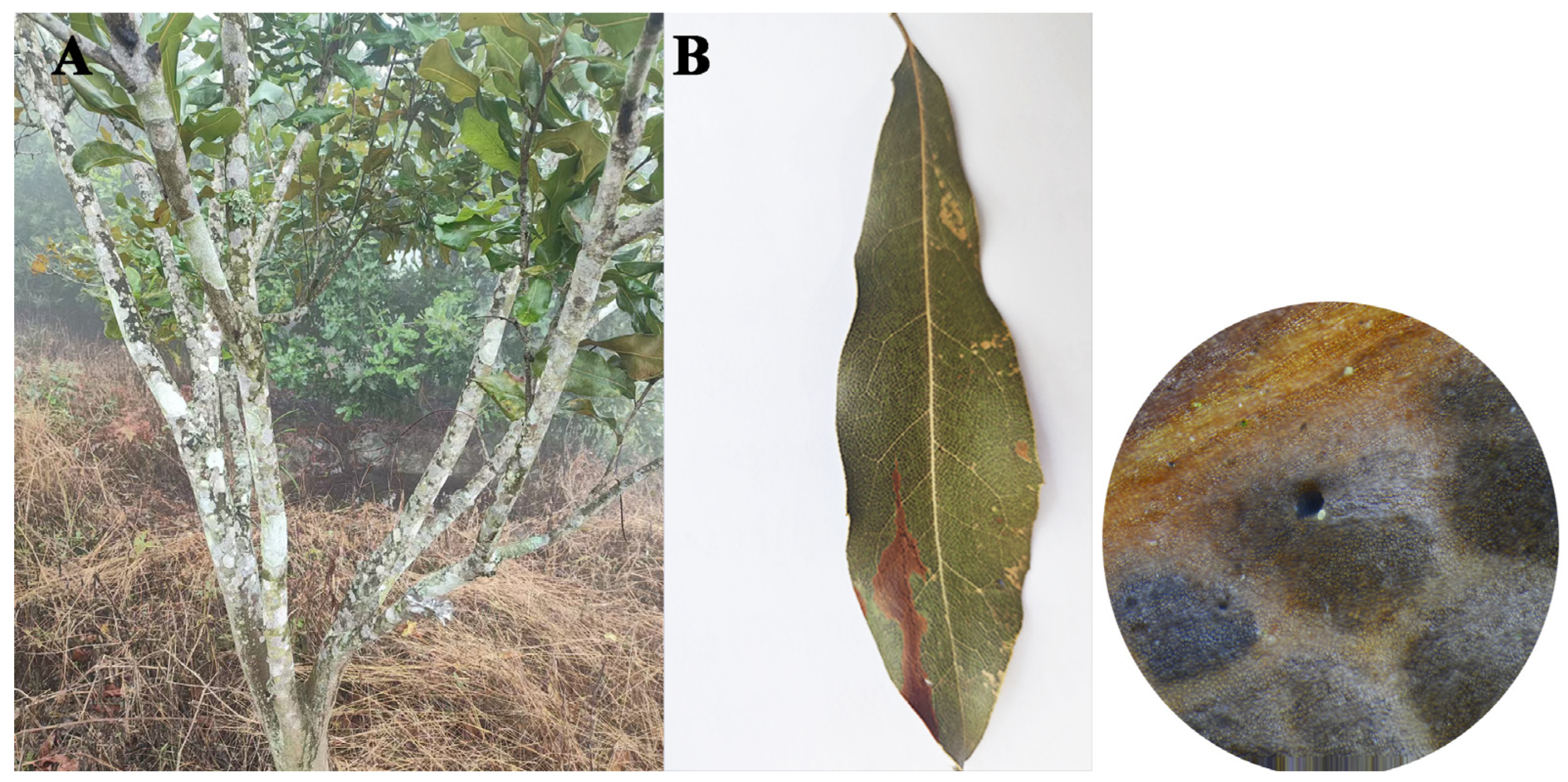
Figure 1. M. integrifolia leaf blight field symptoms. (A) M. integrifolia leaf blight field symptoms; (B) symptoms of M. integrifolia leaf blight.
Jof 11 00613 g001
Figure 2. Symptoms of Pestalotiopsis colombiensis. (A) Conidia with two appendages; (B) conidia with three appendages; (C) colony on PDA after 25 days; scale = 10 μm.
Jof 11 00613 g002
Figure 3. Phylogram inferred from neighbor-joining analysis of combined ITS, LSU and TEF1-a sequence data. The values above the branches indicate bootstrap values. The percentage of replicate trees in which the associated taxa clustered together in the bootstrap test (1000 replicates) is indicated next to the branches. Numbers above branches show bootstrap support values. Newly obtained sequences in this study are depicted in red.
Jof 11 00613 g003
Figure 4. Symptoms of P. colombiensis and symptoms of re-inoculated leaves after 15 days and sterile water processing. (A) Control; (B) re-inoculated.
Jof 11 00613 g004
Table 1. Primer sequences.
LocusPrimerSequence (5′–3′)Tm (℃)Reference
TEFl-αEF1-728FGTACCCGCTGAACTTAAGC52[20]
EF2GGA(G/A)GTACAGT(G/C)ATCATGTT[21]
LSULRORGTACCCGCTGAACTTAAGC47[22]
LR5ATCCTGAGGGAAACTTC[23]
ITSITS1TCCGTAGGTGAACCTGCGG50[24]
ITS4TCCTCCGCTTATTGATAT
Table 2. GeneBank accession numbers used in the phylogenetic analyses.
IsolateCountry/LocationHost/HabitatITSLSUEF
Pestalotiopsis telopeae
JX16b; CBS 113606; CBS 114161China; ThailandUnknown; Telopea sp.OR781522KM116202KM199500
GZ22b; CBS 114137China; ThailandUnknown; Protea sp.OR781519KM116219KM199559
P. trachycarpicola
ZHKUCC 23-1026; AV-108; GUCC 23-0414China; IndiaCave subculture; Persea americana; UnknownPP029403PQ299650PQ141020
PB2-e; AV-244; OP068China; India; ThailandPanax notoginseng; Persea americana; Trachycarpus fortuneiOR056327PQ299673JQ845946
YA3; AV-191; OP143China; India; ThailandUnknown; Persea americana; Podocarpus macrophyllusOR484805PQ299666KC537816
P. oryzae
CBS 171.26ThailandUnknownKM199304MH866374KM199494
MWN35; Y2-3; CBS 111522Kenya; China; ThailandOryza sativa; Unknown; Telopea sp.OM899910PQ432485KM199493
P. knightiae
KoRLI046217; NSW:7213; CBS 111963Korea; Australia; ThailandStereocaulon japonicum; Banksia serrata; Knightia sp.MN341554OP082342KM199406
CBS 114138ThailandKnightia sp.KM199310KM116227KM199408
P. biciliata
CBS 236.38Netherlands; ThailandUnknown; Paeonia sp.MH855953KM116214KM199506
UMAS P16; CBS 124463Malaysia; ThailandShorea macrophylla; Platanus x hispanicaKT337374KM116224KM199505
P. parva
D034; CBS 265.37Malaysia; ThailandCocos nucifera; Delonix regiaOP727543KM116226KM199508
CBS 278.35ThailandLeucothoe fontanesianaKM199313KM116205KM199509
P. humus
CBS 115450ThailandUnknown; Ilex cinereaKM199319KM116208KM199487
CBS 336.97ThailandUnknownKM199317KM116230KM199484
P. australis
CBS 114193; CBS 114474; CBS 111503ThailandGrevillea sp.; Protea susannaeNR145239KM116220KM199557
CBS 114193ThailandGrevillea sp.KM199332KM116197KM199475
P. chamaeropis
CBS 186.71Thailand; NetherlandsChamaerops humilis; UnknownKM199326MH871839KM199473
CBS 237.38; LC3619Australia; Netherlands; ChinaProstanthera rotundifolia; Unknown; Camellia sp.KR259104MH867450KX895208
P. macadamiae
BRIP 63739b; BRIP 70520Australia; IndiaMacadamia sp.; Pongamiea sp.KX186587OP363152MZ327136
BRIP 70519AustraliaMacadamia sp.MZ303757-MZ327135
P. colombiensis
CBS 118553ThailandEucalyptus grandisNR147551NG069213KM199488
5F; CBS 118553Australia; ThailandOryza australiensis; Eucalyptus grandisKU715149KM116222-
SWFUCB2ChinaMacadamia integrifoliaPQ895603PQ895623 PQ997934
SWFUCB1ChinaMacadamia integrifoliaPQ895604PQ895622PQ997935
Neopestalotiopsis saprophytica
CBS 115452ThailandLitsea rotundifoliaKM199345KM116251KM199538
“-” The database did not find this sequence.
Disclaimer/Publisher’s Note: The statements, opinions and data contained in all publications are solely those of the individual author(s) and contributor(s) and not of MDPI and/or the editor(s). MDPI and/or the editor(s) disclaim responsibility for any injury to people or property resulting from any ideas, methods, instructions or products referred to in the content.