Article

The Novel Disease Vicia unijuga Caused by Colletotrichum tofieldiae in China: Implications for Host Growth, Photosynthesis, and Nutritional Quality

State Key Laboratory of Herbage Improvement and Grassland Agro-Ecosystems, Engineering Research Center of Grassland Industry, College of Pastoral Agriculture Science and Technology, Lanzhou University, Lanzhou 730020, China
*
Author to whom correspondence should be addressed.
J. Fungi 2025, 11(8), 567; https://doi.org/10.3390/jof11080567
Submission received: 1 July 2025 / Revised: 27 July 2025 / Accepted: 28 July 2025 / Published: 29 July 2025

Abstract

Vicia unijuga, an important forage legume on China’s Qinghai–Tibetan Plateau, exhibited dark-brown sunken lesions on their stems at the Qingyang Experimental Station of Lanzhou University. The fungus isolated from the diseased tissues was identified as Colletotrichum tofieldiae via a multi-locus phylogeny (ITS-ACT-Tub2-CHS-1-GADPH-HIS3). The pathogenicity was confirmed by Koch’s postulates. The inoculated plants showed significantly reduced (p < 0.05) growth parameters (height, root length, and biomass), photosynthetic indices (net rate, transpiration, and stomatal conductance), and nutritional quality (crude protein, crude fat, crude ash, and crude fiber) compared to the controls. C. tofieldiae additionally infected six legume species (V. sativa, Medicago sativa, Onobrychis viciifolia, Astragalus adsurgens, Trifolium pratense, and T. repens). Optimal in vitro growth occurred on oatmeal agar (mycelium) and cornmeal agar (spores), with D-sucrose and D-peptone as the best carbon and nitrogen sources. This first report of C. tofieldiae causing V. unijuga anthracnose advances the understanding of legume anthracnose pathogens.

1. Introduction

Vicia unijuga (commonly known as perennial vetch), an important forage legume in the Leguminosae family, is broadly distributed in China, North Korea, and Russia [1]. V. unijuga has an extremely extensive utilization value as a medicinal plant, edible wild vegetable, and landscaping plant. Nonetheless, its feed value is most important because of its high protein and soluble sugar content [2]. However, the occurrence of diseases adversely affects the plant growth, yield, and nutritional quality of V. unijuga. Six genera, including Cylindrosporium [3], Erysiphe [4,5,6], Microsphaera [4], Oidium [4,6], Septoriella [7], and Uromyces [4,5,8,9,10], cause fungal diseases in V. unijuga [11]. Of note, five of these fungal pathogens have been reported in China.
Colletotrichum (Glomerellaceae, Glomerellales, Sordariomycetes, and Ascomycota) is a large genus of ascomycete fungi that comprises 280 species, whose molecular data is available and accepted in the genus [12]. The Colletotrichum species include pathogens, endophytes, and saprobes. Most of the Colletotrichum pathogens are the principal causes of plant anthracnose in almost every crop grown globally [13]. More than 97% of the documented Colletotrichum–host associations occur on dicotyledonous and monocotyledonous plants [14], whereas 33 species are endophytes that have mutualistic relationships with their hosts [12,15]. Notably, the Colletotrichum pathogens are the eighth most important group of plant pathogenic fungi globally, based on their scientific and economic importance [13]. These pathogens can infect the stems, leaves, flowers, and fruits, which hinders plant growth and reduces yield [16]. The Colletotrichum species can infect multiple Vicia species. For instance, V. faba has been infected by C. acutatum in Australia [17], while C. lentices and C. spinaciae have caused anthracnose in V. sativa, which has led to serious losses in the Gansu province of China [18,19,20].
Previously, Colletotrichum was classified based on its morphology and host range. However, there are only a few unique morphological characteristics that can be used for identification, which has led to the misidentification of species because of the lack of a clear understanding of host specificity [21]. In recent years, a multi-locus sequence analysis has become the primary method for accurate species identification [22]. Many researchers have provided a comprehensive set of methods to guide the delimitation and classification of Colletotrichum species [21,23]. Currently, the internal transcribed spacers (ITS), glyceraldehyde-3-phosphate dehydrogenase (GAPDH) gene, and actin (ACT) gene are the commonly used genes for the molecular identification of Colletotrichum. Both existing and newly discovered Colletotrichum species can be classified and identified based on a multi-locus sequence analysis of these genes [24,25].
V. unijuga is a wild forage legume widely distributed in the alpine steppe of the Qinghai–Tibetan Plateau of China. It has been selected as a cultivated forage legume for breeding because of its strong adaptability [26,27,28], good palatability, and high feeding value [2]. Noteworthy, the research team from the College of Pastoral Agriculture Science and Technology of Lanzhou University successfully domesticated and selected cultivated V. unijuga varieties after years of experimentation, which have since been approved. Comprehensively studying plant diseases is foundational for large-scale cultivation and is critical for developing effective disease control strategies.
Currently, the pathogen responsible for anthracnose in V. unijuga remains poorly understood. This study thus aimed to (i) identify the causal pathogen through tissue isolation, morphological observation, pathogenicity, and a multi-locus sequence analysis; (ii) explore the host range and biological characteristics of the causal pathogen; and (iii) study its effects on the growth and photosynthesis indicators and nutritional quality of V. unijuga.

2. Materials and Methods

2.1. Sampling and Isolation

V. unijuga plants exhibiting disease symptoms on their stems were identified and collected from the Qingyang Experimental Station of Lanzhou University (35.67° N, 107.85° E) in Gansu Province in September 2022. The stem tissues of the V. unijuga plants at the border between the symptomatic and healthy areas were selected for pathogen isolation. The stem surfaces were first rinsed with running water, disinfected with 75% alcohol for 15 s and 1% NaClO for 3 min, washed thrice with sterile water, and then dried using sterile filter paper. The stem tissues were subsequently cut into appropriate sizes, followed by evenly plating five pieces onto potato dextrose agar (PDA, Beijing Land Bridge Technology Co., Ltd., Beijing, China). Stem sampling was conducted using ten plants. The tissues from each plant were plated onto 3 PDA plates. Fresh mycelium was picked and purified on PDA after the initial colony formation. The purified isolates were stored at 4 °C and −80 °C in the Strain Preservation Library of the College of Pastoral Agriculture Science and Technology of Lanzhou University.

2.2. Morphological Characterization

Fungal plugs were grown on PDA medium at 25 °C to observe colony morphology and measure their growth rates. Micrographs of conidia and sporodochia, 100 and 50, respectively, were randomly taken. Moreover, 50 conidial appressoria [29] were randomly measured. Scanning electron microscope (Apreo S, Thermo Fisher Scientific, Waltham, MA, USA) was used for observations and measurements.

2.3. DNA Extraction, PCR Amplification, Sequencing, and Phylogenetic Analysis

Fungal DNA was extracted from fresh mycelium using an Omega Biotek E.Z.N.A® High Performance (HP) Fungal DNA Kit (Norcross, GA, USA). The fungal isolates were further characterized by amplifying six gene loci: ITS using ITS1/ITS4 primers [30], ACT using ACT-512F/ACT-783R primers [31], β-tubulin (Tub2) using T1/Bt-2b primers [32,33], chitin synthase 1 (CHS-1) using CHS-354R/CHS-79F primers [31], GAPDH using GDF1/GDR1 primers [34], and histone H3 (HIS3) using CYLH3F/CYLH3 primers [35]. The primers used were synthesized by Sangon Biotech Co., Ltd., Shanghai, China.
A polymerase chain reaction (PCR) was performed using a BIO-RAD T100 thermal cycler. The reaction mix comprised 12.5 μL of 2 × Taq PCR Master Mix (Tiangen Biotech Co., Ltd., Beijing, China), 9.5 μL of double-distilled H2O, 1 μL of each primer, and 1 μL of template DNA, totaling 25 μL. The PCR amplification program was as follows: an initial denaturation at 95 °C for 30 s; primer annealing at 56 °C (ITS), 59 °C (GADPH), 57 °C (ACT), 56 °C (Tub2), 59 °C (CHS-1), and 57 °C (HIS3) for 30 s, and extension at 72 °C for 1 min; and a final extension at 72 °C for 10 min. The PCR products were sent to Sangon Biotech (Shanghai) Co., Ltd. for bidirectional sequencing.
The sequence reads were subsequently spliced using SeqMan 5.0 (DNASTAR, Inc®. Madison, WI, USA) and blasted in the National Center for Biotechnology Information (NCBI) database to find regions of local similarity between the sequences. A phylogenetic tree construction was performed using carefully selected type strain sequences (ex-type/ex-epitype) of the target species, validated by Damm et al. [36] and retrieved from GenBank (Table S1). Both the individual and multi-locus genes were aligned and trimmed using MEGA 6.0 software [37]. A multi-gene phylogenetic tree was constructed using the maximum likelihood (ML) method [38] using MEGA 6.0. The tree was based on the concatenated sequences from six genomic regions (ITS-ACT-Tub2-CHS-1-GAPDH-HIS3), with C. lindemuthianum used as the outgroup.

2.4. Koch’s Postulates and Host Range

Seven common leguminous forages, V. unijuga, V. sativa, Medicago sativa, Onobrychis viciifoila, Astragalus adsurgens, Trifolium pratense, and T. repens, were selected to assess the pathogenicity of the fungus in the greenhouse. The forage species were grown for two weeks, and their stems were subsequently sprayed with a conidial suspension of the isolate LYZ0664 (approximately 106 CFU mL−1) [39] and kept in a moist environment at 25 °C. The control plants were sprayed with sterile water. Each forage species had 45 treated and 45 control plants. The plants were covered for 48 h with black plastic bags sprayed with sterile water to create a moist environment for infection to take place.
The disease progression was monitored and recorded, and subsequent statistical calculations of the incubation period, incidence rate (1), disease index (2), and reisolated rate (3) for each plant species were performed. The incubation period means the time elapsed from pathogen inoculation to the first observation of typical disease symptoms (measured in days). The disease severity was classified based on a slightly modified Li and Nan’s [40] disease severity standard (Table S2).
incidence   rate   ( % ) = diseased   palnts total   number   of   investigated   plants × 100 ,
disease   index = number   of   plants / scale   ×   scale   value highest   scale   vale   ×   total   number   of   plants × 100 ,
reisolated   rate   ( % ) = number   of   diseased   plants   reisolated   to   the   target   pathogen total   number   of   diseased   plants × 100 ,

2.5. Biological Characteristics of Isolated Fungi

The isolated fungi were grown on various media, carbon and nitrogen sources, and at different temperatures to determine how their growth rates and sporulation were affected by these changes. Ten types of media were tested: PDA, corn meal agar (CMA), potato carrot agar (PCA), oatmeal agar (OMA), potato sucrose agar (PSA), carnation leaf agar (CLA), water agar (WA), wheat straw decoction agar (WHDA), beef extract peptone agar (NA), and tomato agar (V8). Eight types of carbon sources were tested in 5 g KNO3, 2 g Na3PO4, 1 g Mg2SO4, 17 g agar, and 1000 mL distilled water as the basic medium. The carbon sources were 20 g of glucose, fructose, sucrose, lactose, starch-soluble, maltose, xylose, and mannitol. Seven types of nitrogen sources were tested in 5 g glucose, 2 g Na3PO4, 1 g Mg2SO4, 17 g agar, and 1000 mL distilled water as the basic medium. The nitrogen sources were 5 g of peptone, urea, NaNO3, (NH4)2SO4, aspartate, tryptophan, and glycine. Seven temperatures were tested: 5 °C, 10 °C, 15 °C, 20 °C, 25 °C, 30 °C, and 35 °C. The tests on the different culture media, carbon, and nitrogen sources were performed at 25 °C in the dark. The different temperatures were tested using PDA medium.
Hyphal plugs of the isolate (diameter = 0.5 cm) were inoculated at the center of the culture dishes, with 5 replicates per treatment. The colony diameter was measured using a vernier caliper after 7 days of culture.
The sporulation was assessed using a hemocytometer (25 middle squares × 16 small squares per counting chamber, chamber volume = 0.1 mm3; Anhui Ranjeck Technology Co., Ltd., Hefei, China) after 21 days of fungal growth. To each fungal culture plate, 5 mL of sterile distilled water was added, and the mycelial surface was gently scraped with a sterile spreader to obtain spore suspension. The resulting suspension was filtered through four layers of sterile gauze to remove mycelial debris, and the filtrate was collected in a sterile centrifuge tube. The spores were pelleted by centrifugation and resuspended in sterile water to an appropriate dilution. A calibrated volume of the diluted spore suspension was pipetted onto the hemocytometer, and the spores within the five middle squares (selected via the five-point sampling method) were counted under a light microscope using the “count top and left, exclude bottom and right” rule. This counting process was repeated 5 times per sample. The spore concentration was calculated using Formula (4).
Sporulation   concentration   ( CFU / mL ) = total   spores   in   5   middle   squares 5 × 25 × 10,000 × dilution   multiple

2.6. Influence of the Isolate LYZ0664 on the Growth Indicators of V. unijuga

The growth indicators of V. unijuga were measured on the 21st day post inoculation with the isolate LYZ0664. The plant height was measured from the soil surface to the plant’s highest point. The dry weight of this section was recorded as the aboveground dry weight. The root length extended from the base of the plant to the root tip. The dry weight of this section was recorded as the underground dry weight.

2.7. Influence of the Isolate LYZ0664 on the Photosynthesis Indicators of V. unijuga

The photosynthetic parameters of V. unijuga, such as the photosynthetic rate (Pn), stomatal conductance (Gs), intercellular carbon dioxide concentration (Ci), and transpiration rate (Tr), were measured on the 7th, 14th, and 21st days post inoculation with the isolate LYZ0664. These measurements were performed using a GFS-3000 gas exchange system (WALZ). The measurements were taken for the same leaf position with consistent growth and illumination across 5 plants. The measurements were performed for 3 leaves per plant and each leaf was measured thrice.

2.8. Influence of the Isolate LYZ0664 on the Nutritional Quality of V. unijuga

Thirty-five diseased and control plants of V. unijuga were sampled from the greenhouse and sent to the Forage Quality Testing Center of the Feeding Laboratory of Lanzhou Polison Ecological Science and Technology Co., Ltd., Lanzhou, China in Gansu Province for a conventional nutrient analysis. The nutritional components analyzed included the crude protein, crude ash, crude fat, neutral detergent fiber, acid detergent fiber, crude fiber, and water content [41].

2.9. Data Analysis

A statistical analysis of the various parameters was performed using SPSS 22.0, while Duncan’s method was used for significance testing. The results are expressed as means ± standard error (SE). Photo editing was performed using GraphPad Prism 8 and Photoshop CS6.

3. Results

3.1. Disease Survey and Strain Isolation

A disease survey in the field revealed a disease incidence rate of 78.93% and a disease index of 57.77. The susceptible plants exhibited complete desiccation and death (Figure 1A). The lesions on the stems were linear and dark brown, expanding to a diameter of 1 to 4 cm (Figure 1B). The potential pathogenic fungus was isolated, with a plant carrying rate of 100% and an isolation rate of 46.67%. Three strains with the same cultural characteristics of the fungus were selected for purification and were numbered as LYZ0664, LYZ0665, and LYZ0666.

3.2. Morphological Characterization of LYZ0664

The colony color on the PDA medium was pink-white initially, with black filamentous hyphae appearing at the center after 3 days. The colony color then turned gray-green and eventually became black with gray-white protrusion particles on the surface. The short, dense hyphae grew in a wheel-like pattern (Figure 1C). Conidiomata brown, with setae, directly formed on them. Setae brown, septate, smooth, 97.2 to 151.2 μm long, and were conically enlarged at the base and acutus at the top (Figure 1D). Conidia hyaline, crescent shaped, aseptate, smooth walled, and 15.27 to 19.73 × 3.66 to 5.72 μm in size (Figure 1E). Conidial appressoria, solitary, ellipsoidal to clavate, smooth, aseptate, brown or dark brown to almost black, and 8.5 to 21.5 × 4.5 to 11 μm in size (Figure 1F).

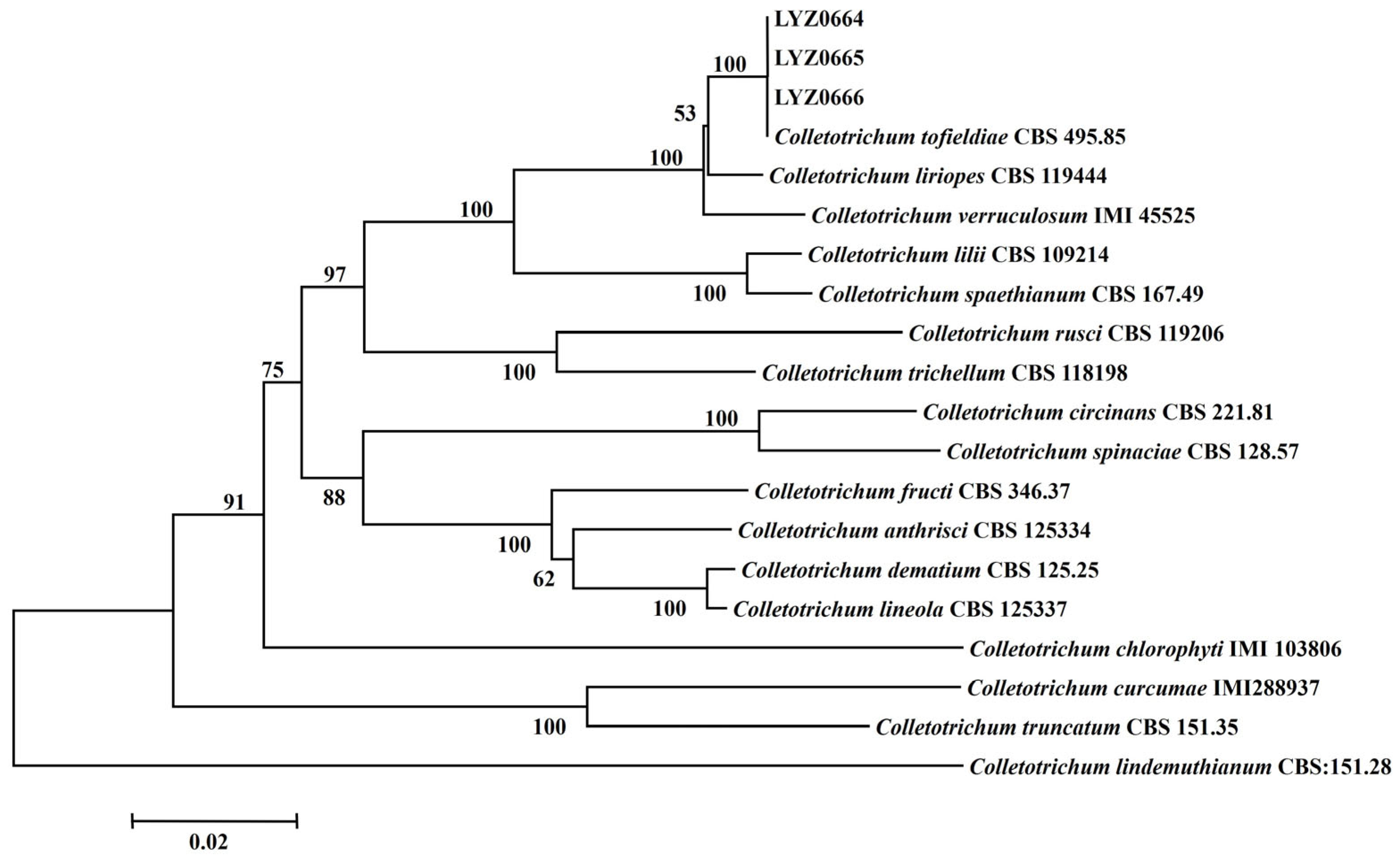
3.3. Phylogenetic Analysis

The BLAST analysis of the ITS, ACT, Tub2, CHS-1, GAPDH, and HIS3 gene sequences of LYZ0664, LYZ0665, and LYZ0666 using the NCBI database revealed the high similarity (>99.8%) of the six DNA sequences to C. tofieldiae. LYZ0664, LYZ0665, LYZ0666, and C. tofieldiae (CBS 495.85) clustered on the same branch, demonstrating a 100% genetic relationship (Figure 2). The strains were identified as C. tofieldiae based on a multi-locus phylogenetic analysis.

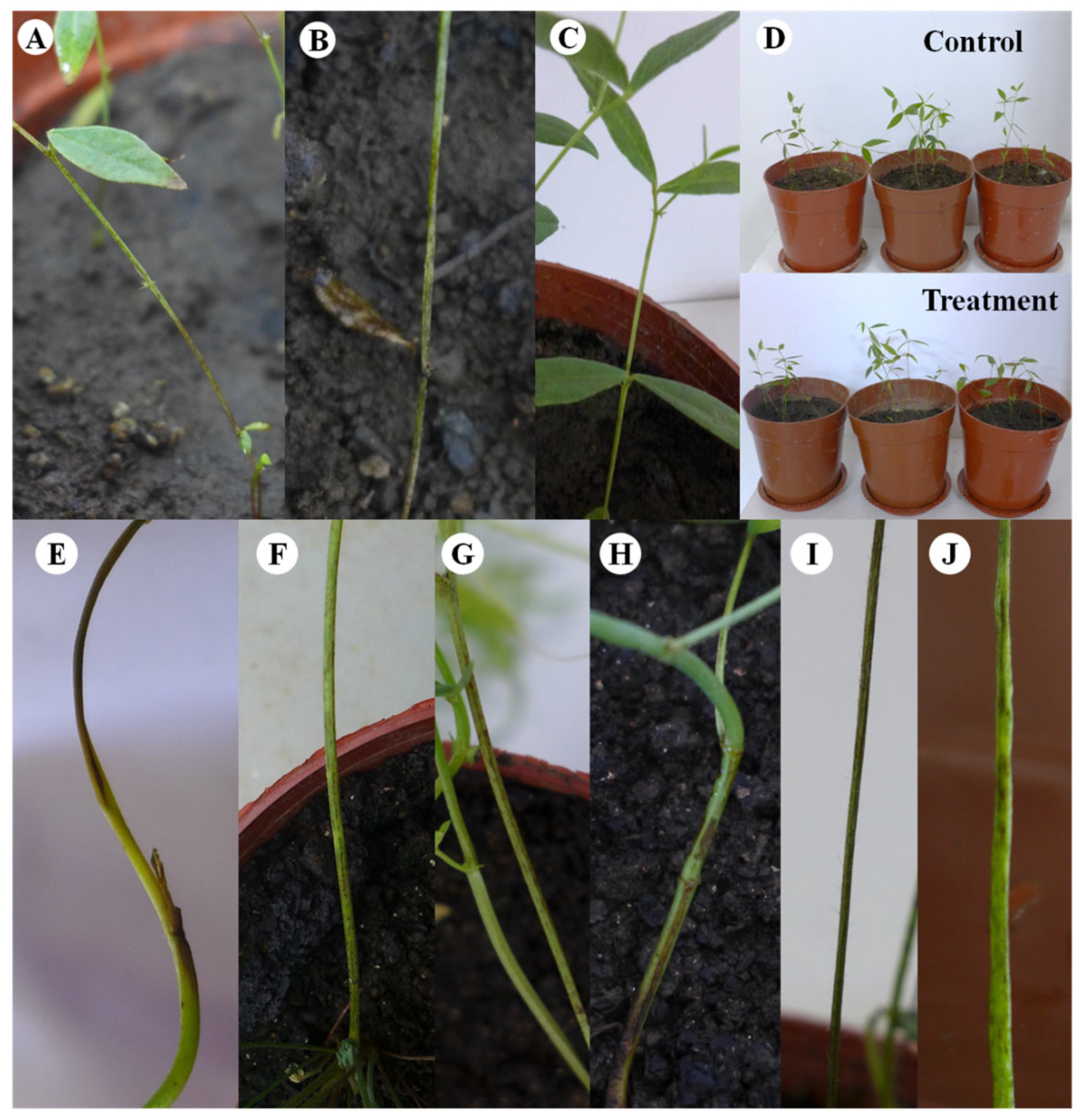
3.4. Koch’s Postulates and Host Range of LYZ0664

Small, round, yellowish-brown, and sunken lesions manifested on the stems of V. unijuga 4 days post inoculation (Figure 3A). The lesions spread into a single mass, with the stems progressively turning black on the 7th day (Figure 3B). In contrast, the control remained unaffected (Figure 3C). Noteworthy, the inoculated plants exhibited reduced vigor (Figure 3D).
The inoculation of V. sativa, M. sativa, O. viciifolia, A. adsurgens, T. pratense, and T. repens with the pathogenic fungus under greenhouse conditions led to all six forages displaying similar symptoms. Small black-brown lesions appeared during the early stage, which subsequently expanded into larger patches (Figure 3E–J).
C. tofieldiae infected all seven tested legume forages, with the infection severity varying among the hosts. Notably, the highest incidence rate (73.33%) and disease index (57.22) (Table 1) were observed on V. sativa, which belongs to the same genus as the pathogen’s original host.

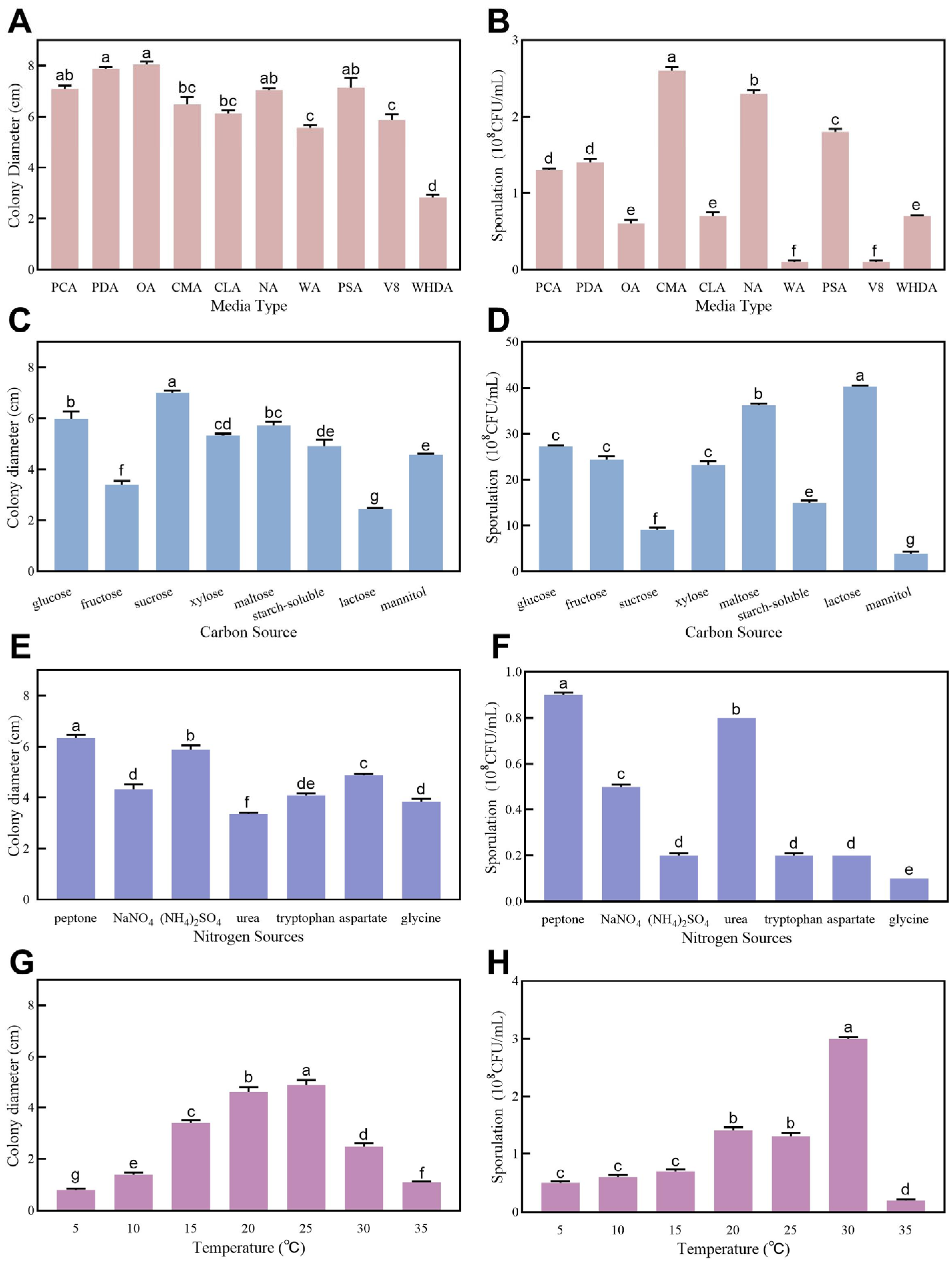
3.5. Biological Characteristics of LYZ0664

The fungal strain demonstrated growth and sporulation capabilities across all the tested conditions: 10 culture media, eight carbon sources, seven nitrogen sources, and temperatures ranging from 5 to 35 °C (Figure 4). Significant variations (p < 0.05) were observed in both the mycelial growth and sporulation yields among the different media types. All the tested media, except WHDA, supported mycelial development (Figure 4A), with CMA proving optimal for sporulation (Figure 4B). The carbon sources exerted significant differential effects (p < 0.05) on fungal development. D-sucrose maximized mycelial growth (Figure 4C), whereas D-lactose enhanced sporulation most effectively (Figure 4D). Among the nitrogen sources, D-peptone significantly outperformed the others (p < 0.05) for both hyphal expansion and spore production (Figure 4E,F). The optimal temperatures were 25 °C for mycelial growth and 30 °C for sporulation (Figure 4G,H).

3.6. Influence of LYZ0664 on Growth Indicators of V. unijuga

All six growth indices, plant height, root length, aboveground fresh weights, aboveground dry weights, underground fresh weights, and underground dry weights, exhibited a decline post inoculation. Specifically, inoculation with the pathogen significantly reduced the underground dry weight by 61.8%, the underground fresh weight by 39.0%, and the aboveground fresh weight by 33.7% (Table 2).

3.7. Influence of LYZ0664 on Photosynthesis Indicators of V. unijuga

Inoculation with the pathogen led to a gradual decline in the Pn, Gs, and Tr, with the respective values significantly reduced (p < 0.05) at all tested time points compared to the corresponding controls (Figure 5). Notably, the Pn and Gs showed the highest reductions on day 21 (68.77% and 55.15%, respectively; p < 0.001) compared to the controls. In contrast, inoculation with the pathogen increased the Ci at all time points but was not significantly different (p > 0.05) from that of the control plants.

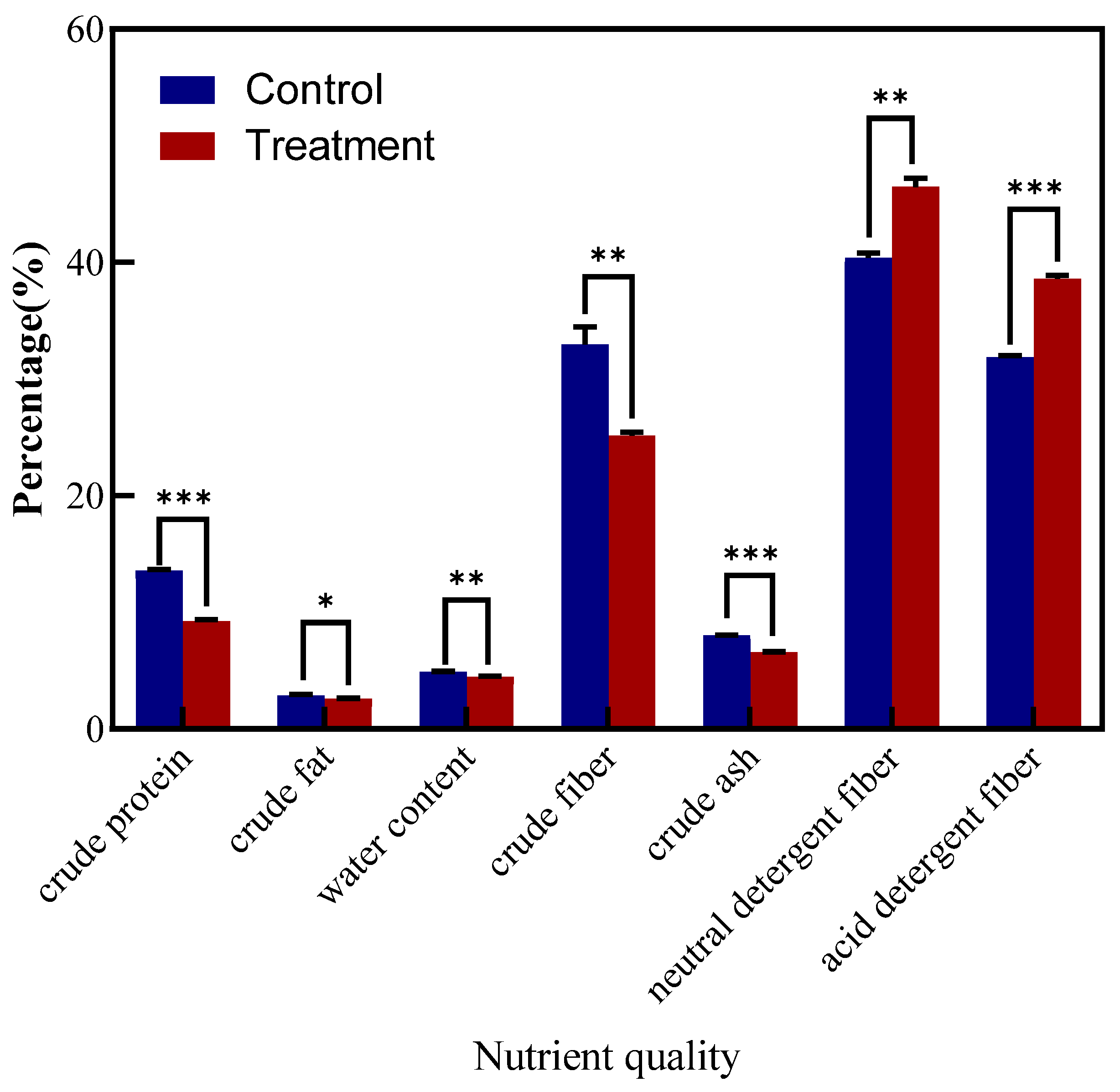
3.8. Influence of LYZ0664 on Nutritional Quality of V. unijuga

Inoculation with the pathogen caused varying changes in the crude protein, crude ash, crude fat, neutral detergent fiber, acid detergent fiber, crude fiber, and water content (Figure 6), with significant differences compared to the controls. Highly significant differences (p < 0.001) were observed in the crude protein (13.6% to 9.3%), crude ash (8.0% to 6.6%), and acid detergent fiber (31.9% to 38.6%) contents among the tested samples.

4. Discussion

The pathogenic fungus C. tofieldiae was first identified as Vermicularia tofieldiae [42] based on its morphology and then later classified as a variant of C. dematium var. minus [43]. In 2009, Damm et al.’s work [36] strongly indicated that it belongs to the C. destructivum rather than the C. dematium clade, and renamed it C. tofieldiae based on the ITS sequences. It was subsequently classified within the spaethianum species complex [21]. The fungus exists on hosts as an endophyte, pathogen, and saprotroph [44]. It has been reported as a pathogen in Dianthus sp. in the UK [36], and in Grevillea crithmifolia and Iris × germanica in Australia [17]. In contrast, it has been found on dead stems and leaves in the form of saprophytic fungi on Lupinus polyphyllus in Germany [36], Tofieldia calyculata in Switzerland [45] and China [36,46], and Ornithogalum umbellatum in Japan [47]. Among the published reports, only Damm et al. [36] have detailed the morphological characteristics of the fungus, including the conidiomata, setae, conidia, and appressoria, among other characteristics. Noteworthy, these descriptions align with the findings of this study, supporting their validity
Despite the numerous species of Colletotrichum, the lack of distinctive morphological traits complicates their identification and classification [48]. For instance, the C. spaethianum clade contains only five species and lacks definitive morphological traits that serve as diagnostic markers [21]. Taxonomic studies of the Colletotrichum genus have demonstrated that certain species exhibit 100% homology in a single gene, resulting in erroneous identification outcomes [49,50]. Consequently, combined multi-gene tree construction has gradually become mainstream in research [51]. The spaethianum clade receives strong support in multi-locus analysis, which incorporates ITS, GAPDH, CHS-1, HIS3, ACT, and TUB2 [21,36]. Herein, the fungal strains were identified using the ITS, GAPDH, CHS-1, HIS3, ACT, and TUB2 genes for joint identification, which determined a 100% similarity to CBS 495.86 (C. tofieldiae). Consequently, the pathogen causing anthracnose in V. unijuga in Gansu Province, China, was identified as C. tofieldiae and was described as a novel disease globally.
There is a growing demand for food production and consumption, particularly for animal proteins [52]. This growing demand underscores the importance of the selection and breeding of high-yielding, nutrient-rich, and locally adapted forage varieties that are disease-resistant [53,54]. V. unijuga exhibits a diverse array of functions because of its high protein and soluble sugar content, which makes it an excellent forage grass suitable for cultivation [2]. Studies have postulated that V. unijuga is a promising candidate for domestication and promotion as a high-altitude forage species [55]. The appropriate field management of V. unijuga can enhance its aboveground dry matter and seed yield [27,28,56]. This study provides the first report on the effects of C. tofieldiae on the nutritional quality of host forage grasses. C. tofieldiae significantly compromised the nutritional quality of the forage grasses, reducing the crude protein, crude fat, crude ash, crude fiber, and water content. Of note, C. tofieldiae significantly reduced the crude protein content by 31.61%.
The existing studies have postulated that the transition between pathogenic and beneficial lifestyles of the same microbial species on the same host depends on the host and environmental conditions, rather than alterations in the microbial genome [57,58]. The evidence supporting this finding includes the symbiotic relationship between Arabidopsis thaliana and C. tofieldiae, which is exclusively activated under phosphate-deficient conditions [57,59]. Noteworthy, this interaction is particularly beneficial for Brassicaceae plants with limited phosphate absorption capabilities [57]. Conversely, transitions in symbiotic relationships is attributable to changes in microbial gene expression. Hiruma et al. [60] revealed that the activation of the ABA-BOT (ABA and the associated sesquiterpene metabolite botrydial) gene cluster in C. tofieldiae induces the transition of the fungus from a mutualistic to a pathogenic lifestyle. Zimmermann et al. [59] reported that the lack of a phosphorylated pathway of serine biosynthesis, particularly the major components, such as indolic glucosinolates, causes C. tofieldiae to behave like a pathogen rather than a beneficial microorganism in plants. Consistent with this, Díaz-González et al. [61] suggested that the strain Ct0861 significantly enhances the shoot length, weight, and yield of Zea mays and Solanum lycopers under optimal nutrient conditions. In summary, the role of C. tofieldiae as either a pathogen or a beneficial microorganism is contingent upon various factors, including environmental conditions and gene expression. This complex mechanism is a subject for further research. Photosynthesis serves as the origin of numerous beneficial substances [62] and is fundamental to nearly all life forms in plants. Enhancing plant photosynthesis is essential because of the limited agricultural land and increasing population [63]. Herein, C. tofieldiae infection significantly reduced the photosynthetic capacity of V. unijuga. The plants were cultivated in nutrient-rich soil without a phosphate deficiency. The decrease in the net photosynthetic rate, transpiration rate, and stomatal conductance in the infected plants indicates that the pathogenic behavior of C. tofieldiae is attributable to the activation of the ABA-BOT gene cluster. This activation potentially results in the production of abscisic acid (ABA), leading to stomatal closure.
C. tofieldiae has been documented as a pathogen in only three plant species, with reports limited to its morphological characteristics. This study is the first to report C. tofieldiae as a pathogen causing anthracnose in V. unijuga by utilizing Koch’s postulates, its morphological characteristics, and a multi-locus sequence analysis. The study also comprehensively analyzes the optimal medium type, carbon and nitrogen sources, and temperature conditions that promote the growth and sporulation of C. tofieldiae, which marks the first such investigation. Additionally, the host range of the pathogen is assessed. Moreover, the impact of C. tofieldiae on the growth, photosynthetic, and nutritional parameters of V. unijuga is evaluated for the first time. The findings of this study underscore the significant infective potential of C. tofieldiae and reveal its wide host range and non-host specificity. Nonetheless, further investigations on the epidemiology, distribution, host range [64], breeding of anthracnose-resistant V. unijuga varieties [65,66], and fungicide selection [67] should be conducted in controlled and natural ecosystems to develop effective control strategies aimed at preventing economic losses.

5. Conclusions

This study establishes Colletotrichum tofieldiae as the causal agent of anthracnose on Vicia unijuga in Gansu Province, China, representing the first global report of this pathogen–host interaction. Its identification was confirmed through Koch’s postulates, a morphological characterization, and a multi-locus phylogenetic analysis. We further delineated the optimal conditions for mycelial growth and sporulation, revealing the pathogen’s adaptability across diverse media, carbon and nitrogen sources, and temperatures. Notably, C. tofieldiae significantly compromised the host’s physiology and nutritional quality. The infected plants exhibited a reduced photosynthetic capacity and a 31.61% reduction in the crude protein content, critically diminishing its forage value. The host-range trials demonstrated non-specific pathogenicity, suggesting a broad infection potential.

Supplementary Materials

The following supporting information can be downloaded at: https://www.mdpi.com/article/10.3390/jof11080567/s1, Table S1: Sequences information for Colletotrichum spp. used in this study; Table S2: The standard of severity levels.

Author Contributions

Conceptualization, Y.-Z.L.; methodology, Y.-Z.L.; software, T.-T.W. and H.L.; validation, T.-T.W., H.L. and Y.-Z.L.; formal analysis, T.-T.W.; investigation, H.L.; resources, Y.-Z.L.; data curation, T.-T.W. and H.L.; writing—original draft preparation, T.-T.W. and H.L.; writing—review and editing, Y.-Z.L.; visualization, T.-T.W.; supervision, Y.-Z.L.; project administration, Y.-Z.L.; funding acquisition, Y.-Z.L. All authors have read and agreed to the published version of the manuscript.

Funding

This work was supported by the Gansu Provincial Science and Technology Plan Program (23ADKA013), the National Key Research and Development Program of China (2022YFD1401103).

Institutional Review Board Statement

Not applicable.

Informed Consent Statement

Not applicable.

Data Availability Statement

All the sequences generated in this study were submitted to GenBank.

Conflicts of Interest

The authors declare no conflicts of interest.

References

  1. Board, C.F.E. Flora of China. Science Press: Beijing, China, 2006. [Google Scholar]
  2. Mao, Z.X.; Fu, H.; Niu, D.C.; Nie, B.; Chen, H. Nutrient variation and forage evaluation of Vicia unijuga in alpine grassland. Acta Prataculturae Sin. 2015, 24, 27–233. [Google Scholar]
  3. Watson, A.J. Foreign Bacterial and Fungus Diseases of Food, Forage, and Fiber Crops: An Annotated List; Department of Agriculture: Washington, DC, USA, 1971. [Google Scholar]
  4. Tai, F.L. Sylloge Fungorum Sinicorum; Science Press: Beijing, China, 1979. [Google Scholar]
  5. List of Plant Diseases in Korea. Available online: https://genebank.rda.go.kr/english/plntDissInfoMain.do (accessed on 16 November 2024).
  6. Zheng, R.Y.; Yu, Y.N. Flora Fungorum Sinicorum; Science Press: Beijing, China, 1987. [Google Scholar]
  7. Andrianova, T.V.; Minter, D.W. New species of Bartalinia and Septoriella from the Altai Mountains. Mycotaxon 2007, 101, 297–313. [Google Scholar]
  8. Cao, Z.M.; Li, Z.Q. Rust Fungi of Qinling Mountains; China Forestry Publishing House: Beijing, China, 1999. [Google Scholar]
  9. Guyot, A.L. Les Rouilles des Legumineuses Fourrageres et Spontanees; Editions Paul Lechevalier: Paris, France, 1957. [Google Scholar]
  10. Zhuang, J.Y. Flora Fungorum Sinicorum; Science Press: Beijing, China, 2005. [Google Scholar]
  11. USDA Fungus Databases. Available online: https://fungi.ars.usda.gov/ (accessed on 16 November 2024).
  12. Liu, F.; Ma, Z.Y.; Hou, L.W.; Diao, Y.Z.; Wu, W.P.; Damm, U.; Song, S.; Cai, L. Updating species diversity of Colletotrichum, with a phylogenomic overview. Stud Mycol. 2022, 101, 1–56. [Google Scholar] [CrossRef] [PubMed]
  13. Dean, R.; Van Kan, J.A.L.; Pretorius, Z.A.; Hammond-Kosack, K.E.; Di Pietro, A.; Spanu, P.D.; Rudd, J.J.; Dickman, M.; Kahmann, R.; Ellis, J.; et al. The top 10 fungal pathogens in molecular plant pathology. Mol. Plant Pathol. 2012, 13, 414–430. [Google Scholar] [CrossRef] [PubMed]
  14. Talhinhas, P.; Baroncelli, R. Hosts of Colletotrichum. Mycosphere 2023, 14, 158–261. [Google Scholar] [CrossRef]
  15. Rodriguez, R.; Redman, R. More than 400 milyears of evolution and some plants still can’t make it on their own: Plant stress tolerance via fungal symbiosis. J. Exp. Bot. 2008, 59, 1109–1114. [Google Scholar] [CrossRef]
  16. Liu, X.B.; Li, B.X.; Cai, J.M.; Zheng, X.L.; Feng, Y.L.; Huang, G.X. Colletotrichum species causing anthracnose of rubber trees in China. Sci. Rep. 2018, 8, 10435. [Google Scholar] [CrossRef]
  17. Shivas, R.G.; Tan, Y.P.; Edwards, J.; Dinh, Q.; Maxwell, A.; Andjic, V.; Liberato, J.R.; Anderson, C.; Beasley, D.R.; Bransgrove, K.; et al. Colletotrichum species in Australia. Australas Plant Pathol. 2016, 45, 447–464. [Google Scholar] [CrossRef]
  18. Wang, Q.; Duan, T.Y.; Nan, Z.B. First report of anthracnose caused by Colletotrichum spinaciae on Vicia sativa in China. Plant Dis. 2019, 103, 2138. [Google Scholar] [CrossRef]
  19. Xu, S.; Li, Y.Z. First report of common vetch anthracnose caused by Colletotrichum lentis in China. Plant Dis. 2015, 99, 1859. [Google Scholar] [CrossRef]
  20. Xu, S.; Christensen, M.J.; Li, Y.Z. Pathogenicity and characterization of Colletotrichum lentis: A causal agent of anthracnose in common vetch (Vicia sativa). Eur. J. Plant Pathol. 2017, 149, 719–731. [Google Scholar] [CrossRef]
  21. Cannon, P.F.; Damm, U.; Johnston, P.R.; Weir, B.S. Colletotrichum: Current status and future directions. Stud Mycol. 2012, 73, 181–213. [Google Scholar] [CrossRef] [PubMed]
  22. Weir, B.S.; Johnston, P.R.; Damm, U. The Colletotrichum gloeosporioides species complex. Stud Mycol. 2012, 73, 115–180. [Google Scholar] [CrossRef] [PubMed]
  23. Cai, L.; Hyde, K.D.; Taylor, P.W.J.; Weir, B.; Waller, J.M.; Abang, M.M.; Zang, J.C.; Yang, Y.L.; Phouliyong, S.; Prihastuti, Z.Y.; et al. A polyphasic approach for studying Colletotrichum. Fungal Divers. 2009, 39, 183–204. [Google Scholar]
  24. Damm, U.; Cannon, P.F.; Woudenberg, J.H.C.; Johnston, P.R.; Weir, B.S.; Tan, Y.P.; Shivas, R.G.; Crous, P.W. The Colletotrichum boninense species complex. Stud Mycol. 2012, 73, 1–36. [Google Scholar] [CrossRef]
  25. Damm, U.; Cannon, P.F.; Liu, F.; Barreto, R.W.; Guatimosim, E.; Crous, P.W. The Colletotrichum orbiculare species complex: Important pathogens of field crops and weeds. Fungal Divers. 2013, 61, 29–59. [Google Scholar] [CrossRef]
  26. Norman, H.C.; Humphries, A.W.; Hulm, E.; Young, P.; Vercoe, P.E. Productivity and nutritional value of 20 species of perennial legumes in a low-rainfall Mediterranean-type environment in southern Australia. Grass Forage Sci. 2021, 76, 134–158. [Google Scholar] [CrossRef]
  27. Tang, W.; Coulter, J.A.; Hu, X.W.; Han, Y.H.; Wang, Y.R.; Zhang, Z.X.; Li, R.; Nan, Z.B. Plastic-film mulch improves Vicia unijuga seed yield and yield components under subalpine climate conditions. Arch Agron Soil Sci. 2020, 66, 1171–1187. [Google Scholar] [CrossRef]
  28. Tang, W.; Christensen, M.J.; Nan, Z.B. Contributions of soil temperature and moisture drivers to variations in perennial vetch (Vicia unijuga) productivity potential in the Qinghai-Tibetan Plateau region of China. J. Agric Sci. 2019, 157, 150–168. [Google Scholar] [CrossRef]
  29. Yang, Y.L.; Liu, Z.Y.; Cai, L.; Hyde, K.D.; Yu, Z.N.; McKenzie, E.H.C. Colletotrichum anthracnose of Amaryllidaceae. Fungal Divers. 2009, 39, 123–146. [Google Scholar]
  30. White, T.J.; Bruns, T.; Lee, S.J.W.T.; Taylor, J. Amplification and direct sequencing of fungal ribosomal RNA genes for phylogenetics. Pcr Protocols. 1990, 18, 315–322. [Google Scholar]
  31. Carbone, I.; Kohn, L.M. A method for designing primer sets for speciation studies in filamentous ascomycetes. Mycologia 1999, 91, 553–556. [Google Scholar] [CrossRef]
  32. O’Donnell, K.; Cigelnik, E. Two divergent intragenomic rDNA ITS2 types within a monophyletic lineage of the fungus Fusarium are nonorthologous. Mol. Phylogenet Evol. 1997, 7, 103–116. [Google Scholar] [CrossRef]
  33. Lousie, G.N.; Donaldson, G.C. Development of primer sets designed for use with the PCR to amplify conserved genes from Filamentous Ascomycetes. Appl. Environ. Microbiol. 1995, 61, 1323–1330. [Google Scholar] [CrossRef]
  34. Guerber, J.C.; Liu, B.; Correll, J.C.; Johnston, P.R. Characterization of diversity in Colletotrichum acutatum sensu lato by sequence analysis of two gene introns, mtDNA and intron RFLPs, and mating compatibility. Mycologia 2003, 95, 872–895. [Google Scholar] [CrossRef] [PubMed]
  35. Crous, P.W.; Groenewald, J.Z.; Risede, J.; Hyweljones, N. Calonectria species and their Cylindrocladium anamorphs: Species with sphaeropedunculate vesicles. Stud Mycol. 2004, 50, 415–430. [Google Scholar]
  36. Damm, U.; Woudenberg, J.H.C.; Cannon, P.F.; Crous, P.W. Colletotrichum species with curved conidia from herbaceous hosts. Fungal Divers. 2009, 37, 45–87. [Google Scholar]
  37. Tamura, K.; Stecher, G.; Peterson, D.; Filipski, A.; Kumar, S. MEGA6: Molecular evolutionary genetics analysis version 6.0. Mol. Biol. Evol. 2013, 30, 2725–2729. [Google Scholar] [CrossRef]
  38. Kuhner, M.; Felsenstein, J. A simulation comparison of phylogeny algorithms under equal and unequal evolutionary rates. Mol. Biol. Evol. 1994, 11, 459–468. [Google Scholar] [CrossRef]
  39. Liu, L.P.; Liu, Y.N.; Yang, L.Y.; Lu, B.H.; Yang, L.N.; Wang, X.; Li, Y.; Gao, J.; Hsiang, T. First report of anthracnose caused by Colletotrichum destructivum on curly dock in China. Plant Dis. 2017, 101, 256. [Google Scholar] [CrossRef]
  40. Li, Y.Z.; Nan, Z.B. The Methods of Diagnose, Investigation and Loss Evaluation for Forage Disease; Phoenix Science Press: Nanjing, China, 2015. [Google Scholar]
  41. Bao, S.D. Soil and Agricultural Chemistry Analysis; China Agriculture Press: Beijing, China, 1999. [Google Scholar]
  42. Patouillard, N.T. Champignons parasites des phanérogames exotiques. Rev. Mycol. 1886, 8, 80–85. [Google Scholar]
  43. Wollenweber, H.W.; Hochapfel, H. Beiträge zur Kenntnis parasitärer und saprophytischer Pilze: VI. Vermicularia, Colletotrichum, Gloeosporium, Glomerella und ihre Beziehung zur Fruchtfäule. Z. Parasitenkd 1949, 14, 181–268. [Google Scholar]
  44. Talhinhas, P.; Baroncelli, R. Colletotrichum species and complexes: Geographic distribution, host range and conservation status. Fungal Divers. 2021, 110, 109–198. [Google Scholar] [CrossRef]
  45. Damm, U.; Sato, T.; Alizadeh, A.; Groenewald, J.Z.; Crous, P.W. The Colletotrichum dracaenophilum, C. magnum and C. orchidearum species complexes. Stud Mycol. 2019, 92, 1–46. [Google Scholar] [CrossRef] [PubMed]
  46. Hacquard, S.; Kracher, B.; Hiruma, K.; Münch, P.C.; Garrido-Oter, R.; Thon, M.R.; Weimann, A.; Damm, U.; Dallery, J.F.; Hainaut, M.; et al. Survival trade-offs in plant roots during colonization by closely related beneficial and pathogenic fungi. Nat Commun. 2016, 7, 11362. [Google Scholar] [CrossRef] [PubMed]
  47. Sato, T.; Moriwaki, J.; Kaneko, S. Anthracnose fungi with curved conidia, Colletotrichum spp. belonging to ribosomal groups 9-13, and their host ranges in Japan. JARQ. 2015, 49, 351–362. [Google Scholar] [CrossRef]
  48. Zhang, W.; Damm, U.; Crous, P.W.; Groenewald, J.Z.; Niu, X.L.; Lin, J.M.; Li, Y.T. Anthracnose disease of carpetgrass (Axonopus compressus) caused by Colletotrichum hainanense sp. nov. Plant Dis. 2020, 104, 1744–1750. [Google Scholar] [CrossRef]
  49. Hyde, K.D.; Cai, L.; McKenzie, E.H.C.; Yang, Y.L.; Zhang, J.Z.; Prihastuti, H. Colletotrichum: A catalogue of confusion. Fungal Divers. 2009, 39, 1–17. [Google Scholar]
  50. Yang, H.C.; Haudenshield, J.S.; Hartman, G.L. Colletotrichum incanum sp. nov., a curved-conidial species causing soybean anthracnose in USA. Mycologia 2014, 106, 32–42. [Google Scholar] [CrossRef]
  51. Guarnaccia, V.; Groenewald, J.Z.; Polizzi, G.; Crous, P.W. High species diversity in Colletotrichum associated with citrus diseases in Europe. Persoonia 2017, 39, 32–50. [Google Scholar] [CrossRef]
  52. Bai, Z.H.; Ma, W.Q.; Ma, L.; Velthof, G.L.; Wei, Z.B.; Havlík, P.; Oenema, O.; Lee, M.R.F.; Zhang, F.S. China’s livestock transition: Driving forces, impacts, and consequence. Sci. Adv. 2018, 4, eaar8534. [Google Scholar] [CrossRef]
  53. Thomas, D.T.; Moore, A.D.; Bell, L.W.; Webb, N.P. Ground cover, erosion risk and production implications of targeted management practices in Australian mixed farming systems: Lessons from the Grain and Graze program. Agric. Syst. 2018, 162, 123–135. [Google Scholar] [CrossRef]
  54. Li, S.; Wu, J.C.; Ma, L. Economic, energy and environmental consequences of shifting from maize-wheat to forage rotation in the North China Plain. J. Clean Prod. 2021, 328, 129760. [Google Scholar] [CrossRef]
  55. Zhou, F.F.; Matthew, C.; Yang, P.F.; Hnag, Y.F.; Nie, B.; Nan, Z.B. Leaf morphology, functional trait and altitude response in perennial vetch (Vicia unijuga A. Braun), alfalfa (Medicago sativa L.) and sainfoin (Onobrychis viciifolia Scop.). Planta 2023, 257, 75. [Google Scholar] [CrossRef] [PubMed]
  56. Tang, W.; Li, R.; Zhang, Z.; Baskin, C.C.; Nan, Z.B. Mulching affects seed set, provisioning, and offspring performance of Vicia unijuga (Fabaceae). Agron. J. 2019, 111, 1341–1357. [Google Scholar] [CrossRef]
  57. Hiruma, K.; Gerlach, N.; Sacristán, S.; Nakano, R.T.; Hacquard, S.; Kracher, B.; Neumann, U.; Ramírez, D.; Bucher, M.; O’Connell, R.J.; et al. Root endophyte Colletotrichum tofieldiae confers plant fitness benefits that are phosphate status dependent. Cell 2016, 165, 464–474. [Google Scholar] [CrossRef]
  58. Morcillo, R.J.; Singh, S.K.; He, D.X.; An, G.; Vílchez, J.I.; Tang, K.; Yuan, F.T.; Sun, Y.Z.; Shao, C.Y.; Zhang, S.; et al. Rhizobacterium-derived diacetyl modulates plant immunity in a phosphate-dependent manner. EMBO J. 2020, 39, e102602. [Google Scholar] [CrossRef]
  59. Zimmermann, S.E.; Blau, S.; Frerigmann, H.; Krueger, S. The phosphorylated pathway of serine biosynthesis is crucial for indolic glucosinolate biosynthesis and plant growth promotion conferred by the root endophyte Colletotrichum tofieldiae. Plant Mol. Biol. 2021, 107, 85–100. [Google Scholar] [CrossRef]
  60. Hiruma, K.; Aoki, S.; Takino, J.; Higa, T.; Utami, Y.D.; Shiina, A.; Okamoto, M.; Nakamura, M.; Kawamura, N.; Ohmori, Y.; et al. A fungal sesquiterpene biosynthesis gene cluster critical for mutualist-pathogen transition in Colletotrichum tofieldiae. Nat Commun. 2023, 14, 5288. [Google Scholar] [CrossRef]
  61. Díaz-González, S.; Marín, P.; Sánchez, R.; Arribas, C.; Kruse, J.; González-Melendi, P.; Brunner, F.; Sacristán, S. Mutualistic fungal endophyte Colletotrichum tofieldiae Ct0861 colonizes and increases growth and yield of maize and tomato plants. Agronomy 2020, 10, 1493. [Google Scholar] [CrossRef]
  62. Ryu, Y.; Berry, J.A.; Baldocchi, D.D. What is global photosynthesis? History, uncertainties and opportunities. Remote Sens Environ. 2019, 223, 95–114. [Google Scholar] [CrossRef]
  63. Stirbet, A.; Lazár, D.; Guo, Y.; Govindjee, G. Photosynthesis: Basics, history and modelling. Ann. Bot. 2020, 126, 511–537. [Google Scholar] [CrossRef]
  64. Liddle, R.L.; Akinsanmi, O.A.; Galea, V.J. Non-host specificity of Botryosphaeriaceae on macadamia and blueberry. Australas. Plant Pathol. 2019, 48, 65. [Google Scholar] [CrossRef]
  65. Mengistu, G.; Shimelis, H.; Laing, M.; Lule, D. Assessment of farmers’perceptions of production constraints, and their trait preferences of sorghum in western Ethiopia: Implications for anthracnose resistance breeding. Acta Agric. Scand. B 2019, 69, 241–249. [Google Scholar]
  66. Ridzuan, R.; Rafii, M.Y.; Ismail, S.I.; Mohammad, Y.M.; Miah, G.; Usman, M. Breeding for anthracnose disease resistance in chili: Progress and prospects. Int. J. Mol. Sci. 2018, 19, 3122. [Google Scholar] [CrossRef] [PubMed]
  67. Tuluhong, M.; Mu, M.Q.; Wang, S.N.; Li, Y.G.; Cui, G.W. Identification and characterization of Colletotrichum truncatum and C. destructivum causing stem rot of white clover in China. Plant Dis. 2024, 108, 1236–1245. [Google Scholar] [CrossRef] [PubMed]
Figure 1. Field symptoms and morphological characteristics of Colletotrichum tofieldiae (LYZ0664). (A,B) Field symptoms. (C) Colony on PDA after 7 days at 25 °C. (D) Acervuli and setae. (E) Conidia on PDA. (F) Appressorium.
Jof 11 00567 g001
Figure 2. Maximum likelihood (ML) phylogenetic analysis of concatenated ITS, ACT, Tub2, CHS-1, GAPDH, and HIS3 sequences reveals the clustering patterns of Colletotrichum tofieldiae strains obtained from Vicia unijuga. GenBank accession numbers are shown in Table S1.
Jof 11 00567 g002
Figure 3. Inoculation symptoms. (AD) Koch’s postulates of Colletotrichum tofieldiae on Vicia unijuga. (A,B) Symptoms of inoculation. (C) Control. (D) Control and infected potted plants. (EJ) Inoculation symptom of other six legumes: (E) Medicago sativa; (F) Onobrychis viciifoila; (G) V. sativa; (H) Astragalus adsurgens; (I) Trifolium pretense; (J) T. repens.
Jof 11 00567 g003
Figure 4. Colony growth and sporulation of Colletotrichum tofieldiae under different media types, carbon sources, nitrogen sources, and temperatures. (A) Effects of media types on colony growth; (B) Effects of media types on sporulation; (C) Effects of carbon sources on colony growth; (D) Effects of carbon sources on sporulation; (E) Effects of nitrogen sources on colony growth; (F) Effects of nitrogen sources on sporulation; (G) Effects of temperatures on colony growth; (H) Effects of temperatures on sporulation. The letters indicate a significant difference between different treatments at the p < 0.05 level.
Jof 11 00567 g004
Figure 5. Effects of Colletotrichum tofieldiae on the net photosynthetic rate, transpiration rate, stomatal conductance, and intercellular CO2 concentration in Vicia unijuga. (A) Effects on the net photosynthetic rate; (B) Effects on the transpiration rate; (C) Effects on the stomatal conductance; (D) Effects on the intercellular CO2 concentration. * indicates a significant difference between the inoculated and control groups at the p < 0.05 level; ** indicates a significant difference between the inoculated and control groups at the p < 0.01 level; *** indicates a significant difference between the inoculated and control groups at the p < 0.001 level; ns indicates not significant difference between the inoculated and control groups.
Jof 11 00567 g005
Figure 6. Influence on nutritional quality of Vicia unijuga inoculated with Colletotrichum tofieldiae. * indicates a significant difference between the inoculated and control groups at the p < 0.05 level; ** indicates a significant difference between the inoculated and control groups at the p < 0.01 level; *** indicates a significant difference between the inoculated and control groups at the p < 0.001 level.
Jof 11 00567 g006
Table 1. Infection index of Colletotrichum tofieldiae on seven legumes.
Forage SpeciesIncubation
Period (d)
Reseparation
Rate (%)
Incidence Rate (%) (Mean ± SE)Disease Index (Mean ± SE)
Vicia unijuga465.0060.00 ± 10.18 ab50.00 ± 2.89 a
Medicago sativa660.0028.89 ± 4.44 b15.00 ± 5.00 bc
Onobrychis viciifoila453.3340.00 ± 7.69 ab20.00 ± 6.67 bc
Vicia sativa376.0073.33 ± 13.87 a57.22 ± 15.48 a
Astragalus adsurgens560.0040.00 ± 11.55 ab15.00 ± 8.66 bc
Trifolium pratense380.0060.00 ± 20.00 ab28.33 ± 8.33 b
Trifolium repens353.3326.67 ± 6.67 b6.67 ± 1.67 c
Different letters in the same column mean significant difference at p < 0.05.
Table 2. Influence of Colletotrichum tofieldiae on the growth of Vicia unijuga.
Measurement IndexControlTreatmentBiomass Loss Rate (%)
Plant height (cm)14.60 ± 0.8110.8 ± 0.7326.0
Root length (cm)9.60 ± 0.408.20 ± 0.5814.6
Aboveground fresh weights (g)0.19 ± 0.020.13 ± 0.0133.7
Aboveground dry weights (g)0.05 ± 0.000.04 ± 0.0019.1
Underground fresh weights (g)0.24 ± 0.030.14 ± 0.0239.0
Underground dry weights (g)0.06 ± 0.000.02 ± 0.0061.8
Disclaimer/Publisher’s Note: The statements, opinions and data contained in all publications are solely those of the individual author(s) and contributor(s) and not of MDPI and/or the editor(s). MDPI and/or the editor(s) disclaim responsibility for any injury to people or property resulting from any ideas, methods, instructions or products referred to in the content.

