Article

The Study of Metschnikowia pulcherrima E1 in the Induction of Improved Gray Spot Disease Resistance in Loquat Fruit

School of Agricultural Engineering, Jiangsu University, Zhenjiang 212013, China
*
Author to whom correspondence should be addressed.
These authors contributed equally to the work.
J. Fungi 2025, 11(7), 497; https://doi.org/10.3390/jof11070497
Submission received: 23 May 2025 / Revised: 26 June 2025 / Accepted: 28 June 2025 / Published: 30 June 2025
(This article belongs to the Special Issue Biological Control of Fungal Plant Pathogens)

Abstract

In this study, the dominant pathogenic fungus of gray spot disease in loquat, which was isolated from postharvest decaying loquat fruits in Zhenjiang, was identified as Pestalotiopsis vismiae (P. vismiae) by morphological characteristics and DNA sequencing. At the same time, a strain of yeast E1, which could effectively inhibit the pathogen, was isolated from the loquat leaves and soil and identified as Metschnikowia pulcherrima (M. pulcherrima) by morphological and molecular biological characteristics. It significantly reduced the natural decay of loquat fruits without affecting fruit quality. Metschnikowia pulcherrima E1 (M. pulcherrima E1) exhibited significant biocontrol efficacy against P. vismiae, the causal agent of gray spot in loquat, reducing disease incidence to 22.73% compared to 100% in the control group. Transcriptome analysis revealed 1444 differentially expressed genes (DEGs) (1111 upregulated, 333 downregulated), with key genes (CML19, XTH23, GSTU10) validated by RT-qPCR. The Kyoto Encyclopedia of Genes and Genomes (KEGG) analysis highlighted enrichment in plant–pathogen interactions, glutathione metabolism, and phenylpropanoid biosynthesis. These findings provided molecular insights into yeast-induced resistance, bridging biocontrol applications with mechanistic studies.

1. Introduction

Loquat (Eriobotrya japonica Lindl) is a subtropical evergreen fruit tree originating in China, and its pulp is rich in potassium, calcium, iron, phosphorus, vitamins and dietary fibre, etc., which has high nutritional and medicinal value [1]. Loquat fruits, however, are susceptible to infection by pathogenic fungi, resulting in fruit decay and economic losses [2,3]. Pestalotiopsis vismiae is a widely distributed plant pathogenic fungus that has been confirmed to infect various subtropical fruits such as loquat, citrus, and grape, causing symptoms like fruit rot and leaf spots. Gray spot caused by Pestalotiopsis vismiae, one of the predominant postharvest diseases of loquat, leads to significant fruit rot and directly affects the commercial value of the fruits. After the fruit is damaged, brown rot occurs first, and the affected parts begin to sag in the later stage, and black dots can be produced, which makes the flesh soften and rot [3]. Currently, chemical pesticides are predominantly used to control fungal diseases. However, their application leads to toxic residue accumulation in crops, fosters pathogen resistance, and increases costs, ultimately impeding sustainable agricultural development and food security [4].
Biocontrol agents (BCAs) provide a sustainable alternative for managing Pestalotiopsis spp. Bacterial antagonists such as Bacillus spp. have demonstrated broad-spectrum efficacy. For instance, rhizospheric Bacillus strains reduced bayberry branch blight (Pestalotiopsis versicolor XJ27) infection by 64–82.9% through lipopeptide production and biofilm formation [5]. Similarly, in large cardamom (Amomum subulatum), Trichoderma spp. and Pseudomonas fluorescens suppressed Pestalotiopsis lesions by 62.6% under in vitro conditions, outperforming conventional fungicides [6]. Antagonistic yeasts have shown remarkable effectiveness in mediating downstream defense-related gene expression by triggering signal transduction pathways to regulate transcription factors [7]. Meyerozyma guilliermondii was reported to enhance broccoli resistance through glutathione metabolism modulation [8]. These BCAs function through three primary mechanisms: (1) direct antagonism (e.g., Bacillus-derived iturins disrupting fungal membranes [9]), (2) competition for nutrients and space in host tissues [10], and (3) host resistance induction by antagonistic yeasts [11,12,13]. Recent studies on Pichia caribbica demonstrated that induced H2O2 accumulation activates fruit defense responses [14]. Similarly, transcriptomic analyses of Sporidiobolus pararoseus Y16 identified key differentially expressed genes (DEGs) involved in glutathione metabolism and oxidative stress response [11]. These findings provide molecular insights that align with our results for M. pulcherrima E1.
Therefore, biological control with antagonistic yeast is considered as a promising alternative to traditional fungicides for postharvest disease [7]. In our previous study, it confirmed that Metschnikowia pulcherrima E1 inhibits loquat gray spot disease through biofilm formation, iron competition, production of volatile organic compounds (VOCs), and induction of defense enzymes (e.g., PPO, POD) [3]. However, it did not explore the molecular mechanisms underlying its induced disease resistance. This study adopted transcriptome analysis to explore the molecular mechanisms of disease resistance induced by Metschnikowia pulcherrima E1 in loquat fruits. It aims to clarify the gene expression dynamics of E1-mediated resistance and fill the knowledge gap in understanding how antagonistic yeasts regulate plant defense at the molecular level.
Transcriptomics is a discipline that studies the transcription and regulation of various genes in organisms at the level of RNA, which has important guiding significance for the growth and development of organisms and the process of disease resistance [15]. While transcriptomics elucidates plant defense mechanisms [16], how M. pulcherrima modulates loquat fruit resistance remains unknown. Although some studies have shown that inducing and increasing the activity of defense-related enzymes in fruits and vegetables is one of the important physiological mechanisms used by antagonistic yeast to exert biocontrol effect, the molecular mechanism of preventing fruit postharvest diseases remains to be elucidated [17].
This study aims to (1) isolate and characterize P. vismiae from loquat fruits infected with gray spot disease, isolate and characterize its antagonist M. pulcherrima E1 from loquat leaves, and determine the control effect of M. pulcherrima E1 on gray spot of postharvest loquat; and (2) decipher the induced resistance mechanisms via transcriptome analysis. Our work bridges the gap between biocontrol application and molecular insights, extending prior findings on M. pulcherrima E1’s enzymatic activities.

2. Materials and Methods

2.1. Fruit

Loquat fruits (Eriobotrya japonica Lindl.) were directly collected from Yangzhong orchard in Zhenjiang City, Jiangsu Province, without any postharvest treatment. In this study, commercial ripe fruits of similar size and without disease, insect damage and mechanical damage were selected as the research objects. Loquat fruits were soaked in 0.1% sodium hypochlorite for 10 min, rinsed twice with tap water and dried at room temperature. The experiment had two treatment groups (one group with M. pulcherrima E1 suspension treatment; another group with sterile water treatment as the control). Each treatment including 30 loquat fruits and three parallel replicates, totaling 180 loquats used in the experiment of antimicrobial effect. Three fruits were randomly selected from each treatment group for transcriptomic experiments. Then, one group of loquat fruits (at least 60 fruits) without 0.1% sodium hypochlorite treatment was used to isolate pathogens. The experimental flow chart is shown in Figure S1 of the Supplementary Materials.

2.2. Fungal Pathogen

Loquat fruits were placed in a sterilized plastic basket, which was sealed with plastic wrap and stored at room temperature for approximately 15 days. Fruit samples suspected to be infected with gray spot disease were collected to isolate pathogens. The separation method was described by Yang [3]. Loquat tissue blocks with a side length of about 5 mm were cut with aseptic scalpel at the junction of disease and health, disinfected with 75% ethanol for about 30 s, then rinsed with sterile water three times. After 5 min of natural drying, these tissue blocks were moved to potato dextrose agar (PDA) medium and cultured at 28 °C. Single colonies of the dominant pathogen were isolated and purified by plate streaking on PDA plate. The isolated pathogen (P2) was identified through combined morphological and molecular approaches. Morphological characterization revealed white villous colonies with orange reverses on PDA plates, producing fusiform 5-celled conidia with apical appendages. For molecular identification, genomic DNA was extracted from fresh mycelia using a Fungal DNA Kit (Tiangen), followed by PCR amplification of the ITS region with primers ITS1 (5′-TCCGTAGGTGAACCTGCGG-3′) and ITS4 (5′-TCCTCCGCTTATTGATATGC-3′) under standard cycling conditions (predenaturation at 95 °C for 30 s, denaturation at 95 °C for 5 s, annealing at 62 °C for 30 s, and finally elongation at 72 °C for 20 s for 40 cycles. The dissolution curve was 95 °C, 15 s; 60 °C, 1 min; and 95 °C, 15 s.). The PCR instrument used was a T100 thermal cycler PCR system with gradient function (Bio-Rad, Shanghai, China). Sequencing results were analyzed using BLAST (2.12.0+) against the NCBI database, and phylogenetic trees were constructed with the Tamura 3-parameter model and the maximum likelihood method in MEGA5.1 (Figure 1B), confirming 99% identity with Pestalotiopsis vismiae (GenBank MH855246).

2.3. Antagonistic Yeast

Loquat leaves were cut into 1 cm pieces and then placed into a conical flask (250 mL) containing 90 mL of potato dextrose broth medium (PDB). The conical flask was placed in a shaker with a rotating speed of 180 rpm for 1 h. Gradient dilution was done on an ultra-clean workbench. Single colonies with different characteristics were selected and purified on nutrient yeast dextrose agar medium (NYDA) plates and stored in a refrigerator at 4 °C for a short time. Primary screening in vitro and secondary screening in vivo were performed through the method described by Yang [3]. The morphology of the selected yeast strain E1 was observed by optical microscope, and the sequence of the 5.8S rDNA-ITS region of E1 was sequenced by Sanger sequencing. Selected yeast strain E1 was identified by a combination of morphological and molecular analyses. Morphological analysis was performed by culturing in malt extract decoction (28 °C, 150 rpm, 2–3 days) and observing cell morphology, pseudohyphae, and spore-forming characteristics [18]. For molecular identification, genomic DNA was extracted from fresh colonies using liquid nitrogen milling and a fungal DNA kit (Tiangen, Beijing, China) followed by PCR amplification (T100 Thermal Cycler PCR system) of the ITS region with primers ITS1 (5′-TCCGTAGGTGAACCTGCGG-3′) and ITS4 (5′-TCCTCCGCTTATTGATATGC-3′) under standard cycling conditions.

2.4. Effect of Metschnikowia pulcherrima E1 on Control of Gray Spot of Loquat

The control effect of M. pulcherrima E1 on gray spot of postharvest loquat was determined by punching holes in the equator of loquat fruits [3]. A hole (3 mm × 3 mm) was punched in the equator of the pretreated loquat fruits. M. pulcherrima E1 suspension (20 μL 1 × 10⁸ cells/mL) was added to the hole (30 fruits each parallel replicate), and loquat fruits with the same amount of sterile water were used as the control (30 fruits each parallel replicate). There were three parallel replicates. After 2 h, a conidial suspension of P. vismiae (20 μL, 1 × 10⁵ spores/mL) was added. The rot rate and spot diameter of loquat fruits were observed and recorded after 5 days of storage at room temperature.

2.5. RNA Extraction and Detection of Loquat Fruit Tissue Samples

2.5.1. Preparation of Loquat Fruit Tissue Samples

Three holes (3 mm × 3 mm) were punched in the equator of the pretreated loquat fruits. M. pulcherrima E1 suspension (20 μL, 1 × 10⁸ cells/mL) was added to each well, and loquat fruits with the same amount of sterile water were used as control. After 2 h standing, the loquat fruits were placed in clean plastic baskets, sealed with plastic wrap, and placed in a constant temperature and humidity incubator (20 °C, 95%). Sampling began on the third day. During sampling, a sterile scalpel was used to remove the fruit wound, and then the loquat fruit tissue samples around the wound were cut, frozen in liquid nitrogen and stored in an ultralow temperature refrigerator at −80 °C.

2.5.2. Extraction and Detection of RNA

The loquat fruits stored in the cryogenic refrigerator were quickly transferred to a mortar and precooled with liquid nitrogen; the total RNA was then extracted according to the instructions of the RNA kit (PrimeScript RT reagent kit with gDNA Eraser). The incubation time was 15–30 min when RNA was extracted. The centrifuge tubes and the heads of the guns used in the experiment were treated without nuclease. The concentration, purity, and integrity of RNA in the samples were detected with a microspectrophotometer, and the qualified samples were sent to Nanjing Jisi Huiyuan Biological Co., Ltd., (Nanjing, China), for sequencing.

2.6. Transcriptome Sequencing and Bioinformatics Analysis of Loquat Fruit

The qualified RNA samples were used for cDNA library construction, library quality detection and computer sequencing. After sequencing, the clean data sequence was assembled to obtain the unigene library of loquat fruit. All sequences were deposited into the NCBI databases with the following accession numbers. For Metschnikowia pulcherrima E1, the BioSample accession is SAMN49174009. For Pestalotiopsis vismiae, the BioSample accession is SAMN49174010. The entire sequencing project is registered under BioProject accession PRJNA1278548, which includes all raw sequencing data deposited in SRA. Then, the quality of the library was evaluated, and a series of bioinformatics analysis, such as gene structure and gene expression quantity, were carried out after evaluation.

2.7. RT–qPCR Verification of DEGs

RNA obtained in Section 2.6 was used for reverse transcription. Two stable reference genes, Actin (TRINITY_DN12345_c0_g1) and EF1α (TRINITY_DN67890_c0_g1), were selected based on their stable expression across treatments (CV < 0.1) and prior validation in loquat [3]. Procedures were performed according to the PrimeScript RT reagent kit with gDNA Eraserr kit. Partial DEGs related to the resistance of loquat fruits were selected from the transcriptome analysis results. Primer design was performed according to Hsu et al. [19], with reference gene primers listed in Table S1 alongside target genes.
Quantitative PCR was performed using SYBR Green I dye for fluorescence detection. Referring to the method of Redshaw [20] and using a 25 μL reaction system, we set the parameters in the real-time fluorescence quantitative PCR analyzer as follows: predenaturation at 95 °C for 30 s, denaturation at 95 °C for 5 s, annealing at 62 °C for 30 s, and finally elongation at 72 °C for 20 s for 40 cycles. The dissolution curve was 95 °C, 15 s; 60 °C, 1 min; and 95 °C, 15 s. The primer structure is shown in Table S1, RT-qPCR Verification of DEGs. The relative expression levels of target genes were calculated using the 2−ΔΔCt method. Briefly:
(1)
Ct values were normalized to the geometric mean of two reference genes (Actin and EF1α).
(2)
ΔΔCt values were derived by comparing treated samples (E1-inoculated) to controls (water-treated).
(3)
Fold changes were expressed as mean ± SD of three biological replicates (n = 3).

2.8. Statistical Analysis

All statistical was analyses were performed using SPSS Software (Version 16.0, SPSS Inc., Chicago, IL, USA). The data were analyzed using a one-way analysis of variance (ANOVA). Mean separations were analyzed by Duncan’s multiple range tests and differences at p < 0.05 were considered statistically significant.

3. Results

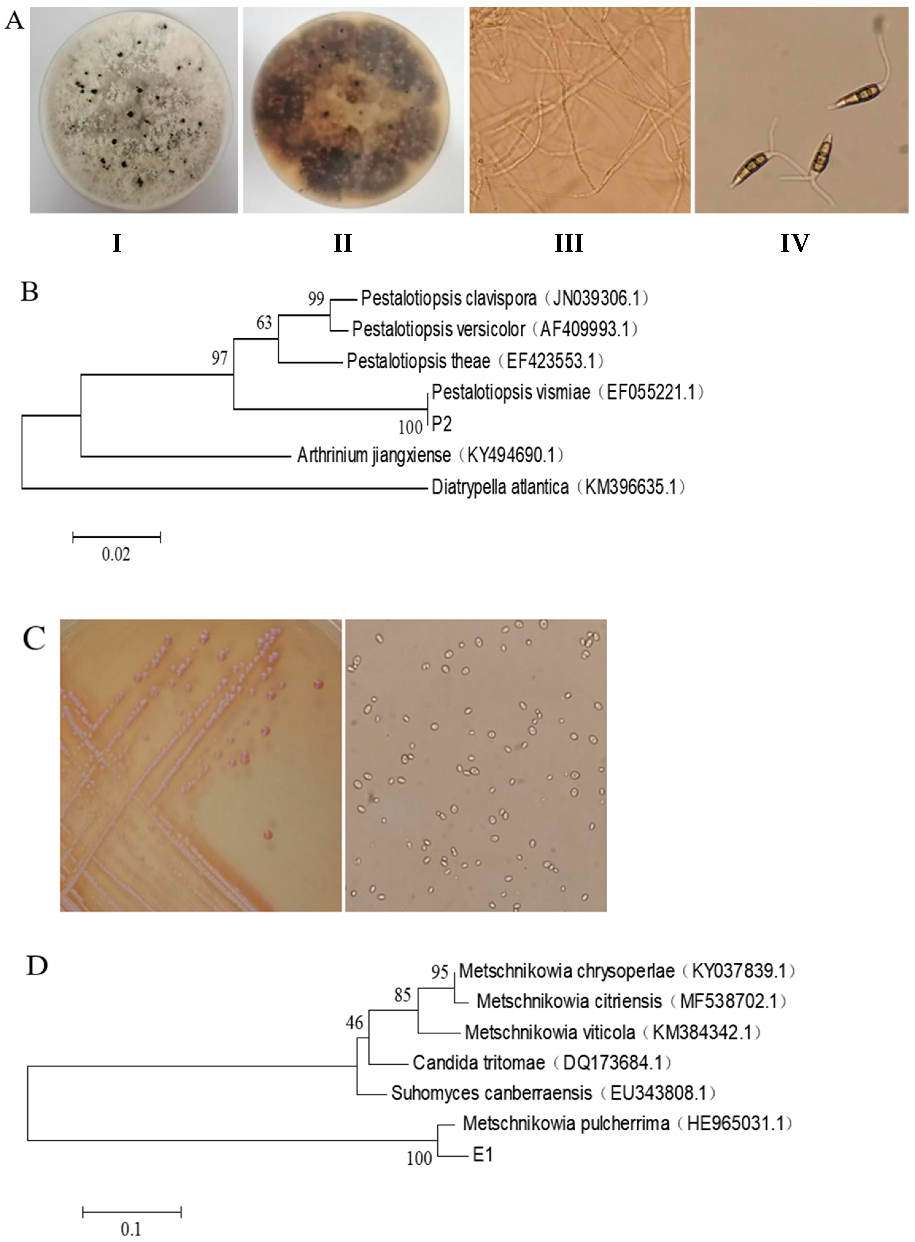
3.1. Isolation, Screening and Identification Results of the Pathogen and Antagonistic Yeast

Strain P2 exhibits different morphological characteristics, as shown in Figure 1A. When incubated on PDA plates, colonies show a pure white villous surface with orange pigmentation on the back. In the later culture stage, small black dots formed on the surface of the mycelium. The conidia of strain P2 showed a straight or slightly curved spindle shape consisting of five cells. The middle three cells were olive, the cells at both ends were conical achromoid cells, the apex had two to three accessory filaments, and the basal cells had a mesocytosis. Phylogenetic analysis (Figure 1B) showed a strong genetic relationship between the pathogen and Pestalotiopsis vismiae. Combined with morphological observations and molecular identification, strain P2 was finally identified as Pestalotiopsis vismiae.
When cultured on NYDA medium at 28 °C for 48 h, strain E1 formed small, round colonies with a creamy-white, opaque appearance (Figure 1C). The colonies exhibited neat edges, a moist and smooth surface, and were easily suspended when stirred. A distinctive red pigment was observed, with the reverse side displaying a reddish-brown coloration. The culture emitted a strong fermentation odor. Microscopic examination revealed spherical to subspherical yeast cells that reproduced through unilateral or multilateral budding. For molecular identification, the 5.8S rDNA-ITS region of strain E1 was sequenced and compared with reference sequences in GenBank. Phylogenetic analysis using the MEGA 5.1 program in DNAStar (Figure 1D) confirmed its close relationship with M. pulcherrima, supporting its identification as this species.

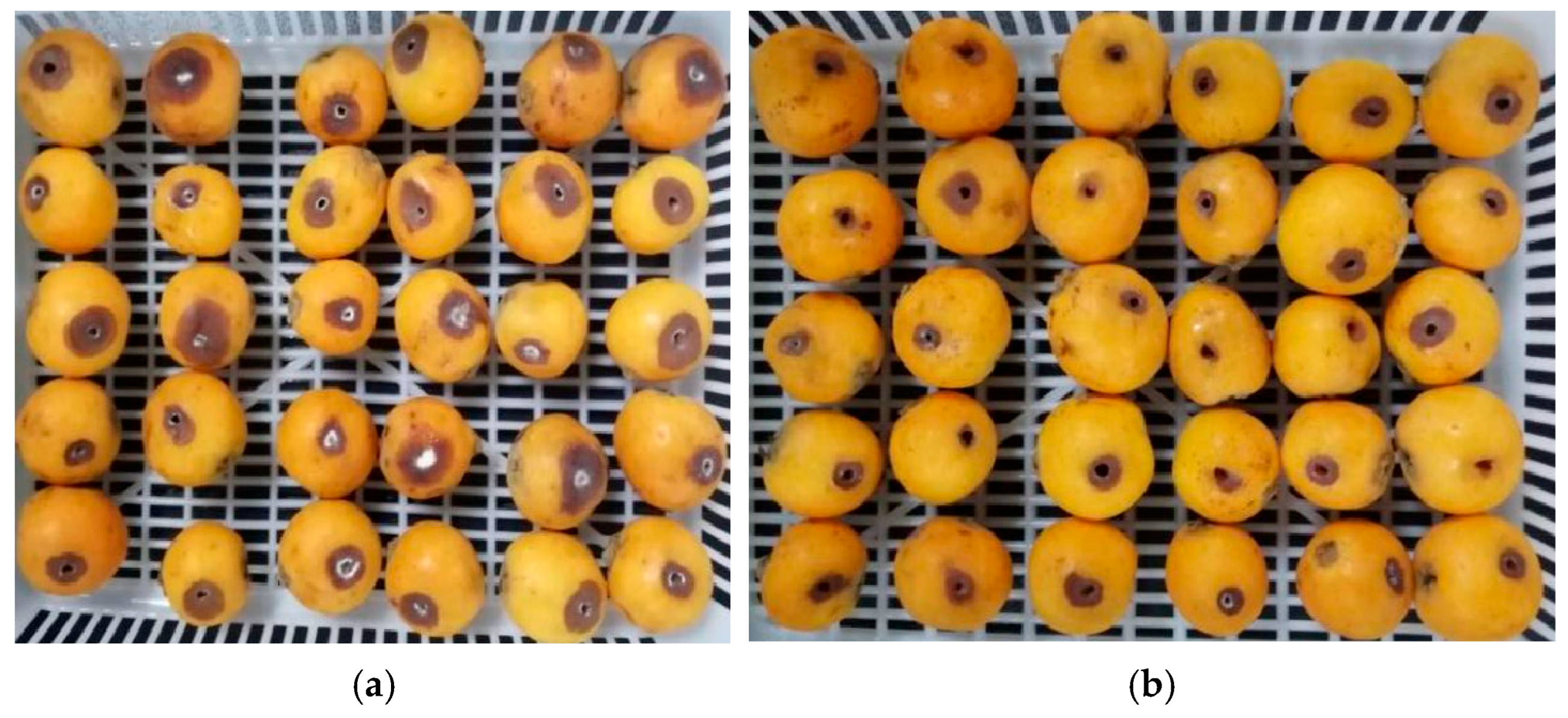
3.2. Effect of M. pulcherrima E1 on Control of Gray Spot of Loquat

M. pulcherrima E1 showed significant efficacy in inhibiting the development of postharvest gray spots and decay in loquat fruits (Figure 2). As shown in Table 1, on the 5th day, all loquat fruits in the control group were diseased, with a spot diameter of 1.59 cm, while those in the M. pulcherrima E1 treatment group were only 22.73%, with a spot diameter of 0.95 cm. The disease incidence and spot diameter in the treatment group were significantly lower than those in the control group (p < 0.05). Therefore, M. pulcherrima E1 can effectively control postharvest gray spot of loquats caused by P. vismiae at room temperature and can be used for storage and preservation of loquat fruits, which is worthy of further research and promotion.

3.3. Sequencing Data and Quality Control
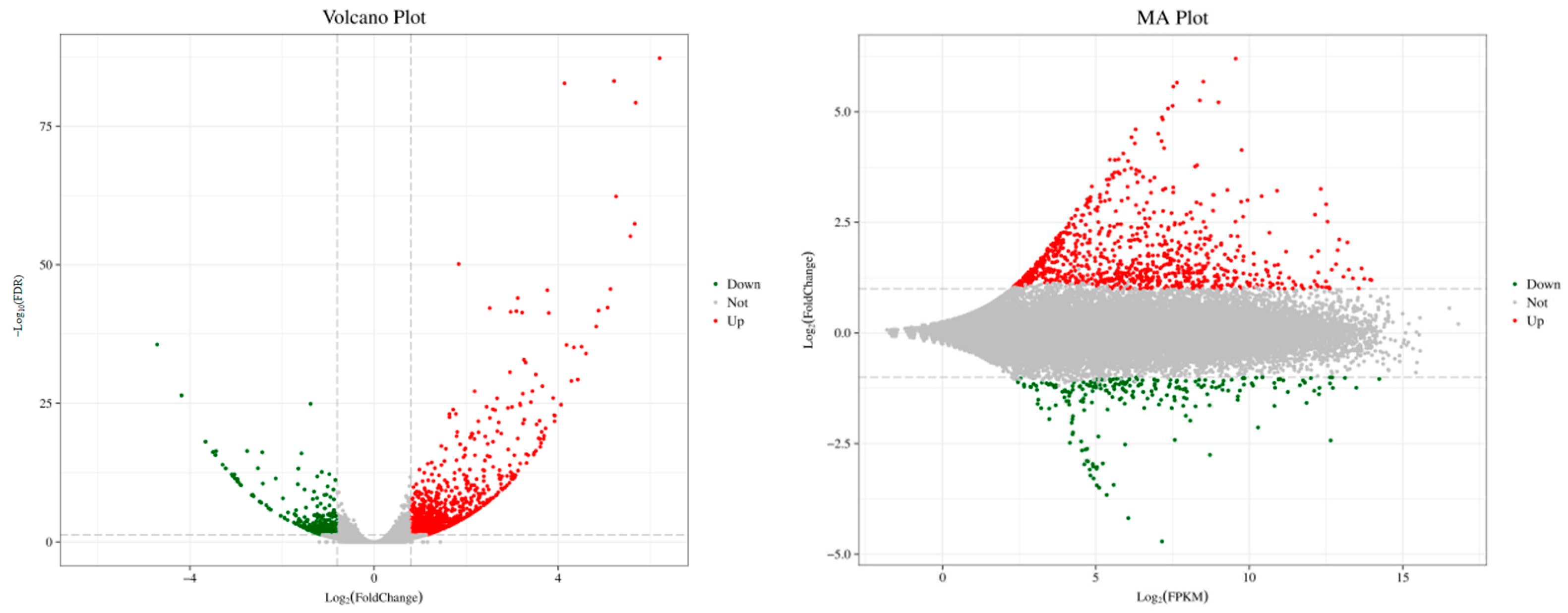
Sequencing result statistics of loquat fruit tissue samples in the sterile water control group (CK) and M. pulcherrima E1 treatment group (T) are shown in Table 2. Through the quality control of the sequencing, a total of 45.33 Gb of clean data was obtained. The statistical evaluation data of the base quality value showed that the percentage of Q30 bases was more than 92.22%. In addition, the GC content in the CK group was 46.56, 46.03 and 46.5, and the GC content in the T group was 46.44, 46.48 and 46.26, indicating that the sequencing results were relatively stable. In summary, the transcriptome sequencing results were stable and reliable and can be used for subsequent bioinformatic analysis.

3.4. DEG Analysis

The DEGs between the control and the treatment groups were shown in Figure 3. A total of 1444 DEGs were screened with the criteria of |log2(Fold Change)| ≥ 0.8 and FDR < 0.05, among which 1111 genes were upregulated, and 333 genes were downregulated.

3.5. GO Enrichment Analysis of DEGs

GO enrichment analysis of DEGs of loquat fruit tissue treated with M. pulcherrima E1 was performed. Figure 4 showed the GO classification of differentially expressed genes (DIFF genes/DEGs), which can be divided into three main categories as follows: cell component, molecular function, and biological process. These three categories contain 15, 12 and 24 secondary functions, respectively, each of which contains many DEGs. The cell components with the highest number of DEGs were cells (enriched by 588 DEGs), cell components (enriched by 582 DEGs), organelles (enriched by 359 DEGs) and membranes (enriched by 322 DEGs). The molecular functions with the highest number of DEGs were catalytic activity (enriched by 435 DEGs) and cell binding (enriched by 413 DEGs). The biological processes with the largest number of DEGs were cellular processes (enriched by 499 DEGs), metabolic processes (enriched by 465 DEGs), stress response (enriched by 296 DEGs) and biological regulation (enriched by 225 DEGs).

3.6. KEGG Enrichment Analysis of DEGs

KEGG enrichment analysis was performed to investigate the biological pathways and functional networks associated with the differentially expressed genes (DEGs). The analysis revealed that 221 DEGs participated in 86 distinct metabolic pathways. As shown in Figure 5, these DEGs were primarily enriched in five major functional categories: genomic processes, cellular processes, basic metabolic processes, biological systems, and environmental information processing. These primary categories could be further divided into 17 secondary metabolic pathways. The genomic processes category encompassed DNA replication and repair, translation, and transcription. Cellular processes mainly involved transport and catabolism activities, while environmental information processing included membrane transport and signal transduction. Basic metabolic processes covered lipid metabolism, biosynthesis of secondary metabolites, and metabolism of various amino acids. The biological systems category was primarily associated with environmental adaptation functions. Among all identified pathways, the five most significantly enriched KEGG pathways were zein biosynthesis, stilbene/diarylheptane/gingerol biosynthesis, riboflavin metabolism, and plant–pathogen interaction.

3.6.1. Effects on Plant–Pathogen Interaction Pathways

The treatment with Metschnikowia pulcherrima E1 induced significant differential gene expression in the plant–pathogen interaction pathway, as evidenced in Table 3. Notably, genes encoding calcium-binding proteins including CML5, CML19, and CML23 showed upregulated expression levels. These molecular changes are functionally linked to enhanced plant hypersensitivity responses, cell wall fortification, and stomatal closure regulation. Further analysis revealed upregulation of several key defense-related genes in E1-treated loquat fruits: the defense-associated transcription factor WRKY29, the pathogenesis-related protein-coding gene PTI5, the hypersensitive response mediator RPS2, the programmed cell death regulator EDS1, and RBOHC which encodes a NADPH oxidase component involved in oxidative burst during plant defense responses.

3.6.2. Effects on Secondary Metabolic Pathways

M. pulcherrima E1 treatment caused changes in secondary metabolism-related genes in pulp tissues of loquat, as shown in Table 4. In the biosynthetic pathway of benzene propane, PER52 and PER63, which are involved in the synthesis of the resistant substances P-hydroxyphenyl lignin, guaiacol lignin, 5-hydroxyl guaiacyl lignin and syringyl lignin, the shikimate O-hydroxyl cinnamyl transferase gene (HST and ACT) and the coffee acyl coenzyme A-O-methyltransferase gene (ATOMT) were upregulated to varying degrees. In the biosynthesis pathway of flavonoids, FLS, ACT and HST jointly participated in the synthesis of the flavonoids myricetin, quercetin, kaempferol and galangin, and UGT88A1 was involved in the synthesis of phlorizin. The expression of FLS and UGT88A1 was upregulated.

3.6.3. Effects on the Related Pathways of Plant Hormone Metabolism

Table 5 shows that M. pulcherrima E1 treatment caused differential expression of plant hormone metabolism-related genes in pulp tissues of loquat. For example, in the auxin metabolic pathway, the auxin response genes IAA17 and AHP1, which encode histidine phosphate transfer proteins and are involved in cytokinin signal transmission, the cytokinin response regulating gene ARR3 and the gibberellin receptor-related gene GID1C were upregulated to varying degrees. In addition, the expression of XTH22 and XTH23, the key genes in the synthesis and metabolism of brassinosteroids, was upregulated, while the expression of CYP749A22, the inactivated brassinolide gene, was downregulated. However, the expression level of the NPR1 gene, a regulatory protein in the SA signal transduction pathway, was downregulated, and the expression of the TIFY9 and TIFY10A genes encoding the negative regulatory factor JAZ enzyme in the synthesis of JA was upregulated. This indicated that the antagonistic yeast inhibited the expression of genes related to the SA and JA signaling pathways, which may not play a role in this experiment.

3.6.4. Effects of Glutathione Metabolic Pathways

As shown in Table 6, in the glutathione metabolic pathway, M. pulcherrima E1 treatment caused varying degrees of upregulated expression of GSTU8 and GSTU10 in loquat pulp, which are involved in coding the key enzyme GSTs catalyzing the glutathione binding reaction. In addition, the APX3 gene, which encodes ascorbic acid peroxidase, was also upregulated in loquat fruits treated with antagonistic yeast.

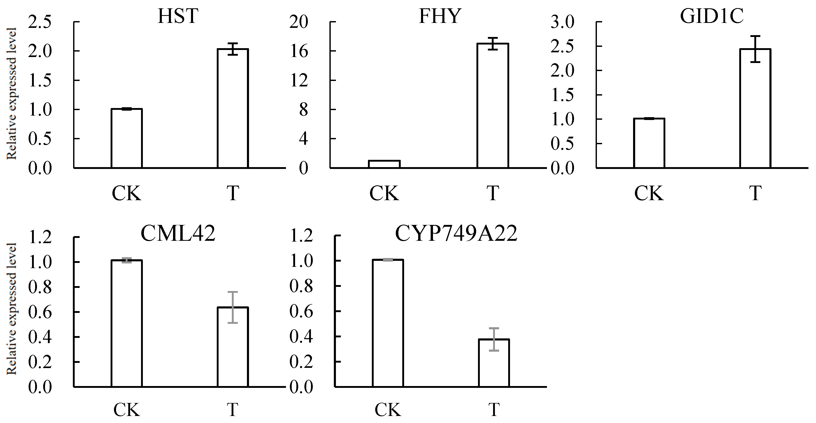
3.7. Results of RT–qPCR Verification of DEGs

RT–qPCR was used to verify the expression levels of the DEGs related to plant resistance, as shown in Figure 6. The vast majority of DEGs are upregulated at expression levels, including CML19 (59.8%), FLS (280%), ACT (162.3%), XTH23 (551.5%), AFS1 (175.6%), CP1 (95%), HST (101%), FHY (158.4%), and GID1C (141.6%). The expression levels of CML42 and CYP74A22 were downregulated by 36.6% and 62.4%, respectively, which was basically consistent with the results of transcriptome analysis.

4. Discussion

4.1. The Biocontrol Efficacy of Antagonistic Yeasts

As mentioned above, loquats suffer from various pathogenic fungi in postharvest and storage processes, leading to decay and economic losses. As shown in Table 1 and Figure 2, M. pulcherrima E1 exhibited significant inhibitory effects against postharvest loquat gray spot disease, indicating that biological control with antagonistic yeast provides a promising alternative to traditional fungicides. Recent studies have highlighted the biocontrol potential of M. pulcherrima, though its mechanisms vary across applications. While a previous study demonstrated its biocontrol efficacy in winemaking through nutrient competition and volatile production [21], our study reveals a novel role in postharvest fruit protection by inducing resistance pathways (e.g., upregulation of CML19, GSTU10) against fungal pathogens. This expands the known biocontrol applications of M. pulcherrima beyond microbial antagonism to include host defense priming, bridging the gap between applied biocontrol and molecular insights.
The experiment employed a punch-inoculation method in loquat fruits to infect pathogenic fungi and inoculate antagonistic yeast. Fungal infection of fruits commonly occurs via mechanical wounds or natural orifices, where germinated spores form mycelial tubes to invade pulp tissues. A previous study have adopted the same method to explore the control effect of Wickerhamomyces anomalus on postharvest primary diseases of peach fruits [22]. Another study also punched holes on the surface of eggplants to infect pathogenic fungi and explored the control effect of Bacillus velezensis on eggplant soft rot [23]. Thus, it can be seen that the method of punching holes in fruits has little effect on the exploration of antagonistic yeasts in improving fruit disease resistance.

4.2. KEGG Pathway Enrichment Analysis of DEGs

In this study, the predominant pathogenic fungus causing loquat gray spot was isolated from rotten loquat fruits, followed by screening for antagonistic yeasts effective against this pathogen. To decipher the underlying mechanisms of antagonistic yeast-mediated disease resistance, comprehensive GO and KEGG enrichment analyses were performed. In the KEGG enrichment classification of DEGs, the plant–pathogen interaction pathway, secondary metabolic pathway, hormone-related pathway and glutathione metabolic pathway in the top 20 KEGG metabolic pathways with the most significant enrichment were analyzed.

4.2.1. Analysis of Plant–Pathogen Interaction Pathways

In the plant–pathogen interaction pathway, antagonistic yeasts, as biological activators, can induce a series of disease-resistant defense responses in plants, such as oxygen explosion and plant signal transduction [24]. Reactive oxygen species (ROS) exhibit dual functions in plants. When an organism is under stress, plants can kill foreign substances by producing ROS, which can act as important signaling molecules and prompt plants to initiate their own defense response [25]. However, excessive accumulation of ROS will cause serious oxidative stress in plants [26]. Plant respiratory burst oxidase Rboh (NADPH oxidase) can trigger the immune response and affect ROS production through pathogen-related molecular patterns [27]. ROS can also act as signal molecules for plant hypersensitivity reactions [28]. The hypersensitive response (HR) is a defense action against pathogen ingression. Typically, HR is predictable with the appearance of dead, brown cells along with visible lesions [29]. Plant hypersensitivity is a form of programmed cell death that can stimulate defense responses and systemic acquired resistance in plants, which leads to a significant reduction in symptoms caused by many pathogens [30]. The process of plant cell wall reinforcement and stomatal closure can prevent pathogen infection, which is of great significance for improving plant disease resistance [31]. Upon pathogen infection, WRKY transcription factors activate W-box elements in target genes, reinforcing the plant’s capacity to combat pathogen attack and withstand stress stimuli [32].
Table 3 shows that in the treatment with M. pulcherrima E1, many DEGs in the plant–pathogen interaction pathway were produced, which induced the defense response of loquat pulp tissues. For example, the upregulation of CML5, CML19, CML23 and other genes encoding calcium binding proteins can affect the process of hypersensitivity, cell wall enhancement and stomatal closure in plants. The expression of the transcription factor WRKY29, which is related to defense genes, PTI5, which encodes a resistance protein, RPS2, which is related to plant hypersensitivity, EDS1, which is related to programmed cell death, and RBOHC, which is involved in coding NADPH oxidase, was upregulated in loquat fruits treated with M. pulcherrima E1. These results suggest that antagonistic yeast enhances loquat fruit disease resistance through ROS accumulation, oxidative signal transduction, hypersensitive reaction induction, and reinforcement of plant cell walls and stomatal closure. These findings are consistent with our previous study on M. pulcherrima E1, which showed enhanced activities of defense-related enzymes including polyphenol oxidase (PPO), peroxidase (POD), catalase (CAT), ascorbate peroxidase (APX), and phenylalanine ammonia-lyase (PAL) in loquat fruits [10].

4.2.2. Analysis of Secondary Metabolic Pathways

When plants encounter pathogen infection, they activate the biosynthesis of diverse secondary metabolites as part of their defense response. These protective compounds primarily consist of phenylpropanoids, flavonoids, terpenoids, and alkaloids, which accumulate significantly at infection sites to combat microbial invasion [33]. Lignin is an important structural component of the plant cell wall. When plants are infected by pathogens, lignin is synthesized in large quantities and lignifies the cell wall, forming a physical barrier to resist the invasion of pathogens [34]. As shown in Table 4, M. pulcherrima E1 treatment caused changes in secondary metabolism-related genes in pulp tissues of loquat. In the biosynthetic pathway of phenylpropanoid, PER52 and PER63, which are involved in the synthesis of the resistant substances P-hydroxyphenyl lignin, guaiacol lignin, 5-hydroxyl guaiacyl lignin and syringyl lignin, the shikimate O-hydroxyl cinnamyl transferase gene (HST and ACT) and the coffee acyl coenzyme A-O-methyltransferase gene (ATOMT) were upregulated to varying degrees. In the flavonoid biosynthesis pathway, FLS, ACT and HST jointly participated in the synthesis of myricetin, quercetin, kaempferol and galangin. UGT88A1 is involved in the synthesis of phlorizin, and FLS and UGT88A1 were significantly upregulated. The above results showed that M. pulcherrima E1 could resist pathogen infection by inducing loquat fruit to synthesize more defensive secondary metabolites, such as lignin and flavonoids.

4.2.3. Analysis of the Related Pathways of Plant Hormone Metabolism

Plant hormones serve crucial functions in both developmental processes and stress responses, regulating not only growth, development, and senescence but also mediating defense signaling pathways during plant immunity [35]. There is a set of complex and intersecting hormone signaling networks in plants, and many hormone-mediated stress-related pathways may have antagonistic and synergistic effects, which can coordinate plant resistance alone or together with other signaling pathways [36]. The hormones related to plant disease resistance include ethylene, jasmonic acid (JA), salicylic acid (SA), brassinosterol (BR), cytokinin (CTK), auxin (IAA) and gibberellin (GA) [37,38]. Salicylic acid can interact with the downstream positive regulator NPR1 to activate the expression of resistance genes such as WRKY transcription factors and disease-initiating proteins, thus enhancing the resistance of plants to pathogens [39]. In the JA signaling pathway, JA binding to the receptor protein COI1 can cause the negative regulator JAZ to release the positively regulated transcription factor MYC2, thereby initiating downstream expression [40]. In this experiment, the expression level of the NPR1 gene, a regulatory protein in the SA signal transduction pathway, was downregulated, and the expression of the TIFY9 and TIFY10A genes encoding the negative regulatory factor JAZ enzyme in the synthesis of JA was upregulated. This indicated that the antagonistic yeast inhibited SA- and JA-signaling-related gene expression, suggesting that these pathways may not contribute to loquat fruit resistance induced by the yeast.
The auxin signaling pathway is pivotal in programming growth and development and coordinating immunity in plant [41]. The repressor 1 gene of the PHYB activation label (CYP749A22/BAS1) is the inactivated gene of brassinolide. Excessive expression of the BAS1 gene will produce brassinolide C-26 hydroxyl hydroxylate, resulting in a decrease in the content of active brassinolide in plants [42]. Table 5 shows that M. pulcherrima E1 treatment caused differential expression of plant hormone metabolism-related genes in pulp tissues of loquat. For example, in the auxin metabolic pathway, the auxin response genes IAA17 and AHP1, which encode histidine phosphate transfer proteins and are involved in cytokinin signal transduction, the cytokinin response regulating gene ARR3 and the gibberellin receptor-related gene GID1C were upregulated to varying degrees. In addition, the expression of XTH22 and XTH23, the key genes in the synthesis and metabolism of BR, was upregulated, while the expression of CYP749A22, the inactivated brassinolide gene, was downregulated. This indicated that antagonistic yeast treatment inhibited the expression of genes related to the SA and JA signaling pathways and mainly improved the disease resistance of loquat fruits by activating the expression of IAA, GA, CTK and BR related to signal transduction.

4.2.4. Analysis of Glutathione Metabolic Pathways

Glutathione is a vital antioxidant in plants, primarily functioning to scavenge excess ROS, thereby offering multifaceted protection. It can eliminate excessive reactive oxygen radicals in plants and has detoxification and anti-aging effects, which are closely related to the stress resistance of plants [28,43]. Glutathione S-transferase (GST) is a multifunctional superfamily enzyme that catalyzes the binding interaction between some harmful electron-loving compounds and glutathione. It is the main detoxification system in many plants [44]. It has been reported that GSTs are involved in the accumulation and transport of resistant flavonoid substances in plants [45], and HaGSTU1 protein can play an important role in improving the resistance of sunflower against Paracoccus nucleuses [46]. APX also plays a very important role in the plant response to stress. It can protect cells from the toxicity of ROS by removing excessive H2O2 produced in cells [47]. Studies have shown that inducing the expression of the APX gene in poplar trees is closely related to the enhancement of its resistance to the ulcerative bacteria Botryosphaeria dothidea [48]. According to Table 6, in the glutathione metabolic pathway, M. pulcherrima E1 treatment caused the upregulated expression of GSTU8 and GSTU10, which are involved in coding the key enzyme GSTs catalyzing the glutathione binding reaction. In addition, the expression of APX3, a gene involved in encoding ascorbate peroxidase, was also upregulated in the treatment with antagonistic yeast. Our previous study also demonstrated that the activity of ascorbate peroxidase (APX) was significantly enhanced in loquats treated with Metschnikowia pulcherrima E1 [10]. This suggests that the antagonistic yeast treatment enhances disease resistance by activating glutathione metabolism on loquat fruits.

5. Conclusions

In this study, the pathogen of gray spot disease in loquat was identified as Pestalotiopsis vismiae from rotten loquat fruits, and Metschnikowia pulcherrima E1 was screened as an efficient antagonistic yeast, which reduced the fruit rot rate to 22.73% through dual mechanisms of iron competition and biofilm formation.
Then, transcriptome sequencing was performed on loquat fruits in the antagonistic yeast M. pulcherrima E1 treatment group and the sterile water control group, and 1444 differentially expressed genes were screened out. The results showed that these DEGs were involved in various signaling pathways and metabolic pathways related to fruit resistance, including plant–pathogen interaction, plant hormone metabolism, secondary metabolism, and glutathione metabolism. In addition, the RT–qPCR validation results of DEGs were consistent with the basic trend of sequencing results: the expression levels of CML19, FLS, ACT, XTH23, AFS1, CP1, HST, FHY, and GID1C were upregulated, while the expression levels of CML42 and CYP74A22 were downregulated. These findings validate the molecular mechanisms of yeast-induced plant resistance and provide a theoretical basis for biocontrol applications.

Supplementary Materials

The following supporting information can be downloaded at: https://www.mdpi.com/article/10.3390/jof11070497/s1, Figure S1: Experimental flow chart; Table S1: Primer design of differentially expressed genes.

Author Contributions

Methodology, X.L. (Xiaoya Li) and K.W.; software, X.L. (Xin Li) and Y.Z.; formal analysis, X.L. (Xiaoya Li) and K.W.; investigation, X.L. (Xin Li); data curation, Y.Z.; writing—original draft preparation, X.L. (Xiaoya Li) and K.W.; writing—review and editing, W.S.; visualization, X.L. (Xiaoya Li); supervision, K.W.; project administration, W.S.; funding acquisition, W.S. All authors have read and agreed to the published version of the manuscript.

Funding

This research was funded by Key R&D Program of Jiangsu Province (BE2021338).

Institutional Review Board Statement

Not applicable.

Informed Consent Statement

Not applicable.

Data Availability Statement

The data shown in this study are contained within the article.

Acknowledgments

All authors are thankful to their representative universities/institutes for the support and services used in this study.

Conflicts of Interest

The authors declare no conflicts of interest.

References

  1. Yan, F.; Zhang, D.; Wang, X.; Liu, C.; Zhang, F. Reduction of postharvest diseases of loquat fruit by serine protease and possible mechanisms involved. Sci. Hortic. 2022, 304, 111246. [Google Scholar] [CrossRef]
  2. Yan, F.; Zhang, D.; Ye, X.; Wu, Y.; Fang, T. Potential of Saturnispora diversa MA as a postharvest biocontrol agent against anthracnose in loquat fruit. Biol. Control 2022, 173, 105006. [Google Scholar] [CrossRef]
  3. Ye, W.Q.; Sun, Y.F.; Tang, Y.J.; Zhou, W.W. Biocontrol potential of a broad-spectrum antifungal strain Bacillus amyloliquefaciens B4 for postharvest loquat fruit storage. Postharvest Biol. Technol. 2021, 174, 111439. [Google Scholar] [CrossRef]
  4. Sun, X.W.; Xu, Y.; Chen, L.; Jin, X.M.; Ni, H. The salt-tolerant phenazine-1-carboxamide-producing bacterium Pseudomonas aeruginosa NF011 isolated from wheat rhizosphere soil in dry farmland with antagonism against Fusarium graminearum. Microbiol. Res. 2021, 245, 126673. [Google Scholar] [CrossRef]
  5. Ali, M.A.; Ren, H.; Ahmed, T.; Luo, J.; An, Q.; Qi, X.; Li, B. Antifungal effects of rhizospheric Bacillus species against bayberry twig blight pathogen Pestalotiopsis versicolor. Agronomy 2020, 10, 1811. [Google Scholar] [CrossRef]
  6. Saju, K.A.; Mech, S.; Deka, T.N.; Biswas, A.K. In vitro evaluation of biocontrol agents, botanicals and fungicides against Pestalotiopsis sp. infecting large cardamom (Amomum subulatum Roxb.). J. Spices Aromat. Crops 2012, 20, 89–92. [Google Scholar]
  7. Zhang, X.; Wu, F.; Gu, N.; Yan, X.; Wang, K.; Dhanasekaran, S.; Zhang, H.; Gu, X.; Zhao, L. Postharvest biological control of Rhizopus rot and the mechanisms involved in induced disease resistance of peaches by Pichia membranefaciens. Postharvest Biol. Technol. 2020, 163, 111146. [Google Scholar] [CrossRef]
  8. Zhang, X.; Yao, Y.; Dhanasekaran, S.; Li, J.; Ngea, G.L.N.; Gu, X.; Zhang, H. Controlling black spot of postharvest broccoli by Meyerozyma guilliermondii and its regulation on ROS metabolism of broccoli. Biol. Control 2022, 170, 104938. [Google Scholar] [CrossRef]
  9. Bonaterra, A.; Badosa, E.; Daranas, N.; Francés, J.; Roselló, G.; Montesinos, E. Bacteria as Biological Control Agents of Plant Diseases. Microorganisms 2022, 10, 1759. [Google Scholar] [CrossRef] [PubMed]
  10. Yang, H.H.; Wang, L.; Li, S.J.; Gao, X.B.; Wu, N.N.; Zhao, Y.F.; Sun, W.H. Control of postharvest grey spot rot of loquat fruit with Metschnikowia pulcherrima E1 and potential mechanisms of action. Biol. Control 2021, 152, 104406. [Google Scholar] [CrossRef]
  11. Zhao, L.; Shu, Y.; Xiao, J.; Lin, R.; Godana, E.A.; Zhang, X.; Zhang, H. Transcriptome analysis reveals mechanisms involved in the enhanced antagonistic efficacy of Sporidiobolus pararoseus Y16 treated by γ-aminobutyric acid. Biol. Control 2022, 176, 105089. [Google Scholar] [CrossRef]
  12. Di Francesco, A.; Martini, C.; Mari, M. Biological control of postharvest diseases by microbial antagonists: How many mechanisms of action? Eur. J. Plant Pathol. 2016, 145, 711–717. [Google Scholar] [CrossRef]
  13. Li, Q.; Zhang, H.; Yang, Q.; Lin, Z.; Cheng, Y.; Sun, Y. Progress in biocontrol yeast agents for preventing and treating diseases of fruits. Food Sci. 2018, 39, 291–296. [Google Scholar] [CrossRef]
  14. Zhang, X.; Zhou, Y.; Li, J.; Gu, X.; Zhao, L.; Li, B.; Zhang, H. Pichia caribbica improves disease resistance of cherry tomatoes by regulating ROS metabolism. Biol. Control 2022, 169, 104870. [Google Scholar] [CrossRef]
  15. Costa, V.; Angelini, C.; De Feis, I.; Ciccodicola, A. Uncovering the Complexity of Transcriptomes with RNA-Seq. J. Biomed. Biotechnol. 2010, 1, 853916. [Google Scholar] [CrossRef]
  16. Strauss, T.; van Poecke, R.M.P.; Strauss, A.; Romer, P.; Minsavage, G.V.; Singh, S.; Wolf, C.; Strauss, A.; Kim, S.; Lee, H.A.; et al. RNA-seq pinpoints a Xanthomonas TAL-effector activated resistance gene in a large-crop genome. Proc. Natl. Acad. Sci. USA 2012, 109, 19480–19485. [Google Scholar] [CrossRef] [PubMed]
  17. Qi, Y.W.; Lei, Q.; Zhang, Y.J.; Liu, X.R.; Zhou, B.; Liu, C.H.; Ren, X.L. Comparative transcripome data for commercial maturity and physiological maturity of ‘Royal Gala’ apple fruit under room temperature storage condition. Sci. Hortic. 2017, 225, 386–393. [Google Scholar] [CrossRef]
  18. Kurtzman, C.; Fell, J.W. The Yeasts—A Taxonomic Study; Elsevier Science: Amsterdam, The Netherlands, 1998. [Google Scholar]
  19. Hsu, S.C.; Tsen, H.Y. PCR primers designed from malic acid dehydrogenase gene and their use for detection of Escherichia coli in water and milk samples. Int. J. Food Microbiol. 2001, 64, 1–11. [Google Scholar] [CrossRef]
  20. Redshaw, N.; Wilkes, T.; Whale, A.; Cowen, S.; Huggett, J.; Foy, C.A. A comparison of miRNA isolation and RT-qPCR technologies and their effects on quantification accuracy and repeatability. Biotechniques 2013, 54, 155–164. [Google Scholar] [CrossRef]
  21. Puyo, M.; Simonin, S.; Bach, B.; Klein, G.; Alexandre, H.; Tourdot-Maréchal, R. Bio-protection in oenology by Metschnikowia pulcherrima: From field results to scientific inquiry. Front. Microbiol. 2023, 14, 1252973. [Google Scholar] [CrossRef]
  22. Zhou, Y.; Zhao, L.; Chen, Y.; Dhanasekaran, S.; Chen, X.; Zhang, X.; Zhang, H. Study on the control effect and physiological mechanism of Wickerhamomyces anomalus on primary postharvest diseases of peach fruit. Int. J. Food Microbiol. 2024, 413, 110575. [Google Scholar] [CrossRef]
  23. Zhou, Y.; Zhao, L.; Chen, Y.; Dhanasekaran, S.; Chen, X.; Zhang, X.; Zhang, H. Characterization of a Bacillus velezensis strain as a potential biocontrol agent against soft rot of eggplant fruits. Int. J. Food Microbiol. 2024, 410, 110480. [Google Scholar] [CrossRef]
  24. Wang, W.; Feng, B.; Zhou, J.-M.; Tang, D. Plant immune signaling: Advancing on two frontiers. J. Integr. Plant Biol. 2020, 62, 2–24. [Google Scholar] [CrossRef] [PubMed]
  25. Ma, Z.; Bykova, N.V.; Igamberdiev, A.U. Cell signaling mechanisms and metabolic regulation of germination and dormancy in barley seeds. Crop J. 2017, 5, 459–477. [Google Scholar] [CrossRef]
  26. Dietz, K.-J.; Mittler, R.; Noctor, G. Recent Progress in Understanding the Role of Reactive Oxygen Species in Plant Cell Signaling. Plant Physiol. 2016, 171, 1535–1539. [Google Scholar] [CrossRef] [PubMed]
  27. Miller, G.; Shulaev, V.; Mittler, R. Reactive oxygen signaling and abiotic stress. Physiol. Plant. 2008, 133, 481–489. [Google Scholar] [CrossRef]
  28. Feng, J.; Chen, L.; Zuo, J. Protein S-Nitrosylation in plants: Current progresses and challenges. J. Integr. Plant Biol. 2019, 61, 1206–1223. [Google Scholar] [CrossRef]
  29. Noman, A.; Aqeel, M.; Qari, S.H.; Al Surhanee, A.A.; Yasin, G.; Alamri, S.; Hashem, M.; Al-Saadi, A.M. Plant hypersensitive response vs. pathogen ingression: Death of few gives life to others. Microb Pathog 2020, 145, 104224. [Google Scholar] [CrossRef]
  30. Suzuki, N.; Katano, K. Coordination Between ROS Regulatory Systems and Other Pathways Under Heat Stress and Pathogen Attack. Front. Plant Sci. 2018, 9, 490. [Google Scholar] [CrossRef]
  31. Lucas, W.J.; Groover, A.; Lichtenberger, R.; Furuta, K.; Yadav, S.-R.; Helariutta, Y.; He, X.-Q.; Fukuda, H.; Kang, J.; Brady, S.M.; et al. The Plant Vascular System: Evolution, Development and Functions. J. Integr. Plant Biol. 2013, 55, 294–388. [Google Scholar] [CrossRef]
  32. Zhang, Q.L.; Li, Y.; Zhang, Y.; Wu, C.B.; Wang, S.N.; Hao, L.; Wang, S.Y.; Li, T.Z. Md-miR156ab and Md-miR395 Target WRKY Transcription Factors to Influence Apple Resistance to Leaf Spot Disease. Front. Plant Sci. 2017, 8, 526. [Google Scholar] [CrossRef] [PubMed]
  33. Ballester, A.R.; Marcet-Houben, M.; Levin, E.; Sela, N.; Selma-Lazaro, C.; Carmona, L.; Wisniewski, M.; Droby, S.; Gonzalez-Candelas, L.; Gabaldon, T. Genome, Transcriptome, and Functional Analyses of Penicillium expansum Provide New Insights into Secondary Metabolism and Pathogenicity. Mol. Plant Microbe Interact. 2015, 28, 232–248. [Google Scholar] [CrossRef]
  34. Zhao, Q.; Dixon, R.A. Transcriptional networks for lignin biosynthesis: More complex than we thought? Trends Plant Sci. 2011, 16, 227–233. [Google Scholar] [CrossRef] [PubMed]
  35. Pacifici, E.; Polverari, L.; Sabatini, S. Plant hormone cross-talk: The pivot of root growth. J. Exp. Bot. 2015, 66, 1113–1121. [Google Scholar] [CrossRef]
  36. Gong, Z.; Xiong, L.; Shi, H.; Yang, S.; Herrera-Estrella, L.R.; Xu, G.; Chao, D.-Y.; Li, J.; Wang, P.-Y.; Qin, F.; et al. Plant abiotic stress response and nutrient use efficiency. Sci. China Life Sci. 2020, 63, 635–674. [Google Scholar] [CrossRef]
  37. Pieterse, C.M.J.; Van der Does, D.; Zamioudis, C.; Leon-Reyes, A.; Van Wees, S.C.M. Hormonal Modulation of Plant Immunity. In Annual Review of Cell and Developmental Biology; Schekman, R., Ed.; Annual Reviews: San Mateo, CA, USA, 2012; Volume 28, pp. 489–521. [Google Scholar]
  38. Robert-Seilaniantz, A.; Grant, M.; Jones, J.D.G. Hormone Crosstalk in Plant Disease and Defense: More Than Just JASMONATE-SALICYLATE Antagonism. In Annual Review of Phytopathology; VanAlfen, N.K., Bruening, G., Leach, J.E., Eds.; Annual Reviews: Palo Alto, CA, USA, 2011; Volume 49, pp. 317–343. [Google Scholar]
  39. Thakur, M.; Sohal, B.S. Role of Elicitors in Inducing Resistance in Plants against Pathogen Infection: A Review. ISRN Biochem. 2013, 2013, 762412. [Google Scholar] [CrossRef] [PubMed]
  40. Shigenaga, A.M.; Argueso, C.T. No hormone to rule them all: Interactions of plant hormones during the responses of plants to pathogens. Semin. Cell Dev. Biol. 2016, 56, 174–189. [Google Scholar] [CrossRef]
  41. Yu, X.Y.; Hu, K.X.; Geng, X.Y.; Cao, L.F.; Zhou, T.T.; Lin, X.X.; Liu, H.C.; Chen, J.R.; Luo, C.G.; Qu, S.C. The Mh-miR393a-TIR1 module regulates Alternaria alternata resistance of Malus hupehensis mainly by modulating the auxin signaling. Plant Sci. 2024, 341, 112008. [Google Scholar] [CrossRef]
  42. Peng, H.; Neff, M.M. CIRCADIAN CLOCK ASSOCIATED 1 and ATAF2 differentially suppress cytochrome P450-mediated brassinosteroid inactivation. J. Exp. Bot. 2020, 71, 970–985. [Google Scholar] [CrossRef]
  43. Venturas, M.D.; Sperry, J.S.; Hacke, U.G. Plant xylem hydraulics: What we understand, current research, and future challenges. J. Integr. Plant Biol. 2017, 59, 356–389. [Google Scholar] [CrossRef] [PubMed]
  44. Lecourieux, D.; Kappel, C.; Claverol, S.; Pieri, P.; Feil, R.; Lunn, J.E.; Bonneu, M.; Wang, L.; Gomes, E.; Delrot, S.; et al. Proteomic and metabolomic profiling underlines the stage- and time-dependent effects of high temperature on grape berry metabolism. J. Integr. Plant Biol. 2020, 62, 1132–1158. [Google Scholar] [CrossRef] [PubMed]
  45. Zhao, J.; Dixon, R.A. The ‘ins’ and ‘outs’ of flavonoid transport. Trends Plant Sci. 2010, 15, 72–80. [Google Scholar] [CrossRef] [PubMed]
  46. Kong, F.; Deng, Y.; Wang, G.; Wang, J.; Liang, X.; Meng, Q. LeCDJ1, a chloroplast DnaJ protein, facilitates heat tolerance in transgenic tomatoes. J. Integr. Plant Biol. 2014, 56, 63–74. [Google Scholar] [CrossRef] [PubMed]
  47. Jiang, J.J.; Ma, S.H.; Ye, N.H.; Jiang, M.; Cao, J.S.; Zhang, J.H. WRKY transcription factors in plant responses to stresses. J. Integr. Plant Biol. 2017, 59, 86–101. [Google Scholar] [CrossRef]
  48. Guo, B.; Chen, Z.-Y.; Lee, R.D.; Scully, B.T. Drought stress and preharvest aflatoxin contamination in agricultural commodity: Genetics, genomics and proteomics. J. Integr. Plant Biol. 2008, 50, 1281–1291. [Google Scholar] [CrossRef]
Figure 1. (A): Morphological characteristics of strain P2 and strain E1. (I) Colony morphology of P2 on the front of a PDA plate; (II) colony morphology of P2 on the back of a PDA plate; (III) image of mycelia of P2; (IV) image of conidia of P2. (B): Molecular evolutionary tree of P2 (SAMN49174010). The phylogenetic tree was constructed by the neighbor-joining method using MEGA 5.1 software. The bootstrap values are shown at the branch points. (C): Colony (left) and cell (right) morphology of strain E1. (D): Molecular evolutionary tree of Metschnikowia pulcherrima E1(SAMN49174009). The phylogenetic tree was constructed by the neighbor-joining method using MEGA 5.1 software. The bootstrap values are shown at the branch points.
Jof 11 00497 g001
Figure 2. Effect of M. pulcherrima E1 on the decay rate of gray spot in loquat fruit. (a) Control group: treated with sterile water of one parallel replicate. (b) Treatment group: treated with M. pulcherrima E1 of one parallel replicate.
Jof 11 00497 g002
Figure 3. Volcano plot and MA plot of DEGs. The Volcano plot shows the relationship between the significance (Y-axis) and fold change (X-axis) of differentially expressed genes (DEGs). The MA plot displays the distribution of gene expression levels (X-axis) and fold changes (Y-axis). Green points represent downregulated DEGs, red points represent upregulated DEGs, and black points represent non-differentially expressed genes.
Jof 11 00497 g003
Figure 4. Classification of DEGs based on GO annotation. DIFF gene: differentially expressed gene (DEG), defined as genes with |log2(FC)| ≥ 0.8 and FDR < 0.05.
Jof 11 00497 g004
Figure 5. KEGG pathway classification of DEGs.
Jof 11 00497 g005
Figure 6. Results of verification of some DEGs by RT–qPCR.
Jof 11 00497 g006aJof 11 00497 g006b
Table 1. Effect of M. pulcherrima E1 on the incidence and diameter of gray spot in loquat.
ExperimentIncident Rate (%)Spot Diameter (cm)
treatment group22.730.95 ± 0.06 b
control group1001.59 ± 0.16 a
Different letters in the same column indicate significant differences (p < 0.05). The standard error of the treatment group is 0.01, and that of the control group is 0.03 (The standard error is calculated by dividing the standard deviation by the square root of the sample size, n = 30).
Table 2. Sequencing data tables of loquat tissues.
SamplesJSHY-IDReadSumBaseSumGC (%)Q30 (%)
CK1T0124562436736873080046.5692.22
CK2T0225701745771052350046.0392.84
CK3T0323718990711569700046.592.95
T1T0428165336844960080046.4494.37
T2T0524068412722052360046.4894.24
T3T0623280409698412270046.2692.62
Table 3. Effects of M. pulcherrima E1 on loquat genes related to plant–pathogen interactions.
Gene IDGene Namelog2FCDefinition
TRINITY_DN14842_c0_g1RBOHC0.96respiratory burst oxidase
TRINITY_DN12258_c0_g1CML51.40calcium-binding protein
TRINITY_DN14083_c0_g1HSP82−1.16molecular chaperone HtpG
TRINITY_DN15797_c0_g1CML231.56calcium-binding protein
TRINITY_DN15820_c0_g1CML271.57calcium-binding protein
TRINITY_DN15820_c0_g3CML271.17calcium-binding protein
TRINITY_DN16219_c0_g1CML192.00calcium-binding protein
TRINITY_DN16219_c0_g2CML190.97calcium-binding protein
TRINITY_DN17371_c0_g3EDS11.53enhanced disease susceptibility 1 protein
TRINITY_DN17724_c5_g2CP11.20Calcium-binding protein
TRINITY_DN17724_c5_g8CP11.74Calcium-binding protein
TRINITY_DN18123_c0_g1CML42−1.64calcium-binding protein
TRINITY_DN18265_c0_g4CML361.04calcium-binding protein
TRINITY_DN24035_c0_g1RPS21.06disease resistance protein
TRINITY_DN20679_c0_g1WRKY290.86WRKY transcription factor 29
TRINITY_DN23615_c1_g2PTI50.93pathogenesis-related genes transcriptional activator
Table 4. Effects of M. pulcherrima E1 on loquat genes related to secondary metabolism. All log2FC values represent statistically significant differentially expressed genes (|log2FC| ≥ 0.8, FDR < 0.05), as defined in Figure 3.
Gene IDGene Namelog2FCDefinition
Phenylpropanoid biosynthesis
TRINITY_DN15146_c0_g1PER521.82peroxidase
TRINITY_DN15166_c0_g1-0.97caffeoyl-CoA O-methyltransferase
TRINITY_DN15653_c0_g1PER521.23peroxidase
TRINITY_DN16201_c0_g1MEE23−1.06cinnamyl-alcohol dehydrogenase
TRINITY_DN17214_c1_g1ACT1.55shikimate O-hydroxycinnamoyltransferase
TRINITY_DN18626_c3_g2PER630.92peroxidase
TRINITY_DN22855_c1_g4HST1.06shikimate O-hydroxycinnamoyltransferase
TRINITY_DN23721_c0_g2F6&apos; H1−1.52feruloyl-CoA 6-hydroxylase
Flavonoid biosynthesis
TRINITY_DN15166_c0_g1-0.97caffeoyl-CoA O-methyltransferase
TRINITY_DN17214_c1_g1ACT1.55shikimate O-hydroxycinnamoyltransferase
TRINITY_DN21813_c0_g1PKS50.88chalcone synthase
TRINITY_DN21935_c3_g2SALAT−0.88shikimate O-hydroxycinnamoyltransferase
TRINITY_DN22199_c0_g1UGT88A11.10phlorizin synthase
TRINITY_DN22855_c1_g4HST1.06shikimate O-hydroxycinnamoyltransferase
TRINITY_DN9789_c0_g1FLS1.26flavonol synthase
Diterpenoid biosynthesis
TRINITY_DN15372_c0_g1GA2OX81.36gibberellin 2beta-dioxygenase
TRINITY_DN15922_c0_g2GA2OX11.80gibberellin 2beta-dioxygenase
TRINITY_DN20240_c0_g1GA2OX20.97gibberellin 2beta-dioxygenase
TRINITY_DN6982_c0_g1GES1.05geranyllinalool synthase
Sesquiterpenoid and triterpenoid biosynthesis
TRINITY_DN12696_c0_g1AFS12.38alpha-farnesene synthase
TRINITY_DN22548_c2_g1-−1.01squalene monooxygenase
Riboflavin metabolism
TRINITY_DN10940_c0_g1PAP32.91tartrate-resistant acid phosphatase type 5
TRINITY_DN12491_c0_g1FHY2.59riboflavin kinase/FMN hydrolase
TRINITY_DN14884_c0_g1PAP173.41tartrate-resistant acid phosphatase type 5
Stilbenoid, diarylheptanoid and gingerol biosynthesis
TRINITY_DN15166_c0_g1-0.97caffeoyl-CoA O-methyltransferase
TRINITY_DN17214_c1_g1ACT1.55shikimate O-hydroxycinnamoyltransferase
TRINITY_DN21935_c3_g2SALAT−0.88shikimate O-hydroxycinnamoyltransferase
TRINITY_DN22855_c1_g4HST1.06shikimate O-hydroxycinnamoyltransferase
Table 5. Effects of M. pulcherrima E1 on loquat genes related to phytohormone metabolism.
Gene IDGene Namelog2FCDefinition
Auxin metabolism
TRINITY_DN26772_c0_g1IAA171.14auxin-responsive protein IAA
TRINITY_DN14799_c0_g1SAUR32−0.84SAUR family protein
Cytokinine metabolism
TRINITY_DN21313_c0_g1AHP10.81histidine-containing phosphotransfer protein
TRINITY_DN17952_c2_g1ARR30.94two-component response regulator ARR-A
Gibberellin metabolism
TRINITY_DN13496_c0_g1GID1C1.00gibberellin receptor GID1
Brassinosteroid biosynthesis and metabolism
TRINITY_DN18076_c3_g4CYP749A22−1.00PHYB activation tagged suppressor 1
TRINITY_DN20436_c1_g2CYP749A22−1.11PHYB activation tagged suppressor 1
TRINITY_DN17619_c0_g1XTH232.95xyloglucan:xyloglucosyl transferase TCH4
TRINITY_DN19437_c1_g1XTH221.51xyloglucan:xyloglucosyl transferase TCH4
TRINITY_DN21110_c1_g4XTH233.79xyloglucan:xyloglucosyl transferase TCH4
TRINITY_DN21863_c1_g1XTH222.63xyloglucan:xyloglucosyl transferase TCH4
Jasmonic acid
TRINITY_DN16088_c0_g2TIFY10A1.86jasmonate ZIM domain-containing protein
TRINITY_DN17853_c2_g4TIFY91.58jasmonate ZIM domain-containing protein
Salicylic acid
TRINITY_DN14251_c0_g1NPR1−1.29regulatory protein NPR1
Table 6. Effects of M. pulcherrima E1 on loquat genes related to glutathione metabolism.
Gene IDGene Namelog2FCDefinition
TRINITY_DN17157_c0_g3-0.94glutathione S-transferase
TRINITY_DN17817_c0_g1-0.82glutathione S-transferase
TRINITY_DN14083_c0_g1-2.18glutathione S-transferase
TRINITY_DN21103_c2_g4-0.97glutathione S-transferase
TRINITY_DN21103_c2_g5-3.23glutathione S-transferase
TRINITY_DN21174_c0_g1-0.82glutathione S-transferase
TRINITY_DN22846_c1_g1CDKD-10.94glutathione S-transferase
TRINITY_DN24039_c0_g1GSTU103.60glutathione S-transferase
TRINITY_DN11198_c0_g1APX31.58L-ascorbate peroxidase
Disclaimer/Publisher’s Note: The statements, opinions and data contained in all publications are solely those of the individual author(s) and contributor(s) and not of MDPI and/or the editor(s). MDPI and/or the editor(s) disclaim responsibility for any injury to people or property resulting from any ideas, methods, instructions or products referred to in the content.

