Article

Six Novel Species of Distoseptispora (Distoseptisporaceae, Distoseptisporales) and Helminthosporium (Massarinaceae, Pleosporales) Isolated from Terrestrial Habitats in Southern China

1
College of Agronomy, Jiangxi Agricultural University, Nanchang 330045, China
2
College of Agriculture and Forestry, Linyi University, Linyi 276300, China
3
Shandong Provincial Key Laboratory for Biology of Vegetable Diseases and Insect Pests, College of Plant Protection, Shandong Agricultural University, Tai’an 271018, China
4
Jiangxi Key Laboratory for Excavation and Utilization of Agricultural Microorganisms, Jiangxi Agricultural University, Nanchang 330045, China
*
Author to whom correspondence should be addressed.
J. Fungi 2025, 11(7), 494; https://doi.org/10.3390/jof11070494
Submission received: 19 May 2025 / Revised: 24 June 2025 / Accepted: 27 June 2025 / Published: 29 June 2025

Abstract

Saprobic hyphomycetous fungi exhibit high colonization density and diversity on rotting woody plant material. During our continuing mycological research in the forest ecosystem of Jiangxi, Fujian and Zhejiang provinces, China, several Distoseptispora-like and Helminthosporium-like strains were isolated from unidentified dead branches in terrestrial habitats. Based on morphological comparisons and multi-locus phylogenetic analyses using maximum-likelihood (ML) and Bayesian inference (BI), six novel species of Distoseptispora (D. terrestris, D. wuyishanensis, D. zhejiangensis) and Helminthosporium (H. ganzhouense, H. jiangxiense, H. saprophyticum) were introduced, and one known species, H. velutinum was reported. The species diversity within Distoseptispora and Helminthosporium was supplemented in this study.

1. Introduction

Fungi represent a critical component of forest ecosystems, functioning as decomposers, mutualists, or pathogens that significantly influence plant nutrition and carbon cycling within these systems [1]. The earliest conservative estimate by Hawksworth [2] suggested a global fungal species count of 1.5 million. Subsequently, DNA sequencing technologies have led to a shift in the estimated number of fungal species, from a range of 2.2 to 3.8 million based on host association to a range of 11.7 to 13.2 million based on high-throughput sequencing [3,4]. However, current scientific consensus estimates that only approximately 150,000 fungal species have been discovered and formally described [5,6].
The genus Distoseptispora K.D. Hyde, McKenzie & Maharachch. was established by Su et al. [7] based on molecular support and morphological characteristics. The majority of Distoseptispora species was described based on morphological characteristics of asexual morphs, and is mainly characterized by acrogenous, solitary, distoseptate or euseptate conidia seceding schizolytically from monoblastic or polyblastic, integrated, terminal, determinate or percurrently extending conidiogenous cells on macronematous, unbranched, septate conidiophores [8,9,10]. Only four species, D. euseptata W.L. Li, H.Y. Su & Jian K. Liu, D. hyalina Monkai & Phookamsak, D. licualae Konta & K.D. Hyde, and D. suoluoensis J. Yang, Maharachch. & K.D. Hyde were observed from their sexual morphs [11,12,13]. However, the teleomorph–anamorph relationship remains unverified in D. euseptata and D. hyalina due to lack of cultural studies and molecular data [11,12,14]. To date, 94 epithets for Distoseptispora are listed in Index Fungorum [15]. However, D. submersa Z.L. Luo, K.D. Hyde & H.Y. Su was synonymized with D. tectonae Doilom & K.D. Hyde by Dong et al. [16] based on minimal nucleotide divergence and high morphological similarity. Thus, Distoseptispora currently includes 93 valid taxa, and all species identified based on morphological and phylogenetic analyses.
The genus Helminthosporium Link was established by Link [17] with an asexual saprobe, H. velutinum Link as the type species, and is mainly characterized by distinct, unbranched or sparingly branched, determinate or percurrently extending conidiophores, and tretic, integrated, terminal or intercalary conidiogenous cells with small conspicuous pores beneath the septa, and solitary (rarely in short chains), acropleurogenous, usually obclavate, sometimes rostrate, distoseptate conidia frequently with a prominent, dark brown to black scar at the base [18,19,20]. Most of Helminthosporium species are described as asexual morph on dead branches and submerged wood, and only seven species, namely H. massarinum Kaz. Tanaka, K. Hiray. & Shirouzu, H. microsorum D. Sacc., H. oligosporum (Corda) S. Hughes, H. puerense L. Lu & Tibpromma, H. quercicola (M.E. Barr) Voglmayr & Jaklitsch, H. quercinum Voglmayr & Jaklitsch, and H. tiliae (Link) Fr., were proposed to be related massaria- or splachnonema-like teleomorphs [18,21]. To date, more than 780 epithets have been listed in Helminthosporium [15]. Of these, many helminthosporoid taxa known as parasites on graminicolous plants, or saprobes on woody substrates, were excluded due to their atypical features in Helminthosporium [18,22,23]. Nowadays, only 72 species have been accepted followed Siboe et al.’s [22] treatment [23,24,25,26,27,28,29,30], and one species, H. solani is an economically important pathogen that attacks the periderm of the potato tuber causing silver scurf disease [31]. Currently, genomic resources are limited in Helminthosporium, with only 42 species represented by DNA sequences in GenBank.
The southern region of China boasts complex geography, warm and humid climates, and abundant plant resources, which led to an extensive accumulation of various microbial species in the forest ecosystems. During our continuing surveys of saprobic fungi associated with plant debris in southern China, over 270 strains were isolated from dead branches, including several Distoseptispora-like and Helminthosporium-like fungi. Based on phylogenetic analyses combined with morphological comparisons, seven species of Distoseptispora and Helminthosporium were introduced, including six new species, namely D. terrestris, D. wuyishanensis, D. zhejiangensis, H. ganzhouense, H. jiangxiense and H. saprophyticum, as well as one known species, H. velutinum.

2. Materials and Methods

2.1. Sample Collection, Fungal Isolation and Morphological Observation

Dead branches were obtained from the subtropical forest ecosystems in southern China. The samples were placed in Ziploc™ sealed bags and taken to the laboratory. Subsequent treatment of the samples were performed according to the methods in Ma et al. [32]. Microscopic characters of colonies on samples surface was observed using a Motic SMZ-168 stereomicroscope (Motic China Group Co., Ltd., Xiamen, China), and then transferred onto a slide with lactic acid–phenol solutions (lactic acid, phenol, glycerin, sterile water in a ratio of 1:1:2:1) using sterile needles. The microscopic morphological characteristics was examined and recorded by the slide under an Olympus BX 53 light microscope equipped with an Olympus DP 27 digital camera (Olympus Optical Co., Ltd., Tokyo, Japan). A toothpick dipped in sterile water was used to collect conidia from the target colony on the surface of the dead branches under 5 × magnification. The culture and purification of the conidia were performed according to the method described by Liu et al. [14]. The morphological characteristics of the cultures (n = 3) were observed and documented after four weeks of incubation, including mycelial growth patterns, shape, size, and color. The voucher specimens and cultures were kept in the Herbarium of Jiangxi Agricultural University, Plant Pathology, Nanchang, China (HJAUP).

2.2. DNA Extraction, PCR Amplification and Sequencing

Genomic DNA of studied strains was extracted using a fungal genomic DNA extraction kit (Beijing Solarbio Science & Technology Co., Ltd., Beijing, China) following the method provided by instructions for use. Polymerase chain reaction (PCR) was performed by Genomic DNA of five loci including the internal transcribed spacer (ITS: ITS5/ITS4) [33], the large subunit ribosomal DNA (LSU: 28S1–F/28S3–R) [8], the small subunit ribosomal RNA (SSU: 18S–F/18S–R) [8], the partial second largest subunit of RNA polymerase II (RPB2: RPB2–5F2 [34] /fRPB2–7cR) [35], and the partial translation elongation factor 1-alpha (TEF1: EF1–983F/EF1–2218R) [36,37]. The PCR mixture (25 µL total volume) consisted of 9.5 µL of double-distilled water (ddH2O), 12.5 µL of 2× Power Taq PCR MasterMix, 1 µL per primer (forward and reverse), and 1 µL of DNA template. The PCR thermal cycling conditions are shown in Table 1. The quality of PCR products were verified on 1% agarose gel electrophoresis stained with ethidium bromide. PCR products were outsourced to Beijing Tsingke Biotechnology Co., Ltd., Beijing, China, for purification and DNA sequencing.

2.3. Phylogenetic Analyses

The newly obtained sequences were aligned with related sequences downloaded from GenBank (Table 2 includes ITS, LSU, RPB2, and TEF1; Table 3 includes ITS, LSU, RPB2, SSU, and TEF1) using the online program MAFFTv.7 [38] (https://mafft.cbrc.jp/alignment/server/index.html, accessed on 10 March 2025), followed by manual optimization to enhance the alignment quality and sequence approximation. Each single-locus phylogenetic analyses was performed, after which the aligned datasets of Table 2 (ITS, LSU, RPB2, and TEF1) and the aligned datasets of Table 3 (ITS, LSU, RPB2, SSU, and TEF1) were concatenated, respectively, using Phylosuite software v1.2.2 [39]. Based on the concatenated ITS–LSU–RPB2TEF1 sequence data as well as ITS–LSU–RPB2–SSU–TEF1 sequence data, the multigene phylogenetic trees were constructed using Phylosuite software v1.2.2 [39]. Phylogenetic analyses were conducted using both maximum likelihood (ML) and Bayesian inference (BI) on the concatenated aligned datasets. The ModelFinder function was used to select the best fitting partition model [40], using the BIC criterion for constructing IQ-TREE and the AlCc criterion for MrBayes. IQ-TREE was used to infer the ML phylogenies [41] by a best partition model with 10,000 ultrafast bootstraps [42]. MrBayes 3.2.6 [43] was used to infer the BI phylogenies by a best partition model (two parallel runs, 2,000,000 generations), discarding the initial 25% of sampled data as burn-in. For the genus Distoseptispora, the likelihood scores were compared to select the final tree from suboptimal trees of each run in ML phylogenies, with the SYM + I + G4 model used for ITS, TIM2 + F + R4 for LSU and TEF1, and TIM3 + F + R3 for RPB2. The best-fit model of BI phylogenies was SYM + I + G4 for ITS, GTR + F + I + G4 for LSU, RPB2 and TEF1. For the genus Helminthosporium, the best model of ML phylogenies was the TIM2e + I + G4 model used for ITS and LSU, TIM2 + F + I + G4 for RPB2, K2P + R2 for SSU, and TN + F + I + G4 for TEF1. The best-fit model of BI phylogenies was SYM + I + G4 for ITS, GTR + F + I + G4 for LSU, RPB2 and TEF1, and HKY + F + G4 for SSU. The final tree were visualized using FigTree v. 1.4.4. And graphical refinement were using Adobe Illustrator CS v. 5. The newly obtained sequences in this study were submitted to GenBank.

3. Results

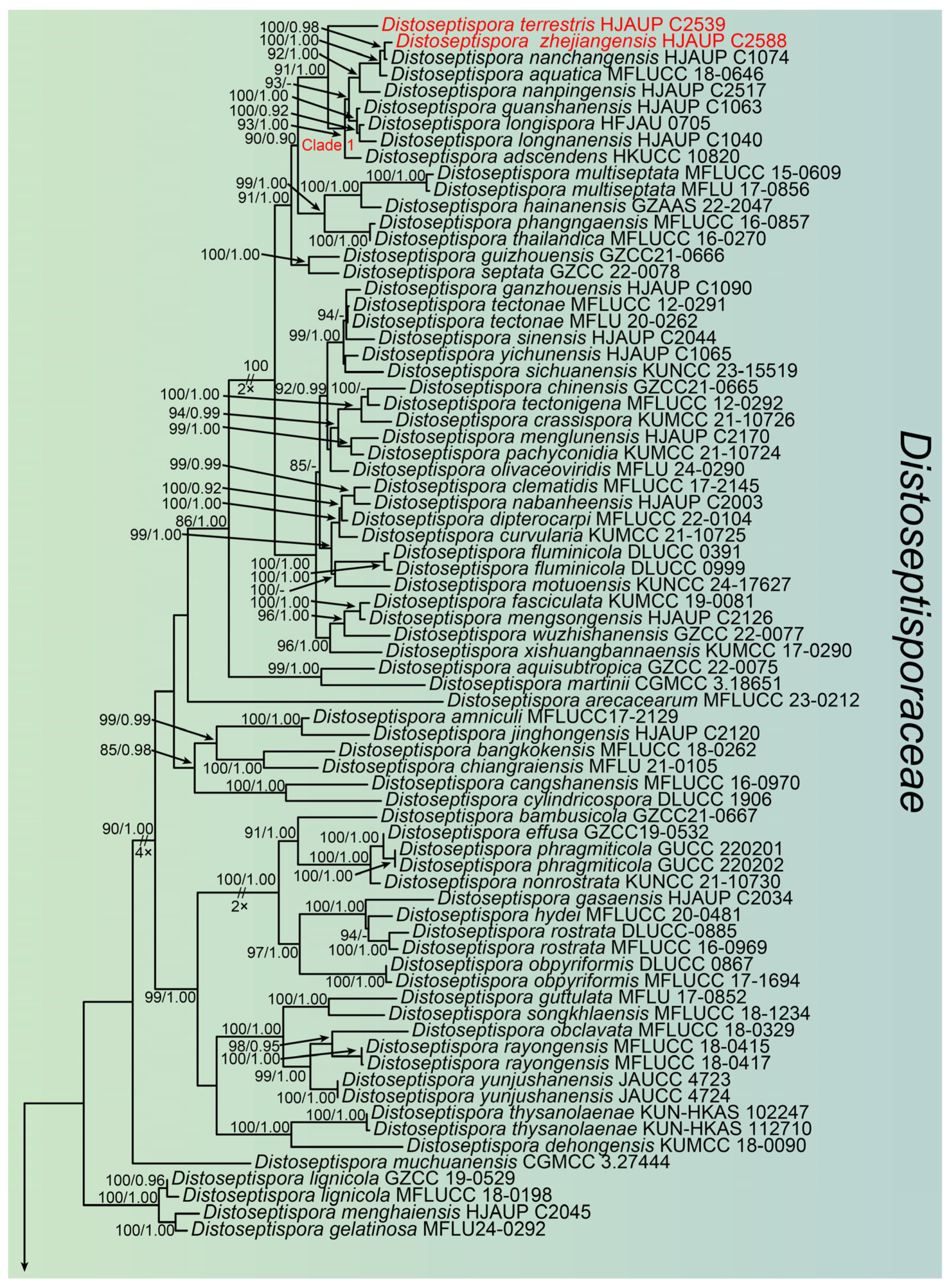
3.1. Molecular Phylogeny
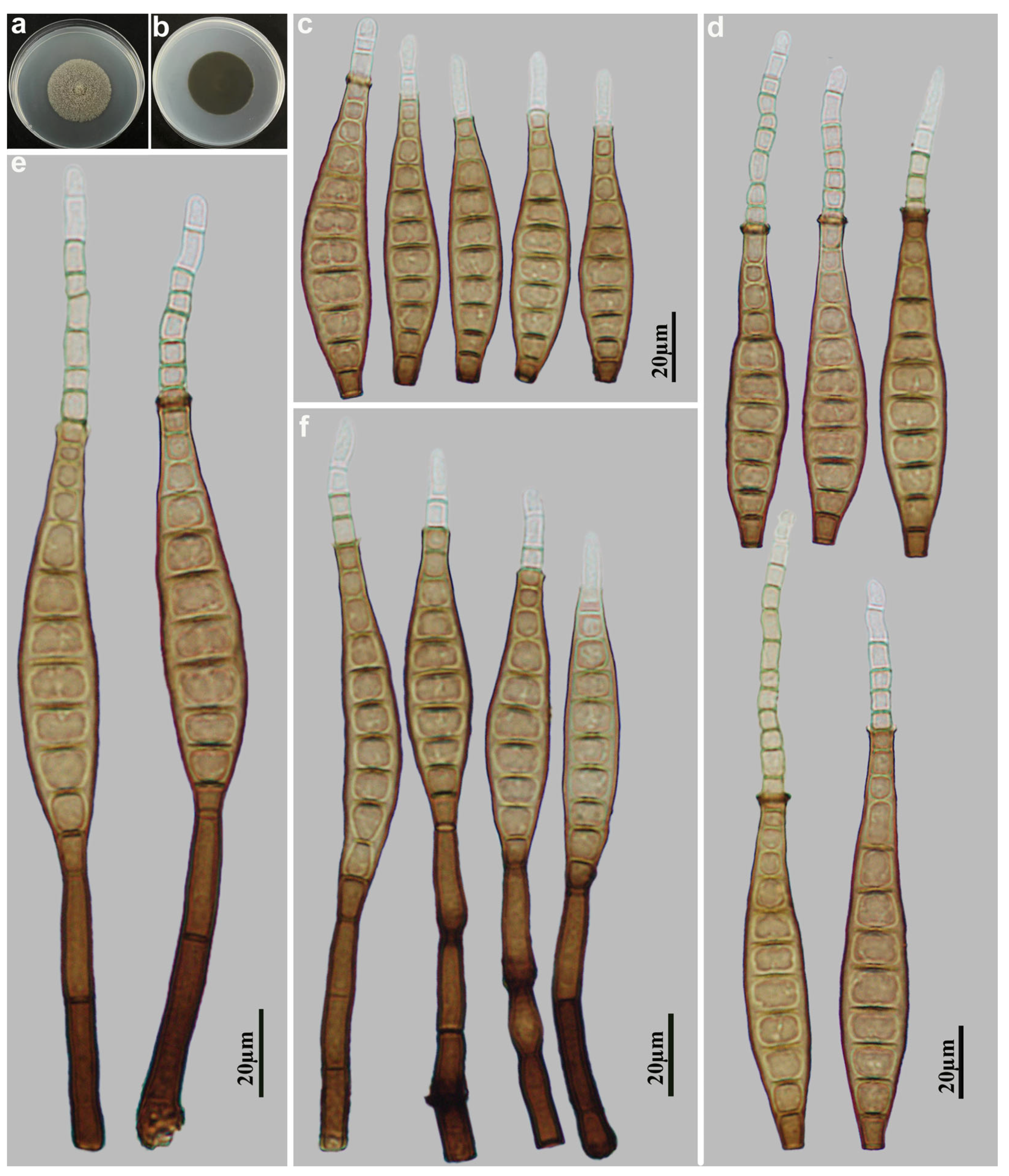
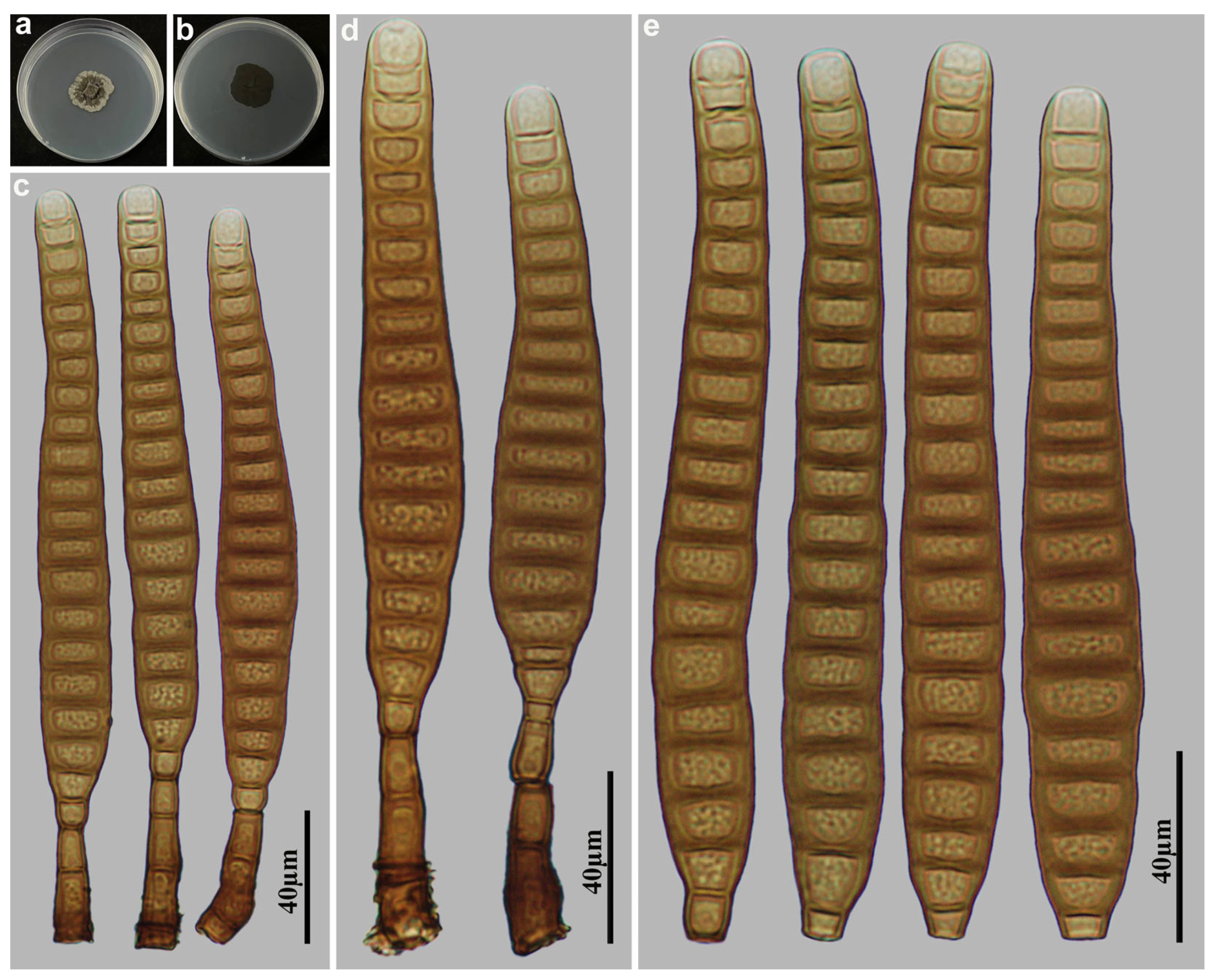
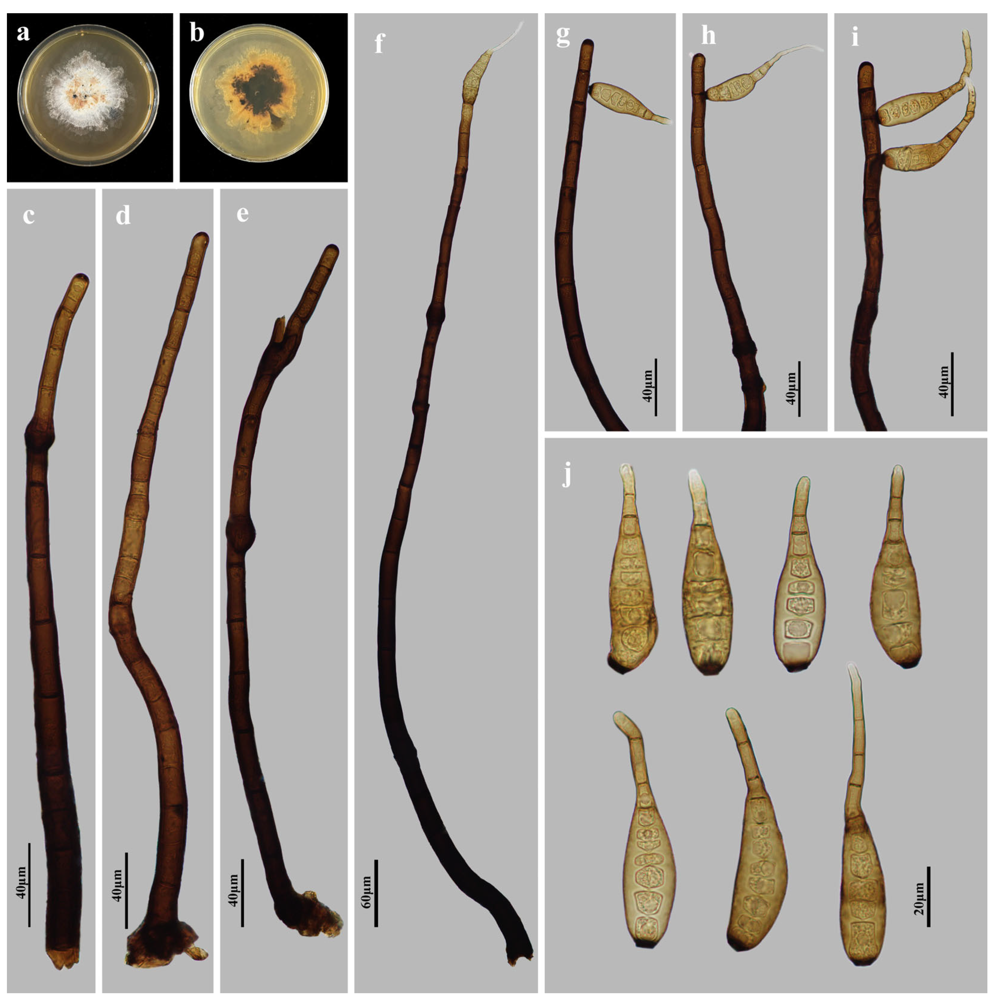
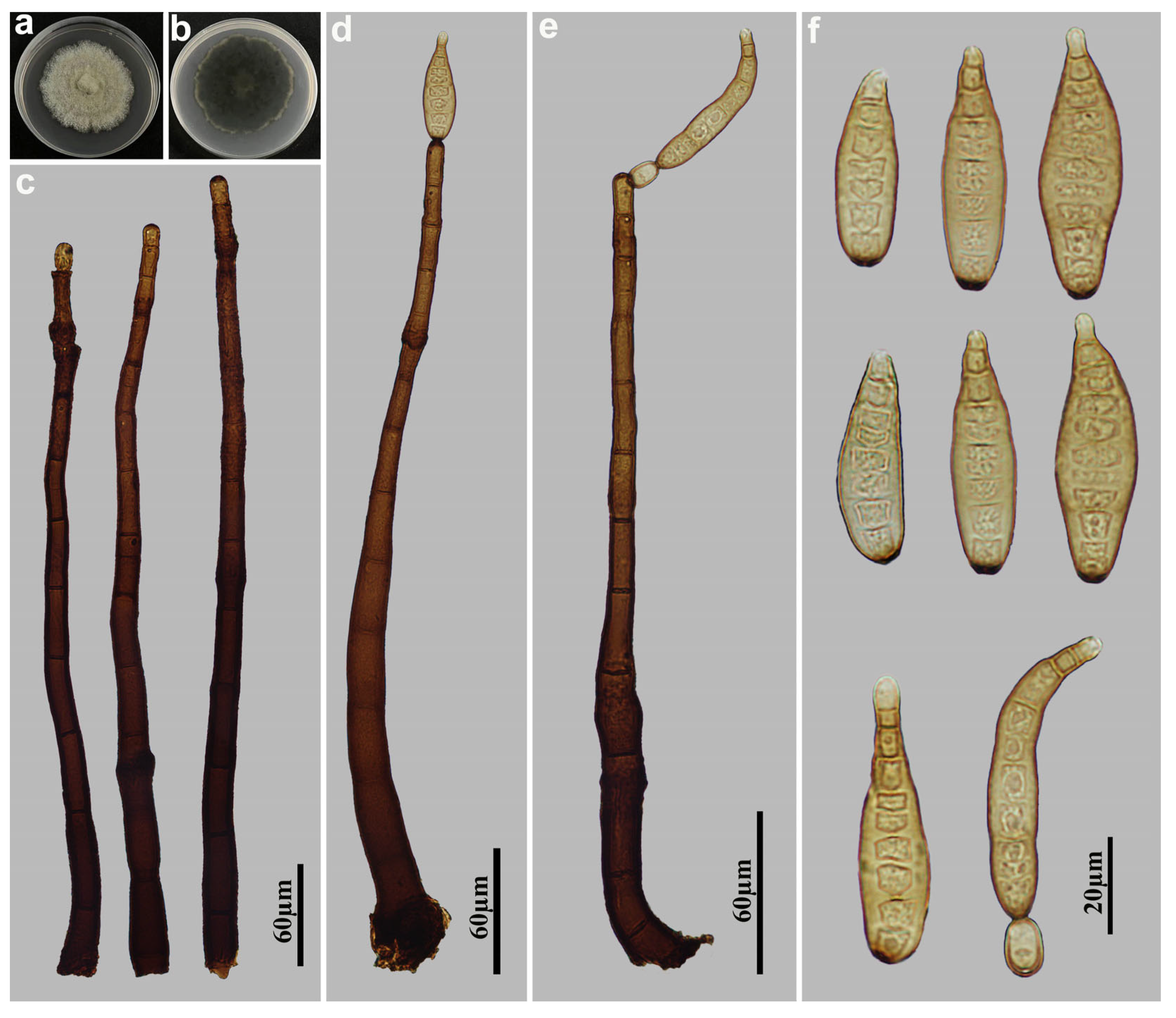
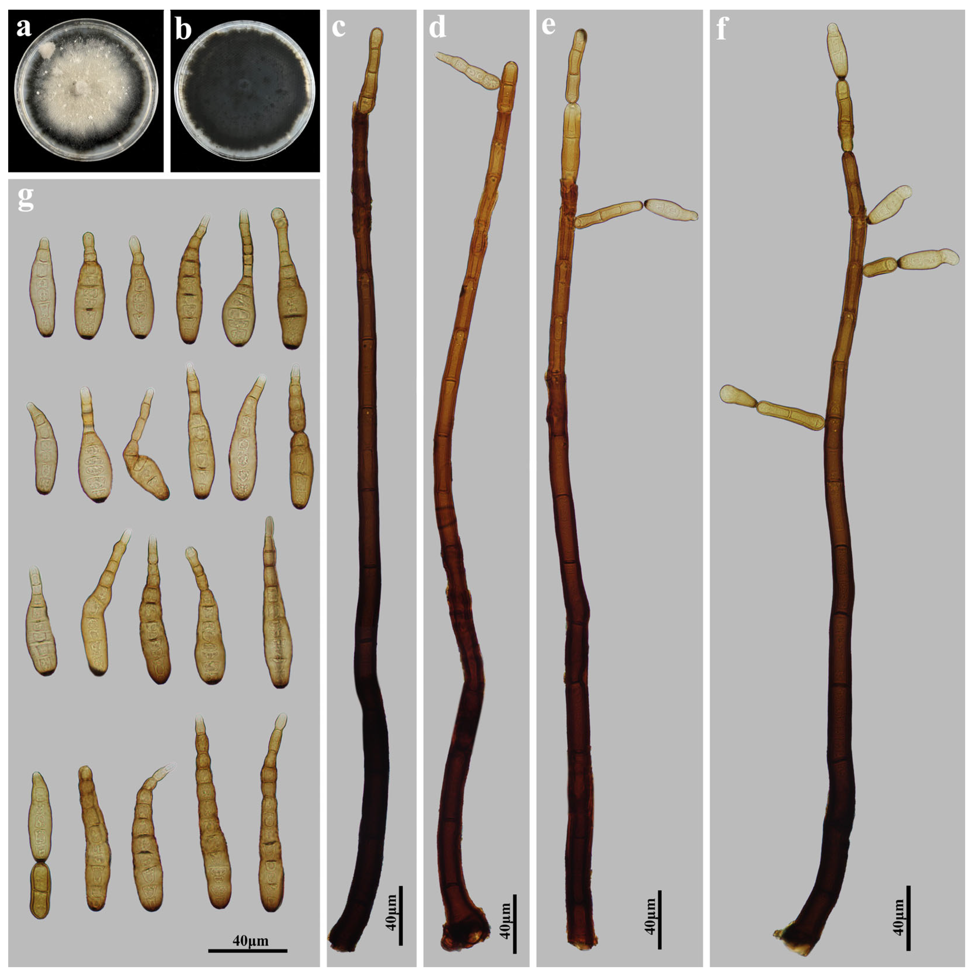
Based on the combined sequences of ITS, LSU, RPB2 and TEF1, the phylogenetic tree was constructed to analyze the phylogenetic relationships of the three strains in this study. The family Distoseptisporaceae and related families (Acrodictyaceae, Aquapteridosporaceae, Bullimycetaceae, Cancellidiaceae, Papulosaceae, and Pseudostanjehughesiaceae) was used a total of 131 strains representing 109 species (Table 2). Myrmecridium banksiae (CBS 132536) and M. schulzeri (CBS 100.54) were used as the outgroup. The combined sequence consisted of 2759 nucleotide positions (ITS:1–581, LSU:582–1095, RPB2:1096–1872, and TEF1:1873–2759), comprising 1115 parsimony-informative sites, 192 singleton sites, 1452 constant sites, and 1466 distinct patterns. The phylogenetic reconstruction obtained from ML and BI analyses of the combined sequences showed essentially congruent topological structures. The best-scoring ML tree (lnL = −37642.042) was shown in Figure 1, with node support values indicated above each branch. The first and second values at each node represent ultrafast bootstrap support values from ML analysis and posterior probabilities from MrBayes analysis, respectively. Based on multi-locus phylogenetic analyses combined with morphological characteristics, the three strains in this study were classified into three novel species, namely D. terrestris, D. wuyishanensis, and D. zhejiangensis.
Based on the combined sequences of ITS, LSU, RPB2, SSU and TEF1, the phylogenetic tree was constructed to analyze the phylogenetic relationships of the five strains in this study. The family Massarinaceae and related families (Periconiaceae, Corynesporascaceae and Cyclothyriellaceae) was used a total of 86 strains representing 55 species (Table 3). Cyclothyriellaceae rubronotata (CBS 121892) and C. rubronotata (141486) were used as the outgroup. The combined sequence consisted of 3744 nucleotide positions (ITS:1–532, LSU:533–1054, RPB2:1055–2087, SSU:2088–2861, and TEF1:2862–3730), comprising 1058 parsimony-informative sites, 201 singleton sites, 2485 constant sites, and 1435 distinct patterns. The phylogenetic reconstruction obtained from ML and BI analyses of the combined sequences showed essentially similar topological structures. The best-scoring ML tree (lnL = −25759.369) was shown in Figure 2, with node support values indicated above each branch. The first and second values at each node represent ultrafast bootstrap support values from ML analysis and posterior probabilities from MrBayes analysis, respectively. Based on multi-locus phylogenetic analyses combined with morphological characteristics, the five strains in this study were classified into three new species, namely H. ganzhouense, H. jiangxiense, and H. saprophyticum and one known species, H. velutinum.

3.2. Taxonomy

Distoseptispora terrestris M.G. Liao & Jian Ma, sp. nov., Figure 3.
Index Fungorum number: IF903855
Etymology: The name refers to the fact that this fungus was isolated from a terrestrial habitat.
Holotype: HJAUP M2539
Description: Asexual morph on natural substrate. Colonies effuse, scattered, smooth, hairy, dark brown or black. Mycelium immersed and superficial, composed of septate, branched, pale brown hyphae. Conidiophores macronematous, mononematous, solitary or in groups of 2–3, unbranched, stralight or flexuous, smooth, 2–3-septate, cylindrical, brown to dark brown, paler towards the apex, determinate or sometimes with cylindrical, enteroblastic percurrent extensions, 52.5–128 × 6.3–8.8 µm ( x ¯ = 76 × 7.3 µm, n = 15). Conidiogenous cells monoblastic, terminal, integrated, smooth, cylindrical, brown, with a flat apex. Conidial secession schizolytic. Conidia acrogenous, solitary, obclavate, straight or slightly curved, smooth, brown, 9–11-distoseptate, with a subhyaline to pale brown, euseptate rostrate regenerated from the terminal fracture of conidia, 54.5–91 × 11–18 µm ( x ¯ = 71.8 × 14 µm, n = 30), 8.8–10 µm in width at the truncate base, gradually tapering to 5–7.5 µm towards the apex. Sexual morph are unknown.
Culture characteristics: Colonies grown on PDA reaching 44 mm diameter from 28 to 42 days at 25 °C in dark conditions, circular, flat, surface grayish brown, with dense mycelium, and entire margin; reverse dark brown to black.
Material examined: China, Fujian Province, Nanping City, Wuyishan National Nature Reserve, 27°43′ N, 117°41′ E, on dead branches of unidentified plants under broad-leaved forest, 16 October 2023, Y.F. Hu (HJAUP M2539, holotype; HJAUP C2539, ex-type living culture).
Notes: Phylogenetic analyses shows that D. terrestris (HJAUP C2539) formed an independent lineage basal to Clade 1 with 91%ML/1.00BI bootstrap support. However, D. terrestris can be distinguished from the morphologically most similar species, D. longnanensis Y.F. Hu & Jian Ma [44] by its 9–11-distoseptate, wider conidia (11–18 µm vs. 8.2–14 µm wide) with euseptate rostrate regenerated from its terminal fracture, and further from D. longnanensis by 66 nucleotide differences (34/613 in ITS including 9 gaps, 7/555 in LSU including one gap, and 25/943 in TEF1 including 17 gaps). In addition, D. terrestris also differs from other taxa in Clade 1 in the shape and size of conidia and conidiophores.
Distoseptispora wuyishanensis M.G. Liao & Jian Ma, sp. nov., Figure 4.
Index Fungorum number: IF903856
Etymology: The name refers to Wuyishan National Nature Reserve, the locality where the fungus was collected.
Holotype: HJAUP M2515
Description: Asexual morph on natural substrate. Colonies effuse, scattered, smooth, hairy, dark brown or black. Mycelium immersed and superficial, composed of septate, branched, pale brown hyphae. Conidiophores macronematous, mononematous, solitary, unbranched, cylindrical, stralight or flexuous, 5–11-septate, brown to dark brown, paler towards the apex, determinate or with several cylindrical, enteroblastic percurrent extensions, 85.5–192 × 5.5–9.1 µm ( x ¯ = 130.9 × 7.3 µm, n = 15). Conidiogenous cells monoblastic, terminal, integrated, cylindrical, smooth, brown, with a flat apex. Conidial secession schizolytic. Conidia acrogenous, solitary, obclavate, fusiform, straight or slightly curved, brown, 5–8-euseptate and 5-distoseptate, smooth, rostrate, 54.5–91 × 11–18 µm ( x ¯ = 71.8 × 14 µm, n = 30), 4.2–5.5 µm in width at the truncate base, gradually tapering to 3.3–4.5 µm towards the apex. Sexual morph are unknown.
Culture characteristics: Colonies grown on PDA reaching 57 mm diameter from 28 to 42 days at 25 °C in dark conditions, circular, flat, surface yellowish brown, with dense mycelium, dark brown at the margin; reverse black with sparse edges.
Material examined: China, Fujian Province, Nanping City, Wuyishan National Nature Reserve, 27°43′ N, 117°41′ E, on dead branches of unidentified plants under broad-leaved forest, 16 October 2023, Y.F. Hu (HJAUP M2515, holotype; HJAUP C2515, ex-type living culture).
Notes: Phylogenetic analyses shows that D. wuyishanensis (HJAUP C2515) clusters as an independent clade sister to D. fujianensis (HJAUP C2509) and D. lanceolatispora (GZCC 22–2045) with 100%ML/1.00BI bootstrap support. The BLASTn shows that the nucleotide comparison of D. wuyishanensis (HJAUP C2515) and D. fujianensis (HJAUP C2509) in 256 nucleotide differences (55/608 in ITS including 16 gaps, 39/582 in LSU including 4 gaps, 116/946 in RPB2 including one gap, 46/922 in TEF1 including one gap). The sequences comparison of D. lanceolatispora (GZCC 22–2045) share 237 nucleotide differences (53/528 in ITS including 20 gaps, 40/572 in LSU including 3 gaps, 107/896 in RPB2 including one gap, 37/906 in TEF1 including one gap). Moreover, D. wuyishanensis differs from D. fujianensis M.G. Liao & Jian Ma [45] and D. lanceolatispora X.M. Chen & Y.Z. Lu [46] by its conidia with both eusepta and distosepta, and further from D. fujianensis by its bigger conidiophores (85.5–192 × 5.5–9.1 µm vs. 86–127 × 4.5–6 μm) and conidia (54.5–91 × 11–18 µm vs. 28–47 × 8–14 μm), and from D. lanceolatispora by its fusiform, smooth, wider conidia (11–18 µm vs. 9.5–15 μm).
Distoseptispora zhejiangensis M.G. Liao & Jian Ma, sp. nov., Figure 5.
MycoBank number: MB859236
Etymology: The name refers to Zhejiang, the province where the fungus was collected.
Holotype: HJAUP M2588
Description: Asexual morph on natural substrate. Colonies effuse, scattered, smooth, hairy, dark brown or black. Mycelium immersed and superficial, composed of septate, branched, pale brown hyphae. Conidiophores macronematous, mononematous, solitary, unbranched, smooth, cylindrical, stralight, 1–3-septate, brown, 16–56 × 13.5–20 µm ( x ¯ = 38 × 9.7 µm, n = 15). Conidiogenous cells monoblastic, terminal, integrated, cylindrical, smooth, brown, with a flat apex. Conidial secession schizolytic. Conidia acrogenous, solitary, obclavate, straight or slightly curved, 13–20-distoseptate, smooth, brown, paler towards the apex, 128.1–261.7 × 18.7–26.7 µm ( x ¯ = 161.1 × 23.5 µm, n = 30), 5.7–8.6 µm in width at the truncate base, gradually tapering to 7.1–14 µm towards the apex. Sexual morph are unknown.
Culture characteristics: Colonies grown on PDA reaching 27 mm diameter from 28 to 42 days at 25 °C in dark conditions, irregular circular, flat, surface velvety, with abundant aerial mycelium, dark olivaceous, paler towards the margin; reverse black.
Material examined: China, Zhejiang Province, Quzhou City: Meishudi Scenic, 29°08′ N, 118°69′ E, on dead branches of unidentified plants under broad-leaved forest, 21 June 2022, Y.F. Hu (HJAUP M2588, holotype; HJAUP C2588, ex-type living culture).
Notes: Phylogenetic analyses shows that D. zhejiangensis (HJAUP C2588) clusters with D. nanchangensis (HJAUP C1074) with 100%ML/0.98BI bootstrap support. The BLASTn shows that the nucleotide comparison of D. zhejiangensis (HJAUP C2588) and D. nanchangensis (HJAUP C1074) in 6 nucleotide differences (5/588 in ITS including one gap, 1/932 in TEF1). Moreover, D. zhejiangensis differs from D. nanchangensis Y.F. Hu & Jian Ma [44] in its shorter and wider conidiophores (16–56 × 13.5–20 µm vs. 18.2–76.4 × 5.5–8 µm) and shorter and wider conidia (128.1–261.7 × 18.7–26.7 µm vs. 149.1–292.7 × 10.9–17.8 µm) with fewer distosepta (13–20 vs. 17–43). In addition, the conidia of D. nanchangensis sometimes bear percurrent regeneration forming a secondary conidium, or a germ tube or short germination hypha from the conidial apex.
Helminthosporium ganzhouense M.G. Liao & Jian Ma, sp. nov., Figure 6.
MycoBank number: MB859237
Etymology: The name refers to Ganzhou city, the locality where the fungus was collected.
Holotype: HJAUP M1086
Description: Asexual morph on natural substrate. Colonies effuse, scattered, smooth, hairy, dark brown or black. Mycelium immersed and superficial, composed of septate, branched, pale brown hyphae. Conidiophores macronematous, mononematous, solitary, unbranched, erect, curved, smooth, cylindrical, dark brown, paler towards the apex, 10–24-septate, enteroblastic percurrent extensions, with distinct pores in subapical region, 312–428–(744) × 10.5–20 µm ( x ¯ = 420 × 14.4 µm, n = 15). Conidiogenous cells polytretic, terminal, cylindrical, integrated, brown. Conidial secession schizolytic. Conidia acropleurogenous, solitary, rostrate, obclavate, straight or curved, pale brown, 8–13-distoseptate, 62–96 × 12–16 µm ( x ¯ = 78 × 14.4 µm, n = 30), 4–5.9 µm in width at the truncate base, gradually tapering to 1.2–4 µm towards the apex. Sexual morph are unknown.
Culture characteristics: Colonies grown on PDA reaching 54 mm diameter from 28 to 42 days at 25 °C in dark conditions, irregular circular, surface gray-white with yellow-brown in the center; reverse rosy-brown with dark brown center.
Material examined: China, Jiangxi Province, Ganzhou City: Jiulianshan National Nature Reserve, 24°31′ N, 114°27′ E, on dead branches of unidentified plants under broad-leaved forest, 29 June 2022, Y.F. Hu (HJAUP M1086, holotype; HJAUP C1086, ex-type living culture).
Notes: Phylogenetic analyses shows that H. ganzhouense (HJAUP C1086) belongs to Helminthosporium and forms a distinct lineage sister to Clade 2 with 98%ML/1.00BI bootstrap support. However, H. ganzhouense differs from the morphologically most similar species, H. solani Durieu & Mont. [19,47] in its shorter and wider conidiophores (312–744 × 10.5–20 vs. 120–600 × 9–15 µm) and bigger conidia (62–96 × 12–16 µm vs. 24–85 × 7–11 µm) with more distosepta (8–13 vs. 2–8), and further from H. solani (CBS 365.75) by 93 nucleotide differences (27/568 in ITS including 3 gaps, 9/558 in LSU including 2 gaps, and 57/985 in TEF1). In addition, H. ganzhouense also differs from other taxa in Clade 2 in the size of conidiophores and conidia.
Helminthosporium jiangxiense M.G. Liao & Jian Ma, sp. nov., Figure 7.
MycoBank number: MB859238
Etymology: The name refers to Jiangxi, the province where the fungus was collected.
Holotype: HJAUP M1325
Description: Asexual morph on natural substrate. Colonies effuse, scattered, smooth, hairy, dark brown or black. Mycelium immersed and superficial, composed of septate, branched, pale brown hyphae. Conidiophores macronematous, mononematous, solitary, unbranched, erect, cylindrical, flexuous, 10–16-septate, dark brown to brown, paler towards the apex, enteroblastic percurrent extensions, with distinct pores in subapical region, 222.5–350 × 6.3–8 µm ( x ¯ = 278.1 × 7.3 µm, n = 10). Conidiogenous cells polytretic, terminal and intercalary, integrated, cylindrical, brown. Conidial secession schizolytic. Conidia solitary or in short chains, acropleurogenous, obclavate, straight or curved, pale brown, smooth, 2–10-distoseptate, 30–132.5 × 5–9.5 µm ( x ¯ = 55 × 6.5 µm, n = 30), 1.3–2.5 µm in width at the truncate base, gradually tapering to 2.5–3.8 µm towards the apex. Sexual morph are unknown.
Culture characteristics: Colonies grown on PDA reaching 54 mm diameter from 28 to 42 days at 25 °C in dark conditions, irregular circular, surface gray-white with dark brown; reverse rosy-brown with dark brown center.
Material examined: China, Jiangxi Province, Ganzhou City: Jiulianshan National Nature Reserve, 24°31′ N, 114°27′ E, on dead branches of unidentified plants under broad-leaved forest, 29 June 2022, Y.F. Hu (HJAUP M1325, holotype; HJAUP C1325, ex-type living culture).
Notes: Phylogenetic analyses shows that H. jiangxiense (HJAUP C1325) clusters with H. endiandrae (CBS 138902) with 99%ML/0.97BI bootstrap support. The BLASTn shows that the nucleotide comparison of H. jiangxiense (HJAUP C1325) and H. endiandrae (CBS 138902) show 93 nucleotide differences (73/593 in ITS including 30 gaps, 20/575 in LSU including 2 gaps). Moreover, H. jiangxiense differs from H. endiandrae (Crous & Summerell) Voglmayr & Jaklitsch [18] in its polytretic conidiogenous cell, bigger conidiophores (222.5–350 × 6.3–8 µm vs. 200–300 × 5–7 µm) and longer conidia (30–132.5 µm vs. 35–57 µm) with more distosepta (2–10 vs. 3–4).
Helminthosporium saprophyticum M.G. Liao & Jian Ma, sp. nov., Figure 8.
MycoBank number: MB859239
Etymology: The epithet refers to the saprophytic habit on dead branches.
Holotype: HJAUP M2572
Description: Asexual morph on natural substrate. Colonies effuse, scattered, smooth, hairy, dark brown or black. Mycelium immersed and superficial, composed of septate, branched, pale brown hyphae. Conidiophores macronematous, mononematous, solitary, unbranched, cylindrical, erect, flexuous, dark brown, paler towards the apex, enteroblastic percurrent extensions, with distinct pores in subapical region, 10–16 septate, 285.7–542 × 14.3–20 µm ( x ¯ = 417.1 × 18.8 µm, n = 15). Conidiogenous cells polytretic, terminal and intercalary, integrated, cylindrical, brown. Conidial secession schizolytic. Conidia acropleurogenous, solitary, rostrate, obclavate, straight or curved, pale brown, 6–10-distoseptate, 37.1–57.1 × 10–17.1 µm ( x ¯ = 47.7 × 13.1 µm, n = 30), 3.2–5.8 µm in width at the truncate base, gradually tapering to 2.9–3.4 µm towards the apex. Sexual morph are unknown.
Culture characteristics: Colonies grown on PDA reaching 65 mm diameter from 28 to 42 days in the incubation at 25 °C and dark conditions, subcircular, surface velvety, with abundant aerial mycelium, grayish brown, pale at the margin; reverse black with pale margin.
Material examined: China, Jiangxi Province, Ganzhou City: Jiulianshan National Nature Reserve, 24°31′ N, 114°27′ E, on dead branches of unidentified plants under broad-leaved forest, 29 June 2022, Y.F. Hu (HJAUP M2572, holotype; HJAUP C2572 = HJAUP C2573, ex-type living culture).
Notes: Phylogenetic analyses shows that H. saprophyticum (HJAUP C2572) and H. saprophyticum (HJAUP C2573) clusters with H. velutinum (H 4626 and HJAUP C1289) with 100%ML/1.00BI bootstrap support. The BLASTn shows that the nucleotide comparison of H. saprophyticum (HJAUP C2572) and H. velutinum (H 4626) in 11 nucleotide differences (2/524 in ITS, 1/579 in LSU, 3/472 in SSU including one gap, 5/923 in TEF1). Moreover, H. saprophyticum differs from H. velutinum Link [17,18] in having smaller conidiophores (285.7–542 × 14.3–20 µm vs. 340–698 × 14–26 µm), and smaller conidia (37.1–57.1 × 10–17.1 µm vs. 56–89 ×14.3–18.5 µm) with fewer distosepta (6–10 vs. 6–18).
Helminthosporium velutinum Link [as ‘Helmisporium’], Mag. Gesell. naturf. Freunde, Berlin 3(1–2): 10, (1809), Figure 9.
Description: Asexual morph on natural substrate. Colonies effuse, scattered, smooth, hairy, dark brown or black. Mycelium immersed and superficial, composed of septate, branched, pale brown hyphae. Conidiophores macronematous, mononematous, solitary, erect, unbranched, cylindrical, flexuous, dark brown, paler towards the apex, 13–20-septate, enteroblastic percurrent extensions, with distinct pores in subapical region, 464–616 × 12–16 µm, ( x ¯ = 552 × 14.8 µm, n = 10). Conidiogenous cells polytretic, terminal and intercalary, integrated, cylindrical, brown. Conidial secession schizolytic. Conidia acropleurogenous, solitary, rostrate, obclavate, straight or curved, smooth, pale brown, 4–13-distoseptate, constricted at the distoseptate, especially at the apex, 40–96 × 8–16 µm ( x ¯ = 68 × 12 µm, n = 30), 4.2–5.9 µm in width at the truncate base, gradually tapering to 2.8–5.2 µm toward the apex. Sexual morph are unknown.
Culture characteristics: Colonies grown on PDA reaching 81 mm diameter from 28 to 42 days at 25 °C in dark conditions, circular, surface creamish white with black margins, velvety mycelium in center; reverse black with sparse edges.
Material examined: China, Jiangxi Province, Ganzhou City: Jiulianshan National Nature Reserve, 24°31′ N, 114°27′ E, on dead branches of unidentified plants under broad-leaved forest, 29 June 2022, Y.F. Hu, HJAUP M1289; HJAUP C1289 (living culture).
Notes: Helminthosporium velutinum, the generic type, is a cosmopolitan species, and has been recorded from a wide range of woody and herbaceous plants [18]. Phylogenetic analyses shows that our new isolate (HJAUP C1289) clusters with H. velutinum (H 4626) with 100%ML/1.00BI bootstrap support. Morphologically, our isolate fits well with the description of H. velutinum by Voglmayr & Jaklitsch [18], except for the narrower conidiophores (12–16 µm vs. 14–26 µm). Comparative analysis of their nucleotide sequences revealed no genetic differences in the ITS (524/524, no gaps), LSU (552/552, no gaps), SSU (421/421, no gaps), and TEF1 (923/923, no gaps) regions. Based on the high morphological similarity and no nucleotide differences, we identified our isolate (HJAUP M1289) as H. velutinum.

4. Discussion

In recent years, studies on microfungi have received extensive attention, and numerous published information about their taxonomy are recorded in China [48,49,50,51,52]. During the ongoing study, we collected saprophytic microfungi from dead branches in southern China, and highly phylogenetic support and significant morphological comparison showed six novel species, namely Distoseptispora terrestris, D. wuyishanensis, D. zhejiangensis, Helminthosporium ganzhouense, H. jiangxiense, and H. saprophyticum, and one known species, H. velutinum.
Distoseptispora is one of the sporidesmium-like genera introduced by Su et al. [7] based on molecular phylogenetic analyses combined with morphology. Distoseptispora species were well described in terms of their taxonomic placements and genetic relationship by significant morphological characteristics and phylogenetic analyses. However, the research conducted on Distoseptispora has no universally accepted Standards for genetic barcode selection in phylogenetic analyses [14], which result in a lack of correlation between phylogenetic relationships and morphological analysis for some Distoseptispora species. For instance, D. lanceolatispora cluster with D. neorostrata in a subclade with highly genetic relationship, but significant morphological variations were observed, specifically in conidiophores, conidiogenous cells, and conidia [46,53]. The limited phylogenetic resolution at the genus level may be attributed to the exclusive use of ribosomal genes in the analysis of D. neorostrata. The holotype of D. bambusae (MFLU 20-0261) exhibited distinct morphological differences from phylogenetic strains (MFLU 17-1653 and HKAS 125826) in the characteristics of conidiophores, conidiogenous cells, and conidia [54,55]. Under the current phylogenetic framework, traditional taxonomic characteristics such as conidiogenesis may no longer play a decisive role in species delimitation within sporidesmium-like genera [45]. A comprehensive investigation incorporating ribosomal genes and additional genomic loci is required to clarify the relationship between phylogenetic affiliations and morphological traits.
The genus Helminthosporium was introduced by Link based on morphological characters. To date, more than 780 epithets for Helminthosporium are listed in Index Fungorum [15], and only 45 species, including our three new species, were provided with DNA sequences. Most Helminthosporium species were reported before sequencing technology, resulting in a lack of molecular data. It is questionable to identify Helminthosporium species and related genera only by reference to morphological characteristics, host, and environment. Voglmayr & Jaklitsch [18] investigate the phylogenetic relationships of Helminthosporium and its related genera, Corynespora and Exosporium based on phylogenetic analyses of combined SSU, ITS, LSU, RPB2 and TEF1 sequence data, synonymised Exosporium with Helminthosporium, and transformed four Corynespora species to Helminthosporium. Since then, the number of Helminthosporium species is steadily increasing, and all described species were introduced using multilocus phylogenetic analyses. However, recent studies indicated that the phylogenetic analysis for Helminthosporium species have no universally accepted standards in selecting barcodes [56] and based on only ribosomal genes may be insufficient in resolving the phylogeny of Helminthosporium and related genera within Massarinaceae, as together with RPB2 and/ or TEF1 showed more powerful resolution in species delineation and higher bootstrap support values for most clades [18,23,27,29]. Considering this phenomenon, we conducted phylogenetic analyses using ITS, LSU, RPB2, SSU and TEF1, and proposed three new Helminthosporium species, and one known species, H. velutinum in this study.
Current studies on Distoseptispora and Helminthosporium have predominantly focused on the taxonomy of their asexual morphs, with most specimens collected from submerged wood, dead twigs and plant leaves. However, few studies have investigated their ecological functions in relation to the specific substrates. Current knowledge regarding their functional roles in decomposition, nutrient cycling, geographical distribution patterns, ecological adaptability, host associations, substrate specificity, and teleomorph-anamorph relationships remains relatively limited. As a result, their precise contributions to ecosystem functioning cannot yet be quantitatively assessed [44]. Several Helminthosporium species have been isolated from plant leaves as endophytes or pathogens. By colonizing plant tissues, endophytic fungi support host development and increase resilience to environmental stresses [57,58], thereby improving the community structure of ecosystems. In contrast, H. solani as a pathogen mainly causes blemishes in the periderm of potato tubers [22], which has emerged as a significant economically important plant disease in 1990s [59,60]. Therefore, conducting surveys across diverse geographical regions, ecological environments, and vegetation types will contribute to the conservation of species diversity in Distoseptispora and Helminthosporium, while facilitating the clarification of their taxonomic status based on phylogenetic analyses combined with morphological characterization.

Author Contributions

Conceptualization, M.-G.L. and X.-X.L.; methodology, M.-G.L.; software, M.-G.L. and Z.-H.X.; validation, J.-W.X.; formal analysis, M.-G.L.; investigation, Y.-F.H.; resources, Y.-F.H.; data curation, M.-G.L.; writing—original draft preparation, M.-G.L.; writing—review and editing, X.-G.Z., L.-H.Z. and J.M.; visualization, M.-G.L. and X.-P.Z.; supervision, J.M.; project administration, J.M.; funding acquisition, J.M. All authors have read and agreed to the published version of the manuscript.

Funding

This work was supported by the National Natural Science Foundation of China (Nos. 32160006, 31970018).

Institutional Review Board Statement

Not applicable.

Informed Consent Statement

Not applicable.

Data Availability Statement

All sequences generated in this study were submitted to GenBank.

Conflicts of Interest

The authors declare no conflicts of interest.

References

  1. Schimann, H.; Bach, C.; Lengelle, J.; Louisanna, E.; Barantal, S.; Murat, C.; Buée, M. Diversity and structure of fungal communities in neotropical rainforest soils: The effect of host recurrence. Microb. Ecol. 2017, 73, 310–320. [Google Scholar] [CrossRef]
  2. Hawksworth, D.L. The fungal dimension of biodiversity: Magnitude, significance, and conservation. Mycol. Res. 1991, 95, 641–655. [Google Scholar] [CrossRef]
  3. Hawksworth, D.L.; Lücking, R. Fungal diversity revisited: 2.2 to 3.8 million species. Microbiol. Spectr. 2017, 5, 79–95. [Google Scholar] [CrossRef] [PubMed]
  4. Hyde, K.D. The numbers of fungi. Fungal Divers. 2022, 114, 1. [Google Scholar] [CrossRef]
  5. Phukhamsakda, C.; Nilsson, R.H.; Bhunjun, C.S.; De Farias, A.R.G.; Sun, Y.-R.; Wijesinghe, S.N.; Raza, M.; Bao, D.-F.; Lu, L.; Tibpromma, S.; et al. The numbers of fungi: Contributions from traditional taxonomic studies and challenges of metabarcoding. Fungal Divers. 2022, 114, 327–386. [Google Scholar] [CrossRef]
  6. Bhunjun, C.S.; Niskanen, T.; Suwannarach, N.; Wannathes, N.; Chen, Y.-J.; McKenzie, E.H.C.; Maharachchikumbura, S.S.N.; Buyck, B.; Zhao, C.-L.; Fan, Y.-G.; et al. The numbers of fungi: Are the most speciose genera truly diverse? Fungal Divers. 2022, 114, 387–462. [Google Scholar] [CrossRef]
  7. Su, H.-Y.; Hyde, K.D.; Maharachchikumbura, S.S.N.; Ariyawansa, H.A.; Luo, Z.-L.; Promputtha, I.; Tian, Q.; Lin, C.-G.; Shang, Q.-J.; Zhao, Y.-C.; et al. The families Distoseptisporaceae fam. nov., Kirschsteiniotheliaceae, Sporormiaceae and Torulaceae, with new species from freshwater in Yunnan Province, China. Fungal Divers. 2016, 80, 375–409. [Google Scholar] [CrossRef]
  8. Xia, J.-W.; Ma, Y.-R.; Li, Z.; Zhang, X.-G. Acrodictys-like wood decay fungi from southern China, with two new families Acrodictyaceae and Junewangiaceae. Sci. Rep. 2017, 7, 7888. [Google Scholar] [CrossRef]
  9. Yang, J.; Maharachchikumbura, S.S.N.; Liu, J.-K.; Hyde, K.D.; Jones, E.B.G.; Al-Sadi, A.M.; Liu, Z.-Y. Pseudostanjehughesia aquitropica gen. et sp. nov. and Sporidesmium sensu lato species from freshwater habitats. Mycol. Prog. 2018, 17, 591–616. [Google Scholar] [CrossRef]
  10. Hyde, K.D.; Tennakoon, D.S.; Jeewon, R.; Bhat, D.J.; Maharachchikumbura, S.S.N.; Rossi, W.; Leonardi, M.; Lee, H.B.; Mun, H.Y.; Houbraken, J.; et al. Fungal diversity notes 1036–1150: Taxonomic and phylogenetic contributions on genera and species of fungal taxa. Fungal Divers. 2019, 96, 1–242. [Google Scholar] [CrossRef]
  11. Yang, J.; Liu, L.-L.; Jones, E.B.G.; Li, W.-L.; Hyde, K.D.; Liu, Z.-Y. Morphological variety in Distoseptispora and introduction of six novel species. J. Fungi 2021, 7, 945. [Google Scholar] [CrossRef] [PubMed]
  12. Konta, S.; Tibpromma, S.; Karunarathna, S.C.; Samarakoon, M.C.; Steven, L.S.; Mapook, A.; Boonmee, S.; Senwanna, C.; Balasuriya, A.; Eungwanichayapant, P.D.; et al. Morphology and multigene phylogeny reveal ten novel taxa in Ascomycota from terrestrial palm substrates (Arecaceae) in Thailand. Mycosphere 2023, 14, 107–152. [Google Scholar] [CrossRef]
  13. Manawasinghe, I.S.; Hyde, K.D.; Wanasinghe, D.N.; Karunarathna, S.C.; Maharachchikumbura, S.S.N.; Samarakoon, M.C.; Voglmayr, H.; Pang, K.-L.; Chiang, M.W.-L.; Jones, E.B.G.; et al. Fungal diversity notes 1818–1918: Taxonomic and phylogenetic contributions on genera and species of fungi. Fungal Divers. 2025, 130, 1–261. [Google Scholar] [CrossRef]
  14. Liu, J.-W.; Hu, Y.-F.; Luo, X.-X.; Xu, Z.-H.; Castañeda-Ruíz, R.F.; Xia, J.-W.; Zhang, X.-G.; Zhang, L.-H.; Cui, R.-Q.; Ma, J. Morphological and phylogenetic analyses reveal three new species of Distoseptispora (Distoseptisporaceae, Distoseptisporales) from Yunnan, China. J. Fungi 2023, 9, 470. [Google Scholar] [CrossRef] [PubMed]
  15. Index Fungorum. Available online: http://www.indexfungorum.org/Names/Names.asp (accessed on 10 March 2025).
  16. Dong, W.; Hyde, K.D.; Jeewon, R.; Doilom, M.; Yu, X.-D.; Wang, G.-N.; Liu, N.-G.; Hu, D.-M.; Nalumpang, S.; Zhang, H. Towards a natural classification of annulatascaceae-like taxa II: Introducing five new genera and eighteen new species from freshwater. Mycosphere 2021, 12, 1–88. [Google Scholar] [CrossRef]
  17. Link, H.F. Observationes in Ordines plantarum naturales. Dissertatio prima complectens Anandrarum ordines Epiphytas, Mucedines, Gastromycos et Fungos. Ges. Natur. 1809, 3, 3–42. [Google Scholar]
  18. Voglmayr, H.; Jaklitsch, W.M. Corynespora, Exosporium and Helminthosporium revisited—New species and generic reclassification. Stud. Mycol. 2017, 87, 43–76. [Google Scholar] [CrossRef]
  19. Ellis, M.B. Dematiaceous hyphomycetes. III. Mycol. Pap. 1961, 82, 55. [Google Scholar]
  20. Luttrell, E.S. Systematics of Helminthosporium and related genera. Mycologia 1964, 56, 119–132. [Google Scholar] [CrossRef]
  21. Tanaka, K.; Hirayama, K.; Yonezawa, H.; Sato, G.; Toriyabe, A.; Kudo, H.; Hashimoto, A.; Matsumura, M.; Harada, Y.; Kurihara, Y.; et al. Revision of the Massarineae (Pleosporales, Dothideomycetes). Stud. Mycol. 2015, 82, 75–136. [Google Scholar] [CrossRef]
  22. Siboe, G.M.; Kirk, P.M.; Cannon, P.F. New dematiaceous hyphomycetes from Kenyan rare plants. Mycotaxon 1999, 73, 283–302. [Google Scholar]
  23. Hu, Y.-F.; Liu, J.-W.; Xu, Z.-H.; Castañeda-Ruíz, R.F.; Zhang, K.; Ma, J. Morphology and multigene phylogeny revealed three new species of Helminthosporium (Massarinaceae, Pleosporales) from China. J. Fungi 2023, 9, 280. [Google Scholar] [CrossRef]
  24. Hyde, K.D.; Norphanphoun, C.; Ma, J.; Yang, H.D.; Zhang, J.-Y.; Du, T.-Y.; Gao, Y.; De Farias, A.R.G.; He, S.-C.; He, Y.-K.; et al. Mycosphere notes 387–412–novel species of fungal taxa from around the world. Mycosphere 2023, 14, 663–744. [Google Scholar] [CrossRef]
  25. Hyde, K.D.; Wijesinghe, S.N.; Afshari, N.; Aumentado, H.D.; Bhunjun, C.S.; Boonmee, S.; Camporesi, E.; Chethana, K.W.T.; Doilom, M.; Dong, W.; et al. Mycosphere Notes 469–520. Mycosphere 2024, 15, 1294–1454. [Google Scholar] [CrossRef]
  26. Ilyukhin, E.; Markovskaja, S. Helminthosporium paraoligosporum sp. nov., a new ascomycetous fungus from Ontario, Canada. Phytotaxa 2023, 607, 151–159. [Google Scholar] [CrossRef]
  27. Tian, W.-H.; Jin, Y.; Liao, Y.-C.; Faraj, T.K.; Guo, X.-Y.; Maharachchikumbura, S.S.N. Phylogenetic insights reveal new taxa in Thyridariaceae and Massarinaceae. J. Fungi 2024, 10, 542. [Google Scholar] [CrossRef] [PubMed]
  28. Zou, M.; Al-Otibi, F.; Hyde, K.D.; Wang, Y.; Pan, X.-J. New Helminthosporium (Massarinaceae, Dothideomycetes) and Nigrospora (Incertae sedis, Sordariomycetes) species associated with walnut (Juglans regia L.) in China. MycoKeys 2024, 109, 265–284. [Google Scholar] [CrossRef]
  29. Lu, L.; Karunarathna, S.C.; Rajeshkumar, K.C.; Elgorban, A.M.; Jayawardena, R.S.; Hongsanan, S.; Suwannarach, N.; Kumla, J.; Xiong, Y.-R.; Hyde, K.D.; et al. Unveiling fungal diversity associated with coffee trees in China using a polyphasic approach and a global review of coffee saprobic fungi. IMA Fungus 2025, 16, e144874. [Google Scholar] [CrossRef]
  30. Sun, Y.-R.; Hyde, K.D.; Liu, N.-G.; Jayawardena, R.S.; Wijayawardene, N.N.; Ma, J.; Zhang, Q.; Al-Otibi, F.; Wang, Y. Micro-fungi in southern China and northern Thailand: Emphasis on medicinal plants. Fungal Divers. 2025, 131, 99–299. [Google Scholar] [CrossRef]
  31. Errampalli, D.; Saunders, J.M.; Holley, J.D. Emergence of silver scurf (Helminthosporium solani) as an economically important disease of potato. Plant Pathol. 2001, 50, 141–153. [Google Scholar] [CrossRef]
  32. Ma, J.; Wang, Y.; Ma, L.-G.; Zhang, Y.-D.; Castañeda-Ruíz, R.F.; Zhang, X.G. Three new species of Neosporidesmium from Hainan, China. Mycol. Prog. 2011, 10, 157–162. [Google Scholar] [CrossRef]
  33. White, T.J.; Bruns, T.D.; Lee, S.B.; Taylor, J.W. Amplification and direct sequencing of fungal ribosomal RNA genes for phylogenetics. In PCR Protocols: A Guide to Methods and Applications; Innis, M.A., Gelfand, D.H., Sninsky, J.J., White, T.J., Eds.; Academic Press: New York, NY, USA, 1990; pp. 315–322. [Google Scholar] [CrossRef]
  34. Sung, G.H.; Sung, J.M.; Hywel-Jones, N.L.; Spatafora, J.W. A multi-gene phylogeny of Clavicipitaceae (Ascomycota, Fungi): Identification of localized incongruence using a combinational bootstrap approach. Mol. Phylogenet. Evol. 2007, 44, 1204–1223. [Google Scholar] [CrossRef] [PubMed]
  35. Liu, Y.-J.; Whelen, S.; Hall, B.D. Phylogenetic relationships among ascomycetes: Evidence from an RNA polymerse II subunit. Mol. Biol. Evol. 1999, 16, 1799–1808. [Google Scholar] [CrossRef]
  36. Rehner, S.A.; Buckley, E. A Beauveria phylogeny inferred from nuclear ITS and EF1-alpha sequences: Evidence for cryptic diversification and links to Cordyceps teleomorphs. Mycologia 2005, 97, 84–98. [Google Scholar] [CrossRef]
  37. Zhao, N.; Luo, Z.-L.; Hyde, K.D.; Su, H.-Y.; Bhat, D.J.; Liu, J.-K.; Bao, D.-F.; Hao, Y.-E. Helminthosporium submersum sp. nov. (Massarinaceae) from submerged wood in north-western Yunnan Province. China. Phytotaxa 2018, 348, 269–278. [Google Scholar] [CrossRef]
  38. Katoh, K.; Standley, D.M. MAFFT multiple sequence alignment software version 7: Improvements in performance and usability. Mol. Biol. Evol. 2013, 30, 772–780. [Google Scholar] [CrossRef] [PubMed]
  39. Zhang, K.; Zhang, H.; Li, D.-W.; Castañeda-Ruíz, R.F. Mirohelminthosporium gen. nov. for an atypical Helminthosporium species and H. matsushimae nom. nov. Mycotaxon 2020, 135, 777–783. [Google Scholar] [CrossRef]
  40. Kalyaanamoorthy, S.; Minh, B.Q.; Wong, T.K.F.; von Haeseler, A.; Jermiin, L.S. ModelFinder: Fast model selection for accurate phylogenetic estimates. Nat. Methods 2017, 14, 587–589. [Google Scholar] [CrossRef]
  41. Nguyen, L.T.; Schmidt, H.A.; von Haeseler, A.; Minh, B.Q. IQ-TREE: A fast and effective stochastic algorithm for estimating maximum-likelihood phylogenies. Mol. Biol. Evol. 2015, 32, 268–274. [Google Scholar] [CrossRef]
  42. Minh, B.Q.; Nguyen, M.A.; Von Haeseler, A. Ultrafast approximation for phylogenetic bootstrap. Mol. Biol. Evol. 2013, 30, 1188–1195. [Google Scholar] [CrossRef]
  43. Ronquist, F.; Teslenko, M.; van der Mark, P.; Ayres, D.L.; Darling, A.; Höhna, S.; Larget, B.; Liu, L.; Suchard, M.A.; Huelsenbeck, J.P. MrBayes 3.2: Efficient bayesian phylogenetic inference and model choice across a large model space. Syst. Biol. 2012, 61, 539–542. [Google Scholar] [CrossRef] [PubMed]
  44. Hu, Y.-F.; Liu, J.-W.; Luo, X.-X.; Xu, Z.-H.; Xia, J.-W.; Zhang, X.-G.; Castañeda-Ruíz, R.F.; Ma, J. Multi-locus phylogenetic analyses reveal eight novel species of Distoseptispora from southern China. Microbiol. Spectr. 2023, 11, e0246823. [Google Scholar] [CrossRef]
  45. Liao, M.-G.; Luo, X.-X.; Hu, Y.-F.; Castañeda-Ruíz, R.F.; Xu, Z.-H.; Ma, J. Morphological and phylogenetic analyses reveal four novel species of Distoseptispora (Distoseptisporaceae, Distoseptisporales) from southern China. MycoKeys 2025, 113, 31–55. [Google Scholar] [CrossRef]
  46. Chen, X.-M.; Tang, X.; Ma, J.; Liu, N.-G.; Tibpromma, S.; Karunarathna, S.C.; Xiao, Y.-P.; Lu, Y.-Z. Identification of two new species and a new host record of Distoseptispora (Distoseptisporaceae, Distoseptisporales, Sordariomycetes) from terrestrial and freshwater habitats in Southern China. MycoKeys 2024, 102, 83–105. [Google Scholar] [CrossRef]
  47. De Bory, S.V.; De Durieu, M. Exploration Scientifique de l’Algérie Pendant les Années 1840, 1841, 1842 Publiée par Ordre du Gouvernement et avec le Concours d’une Commission Académique; Sciences physiques; Botanique par MM Bory de St-.Vincent et Durieu de Maisonneuve membres de la Commission scientifique d’Algérie; Imprimerie Nationale: Paris, France, 1846–1869. [Google Scholar]
  48. Zhang, Z.-X.; Shang, Y.-X.; Liu, Q.-Y.; Li, D.-H.; Yin, X.-Y.; Liu, X.-Y.; Tao, M.-F.; Jiang, Y.; Wang, Y.-X.; Zhang, M.-Y.; et al. Deciphering the evolutionary and taxonomic complexity of Diaporthales (Sordariomycetes, Ascomycota) through integrated phylogenomic and divergence time estimation. Fungal Divers. 2025, 132, 1–125. [Google Scholar] [CrossRef]
  49. Liu, N.-G.; Hyde, K.D.; Sun, Y.-R.; Bhat, D.J.; Jones, E.B.G.; Jumpathong, J.J.; Lin, C.-G.; Lu, Y.-Z.; Yang, J.; Liu, J.-J.; et al. Notes, outline, taxonomy and phylogeny of brown-spored hyphomycetes. Fungal Divers. 2024, 129, 1–281. [Google Scholar] [CrossRef]
  50. Liu, J.-W.; Hu, Y.-F.; Luo, X.-X.; Castañeda-Ruíz, R.F.; Xia, J.-W.; Xu, Z.-H.; Cui, R.-Q.; Shi, X.-G.; Zhang, L.-H.; Ma, J. Molecular Phylogeny and Morphology Reveal Four Novel Species of Corynespora and Kirschsteiniothelia (Dothideomycetes, Ascomycota) from China: A Checklist for Corynespora Reported Worldwide. J. Fungi 2023, 9, 107. [Google Scholar] [CrossRef] [PubMed]
  51. Tian, W.-H.; Jin, Y.; Liao, Y.-C.; Faraj, T.K.; Guo, X.-Y.; Maharachchikumbura, S.S.N. New and Interesting Pine-Associated Hyphomycetes from China. J. Fungi 2024, 10, 546. [Google Scholar] [CrossRef]
  52. Luo, X.-X.; Liao, M.-G.; Hu, Y.-F.; Zhang, X.-G.; Xu, Z.-H.; Ma, J. Identification of three novel species and one new record of Kirschsteiniothelia (Kirschsteiniotheliaceae, Kirschsteiniotheliales) from Jiangxi, China. MycoKeys 2025, 112, 277–306. [Google Scholar] [CrossRef]
  53. Luo, Z.-L.; Hyde, K.D.; Liu, J.-K.; Maharachchikumbura, S.S.N.; Jeewon, R.; Bao, D.-F.; Bhat, D.J.; Lin, C.-G.; Li, W.-L.; Yang, J.; et al. Freshwater Sordariomycetes. Fungal Divers. 2019, 99, 451–660. [Google Scholar] [CrossRef]
  54. Sun, Y.; Goonasekara, I.D.; Thambugala, K.M.; Jayawardena, R.S.; Wang, Y.; Hyde, K.D. Distoseptispora bambusae sp. nov. (Distoseptisporaceae) on bamboo from China and Thailand. Biodivers. Data J. 2020, 8, e53678. [Google Scholar] [CrossRef] [PubMed]
  55. Shen, H.-W.; Bao, D.-F.; Boonmee, S.; Lu, Y.-Z.; Su, X.-J.; Li, Y.-X.; Luo, Z.-L. Diversity of Distoseptispora (Distoseptisporaceae) taxa on submerged decaying wood from the Red River in Yunnan, China. MycoKeys 2024, 102, 1–28. [Google Scholar] [CrossRef] [PubMed]
  56. Chen, Y.; Tian, W.; Guo, Y.; Madrid, H.; Maharachchikumbura, S.S.N. Synhelminthosporium gen. et sp. nov. and Two New Species of Helminthosporium (Massarinaceae, Pleosporales) from Sichuan Province, China. J. Fungi 2022, 8, 712. [Google Scholar] [CrossRef]
  57. Hassani, M.A.; Durán, P.; Hacquard, S. Microbial interactions within the plant holobiont. Microbiome 2018, 6, 58. [Google Scholar] [CrossRef] [PubMed]
  58. Li, H.; Yan, C.-L.; Tang, Y.-Q.; Ma, X.; Chen, Y.-H.; Chen, S.-B.; Lin, M.; Liu, Z. Endophytic bacterial and fungal microbiota in different cultivars of cassava (Manihot esculenta Crantz). J. Microbiol. 2020, 58, 614–623. [Google Scholar] [CrossRef]
  59. Ivanyuk, V.; Zezyulina, G. Helminthosporium solani causing silver scab is reported from Belarus. Zashch. Rastenii 1991, 3, 45. [Google Scholar]
  60. El Immane-Collet, R.; Elakel, M.; Jouan, B. Comparative study of the agronomical incidence of the silver scurf disease of potato in Morocco and in France. Al Awamia 1995, 91, 1–8. [Google Scholar]
Figure 1. The phylogenetic tree of Distoseptispora and related families based on the combined ITS, LSU, RPB2 and TEF1 sequences. Myrmecridium schulzeri (CBS 100.54) and M. banksiae (CBS 132536) was outgroup. The ML bootstrap support values (>75%) and BI posterior probabilities (>0.90) are shown in the first and second positions, respectively. New species are shown in red. Some branches were shortened according to the indicated multipliers, and these are indicated by the symbol (//).
Jof 11 00494 g001aJof 11 00494 g001b
Figure 2. The phylogenetic tree of Helminthosporium and related families based on the combined ITS, LSU, RPB2, SSU and TEF1 sequences. Cyclothyriella rubronotata (CBS 121892) and C. rubronotata (CBS 141486) was outgroup. The ML bootstrap support values (>75%) and BI posterior probabilities (>0.90) are shown in the first and second positions, respectively. New strains identified in this study are shown in blue; new species are shown in red. Some branches were shortened according to the indicated multipliers, and these are indicated by the symbol (//).
Jof 11 00494 g002
Figure 3. Distoseptispora terrestris (HJAUP M2539, holotype): (a) Colony on PDA after 4 weeks (from above); (b) Colony on PDA after 4 weeks (from below); (c,d) Conidia; (e,f) Conidiophores, conidiogenous cells and conidia.
Jof 11 00494 g003
Figure 4. Distoseptispora wuyishanensis (HJAUP M2515, holotype): (a) Colony on PDA after 4 weeks (from above); (b) Colony on PDA after 4 weeks (from below); (c,d) Conidiophores; (e) Conidiophores, conidiogenous cells and conidia; (f) Conidia.
Jof 11 00494 g004
Figure 5. Distoseptispora zhejiangensis (HJAUP M2588, holotype): (a) Colony on PDA after 4 weeks (from above); (b) Colony on PDA after 4 weeks (from below); (c,d) Conidiophores, conidiogenous cells and conidiaconidia; (e) Conidia.
Jof 11 00494 g005
Figure 6. Helminthosporium ganzhouense (HJAUP M1086, holotype): (a) Colony on PDA after 4 weeks (from above); (b) Colony on PDA after 4 weeks (from below); (ce) Conidiophores; (fi) Conidiophores, conidiogenous cells and conidia; (j) Conidia.
Jof 11 00494 g006
Figure 7. Helminthosporium jiangxiense (HJAUP M1325, holotype): (a) Colony on PDA after 4 weeks (from above); (b) Colony on PDA after 4 weeks (from below); (c) Conidiophores and conidiogenous cells; (df) Conidiophores, conidiogenous cells and conidia; (g) Conidia.
Jof 11 00494 g007
Figure 8. Helminthosporium saprophyticum (HJAUP M2572, holotype): (a) Colony on PDA after 4 weeks (from above); (b) Colony on PDA after 4 weeks (from below); (c) Conidiophores; (d,e) Conidiophores, conidiogenous cells and conidia; (f) Conidia.
Jof 11 00494 g008
Figure 9. Helminthosporium velutinum (HJAUP M1289): (a) Colony on PDA after 4 weeks (from above); (b) Colony on PDA after 4 weeks (from below); (c) Conidiophore and conidiogenous cells; (df) Conidiophores, conidiogenous cells and conidia; (g) Conidia.
Jof 11 00494 g009
Table 1. The PCR thermal cycling conditions.
LocusInitial
Denaturation
DenaturationAnnealingElongationFinal
Extension
ITS94 °C/3 min94 °C/15 s54 °C/15 s72 °C/30 s72 °C/10 min
LSU94 °C/3 min94 °C/15 s54 °C/15 s72 °C/30 s72 °C/10 min
RPB294 °C/3 min94 °C/15 s59 °C/50 s72 °C/30 s72 °C/10 min
SSU94 °C/3 min94 °C/15 s54 °C/15 s72 °C/30 s72 °C/10 min
TEF194 °C/3 min94 °C/15 s54 °C/15 s72 °C/30 s72 °C/10 min
Note: Repeat steps of denaturation, annealing, elongation for 35 cycles.
Table 2. Distoseptispora species and relevant species with their corresponding GenBank accession numbers used in the phylogenetic analyses of this study. The ex-type strain are indicated using “T” after strain numbers; “—” stands for no sequence data in GenBank.
TaxonStrain NumberGenBank Accession Numbers
ITSLSURPB2TEF1
Acrodictys bambusicolaCGMCC 3.18641 TKU999973KX033564
A. elaeidicolaCGMCC 3.18642KU999978KX033569
Aquapteridospora aquaticaMFLUCC 17–2371 TMW286493MW287767
A. fusiformisMFLUCC 18–1606 TMK828652MK849798MN194056
A. lignicolaMFLUCC 15–0377 TMZ868774KU221018MZ892986MZ892980
Bullimyces aurisporusAF316–1b TJF775590
B. communisAF281–5JF775587
Cancellidium applanatumCBS 337.76 TMH860985MH872755
Cancellidium cinereumMFLUCC 18–0424 TMT370353MT370363MT370486MT370488
Distoseptispora adscendensHKUCC 10820DQ408561DQ435092
D. amniculiMFLUCC 17–2129 TMZ868770MZ868761MZ892982
D. appendiculataMFLUCC 18–0259 TMN163009MN163023MN174866
D. aqualignicolaKUNCC 21–10729 TOK341186ON400845OP413474OP413480
D. aquamycesKUNCC 21–10732 TOK341187OK341199OP413476OP413482
D. aquaticaMFLUCC 18–0646MK828648MK849793MN194052
D. aquisubtropicaGZCC 22–0075 TON527933ON527941ON533685ON533677
D. arecacearumMFLUCC 23–0212OR354399OR510860OR481048OR481045
D. atroviridisGZCC 20–0511 TMZ868772MZ868763MZ892984MZ892978
D. atroviridisGZCC 19–0531MW133915MZ227223MZ206155
D. bambusaeMFLUCC 20–0091 TMT232713MT232718MT232881MT232880
D. bambusicolaGZCC 21–0667 TMZ474873MZ474872OM272845
D. bangkokensisMFLUCC 18–0262 TMZ518205MZ518206OK067246
D. cangshanensisMFLUCC 16–0970 TMG979754MG979761MG988419
D. caricisCPC 36498 TMN562124MN567632MN556805
D. chinensisGZCC 21–0665 TMZ474871MZ474867MZ501609
D. clematidisMFLUCC 17–2145 TMT310661MT214617MT394721
D. chengduensisCGMCC 3.27439 TPQ067828PQ067744PQ278565
D. chiangraiensisMFLU 21-0105 TMZ890145MZ890139MZ892970
D. chishuiensisGZCC 23-0729 TPP663310PP584670PP584767
D. crassisporaKUMCC 21–10726 TOK310698OK341196OP413473OP413479
D. curvulariaKUMCC 21–10725 TOK310697OK341195OP413472OP413478
D. cylindricosporaDLUCC 1906 TOK491122OK513523OK524220
D. dehongensisKUMCC 18–0090 TMK085061MK079662MK087659
D. dipterocarpiMFLUCC 22–0104 TOP600053OP600052OP595140
D. effusaGZCC 19–0532 TMW133916MZ227224MZ206156
D. eleiodoxaeMFLUCC 23–0214OR354398OR510859OR481047OR481044
D. euseptataMFLUCC 20–0154 TMW081539MW081544MW151860
D. euseptataDLUCC S2024 MW081540MW081545MW084996MW084994
D. fasciculataKUMCC 19–0081 TMW286501MW287775MW396656
D. fluminicolaDLUCC 0391MG979755MG979762MG988420
D. fluminicolaDLUCC 0999MG979756MG979763MG988421
D. fujianensisHJAUP C2509 TPQ211095PQ211103PQ303679PQ303682
D. fusiformisGZCC 20–0512 TMZ868773MZ868764MZ892985MZ892979
D. ganzhouensisHJAUP C1090 TPQ211100PQ211108PQ303687
D. gasaensisHJAUP C2034 TOQ942896OQ942891OQ944455
D. guanshanensisHJAUP C1063 TOQ942894OQ942898OQ944458OQ944452
D. guizhouensisGZCC 21–0666 TMZ474868MZ474869MZ501611MZ501610
D. guttulataMFLU 17–0852 TMF077543MF077554MF135651
D. hainanensisGZCC 22-2047 TOR427328OR438894OR449119OR449122
D. hyalinaMFLUCC 17–2128 TMZ868769MZ868760MZ892981MZ892976
D. hydeiMFLUCC 20–0481 TMT734661MT742830MT767128
D. jinghongensisHJAUP C2120 TOQ942897OQ942893OQ944456
D. lancangjiangensisDLUCC 1864 TMW723055MW879522
D. lanceolatisporaGZCC 22-2045 TOR427329OR43BB95OR449120OR449123
D. leonensisHKUCC 10822DQ408566DQ435089
D. licualaeMFLUCC 14–1163A TON650686ON650675ON734007
D. licualaeMFLUCC 14–1163B TON650687ON650676ON734008
D. licualaeMFLUCC 14–1163C TON650688ON650677
D. lignicolaMFLUCC 18–0198 TMK828651MK849797
D. lignicolaGZCC 19–0529MW133911MZ227219MZ206152
D. liupanshuiensisGZCC 23-0730 TPP663309PP584669PP584766
D. longisporaHFJAU 0705 TMH555359MH555357
D. longnanensisHJAUP C1040 TOQ942887OQ942886OQ944451
D. martiniiCGMCC 3.18651 TKU999975KX033566
D. meilingensisJAUCC 4727 TOK562390OK562396OK562408
D. meilingensisJAUCC 4728 TOK562391OK562397OK562409
D. menghaiensisHJAUP C2045 TOQ942890OQ942900
D. menglunensisHJAUP C2170 TOQ942899OQ942888OQ944461OQ944457
D. mengsongensisHJAUP C2126 TOP787876OP787874OP961937
D. motuoensisKUNCC24-17628 TPP600327PP621731PP639546
D. muchuanensisCGMCC 3.27444 TPQ067834PQ067750PQ278571
D. multiseptataMFLUCC 15–0609 TKX710145KX710140MF135659
D. multiseptataMFLU 17–0856MF077544MF077555MF135644MF135652
D. nabanheensisHJAUP C2003 TOP787873OP787877OP961935
D. nanchangensisHJAUP C1074 TOQ942889OQ942895OQ944460OQ944454
D. nanpingensisHJAUP C2517 TPQ211096PQ211104PQ303678
D. narathiwatensisMFLUCC 23–0216OR354400OR510861OR481049OR481046
D. neorostrataMFLUCC 18–0376 TMN163008MN163017
D. nonrostrataKUNCC 21–10730 TOK310699OK341198OP413475OP413481
D. obclavataMFLUCC 18–0329 TMN163012MN163010
D. obpyriformisMFLUCC 17–1694 TMG979764MG988415MG988422
D. obpyriformisDLUCC 0867MG979757MG979765MG988416MG988423
D. pachyconidiaKUMCC 21–10724 TOK310696OK341194OP413471OP413477
D. palmarumMFLUCC 18–1446 TMK085062MK079663MK087670MK087660
D. phangngaensisMFLUCC 16–0857 TMF077545MF077556MF135653
D. phragmiticolaGUCC 220201 TOP749887OP749880OP752699OP749891
D. phragmiticolaGUCC 220202 TOP749888OP749881OP752700OP749892
D. rayongensisMFLUCC 18–0415 TMH457172MH457137MH463255MH463253
D. rayongensisMFLUCC 18–0417MH457173MH457138MH463256MH463254
D. rostrataMFLUCC 16–0969 TMG979758MG979766MG988417MG988424
D. rostrataDLUCC 0885MG979759MG979767MG988425
D. saprophyticaMFLUCC 18–1238 TMW286506MW287780MW504069MW396651
D. septataGZCC 22–0078 TON527939ON527947ON533690ON533683
D. sinensisHJAUP C2044 TOP787878OP787875OP961936
D. sichuanensisKUNCC 23-15519 TPP584672PP584769PP663312
D. songkhlaensisMFLUCC 18–1234 TMW286482MW287755MW396642
D. suaeCGMCC 3.24262 TOQ874968OQ732679OQ870341OR367670
D. subtropicaHJAUP C2528 TPQ211099PQ211107PQ303677PQ303684
D. suoluoensisMFLUCC 17–0224 TMF077546MF077557MF135654
D. suoluoensisMFLUCC 17–0854MF077547MF077558MZ945510
D. tectonaeMFLUCC 12–0291 TKX751711KX751713KX751708KX751710
D. tectonaeMFLU 20–0262MT232714MT232719
D. tectonigenaMFLUCC 12–0292 TKX751712KX751714KX751709
D. terrestrisHJAUP C2539 TPV448667PV450538PV469764
D. thailandicaMFLUCC 16–0270 TMH275060MH260292MH412767
D. thysanolaenaeKUN–HKAS 112710MW723057MW879524MW729783
D. thysanolaenaeKUN–HKAS 102247 TMK045851MK064091MK086031
D. tropicaGZCC 22–0076 TON527935ON527943ON533687ON533679
D. verrucosaGZCC 20–0434 TMZ868771MZ868762MZ892983MZ892977
D. wuyishanensisHJAUP C2515 TPV448666PV450537PV469759PV469763
D. wuzhishanensisGZCC 22–0077 TON527938ON527946ON533682
D. xingpingensisKUNCC 22–12669OQ874969OQ732680
D. xingpingensisKUNCC 22–12667 TOQ874970OQ732681OQ870340OR367671
D. xishuangbannaensisKUMCC 17–0290 TMH275061MH260293MH412754MH412768
D. yichunensisHJAUP C1065 TOQ942885OQ942892OQ944459OQ944453
D. yongxiuensisJAUCC 4725 TOK562388OK562394OK562406
D. yongxiuensisJAUCC 4726 TOK562389OK562395OK562407
D. yunjushanensisJAUCC 4723 TOK562392OK562398OK562410
D. yunjushanensisJAUCC 4724 TOK562393OK562399OK562411
D. yunnansisMFLUCC 20–0153 TMW081541MW081546MW151861MW084995
D. zhejiangensisHJAUP C2588 TPV448668PV450539PV469765
Fluminicola saprophyticaMFLUCC 15–0976 TMF374358MF374367MF370954MF370956
Myrmecridium banksiaeCBS 132536 TJX069871JX069855
M. schulzeriCBS 100.54EU041769EU041826
Papulosa amerosporaAFTOL–ID 748DQ470950DQ470901DQ471069
Pleurophragmium bambusinumMFLUCC 12–0850KU940161KU863149KU940213
Pseudostanjehughesia aquitropicaMFLUCC 16–0569 TMF077548MF077559MF135655
P. lignicolMFLUCC 15–0352 TMK828643MK849787MN124534MN194047
Wongia griffiniiDAR 80512 TKU850473KU850471
Table 3. The Helminthosporium species and relevant species with their corresponding GenBank accession numbers used in the phylogenetic analyses of this study. The ex-type cultures are indicated using “T” after strain numbers; “—” stands for no sequence data in GenBank.
TaxonStrain Number GenBank Accession Numbers
ITSLSURPB2SSUTEF1
Byssothecium circinansCBS 675.92OM337536GU205217DQ767646GU205235GU349061
Corynespora cassiicolaCBS 100822GU301808GU371742GU296144GU349052
C. smithiiL120KY984297KY984297KY984361KY984435
C. smithiiL130KY984298KY984298KY984362KY984419KY984436
Cyclothyriella rubronotataCBS 121892KX650541KX650541KX650571KX650516
C. rubronotataCBS 141486 TKX650544KX650544KX650574KX650507KX650519
Helminthosporium aquaticumMFLUCC 15–0357 TKU697302KU697306KU697310
H. austriacumCBS 139924 TKY984301KY984301KY984365KY984420KY984437
H. austriacumCBS 142388KY984303KY984303KY984367KY984439
H. caespitosumCBS 484.77 TJQ044429JQ044448KY984370KY984421KY984440
H. caespitosumL141KY984305KY984305KY984368
H. chengduenseUESTC 22.0024 TON557751ON557745ON563073ON557757ON600598
H. chengduenseUESTC 22.0025ON557750ON557744ON563072ON557756ON600597
H. chiangraienseMFLUCC 21–0087 TMZ538504MZ538538
H. chinenseCGMCC 3.23570 TON557754ON557748ON557760ON600601
H. chlorophoraeBRIP 14521AF120259
H. dalbergiaeMAFF 243853LC014555AB807521AB797231AB808497
H. endiandraeCBS 138902 TKP004450KP004478
H. erythrinicolaCBS 145569 TNR_165563MK876432MK876486
H. filamentosumUESTCC 24.0132 TPP835322PP835316PP844886PP835319PP844888
H. ganzhouenseHJAUP C1086 TPV448665PV450532PP501323PV450540PP501325
H. genistaeCBS 139922KY984309KY984309KY984373KY984423
H. genistaeCBS 142597 TKY984310KY984310KY984374
H. genistaeCBS 139927KY984311KY984311KY984375
H. genistaeCBS 139928KY984312KY984312KY984376
H. genistaeCBS 139929KY984315KY984315KY984379
H. genistaeCBS 139930KY984316KY984316KY984380
H. guanshanenseHJAUP C1022 TOQ172249OQ172239OQ234978OQ172247OQ256247
H. guizhouenseGUCC24–0011 TPP915799PP949847PP947940PP949912
H. guizhouenseHKAS 130313PP981907PP981904PP977159PQ041228
H. hispanicumCBS 136917 TKY984318KY984318KY984381KY984424KY984441
H. italicumMFLUCC 17–0241 TKY797638KY815015KY815021
H. jiangxienseHJAUP C1325 TPV448662PV450534PV450542PV469760
H. jiulianshanenseHJAUP C1057 TOQ172245OQ172253OQ234979
H. juglandinumCBS 136912KY984319KY984319KY984382KY984442
H. juglandinumCBS 136913KY984320KY984320
H. juglandinumCBS 136922 TKY984321KY984321KY984383KY984443
H. juglandinumCBS 136911KY984322KY984322KY984425
H. leucadendriCBS 135133 TKF251150KF251654KF252159KF253110
H. lignicolaMFLUCC 22–0118 TOP740252OP757656OP740253OP757657
H. livistonaeCBS144413 TNR_160348NG_064539
H. magnisporumMAFF 239278 TAB811452AB807522AB797232AB808498
H. massarinumJCM 13094AB809628AB807523AB797233AB808499
H. massarinumCBS 139690 TAB809629AB807524AB797234AB808500
H. meilingenseHJAUP C1076 TOQ172244OQ172238OQ234980OQ172246OQ234981
H. microsorumL94KY984327KY984327KY984388KY984426KY984446
H. microsorumL95KY984328KY984328KY984389KY984447
H. microsorumCBS 136910 TKY984329KY984329KY984390KY984427KY984448
H. nabanhenseHJAUP C2054 TOP555394OP555398OP555400OP961931
H. nanjingensisHHAUF020380KF192322
H. oligosporumCBS 136908KY984332KY984332KY984393KY984428KY984450
H. oligosporumCBS 136909 TKY984333KY984333KY984394KY984451
H. paraoligosporumEI–418ON206009OP888100
H. puerensisKUNCC 24–18347 TOR428391OR428413OR515515OR428371OR509745
H. piniHKAS 135177 TPP835323PP835317PP844887PP835320PP844889
H. quercinumCBS 112393KY984334KY984334KY984395KY984452
H. quercinumCBS 136915KY984336KY984336KY984397
H. quercinumCBS 136921 TKY984339KY984339KY984400KY984429KY984453
H. saprophyticumHJAUP C2572 TPV448663PV450536PV469758PV450544PV469761
H. saprophyticumHJAUP C2573PV448664PV450535PV450543PV469762
H. shangrilaenseKUNCC 22–12540 TOP767128OP767126OP767127
H. sinenseHJAUP C2121 TOP555393OP555397OP555399OP961932
H. solaniCBS 365.75KY984341KY984341KY984402KY984430KY984455
H. solaniCBS 640.85KY984342KY984342KY984403
H. submersumMFLUCC 16–1360 TMG098787MG098796MG098586
H. submersumMFLUCC 16–1290MG098780MG098788MG098592MG098797MG098587
H. syzygiiCBS 145570 TNR_165564MK876433MK876487
H. thailandicumMFLU 24-0275 TPQ325264PQ578298PQ651973
H. tiliaeCBS 136906KY984344KY984344KY984405
H. tiliaeCBS 136907 TKY984345KY984345KY984406KY984431KY984457
H. velutinumH 4626LC014556AB807530AB797240AB808505
H. velutinumCBS 136924KY984347KY984347KY984408KY984458
H. velutinumCBS 139923 TKY984352KY984352KY984413KY984432KY984463
H. velutinumL98KY984359KY984359KY984417KY984433KY984466
H. velutinumHJAUP C1289PV448661PV450533PP501324PV450541PP501326
H. yunnanenseHJAUP C2071 TOP555395OP555396OP961934OP555392OP961933
Massarina cistiCBS 266.62 TLC014568AB807539AB797249AB808514
M. eburneaCBS 473.64AF383959GU301840GU371732AF164367GU349040
M. eburneaCBS 139697LC014569AB521735AB521718AB808517
Periconia byssoidesMAFF243872LC014581AB807570AB797280AB808546
P. digitataCBS 510.77LC014584AB807561AB797271AB808537
P. macrospinosaCBS 135663KP183999KP184038KP184080
P. pseudodigitataCBS139699NR_153490NG_059396NG_064850AB808540
Pseudosplanchnonema phorcioidesCBS 122935KY984360KY984360KY984418KY984434KY984467
Stagonospora paludosaCBS 135088KF251257KF251760KF252262KF253207
S. perfectaMAFF 239609AB809642AB807579AB797289AB808555
S. pseudoperfectaCBS 120236 TAB809641AB807577AB797287AB808553
S. tainanensisMAFF 243860AB809643AB807580AB797290AB808556
Disclaimer/Publisher’s Note: The statements, opinions and data contained in all publications are solely those of the individual author(s) and contributor(s) and not of MDPI and/or the editor(s). MDPI and/or the editor(s) disclaim responsibility for any injury to people or property resulting from any ideas, methods, instructions or products referred to in the content.

