Article

Streptomyces-Based Bioformulation to Control Wilt of Morchella sextelata Caused by Pestalotiopsis trachicarpicola

1
Key Laboratory of Southwest China Wildlife Resource Conservation, China West Normal University, Ministry of Education, Nanchong 637002, China
2
College of Life Sciences, China West Normal University, Nanchong 637002, China
3
Department of Biology, College of Science, Shantou University, Shantou 515063, China
4
Guangdong Provincial Key Laboratory of Marine Disaster Prediction and Prevention, Shantou University, Shantou 515063, China
*
Author to whom correspondence should be addressed.
J. Fungi 2025, 11(6), 452; https://doi.org/10.3390/jof11060452
Submission received: 21 May 2025 / Revised: 7 June 2025 / Accepted: 10 June 2025 / Published: 13 June 2025
(This article belongs to the Section Fungal Pathogenesis and Disease Control)

Abstract

In recent years, there has been extensive documentation of pathogenic fungi infecting Morchella sextelata. However, investigations of microorganisms with antagonistic properties against these pathogens are limited. This study successfully isolated two isolates of the genus Streptomyces (F16 and F19) from the rhizosphere soil of M. sextelata fruiting bodies, both of which exhibit potent antagonistic activity against Pestalotiopsis trachicarpicola, the causative agent of M. sextelata wilt disease. Comprehensive characterization, including physiological–biochemical tests and 16S rDNA sequence analysis, led to the identification of these isolates as Streptomyces sp. F16 and Streptomyces sp. F19. Both isolates significantly inhibited P. trachicarpicola through multiple mechanisms. The volatile compounds produced by these isolates effectively suppressed the conidial germination of P. trachicarpicola in vitro. Furthermore, fermentation filtrates at various dilutions exhibited pronounced antifungal activity against conidial germination, with Streptomyces sp. F16 showing 66.93% inhibition at a 50× dilution and Streptomyces sp. F19 achieving 49.22% inhibition under identical conditions. Field experiments have demonstrated the practical applicability of these antagonists. The topical application of fermentation filtrates (diluted 50×) from both isolates significantly reduced the incidence and severity of disease in M. sextelata cultivation. Notably, the yield improvements were substantial: fields treated with Streptomyces sp. F16 produced 299.6 g/m2, whereas those treated with Streptomyces sp. F19 yielded 277.65 g/m2. These yields significantly surpassed those of both the untreated control group (231 g/m2) and the P. trachicarpicola-inoculated group (134.93 g/m2). These findings indicate that the two isolates not only effectively control P. trachicarpicola but also increase the yield of M. sextelata.

1. Introduction

Fungi known as Morchella spp. are prized for their use in both culinary and medicinal applications, with the term ‘morels’ encompassing all species within this genus [1]. Morels are rich in amino acids, adenosine, and essential flavor compounds, and they also have high amounts of protein, lipids, dietary fiber, vitamins, minerals, and various bioactive nutrients [2]. Researchers have focused on the bioactive compounds found in morels, such as phenolics, ascorbic acid, and carotenoids, because of their strong antioxidant effects and related biological functions [3]. Owing to their high nutritional and commercial value, the indoor and outdoor cultivation of morels has garnered significant global attention. Since 2012, the application of external nutrients and the successful breeding of improved morel strains have enabled mainland China to produce 16,466 hectares outdoors during the 2021–2022 season [4]. However, with the rapid expansion of morel cultivation, diseases have emerged as the most significant threat to production, primarily caused by bacterial and fungal pathogens [5]. Recent studies have focused on fungal pathogens affecting morel cultivation, including stipe rot caused by Fusarium spp. [6], cobweb disease induced by Cladobotryum spp. [7], white mold disease attributed to Paecilomyces penicillatus [8], etc.
In recent years, morel wither diseases have caused severe outbreaks across multiple cultivation bases in China, with incidence rates reaching 80% [9]. It has been reported that the pathogens causing morels wither diseases include Diploöspora longispora [10], Paecilomyces penicillatus [8], and Aspergillus spp. [11]. Our recent research identified Pestalotiopsis trachicarpicola as the causative agent of apothecium wither in cultivated Morchella sextelata for the first time [12]. The disease primarily manifests on the pileus of morels, with rare occurrence on the stipe. Initial symptoms appear as irregular white fuzzy lesions on the fruit surface, which progressively expand to cause localized atrophy, desiccation, and perforation. Severe infections may lead to rot and wither of the pileus, accompanied by abnormal morphogenesis of the fruit [10]. Notably, the symptoms caused by different pathogens are slightly different. The pathogenic fungus P. trachicarpicola induces wither disease in M. sextelata through a two-stage infection process. During the initial phase, hyphal colonization results in the formation of white feathery lesions on the pileus surface, whereas in the second stage, mycelial webs spread along vertical and horizontal ridges, progressively penetrating the fruit parenchyma [12]. This leads to severe symptoms including desiccation, cracking, and pore formation on the pileus, ultimately resulting in large-scale yield reduction and significant economic losses for morels cultivators.
Actinomycetes, predominantly saprophytic aerobic microorganisms, are widely distributed in the environment and serve as important biocontrol agents with extensive applications. Actinomycetes are capable of producing a diverse array of high-value secondary metabolites, including β-lactams, peptides, glycopeptides, piperazines, polyketides, nucleosides, and other bioactive compounds [13]. These metabolites exhibit a wide spectrum of biological activities, such as antimicrobial, anticancer, immunosuppression, and insecticidal properties, making them critical resources for drug discovery and agricultural applications. The application of actinomycetes in the biocontrol of plant diseases has received more and more attention, with demonstrated efficacy against pathogens causing apple tree rot [14], bitter gourd wither [15], and cucumber wither [16]. However, research on biocontrol microorganisms that target fungal diseases of morels remains scarce, particularly with respect to the development of biocontrol strategies against P. trachicarpicola induced M. sextelata wither, a condition that currently lacks systematic investigation. In this study, actinomycetes from M. sextelata rhizoplane soil, which exhibited significant inhibitory activity against P. trachicarpicola, were isolated and subjected to preliminary investigations of their antagonistic mechanisms.

2. Materials and Methods

2.1. Screening of Antagonistic Actinomycetes

Both P. trachicarpicola and M. sextelata strains [12] were isolated and preserved by the Fungal Resources Research Section, Key Laboratory of Southwest China Wildlife Resource Conservation, China. Soil samples (at a 2–5 cm depth immediately below the stipe) were collected from 20 M. sextelata sporocarps, followed by serial dilution isolation using Gause’s No.1 medium (containing (per liter): soluble starch, 20 g; KNO3, 1 g; K2HPO4, 0.5 g; MgSO4·7H2O, 0.5 g; NaCl, 0.5 g; FeSO4·7H2O, 0.01 g; and agar, 20 g, with pH adjusted to 7.4–7.6 before sterilization) to screen for actinomycetes. These reagents were purchased from Sangon Biotech (Shanghai) Co., Ltd. (Shanghai, China). The antifungal activity of the isolated actinomycetes against P. trachicarpicola in M. sextelata was evaluated via the plate confrontation method. P. trachicarpicola disks (5 mm diameter) were transferred to the center of potato dextrose agar (PDA) medium plates, while actinomycetes (5 mm diameter) were transferred to two parallel sites 2.5 cm away from the P. trachicarpicola. A control group containing only P. trachicarpicola was established. All plates were incubated inverted at 28 °C. When the P. trachicarpicola mycelium in the control group had completely colonized the plate surface, mycelial growth inhibition in the experimental groups was assessed. The P. trachicarpicola colony diameters were measured, and antimicrobial activity was calculated using Formula (1) [17]. Three biological replicates were conducted per experimental group.
T h e   m y c e l i u m   g r o w t h   i n h i b i t i o n   r a t e % = A B 1 A B 2 × 100 %
where A is the colony diameter of the control P. trachicarpicola; B1 is the colony diameter of the experimental P. trachicarpicola; and B2 is the initial colony diameter of P. trachicarpicola (5 mm).
The isolated actinomycetes were cultured in Gause’s No.1 medium for fermentation at 28 °C for 6 days, and the fermentation supernatant was collected by centrifugation. A secondary-metabolite-enriched coculture medium was prepared by adding 10% (v/v) sterile fermentation supernatant to the PDA medium. P. trachicarpicola (5 mm diameter) was transferred to a coculture medium, followed by incubation at 28 °C in constant darkness. Sterile water was used as a control substitute for the fermentation supernatant. When P. trachicarpicola mycelium fully covered the plate in the control group, the experimental colony diameters were measured via the cross-hatching method [18]. Three biological replicates were conducted per group. The mycelial growth inhibition rate was calculated using Formula (1).

2.2. Identification of Antagonistic Actinomycetes

The screened antagonistic actinomycetes were cultured in Gause’s No.1 medium at 28 °C for 6 days. The morphological characteristics, including colony shape, pigmentation, and growth kinetics, were systematically documented. The basic physiological and biochemical characteristics of the isolated antagonistic actinomycetes were determined by referring to the bacterial identification method presented in the Manual of Identification of Common Bacterial Systems [19]. After culturing these isolates at 28 °C for 6 days, the biomass was subsequently harvested through centrifugation (12,000× g, 10 min), and genomic DNA was extracted using a commercial bacterial DNA extraction kit (Tiangen Biotech, Beijing, China). The 16S rRNA gene was amplified via PCR with primers 27F (5′-AGAGTTTGATCMTGGCTCAG-3′) and 1492R (5′-TACGGYTACCTTGTTACGACTT-3′). Amplification was carried out under the following conditions: 95 °C for 5 min (initial denaturation); 30 cycles of 95 °C for 1 min, 55 °C for 30 s, and 72 °C for 1 min 30 s; and a final extension at 72 °C for 10 min [20]. The PCR products were sent to Sangon Biotech (Shanghai) Co., Ltd., (Shanghai, China) for sequencing. The sequences were compared with known sequences in the National Center of Biotechnology Information (NCBI) database via BLAST+ 2.14.1. A phylogenetic tree was constructed via the neighbor-joining method with MEGA 7.0 software [21].

2.3. Assessment of Antimicrobial Efficacy of Volatile Compounds

Antagonistic actinomycetes (5 mm in diameter) were transferred to the center of PDA medium plates and incubated at 28 °C for 3 days. A new PDA plate was subsequently prepared by inoculating P. trachicarpicola (5 mm diameter). After the lids of the two plates had been removed, the two plates were joined using sealing film, sealed securely, and positioned with a P. trachicarpicola plate as the lower layer [22]. The coculture was maintained at 28 °C. A control group containing only P. trachicarpicola was established. When the control P. trachicarpicola mycelium had completely colonized the plate surface, the experimental colony diameters were measured via the cross-hatching method. Three biological replicates were conducted per group. The mycelial growth inhibition rate was calculated using Formula (1).

2.4. Suppression of Conidial Germination in P. trachicarpicola by Fermentation Supernatant

The fermentation supernatant of antagonistic actinomycetes was serially diluted (0-, 5-, 10-, 20-, 50-, and 100-fold) with sterile water. P. trachicarpicola conidium was transferred to each dilution and adjusted to a final concentration of 1 × 10⁵ cell/mL via a hemocytometer. Sterile water served as the negative control. The spore suspensions (25 μL) were spotted onto sterile microscope slides placed within 1% agar medium plates. The plates were incubated at 28 °C for 36 h. Spore germination was observed under a microscope, and each treatment was repeated five times. The germination inhibition rate was calculated via Formula (2).
T h e   g e r m i n a t i o n   i n h i b i t i o n   r a t e % = C D C × 100 %
where C is the diameter of the P. trachicarpicola colony, and D is the diameter of the P. trachicarpicola colony after growth inhibition by antagonistic actinomycetes.

2.5. Biocontrol of the Fermentation Filtrate of Antagonistic Actinomycetes Against P. trachicarpicola in M. sextelata Fruiting Bodies

M. sextelata fruiting bodies (4–6 cm in height) were selected for the biocontrol test. After the fruiting bodies had been treated as follows, the infection was observed one week after growth. A 5 mm diameter hole was created on each fruiting body pileus. The following treatments were applied to each hole: Treatment 1: sterile water (25 μL) was added to each hole; Treatment 2: P. trachicarpicola spore suspension was inoculated into each hole; Treatment 3: P. trachicarpicola spore suspension was inoculated first, followed by the addition of 25 μL fermentation filtrate of antagonistic actinomycetes 24 h later; and Treatment 4: the fermentation filtrate of antagonistic actinomycetes was added first and allowed to air-dry for 2 h before the addition of P. trachicarpicola spore suspension. Each treatment test included 10 independent M. sextelata fruiting bodies.

2.6. Field Cultivation Assay

The field cultivation assay utilized fermentation filtrates of antagonistic actinomycetes at 5-, 10-, 20-, and 50-fold dilutions. The spores of P. trachicarpicola were aseptically transferred into fermentation filtrates of antagonistic actinomycetes at varying dilution levels using an inoculation needle. The mixture was vortex-mixed and adjusted to a final concentration of 1 × 10⁵ spore/mL using a hemocytometer. Sterile water served as the negative control. The addition protocols of each assay are shown in Table S1. Each assay received 167 g/m2 of M. sextelata and a 10 mL/m2 mixture. The duration of the M. sextelata field cultivation assay was approximately three months. The field trial was configured with optimized bed and ditch dimensions to maximize M. sextelata cultivation efficiency. Each cultivation bed measured 3 m in length and 1 m in width (3 m2 surface area), while associated drainage ditches were constructed with the following dimensions: 3 m length × 0.2 m width × 0.2 m depth. The experimental layout adopted a randomized complete block design comprising 5 replicate plots per treatment block, with each experimental unit standardized to 3 m2. In addition, the fields were covered with black plastic film to maintain the humidity and temperature. The growth and disease incidence were continuously monitored throughout the growth period of the fruiting bodies. Upon complete fruiting body maturation, the total harvest yields per treatment block were quantified using electronic scales. The average yield of M. sextelata per square meter (g/m2) was calculated for 5 replicates of each treatment group, and the yields of the different treatment groups were compared.

3. Results

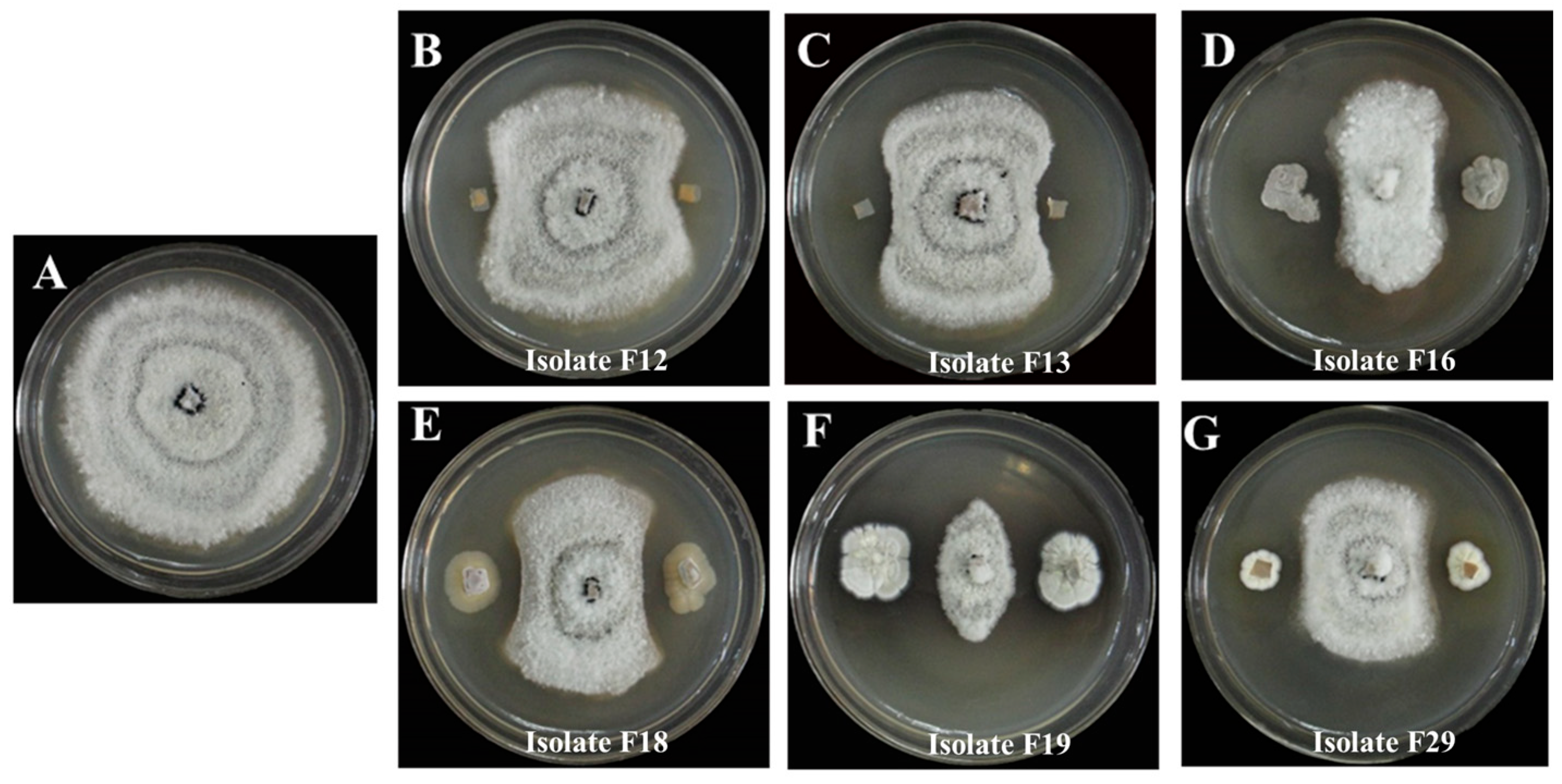
3.1. Isolation of Antagonistic Actinomycetes

A total of 29 colonies were isolated from the soil samples collected from the stipe of M. sextelata and preserved on Gao’s No. 1 slant medium. The isolates are numbered from F1 to F29. Using P. trachicarpicola as a test indicator species, six antagonistic isolates (F12, F13, F16, F18, F19, and F29) that exhibited significant inhibitory effects (antifungal rates > 50%) against P. trachicarpicola were initially identified through plate confrontation assays (Table S2). The highest antifungal rate among the six isolates was observed for isolate F19, reaching 80.59%, followed by isolate F16, with an antifungal rate of 70.04%. Figure 1 provides a clearer demonstration of the inhibitory effects of the six isolates on P. trachicarpicola growth. The inhibitory activity of the fermentation filtrates from the six antagonistic isolates was subsequently analyzed. The results revealed that the mycelium growth of P. trachicarpicola was significantly inhibited (Figure 2). Among the six isolates tested, the antifungal rates ranged from 19.61% to 62.54% (Table 1), with isolates F16 and F19 exhibiting antifungal rates exceeding 60%, which was statistically significant compared with those of the other isolates (p < 0.05). On the basis of these results, isolates F16 and F19 were selected as the dominant antagonistic isolates.

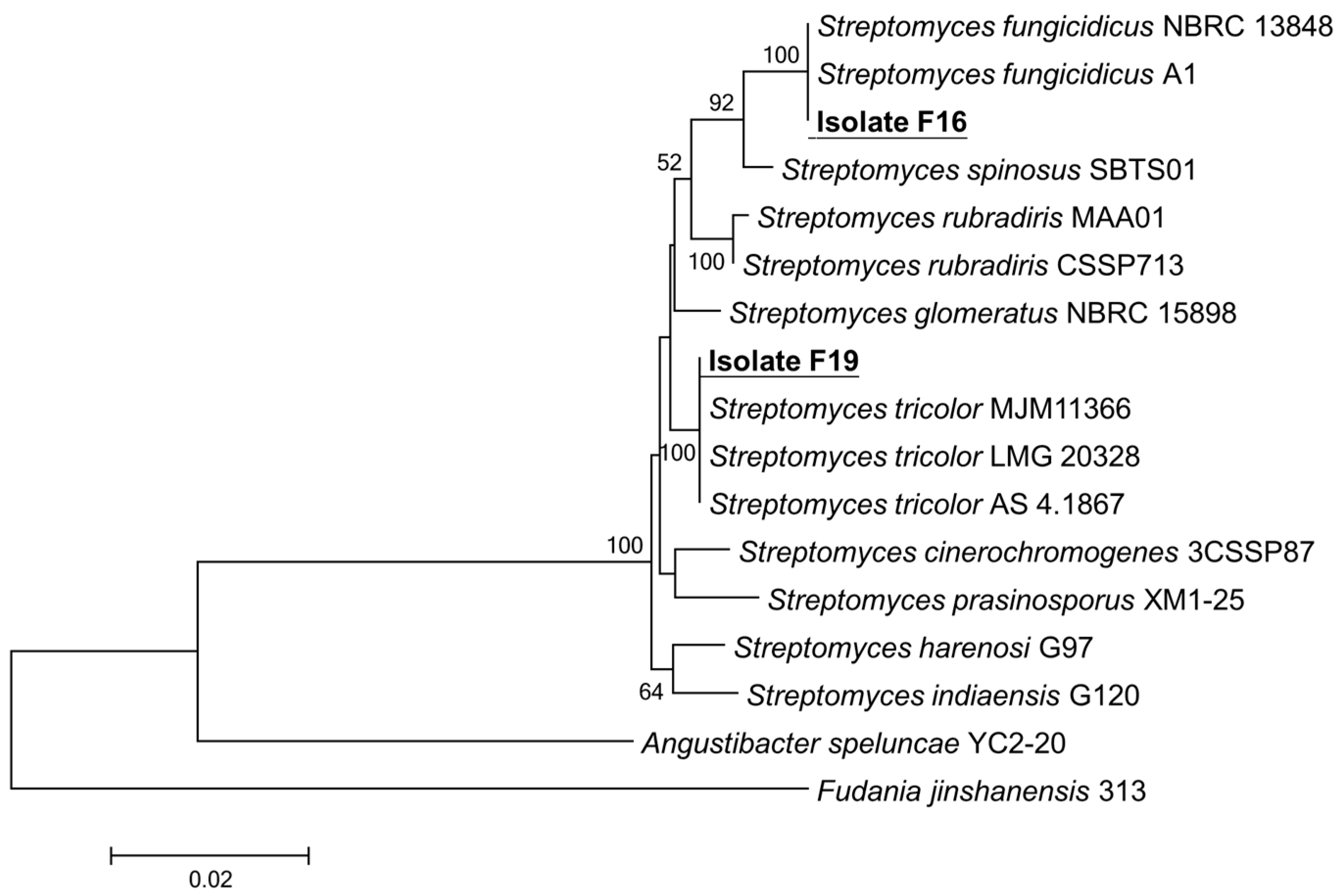
3.2. Identification and Biochemical Characteristics of Antagonistic Actinomycetes

As shown in Figure 3A–C, isolate F16 exhibited distinct morphological features on Gao’s No. 1 medium: Initially, the aerial mycelium was white, and the subsequent formation of spore masses resulted in a light purple color. The spores were elliptical with a smooth surface. In contrast, isolate F19 displayed similar initial white aerial mycelium but developed blue-gray spore masses upon maturation, producing elliptical spores without surface ornamentation (Figure 3D–F). The carbon and nitrogen source utilization analysis revealed that both isolates F16 and F19 utilized all 11 tested carbon sources and 4 nitrogen sources. It demonstrated the strongest utilization capacity for glucose, sucrose, glycerol and peptone, followed by moderate utilization of fructose, lactose, inositol, maltose, D-mannitol, KNO3, and (NH4)2SO4. The poorest utilization was observed for trehalose, starch, and cellulose (Table S3). Notably, neither isolate F16 nor isolate F19 exhibited hydrogen sulfide production or nitrate reduction activity. Furthermore, isolate F16 lacked gelatin liquefaction activity, whereas isolate F19 exhibited this capability, highlighting a notable distinction in metabolic functions between the two isolates.
After 16S rRNA gene sequencing was completed, the sequences of isolates F16 and F19 were compared with the known sequences in the NCBI database by using BLAST. Meanwhile, the GenBank accession numbers for the 16S rRNA gene sequence of isolates F16 and F19 were PV653593 and PV653594, respectively. The 16S rRNA gene sequences with high homology were downloaded and compared using MEGA 7.0 software to construct a phylogenetic tree by using the neighbor-joining method. The results revealed (Figure 4) that two isolates belong to the genus Streptomyces. Isolate F16 clustered with Streptomyces fungicidicus in a cluster, whereas isolate F19 clustered with Streptomyces tricolor. Finally, the two isolates were tentatively identified as Streptomyces sp. F16 and Streptomyces sp. F19.

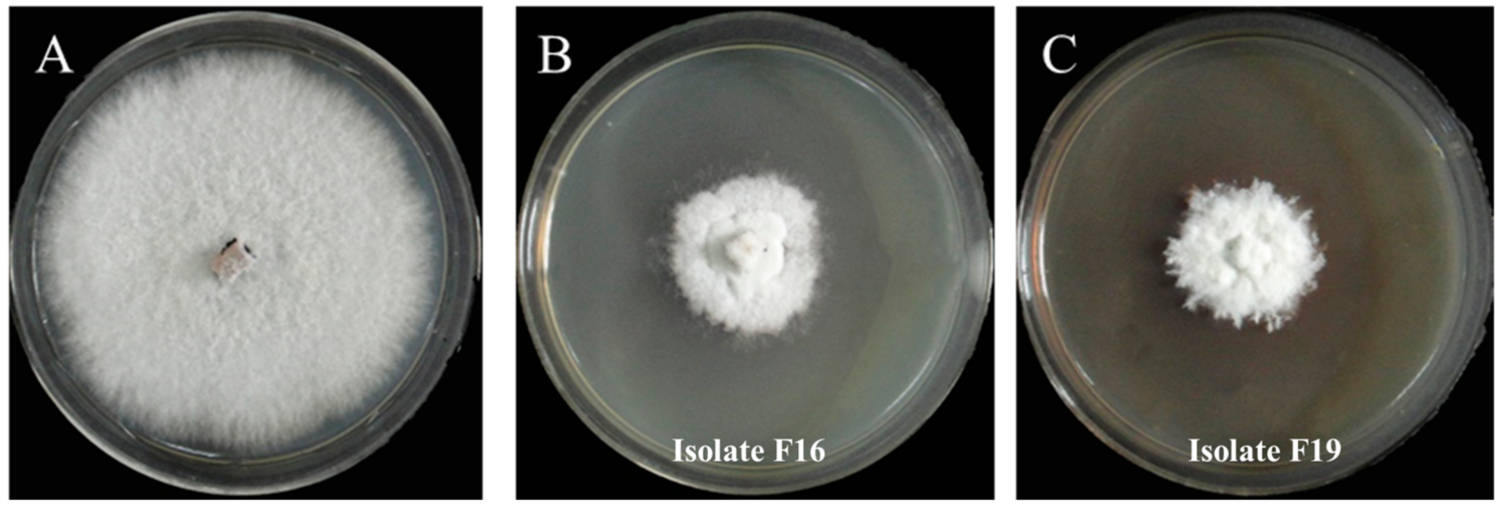
3.3. Determination of Antifungal Effects of Volatile Substances from Isolates F16 and F19 Against P. trachicarpicola

When P. trachicarpicola was cocultured with isolates F16 and F19 under various conditions, P. trachicarpicola presented consistent average growth rates across all of the tested cultures, completely covering the agar surface by day 8. However, notable differences were observed in colony morphology on day 14: while the control group produced conidia (Figure 5B), no conidiation was detected in the coculture systems with Streptomyces sp. F16 (Figure 5C) or Streptomyces sp. F19 (Figure 5D). These observations suggest that both antagonistic isolates may secrete volatile compounds that specifically inhibit conidiation in P. trachicarpicola.

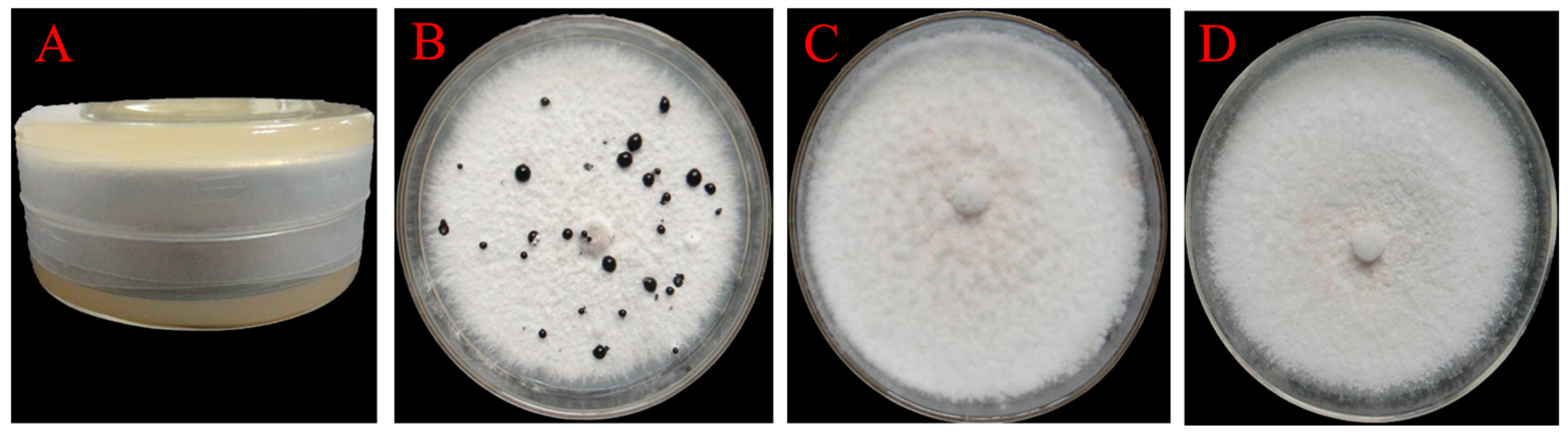
3.4. Determination of the Inhibitory Effects of Fermentation Filtrates of Isolates F16 and F19 Against P. trachicarpicola

The experimental investigation of the inhibitory effects of fermentation filtrates from Streptomyces sp. F16 and Streptomyces sp. F19 at various dilutions on the conidial germination of P. trachicarpicola revealed significant morphological alterations in treated conidia, including pronounced swelling and an abnormal morphology (Figure 6). The fermentation filtrates of Streptomyces sp. F16 and Streptomyces sp. F19 potently suppressed P. trachicarpicola conidial germination when diluted 5-fold. Notably, both isolates demonstrated concentration-dependent inhibition, with 50-fold dilutions achieving inhibition rates of 66.93% and 49.22%, respectively. However, when the mixture was diluted 100-fold, the inhibitory effects were substantially reduced, resulting in inhibition rates of only 36.96% and 21.21%, respectively (Table 2). These results suggest that the antifungal compounds secreted by Streptomyces F16/F19 are likely the cause of inhibited germination, and highlight that antifungal activity significantly decreases with an increase in dilution.

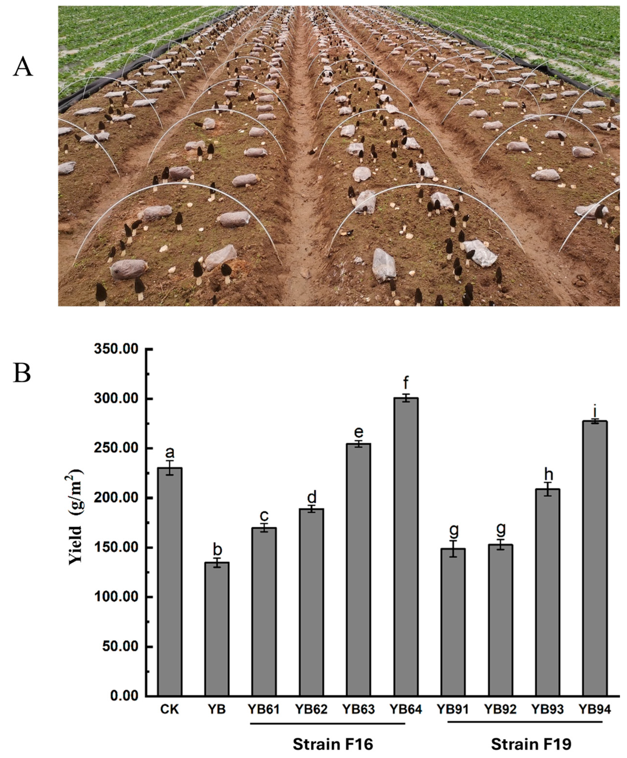
3.5. Field Experiment for Evaluating the Efficacy of the Isolates F16 and F19 on P. trachicarpicola in M. sextelata

The preventive effects of fermentation filtrates from the antagonistic isolates Streptomyces sp. F16 and Streptomyces sp. F19 against P. trachicarpicola on M. sextelata fruiting bodies were investigated. The in vitro inhibition rates of the two isolates against P. trachicarpicola reached 89.57% and 75.64%, respectively. (1) Preventive efficacy of Streptomyces sp. F16 on M. sextelata fruiting bodies: When P. trachicarpicola was inoculated onto fruiting bodies followed by the application of Streptomyces sp. F16 filtrate 24 h later, no disease symptoms were observed compared with those in the control group (Figure 7C). These results show that the method significantly inhibited the growth of P. trachicarpicola. The application of Streptomyces sp. F16 filtrate 2 h before P. trachicarpicola inoculation also completely prevented infection, with P. trachicarpicola growth inhibited under the fruiting skin (Figure 7D). (2) Comparative efficacy of Streptomyces sp. F19: Similarly to Streptomyces sp. F16, Streptomyces sp. F19 filtrate application 24 h after inoculation partially reduced disease severity, although limited lesion formation was still observed on fruiting bodies compared with the control (Figure 7E). Pretreatment with Streptomyces sp. F19 filtrate 2 h before infection effectively suppressed P. trachicarpicola colonization (Figure 7F), albeit with weaker protective effects than those of Streptomyces sp. F16. Both fermentation filtrates demonstrated antifungal activity against P. trachicarpicola on M. sextelata fruiting bodies. Notably, Streptomyces sp. F16 exhibited superior efficacy in both pre- and postinoculation treatments, achieving complete disease prevention and robust P. trachicarpicola suppression, whereas Streptomyces sp. F19 showed partial protection under specific conditions. These results highlight the potential of isolate-specific fermentation products for developing targeted biocontrol strategies against M. sextelata wilt disease.
Field cultivation experiments were carried out according to Table S1. Field observations revealed that the experimental field YB presented significantly fewer M. sextelata primordia formations than did the control field (CK). During the early fruiting stage, young fruiting bodies in the experimental field YB were highly susceptible to P. trachicarpicola invasion. Notably, the experimental groups treated with 20-fold and 50-fold diluted fermentation filtrates presented increased primordium counts relative to those in the experimental field YB. However, the immature ascocarps in these groups remained vulnerable to infection. In contrast, the experimental groups receiving 5-fold and 10-fold dilutions not only presented increased primordium formation but also presented substantially reduced infection rates in young fruiting bodies (Figure 8B). These results indicate the concentration-dependent efficacy of fermentation filtrates in promoting primordium development while suppressing pathogen infection during critical growth stages.
Field observations demonstrated that inoculation with P. trachicarpicola alone resulted in a significant reduction in M. sextelata yield. The yields of the experimental fields YB61, YB62, YB91, and YB92 were intermediate, exceeding those of the P. trachicarpicola-inoculated field YB but remaining lower than those of the noninoculated control group (field CK). These findings suggested that 20-fold and 50-fold dilutions of the fermentation filtrate from antagonistic isolates exhibited limited antifungal efficacy. In contrast, the experimental fields treated with 5-fold or 10-fold dilutions of Streptomyces sp. F16 or 5-fold dilutions of Streptomyces sp. F19 surpassed both the experimental field YB yield and the CK yield, indicating exceptional productivity (Figure 8B). Notably, the 5-fold diluted filtrates from Streptomyces sp. F16 and Streptomyces sp. F19 yielded 299.6 g/m2 and 277.65 g/m2, respectively, which were significantly greater values than the value of 231 g/m2 observed in the M. sextelata-alone group (CK).

4. Discussion

A series of physical control methods for Morchella wilt disease, including field-based prevention, pest attraction and elimination, the timely removal of contaminated nutrient bags, and the prompt removal of infected fruiting bodies, have shown limited efficacy. With the increasing focus on sustainable agricultural practices, biological control has emerged as a promising alternative [23]. Actinomycetes are ubiquitous microorganisms that inhabit diverse ecological niches such as soil, air, plant debris, and sedimentary environments [24]. The ability of the Streptomyces genus to produce a diverse array of secondary metabolites is a well-established characteristic within the actinomycetes order. The secondary metabolites present antibacterial properties against diverse plant diseases [25]. For example, Streptomyces lunalinharesii was isolated from the rhizosphere soil of Illicium verum and exhibited significant antifungal activity against Colletotrichum gloeosporioides, the causative agent of I. verum anthracnose [26]. Similarly, Streptomyces olivoreticuli screened from the rhizosphere soils of plants had a strong antagonistic effect on Rhizoctonia solani, which is a soilborne pathogen that causes root rot in various crops [27].
Extensive studies have demonstrated that strains of the genus Streptomyces exhibit significant suppressive effects against diverse plant pathogens, indicating great potential for the development of eco-friendly antifungal strategies targeting Morchella diseases [28]. In this work, we isolated 29 actinomycetes from the rhizosphere soil of M. sextelata through a multistage screening process. Initially, plate confrontation assays and fermentation filtrate rescreening identified antagonistic actinomycetes with significant antifungal activity against P. trachicarpicola. Subsequent characterization on the basis of morphological features, culture patterns, physiological and biochemical tests, and 16S rRNA gene sequences confirmed that two isolates, isolates F16 and F19, belong to the Streptomyces genus.
The plate confrontation assays revealed that two antagonistic strains, Streptomyces sp. F16 and Streptomyces sp. F19, produce antimicrobial substances capable of significantly inhibiting P. trachicarpicola mycelial growth. To elucidate whether these substances are soluble or volatile, we systematically investigated the effects of fermentation filtrates and volatile compounds from the two isolates. The results demonstrated that (1) the fermentation filtrates suppressed P. trachicarpicola mycelial growth through soluble antimicrobial components, and that (2) the volatile compounds inhibited the conidial germination of P. trachicarpicola. This dual-mode inhibition mechanism aligns with findings by Sudha et al., who reported that volatile metabolites from S. rochei ASH disrupted fungal hyphal structures, achieving inhibition rates of 63.75% and 68.52% against Fusarium moniliforme and Curvularia lunata, respectively [29].
Spores are resilient to harsh environmental conditions and can germinate once favorable conditions are restored [30]. Therefore, evaluating the inhibitory effects of antagonistic actinobacteria on P. trachicarpicola spore germination is critical for disease control [31]. In this work, fermentation filtrates of Streptomyces sp. F16 and F19 significantly impaired P. trachicarpicola conidial growth, resulting in swelling and rupturing of the spores. These results align with previous research on Streptomyces species [32], which demonstrated similar conidial inhibition patterns through secreted antimicrobial compounds.
Streptomyces strains can further alleviate disease pressure by stimulating elements of the plant immune system and increasing plant productivity [33]. On the basis of field yield, 5- and 10-fold dilutions of the fermentation filtrates from both strains not only suppressed P. trachicarpicola growth but also significantly promoted an increase in M. sextelata yield. Similar research results show that Streptomyces flavovirens can be used to increase mushroom yield and contribute to disease control against green mold disease caused by Trichoderma aggressivum [34]. This dual functionality suggests that these strains exhibit mycelial-growth-promoting effects alongside P. trachicarpicola inhibition. Therefore, Streptomyces can be used not only as a biological control agent for pathogens of M. sextelata, but also as a growth promoter for M. sextelata.

5. Conclusions

Overall, this study provides basic data for the accurate identification of the causative agents and prevention and control of diseases affecting M. sextelata in the field. Streptomyces-based bio-formulation has emerged as the most effective strategy for decreasing disease severity and enhancing fruit yield in M. sextelata cultivation. These results provide valuable insights into sustainable agricultural practices, suggesting that integrating biocontrol agents into crop management can lead to improved disease resistance and yield stability in the face of environmental challenges.

Supplementary Materials

The following supporting information can be downloaded at: https://www.mdpi.com/article/10.3390/jof11060452/s1. Table S1: Inoculation methods of each test group in the field; Table S2: Inhibitory effect of primary screening of actinomycetota strains against P. trachicarpicola; Table S3: Biochemical characteristics of antagonistic isolates F16 and F19.

Author Contributions

Conceptualization, J.L. and B.L.; methodology, J.L.; investigation, B.L. and Y.L.; writing—original draft preparation, B.L.; writing—review and editing, Z.H. and J.L.; visualization, Y.L. and A.M.; supervision, J.L.; project administration, J.L.; funding acquisition, J.L. All authors have read and agreed to the published version of the manuscript.

Funding

This research was funded by the National Natural Science Foundation of China (32300113), Doctoral Start-up Funds of China West Normal University (23KE020), and the Open Fund of the Guangdong Provincial Key Laboratory of Marine Disaster Prediction and Prevention (GPKLMD2023003).

Institutional Review Board Statement

Not applicable.

Informed Consent Statement

Not applicable.

Data Availability Statement

The original contributions presented in this study are included in the article and Supplementary Materials. Further inquiries can be directed to the corresponding author.

Conflicts of Interest

The authors declare no conflicts of interest.

Abbreviations

The following abbreviations are used in this manuscript:
PDAPotato dextrose agar medium
NCBINational Center of Biotechnology Information

References

  1. Du, X.H.; Wu, D.; Kang, H.; Wang, H.; Xu, N.; Li, T.; Chen, K. Heterothallism and potential hybridization events inferred for twenty-two yellow morel species. IMA Fungus 2020, 11, 4. [Google Scholar] [CrossRef] [PubMed]
  2. Wang, W.K.; Zhu, Y.; Tang, Y.; Lu, N.; Song, J.L.; Yuan, W.D.; Jia, Y. Non-volatile taste components of different cultivated mushrooms at mycelia, primordium, and fruit body cultivation stages. Int. J. Food Prop. 2016, 19, 1938–1948. [Google Scholar] [CrossRef]
  3. Vieira Gomes, D.C.; de Alencar, M.V.O.B.; dos Reis, A.C.; de Lima, R.M.T.; de Oliveira Santos, J.V.; da Mata, A.M.O.F.; Soares Dias, A.C.; da Costa, J.S.; de Medeiros, M.d.G.F.; Paz, M.F.C.J.; et al. Antioxidant, anti-inflammatory and cytotoxic/antitumoral bioactives from the phylum Basidiomycota and their possible mechanisms of action. Biomed. Pharmacother. 2019, 112, 108643. [Google Scholar] [CrossRef]
  4. Shi, X.; Liu, D.; He, X.; Liu, W.; Yu, F. Epidemic identification of fungal diseases in morchella cultivation across China. J. Fungi 2022, 8, 1107. [Google Scholar] [CrossRef] [PubMed]
  5. Tan, H.; Liu, T.; Yu, Y.; Tang, J.; Jiang, L.; Martin Francis, M.; Peng, W. Morel production related to soil microbial diversity and evenness. Microbiol. Spectr. 2021, 9, e00229-21. [Google Scholar] [CrossRef] [PubMed]
  6. Guo, M.P.; Chen, K.; Wang, G.Z.; Bian, Y.B. First report of stipe rot disease on morchella importuna caused by Fusarium incarnatumF. equiseti species complex in China. Plant Dis. 2016, 100, 2530. [Google Scholar] [CrossRef]
  7. Lan, Y.F.; Cong, Q.Q.; Wang, Q.W.; Tang, L.N.; Li, X.M.; Yu, Q.W.; Cui, X.; An, X.R.; Yu, C.X.; Kong, F.H.; et al. First report of Cladobotryum protrusum causing cobweb disease on cultivated Morchella importuna. Plant Dis. 2019, 104, 977. [Google Scholar] [CrossRef]
  8. Yu, Y.; Liu, T.; Liu, L.; Chen, Y.; Tang, J.; Peng, W.; Tan, H. Application of the mushroom volatile 1-octen-3-ol to suppress a morel disease caused by Paecilomyces penicillatus. Appl. Microbiol. Biotechnol. 2022, 106, 4787–4799. [Google Scholar] [CrossRef]
  9. He, X.L.; Peng, W.H.; Miao, R.Y.; Tang, J.; Chen, Y.; Liu, L.X.; Wang, D.; Gan, B.C. White mold on cultivated morels caused by Paecilomyces penicillatus. FEMS Microbiol. Lett. 2017, 364, fnx037. [Google Scholar] [CrossRef]
  10. He, P.; Li, C.; Cai, Y.; Zhang, Y.; Bian, Y.; Liu, W. First report of pileus rot disease on cultivated Morchella importuna caused by Diploöspora longispora in China. J. Gen. Plant Pathol. 2018, 84, 65–69. [Google Scholar] [CrossRef]
  11. Tu, S.; Zhang, Y.; Chen, X.; Song, L.; Chen, Y.; Lv, B. First report of Aspergillus niger causing rot of Morchella sextelata in China. Plant Dis. 2024, 108, 804. [Google Scholar] [CrossRef] [PubMed]
  12. Liang, Z.; Liu, Y.; Zhao, K.; Li, L.; Jiang, S. First report of Pestalotiopsis trachicarpicola causing apothecium wither on cultivated Morchella sextelata in Sichuan, China. Plant Dis. 2023, 107, 3631. [Google Scholar] [CrossRef]
  13. Manivasagan, P.; Venkatesan, J.; Sivakumar, K.; Kim, S.K. Pharmaceutically active secondary metabolites of marine actinobacteria. Microbiol. Res. 2014, 169, 262–278. [Google Scholar] [CrossRef] [PubMed]
  14. João Frederico, M.; dos Passos, P.B.D.C.; Murilo, D.; Costa, G.R.; Zaffari, G.N.; José Itamar Boneti, A.M.R.; de Oliveira, L.M.P. Cultivable bacteria isolated from apple trees cultivated under different crop systems: Diversity and antagonistic activity against Colletotrichum gloeosporioides. Genet. Mol. Biol. 2014, 37, 560–572. [Google Scholar]
  15. Bhat, P.S.; Kumar, N.R.P.; Ranganath, H.R.; Saroja, S. Pests and their management in cucurbits. In Trends in Horticultural Entomology; Mani, M., Ed.; Springer Nature: Singapore, 2022; pp. 1013–1030. [Google Scholar]
  16. Bondarenko, S.V.; Stankevych, S.V.; Matsyura, A.V. Major cucumber diseases and the crop immunity. Ukr. J. Ecol. 2021, 11, 46–54. [Google Scholar] [CrossRef]
  17. Li, X.; Wang, J.; Lv, Y.; Zhao, L.; Jiang, W.; Lv, J.; Xu, X.; Yu, Y.; Liu, Y.; Chen, X.; et al. Screening and identification of Paenibacillus polymyxa GRY-11 and its biological control potential against apple replant disease. Folia Microbiol. 2024, 70, 475–487. [Google Scholar] [CrossRef]
  18. Yang, D.; Luo, J.; Zhou, Y.; Zhou, S.; Liu, X.; Liu, C. Identification and biological characterization of pathogen causing sooty blotch of Ardisia crispa (Thunb.) A.DC. PeerJ 2025, 13, e19130. [Google Scholar] [CrossRef]
  19. Conville Patricia, S.; Brown-Elliott Barbara, A.; Smith, T.; Zelazny Adrian, M. The complexities of Nocardia taxonomy and identification. J. Clin. Microbiol. 2017, 56, 10–1128. [Google Scholar]
  20. Zhu, X.; Ma, K.; Sun, M.; Zhang, J.; Liu, L.; Niu, S. Isolation and identification of pathogens of Morchella sextelata bacterial disease. Front. Microbiol. 2023, 14, 1231353. [Google Scholar] [CrossRef]
  21. Kumar, S.; Stecher, G.; Tamura, K. MEGA7: Molecular evolutionary genetics analysis version 7.0 for bigger datasets. Mol. Biol. Evol. 2016, 33, 1870–1874. [Google Scholar] [CrossRef]
  22. Wang, X.; Peng, J.; Sun, L.; Bonito, G.; Guo, Y.; Li, Y.; Fu, Y. Genome sequencing of Paecilomyces penicillatus provides insights into its phylogenetic placement and mycoparasitism mechanisms on morel mushrooms. Pathogens 2020, 9, 834. [Google Scholar] [CrossRef]
  23. Chen, X.; Zhang, Y.; Chao, S.; Song, L.; Wu, G.; Sun, Y.; Chen, Y.; Lv, B. Biocontrol potential of endophytic Bacillus subtilis A9 against rot disease of Morchella esculenta. Front. Microbiol. 2024, 15, 1388669. [Google Scholar] [CrossRef]
  24. Bhatti, A.A.; Haq, S.; Bhat, R.A. Actinomycetes benefaction role in soil and plant health. Microb. Pathog. 2017, 111, 458–467. [Google Scholar] [CrossRef] [PubMed]
  25. Viaene, T.; Langendries, S.; Beirinckx, S.; Maes, M.; Goormachtig, S. Streptomyces as a plant’s best friend? FEMS Microbiol. Ecol. 2016, 92, fiw119. [Google Scholar] [CrossRef] [PubMed]
  26. Pan, J.; Geng, X.; Cai, Y.; Yu, Y.; Hou, Y.; Liu, Y.; Ya, C.; Liu, Q. Identification, fermentation optimization, and biocontrol efficacy of actinomycete YG-5 for the prevention of Alternaria leaf spot disease in star anise. Sci. Rep. 2024, 14, 18621. [Google Scholar] [CrossRef]
  27. Zhong, J.; Sui, W.W.; Bai, X.Y.; Qiu, Z.L.; Li, X.G.; Zhu, J.Z. Characterization and biocontrol mechanism of Streptomyces olivoreticuli as a potential biocontrol agent against Rhizoctonia solani. Pestic. Biochem. Physiol. 2023, 197, 105681. [Google Scholar] [CrossRef] [PubMed]
  28. Khan, S.; Srivastava, S.; Karnwal, A.; Malik, T. Streptomyces as a promising biological control agents for plant pathogens. Front. Microbiol. 2023, 14, 1285543. [Google Scholar] [CrossRef]
  29. Sudha, A.; Durgadevi, D.; Archana, S.; Muthukumar, A.; Suthin Raj, T.; Nakkeeran, S.; Poczai, P.; Nasif, O.; Ansari, M.J.; Sayyed, R.Z. Unraveling the tripartite interaction of volatile compounds of Streptomyces rochei with grain mold pathogens infecting sorghum. Front. Microbiol. 2022, 13, 923360. [Google Scholar] [CrossRef]
  30. Liu, W.; He, P.; Zhang, J.; Wu, L.; Er, L.; Shi, X.; Gu, Z.; Yu, F.; Pérez-Moreno, J. Ultrastructure and physiological characterization of Morchella mitospores and their relevance in the understanding of the morel life cycle. Microorganisms 2023, 11, 345. [Google Scholar] [CrossRef]
  31. Shao, J.; Pei, Z.; Jing, H.; Wang, L.; Jiang, C.; Du, X.; Jiang, C.; Lou, Z.; Wang, H. Antifungal activity of myriocin against Fusarium graminearum and its inhibitory effect on deoxynivalenol production in wheat grains. Physiol. Mol. Plant Pathol. 2021, 114, 101635. [Google Scholar] [CrossRef]
  32. Kamil, F.H.; Saeed, E.E.; El-Tarabily, K.A.; AbuQamar, S.F. Biological control of mango dieback disease caused by Lasiodiplodia theobromae using Streptomycete and non-streptomycete Actinobacteria in the united arab emirates. Front. Microbiol. 2018, 9, 829. [Google Scholar] [CrossRef] [PubMed]
  33. Olanrewaju, O.S.; Babalola, O.O. Streptomyces: Implications and interactions in plant growth promotion. Appl. Microbiol. Biotechnol. 2019, 103, 1179–1188. [Google Scholar] [CrossRef] [PubMed]
  34. Šantrić, L.; Ivana, P.; Ljiljana, R.; Gajić, U.J.; Emil, R.; Bojan, D.; Svetlana, M. Impact of a native Streptomyces flavovirens from mushroom compost on green mold control and yield of Agaricus bisporus. J. Environ. Sci. Health Part B 2018, 53, 677–684. [Google Scholar] [CrossRef] [PubMed]
Figure 1. Inhibitory effects of the 6 isolates on mycelium growth of P. trachicarpicola. (A) P. trachicarpicola; (B) P. trachicarpicola and isolate F12; (C) P. trachicarpicola and isolate F13; (D) P. trachicarpicola and isolate F16; (E) P. trachicarpicola and isolate F18; (F) P. trachicarpicola and isolate F19; (G) P. trachicarpicola and isolate F29.
Jof 11 00452 g001
Figure 2. Inhibitory effects of fermentation filtrates of antagonistic isolates on P. trachicarpicola. (A) P. trachicarpicola; (B) P. trachicarpicola and the fermentation filtrates of isolate F16; (C) P. trachicarpicola and the fermentation filtrates of isolate F19.
Jof 11 00452 g002
Figure 3. Morphology of colonies and spores of antagonistic isolates F16 and F19 (1000×). (AC) Isolate F16; (DF) isolate F19; (A,D) front of the medium; (B,E) reverse side of the medium; (C,F) spores.
Jof 11 00452 g003
Figure 4. Phylogenetic tree of isolates F16 and F19 based on the 16S rRNA gene sequence.
Jof 11 00452 g004
Figure 5. Inhibitory effects of volatile products of antagonistic isolates F16 and F19 against P. trachicarpicola. (A) Schematic of dual-plate co-culture assay; (B) P. trachicarpicola control; (C) P. trachicarpicola cultured with antagonistic isolate F16; (D) P. trachicarpicola cultured with antagonistic isolate F19.
Jof 11 00452 g005
Figure 6. The effect of 5-fold dilution of fermentation filtrate on the conidial morphology of P. trachicarpicola. (A) CK; (B) the fermentation filtrates from Streptomyces sp. F16; (C) the fermentation filtrates from Streptomyces sp. F19.
Jof 11 00452 g006
Figure 7. Inhibitory effects of fermentation filtrates of antagonistic isolates of P. trachicarpicola on M. sextelata fruiting bodies. (A) Sterile-water treatment; (B) inoculate with only P. trachicarpicola; (C) Streptomyces sp. F16 fermentation filtrate 24 h after inoculation with P. trachicarpicola; (D) P. trachicarpicola inoculated 2 h after Streptomyces sp. F16 fermentation filtrate was applied; (E) Streptomyces sp. F19 fermentation filtrate applied 24 h after inoculation with P. trachicarpicola; (F) P. trachicarpicola inoculated after Streptomyces sp. F19 fermentation filtrate was applied for 2 h.
Jof 11 00452 g007
Figure 8. Effects of two antagonistic isolates on yield of M. sextelata. (A) Cultivation fields of M. sextelata; (B) yield of M. sextelata in different test fields. Superscript lowercase letters (a, b, c…) were assigned to denote statistically significant differences (p < 0.05) in mean yields among test fields.
Jof 11 00452 g008
Table 1. Antifungal rates of 6 isolates with antagonistic activity.
Isolate NumberColony Diameter (cm)Antifungal Rate (%)
CK8.42 ± 0.08 a
F126.47 ± 0.15 b24.66% ± 0.02 a
F136.87 ± 0.18 c19.61% ± 0.02 b
F163.47 ± 0.15 d62.54% ± 0.02 c
F186.45 ± 0.15 b24.87% ± 0.02 a
F193.65 ± 0.14 e60.23% ± 0.02 d
F294.82 ± 0.12 f45.50% ± 0.01 e
Note: Lowercase letters (a, b, c…) after the numbers were assigned to denote statistically significant differences (p < 0.05).
Table 2. Effects of different dilution ratios of fermentation filtrate from isolates F16 and F19 on the conidial germination of P. trachicarpicola.
Dilution TimesInhibition Rate of Conidial Germination (%)
Streptomyces sp. F16Streptomyces sp. F19
099.81% ± 0.00 a97.86% ± 0.01 a
599.22% ± 0.01 a94.94% ± 0.01 a
1088.91% ± 0.02 b79.96% ± 0.01 b
2078.40% ± 0.03 c66.73% ± 0.03 c
5066.93% ± 0.05 d49.22% ± 0.03 d
10036.96% ± 0.03 e21.21% ± 0.04 e
Note: the lowercase letters (a, b, c…) after the numbers were assigned to denote statistically significant differences (p < 0.05).
Disclaimer/Publisher’s Note: The statements, opinions and data contained in all publications are solely those of the individual author(s) and contributor(s) and not of MDPI and/or the editor(s). MDPI and/or the editor(s) disclaim responsibility for any injury to people or property resulting from any ideas, methods, instructions or products referred to in the content.

