Article

Whole-Genome Sequencing and Comparative Genomic Analysis of Three Clinical Bloodstream Infection Isolates of Trichosporon austroamericanum

by
Takanori Horiguchi
1,2,
Takashi Umeyama
1,*,
Hiroko Tomuro
1,
Amato Otani
1,
Takayuki Shinohara
1,
Masahiro Abe
1,
Shogo Takatsuka
1,
Ken Miyazawa
1,
Minoru Nagi
3,
Yasunori Muraosa
1,
Yasutaka Hoshino
1,
Takashi Sakoh
4,
Hideki Araoka
4,
Naoyuki Uchida
5,
Tomoyoshi Kaneko
6,
Yuko Nagano
6,
Hiroki Tsukada
6,
Taiga Miyazaki
2 and
Yoshitsugu Miyazaki
1
1
Department of Fungal Infection, National Institute of Infectious Diseases, Japan Institute for Health Security, Tokyo 162-8640, Japan
2
Division of Respirology, Rheumatology, Infectious Diseases and Neurology, Department of Internal Medicine, Faculty of Medicine, University of Miyazaki, Miyazaki 889-1692, Japan
3
Antimicrobial Resistance Research Center, National Institute of Infectious Diseases, Japan Institute for Health Security, Tokyo 189-0002, Japan
4
Department of Infectious Diseases, Toranomon Hospital, Tokyo 105-8470, Japan
5
Department of Hematology, Toranomon Hospital, Tokyo 105-8470, Japan
6
The Jikei University School of Medicine Kashiwa Hospital, Chiba 277-8567, Japan
*
Author to whom correspondence should be addressed.
J. Fungi 2025, 11(5), 401; https://doi.org/10.3390/jof11050401
Submission received: 18 April 2025 / Revised: 16 May 2025 / Accepted: 19 May 2025 / Published: 21 May 2025
(This article belongs to the Section Fungal Genomics, Genetics and Molecular Biology)

Abstract

:
Trichosporon austroamericanum is a recently described yeast species related to Trichosporon inkin and exclusively isolated from clinical specimens. However, its genomic features and pathogenic potential remain poorly understood. In this study, we performed whole-genome sequencing on three blood-derived isolates from patients with invasive fungal infections and comparative analyses with 13 related Trichosporon species. The three isolates yielded high-quality assemblies of 9–10 scaffolds (~21 Mb), facilitating reliable comparisons. While most species had comparable genome sizes, Trichosporon ovoides, Trichosporon coremiiforme, and Cutaneotrichosporon mucoides displayed large, fragmented genomes, suggestive of polyploidy. ANI analysis and phylogenetic trees based on ANI and single-copy orthologs supported the classification of T. austroamericanum as a distinct clade with moderate intraspecific divergence. Using the Galleria mellonella, a model for fungal pathogenicity, all T. austroamericanum strains reduced larval survival, and NIIDF 0077300 exhibited virulence comparable to T. asahii and greater than T. inkin. To explore the gene-level differences associated with pathogenicity, we performed ortholog analysis based on single-copy genes. This revealed a unique Zn(II)2Cys6-type transcription factor gene (OG0010545) present only in NIIDF 0077300 and T. asahii. These findings highlight the genomic diversity and infection-associated traits of T. austroamericanum, providing a framework for future functional studies.

1. Introduction

Trichosporon is a genus of basidiomycetous yeasts within the phylum Basidiomycota, widely distributed in natural environments, such as soil, water, and decaying organic matter, and is found as a commensal organism in the human microbiota [1,2]. While generally regarded as harmless, several Trichosporon species have emerged as opportunistic pathogens capable of causing invasive infections, particularly in immunocompromised individuals [1,2,3,4]. Among these, Trichosporon inkin is considered a clinically important species that is commonly associated with superficial infections, although cases of invasive trichosporonosis have also been reported in patients with hematologic malignancies or neutropenia. However, such systemic infections are exceedingly rare [4,5,6,7,8].
Trichosporon austroamericanum is a recently described yeast species isolated from clinical specimens and is closely related to T. inkin, based on sequence analysis of the intergenic spacer 1 (IGS1) region [9]. Standard barcoding regions, such as the Internal Transcribed Spacer and D1/D2, share >99% identity between these two species, making it difficult to reliably distinguish them using conventional markers [9]. In contrast, IGS1 analysis enables reliable species-level differentiation and has proven useful in refining the Trichosporon taxonomy. Some isolates previously identified as T. inkin have been reassigned to T. austroamericanum [9]. T. austroamericanum has been isolated exclusively from clinical specimens, including urine and skin, and has been reported in geographically diverse regions, such as China, Brazil, France, Argentina, and India [9]. In Japan, three isolates with IGS1 sequences identical to those of T. austroamericanum were recently recovered from the blood of patients with bloodstream infections, suggesting that this species may possess a previously unrecognized pathogenic potential.
Despite its isolation in patients with invasive infections and close genetic relatedness to clinically important Trichosporon species, genomic insights into T. austroamericanum are currently lacking, hindering the understanding of its evolutionary relationships and pathogenic potential. Whole-genome sequencing (WGS) has become the cornerstone of modern phylogenetic classification, enabling precise species delineation at the genomic level. Beyond taxonomy, WGS further reveals lineage-specific genomic features that may underlie phenotypic traits such as pathogenicity, drug resistance, and environmental adaptation [10,11]. We selected WGS as our primary approach because conventional barcoding markers such as ITS and D1/D2 are insufficient to resolve the taxonomic ambiguity between T. austroamericanum and closely related species. In addition to WGS, we applied genome-wide average nucleotide identity (ANI) analysis and ortholog-based phylogenetics to assess interspecies relatedness from both nucleotide and protein perspectives. Comparative genomics was also employed to explore strain-specific features that may explain clinical adaptation. Together, these approaches enabled us to clarify the taxonomic position of T. austroamericanum and gain insight into its genomic features.

2. Materials and Methods

2.1. Fungal Isolates

All strains used in this study are listed in Table 1.
Three strains of T. austroamericanum (NIIDF 0077000, NIIDF 0077300, and NIIDF 0079200) were isolated from blood cultures collected between 1 April 2023 and 31 March 2024. All three strains were detected in blood culture specimens obtained from different patients. NIIDF 0077000 and NIIDF 0079200 were isolated at the same hospital, while NIIDF 0077300 was obtained from a hospital located in a different city. Thirteen Trichosporon-related strains, excluding T. austroamericanum, were obtained from the Japan Collection of Microorganisms (JCM), RIKEN BRC.

2.2. DNA Extraction and Genome Sequencing

Each strain used in this study was cultured on Potato Dextrose Agar (PDA; Difco™, Becton, Dickinson and Company, Franklin Lakes, NJ, USA) at 30 °C for 48 h. A single colony was aseptically transferred to 10 mL of Yeast Extract Peptone Dextrose (YPD) broth (Difco™, Becton, Dickinson and Company, Franklin Lakes, NJ, USA) and incubated at 30 °C with shaking at 150 rpm for 24 h. After cultivation, cells were centrifuged at 3000× g for 10 min, and the resulting pellet was washed once with 1× phosphate-buffered saline (PBS) by centrifugation. The resulting pellet was frozen in liquid nitrogen, ground using a mortar and pestle, and subjected to genomic DNA extraction using the Maxwell® RSC Genomic DNA Kit (Promega, Madison, WI, USA) according to the manufacturer’s protocol. DNA purity (A260/A280 ratio) was assessed using a Nanodrop spectrophotometer (Thermo Fisher, Waltham, MA, USA), and DNA concentration was quantified using a Quantus™ Fluorometer (Promega, Madison, WI, USA). The extracted DNA was barcoded using the Native Barcoding Kit 24 V14 (Oxford Nanopore Technologies, Didcot, UK) according to the manufacturer’s protocol, and sequencing was performed for 24–72 h on an R10.4.1 flow cell (Oxford Nanopore Technologies, Didcot, UK). Basecalling was conducted using Dorado (v0.7.1), and the processed reads were used for subsequent analyses [12].

2.3. Genome Assembly

The analysis was performed using the NIG supercomputer at the ROIS National Institute of Genetics (Mishima, Shizuoka, Japan). Nanopore sequencing data were filtered to remove low-quality reads using Filtlong (v0.2.1) with the following parameters: --keep_percent 95--min_length 1000 [13]. Genome assembly was performed using Flye (v2.9.5) with default parameters, followed by read mapping using Minimap2 (v2.24) [14,15]. Contig polishing and consensus generation were conducted using two iterations of Racon (v1.5.0) and one iteration of Medaka (v1.8.0), with default settings [16,17]. Assembly quality was evaluated using QUAST (v5.0.2), whereas genome completeness was assessed using BUSCO (v5.2.2) and the tremellomycetes_odb10 reference database [18,19].

2.4. Gene Prediction and Functional Annotation

Gene prediction and functional annotation were performed using Funannotate (v1.8.17) [20]. A combination of evidence-based and ab initio gene prediction methods was used in this study. The tremellomycetes_odb10 BUSCO database was used to extract conserved gene models, which were then utilized to train the ab initio prediction tools AUGUSTUS (v4.0.0), GlimmerHMM (v3.0.4), and SNAP [21,22,23]. Additionally, self-training gene prediction was performed using GeneMark-ES (v4.72) with parameters optimized for fungal genomes [24]. Functional annotation was conducted using InterProScan (v5.70-102.0), which compares the predicted gene models against the Pfam, PROSITE, and SMART databases to identify conserved domains and assign functional descriptions. Gene Ontology (GO) terms were assigned based on InterProScan GO mapping as well as functional categorization using EggNOG-mapper (v2.1.12) [25,26]. Secondary metabolite biosynthetic gene clusters were predicted using AntiSMASH (v7.0.0) to identify putative non-ribosomal peptide synthetases (NRPSs), polyketide synthases (PKSs), and other secondary metabolite pathways. Signal peptides were predicted using SignalP (v5.0b), whereas Phobius (v1.01) was used to predict transmembrane domains and additional signal peptides, facilitating protein localization analysis [27,28,29].

2.5. Phylogenetic Analysis

We used two complementary phylogenetic approaches based on genome-wide nucleotide identity and single-copy orthologous genes (SCOs). Pairwise Average Nucleotide Identity (ANI) values were calculated using FastANI (v1.33) to quantify genome-wide similarity [30]. The resulting output was processed via a custom Python (v3.12.3) script into a CSV-formatted ANI matrix that was used in the two downstream analyses. First, the ANI values were transformed into a distance matrix (1−ANI/100), and a distance-based clustering tree was generated using the neighbor-joining (NJ) method implemented in the Bio.Phylo module of Biopython (v1.81) [31,32]. The resulting tree, exported in Newick format, represented hierarchical relationships based on genomic dissimilarity and was used to visualize the overall patterns of relatedness among the analyzed strains. In parallel, the same ANI matrix was imported into R (v4.3.3) and displayed as a heatmap using the ComplexHeatmap package (v2.18.0) to provide an overview of genome-wide similarity patterns across strains [33]. Protein-level phylogenetic inference was performed using OrthoFinder (v2.5.5) [34]. Orthologous groups (OGs) were inferred from predicted protein sequences based on an all-versus-all DIAMOND (v 2.1.9) search [35]. Multiple sequence alignments were generated for each orthogroup using MAFFT (v7.505), and gene trees were constructed with IQ-TREE2 (v2.4.0) using the -M msa -T iqtree option, which performs maximum likelihood (ML) inference from alignments [36,37]. Based on these gene trees, OrthoFinder automatically identified SCOs shared across all analyzed strains and generated a concatenated alignment for species tree estimation. This alignment was subjected to further ML inference using IQ-TREE2 with the best-fit model selected by ModelFinder (-m MFP) implemented in IQ-TREE version 2.4.0 and 1000 ultrafast bootstrap replicates (-bb 1000) to assess branch support [37,38,39]. The final species tree was exported in Newick format. The resulting tree was visualized using FigTree (v1.4.4) [40].

2.6. Ortholog Analysis

Ortholog analysis was performed using OrthoFinder [34]. In this analysis, orthologous groups (OGs) that were shared by at least one strain of T. asahii and T. austroamericanum but absent in all other analyzed Trichosporon species were extracted. A list of protein-coding gene identifiers (FUN IDs) corresponding to these OGs was generated for relevant strains. Functional annotations of these genes were retrieved from the Funannotate output using a custom script focusing on the presence of domains retrieved from InterProScan results via Funannotate. The corresponding protein sequences were obtained in the FASTA format, and a homology search was conducted against the PHI-base (v4.17) using BLASTP (v2.12.0+) with thresholds of E-value ≤ 1 × 10⁻⁵, sequence identity ≥ 30%, and query coverage ≥ 50% [41].

2.7. Galleria mellonella Infection Model

Each strain was cultured as described above, and the cells were centrifuged at 3000× g for 10 min to collect the pellet. The cells were then resuspended in PBS, and cell density was manually determined and adjusted using a disposable hemocytometer. The fungal suspension was prepared at a final concentration of 1 × 10⁶ CFU per larva. Groups of Galleria melonella larvae (n = 30 per strain per experiment) were inoculated with 10 µL of the suspension in the left proleg using a Hamilton syringe (Trajan Scientific and Medical, Ringwood, Victoria, Australia). After injection, the larvae were placed in Petri dishes and maintained at 37 °C in the dark throughout the observation period [42]. Survival curves of G. mellonella larvae were constructed using the Kaplan–Meier method based on daily survival monitoring for up to 10 days post-infection. To compare survival distributions between groups, the log-rank test (Mantel–Cox test) was performed. To account for multiple pairwise comparisons among strains and the control group, the Bonferroni correction was applied to the resulting p-values. Statistical analyses were conducted using the Python lifelines package (version 0.30.0), and survival curves were visualized using matplotlib (v 3.10.0) [43,44].

3. Results

3.1. Genome Assembly and Gene Annotation

The genome structure and gene content of T. austroamericanum strains NIIDF 0077000, NIIDF 0077300, and NIIDF 0079200 were evaluated to assess assembly quality and genomic stability. The genomes were assembled into 9–10 scaffolds, with an average size of 21.04 Mb and an N50 of 3.19–3.88 Mb (Table 2).
Each strain encoded approximately 7427–7574 protein-coding genes and 323–344 tRNA genes. In comparison, there were ten other Trichosporon and related strains (excluding Cutaneotrichosporon mucoides JCM 9939, Trichosporon coremiiforme JCM 2938, and Trichosporon ovoides JCM 9940), which showed genome sizes of 18.8–22.3 Mb and 6946–9054 protein-coding genes, indicating similar gene content and size. The three excluded strains displayed much larger genomes (41.3–42.2 Mb), approximately double the gene counts, and elevated tRNA gene numbers (759–930), suggesting possible polyploidy. BUSCO analysis confirmed the high completeness and low duplication of T. austroamericanum genomes, with Complete BUSCOs ranging from 90.3% to 91.4%, and Duplicated Complete BUSCOs at only 0.1% (Table 3).
These values were comparable to those of other non-polyploid strains. In contrast, the three suspected polyploid strains exhibited markedly high duplication rates (63.9–70.6%). The annotation coverage of T. austroamericanum was also stable and consistent across the strains: 75.7–76.4% for InterProScan, 83.1–83.6% for eggNOG, and 66.5–66.9% for Pfam. Similar values were observed for other non-polyploid strains (Table 4).
Functional classification using CAZyme (annotated using dbCAN HMMdb release 13.0), MEROPS (v12.5), and Phobius (v1.01) revealed no unusual deviations.

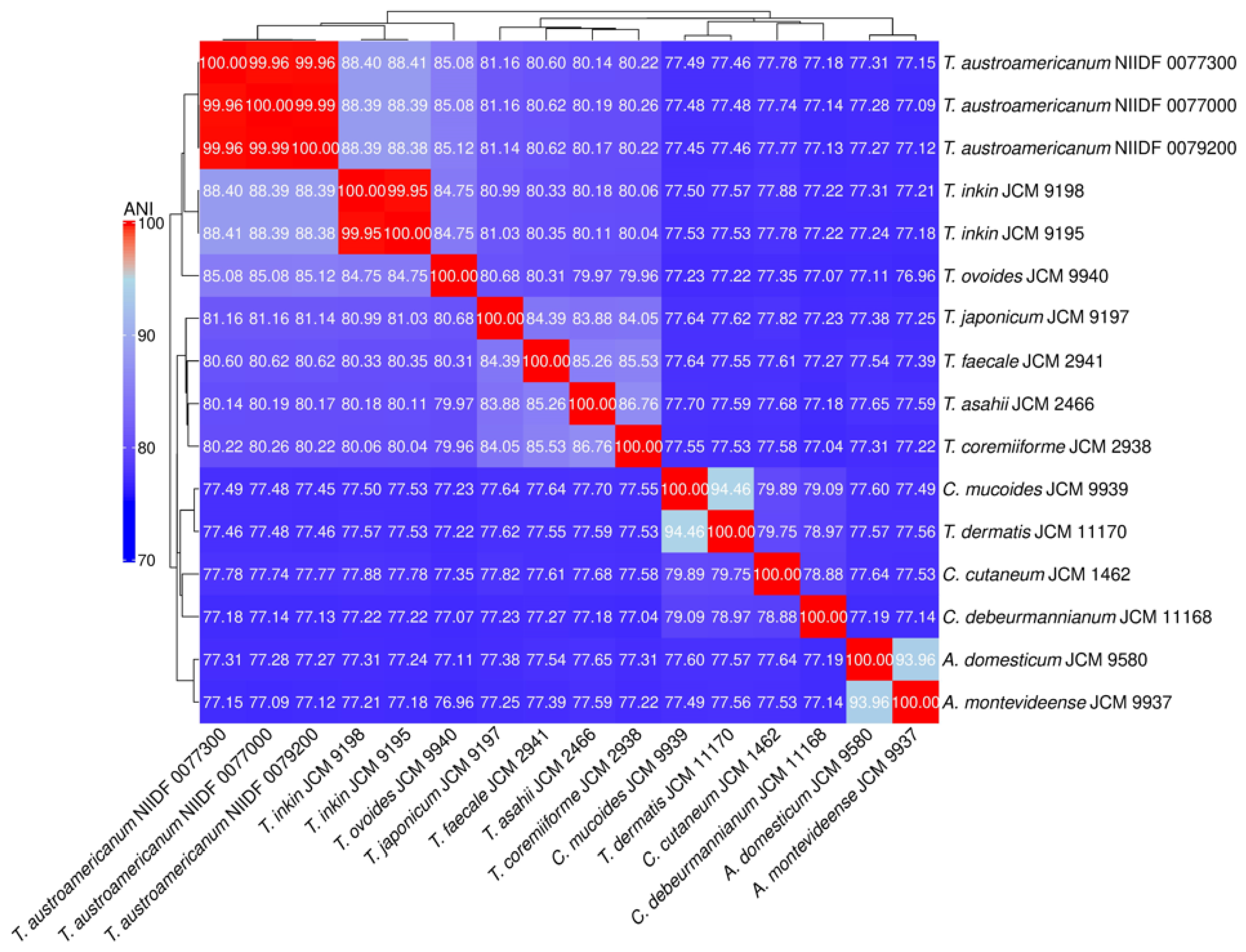
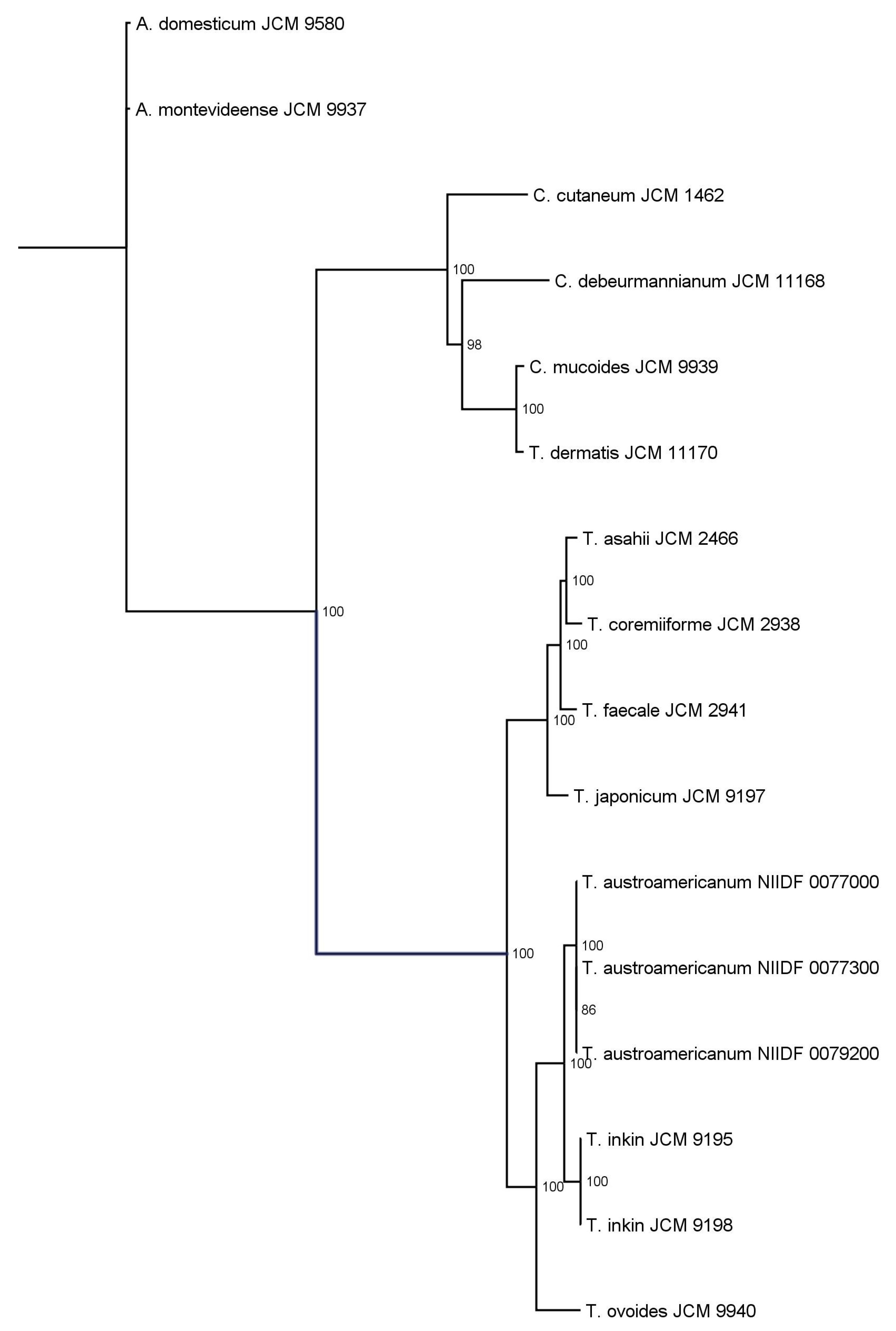
3.2. Phylogenetic Analysis

To characterize the genomic distinctiveness of T. austroamericanum, we conducted phylogenetic analyses using two complementary approaches: whole-genome ANI (Figure 1) and single-copy ortholog (SCO)-based ML phylogeny (Figure 2).
Pairwise Average Nucleotide Identity (ANI) values among the 16 Trichosporonales strains were calculated using FastANI and visualized as a heatmap. Red shading indicates high genomic similarity (higher ANI values), whereas blue shading indicates low similarity. The dendrogram represents hierarchical clustering based on a distance matrix derived from ANI values (1–ANI/100).
Orthologous gene groups were identified using OrthoFinder, and a set of single-copy ortholog genes conserved across all strains was extracted. Multiple sequence alignments were performed using MAFFT, concatenated into a supermatrix, and subjected to phylogenetic inference using the IQ-TREE2. The best-fit model was selected using ModelFinder, and branch support was assessed using 1000 ultrafast bootstrap replicates. Ultrafast bootstrap support (UFBoot) values are shown at internal nodes as percentages, with only values ≥70% displayed. The tree resolved the genera Apiotrichum, Cutaneotrichosporon, and Trichosporon into distinct clades. Notably, the node separating Apiotrichum domesticum and A. montevideense lacked bootstrap support, indicating a low statistical confidence in their divergence.
ANI analysis provided a measure of genome-wide nucleotide similarity, whereas SCO-based phylogeny inferred evolutionary relationships based on conserved protein-coding genes. The three T. austroamericanum strains exhibited extremely high similarities (ANI = 99.96–99.99%), indicating that they constituted a single genomic lineage. ANI values between T. austroamericanum and T. inkin ranged from 88.38% to 88.44%, well below the 95% species threshold, supporting their separation. Lower ANI values (~84.68–85.15%) were observed for T. ovoides, T. japonicum, and T. faecale, and even lower ANI values (<78%) for Cutaneotrichosporon and Apiotrichum species, reflecting increasing evolutionary distance. The SCO-based ML phylogeny similarly recovered T. austroamericanum as a strongly supported monophyletic clade (ultrafast bootstrap support [UFBoot] = 100), indicating a highly robust grouping. Within this group, strains 0077000 and 0077300 formed a sister pair with moderate support (UFBoot = 86.0), suggesting early-stage intraspecific divergence. The tree clearly separated the genera Apiotrichum, Cutaneotrichosporon, and Trichosporon, which was consistent with the current taxonomy. However, the node separating Apiotrichum domesticum and Apiotrichum montevideense lacks statistical support, leaving the relationship unresolved. Together, the ANI and ML trees provided complementary perspectives on both species- and strain-level relationships within the Trichosporonales and reinforced the distinctiveness of T. austroamericanum.

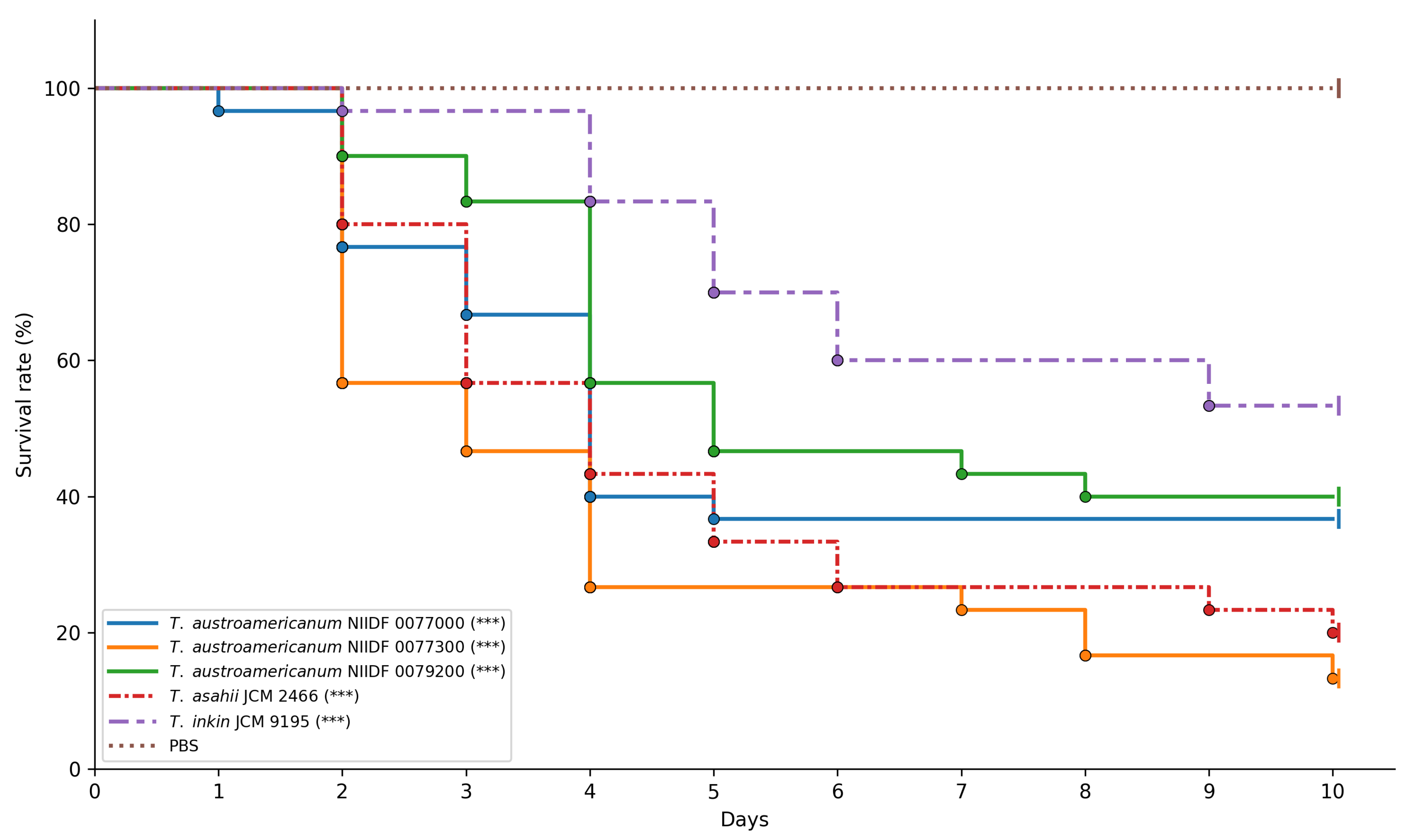
3.3. Virulence Assessment Using Galleria mellonella Infection Model

To assess strain-specific differences in virulence and provide a phenotypic context for orthologous gene analysis, we used the G. mellonella infection model [41]. Three bloodstream-derived isolates of T. austroamericanum (NIIDF 0077000, 0077300, and 0079200) were compared with T. asahii JCM 2466, a well-documented cause of invasive infections, and T. inkin JCM 9195, a phylogenetically related species generally associated with superficial infections. At an inoculum of 1 × 10⁶ CFU per larva, all strains caused markedly increased larval mortality relative to baseline survival (Figure 3).
Pairwise comparisons revealed that T. austroamericanum NIIDF 0077300 and T. asahii JCM 2466 exhibited significantly higher virulence than T. inkin JCM 9195 (adjusted p = 0.0005 and 0.0154, respectively) (Table 5).
In contrast, NIIDF 0077000 and NIIDF 0079200 showed no statistically significant differences from T. inkin (adjusted p = 0.5687 and 1.0000, respectively). No significant differences were observed among the T. austroamericanum strains or between any of them and T. asahii. Among the strains tested, NIIDF 0077300 and T. asahii JCM 2466 demonstrated relatively high virulence, with larval survival below 30% by day 10 (Figure 3). In contrast, T. inkin JCM 9195 and the remaining T. austroamericanum strains (NIIDF 0077000 and 0079200) exhibited moderate virulence under the same conditions.

3.4. Ortholog Analysis

As shown in the survival curve (Figure 3), all three strains of T. austroamericanum showed higher virulence in the G. mellonella infection model than in the PBS control. Notably, the virulence profiles of these strains were comparable to those of T. asahii JCM 2466, a well-characterized pathogenic species. Based on these phenotypic similarities, we selected three strains of T. austroamericanum and T. asahii for comparative ortholog analysis to examine the shared gene content that might be related to their observed virulence profiles. This approach was designed to link phenotypic observations with genomic features identified through comparative analysis. Using OrthoFinder, 15432 OGs were identified across 16 Trichosporon-related strains. Among them, we extracted OGs that were common to the two species but absent from all other Trichosporon species. Consequently, a single OG (OG0010545) was identified. OG0010545 was found only in T. asahii JCM 2466 and T. austroamericanum strain NIIDF 0077300 and was absent in the other two T. austroamericanum strains (NIIDF 0077000 and NIIDF 0079200) as well as in T. inkin JCM 9195. The genes were annotated by Funannotate as FUN_006829-T1 and FUN_007303-T1, respectively. Although both genes were labeled as “hypothetical protein” with no clearly assigned function, they possessed conserved domains associated with Zn(II)2Cys6 fungal-type DNA-binding transcription factors (IPR001138, IPR036864). GO annotations for both genes included terms such as zinc ion binding (GO:0008270), RNA polymerase II-specific DNA-binding transcription factor activity (GO:0000981), and regulation of DNA-templated transcription (GO:0006355). EggNOG analysis further classified both genes into the same orthologous group (ENOG503P0AT), indicating evolutionary conservation. A PHI-based search yielded no matches with the annotated virulence functions. These results highlight that OG0010545 is a conserved orthologous group uniquely shared between T. asahii and T. austroamericanum strain NIIDF 0077300, with features consistent with those of fungal transcription factors.

4. Discussion

In this study, we explored the genomic features and infection model responses of T. austroamericanum, a yeast species recently isolated from blood culture. Taken together, the results presented in Table 2, Table 3 and Table 4—covering assembly metrics, genome completeness, and functional annotation—demonstrate that all the analyzed strains possess high-quality, contiguous, and stable genomes with a conserved functional composition, supporting their suitability for comparative and functional genomic studies. WGS and comparative analyses were conducted to characterize the genome structure and clarify its phylogenetic position within the genus Trichosporon. To this end, we employed two complementary phylogenomic approaches: ANI (Figure 1), which quantifies genome-wide sequence similarity at the nucleotide level, and ML phylogeny based on SCOs (Figure 2), which reconstructs evolutionary relationships from conserved protein-coding genes. In the ANI heatmap (Figure 1), the three T. austroamericanum strains clustered tightly, exhibiting pairwise ANI values of 99.96–99.99%, consistent with extremely high genomic similarity. These strains formed a visually distinct block, clearly separated from other Trichosporon species, most notably from T. inkin, with ANI values ranging from 88.38% to 88.44%. This value fell well below the 95% threshold commonly used for species delineation, supporting the interpretation that T. austroamericanum is genomically distinct. A gradual decline in ANI values with increasing evolutionary distance was also observed, which aligned with the known taxonomic relationships. Similarly, the SCO-based ML tree (Figure 2) identified T. austroamericanum as a strongly supported monophyletic clade (UFBoot = 100). Within this clade, strains NIIDF 0077000 and 0077300 formed a moderately supported sister group (UFBoot = 86.0), suggesting early-stage divergence despite their nearly identical ANI values. This internal structuring within T. austroamericanum may reflect incipient genomic diversification, potentially resulting in strain-specific adaptation or functional divergence. The overall tree topology further classified the genera Apiotrichum, Cutaneotrichosporon, and Trichosporon into well-supported clades. However, the node separating A. domesticum and A. montevideense lacked bootstrap support, indicating that their relationship remains unresolved at the current resolution. Taken together, the ANI-based similarity landscape and protein-level ML phylogeny offer complementary perspectives on evolutionary divergence, highlighting both inter-species boundaries and emerging intraspecific variation. These findings reinforce the genomic distinctiveness of T. austroamericanum and underscore its status as a separate lineage that is closely related to T. inkin.
To assess strain-specific differences in virulence and provide a phenotypic context for orthologous gene analysis, we used the G. mellonella infection model. We compared the virulence of three clinical isolates of T. austroamericanum—strains NIIDF 0077000, 0077300, and 0079200—with T. asahii JCM 2466, a well-documented cause of invasive infections, and T. inkin JCM 9195, a phylogenetically related species typically associated with superficial infections [42]. At an inoculum of 1 × 10⁶ CFU per larva, all fungal strains tested induced significantly increased G. mellonella mortality compared to the PBS control (log-rank test, *** p < 0.001; Figure 3). Among the T. austroamericanum strains, NIIDF 0077300 exhibited a virulence profile closely resembling that of T. asahii and significantly different from that of T. inkin (adjusted p = 0.0005). However, no statistically significant differences were observed between NIIDF 0077300 and the other T. austroamericanum strains (NIIDF 0077000 and 0079200) or between these strains and T. inkin (adjusted p = 0.5687 and 1.0000, respectively). Additionally, there were no significant differences among the T. austroamericanum strains or between any of them and T. asahii. These findings demonstrate that all tested T. austroamericanum strains are capable of inducing lethal infection in the Galleria mellonella model, while also exhibiting strain-dependent variability in virulence. Furthermore, the observed mortality profiles suggest that T. austroamericanum exhibits strain-dependent virulence potential, with NIIDF 0077300 displaying pathogenicity comparable to that of T. asahii, whereas the other two strains showed levels of virulence similar to T. inkin. This is consistent with previous work by Mariné et al. [42], who reported that T. asahii was significantly more virulent than T. inkin in the G. mellonella model, as measured by survival curves and time to death. Our findings parallel this pattern, suggesting that T. austroamericanum may encompass strains with divergent virulence potential comparable to either species. Although the genetic basis of this phenotypic diversity remains to be elucidated, it may partly reflect underlying genomic differences. These observations highlight the importance of integrating phenotypic evidence with ortholog-based comparative analyses in future studies.
In ortholog analysis, OG0010545 was identified as an orthologous group shared between T. asahii and the most virulent T. austroamericanum strain NIIDF 0077300. It was absent in all the other strains examined. All genes within this ortholog group encode proteins with conserved Zn(II)2Cys6-type DNA-binding domains (IPR001138, IPR036864), suggesting that they likely encode fungal-specific transcription factors of the Zn(II)2Cys6 family. Zn(II)2Cys6-type transcription factors (zinc cluster proteins) are fungal-specific regulatory proteins known to perform diverse functions depending on the species, including metabolic regulation, drug resistance, ergosterol biosynthesis, and control of morphological transitions [45]. Structurally, Zn(II)2Cys6 transcription factors possess a conserved N-terminal DNA-binding domain (DBD) characterized by six cysteines arranged in the consensus Cys-X2-Cys-X6-Cys-X5–12-Cys-X2-Cys-X6–8-Cys motif. This motif coordinates two zinc ions, stabilizing the domain for specific DNA binding, typically to CGG triplet motifs. DBD includes a zinc finger, linker, and dimerization region that facilitates DNA recognition and protein interactions. Additional regulatory and activation domains enable transcriptional control through cofactors and chromatin-modifying complexes. This modular architecture allows for the precise condition-dependent regulation of target genes in fungi [45]. The functional relevance of this transcription factor family in pathogenicity is particularly well studied in Candida albicans, a dimorphic fungus capable of switching between yeast and hyphal forms, a morphological transition closely linked to virulence and tissue invasion [46,47,48]. Hyphal development facilitates tissue invasion, endothelial translocation, and immune evasion during systemic infection. Among the 77 candidate transcription factors with Zn(II)2Cys6 DNA-binding motifs identified in C. albicans, several, including CZF1, FGR17, and FGR27, regulate this yeast-to-hypha transition and are directly implicated in its pathogenic potential [45]. Given the structural and functional conservation of this transcription factor family, it is conceivable that the Zn(II)2Cys6-type transcription factor encoded by OG0010545 may regulate downstream genes involved in processes relevant to systemic infection, such as host tissue invasion, endothelial translocation, and immune evasion. Although based solely on in silico predictions, the identification of OG0010545, which encodes a Zn(II)2Cys6-type transcription factor, in both T. asahii and the most virulent T. austroamericanum strain suggests a potential role in infection-related processes. Its presence in these pathogenic strains represents only a single example but raises the possibility that similar regulatory mechanisms contribute to traits associated with persistence or dissemination within the host. Its strain-specific presence in T. austroamericanum suggests intraspecific genomic variation that may underlie phenotypic differences in host interactions. The presence of this ortholog in phylogenetically distinct, yet pathogenic strains may reflect shared selective pressures associated with host adaptation, potentially involving convergent evolution or horizontal gene retention under similar environmental constraints. This is consistent with the observed phylogenetic substructure and variability among the three isolates. However, the absence of OG0010545 in the other two T. austroamericanum strains highlights the need for further investigation. Future studies incorporating RNA-Seq under infection-relevant conditions and gene disruption analyses are essential to clarify its potential functional role. It is also likely that additional genomic elements contribute to strain-level variations, warranting further exploration.
This study had several limitations that warrant consideration. First, our conclusions regarding species-specific orthologs were based solely on in silico predictions, including domain architecture and sequence homology, without supporting the transcriptomic data. Although such predictions provide a useful foundation for hypothesis generation, they do not directly confirm the gene function or expression under biologically relevant conditions. To address this limitation, future studies should incorporate transcriptomic analyses, such as RNA-seq, to assess the expression of candidate orthologs under infection-relevant conditions and functional assays, such as gene disruption or complementation, to experimentally validate their biological roles. Second, this study did not use vertebrate infection models. Invertebrate models such as G. mellonella are widely used for screening fungal pathogenicity and virulence factors, offering practical advantages, including low cost, ease of handling, and exemption from ethical approval [49,50]. However, these models do not fully recapitulate the mammalian immune responses. Although valuable for initial pathogenicity assessment, validation in vertebrate systems, such as murine models, will be important to contextualize the findings within a mammalian host environment. Third, our genome assemblies were generated using long-read sequencing only, without incorporating short-read correction or hybrid assembly approaches. While the overall assembly quality was high for most strains (9–10 scaffolds), three species showed highly fragmented assemblies. This could reflect either biological features such as polyploidy or technical issues such as degraded or sheared input DNA. Moreover, the absence of short-read polishing may allow residual base-level errors to persist. Future studies should consider hybrid assembly methods, combining long- and short-read sequencing to improve assembly accuracy and completeness, particularly for structurally complex or repetitive regions.
Phylogenetic analyses based on ANI and SCOs indicated that T. austroamericanum constitutes a distinct lineage within Trichosporonales, despite its close relationship with T. inkin. The identification of a strain-specific ortholog opens avenues for future research on intraspecific variation in host interaction traits.

Author Contributions

Conceptualization, T.H. and T.U.; methodology, T.H., T.U. and H.T. (Hiroko Tomuro); software, T.H. and T.U.; validation, T.H. and T.U.; formal analysis, T.H. and T.U.; investigation, H.T. (Hiroko Tomuro), T.H., A.O., T.S. (Takayuki Shinohara), S.T., K.M., M.N., M.A., Y.M.(Yasunori Muraosa), and Y.H.; resources, T.H., T.S. (Takashi Sakoh), H.A., N.U., T.K., Y.N., H.T. (Hiroki Tsukada), and T.U.; writing—original draft preparation, T.H.; writing—review and editing, T.H. and T.U.; supervision, T.M. and Y.M. (Yoshitsugu Miyazaki); project administration, Y.M. (Yoshitsugu Miyazaki); funding acquisition, Y.M. (Yoshitsugu Miyazaki) and T.U. All authors have read and agreed to the published version of the manuscript.

Funding

This work was financially supported by the MHLW Emerging and Re-emerging Infectious Diseases and Immunization Policy Promotion Program (grant number JPMH22HA2001), the Japan Society for the Promotion of Science (JSPS) KAKENHI grant JP23K07936, and the Japan Agency for Medical Research and Development (AMED) under grant number JP25fk0108700. The funders had no role in the study design, collection, analysis, interpretation of data, writing of the manuscript, or decision to publish the results.

Institutional Review Board Statement

The use of clinical isolates (NIIDF 0077000, NIIDF 0077300, and NIIDF 0079200) was approved by the ethics committee of the National Institute of Infectious Diseases (Approval No: 1922, approved on 18 March 2025).

Informed Consent Statement

Not applicable.

Data Availability Statement

The generated dataset for this study can be found in the NCBI database under the BioProject accession number PRJNB20459. The data are currently under embargo and will be made publicly available upon acceptance of the manuscript.

Acknowledgments

During the preparation of this manuscript/study, the authors used ChatGPT o1 and 4o, Claude 3.7 Sonnet, and Gemini 2.5 Pro for the purposes of generating genomic analysis scripts. The authors have reviewed and edited the output and take full responsibility for the content of this publication.

Conflicts of Interest

The authors declare no conflicts of interest.

References

  1. Colombo, A.L.; Padovan, A.C.B.; Chaves, G.M. Current knowledge of Trichosporon spp. and Trichosporonosis. Clin. Microbiol. Rev. 2011, 24, 682–700. [Google Scholar] [CrossRef]
  2. de Almeida, J.N.; Francisco, E.C.; Ruiz, A.H.; Cuéllar, L.E.; Aquino, V.R.; Mendes, A.V.; Queiroz-Telles, F.; Santos, D.W.; Guimarães, T.; Chaves, G.M.; et al. Epidemiology, Clinical Aspects, Outcomes and Prognostic Factors Associated with Trichosporon Fungaemia: Results of an International Multicentre Study Carried Out at 23 Medical Centres. J. Antimicrob. Chemother. 2021, 76, 1907–1915. [Google Scholar] [CrossRef]
  3. de Almeida Júnior, J.N.; Hennequin, C. Invasive Trichosporon Infection: A Systematic Review on a Re-emerging Fungal Pathogen. Front. Microbiol. 2016, 7, 1629. [Google Scholar] [CrossRef]
  4. Francisco, E.C.; de Almeida Junior, J.N.; de Queiroz Telles, F.; Aquino, V.R.; Mendes, A.V.A.; de Andrade Barberino, M.G.M.; de Tarso O Castro, P.; Guimarães, T.; Hahn, R.C.; Padovan, A.C.B.; et al. Species distribution and antifungal susceptibility of 358 Trichosporon clinical isolates collected in 24 medical centres. Clin. Microbiol. Infect. 2019, 25, 909.e1–909.e5. [Google Scholar] [CrossRef]
  5. Guo, L.N.; Yu, S.Y.; Hsueh, P.R.; Al-Hatmi, A.M.S.; Meis, J.F.; Hagen, F.; Xiao, M.; Wang, H.; Barresi, C.; Zhou, M.L.; et al. Invasive Infections Due to Trichosporon: Species Distribution, Genotyping, and Antifungal Susceptibilities from a Multicenter Study in China. J. Clin. Microbiol. 2019, 57, e01505-18. [Google Scholar] [CrossRef]
  6. Dabas, Y.; Xess, I.; Kale, P. Molecular and antifungal susceptibility study on trichosporonemia and emergence of Trichosporon mycotoxinivorans as a bloodstream pathogen. Med. Mycol. 2017, 55, 518–527. [Google Scholar] [CrossRef]
  7. Santos, F.A.; Leite-Andrade, M.C.; Vasconcelos, M.A.; Alves, A.I.; Buonafina-Paz, M.D.; Araújo-Neto, L.N.; Macêdo, D.P.; Neves, R.P. Trichosporon inkin fungemia case report: Clinical and laboratory management. Future Microbiol. 2022, 17, 81–87. [Google Scholar] [CrossRef]
  8. Rodriguez-Tudela, J.L.; Diaz-Guerra, T.M.; Mellado, E.; Cano, V.; Tapia, C.; Perkins, A.; Gomez-Lopez, A.; Rodero, L.; Cuenca-Estrella, M. Susceptibility patterns and molecular identification of Trichosporon species. Antimicrob. Agents Chemother. 2005, 49, 4026–4034. [Google Scholar] [CrossRef]
  9. Francisco, E.C.; Desnos-Ollivier, M.; Dieleman, C.; Boekhout, T.; Santos, D.W.C.L.; Medina-Pestana, J.O.; Colombo, A.L.; Hagen, F. Unveiling Trichosporon austroamericanum sp. nov.: A Novel Emerging Opportunistic Basidiomycetous Yeast Species. Mycopathologia 2024, 189, 43. [Google Scholar] [CrossRef]
  10. Libkind, D.; Čadež, N.; Opulente, D.A.; Langdon, Q.K.; Rosa, C.A.; Sampaio, J.P.; Gonçalves, P.; Hittinger, C.T.; Lachance, M.A. Towards yeast taxogenomics: Lessons from novel species descriptions based on complete genome sequences. FEMS Yeast Res. 2020, 20, foaa042. [Google Scholar] [CrossRef]
  11. Kobayashi, Y.; Kayamori, A.; Aoki, K.; Shiwa, Y.; Matsutani, M.; Fujita, N.; Sugita, T.; Iwasaki, W.; Tanaka, N.; Takashima, M. Chromosome-level genome assemblies of Cutaneotrichosporon spp. (Trichosporonales, Basidiomycota) reveal imbalanced evolution between nucleotide sequences and chromosome synteny. BMC Genom. 2023, 24, 609. [Google Scholar] [CrossRef]
  12. Oxford Nanopore Technologies. Dorado: A High-Performance Basecaller for Nanopore Reads. Available online: https://github.com/nanoporetech/dorado (accessed on 5 March 2025).
  13. Wick, R. Filtlong: Quality Filtering Tool for Nanopore Reads. Available online: https://github.com/rrwick/Filtlong (accessed on 5 March 2025).
  14. Kolmogorov, M.; Yuan, J.; Lin, Y.; Pevzner, P.A. Assembly of long, error-prone reads using repeat graphs. Nat. Biotechnol. 2019, 37, 540–546. [Google Scholar] [CrossRef]
  15. Li, H. Minimap2: Pairwise alignment for nucleotide sequences. Bioinformatics 2018, 34, 3094–3100. [Google Scholar] [CrossRef]
  16. Vaser, R.; Sović, I.; Nagarajan, N.; Šikić, M. Fast and accurate de novo genome assembly from long uncorrected reads. Genome Res. 2017, 27, 737–746. [Google Scholar] [CrossRef]
  17. Oxford Nanopore Technologies. Medaka: Sequence Consensus Polishing Tool. Available online: https://github.com/nanoporetech/medaka (accessed on 5 March 2025).
  18. Gurevich, A.; Saveliev, V.; Vyahhi, N.; Tesler, G. QUAST: Quality assessment tool for genome assemblies. Bioinformatics 2013, 29, 1072–1075. [Google Scholar] [CrossRef]
  19. Simão, F.A.; Waterhouse, R.M.; Ioannidis, P.; Kriventseva, E.V.; Zdobnov, E.M. BUSCO: Assessing genome assembly and annotation completeness with single-copy orthologs. Bioinformatics 2015, 31, 3210–3212. [Google Scholar] [CrossRef]
  20. Funannotate. Funannotate: Fungal Genome Annotation Pipeline. Available online: https://github.com/nextgenusfs/funannotate (accessed on 5 March 2025).
  21. Stanke, M.; Morgenstern, B. AUGUSTUS: A web server for gene prediction in eukaryotes that allows user-defined constraints. Nucleic Acids Res. 2005, 33, W465–W467. [Google Scholar] [CrossRef]
  22. Majoros, W.H.; Pertea, M.; Salzberg, S.L. TigrScan and GlimmerHMM: Two open-source ab initio eukaryotic gene finders. Bioinformatics 2004, 20, 2878–2879. [Google Scholar] [CrossRef]
  23. Korf, I. Gene finding in novel genomes. Bioinformatics 2004, 5, 59. [Google Scholar] [CrossRef]
  24. Borodovsky, M.; Lomsadze, A. Gene identification in novel eukaryotic genomes by self-training algorithm. Nucleic Acids Res. 2011, 39, e147. [Google Scholar] [CrossRef]
  25. Jones, P.; Binns, D.; Chang, H.-Y.; Fraser, M.; Li, W.; McAnulla, C.; McWilliam, H.; Maslen, J.; Mitchell, A.; Nuka, G.; et al. InterProScan 5: Genome-scale protein function classification. Bioinformatics 2014, 30, 1236–1240. [Google Scholar] [CrossRef]
  26. Cantalapiedra, C.P.; Hernández-Plaza, A.; Letunic, I.; Bork, P.; Huerta-Cepas, J. eggNOG-mapper v2: Functional annotation, orthology assignments, and domain prediction at the metagenomic scale. Mol. Biol. Evol. 2021, 38, 5825–5829. [Google Scholar] [CrossRef]
  27. Blin, K.; Shaw, S.; Steinke, K.; Villebro, R.; Ziemert, N.; Lee, S.Y.; Medema, M.H.; Weber, T. antiSMASH 7.0: New and improved predictions for detection, regulation, chemical structures, and visualisation. Nucleic Acids Res. 2023, 51, W46–W50. [Google Scholar] [CrossRef]
  28. Almagro Armenteros, J.J.; Tsirigos, K.D.; Sønderby, C.K.; Petersen, T.N.; Winther, O.; Brunak, S.; von Heijne, G.; Nielsen, H. SignalP 5.0 improves signal peptide predictions using deep neural networks. Nat. Biotechnol. 2019, 37, 420–423. [Google Scholar] [CrossRef]
  29. Käll, L.; Krogh, A.; Sonnhammer, E.L. A combined transmembrane topology and signal peptide prediction method. J. Mol. Biol. 2004, 338, 1027–1036. [Google Scholar] [CrossRef]
  30. Jain, C.; Rodriguez, R.L.; Phillippy, A.M.; Konstantinidis, K.T.; Aluru, S. High throughput ANI analysis of 90K prokaryotic genomes reveals clear species boundaries. Nat. Commun. 2018, 9, 5114. [Google Scholar] [CrossRef]
  31. Saitou, N.; Nei, M. The neighbor-joining method: A new method for reconstructing phylogenetic trees. Mol. Biol. Evol. 1987, 4, 406–425. [Google Scholar] [CrossRef]
  32. Cock, P.J.A.; Antao, T.; Chang, J.T.; Chapman, B.A.; Cox, C.J.; Dalke, A.; Friedberg, I.; Hamelryck, T.; Kauff, F.; Wilczynski, B.; et al. Biopython: Freely available Python tools for computational molecular biology and bioinformatics. Bioinformatics 2009, 25, 1422–1423. [Google Scholar] [CrossRef]
  33. Gu, Z.; Eils, R.; Schlesner, M. Complex heatmaps reveal patterns and correlations in multidimensional genomic data. Bioinformatics 2016, 32, 2847–2849. [Google Scholar] [CrossRef]
  34. Emms, D.M.; Kelly, S. OrthoFinder: Phylogenetic orthology inference for comparative genomics. Genome Biol. 2019, 20, 238. [Google Scholar] [CrossRef]
  35. Buchfink, B.; Xie, C.; Huson, D.H. Fast and sensitive protein alignment using DIAMOND. Nat. Methods 2015, 12, 59–60. [Google Scholar] [CrossRef]
  36. Katoh, K.; Standley, D.M. MAFFT multiple sequence alignment software version 7: Improvements in performance and usability. Mol. Biol. Evol. 2013, 30, 772–780. [Google Scholar] [CrossRef]
  37. Minh, B.Q.; Schmidt, H.A.; Chernomor, O.; Schrempf, D.; Woodhams, M.D.; von Haeseler, A.; Lanfear, R. Corrigendum to: IQ-TREE 2: New Models and Efficient Methods for Phylogenetic Inference in the Genomic Era. Mol. Biol. Evol. 2020, 37, 2461. [Google Scholar] [CrossRef]
  38. Kalyaanamoorthy, S.; Minh, B.Q.; Wong, T.K.F.; von Haeseler, A.; Jermiin, L.S. ModelFinder: Fast model selection for accurate phylogenetic estimates. Nat. Methods. 2017, 14, 587–589. [Google Scholar] [CrossRef]
  39. Hoang, D.T.; Chernomor, O.; von Haeseler, A.; Minh, B.Q.; Vinh, L.S. UFBoot2: Improving the Ultrafast Bootstrap Approximation. Mol. Biol. Evol. 2018, 35, 518–522. [Google Scholar] [CrossRef]
  40. Rambaut, A. FigTree v1.4.4. Institute of Evolutionary Biology, University of Edinburgh. Available online: http://tree.bio.ed.ac.uk/software/figtree/ (accessed on 13 May 2025).
  41. Urban, M.; Cuzick, A.; Seager, J.; Wood, V.; Rutherford, K.; Venkatesh, S.Y.; De Silva, N.; Martinez, M.C.; Pedro, H.; Yates, A.D.; et al. PHI-base: The pathogen–host interactions database. Nucleic Acids Res. 2020, 48, D613–D620. [Google Scholar] [CrossRef] [PubMed]
  42. Mariné, M.; Bom, V.L.P.; de Castro, P.A.; Winkelstroter, L.K.; Ramalho, L.N.; Brown, N.A.; Goldman, G.H. The development of animal infection models and antifungal efficacy assays against clinical isolates of Trichosporon asahii, T. asteroides and T. inkin. Virulence 2015, 6, 476–486. [Google Scholar] [CrossRef]
  43. Davidson-Pilon, C. Lifelines: Survival Analysis in Python. Available online: https://lifelines.readthedocs.io (accessed on 24 March 2025).
  44. Hunter, J.D. Matplotlib: A 2D Graphics Environment. Comput. Sci. Eng. 2007, 9, 90–95. [Google Scholar] [CrossRef]
  45. MacPherson, S.; Larochelle, M.; Turcotte, B. A fungal family of transcriptional regulators: The zinc cluster proteins. Microbiol. Mol. Biol. Rev. 2006, 70, 583–604. [Google Scholar] [CrossRef]
  46. Pla, J.; Gil, C.; Monteoliva, L.; Navarro-García, F.; Sánchez, M.; Nombela, C. Understanding Candida albicans at the molecular level. Yeast 1996, 12, 1677–1702. [Google Scholar] [CrossRef]
  47. Leberer, E.; Ziegelbauer, K.; Schmidt, A.; Harcus, D.; Dignard, D.; Ash, J.; Johnson, L.; Thomas, D.Y. Virulence and hyphal formation of Candida albicans require the Ste20p-like protein kinase CaCla4p. Curr. Biol. 1997, 7, 539–546. [Google Scholar] [CrossRef] [PubMed]
  48. Lo, H.J.; Köhler, J.R.; DiDomenico, B.; Loebenberg, D.; Cacciapuoti, A.; Fink, G.R. Nonfilamentous Candida albicans mutants are avirulent. Cell 1997, 90, 939–949. [Google Scholar] [CrossRef] [PubMed]
  49. Giammarino, A.; Bellucci, N.; Angiolella, L. Galleria mellonella as a Model for the Study of Fungal Pathogens: Advantages and Disadvantages. Pathogens 2024, 13, 233. [Google Scholar] [CrossRef]
  50. Serrano, I.; Verdial, C.; Tavares, L.; Oliveira, M. The Virtuous Galleria mellonella Model for Scientific Experimentation. Antibiotics 2023, 12, 505. [Google Scholar] [CrossRef]
Figure 1. Heatmap and hierarchical clustering of genome-wide ANI among Trichosporonales strains.
Jof 11 00401 g001
Figure 2. Maximum likelihood phylogenetic tree inferred from single-copy orthologs identified in 16 Trichosporonales strains.
Jof 11 00401 g002
Figure 3. Galleria mellonella survival after fungal infection. Kaplan–Meier survival curves of Galleria mellonella larvae (n = 30 per strain) infected with Trichosporon isolates at an inoculum of 1 × 10⁶ CFU per larva. Larvae injected with PBS served as the control group. Asterisks indicate statistically significant differences compared to the PBS group (log-rank test with Bonferroni correction): *** p < 0.001. Censored individuals (alive on day 10) are indicated by the vertical tick marks on the curves.
Jof 11 00401 g003
Table 1. Strains used in this study.
StrainsStrain IDSource
Trichosporon austroamericanum NIIDF 0077000 human blood
Trichosporon austroamericanum NIIDF 0077300 human blood
Trichosporon austroamericanum NIIDF 0079200 human blood
Trichosporon inkin JCM 9195 human skin
Trichosporon inkin JCM 9198 surgical wound
Trichosporon ovoides JCM 9940 scalp
Trichosporon asahii JCM 2466 human skin
Trichosporon coremiiforme JCM 2938 lesion on farmer head
Trichosporon dermatis JCM 11170 human skin
Trichosporon faecale JCM 2941 human feces
Trichosporon japonicum JCM 9197 unknown
Apiotrichum domesticum JCM 9580 damp and rotten wooden sideboard
Apiotrichum montevideense JCM 9937 water purification tank
Cutaneotrichosporon cutaneum JCM 1462 unknown
Cutaneotrichosporon debeurmannianum JCM 11168 human bronchial secretion
Cutaneotrichosporon mucoides JCM 9939 case of meningitis
Table 2. Genome assembly statistics and repeat content in Trichosporon and related species sequenced in this study based on QUAST analysis.
StrainsScaffoldsTotal Length (Mb)Largest Scaffold (Mb)GC (%)N50 (Mb)Repeat Sequences (%)
T. austroamericanum NIIDF 0077000 921.023.9350.033.1919.37
T. austroamericanum NIIDF 0077300 1021.055.6950.013.8819.39
T. austroamericanum NIIDF 0079200 921.063.9250.043.4119.32
T. inkin JCM 9195 3620.363.6650.012.7118.62
T. inkin JCM 9198 3620.444.5050.022.7418.68
T. ovoides JCM 9940 5540.403.1950.011.8719.89
T. asahii JCM 2466 4024.824.0449.992.2920.28
T. coremiiforme JCM 2938 15343.001.5350.000.9120.13
T. dermatis JCM 11170 2423.423.8949.902.7820.01
T. faecale JCM 2941 5824.812.9050.061.3719.84
T. japonicum JCM 9197 2823.554.6050.073.2818.57
A. domesticum JCM 9580 2024.657.9249.983.5320.77
A. montevideense JCM 9937 2425.183.7649.962.1720.87
C. cutaneum JCM 1462 12224.221.6550.060.3718.73
C. debeurmannianum JCM 11168 2819.153.8049.922.9720.34
C. mucoides JCM 9939 3642.754.2149.963.3519.90
Table 3. BUSCO-based assessment of genome completeness, duplication, and gene content integrity in Trichosporon related species.
StrainsComplete
(%)
Single-Copy
(%)
Duplicated
(%)
Fragmented
(%)
Missing
(%)
T. austroamericanum NIIDF 0077000 91.491.30.12.85.8
T. austroamericanum NIIDF 0077300 91.391.20.12.95.8
T. austroamericanum NIIDF 0079200 90.390.20.12.86.5
T. inkin JCM 9195 88.387.90.44.17.6
T. inkin JCM 9198 88.387.90.44.17.6
T. ovoides JCM 9940 95.324.770.61.13.6
T. asahii JCM 2466 88.487.70.74.86.8
T. coremiiforme JCM 2938 91.023.467.63.45.6
T. dermatis JCM 11170 92.191.20.92.85.1
T. faecale JCM 2941 88.387.70.64.96.8
T. japonicum JCM 9197 88.287.70.54.87.0
A. domesticum JCM 9580 89.088.20.82.78.3
A. montevideense JCM 9937 88.788.00.72.88.5
C. cutaneum JCM 1462 93.292.50.71.75.1
C. debeurmannianum JCM 11168 91.090.40.62.96.1
C. mucoides JCM 9939 93.930.063.91.94.2
Table 4. Number and proportion of functionally annotated genes in Trichosporon related species.
StrainsGenesInterProScan
(%)
eggNOG
(%)
Pfam
(%)
CAZyme
(%)
MEROPS
(%)
Phobius
(%)
T. austroamericanum NIIDF 0077000 74275674
(76.4%)
6206
(83.6%)
4974
(67.0%)
185
(2.5%)
232
(3.1%)
517
(7.0%)
T. austroamericanum NIIDF 0077300 74425653
(76.0%)
6202
(83.3%)
4959
(66.6%)
185
(2.5%)
232
(3.1%)
511
(6.9%)
T. austroamericanum NIIDF 0079200 75745736
(75.7%)
6291
(83.1%)
5037
(66.5%)
187
(2.5%)
231
(3.0%)
528
(7.0%)
T. inkin JCM 9195 73255715
(78.0%)
6267
(85.6%)
5031
(68.7%)
184
(2.5%)
230
(3.1%)
494
(6.7%)
T. inkin JCM 9198 71785612
(78.2%)
6149
(85.7%)
4913
(68.4%)
182
(2.5%)
228
(3.2%)
485
(6.8%)
T. ovoides JCM 9940 1432110864
(75.9%)
11193
(78.2%)
9494
(66.3%)
348
(2.4%)
440
(3.1%)
943
(6.6%)
T. asahii JCM 2466 85305932
(69.5%)
6530
(76.6%)
5143
(60.3%)
194
(2.3%)
245
(2.9%)
639
(7.5%)
T. coremiiforme JCM 2938 1523510972
(72.0%)
12053
(79.1%)
9533
(62.6%)
350
(2.3%)
459
(3.0%)
1063
(7.0%)
T. dermatis JCM 11170 82856246
(75.4%)
6816
(82.3%)
5469
(66.0%)
233
(2.8%)
270
(3.3%)
540
(6.5%)
T. faecale JCM 2941 87745950
(67.8%)
6539
(74.5%)
5147
(58.7%)
191
(2.2%)
242
(2.8%)
681
(7.8%)
T. japonicum JCM 9197 81995874
(71.6%)
6405
(78.1%)
5096
(62.2%)
196
(2.4%)
248
(3.0%)
652
(8.0%)
A. domesticum JCM 9580 79755871
(73.6%)
6430
(80.6%)
5148
(64.6%)
208
(2.6%)
244
(3.1%)
613
(7.7%)
A. montevideense JCM 9937 1431510887
(76.1%)
11874
(82.9%)
9560
(66.8%)
403
(2.8%)
466
(3.3%)
959
(6.7%)
C. cutaneum JCM 1462 89826315
(70.3%)
6903
(76.9%)
5524
(61.5%)
202
(2.2%)
296
(3.3%)
597
(6.6%)
C. debeurmannianum JCM 11168 69465553
(79.9%)
6069
(87.4%)
4897
(70.5%)
171
(2.5%)
233
(3.4%)
380
(5.5%)
C. mucoides JCM 9939 1431510,887
(76.1%)
11,874
(82.9%)
9560
(66.8%)
403
(2.8%)
466
(3.3%)
959
(6.7%)
Table 5. Pairwise comparison of virulence among Trichosporon strains based on Galleria mellonella survival assay. Pairwise comparisons of Galleria mellonella survival curves were conducted using the log-rank (Mantel–Cox) test. Adjusted p-values were obtained by applying the Bonferroni correction for multiple tests.
ComparisonAdjusted p-Value
T. asahii JCM 2466 vs. T. austroamericanum NIIDF 00770001.0000
T. asahii JCM 2466 vs. T. austroamericanum NIIDF 00773001.0000
T. asahii JCM 2466 vs. T. austroamericanum NIIDF 00792000.9312
T. asahii JCM 2466 vs. T. inkin JCM 91950.0154
T. inkin JCM 9195 vs. T. austroamericanum NIIDF 00770000.5687
T. inkin JCM 9195 vs. T. austroamericanum NIIDF 00773000.0005
T. inkin JCM 9195 vs. T. austroamericanum NIIDF 00792001.0000
T. austroamericanum NIIDF 0077000 vs. T. austroamericanum NIIDF 00773000.7089
T. austroamericanum NIIDF 0077000 vs. T. austroamericanum NIIDF 00792001.0000
T. austroamericanum NIIDF 0077300 vs. T. austroamericanum NIIDF 00792000.0707
Disclaimer/Publisher’s Note: The statements, opinions and data contained in all publications are solely those of the individual author(s) and contributor(s) and not of MDPI and/or the editor(s). MDPI and/or the editor(s) disclaim responsibility for any injury to people or property resulting from any ideas, methods, instructions or products referred to in the content.

