Article

Phenotypic and Molecular Characterization of Multidrug-Resistant Clinical Isolates of the Candidozyma haemuli Species Complex (Formerly Candida haemulonii Species Complex) from the Brazilian Amazon Reveals the First Case of Candidozyma pseudohaemuli in Brazil

by
Sérgio Lobato França
1,2,3,*,
Rodrigo Santos de Oliveira
3,
Gabriel Silas Marinho Sousa
2,3,
Sarah Rodrigues de Sá
3,
Walber da Silva Nogueira
1,2,3,
Elaine Patrícia Tavares do Espírito Santo
3,
Daniel dos Santos Caldas
2,3 and
Silvia Helena Marques da Silva
3
1
Hospital Universitário João de Barros Barreto, Belém 66073-000, Pará, Brazil
2
Programa de Pós-Graduação em Biologia de Agentes Infecciosos e Parasitários, Instituto de Ciências Biológicas, Universidade Federal do Pará, Belém 66075-110, Pará, Brazil
3
Laboratório de Micoses Superficiais e Sistêmicas, Seção de Bacteriologia e Micologia, Instituto Evandro Chagas, Ananindeua 67030-000, Pará, Brazil
*
Author to whom correspondence should be addressed.
J. Fungi 2025, 11(5), 394; https://doi.org/10.3390/jof11050394
Submission received: 9 April 2025 / Revised: 16 May 2025 / Accepted: 16 May 2025 / Published: 20 May 2025

Abstract

:
This study included 24 isolates of the Candidozyma haemuli species complex from patients in the Brazilian Amazon between 2021 and 2024. These isolates were identified by sequencing as C. duobushaemuli (54.2%), C. haemuli sensu stricto (29.2%), C. haemuli var. vulneris (12.5%), and C. pseudohaemuli (4.2%). The finding of C. pseudohaemuli represents the first case reported in Brazil. Haplotype and phylogenetic analysis of these species, along with other isolates from Brazil, revealed low intraspecific genetic diversity. Resistance to at least one antifungal was observed in 83.3% of isolates, with multidrug resistance in 58.3%, including one isolate resistant to all tested antifungals. The isolates demonstrated active biofilm production, lytic enzyme activity, and thermotolerance. Notably, one C. duobushaemuli isolate exhibited tolerance to 42 °C, a phenotype not previously described. It is crucial for Brazil and other countries to recognize the emergence of these species as a public health threat and to take proactive measures to prevent outbreaks.

1. Introduction

Recently, a new genus called Candidozyma has been proposed to aggregate the Candidozyma haemuli species complex (CHSC) (formerly known as the Candida haemulonii species complex), together with the multidrug-resistant fungus Candidozyma auris (formerly Candida auris) [1].
This new genus includes both species with significant clinical relevance and those isolated from natural environments, indicating a broad ecological and adaptive diversity [1]. These fungi are notable not only for their resistance to antifungal drugs but also for their ability to cause invasive fungal infections, which can be difficult to treat and have high mortality rates, especially in immunocompromised patients [2,3].
Although the global prevalence of the CHSC is still relatively low compared to other fungal infections, the number of cases has increased worryingly in recent years [2,3,4,5,6]. It is interesting to note that in South America, infections with this complex are more frequently associated with candidemia, while in Asia and the United States, cases of cutaneous and non-invasive infections predominate [7,8].
Brazil has stood out as one of the countries most affected by this increase in cases of resistant fungal infections, with the number of diagnoses almost doubling over the last 11 years [9]. This reflects both the growing resistance of fungi to conventional treatments and the lack of early diagnosis of these infections [5].
In contrast to C. auris, which is a focus of intensive global study and monitoring, knowledge about the CHSC is still limited. This includes not only an understanding of its biology but also the paucity of epidemiological data on its geographical distribution and the factors that contribute to its pathogenicity [10,11].
Recent studies have shown that, in particular, clinical isolates of the CHSC in the Brazilian Amazon region remain virtually unexplored [4,10]. Considering that the distribution and frequency of infections caused by different species of the complex may vary according to geographical region [12], it is possible that ecological and structural factors specific to the Amazon may influence the circulation and dissemination of these species.
Therefore, the main objective of this study was to identify and characterize clinical isolates of the Candidozyma haemuli species complex from patients in the Brazilian Amazon region in order to increase understanding of the presence, diversity, virulence, and antifungal susceptibility of these species in the region.

2. Materials and Methods

2.1. This Study

This is a cross-sectional observational study that included CHSC isolates obtained from patients treated at a referral hospital in the state of Pará, Brazilian Amazon, from January 2021 to May 2024. Sociodemographic and clinical information of the patients was collected by reviewing medical records.

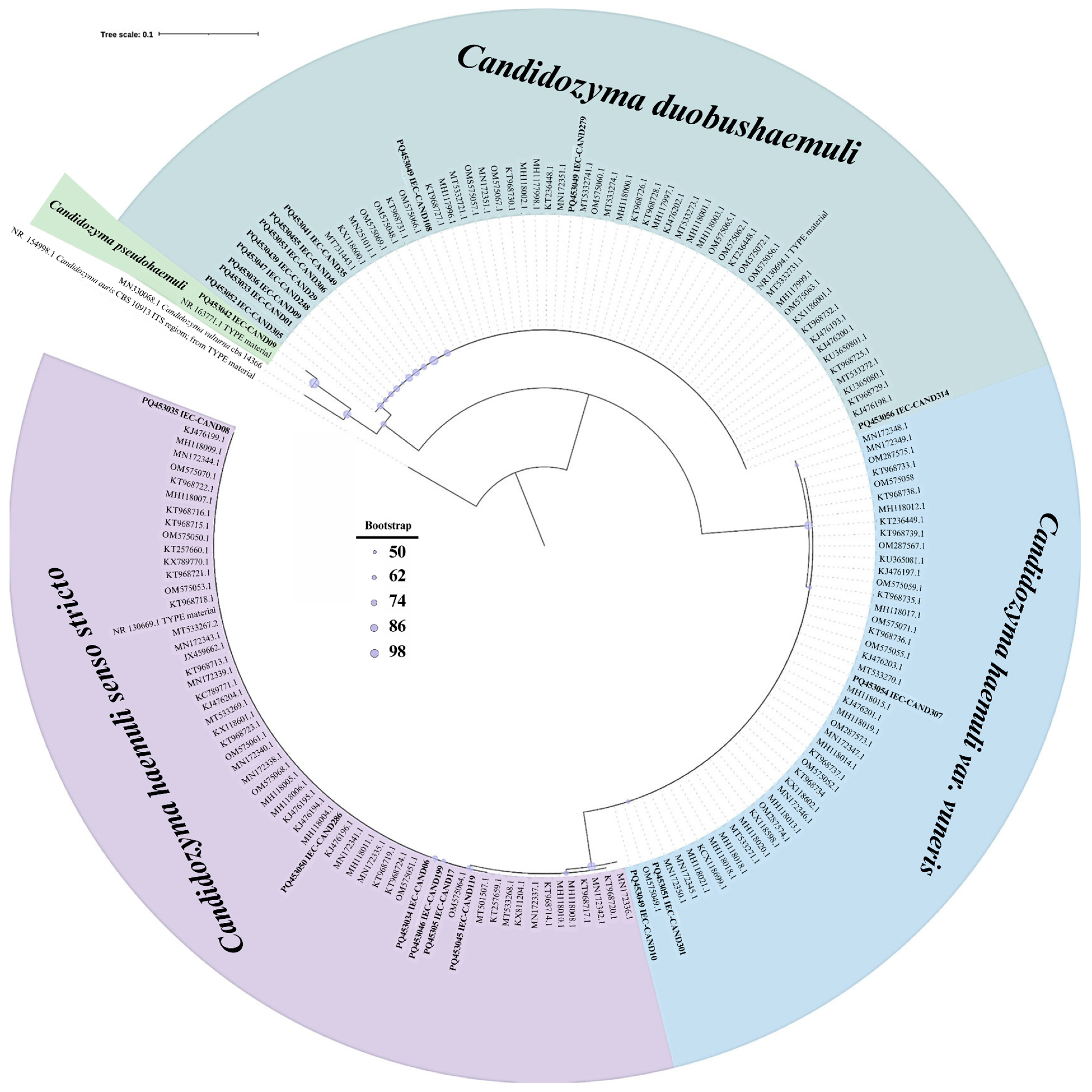
2.2. Species Identification

CHSC isolates were initially identified using the Vitek® 2 compact system (BioMérieux, Durham, NC, USA) and the MALDI-TOF Biotyper® sirius (Bruker, Billerica, MA, USA), following the manufacturers’ recommendations. Final species identification, phylogenetic analyses, and haplotype studies were conducted through genetic sequencing of the ITS1-5.8S-ITS2 rDNA regions using the ABI 3130 genetic analyzer (Thermo Fisher Scientific, Waltham, MA, USA). Sequences were aligned with those deposited in the GenBank database (NCBI, https://www.ncbi.nlm.nih.gov, accessed on 12 October 2024) referencing type strains, with species determination based on a similarity and coverage threshold of >99%. Phylogenetic and haplotype analyses incorporated sequences from this study along with non-redundant Brazilian sequences available in GenBank.
Sequences were first organized into a multifasta file using NotePad++ v8.5.4 (Don HO, https://notepad-plus-plus.org, accessed on 5 October 2024) and then aligned using MAFFT v7 (RIMD, https://mafft.cbrc.jp/alignment/server, accessed on 9 October 2024). Phylogenetic tree reconstruction was performed using IQ-TREE (http://www.iqtree.org, accessed on 7 October 2024), applying the maximum likelihood method with the TNe+G4 substitution model and 1000 bootstrap replicates. Haplotype analysis included the evaluation of polymorphic diversity using DnaSP v5.10 (http://www.ub.edu/dnasp/index_v5.html, accessed on 10 October 2024) and haplotype network construction was done using Cytoscape v3.10.2 (https://cytoscape.org, accessed on 14 October 2024).

2.3. Antifungal Susceptibility Testing

Antifungal susceptibility testing was conducted for amphotericin B (AB), fluconazole (FLU), itraconazole (ITC), and flucytosine (FC) (all from Sigma-Aldrich, Burlington, MA, USA) following the European Committee on Antimicrobial Susceptibility Testing (EUCAST) broth microdilution protocol E.DEF.7.4 [13]. Since clinical susceptibility breakpoints for CHSC are not established, we used provisional thresholds for C. auris suggested by the Centers for Disease Control and Prevention (CDC) [14] for FLU (≥32 µg/mL) and AB (≥2 µg/mL). For ITR (≥1 µg/mL) and FC (≥32 µg/mL), the general Candida spp. breakpoints from the Clinical and Laboratory Standards Institute (CLSI) were applied (M27-A3) [15].

2.4. Evaluation of Virulence Factors

Virulence factors evaluated included thermotolerance at 30 °C, 37 °C, and 42 °C on Sabouraud dextrose agar plates after 48 h of incubation, biofilm formation, biomass quantification, and enzymatic activities of lipase, phospholipase, and proteinase.

2.4.1. Biofilm Formation and Biomass Quantification

The assays for biofilm formation, biomass quantification, and metabolic activity assessment were performed as described by Ramos et al. [16] and in technical triplicate. For this, fungal cell suspensions were prepared in Sabouraud broth, and 200 µL of each suspension (containing 106 cells) were transferred into the wells of a 96-well microplate and incubated at 37 °C for 48 h without agitation. After the incubation period, the supernatant was carefully removed, and the wells were washed with PBS to eliminate non-adherent cells. The biofilms were then fixed with 99% methanol and allowed to air dry.
To quantify the biomass, the biofilms were stained with 0.4% crystal violet solution, washed with PBS, and subsequently decolorized with 33% acetic acid. The absorbance of the resulting solution was measured at 560 nm using a microplate reader to estimate the amount of biomass present in the biofilms.
Finally, the metabolic activity of the biofilms was assessed using the XTT/menadione colorimetric assay. After preparation of the XTT/menadione solution and its addition to the wells containing the biofilms, the plates were incubated at 37 °C for 3 h in the dark. The supernatant was then transferred to a new 96-well microplate, and absorbance was measured at 492 nm, enabling the quantification of the fungal biofilms’ metabolic activity.
To assess differences in biomass among species, the non-parametric Kruskal–Wallis test was applied due to unequal sample sizes and non-normally distributed data.

2.4.2. Phospholipase Activity

Phospholipase activity was assessed following the method described by Price et al. [17]. Sabouraud agar supplemented with 8% egg yolk, 1 M NaCl, and 5 mM CaCl2 was used to prepare the phospholipase agar medium. The test samples were initially cultured on Sabouraud agar for 48 h. Subsequently, colonies were inoculated onto phospholipase agar at equidistant points and incubated at 37 °C for 10 days. The diameters of the colonies with precipitation zones were measured according to the method described by Price et al. [17].

2.4.3. Lipase Activity

Lipase activity was evaluated according to the method described by Muhsin et al. [18]. The lipase agar medium was prepared by supplementing a base medium with the following components: 10 g of peptone (Merck, Darmstadt, HE, Germany), 5 g of NaCl (VETEC, Brazil), 0.1 g of CaCl2 (Sigma-Aldrich, Burlington, MA, USA), 20 g of agar (Merck, Darmstadt, HE, Germany), and 10 mL of Tween 20 (Sigma-Aldrich, Burlington, MA, USA). A portion of each colony was inoculated onto sterile Petri dishes containing the lipase agar and incubated at 27 °C for 10 days. Samples were considered lipase producers when an opaque halo was observed around the colony.

2.4.4. Proteinase Activity

Proteinase activity was assessed according to the method described by Rüchel et al. [19]. The proteinase agar medium was prepared by supplementing the base medium with the following components: 11.7 g of Yeast Carbon Base (HIMEDIA, Mumbai, MH, India), 2 g of bovine serum albumin—fraction V (Sigma-Aldrich, Burlington, MA, USA), 2.5 mL of Protovit® (Roche, Basel, Switzerland), and 100 mL of sterile distilled water. Samples were inoculated onto the medium and incubated at 30 °C for 10 days. Proteinase production was indicated by the formation of a clear halo around the colony.
All enzymatic activity assays were performed in technical duplicate and quantified using the activity index (Pz) [17].

2.5. Statistical Analysis

ANOVA was employed to analyze biofilm results, while the Kruskal–Wallis test followed by the Dwass–Steel–Critchlow–Fligner post hoc test was used for enzymatic activity data. Pearson’s correlation coefficient was used for the correlation analysis. Statistical analyses were performed using JAMOVI software (v2.3, https://www.jamovi.org, accessed on 15 November 2024), with statistical significance set at p < 0.05.

2.6. Ethics Approval

This study was approved by the research ethics committee for human studies of the João de Barros Barreto University Hospital (No. 5.514.367) and the Evandro Chagas Institute (No. 5.799.031) in compliance with Brazilian legislation.

3. Results

3.1. Patient Characteristics and Isolate Identification

We analyzed 24 clinical CHSC isolates from 24 patients (Table 1). A comparison of identification methods, using sequencing as the reference standard, revealed discrepancies in results obtained with Vitek® 2 (66.7%) and MALDI-TOF (13.3%) (Table 2).
Phylogenetic reconstruction showed no genetic divergence among Brazilian isolates (Figure 1). A similar result was observed in haplotype diversity analysis. Although two haplotypes were identified for C. pseudohaemuli, this separation was considered insignificant as it resulted from a single mutation (Figure 2). Notably, no C. pseudohaemuli sequences from Brazil were available in GenBank.

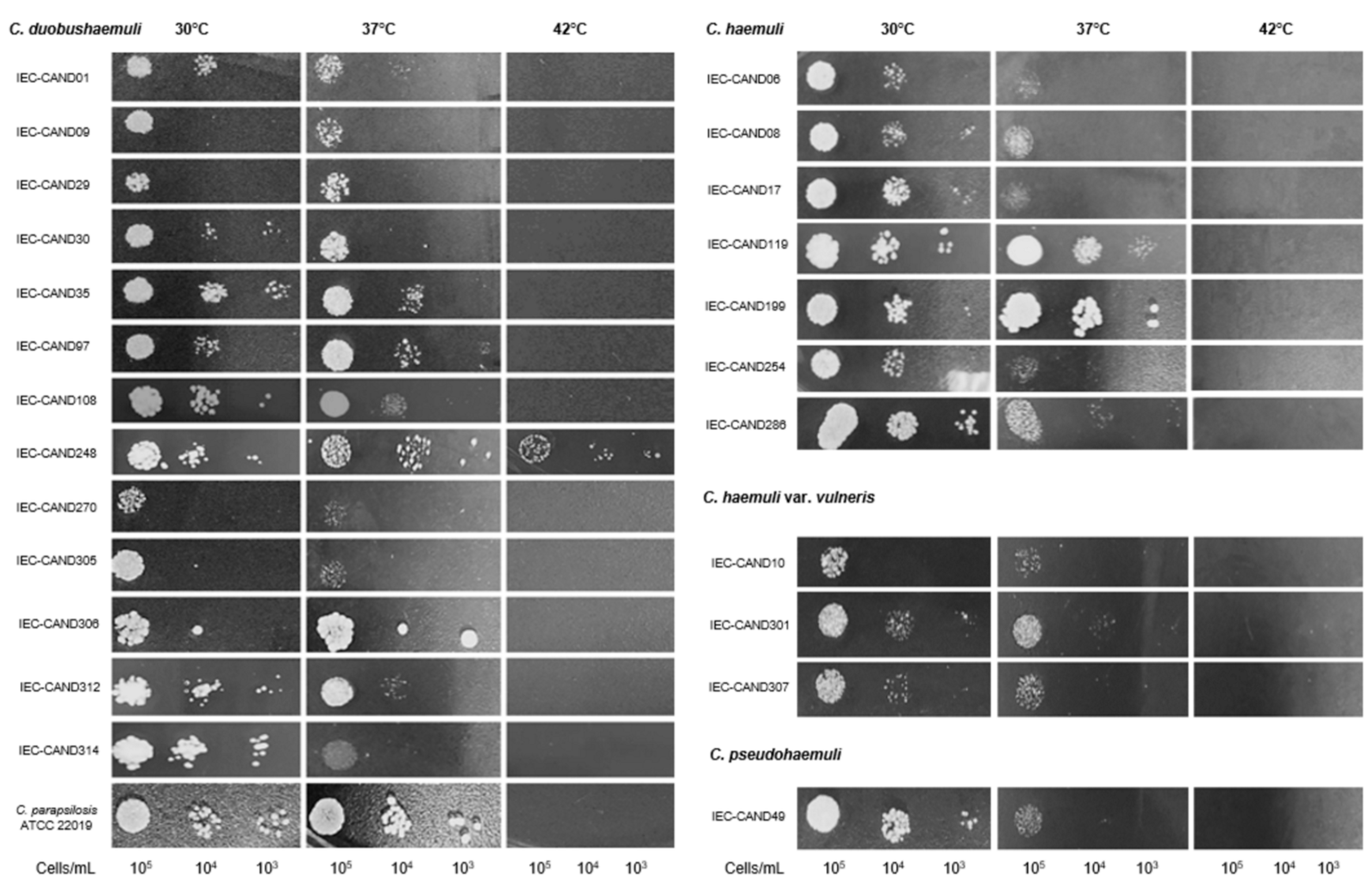
3.2. Phenotypic Virulence Features of the Isolates

All isolates were able to grow at 35 °C and 37 °C; however, at 42 °C, only one isolate of C. duobushaemuli (IEC-CAND248) exhibited growth (Figure 3).
Regarding biofilm formation, all isolates demonstrated production capacity, and there was a positive correlation between biofilm biomass and metabolic activity (Pearson’s r = 0.547) (Figure 4). The analysis of mean biomass among species, conducted using the Kruskal–Wallis test, did not indicate a statistically significant difference between the evaluated groups (H = 1.27; p = 0.736). This suggests that, under the assay conditions, the analyzed species exhibited similar performance in terms of biomass production. The results of the quantification of biofilm biomass and metabolic activity in clinical isolates of the Candidozyma haemuli species complex and reference strains are presented in Supplementary Material Table S1.
In enzymatic activity tests, isolates showed varying capacities to produce proteinase (p = 0.017) and lipase (p = 0.005). Pairwise analysis for proteinase revealed differences between C. duobushaemuli and C. haemuli sensu stricto (p = 0.015), primarily due to 85.7% of C. haemuli sensu stricto isolates being strong producers of this enzyme, while C. duobushaemuli isolates were either moderate producers (53.8%) or non-producers (30.8%). For lipase, differences were observed between C. duobushaemuli and C. haemuli sensu stricto (p = 0.004), as well as between C. duobushaemuli and C. haemuli var. vulneris (p = 0.013). These differences were mainly attributed to the fact that none of the C. duobushaemuli isolates produced lipase (Table 3).

3.3. Antifungal Resistance Patterns in Clinical Isolates

Antifungal susceptibility testing revealed that most isolates displayed some level of resistance (83.3%). Multidrug resistance (MDR) was observed in half of the isolates, a common finding for C. duobushaemuli (69.2%) and C. pseudohaemuli (100%) (Table 4 and Table 5). Multidrug-resistant isolates were at least resistant to azoles and amphotericin B (AB), with the following resistance combinations identified: FLU+ITC+AB (20.8%); FLU+AB (16.7%); ITC+AB (8.3%); FLU+ITC (8.3%); and FLU+ITC+AB+FC (4.2%).

4. Discussion

This is the first study of clinical isolates of CHSC from the Brazilian Amazon, highlighting the discovery of C. pseudohaemuli, which represents the first report of this species in Brazil, as there were no previous records [9,20,21,22,23,24,25,26,27,28,29,30]. This report represents a significant advance in the understanding of clinical fungal diversity in the country. The absence of previous reports suggests that this species may have been underdiagnosed due to limitations in conventional identification methods or that its emergence is recent in the region.
Furthermore, the identification of C. pseudohaemuli in the Brazilian Amazon highlights the urgency of an effective surveillance system for monitoring emerging fungal infections. Epidemiological surveillance contributes to the early detection of new cases, the identification of dissemination patterns, and the implementation of appropriate control measures, especially in hospital environments, where species of the Candidozyma genus can cause outbreaks that are difficult to manage due to their recognized resistance to antifungal drugs [31,32].
With regard to C. pseudohaemuli, only a slightly over a dozen cases have been reported worldwide to date, with cases identified in South Korea, Thailand, the United States, Venezuela, Panama, Colombia, and French Guiana, involving fungemias and antifungal resistance profiles [30,33,34,35]. The limited distribution of C. pseudohaemuli positions it as one of the rarest pathogens in the field of CHSC. The detection of this fungus in the Amazon underscores the importance of microbiological surveillance in the region, especially given the growing emergence of opportunistic pathogens in tropical environments [30].
Genetic analysis of the rDNA regions of the isolates in this study, including other Brazilian isolates, revealed low intraspecific diversity within CHSC in Brazil, consistent with a Brazilian study that used whole genome sequencing [29].
This low diversity suggests a possible clonal expansion of these species in the country [36], which could have important implications for public health, facilitating the spread of well-adapted strains and increasing the risk of outbreaks. The stability of genotype frequencies may be associated with the small number of sequenced isolates, low mutation rates, or possible clonal origins [37], highlighting the need for continuous genetic surveillance to monitor evolutionary dynamics and implement more effective control strategies.
In terms of virulence, in addition to tolerance at 35–37 °C, one isolate of C. duobushaemuli showed growth at 42 °C, which is, to our knowledge, the first global report of this phenotype for the species. Previous studies have not demonstrated this characteristic [6,38,39,40,41,42,43]. Until now, it was only known that C. auris, C. khanbhai, and C. ruelliae, within the genus Candidozyma, could withstand 42 °C [6,44,45]. However, genomic studies have shown that C. duobushaemuli and other CHSC species possess genes involved in thermotolerance [10].
This characteristic could increase the virulence and evolutionary advantage of C. duobushaemuli, allowing it to survive in hosts during febrile episodes and in high-temperature environments. Although more studies are needed, we infer that local environmental conditions and the environmental adaptability of CHSC suggest that this high thermotolerance may represent an adaptation of C. duobushaemuli to rising temperatures in the Amazon [46,47,48].
We also observed the ability of the isolates to form biofilms. Biofilm formation by Candidozyma spp. is particularly relevant due to its association with increased antifungal resistance, evasion of host immune responses, and adaptation to stress conditions, which complicate treatment and may promote infection persistence [16].
Regarding enzymatic activity, our analysis revealed notable differences among the species evaluated. C. haemuli and its varieties exhibited higher levels of proteinase and lipase activity compared to C. duobushaemuli isolates. These findings are consistent with previous studies that also reported elevated enzymatic activity in C. haemuli [16,26,27,49], suggesting a possible role for these enzymes in virulence and tissue invasion. For C. duobushaemuli, the absence of lipase production in the isolates may represent a local pattern, although a Brazilian isolate with very high lipase activity has been described [44]. Phospholipase activity was low in all isolates of this study, in agreement with the results of other authors [23,26,27,49], which may suggest a less significant role for this enzyme in the pathogenicity of these isolates. However, further studies are needed to better investigate this aspect.
The low prevalence of CHSC and the limited studies on its phenotypic characteristics may be related to the difficulty of laboratory identification using commercial methods. In our comparative analysis of identification methods, frequent failures of Vitek® 2 were observed, which is consistent with the results of Ambaraghassi et al. [50]. Although MALDI-TOF performed better, discrepancies in results were also observed, which highlights the need to expand mass spectrometry databases for CHSC identification. Given the limitations of conventional identification methods, we believe that many CHSC infections in hospitals in Brazil and around the world are underreported.
Clinically, the main challenge of CHSC infections in the Brazilian Amazon is antifungal resistance, particularly due to the high number of isolates resistant to azoles and AB, a common feature in C. duobushaemuli and C. pseudohaemuli. This resistance limits therapeutic options, making the management of these infections more complex, especially in immunocompromised patients or those with associated comorbidities [11,13]. However, in the present study, a favorable observation was the broad susceptibility of these isolates to FC, encouraging its use as a therapeutic alternative.
Despite the potential of FC, its isolated use has limitations, since monotherapy can lead to the rapid development of resistance and has variable efficacy [51]. It is therefore recommended that FC be administered in combination with other antifungals to ensure a synergistic effect and broaden the therapeutic response, reducing the fungal load and minimizing the likelihood of emerging resistance [52].
The resistance profile of the C. pseudohaemuli isolate (IEC-CAND49) to FLU and AB was similar to that of the first recorded isolate [33] and other global isolates [30], potentially indicating an intrinsic resistance to these antifungals. A worrying finding was the extensive resistance exhibited by an isolate of C. duobushaemuli to three classes of antifungals. Locally, this isolate can be considered the true “superfungus”, since all the strains of C. auris identified in Brazil to date have not shown resistance [53,54,55]. Regarding the country’s therapeutic capacity to combat these fungi, there is cause for concern, as a recent survey indicated that, in 2019, half of Brazilian hospitals had no echinocandins and less than 20% had FC available [56]. In this context, the absence of echinocandins in our testing is recognized as a limitation, which occurred due to constraints within our study.
In conclusion, the Brazilian Amazon emerges as a region with a diverse presence of CHSC species in hospital environments, characterized by frequent multidrug resistance and isolates exhibiting distinct virulence traits, such as elevated thermotolerance, which pose a potential global health threat. The socio-economic profile of the region, characterized by limited access to healthcare and inadequacies in hospital infrastructure, may facilitate the dissemination of these pathogens and complicate their timely diagnosis and treatment. Our findings also highlight the relevance of considering both environmental and hospital-associated transmission routes in the epidemiology of CHSC infections. In this context, the implementation of regional genomic surveillance programs becomes essential to monitor strain circulation, detect emerging variants, and inform public health strategies. These results reinforce the need for continuous monitoring efforts in the region to enable early detection of CHSC cases, guide therapeutic decision-making, and mitigate the burden of infection on vulnerable populations. To effectively limit CHSC infections, hospitals must strengthen their diagnostic capacity and ensure equitable access to antifungal agents.

Supplementary Materials

The following supporting information can be downloaded at: https://www.mdpi.com/article/10.3390/jof11050394/s1, Supplementary Table S1: Biofilm Biomass and Metabolic Activity in Candidozyma haemuli species complex and reference strains.

Author Contributions

Conceptualization, S.L.F., R.S.d.O. and S.H.M.d.S.; methodology, S.L.F., R.S.d.O., G.S.M.S., S.R.d.S. and W.d.S.N.; formal analysis, S.L.F. and R.S.d.O.; investigation, S.L.F.; data curation, E.P.T.d.E.S. and S.L.F.; writing—original draft preparation, S.L.F.; writing—review and editing, R.S.d.O., G.S.M.S., D.d.S.C. and S.H.M.d.S.; supervision, S.H.M.d.S. All authors have read and agreed to the published version of the manuscript.

Funding

The authors acknowledge the financial support from the Institute Evandro Chagas (IEC), Department of Health Surveillance, Ministry of Health: Laboratory of Superficial and Systemic Mycoses.

Institutional Review Board Statement

This study was approved by the Research Ethics Committee for Human Studies of the João de Barros Barreto University Hospital (approval No. 5.514.367) and the Evandro Chagas Institute (approval No. 5.799.031) on 9 November 2021, in accordance with Brazilian regulations.

Informed Consent Statement

Informed consent was obtained from all subjects involved in the study.

Data Availability Statement

The original contributions presented in this study are included in the article and Supplementary Materials. Further inquiries can be directed to the corresponding author.

Acknowledgments

We thank the João de Barros Barreto University Hospital, the Evandro Chagas Institute, and the Graduate Program in Biology of Infectious and Parasitic Agents at the Federal University of Pará for their support.

Conflicts of Interest

The authors declare no conflicts of interest.

References

  1. Liu, F.; Hu, Z.-D.; Zhao, X.-M.; Zhao, W.-N.; Feng, Z.-X.; Yurkov, A.; Alwasel, S.; Boekhout, T.; Bensch, K.; Hui, F.-L.; et al. Phylogenomic Analysis of the Candida auris-Candida haemuli Clade and Related Taxa in the Metschnikowiaceae, and Proposal of Thirteen New Genera, Fifty-Five New Combinations and Nine New Species. Persoonia 2024, 52, 22–43. [Google Scholar] [CrossRef] [PubMed]
  2. Fisher, M.C.; Gurr, S.J.; Cuomo, C.A.; Blehert, D.S.; Jin, H.; Stukenbrock, E.H.; Stajich, J.E.; Kahmann, R.; Boone, C.; Denning, D.W.; et al. Threats Posed by the Fungal Kingdom to Humans, Wildlife, and Agriculture. mBio 2020, 11, 10–1128. [Google Scholar] [CrossRef]
  3. Brown, G.D.; Denning, D.W.; Gow, N.A.R.; Levitz, S.M.; Netea, M.G.; White, T.C. Hidden Killers: Human Fungal Infections. Sci. Transl. Med. 2012, 4, 165rv13. [Google Scholar] [CrossRef]
  4. Francisco, E.C.; de Jong, A.W.; Colombo, A.L. Candida haemulonii Species Complex: A Mini-Review. Mycopathologia 2023, 188, 909–917. [Google Scholar] [CrossRef]
  5. Cao, C.; Bing, J.; Liao, G.; Nobile, C.J.; Huang, G. Candida haemulonii Species Complex: Emerging Fungal Pathogens of the Metschnikowiaceae Clade. Zoonoses 2023, 3, 957. [Google Scholar] [CrossRef]
  6. de Jong, A.W.; Al-Obaid, K.; Mohd Tap, R.; Gerrits van den Ende, B.; Groenewald, M.; Joseph, L.; Ahmad, S.; Hagen, F. Candida khanbhai sp. Nov., a New Clinically Relevant Yeast within the Candida haemulonii Species Complex. Med. Mycol. 2023, 61, myad009. [Google Scholar] [CrossRef] [PubMed]
  7. Chow, N.A.; Gade, L.; Tsay, S.V.; Forsberg, K.; Greenko, J.A.; Southwick, K.L.; Barrett, P.M.; Kerins, J.L.; Lockhart, S.R.; Chiller, T.M.; et al. Multiple Introductions and Subsequent Transmission of Multidrug-Resistant Candida auris in the USA: A Molecular Epidemiological Survey. Lancet Infect. Dis. 2018, 18, 1377–1384. [Google Scholar] [CrossRef]
  8. Garcia, J.A. Distribuição Regional da Resistência Antifúngica em Cepas Causadoras de Candidemia no Brasil. Ph.D. Thesis, Secretaria de Estado da Saúde de São Paulo, São Paulo, Brazil, 2024. Available online: https://fi-admin.bvsalud.org/document/view/pgzh2 (accessed on 20 March 2024).
  9. Lima, S.L.; Francisco, E.C.; de Almeida Júnior, J.N.; Santos, D.W.d.C.L.; Carlesse, F.; Queiroz-Telles, F.; Melo, A.S.d.A.; Colombo, A.L. Increasing Prevalence of Multidrug-Resistant Candida haemulonii Species Complex among All Yeast Cultures Collected by a Reference Laboratory over the Past 11 Years. J. Fungi 2020, 6, 110. [Google Scholar] [CrossRef] [PubMed]
  10. Gómez-Gaviria, M.; Martínez-Álvarez, J.A.; Chávez-Santiago, J.O.; Mora-Montes, H.M. Candida haemulonii Complex and Candida auris: Biology, Virulence Factors, Immune Response, and Multidrug Resistance. Infect. Drug Resist. 2023, 16, 1455–1470. [Google Scholar] [CrossRef]
  11. Chen, X.; Jia, X.; Bing, J.; Zhang, H.; Hong, N.; Liu, Y.; Xi, H.; Wang, W.; Liu, Z.; Zhang, Q.; et al. Clonal Dissemination of Antifungal-Resistant Candida haemulonii, China. Emerg. Infect. Dis. 2023, 29, 576–584. [Google Scholar] [CrossRef]
  12. Lima, S.L. Caracterização Molecular, Avaliação do Perfil de Susceptibilidade a Antifúngicos e Formação de Biofilme de Isolados Clínicos Pertencentes ao Complexo Candida haemulonii e Candida auris. Master’s Thesis, Universidade Federal de São Paulo (Escola Paulista de Medicina), São Paulo, Brazil, 2017. Available online: https://repositorio.unifesp.br/handle/11600/50775 (accessed on 25 March 2024).
  13. Subcommittee on Antifungal Susceptibility Testing (AFST) of the ESCMID European Committee for Antimicrobial Susceptibility Testing (EUCAST). EUCAST Definitive Document EDef 7.1: Method for the Determination of Broth Dilution MICs of Antifungal Agents for Fermentative Yeasts. Clin. Microbiol. Infect. 2008, 14, 398–405. [Google Scholar] [CrossRef]
  14. Centers for Disease Control and Prevention. Antifungal Susceptibility Testing for C. auris. Updated 23 April 2024. Available online: https://www.cdc.gov/candida-auris/hcp/laboratories/antifungal-susceptibility-testing.html (accessed on 27 November 2024).
  15. Approved Standard M27-A3; Reference Method for Broth Dilution Antifungal Susceptibility Testing of Yeasts. Clinical and Laboratory Standards Institute: Malvern, PA, USA, 2008.
  16. Ramos, L.S.; Figueiredo-Carvalho, M.H.G.; Silva, L.N.; Siqueira, N.L.M.; Lima, J.C.; Oliveira, S.S.; Almeida-Paes, R.; Zancopé-Oliveira, R.M.; Azevedo, F.S.; Ferreira, A.L.P.; et al. The Threat Called Candida haemulonii Species Complex in Rio de Janeiro State, Brazil: Focus on Antifungal Resistance and Virulence Attributes. J. Fungi 2022, 8, 574. [Google Scholar] [CrossRef] [PubMed]
  17. Price, M.F.; Wilkinson, I.D.; Gentry, L.O. Plate Method for Detection of Phospholipase Activity in Candida Albicans. Sabouraudia 1982, 20, 7–14. [Google Scholar] [CrossRef]
  18. Muhsin, T.M.; Aubaid, A.H.; al-Duboon, A.H. Extracellular Enzyme Activities of Dermatophytes and Yeast Isolates on Solid Media. Mycoses 1997, 40, 465–469. [Google Scholar] [CrossRef] [PubMed]
  19. Rüchel, R.; Tegeler, R.; Trost, M. A Comparison of Secretory Proteinases from Different Strains of Candida Albicans. Sabouraudia 1982, 20, 233–244. [Google Scholar] [CrossRef] [PubMed]
  20. Ferreira, N.; Belloch, C.; Querol, A.; Manzanares, P.; Vallez, S.; Santos, A. Yeast Microflora Isolated from Brazilian Cassava Roots: Taxonomical Classification Based on Molecular Identification. Curr. Microbiol. 2010, 60, 287–293. [Google Scholar] [CrossRef]
  21. Almeida, J.N.d.; Motta, A.L.; Rossi, F.; Abdala, E.; Pierrotti, L.C.; Kono, A.S.G.; Diz, M.D.P.E.; Benard, G.; Del Negro, G.M.B. First Report of a Clinical Isolate of Candida haemulonii in Brazil. Clinics 2012, 67, 1229–1231. [Google Scholar] [CrossRef]
  22. Ramos, L.S.; Figueiredo-Carvalho, M.H.G.; Barbedo, L.S.; Ziccardi, M.; Chaves, A.L.S.; Zancopé-Oliveira, R.M.; Pinto, M.R.; Sgarbi, D.B.G.; Dornelas-Ribeiro, M.; Branquinha, M.H.; et al. Candida haemulonii Complex: Species Identification and Antifungal Susceptibility Profiles of Clinical Isolates from Brazil. J. Antimicrob. Chemother. 2015, 70, 111–115. [Google Scholar] [CrossRef]
  23. de Souza Ramos, L.; Barbedo, L.S.; Braga-Silva, L.A.; dos Santos, A.L.S.; Pinto, M.R.; da Graça Sgarbi, D.B. Protease and Phospholipase Activities of Candida spp. Isolated from Cutaneous Candidiasis. Revi. Iberoam. Micol. 2015, 32, 122–125. [Google Scholar] [CrossRef]
  24. Almeida, J.N.d.; Assy, J.G.P.L.; Levin, A.S.; Negro, G.M.B.D.; Giudice, M.C.; Tringoni, M.P.; Thomaz, D.Y.; Motta, A.L.; Abdala, E.; Pierroti, L.C.; et al. Candida haemulonii complex species, Brazil, January 2010–March 2015. Emerg. Infect. Dis. 2016, 22, 561–563. [Google Scholar] [CrossRef]
  25. Boatto, H.F.; Cavalcanti, S.D.B.; Del Negro, G.M.; Girão, M.J.B.; Francisco, E.C.; Ishida, K.; Gompertz, O.F. Candida Duobushaemulonii: An Emerging Rare Pathogenic Yeast Isolated from Recurrent Vulvovaginal Candidiasis in Brazil. Mem. Inst. Oswaldo Cruz 2016, 111, 407–410. [Google Scholar] [CrossRef]
  26. Pagani, D.M.; Heidrich, D.; Paulino, G.V.B.; de Oliveira Alves, K.; Dalbem, P.T.; de Oliveira, C.F.; Andrade, Z.M.M.; Silva, C.; Correia, M.D.; Scroferneker, M.L.; et al. Susceptibility to Antifungal Agents and Enzymatic Activity of Candida Haemulonii and Cutaneotrichosporon Dermatis Isolated from Soft Corals on the Brazilian Reefs. Arch. Microbiol. 2016, 198, 963–971. [Google Scholar] [CrossRef]
  27. Ramos, L.S.; Branquinha, M.H.; Santos, A.L.S. Different Classes of Hydrolytic Enzymes Produced by Multidrug-Resistant Yeasts Comprising the Candida haemulonii Complex. Med. Mycol. 2017, 55, 228–232. [Google Scholar] [CrossRef] [PubMed]
  28. Ramos, L.S.; Mello, T.P.; Branquinha, M.H.; Santos, A.L.S. Biofilm Formed by Candida Haemulonii Species Complex: Structural Analysis and Extracellular Matrix Composition. J. Fungi 2020, 6, 46. [Google Scholar] [CrossRef]
  29. de Barros Rodrigues, D.K.; Lockhart, S.R.; Berkow, E.L.; Gade, L.; Bonfietti, L.X.; Mazo Fávero Gimenes, V.; Silva Ruiz, L.; Bronze Macioni, M.; de Souza Carvalho Melhem, M. Whole-Genome Sequencing of Candida haemulonii Species Complex from Brazil and the United States: Genetic Diversity and Antifungal Susceptibility. Med. Mycol. 2023, 61, myad030. [Google Scholar] [CrossRef] [PubMed]
  30. Françoise, U.; Desnos-Ollivier, M.; Le Govic, Y.; Sitbon, K.; Valentino, R.; Peugny, S.; Chouaki, T.; Mazars, E.; Paugam, A.; Nicolas, M.; et al. Candida haemulonii Complex, an Emerging Threat from Tropical Regions? PLoS Negl. Trop. Dis. 2023, 17, e0011453. [Google Scholar] [CrossRef] [PubMed]
  31. Poulopoulou, A.; Sidiropoulou, A.; Sarmourli, T.; Zachrou, E.; Michailidou, C.; Zarras, C.; Vagdatli, E.; Massa, E.; Mouloudi, E.; Pyrpasopoulou, A.; et al. Candida auris: Outbreak, Surveillance and Epidemiological Monitoring in Northern Greece. Med. Mycol. 2024, 62, myae062. [Google Scholar] [CrossRef]
  32. Bilal, H.; Shafiq, M.; Hou, B.; Islam, R.; Khan, M.N.; Khan, R.U.; Zeng, Y. Distribution and Antifungal Susceptibility Pattern of Candida Species from Mainland China: A Systematic Analysis. Virulence 2022, 13, 1573–1589. [Google Scholar] [CrossRef]
  33. Sugita, T.; Takashima, M.; Poonwan, N.; Mekha, N. Candida pseudohaemulonii sp. Nov., an Amphotericin B-and Azole-Resistant Yeast Species, Isolated from the Blood of a Patient from Thailand. Microbiol. Immunol. 2006, 50, 469–473. [Google Scholar] [CrossRef]
  34. Kim, M.-N.; Shin, J.H.; Sung, H.; Lee, K.; Kim, E.-C.; Ryoo, N.; Lee, J.-S.; Jung, S.-I.; Park, K.H.; Kee, S.J.; et al. Candida haemulonii and Closely Related Species at 5 University Hospitals in Korea: Identification, Antifungal Susceptibility, and Clinical Features. Clin. Infect. Dis. 2009, 48, e57–e61. [Google Scholar] [CrossRef]
  35. Gade, L.; Muñoz, J.F.; Sheth, M.; Wagner, D.; Berkow, E.L.; Forsberg, K.; Jackson, B.R.; Ramos-Castro, R.; Escandón, P.; Dolande, M.; et al. Understanding the Emergence of Multidrug-Resistant Candida: Using Whole-Genome Sequencing to Describe the Population Structure of Candida haemulonii Species Complex. Front. Genet. 2020, 11, 554. [Google Scholar] [CrossRef] [PubMed]
  36. Excoffier, L.; Foll, M.; Petit, R.J. Genetic Consequences of Range Expansions. Annu. Rev. Ecol. Evol. Syst 2009, 40, 481–501. [Google Scholar] [CrossRef]
  37. Merseguel, K.B.; Nishikaku, A.S.; Rodrigues, A.M.; Padovan, A.C.; e Ferreira, R.C.; de Azevedo Melo, A.S.; Briones, M.R.d.S.; Colombo, A.L. Genetic Diversity of Medically Important and Emerging Candida Species Causing Invasive Infection. BMC Infect. Dis. 2015, 15, 57. [Google Scholar] [CrossRef]
  38. Cendejas-Bueno, E.; Kolecka, A.; Alastruey-Izquierdo, A.; Theelen, B.; Groenewald, M.; Kostrzewa, M.; Cuenca-Estrella, M.; Gómez-López, A.; Boekhout, T. Reclassification of the Candida Haemulonii Complex as Candida haemulonii (C. haemulonii Group I), C. duobushaemulonii sp. Nov. (C. haemulonii Group II), and C. Haemulonii Var. Vulnera Var. Nov.: Three Multiresistant Human Pathogenic Yeasts. J. Clin. Microbiol. 2012, 50, 3641–3651. [Google Scholar] [CrossRef] [PubMed]
  39. Kathuria, S.; Singh, P.K.; Sharma, C.; Prakash, A.; Masih, A.; Kumar, A.; Meis, J.F.; Chowdhary, A. Multidrug-Resistant Candida auris Misidentified as Candida haemulonii: Characterization by Matrix-Assisted Laser Desorption Ionization—Time of Flight Mass Spectrometry and DNA Sequencing and Its Antifungal Susceptibility Profile Variability by Vitek 2, CLSI Broth Microdilution, and Etest Method. J. Clin. Microbiol. 2015, 53, 1823–1830. [Google Scholar] [CrossRef]
  40. Kumar, A.; Sachu, A.; Mohan, K.; Vinod, V.; Dinesh, K.; Karim, S. Simple Low Cost Differentiation of Candida auris from Candida haemulonii Complex Using CHROMagar Candida Medium Supplemented with Pal’s Medium. Rev. Iberoam. Micol. 2017, 34, 109–111. [Google Scholar] [CrossRef] [PubMed]
  41. Jurado-Martín, I.; Marcos-Arias, C.; Tamayo, E.; Guridi, A.; de Groot, P.W.J.; Quindós, G.; Eraso, E. Candida Duobushaemulonii: An Old But Unreported Pathogen. J. Fungi 2020, 6, 374. [Google Scholar] [CrossRef]
  42. Pharkjaksu, S.; Boonmee, N.; Mitrpant, C.; Ngamskulrungroj, P. Immunopathogenesis of Emerging Candida auris and Candida haemulonii Strains. J. Fungi 2021, 7, 725. [Google Scholar] [CrossRef]
  43. Tamura, T.; Alshahni, M.M.; Makimura, K. Evaluation of CHROMagarTM Candida Plus Chromogenic Agar for the Presumptive Identification of Candida auris. Microbiol. Immunol. 2022, 66, 292–298. [Google Scholar] [CrossRef]
  44. Saluja, P.; Prasad, G.S. Candida ruelliae sp. Nov., a Novel Yeast Species Isolated from Flowers of Ruellia sp. (Acanthaceae). FEMS Yeast Res. 2008, 8, 660–666. [Google Scholar] [CrossRef]
  45. Satoh, K.; Makimura, K.; Hasumi, Y.; Nishiyama, Y.; Uchida, K.; Yamaguchi, H. Candida auris sp. Nov., a Novel Ascomycetous Yeast Isolated from the External Ear Canal of an Inpatient in a Japanese Hospital. Microbiol. Immunol. 2009, 53, 41–44. [Google Scholar] [CrossRef]
  46. Ruv Lemes, M.; Sampaio, G.; Fisch, G.; Alves, L.M.; Maksic, J.; Guatura, M.; Shimizu, M. Impacts of Atmospheric CO2 Increase and Amazon Deforestation on the Regional Climate: A Water Budget Modelling Study. Int. J. Climatol. 2023, 43, 1497–1513. [Google Scholar] [CrossRef]
  47. Butt, E.W.; Baker, J.C.A.; Bezerra, F.G.S.; von Randow, C.; Aguiar, A.P.D.; Spracklen, D.V. Amazon Deforestation Causes Strong Regional Warming. Proc. Natl. Acad. Sci. USA 2023, 120, e2309123120. [Google Scholar] [CrossRef]
  48. Bottino, M.J.; Nobre, P.; Giarolla, E.; da Silva Junior, M.B.; Capistrano, V.B.; Malagutti, M.; Tamaoki, J.N.; de Oliveira, B.F.A.; Nobre, C.A. Amazon Savannization and Climate Change Are Projected to Increase Dry Season Length and Temperature Extremes over Brazil. Sci. Rep. 2024, 14, 5131. [Google Scholar] [CrossRef]
  49. Alves, V.; de Andrade, I.B.; Corrêa-Junior, D.; Avellar-Moura, I.; Passos, K.; Soares, J.; Pontes, B.; Almeida, M.A.; Almeida-Paes, R.; Frases, S. Revealing the Impact of Rapamycin on the Virulence Factors of the Candida haemulonii Complex. Curr. Res. Microb. Sci. 2024, 7, 100247. [Google Scholar] [CrossRef]
  50. Ambaraghassi, G.; Dufresne, P.J.; Dufresne, S.F.; Vallières, É.; Muñoz, J.F.; Cuomo, C.A.; Berkow, E.L.; Lockhart, S.R.; Luong, M.-L. Identification of Candida auris by Use of the Updated Vitek 2 Yeast Identification System, Version 8.01: A Multilaboratory Evaluation Study. J. Clin. Microbiol. 2019, 57, e00884-19. [Google Scholar] [CrossRef]
  51. Novak, A.R.; Bradley, M.E.; Kiser, T.H.; Mueller, S.W. Azole-Resistant Aspergillus and Echinocandin-Resistant Candida—What Are the Treatment Options? Curr. Fungal Infect. Rep. 2020, 14, 141–152. [Google Scholar] [CrossRef]
  52. Ben-Ami, R.; Berman, J.; Novikov, A.; Bash, E.; Shachor-Meyouhas, Y.; Zakin, S.; Maor, Y.; Tarabia, J.; Schechner, V.; Adler, A.; et al. Multidrug-Resistant Candida haemulonii and C. auris, Tel Aviv, Israel. Emerg. Infect. Dis. 2017, 23, 195–203. [Google Scholar] [CrossRef]
  53. Nobrega de Almeida, J.; Brandão, I.B.; Francisco, E.C.; de Almeida, S.L.R.; de Oliveira Dias, P.; Pereira, F.M.; Santos Ferreira, F.; de Andrade, T.S.; de Miranda Costa, M.M.; de Souza Jordão, R.T.; et al. Axillary Digital Thermometers Uplifted a Multidrug-Susceptible Candida auris Outbreak among COVID-19 Patients in Brazil. Mycoses 2021, 64, 1062–1072. [Google Scholar] [CrossRef]
  54. de Almeida, J.N.; Francisco, E.C.; Hagen, F.; Brandão, I.B.; Pereira, F.M.; Presta Dias, P.H.; de Miranda Costa, M.M.; de Souza Jordão, R.T.; de Groot, T.; Colombo, A.L. Emergence of Candida auris in Brazil in a COVID-19 Intensive Care Unit. J. Fungi 2021, 7, 220. [Google Scholar] [CrossRef]
  55. Spruijtenburg, B.; Nobrega de Almeida Júnior, J.; Ribeiro, F.d.C.; Kemmerich, K.K.; Baeta, K.; Meijer, E.F.J.; de Groot, T.; Meis, J.F.; Colombo, A.L.; Brazilian Candida auris collaborative network. Multicenter Candida auris Outbreak Caused by Azole-Susceptible Clade IV in Pernambuco, Brazil. Mycoses 2024, 67, e13752. [Google Scholar] [CrossRef] [PubMed]
  56. Falci, D.R.; Pasqualotto, A.C. Clinical Mycology in Latin America and the Caribbean: A Snapshot of Diagnostic and Therapeutic Capabilities. Mycoses 2019, 62, 368–373. [Google Scholar] [CrossRef] [PubMed]
Figure 1. Maximum likelihood phylogenetic tree of CHSC isolates from Brazil based on ITS rDNA region sequences. The tree was constructed by clustering ITS region sequences from the Amazon region (n = 24) with other Brazilian sequences (n = 140) available in GenBank (https://www.ncbi.nlm.nih.gov, accessed on 12 October 2024), using 1000 bootstrap replicates. Sequences highlighted in bold represent isolates from the Amazon region. A strain of Candida albicans (CBS 562) was used as the outgroup. Maximum likelihood bootstrap values are displayed as circles above the nodes, with circle size proportional to the bootstrap value.
Jof 11 00394 g001
Figure 2. Haplotype diversity of CHSC isolates from Brazil based on ITS rDNA region sequences. The haplotype network was generated by clustering ITS region sequences from the Amazon region (n = 24) with other Brazilian sequences (n = 140) available in GenBank (https://www.ncbi.nlm.nih.gov, accessed on 12 October 2024). Each circle represents a haplotype coded by color (H1–H7). The size of each circle is proportional to the haplotype frequency. Each line in the network represents a mutational step. Red dots (median vectors, mv) indicate haplotypes not displayed or extinct in the population.
Jof 11 00394 g002
Figure 3. Thermotolerance assessment of clinical isolates of the CHSC, Brazilian Amazon, 2021–2024. The isolates were cultured on Sabouraud Dextrose Agar plates at different dilutions and temperatures for 48 h. Candida parapsilosis ATCC 22019 was used as the control strain.
Jof 11 00394 g003
Figure 4. Biofilm formation by clinical isolates of the CHSC, Brazilian Amazon, 2021–2024. Quantification of biofilm biomass was performed for each isolate, stained with crystal violet and measured by optical density (560 nm) after 48 h at 37 °C (A). The mean biofilm biomass quantification for each fungal species is presented (B). The viability of biofilm-associated cells was assessed using the XTT(2,3-bis(2-methoxy-4-nitro-5-sulfophenyl)-5-[(phenylamino)carbonyl]-2H-tetrazolium hydroxide) assay and quantified by optical density (492 nm) after 3 h of incubation at 37 °C (C). The mean results of cell viability tests for biofilm-associated cells are shown by fungal species (D). Species abbreviations: Cd (red), C. duobushaemuli; Ch (blue), C. haemuli; Chv (green), C. haemuli var. vulneris; and Cp (yellow), C. pseudohaemuli.
Jof 11 00394 g004
Table 1. Characteristics of patients with CHSC yeast isolation, Brazilian Amazon, 2021–2024 *.
VariablesNo. (%)
Demographic variables
Age range
 13–181 (4.2)
 19–303 (12.5)
 31–455 (20.8)
 46–607 (29.2)
 61–754 (16.7)
 76+4 (16.7)
 Male gender16 (66.6)
 Female gender8 (33.3)
Sites of isolation
 Bronchoalveolar lavage14 (58.3)
 Sputum4 (16.7)
 Tracheal secretion2 (8.3)
 Urine1 (4.2)
 Skin lesion1 (4.2)
 Blood1 (4.2)
 Pleural fluid1 (4.2)
Main clinical condition
 Tuberculosis5 (20.8)
 Chronic pneumopathy †5 (20.8)
 Neoplasia5 (20.8)
 PLHIV + tuberculosis3 (12.5)
 PLHIV + cryptococcosis1 (4.2)
 PLHIV + histoplasmosis1 (4.2)
 Erysipelas2 (8.3)
 Paracoccidioidomycosis1 (4.2)
 Pneumonia1 (4.2)
Clinical history
 Hospitalization15 (62.5)
 Intensive care5 (20.8)
 Prior antifungal therapy (within 60 days)8 (33.3)
 Prior antibiotic therapy (within 60 days)17 (70.83)
Isolated species
C. duobushaemuli 13 (54.2)
C. haemuli sensu stricto7 (29.2)
C. haemuli var. vulneris3 (12.5)
C. pseudohaemuli 1 (4.2)
* The total number of patients evaluated was 24. † Chronic pneumopathy included any chronic structural, interstitial, or obstructive pulmonary disease. PLHIV, people living with human immunodeficiency virus.
Table 2. Identification of clinical isolates of the CHSC using different methodologies, Brazilian Amazon, 2021–2024 *.
Isolate No.SourceYearAge/
Gender
Identification
Previous Identification (VITEK® 2) **MALDI-TOF MS (Biotyper® Sirius System) **ITS Region SequencingGenBank Accession No.
IEC-CAND01BAL202146/M C. duobushaemulonii NA C. duobushaemuli PQ453033
IEC-CAND09BAL202133/M C. famata C. duobushaemulonii C. duobushaemuli PQ453036
IEC-CAND29Tracheal secretion202184/FNA C. duobushaemulonii C. duobushaemuli PQ453039
IEC-CAND30Urine202160/F C. famata C. duobushaemulonii C. duobushaemuli PQ453040
IEC-CAND35BAL202131/MNA C. duobushaemulonii C. duobushaemuli PQ453041
IEC-CAND97Blood202077/M C. duobushaemulonii C. duobushaemulonii C. duobushaemuli PQ453043
IEC-CAND108BAL202126/M C. duobushaemulonii C. duobushaemulonii C. duobushaemuli PQ453044
IEC-CAND248BAL202243/MNA C. duobushaemulonii C. duobushaemuli PQ453047
IEC-CAND270Sputum202265/FNA C. duobushaemulonii C. duobushaemuli PQ453049
IEC-CAND305BAL202282/F C. duobushaemulonii C. duobushaemulonii C. duobushaemuli PQ453052
IEC-CAND306BAL202257/M C. spherica/Saccharomyces cerevisiae NA C. duobushaemuli PQ453053
IEC-CAND312BAL202240/M C. auris/
C. duobushaemulonii
C. duobushaemulonii C. duobushaemuli PQ453055
IEC-CAND314BAL202418/M C. haemulonii NA C. duobushaemuli PQ453056
IEC-CAND06BAL202130/M C. haemulonii NA C. haemuli PQ453034
IEC-CAND08Tracheal secretion202173/F C. parapsilosis NA C. haemuli PQ453035
IEC-CAND17Sputum202120/MNA C. haemulonii C. haemuli PQ453038
IEC-CAND119BAL202172/FNA C. haemulonii C. haemuli PQ453045
IEC-CAND199Sputum202142/MNANA C. haemuli PQ453046
IEC-CAND254Skin lesion202251/F C. duobushaemulonii NA C. haemuli PQ453048
IEC-CAND286sputum202253/MNA C. haemulonii C. haemuli PQ453050
IEC-CAND10BAL202172/M C. famata C. duobushaemulonii C. haemuli var. vulnerisPQ453037
IEC-CAND301BAL202249/MC. haemulonii/C. haemulonii var. vulnerisC. haemulonii var. vulnerisC. haemuli var. vulnerisPQ453051
IEC-CAND307Pleural fluid202250/FC. haemulonii var. vulnerisC. haemulonii var. vulnerisC. haemuli var. vulnerisPQ453054
IEC-CAND49BAL202181/MNA C. haemulonii C. pseudohaemuli PQ453042
* The total number of patients evaluated was 24. ** Identifications performed by the VITEK® 2 and MALDI-TOF MS systems followed the previous taxonomy of the Candida genus, in which the isolates were classified as part of the Candida haemulonii species complex. † The system was unable to differentiate between the indicated species. BAL, bronchoalveolar lavage; NA, not available.
Table 3. Production of extracellular hydrolytic enzymes by isolates of the CHSC (n = 24) from patients in the Brazilian Amazon, 2021–2024 *.
IsolatesProteinaseLipasePhospholipase
Pz IndexActivityMedian Pz (IQR)Pz IndexActivityMedian Pz
(IQR)
Pz IndexActivityMedian Pz (IQR)
C. duobushaemuli (n = 13)
IEC-CAND090.69Strong0.82
(0.78–1.00) †
1.00None1.0
(1.0–1.10) †
1.00None1.0
(1.0–1.10)
IEC-CAND300.79Moderate1.00None1.00None
IEC-CAND010.75Moderate1.00None1.00None
IEC-CAND2480.82Moderate1.00None1.00None
IEC-CAND2700.73Moderate1.00None1.00None
IEC-CAND3050.82Moderate1.00None1.00None
IEC-CAND3120.78Moderate1.00None1.00None
IEC-CAND3140.78Moderate1.00None1.00None
IEC-CAND290.90Weak1.00None0.85Moderate
IEC-CAND351.00None1.00None1.00None
IEC-CAND971.00None1.00None1.00None
IEC-CAND1081.00None1.00None1.00None
IEC-CAND3061.00None1.00None1.00None
C. haemuli (n = 7)
IEC-CAND060.52Strong0.57
(0.54–0.61) †
0.53Strong0.57
(0.55–0.79) †
1.00None1.0
(1.0–1.10)
IEC-CAND170.57Strong0.56Strong1.00None
IEC-CAND1190.55Strong0.57Strong1.00None
IEC-CAND2860.50Strong0.37Strong1.00None
IEC-CAND080.65Strong1.00None1.00None
IEC-CAND2540.57Strong1.00None0.83Moderate
IEC-CAND1990.89Moderate0.57Strong1.00None
Candida haemuloni var. vulneris (n = 3)
IEC-CAND3010.54Strong0.75
(0.65–0.79) †
0.39Strong0.51
(0.45–0.76) †
1.00None1.0
(1.0–1.10)
IEC-CAND3070.75Moderate0.51Strong1.00None
IEC-CAND100.83Moderate1.00None1.00None
C. pseudohaemuli (n = 1)
IEC-CAND490.69Strong0.69
(0.69–0.69) †
1.00None1.0
(1.0–1.10) †
1.00None1.0
(1.0–1.10)
* Pz index, enzymatic activity zone; IQR, interquartile range. The ranges of activity according to the Pz index were established as follows: high, Pz ≤ 0.69; moderate, Pz = 0.70–0.89; weak, Pz = 0.90–0.99; none, Pz = 1. † Significant differences (p < 0.05), Kruskal–Wallis test.
Table 4. Antifungal susceptibility test results for CHSC isolates (n = 24) from patients in the Brazilian Amazon, 2021–2024 *.
MIC, μg/mL
Clinical IsolatesFLUITCABFC
C. duobushaemuli (n = 13)
IEC-CAND01 >64 0.5 8 0.12
IEC-CAND094 >8 2 0.12
IEC-CAND29160.510.12
IEC-CAND30 64 >8 ≥8 0.25
IEC-CAND3516 >8 10.25
IEC-CAND97 64 1 8 >64
IEC-CAND108 >64 8 >8 0.25
IEC-CAND248 64 0.5 4 0.25
IEC-CAND270 >64 >8 4 0.12
IEC-CAND305 >64 >8 >8 0.25
IEC-CAND306 >64 8 0.50.25
IEC-CAND31240.25 >8 0.25
IEC-CAND314 >64 8 >8 0.12
C. haemuli (n = 7)
IEC-CAND06 64 0.25 >8 0.12
IEC-CAND0820.25 2 0.12
IEC-CAND1780.25 >8 0.12
IEC-CAND11920.510.12
IEC-CAND19920.12510.12
IEC-CAND254 64 1 10.12
IEC-CAND28640.2510.12
C. haemuli var. vulneris (n = 3)
IEC-CAND102 1 10.12
IEC-CAND30110.125 2 0.25
IEC-CAND30716 2 2 0.12
C. pseudohaemuli (n = 1)
IEC-CAND49 64 0.5 2 0.25
* Minimum inhibitory concentration (MIC) assays were performed following the E.Def 7.4 protocol of the European Committee on Antimicrobial Susceptibility Testing (EUCAST) (https://www.eucast.org/astoffungi/methodsinantifungalsusceptibilitytesting/susceptibility_testing_of_yeasts, 4 September 2024). MIC breakpoints were interpreted according to provisional criteria from the Centers for Disease Control and Prevention (CDC) for Candidozyma auris for fluconazole (≥32 µg/mL) and amphotericin B (≥2 µg/mL) (https://www.cdc.gov/candida-auris/hcp/laboratories/antifungal-susceptibility-testing.html, accessed on 7 September 2024). For itraconazole (≥1 µg/mL) and flucytosine (≥32 µg/mL), the M27-S3 protocol of the Clinical and Laboratory Standards Institute (CLSI) was followed. FLU, fluconazole; ITC, itraconazole; AB, amphotericin B; FC, flucytosine; MIC, minimum inhibitory concentration.
Table 5. Percentage of resistant CHSC isolates (n = 24) from patients in the Brazilian Amazon, 2021–2024 *.
% Resistance
Clinical IsolatesFLUITCABFC%MDR
C. duobushaemuli (n = 13)69.269.269.27.769.2
C. haemuli sensu stricto (n = 7)28.614.342.90.014.3
C. haemuli var. vulneris (n = 3)0.066.766.70.033.3
C. pseudohaemuli (n = 1)100.00.0100.00.0100.0
Total (n = 24)50.050.066.74.250.0
* Resistance percentages were calculated based on the minimum inhibitory concentration (MIC) values presented in Table 4. The breakpoints used to define resistance were fluconazole (≥32 µg/mL) and amphotericin B (≥2 µg/mL), according to provisional criteria established by the Centers for Disease Control and Prevention (CDC) for Candidozyma auris; itraconazole (≥1 µg/mL) and flucytosine (≥32 µg/mL), according to the M27-S3 protocol of the Clinical and Laboratory Standards Institute (CLSI). MDR (multidrug resistance) was defined as resistance to two or more classes of antifungal agents. FLU, fluconazole; ITC, itraconazole; AB, amphotericin B; FC, flucytosine; MDR, multidrug resistance.
Disclaimer/Publisher’s Note: The statements, opinions and data contained in all publications are solely those of the individual author(s) and contributor(s) and not of MDPI and/or the editor(s). MDPI and/or the editor(s) disclaim responsibility for any injury to people or property resulting from any ideas, methods, instructions or products referred to in the content.

