Article

Evaluation of the Loop-Mediated Isothermal Amplification Assay (LAMP) Eazyplex® Pneumocystis jirovecii

1
Institute of Medical Microbiology, University Hospital Essen, University of Duisburg-Essen, 45147 Essen, Germany
2
Institute of Medical Microbiology, University Hospital Münster, 48149 Münster, Germany
3
Institute of Medical Microbiology and Hygiene, Medical Center-University of Freiburg, Faculty of Medicine, University of Freiburg, 79104 Freiburg, Germany
4
Institute of Medical Microbiology and Virology, University Hospital Carl Gustav Carus, TU Dresden, 01307 Dresden, Germany
5
Institute of Microbiology—Clinical Microbiology, Immunology and Hygiene, University Hospital Erlangen, 91054 Erlangen, Germany
6
Institute of Clinical Microbiology, Infectious Diseases and Infection Control, Paracelsus Medical University, Nuremberg General Hospital, 90419 Nuremberg, Germany
*
Author to whom correspondence should be addressed.
These authors contributed equally to this work.
J. Fungi 2025, 11(4), 300; https://doi.org/10.3390/jof11040300
Submission received: 25 February 2025 / Revised: 21 March 2025 / Accepted: 8 April 2025 / Published: 10 April 2025

Abstract

:
A commercially available loop-mediated isothermal amplification assay (LAMP) for the detection of Pneumocystis jirovecii (P. jirovecii) has been evaluated for the diagnosis of Pneumocystis pneumonia (PcP) in critically ill patients. Altogether, 109 lower respiratory tract specimens from 95 patients with a positive P. jirovecii test in routine diagnostics were collected from five distinct university hospitals in Germany. All samples were tested with a qPCR and eazyplex® LAMP assay. qPCR was set as the gold standard and was evaluated beforehand with samples from 100 patients categorized to have proven, probable, and possible PcP according to the EORTC/MSGERC guidelines. The sensitivity, specificity, and positive and negative predictive value (PPV and NPV) of the LAMP were assessed. Sensitivity was 68%, specificity was 86%, and PPV and NPV were 99% and 16%, respectively. All patients with proven PcP were positive in the LAMP. There was a weak correlation between the time to positivity and the fungal load (squared Pearson correlation coefficient (r2) = 0.5653). A positive result in the LAMP indicates a PcP. Because of the low sensitivity, negative results do not rule out an infection and should be clarified with further molecular methods. The LAMP should be used in patients in whom a PcP is expected, not for screening only.

1. Introduction

In patients with HIV/AIDS, Pneumocystis pneumonia (PcP) caused by Pneumocystis jirovecii (P. jirovecii) is an important cause of opportunistic fungal infections [1]. In addition, the incidence of PcP in non-HIV/AIDS patients is rising due to the increased use of immunosuppressive agents; thus, a reliable diagnosis of PcP is becoming more important [2]. The diagnosis is crucial due to various criteria, which are non-fungal-specific. The infectious diseases group of the European Organization for Research and Treatment of Cancer and the Mycoses Study Group (EORTC/MSG) defined three classes for the IPA (proven, probable, and possible) depending on which consensus criteria are fulfilled [3]. These criteria include host factors of the patients (e.g., previous illnesses or immunosuppressive therapy), clinical features (radiological signs, abnormalities in typical anatomical sides), and mycological evidence (culture or microscopical detection of fungal structures in respiratory material and detection of biomarkers, like galactomannan (GM) antigen or (1→3)-β-D-glucan (BDG) and DNA detection) [3]. However, once PcP is diagnosed, first-line antibiotic therapy with high-dose co-trimoxazole should be started immediately [4]. Since therapy with high-dose co-trimoxazole can have severe side effects for critically ill patients, it should only be applied if necessary. The guidelines suggest an algorithm for diagnosing P. jirovecii in respiratory samples from patients with hematological malignancies and stem cell transplant recipients [5]. For diagnosis, microscopic immunofluorescence assays (IFAs) and/or the molecular detection of P. jirovecii is recommended, using bronchoalveolar lavage (BAL) fluid. Quantitative real-time PCR (qPCR) is carried out in some laboratories [5,6]. It is often challenging to distinguish between colonization and infection with P. jirovecii if the qPCR is positive. The cutoff for qPCR differs between laboratories, depending on the used assay and the patient cohort [6]. However, due to the high costs and often low number of requirements, this test is not feasible for every laboratory. As an alternative for qPCR, the loop-mediated isothermal amplification assay (LAMP) eazyplex® Pneumocystis jirovecii (AmplexDiagnostics GmbH, Gars am Inn, Germany) has been commercially available since 2018. The LAMP is based on an isothermal amplification of DNA even when a low number of copies are present in the sample [7] and is based on targeting the mitochondrial gene cytochrome c oxidase subunit 2 (cox2). The advantage of the LAMP assay is that no DNA extraction is necessary. Only a specialized heater and reading block are necessary for analysis. The result appears after 25 min. The reagents of the assay are lyophilized, ready to use, and can be stored at room temperature.
Here, we evaluated the performance of the LAMP eazyplex® Pneumocystis jirovecii on its suitability to detect P. jirovecii in human respiratory samples from five university hospitals in Germany.

2. Materials and Methods

2.1. Patients and Specimens

In total, 109 respiratory samples from 95 patients, from five university hospitals in Germany, that each tested positive for P. jirovecii in routine diagnostics (via microscopy and/or molecular detection) were investigated. Classification of the patient cohort was performed according to the EORTC/MSGERC criteria [3,8]. Patients with missing clinical data were defined as not classifiable. Invasive fungal infections are categorized to be proven, probable, and possible depending on host factors, clinical features, and mycological evidence [3,8]. Respiratory samples included bronchoalveolar lavage (BAL, n = 105), bronchial secretions (BS, n = 1), and induced sputum samples (n = 3).
All respiratory samples were investigated via qPCR at the Institute of Medical Microbiology of the University Hospital in Essen, an ECMM Excellence Center with diamond status. The LAMP assays were also performed here, except for the Erlangen samples, for which LAMP assays were performed at the Institute of Microbiology—Clinical Microbiology, Immunology and Hygiene, University Hospital Erlangen. The sampling period for the respiratory material was from 1 September 2020 to 1 September 2021. Samples were stored (a maximum of three days) at −20 °C until molecular testing was performed.
All clinical samples used in the present study were analyzed after conventional microbiological diagnostic tests had been performed. This study did not result in additional constraints for the patients. All analyses were carried out in accordance with the established guidelines. This study was approved by the local ethics committee (Ethics Committee of the Faculty of Medicine Essen, University of Duisburg-Essen, Essen, Germany; reference number 20-9399-BO, 6 August 2020).

2.2. PCR

PCR assays, including the qPCR RealStar Pneumocystis jirovecii PCR Kit 1.0 (altona Diagnostics GmbH, Hamburg, Germany) and the eazyplex® Pneumocystis jirovecii LAMP assay (AmplexDiagnostics GmbH, Gars-Bahnhof, Germany), were performed according to the manufacturer’s guidelines, as previously described [9]. The qPCR targets the multicopy mitochondrial large-subunit rRNA (mtLSU) gene, and the LAMP is based on the mitochondrial gene cytochrome c oxidase subunit 2 (cox2).
In brief, for qPCR, DNA extraction was performed in a Maxwell16 instrument (Promega GmbH, Walldorf, Germany) with the Maxwell16 Tissue LEV Total RNA Purification Kit (Promega Corporation, Madison, WI, USA). The qPCR was performed in a RotorGeneQ thermocycler (Qiagen, Hilden, Germany). The fungal load in copies/mL in the eluate was determined, with a formula provided by the manufacturer regarding the internal standards and the cycle threshold (Ct) of the sample:
F u n g a l   l o a d S a m p l e [ c o p i e s / m L ] = V o l u m e E l u a t e µ L · F u n g a l   l o a d E l u a t e c o p i e s / µ L S a m p l e   I n p u t   [ m L ]
For the LAMP, 25 µL of sample was mixed up with 125 µL of buffer and incubated for 3 min at 99 °C and then added to lyophilized reagents. The LAMP was performed in a Genie® II Mk 2 device (AmplexDiagnostics GmbH). The test run was ready after a maximum of 25 min. Interpretation of the results was automatically performed by the eazyReportTM software (version 2.34.3) of the Genie® II Mk 2 device. The results were reported as positive, negative, or invalid, along with a display of time to positivity (TTP) in minutes (min).
As the positive control, the external quality control assessment schemes (INSTAND, Düsseldorf, Germany) were used. Negative controls were implemented by using induced sputa from healthy individuals (n = 3).
To ensure the lack of influence of storage conditions on assay performance, three samples that tested positive in routine diagnostics were assessed: LAMP assays were performed before storing these samples at −20 °C for two years and then thawing them for re-evaluation. After this procedure, the samples were retested on three consecutive days, during which they were stored at room temperature.

2.3. Evaluation of qPCR as Gold Standard

Receiver operating characteristic (ROC) analysis was performed to define a Ct for the qPCR as a threshold based on clinical features, enabling differentiation between a positive and negative result. For this purpose, 100 respiratory specimens of 100 different patients, which were sent to the laboratory for routine P. jirovecii diagnostics between the years 2020 and 2023, were categorized to have proven, probable, possible, or no PcP, in accordance with the revised EORTC/MSGERC guidelines [3,8]. These patients were different from those used to evaluate the LAMP assay. ROC curve analysis (Figure 1) was performed using GraphPad Prism (version 8.0.0 for Windows, GraphPad Software, San Diego, CA, USA, www.graphpad.com). Ct values from patients categorized to have a proven and probable PcP were summarized as positive and set against Ct values of patients, categorized as possible and not expected PcP, which were summarized in true negative results. The Ct value with the highest sensitivity and specificity was set as the threshold to differentiate between a colonization and an infection with P. jirovecii.

2.4. Statistical Analysis

For quantification purposes, the TTP of the LAMP was plotted against the Ct in the qPCR. The threshold of the qPCR was set as the gold standard. Negative and positive predictive values (NPVs and PPVs), sensitivity, and specificity were calculated according to true and false negative as well as positive results in the LAMP in accordance with the qPCR. A true positive was defined as the Ct of qPCR being <29.29 and the LAMP simultaneously indicating a positive result. Linear correlation was calculated using the squared Pearson correlation coefficient (r2) (GraphPad Prism). p values of <0.05 were considered significant. For this, negative LAMP results were set as TTP 40 min in accordance with a negative result in the qPCR. Since there is no numerical value for a negative LAMP result, we have defined the negative results as TTP 40 min. This was carried out in relation to the Ct value of the qPCR, which is also considered negative at Ct 40.

3. Results

3.1. Evaluation of qPCR as Gold Standard

The categorization of the patients only included in the ROC analysis consisted of 31 probable, 7 possible, and 62 negative patients for PcP. The AUC was 0.9491 (0.8925 to 1.000 95% confidence interval, p value < 0.0001). The ROC analysis is depicted in Figure 1. The optimal Ct cutoff of the qPCR with the highest sensitivity as a leading marker combined with the associated highest sensitivity was 29.29. A Ct of 29.29 showed a sensitivity of 86.84% (95% CI 72.67–94.25%) and a specificity of 100% (95% CI 94.17–100%) and differentiates between a colonization and an infection with P. jirovecii in our cohort. The PcP is possible or not expected if the Ct is tested to be higher than 29.29. In total, 67 samples revealed a Ct higher than 29.29, of which for 62 patients, no PcP was suspected. Four patients were categorized to have a possible PcP (Ct: 30.2, 35.54, 40, 40) and one patient to have a probable PcP (Ct 30.52).

3.2. Characteristics of the Patients from Five Distinct Centers

The qPCR-positive samples (n = 109) consisted of 95 different patients (Table 1). Altogether, 70 non-HIV patients were categorized. A total of 3 patients were classified as proven, 43 as probable, and 13 as possible PcP, while 10 patients were not classifiable according to the revised EORTC/MSGERC criteria, and for 1 patient, no PcP was suspected. Regarding the HIV patients (n = 25), categorization revealed 2 proven, 6 probable, 16 possible, and 1 not categorizable PcP patients. The LAMP-positive samples (n = 70) consisted of 62 different patients, 21 HIV-positive and 41 non-HIV patients (13 transplant recipients, 11 solid tumor, 7 hematological malignancies, 6 other immunocompromising conditions and 4 autoimmune diseases). All samples were positive in the qPCR (Table 2).

3.3. Preanalytical Evaluation of the LAMP

For the positive control, the LAMP revealed a TTP of 14.00 min and 17.75 min in two samples, while the qPCR reached a Ct of 25.23 and 29.15, respectively. The negative controls showed no signal in both assays. The LAMP results for three samples for different storage conditions are listed in Table 3.

3.4. LAMP and qPCR Results

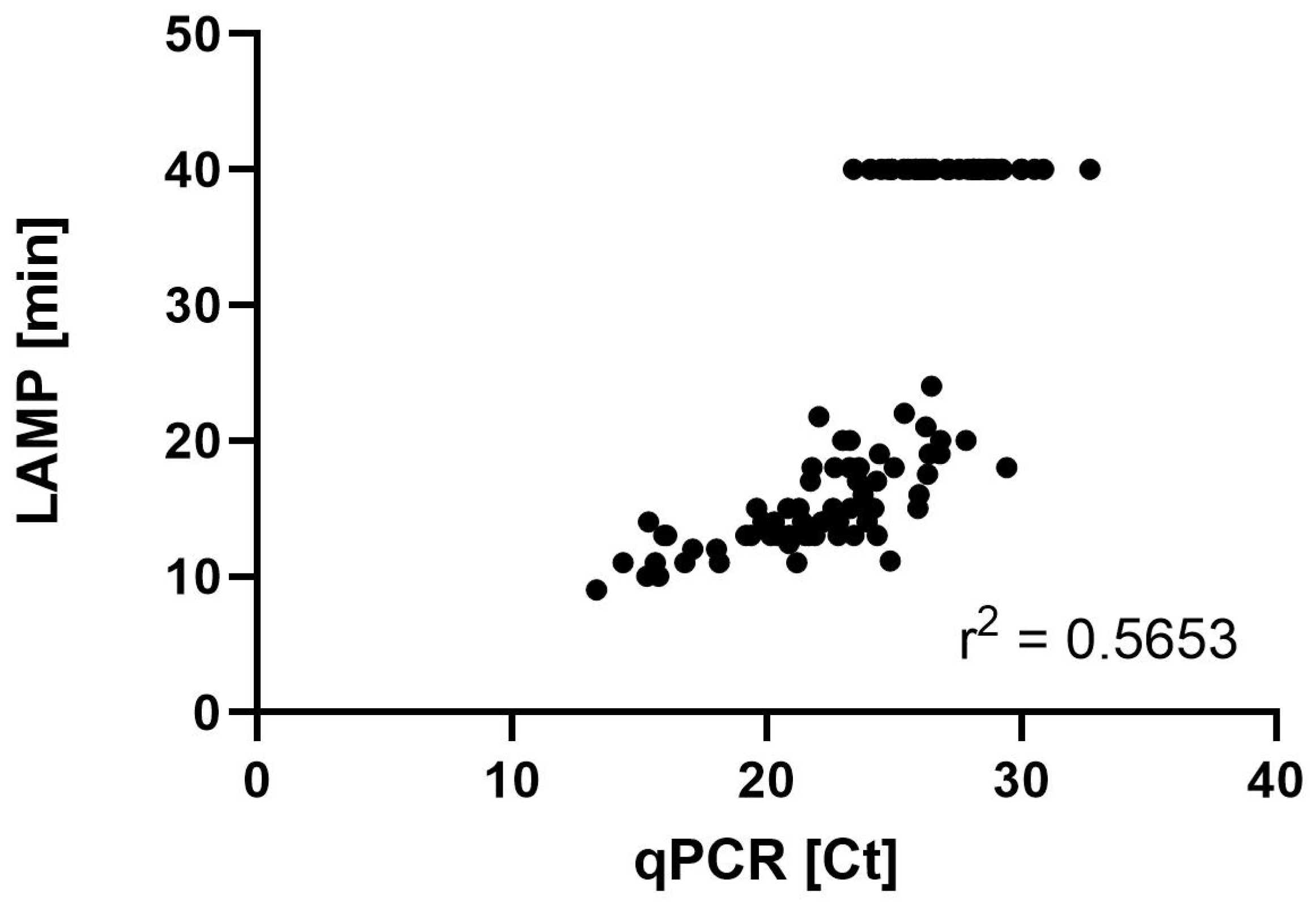
Out of 109 analyzed samples, 6 (5.5%) were negative by qPCR and 38 (34.8%) by the LAMP. In qPCR, positive signals were detected for 103 (94.5%) samples. In contrast, the LAMP resulted in 70 (64.2%) samples with a positive signal, and 1 sample was invalid. Furthermore, 32 samples showed no signal in the LAMP, while qPCR was positive (Table 2). Out of these samples, 6 had a Ct lower than the threshold of 29.29 (Table 2). Consequently, 32 results were found to be false negative, while 1 false positive result was given by the LAMP. The LAMP’s specificity was 86%, the sensitivity was 68%, the PPV was 99%, and the NPV was 16% using Ct 29.29 as the cutoff for the comparison of both methods. Taking the clinical classification into account, all patients with proven PcP resulted in a positive LAMP. The LAMP was negative in 18 of 56 samples, categorized as probable PcP and in 14 of 36 samples classified as possible PcP. Correlation analysis of the samples, plotting Ct against TTP, showed an r2 of 0.5653, which indicated a low linear relationship between the TTP in the LAMP and the Ct obtained by the qPCR (Figure 2).

4. Discussion

The detection of P. jirovecii in respiratory samples is commonly performed via the microscopy of BALs and molecular methods, e.g., qPCR [5,10,11,12,13]. In the diagnostic pathway for the detection of PcP, first a microscopic examination of the respiratory samples is recommended by the guidelines, although this requires highly experienced personnel, irrespective of which staining method is used, which makes the diagnosis difficult to standardize [3,5,8]. The sensitivity of the microscopy methods varies between 26% and 44%, as found by the IFA, which is able to detect cysts and trophic forms [14,15]. For a more rapid and reliable detection of PcP, the LAMP has been established [16,17,18,19,20,21,22]. Since that time, different study groups have evaluated the LAMP [9,16,21,22,23,24]. In contrast to methods such as microscopy and qPCR, the LAMP is easy to handle and requires only a small amount of technical experience in molecular techniques [9,23,24]. At the same time, it offers an advantage to the qPCR regarding the time to the result, the practicability, and the robustness [23]. Here, a commercially available LAMP for PcP detection from respiratory material has been evaluated by testing a total of 109 samples from critically ill patients from five different university hospitals in Germany. The results were compared with a commercial qPCR assay, commonly used in routine diagnostics [23]. By evaluating the LAMP with statistical analysis, the PPV, NPV, sensitivity, and specificity can be calculated. The PPV was 99% and the NPV was 16%, the sensitivity was 68%, and the specificity was 86%, with Ct 29.29 used as a cutoff based on clinical features for the differentiation of colonization and infection with P. jirovecii. A Ct of 29.29 represents a fungal load of approximately 3 × 10³ copies per milliliter of sample. In our previous study, the PPV and NPV were 96% each, and the sensitivity and specificity were 84% and 99%, respectively, while we found a low correlation between the TTP of the LAMP and the fungal load of qPCR and the same a limit of detection (LOD) [9]. The LAMP eazyplex® Pneumocystis jirovecii by AmplexDiagnostics evaluated here targets the cox2 gene (GenBank accession no. MH010440.1), and in the current as well as previous study, we detected an LOD of 4 × 10³ copies/mL sample [9]. Regarding the correlation analysis, the LAMP assay showed a weak correlation between the TTP and the fungal load detected by the qPCR (Pearson correlation r = 0.7519 and r2 = 0.5653).
Huber et al. designed a German multicenter study, evaluating the eazyplex® LAMP, including 49 patients with proven PcP and 126 patients without PcP [23]. For statistical analysis, ROC curve was carried out to evaluate the threshold of the qPCR to distinguish between a PcP and non-infected patients. They found that the sensitivity and specificity of the LAMP were 97.9% and 96.5%, respectively [23]. They set an LOD of 103 copies of the major surface glycoprotein gene per 25 μL of BAL, corresponding to 10 to 20 P. jirovecii cells. This LOD was comparable to the LOD we defined in this study. Huber et al. also found a weak correlation between the TTP of the LAMP and the β-tubulin qPCR assay (Pearson correlation was r = 0.391) as well as the mtLSU rRNA qPCR (r = 0.353) [23]. Uemura et al. compared the performance of another LAMP with a real-time PCR-based method, reporting a diagnostic sensitivity of 87.5% with an LOD of 4 × 10³ copies/mL [16]. Furthermore, they described a decreasing TTP with a rising number of gene copies [16]. The overall diagnostic sensitivity of the LAMP assay using the initially established primer sets was high, and the LOD ranged between 50 and 100 copies/mL [17,18,19,20]. Since then, the LAMP for PcP detection has been modified by designing distinct primer sets. Wang et al. developed a LAMP with a new target site (mitochondrial 16s rRNA) in 2015 [21]. Subsequently, this LAMP assay resulted in an LOD of 10 to 50 copies/mL [21,22]. Varying LODs can probably be explained by different sample numbers and targets used for evaluation, while 103 copies per milliliter of the considered gene seems to be the common LOD. In non-HIV patients, Perret et al. evaluated a cutoff of 5 × 10³ copies/mL to discriminate between a colonization and a PcP [25].
Huber et al. described the eazyplex® LAMP as an appropriate tool to distinguish PcP infection from P. jirovecii-colonized patients [23]. Here, five of six samples turned out to be negative in the LAMP, considering samples with a Ct higher than 29.29. The one sample which was LAMP-positive showed a Ct of 29.44 (4.1 × 10³ copies/mL), just slightly above the cutoff value, and a TTP of 18 min. This patient had a solid tumor (prostate cancer), radiological signs for PcP in the lung, but no immunosuppressive therapy. No antifungal therapy was started after the diagnostic result was known. To investigate the power of the LAMP to distinguish between a colonization and an infection with P. jirovecii, a larger cohort of colonized patients is required. Because of changing fungal loads, depending on the underlying disease of the immunosuppression, there should be a big gray area in which no certain statement can be made. Analyzing the negative LAMP results, 39 (36%) samples were negative, although qPCR was positive. The Ct ranged between 23.41 and 32.7. In 34 samples, a Ct lower than 29.29 was observed. Altogether, 17 patients were categorized to have probable PcP, 14 to have possible PcP, and 7 were not classifiable. Nonetheless, clinical and radiological parameters have to be considered for reliably diagnosing PcP if the laboratory relies solely on the LAMP to detect P. jirovecii. Furthermore, β-1,3-D-glucan, as a fungal biomarker, should be included in the assessment [8]. Regarding only positive results, 69 positive samples from 62 patients were investigated, of whom 5 patients were categorized to have proven PcP, 34 probable, and 18 possible PcP, whereas 4 were not classifiable and 1 had no expected PcP. Regarding the patients’ clinical conditions together with a positive LAMP result, the basis for PcP therapy was given [16]. Taking the classification of patients as a foundation to interpret positive or negative results, all patients with proven PcP (n = 5; 100%) and 66% (37/56) of patients with probable PcP tested positive in the LAMP.

Limitation

The limitations of our study are, among others, the storage conditions of the samples. Only the samples from Essen were analyzed both on arrival at the laboratory and after thawing. In this case, we showed the concordance of the qPCR and LAMP results. The very low NPV and the relatively low sensitivity observed might result from the analytical premises. Only samples from patients in whom PcP was investigated were included. Highly differing numbers of samples were sent from five different hospitals, so as a result, we observed a broadly distributed patient cohort, which might explain our findings. Samples from healthy individuals were not examined over the field. We defined a threshold based on clinical features to differentiate between infection and colonization due to P. jirovecii using an ROC analysis to set a cutoff in the qPCR. The results were not verified using microscopic examination. The variable nature of the specimen was not considered in detail and might result in unintended consequences. Samples from Erlangen could only be examined by qPCR in the Essen laboratory because only DNA already extracted from the original samples was sent to Essen.

5. Conclusions

The eazyplex® LAMP is a useful method for the initial screening of patients suspected of PcP but not for excluding a diagnosis of PcP. The assay’s high detection limit means that most positive patients would not be colonized but would have true PcP.

Author Contributions

Conceptualization, U.S., L.K., J.S. and P.-M.R.; formal analysis, U.S. and L.K.; investigation, U.S. and L.K.; data curation, U.S., L.K., J.B., F.S., A.S., S.R., J.H., T.S., J.S. and P.-M.R.; writing—original draft preparation, U.S.; writing—review and editing, U.S., L.K., J.B., F.S., A.S., S.R., J.H., T.S., J.S. and P.-M.R.; supervision, J.S. and P.-M.R. All authors have read and agreed to the published version of the manuscript.

Funding

This research received no external funding.

Institutional Review Board Statement

This study was conducted in accordance with the Declaration of Helsinki and approved by the Institutional Ethics Committee of the Faculty of Medicine Essen, University of Duisburg-Essen, Essen, Germany (protocol code 20-9399-BO, date of approval 6 August 2020).

Informed Consent Statement

Informed consent for participation is not required, as per local legislation. The patient data were evaluated completely anonymously so that no conclusions can be drawn about the patients.

Data Availability Statement

The original contributions presented in this study are included in the article. Further inquiries can be directed to the corresponding author.

Acknowledgments

We thank Andrea Hain and Silke Dittmer for preparing the laboratory work. We acknowledge support from the Open Access Publication Fund of the University of Duisburg-Essen.

Conflicts of Interest

The authors declare no conflicts of interest.

Abbreviations

The following abbreviations are used in this manuscript:
LAMPloop-mediated isothermal amplification assay
P. jiroveciiPneumocystis jirovecii
PcPPneumocystis pneumonia
NPVnegative predictive value
PPVpositive predictive value
r2squared Pearson correlation coefficient
IFAimmunofluorescence assays
BALbronchoalveolar lavage
qPCRquantitative real-time PCR
cox2mitochondrial gene cytochrome c oxidase subunit 2
mtLSUmulticopy mitochondrial large-subunit rRNA (mtLSU) gene
EORTC/MSGERCEuropean Organization for Research and Treatment of Cancer and the Mycoses Study Group Education and Research Consortium
BSbronchial secretions
Ctcycle threshold
TTPtime to positivity
minminutes
ROCreceiver operating characteristic
LODlimit of detection
FFreiburg
MMunster
NNuremberg
ERErlangen
EEssen
SOsolid tumor
HIVHuman Immunodeficiency Virus
ADautoimmune disease
STsolid organ transplantation
HMhematological malignancy
OOthers

References

  1. Fishman, J.A. Pneumocystis jiroveci. Semin. Respir. Crit. Care Med. 2020, 41, 141–157. [Google Scholar] [CrossRef]
  2. Hitzenbichler, F.; Mohr, A.; Salzberger, B. Pneumocystis jirovecii pneumonia-an opportunistic infection undergoing change. Internist 2019, 60, 669–677. [Google Scholar] [CrossRef] [PubMed]
  3. Donnelly, J.P.; Chen, S.C.; Kauffman, C.A.; Steinbach, W.J.; Baddley, J.W.; Verweij, P.E.; Clancy, C.J.; Wingard, J.R.; Lockhart, S.R.; Groll, A.H.; et al. Revision and Update of the Consensus Definitions of Invasive Fungal Disease From the European Organization for Research and Treatment of Cancer and the Mycoses Study Group Education and Research Consortium. Clin. Infect. Dis. 2020, 71, 1367–1376. [Google Scholar] [CrossRef]
  4. Maschmeyer, G.; Helweg-Larsen, J.; Pagano, L.; Robin, C.; Cordonnier, C.; Schellongowski, P. ECIL guidelines for treatment of Pneumocystis jirovecii pneumonia in non-HIV-infected haematology patients. J. Antimicrob. Chemother. 2016, 71, 2405–2413. [Google Scholar] [CrossRef] [PubMed]
  5. Alanio, A.; Hauser, P.M.; Lagrou, K.; Melchers, W.J.; Helweg-Larsen, J.; Matos, O.; Cesaro, S.; Maschmeyer, G.; Einsele, H.; Donnelly, J.P.; et al. ECIL guidelines for the diagnosis of Pneumocystis jirovecii pneumonia in patients with haematological malignancies and stem cell transplant recipients. J. Antimicrob. Chemother. 2016, 71, 2386–2396. [Google Scholar] [CrossRef] [PubMed]
  6. Jaramillo Cartagena, A.; Asowata, O.E.; Ng, D.; Babady, N.E. An overview of the laboratory diagnosis of Pneumocystis jirovecii pneumonia. J. Clin. Microbiol. 2025, 63, e0036124. [Google Scholar] [CrossRef]
  7. Notomi, T.; Okayama, H.; Masubuchi, H.; Yonekawa, T.; Watanabe, K.; Amino, N.; Hase, T. Loop-mediated isothermal amplification of DNA. Nucleic Acids Res. 2000, 28, E63. [Google Scholar] [CrossRef]
  8. Lagrou, K.; Chen, S.; Masur, H.; Viscoli, C.; Decker, C.F.; Pagano, L.; Groll, A.H. Pneumocystis jirovecii Disease: Basis for the Revised EORTC/MSGERC Invasive Fungal Disease Definitions in Individuals Without Human Immunodeficiency Virus. Clin. Infect. Dis. 2021, 72, S114–S120. [Google Scholar] [CrossRef]
  9. Scharmann, U.; Kirchhoff, L.; Schmidt, D.; Buer, J.; Steinmann, J.; Rath, P.M. Evaluation of a commercial Loop-mediated Isothermal Amplification (LAMP) assay for rapid detection of Pneumocystis jirovecii. Mycoses 2020, 63, 1107–1114. [Google Scholar] [CrossRef]
  10. Wakefield, A.E.; Pixley, F.J.; Banerji, S.; Sinclair, K.; Miller, R.F.; Moxon, E.R.; Hopkin, J.M. Detection of Pneumocystis carinii with DNA amplification. Lancet 1990, 336, 451–453. [Google Scholar] [CrossRef]
  11. Azoulay, E.; Bergeron, A.; Chevret, S.; Bele, N.; Schlemmer, B.; Menotti, J. Polymerase chain reaction for diagnosing pneumocystis pneumonia in non-HIV immunocompromised patients with pulmonary infiltrates. Chest 2009, 135, 655–661. [Google Scholar] [CrossRef] [PubMed]
  12. Oren, I.; Hardak, E.; Finkelstein, R.; Yigla, M.; Sprecher, H. Polymerase chain reaction-based detection of Pneumocystis jirovecii in bronchoalveolar lavage fluid for the diagnosis of Pneumocystis pneumonia. Am. J. Med. Sci. 2011, 342, 182–185. [Google Scholar] [CrossRef]
  13. Thomas, C.F., Jr.; Limper, A.H. Pneumocystis pneumonia. N. Engl. J. Med. 2004, 350, 2487–2498. [Google Scholar] [CrossRef] [PubMed]
  14. Goterris, L.; Mancebo Fernandez, M.A.; Aguilar-Company, J.; Falco, V.; Ruiz-Camps, I.; Martin-Gomez, M.T. Molecular Diagnosis of Pneumocystis jirovecii Pneumonia by Use of Oral Wash Samples in Immunocompromised Patients: Usefulness and Importance of the DNA Target. J. Clin. Microbiol. 2019, 57, e01287-19. [Google Scholar] [CrossRef] [PubMed]
  15. Veintimilla, C.; Alvarez-Uria, A.; Martin-Rabadan, P.; Valerio, M.; Machado, M.; Padilla, B.; Alonso, R.; Diez, C.; Munoz, P.; Marin, M. Pneumocystis jirovecii Pneumonia Diagnostic Approach: Real-Life Experience in a Tertiary Centre. J. Fungi 2023, 9, 414. [Google Scholar] [CrossRef]
  16. Uemura, N.; Makimura, K.; Onozaki, M.; Otsuka, Y.; Shibuya, Y.; Yazaki, H.; Kikuchi, Y.; Abe, S.; Kudoh, S. Development of a loop-mediated isothermal amplification method for diagnosing Pneumocystis pneumonia. J. Med. Microbiol. 2008, 57, 50–57. [Google Scholar] [CrossRef]
  17. Singh, P.; Singh, S.; Mirdha, B.R.; Guleria, R.; Agarwal, S.K.; Mohan, A. Evaluation of Loop-Mediated Isothermal Amplification Assay for the Detection of Pneumocystis jirovecii in Immunocompromised Patients. Mol. Biol. Int. 2015, 2015, 819091. [Google Scholar] [CrossRef]
  18. Xu, L.; Kong, J. A multiplexed nucleic acid microsystem for point-of-care detection of HIV co-infection with MTB and PCP. Talanta 2013, 117, 532–535. [Google Scholar] [CrossRef]
  19. Nakashima, K.; Aoshima, M.; Ohkuni, Y.; Hoshino, E.; Hashimoto, K.; Otsuka, Y. Loop-mediated isothermal amplification method for diagnosing Pneumocystis pneumonia in HIV-uninfected immunocompromised patients with pulmonary infiltrates. J. Infect. Chemother. 2014, 20, 757–761. [Google Scholar] [CrossRef]
  20. Kawano, S.; Maeda, T.; Suzuki, T.; Abe, T.; Mikita, K.; Hamakawa, Y.; Ono, T.; Sonehara, W.; Miyahira, Y.; Kawana, A. Loop-mediated isothermal amplification with the Procedure for Ultra Rapid Extraction kit for the diagnosis of pneumocystis pneumonia. J. Infect. Chemother. 2015, 21, 224–226. [Google Scholar] [CrossRef]
  21. Wang, D.D.; Zheng, M.Q.; Zhang, N.; An, C.L. Investigation of Pneumocystis jirovecii colonization in patients with chronic pulmonary diseases in the People’s Republic of China. Int. J. Chronic Obstr. Pulm. Dis. 2015, 10, 2079–2085. [Google Scholar] [CrossRef]
  22. Xue, T.; Ma, Z.; Liu, F.; Du, W.; He, L.; Wang, J.; An, C. Pneumocystis jirovecii colonization and its association with pulmonary diseases: A multicenter study based on a modified loop-mediated isothermal amplification assay. BMC Pulm. Med. 2020, 20, 70. [Google Scholar] [CrossRef] [PubMed]
  23. Huber, T.; Serr, A.; Geissdorfer, W.; Hess, C.; Lynker-Assmus, C.; von Loewenich, F.D.; Bogdan, C.; Held, J. Evaluation of the Amplex eazyplex Loop-Mediated Isothermal Amplification Assay for Rapid Diagnosis of Pneumocystis jirovecii Pneumonia. J. Clin. Microbiol. 2020, 58, e01739-20. [Google Scholar] [CrossRef] [PubMed]
  24. Ng, W.W.Y.; Ho, Y.I.I.; Wong, A.H.; Leung, E.C.M.; Lee, A.L.H.; Chow, V.C.Y. Comparison of PneumID real-time PCR assay with Amplex eazyplex LAMP assay for laboratory diagnosis of Pneumocystis jirovecii Pneumonia. Med. Mycol. 2022, 60, myac043. [Google Scholar] [CrossRef]
  25. Perret, T.; Kritikos, A.; Hauser, P.M.; Guiver, M.; Coste, A.T.; Jaton, K.; Lamoth, F. Ability of quantitative PCR to discriminate Pneumocystis jirovecii pneumonia from colonization. J. Med. Microbiol. 2020, 69, 705–711. [Google Scholar] [CrossRef]
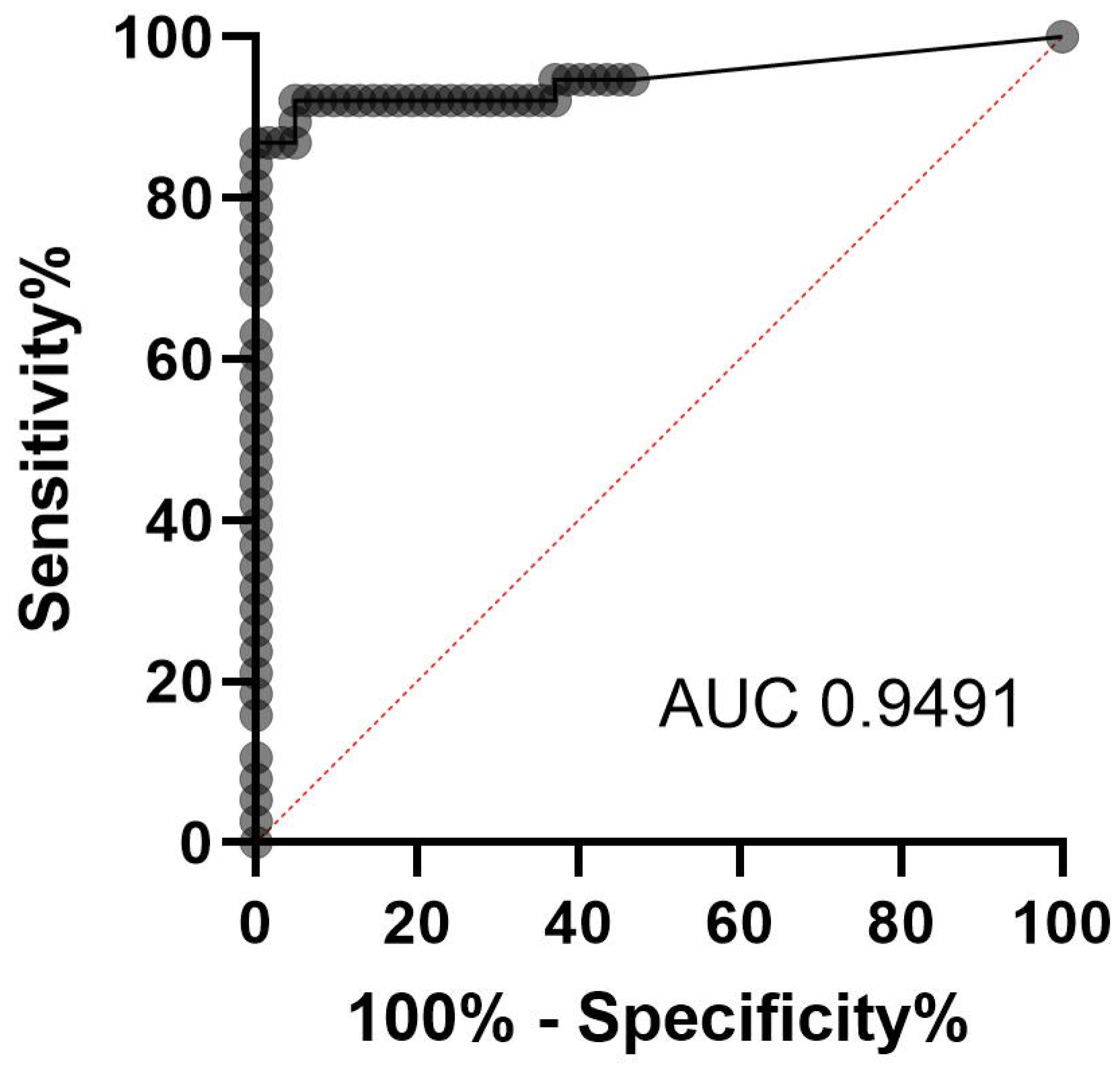
Figure 1. ROC analysis revealed an AUC of 0.9491 and a Ct higher than 29.29 to differentiate between a colonization and an infection with P. jirovecii.
Jof 11 00300 g001
Figure 2. Correlation between qPCR and LAMP. Defining negative LAMP results in a TTP of 40 min in accordance with a negative Ct in qPCR; Pearson’s correlation r = 0.7519 (0.6561 to 0.8238; 95% confidence interval) revealed r2 = 0.5653.
Jof 11 00300 g002
Table 1. Characteristics of patients.
ParameterResults
No. of patients95
Age (mean ± SD) (year)59 ± 16.5
Sex (%)
   Male62 (65.3)
   Female33 (34.7)
Underlying disease (no. of patients (%))
   HIV/AIDS25 (26.3)
   Solid tumor20 (21.1)
   Hematologic malignancy20 (21.1)
   Organ transplant16 (16.8)
   Kidney11 (11.6)
   Liver3 (3.2)
   Heart1 (1.1)
   Lung1 (1.1)
   Immunological disorder7 (7.4)
   Other7 (7.4)
Table 2. Categorization of patient cohort and results in qPCR and LAMP.
Patient IDDiagnosisCategorization EORTCqPCR
(Ct)
Fungal Load
(Copies/mL)
LAMP
(TTP in min)
F-1SOprobable16.812.60 × 10711.87
F-2SOproven18.041.00 × 10712.37
F-3HIVpossible20.891.10 × 10613.30
F-4SOprobable19.623.00 × 10615.32
F-5HIVproven21.735.90 × 10517.68
F-6ADproven22.234.00 × 10514.15
F-7ADprobable23.271.80 × 10520.75
F-8STprobable22.982.20 × 10520.48
F-9HMprobable28.164.20 × 103negative
F-10HIVpossible27.089.50 × 103negative
F-11SOproven26.391.60 × 10419.98
F-12ADpossible32.701.70 × 102negative
M-13SOprobable19.852.50 × 10614.85
M-14STprobable21.905.20 × 10513.93
M-15HIVpossible19.184.20 × 10613.78
M-16STprobable27.925.20 × 103negative
M-17SOprobable24.787.40 × 104negative
N-18Opossible23.312.00 × 10515.33
N-19Opossible21.796.30 × 10518.17
N-20HIVpossible19.393.80 × 10613.65
N-21STprobable24.935.90 × 104negative
N-22SOprobable22.164.70 × 10514.05
N-23HIVpossible15.775.80 × 10710.47
F-24HIVproven15.318.20 × 10710.32
F-25HIVpossible13.333.80 × 1089.90
ER-26STprobable22.612.70 × 10515.75
ER-27STprobable25.862.30 × 104negative
ER-27a 25.872.30 × 104negative
ER-28HMpossible24.516.40 × 104negative
ER-29STprobable28.124.20 × 103negative
ER-30STprobable25.572.90 × 104negative
ER-31STprobable26.811.10 × 10419.75
ER-32STprobable23.451.40 × 10513.75
ER-32a 23.959.80 × 10414.00
ER-32b 28.094.30 × 103negative
ER-32c 22.802.30 × 10513.75
ER-33SOprobable21.526.10 × 10513.00
ER-34STnot classifiable30.885.00 × 102negative
ER-35STnot classifiable22.682.60 × 10518.00
ER-36HMnot classifiable24.079.00 × 104negative
ER-37HMprobable23.781.10 × 10516.25
ER-38Oprobable21.665.50 × 10513.78
ER-38a 27.566.50 × 103negative
ER-38b 24.924.70 × 104negative
ER-39SOnot classifiable29.221.80 × 103negative
ER-40HMpossible26.481.50 × 10424.75
ER-41ADnot classifiable28.852.50 × 103negative
ER-42HMpossible17.091.70 × 10712.67
ER-42a 23.261.90 × 10518.73
ER-42b 26.551.50 × 104negative
ER-43ADnot classifiable26.381.70 × 104negative
ER-44Oprobable15.368.70 × 10714.52
ER-45STnot classifiable23.631.40 × 10518.12
ER-46HMnot classifiable26.251.90 × 10426.00
ER-47Oprobable30.001.00 × 103negative
ER-48STprobable25.383.70 × 104negative
ER-48a 28.333.70 × 103negative
ER-49HMnot classifiable28.672.80 × 103negative
ER-50ADpossible15.637.10 × 10711.18
ER-50a 16.085.00 × 10713.20
ER-51Ono PCP expected24.358.10 × 10413.37
ER-52HIVnot classifiable14.361.90 × 10811.22
ER-53STprobable26.251.90 × 10421.58
E-54HIVpossible28.143.10 × 103negative
E-55SOprobable23.571.10 × 10517.00
E-56HIVpossible27.146.90 × 103negative
E-57HIVpossible20.611.20 × 10613.00
E-57a 22.063.80 × 105522.00
E-58HIVpossible21.286.70 × 10515.00
E-59SOprobable24.864.40 × 10411.25
E-60SOprobable20.879.70 × 10512.75
E-61SOprobable21.416.20 × 10514.00
E-62SOprobable24.207.10 × 10415.00
E-63Opossible26.331.60 × 10417.00
E-64HMprobable28.632.40 × 103negative
E-65SOprobable24.446.60 × 10419.00
E-66HMprobable21.715.30 × 10513.00
E-67HIVprobable25.423.10 × 10422.25
E-68HIVprobable23.431.50 × 10513.25
E-69STprobable25.982.00 × 10416.50
E-70HIVpossible15.971.17 × 10813.25
E-70a 23.411.40 × 105negative
E-71HMpossible28.396.50 × 103negative
E-72HMprobable27.161.70 × 104negative
E-73HIVpossible29.263.30 × 103negative
E-74HIVpossible20.293.90 × 10614.75
E-75HIVpossible23.823.40 × 10515.25
E-76HIVpossible25.956.30 × 10415.38
E-77STprobable29.444.10 × 10318.03
E-77a 23.305.00 × 10515.70
E-78HMprobable26.066.60 × 104negative
E-79HMprobable21.207.50 × 10511.57
E-80HMno classifiable27.844.80 × 10320.10
E-81HMprobable30.022.60 × 103negative
E-82STpossible30.521.80 × 103negative
E-83HIVpossible20.163.90 × 10613.02
E-84HIVprobable25.008.80 × 10418.02
E-84aHIV 18.141.90 × 10711.70
E-85HIVprobable20.842.30 × 10615.43
E-86HIVprobable20.464.70 × 10613.70
E-87ADprobable26.843.50 × 10420.97
E-88STpossible28.761.70 × 103negative
E-89SOprobable25.242.70 × 104invalid
E-90HMprobable26.141.70 × 104negative
E-91HMprobable26.501.60 × 104negative
E-92STprobable22.846.90 × 10514.78
E-93HMprobable28.942.30 × 103negative
E-94HMprobable24.328.30 × 10417.42
E-95HIVprobable16.782.60 × 10711.02
Abbreviations F: Freiburg; M: Munster; N: Nuremberg; ER: Erlangen; E: Essen; SO: solid tumor; HIV: Human Immunodeficiency Virus; AD: autoimmune disease; ST: solid organ transplantation; HM: hematological malignancy; O: other; Ct: cycle threshold; TTP: time to positivity; not classifiable due to lack of clinical data.
Table 3. LAMP results with different storage conditions.
Storage ConditionsSample 1
TTP [min]
Sample 2
TTP [min]
Sample 3
TTP [min]
Initial11:3417:2513:19
after two years of storage in −20 °C14:0416:3715:29
24 h storage in 21 °C14:5320:0613:39
48 h storage in 21 °C14:5420:1612:19
72 h storage in 21 °C14:0418:1912:56
Disclaimer/Publisher’s Note: The statements, opinions and data contained in all publications are solely those of the individual author(s) and contributor(s) and not of MDPI and/or the editor(s). MDPI and/or the editor(s) disclaim responsibility for any injury to people or property resulting from any ideas, methods, instructions or products referred to in the content.