Article

Diversity of Alternaria Section Nimbya in Iran, with the Description of Eight New Species

by
Abdollah Ahmadpour
1,*,
Youbert Ghosta
2,*,
Zahra Alavi
2,
Fatemeh Alavi
2,
Alireza Poursafar
3 and
Pabulo Henrique Rampelotto
4
1
Higher Education Center of Shahid Bakeri, Urmia University, Miyandoab 59781-59111, Iran
2
Department of Plant Protection, Faculty of Agriculture, Urmia University, Urmia 57561-51818, Iran
3
Department of Plant Pathology, North Dakota State University, Fargo, ND 58102, USA
4
Bioinformatics and Biostatistics Core Facility, Institute of Basic Health Sciences, Federal University of Rio Grande do Sul, Porto Alegre 91501-970, Brazil
*
Authors to whom correspondence should be addressed.
J. Fungi 2025, 11(3), 225; https://doi.org/10.3390/jof11030225
Submission received: 27 February 2025 / Revised: 10 March 2025 / Accepted: 12 March 2025 / Published: 15 March 2025
(This article belongs to the Collection Fungal Biodiversity and Ecology)

Abstract

:
Alternaria includes endophytes, saprophytes, and pathogens affecting both plants and animals, with a global distribution across various hosts and substrates. It is categorized into 29 sections, each defined by a type species and six monophyletic lineages. The Alternaria section Nimbya comprises 10 species primarily associated with the families Juncaceae and Cyperaceae, functioning as either saprophytes or plant pathogens. In this study, 189 fungal strains were collected from multiple locations across six provinces in Iran. The isolates were initially classified based on morphological characteristics and ISSR-PCR molecular marker banding patterns. Multi-gene phylogenetic analyses of 38 selected strains, using ITS–rDNA, GAPDH, TEF1, RPB2, and Alt a 1 gene sequences, combined with morphological data, led to the identification of 13 species, including eight new species, namely Alternaria caricifolia, A. cyperi, A. juncigena, A. junci-inflexi, A. persica, A. schoenoplecti, A. salkadehensis, and A. urmiana. In addition, this work identified new host associations (matrix nova) for three previously known species: A. caricicola on Cyperus sp., A. cypericola on Eleocharis sp., and A. junci-acuti on Carex sp. The study provides detailed morphological descriptions and illustrations of all identified species, discusses their habitats, distribution, and phylogenetic relationships within section Nimbya, and presents a key for species identification within this section in Iran. Furthermore, these findings highlight the significance of studying fungal biodiversity in Iran and contribute to a better understanding of species distribution and host range within the Alternaria section Nimbya.

1. Introduction

The genus Nimbya E.G. Simmons was introduced with N. scirpicola (Fuckel) E.G. Simmons as the type species to accommodate fungi with a morphology similar to the genera Alternaria Nees, Sporidesmium Link, and Drechslera S. Ito. However, the most distinct and useful diagnostic character for differentiating Nimbya from these genera is the internal compartmentation of conidia into well-defined, usually angular lumina, surrounded by an abundant distoseptum matrix [1]. The conidial lumina are interconnected by narrow channels through the transverse distosepta, with transverse eusepta inserted at maturity. Sexual morphs of Nimbya spp. were classified under the genus Macrospora Fuckel [1,2]. After the initial description of five Nimbya species, the number of described species, or those transferred to Nimbya from other genera based on morphological characteristics, gradually increased. By 2005, the number of Nimbya had reached 17 species [3,4,5,6,7,8,9]. Various species of Nimbya have been recorded on plants from the families Amaranthaceae, Asteraceae, Caryophyllaceae, Cyperaceae, Euphorbiaceae, Fabaceae, Juncaceae, and Solanaceae [1,9]. Based on conidium morphology, a close relationship between Nimbya and Alternaria was proposed [2,4].
With the advent of genome sequencing and phylogenetic analyses, these methods became valuable tools for identifying species boundaries and phylogenetic relationships. In the first phylogenetic study of Nimbya, the relationships among N. caricis and N. scirpicola, along with species of Embellisa, Alternaria, Ulocladium, and Stemphylium, were examined using the internal transcribed spacer region of rDNA (ITS–rDNA), the mitochondrial small-subunit (mt SSU), and glyceraldehydes-3-phosphate dehydrogenase (GAPDH) gene sequences [10]. The results revealed that the studied species in Alternaria, Embellisia, Nimbya, and Ulocladium formed a large monophyletic clade, with Stemphylium as a sister clade. Moreover, Ulocladium and Embellisia were found to be polyphyletic, suggesting that using only morphological characteristics to differentiate these genera could be misleading, as the key characters used are homoplastic [10]. Similar results from GAPDH and Alternaria major allergen (Alt a 1) genes sequences, also indicated close relationships between Nimbya spp., Alternaria spp. in the infectoria species group, and some Embellisia spp. [11]. A more comprehensive phylogenetic study using a larger set of isolates examined the relationships of Nimbya and Embellisia with other closely related genera, based on ITS–rDNA, Alt a 1, and GAPDH sequences [12]. The study revealed the polyphyletic nature of both Embellisia and Nimbya, dividing species within each genus into four and two clades, respectively. Nimbya group I included four species (N. caricis, N. scirpicola, N. scirpinfestans, and N. scirpivora), while group II contained three species (N. alternantherae, N. celosiae, and N. perpunctulata). Morphological and ecological comparisons among these species indicated that group I species had short-beaked conidia and were associated with the Juncaceae and Cyperaceae plant families, while the group II species, with long-beaked conidia, originated from plants in the family Amarantaceae, suggesting independent evolutionary origins. Three species from group II were transferred to Alternaria, and a new species group, Alternanthera species group, was established to accommodate them [12]. This species group concept was an early attempt at a classification scheme for identifying Alternaria species based on two morphological characteristics: three-dimensional sporulation pattern and conidium morphology, with each species group typified by a representative species [4,13]. In a subsequent study using five phylogenetically informative loci—GAPDH, Alt a 1, Actin (ACT), Plasma membrane ATPase (ATPase), and Calmodulin (CAL)—eight well-supported asexual lineages of Alternaria were identified and grouped into species groups (both previously known and newly identified) [14]. These eight species groups were elevated to the taxonomic rank of “section”, a rank that had previously been used by Neergaard [15] and Joly [16] to classify Alternaria species.
In a landmark study aimed at delineating the phylogenetic lineages and establishing a robust taxonomy within Alternaria and allied genera, six genomic regions (18S nrDNA (SSU), 28SnrDNA (LSU), ITS–rDNA, GAPDH, RPB2, and TEF1) were sequenced [17]. This study recognized 24 internal clades and six monotypic lineages under the Alternaria clade. The 24 clades were called sections (each typified by a type species), thirteen generic names, Allewia, Brachycladium, Chalastospora, Chmelia, Crivellia, Embellisia, Lewia, Nimbya, Sinomyces, Tretispora, Ulocladium, Undifilum, and Ybotromyces were put as synonymy with Alternaria and generic circumscription of Alternaria was amended. In addition, species previously classified as Nimbya proper (Nimbya group I) [12], were reclassified under Alternaria and placed in section Nimbya. A re-examination of morphological characteristics of species formerly identified as Nimbya spp., without phylogenetic analysis, to determine their affiliation with any Alternaria section, resulted in the inclusion of A. heteroschemos and A. juncicola in the Nimbya section [18]. More recently, four new species—A. caricicola, A. cypericola, A. heyranica, and A. junci-acuti—were described in the Nimbya section, based on combined morphological features and multi-gene phylogeny, and the number of species in this section was raised to 10 [19,20].
The plant order Poales is one of the most diverse and ecologically significant groups of angiosperms, encompassing 14 families and over 24,300 species. Members of the Poales families are found worldwide and are notable for their species richness, ecological adaptations, and diverse life forms. They have significantly influenced the evolution of mammals, including hominids, and form the foundation of much of the human diet [21,22]. Among these, the Cyperaceae family, commonly known as sedges, ranks as one of the three largest monocot families and one of the 10 largest plant families overall, with 5687 distributed globally [23]. Cyperaceae plants play crucial roles in various habitats, from wetlands to deserts, dominating wetland vegetation, contributing to nutrient cycling, and providing habitats for numerous species. Additionally, members of this family are used for medicinal purposes, to treat multiple diseases and ailments, as important sources of essential oils, and as a source of food and animal feed [24,25]. However, some Cyperaceae species are invasive weeds, posing threats to the natural ecosystems and agricultural and forest productivity [26]. The family Juncaceae, also known as the rush family, comprises around 500 species of monocotyledonous flowering plants. These plants are integral to aquatic and wetland ecosystems, offering habitats for fauna, helping slow water flow, preventing soil erosion, and improving water quality. Historically, certain Juncaceae species have been used for crafting materials such as baskets and chair bottoms, as well as in traditional herbal medicines due to their bioactive compounds, including phenanthrenes and phenanthrenes-like molecules [27,28].
Both Cyperaceae and Juncaceae plants serve as hosts for a wide variety of fungi, including members of the Alternaria section Nimbya. Some of these fungi are parasitic, causing substantial damage to their host plants, while others function as saprophytes, aiding in decomposition and nutrient cycling. Previous studies on Alternaria species associated with these plant families have already shown a significant level of species diversity [19,20]. In this study, a larger set of fungal isolates were collected from various regions across six provinces in Iran. Through a combination of morphological examination and multi-gene phylogenetic analysis, thirteen species were identified, eight of which are new to science. This paper provides detailed descriptions and illustrations of these species, as well as an analysis of their phylogenetic and host relationships within the section.

2. Materials and Methods

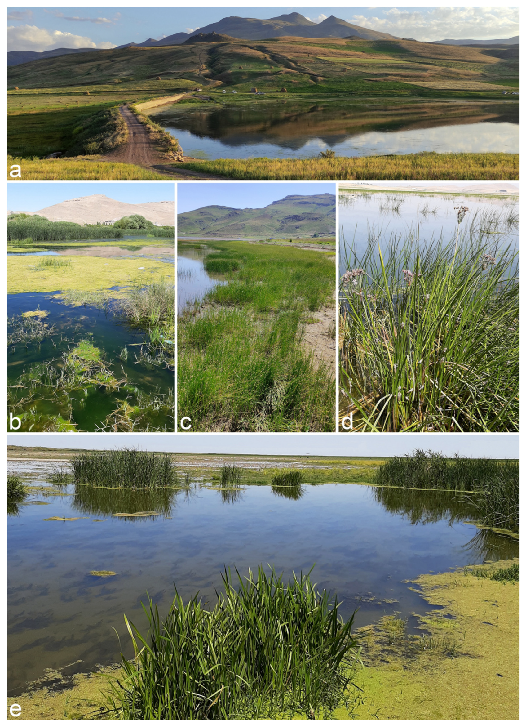
2.1. Fungal Isolates

Plant samples, including leaves and culms from the families Cyperaceae and Juncaceae, exhibiting discolored lesions and blight symptoms, were collected from various wetland areas across six provinces in Iran (Ardebil, East Azarbaijan, Golestan, Guilan, Mazandaran, and West Azarbaijan) during 2019–2021 (Figure 1). The samples were properly labeled, kept cold, and transported to the laboratory. Fungal isolation and purification were followed using the method described by Ahmadpour et al. [20]. Fungal isolates were kept in potato carrot agar (PCA, 20 g potato, 20 g carrot, 20 g agar, and 1000 mL of distilled water) slants at 4 °C for short-term preservation and on sterile filter paper segments at −20 °C for long-term preservation. Pure cultures of all identified isolates were deposited in the fungal culture collections of the Iranian Research Institute of Plant Protection (IRAN) and Urmia University (FCCUU).

2.2. Morphological Observations

Morphological observations were made from the fungal strains growing on potato carrot agar (PCA) medium, incubated at 23–25 °C under Cool White fluorescent light with 8/16 h light/dark cycle for 5–7 days without humidity control [1,2]. Strains showing reduced sporulation under these conditions were transferred to synthetic nutrient-poor agar medium (SNA) [29] containing a small piece of autoclaved filter paper to promote sporulation [17]. Slide mounts were prepared using lactophenol or lactophenol cotton blue, and micro-morphological features of hyphae, as well as asexual and sexual structures (when induced) were examined using an Olympus AX70 compound microscope (Olympus Optical CO., LTD, Tokyo, Japan). Measurements were taken for 30–50 structures, including hyphae, conidiophores, conidia, ascomata, asci, and ascospores, and microphotographs were captured from the slide mounts. Colony morphology was also characterized from cultures grown on Potato Dextrose Agar (PDA, Merck, Darmstadt, Germany), PCA, and V-8A (175 mL of commercial V8 vegetable juice, 3 g CaCO3, 20 g Agar, and 1000 mL of distilled water) media after incubation in the dark at 25 °C for 7 days. Colony color was determined using Rayner’s color charts [30]. To induce ascomata formation, 2% water agar plates containing a 6 cm autoclaved host plant culm segment were inoculated and incubated at three temperatures (15, 18, and 23 °C) for 30–60 days [8]. Taxonomic novelties were deposited in MycoBank [31].

2.3. DNA Extraction and PCR Amplification

DNA was extracted from the mycelial mass of each isolate, which was obtained from 10-day-old PDA plates, using a standard sodium dodecyl sulfate (SDS) lysis buffer. The extraction process involved chloroform extraction followed by isopropanol precipitation [19]. Polymorphic banding patterns were generated for all isolates using inter simple sequence repeat (ISSR)-PCR with the ISSR5 ((GA)5YC) primer for preliminary screening. Each PCR reaction contained 0.4 μM of the primer, 4 μL ready master mix (Taq DNA polymerase 2X Master Mix Red, 2 mM MgCl2, Ampliqon Company, Odense, Denmark), and approximately 10 ng of template DNA in a total volume of 10 μL. The thermal cycling conditions included an initial denaturation step at 95 °C for 5 min, followed by 35 cycles of 95 °C for 45 s, 41 °C for 60 s, and 72 °C for 90 s, with a final extension step at 72 °C for 10 min. Amplicons were visualized on a 1% agarose gel. Isolates with identical banding patterns were classified as closely related taxa [20]. To analyze phylogenetic relationships, 38 isolates were chosen based on ISSR banding pattern and morphological characteristics for multi-gene sequencing. Target regions included the internal transcribed spacer (ITS–rDNA), parts of the glyceraldehyde-3-phosphate dehydrogenase (GAPDH), RNA polymerase II largest subunit (RPB2), translation elongation factor 1-alpha (TEF1) and Alternaria major allergen (Alt a 1) genes, using primers listed in Table 1. PCR conditions and reaction mixtures were consistent with Ahmadpour et al. [20]. The resulting amplicons were visualized on a 1.5% agarose gel stained with GelRedTM (Biotium, Hayward, CA, USA) and viewed under UV light. Amplicon sizes were determined using a HyperLadderTM I molecular marker (Bioline, Memphis, TN, USA). The amplified products were then purified and sequenced using the same primer sets by Macrogen Corp. (Seoul, Republic of Korea).

2.4. Sequence Alignment and Phylogenetic Analyses

DNA sequences generated for the new strains were examined and trimmed using MEGA 6.0 [38], and then exported as FASTA files for subsequent analyses. Published and verified DNA sequences of various loci for the type or representative Alternaria strains were obtained from the GenBank database and incorporated into the phylogenetic analyses (Table 2) [12,17,19,20,39,40,41,42,43]. Multiple sequence alignments for each locus were created using the MAFFT version 7 online tool (https://mafft.cbrc.jp/alignment/server/; accessed on 20 February 2025) [44], with further manual adjustments and trimming conducted in MEGA 6.0 when needed. A five-gene concatenated dataset (ITS–rDNA + GAPDH + TEF1 + RPB2 + Alt a 1) was generated using Mesquite v. 3.61 [45]. Two separate multi-locus phylogenetic analyses were performed: one focusing on Alternaria species within section Nimbya using a concatenated five-gene dataset, and a broader analysis including DNA sequences from 29 Alternaria sections and six monotypic lineages. Bayesian inference (BI) analysis was conducted using MrBayes v. 3.2.7 [46] with the Markov Chain Monte Carlo (MCMC) method, utilizing four chains, 1M generations, and a heated chain temperature of 0.1. Trees were saved every 1000 generations, with a burn-in of 25%, and posterior probabilities (PP) calculated from the remaining trees. The run was considered complete when the average standard deviation of split frequencies dropped below 0.01. MrModeltest 2.3 [47] and the Akaike Information Criterion (AIC) were used to identify the best-fit evolutionary models for BI (Table 3). Maximum-likelihood analyses were performed with the RAxML-HPC BlackBox v. 8.2.12 [48] on the CIPRES Science Gateway version 3.3 (accessible at https://www.phylo.org/) [49], using the GTRGAMMA + I substitution model. Maximum Parsimony (MP) analysis was carried out in PAUP v. 4.0b10 [50], using a heuristic search with 1000 random sequence additions and tree-bisection-reconnection (TBR) branch swapping, treating gaps as missing data. Bootstrap support values were calculated from 1000 replicates, and descriptive tree statistics [Tree Length (TL), Consistency Index (CI), Retention Index (RI), and Homoplasy Index (HI)] were determined from MP analysis. Outgroup taxa included sequences of Alternaria chlamydospora (CBS 491.72), A. phragmospora (CBS 274.70) from section Phragmosporae, and Stemphylium botryosum (CBS 714.68) and S. vesicarium (CBS 191.86) for small- and large-scale analyses, respectively. The resulting phylogenetic trees were visualized in FigTree v. 1.4.4 [51] and refined using Adobe Illustrator® CC 2021. The newly generated sequences were submitted to GenBank (Table 2) and the concatenated alignments were deposited in TreeBASE (https://www.treebase.org) under the Submission ID 26865.

2.5. Genealogical Concordance Phylogenetic Species Recognition Analysis

The Genealogical Concordance Phylogenetic Species Recognition (GCPSR) was employed to identify significant recombinant events [52]. Five-locus concatenated dataset (including ITS–rDNA, GAPDH, TEF1, RPB2, and Alt a 1) was used to determine the recombination level within phylogenetically closely related species. The data were analyzed using SplitsTree 5 software, applying the pairwise homoplasy index (PHI or Φw) test [53,54]. Results from the PHI test, with a value less than 0.05 (Φw < 0.05), indicate a significant presence of recombination within the dataset. To visualize the relationships between new taxa and their closely related species, split graphs were constructed and visualized using both the LogDet transformation and split decomposition options.

3. Results

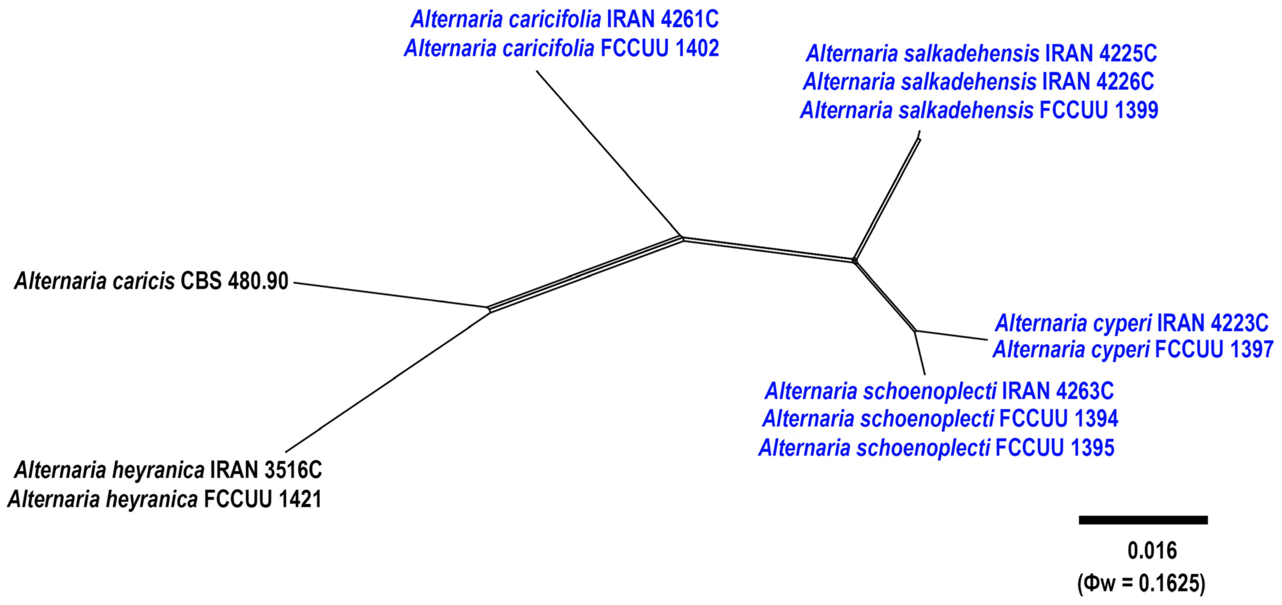
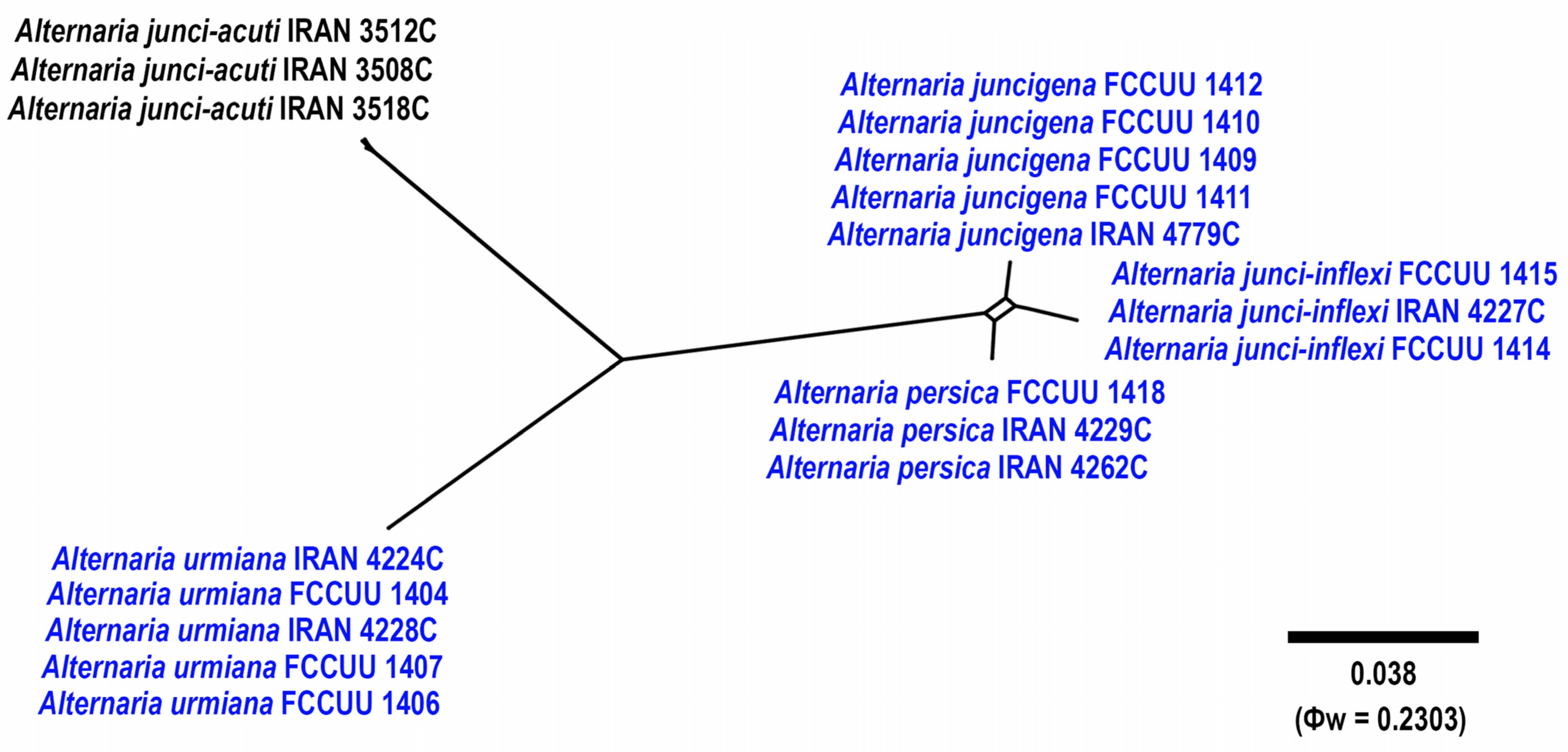
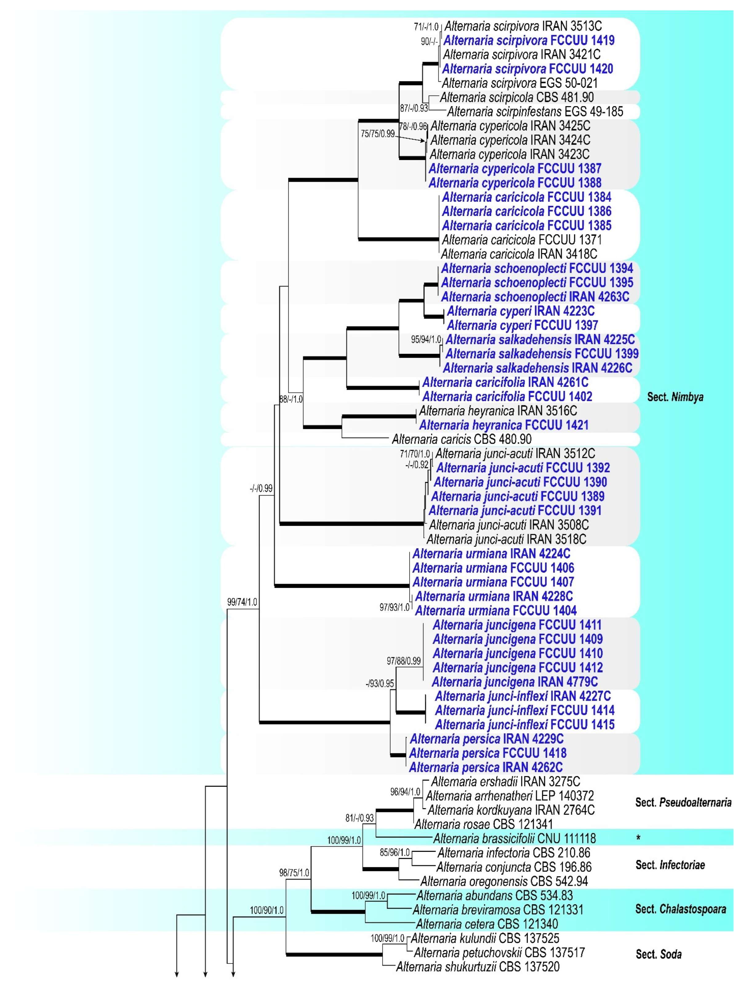
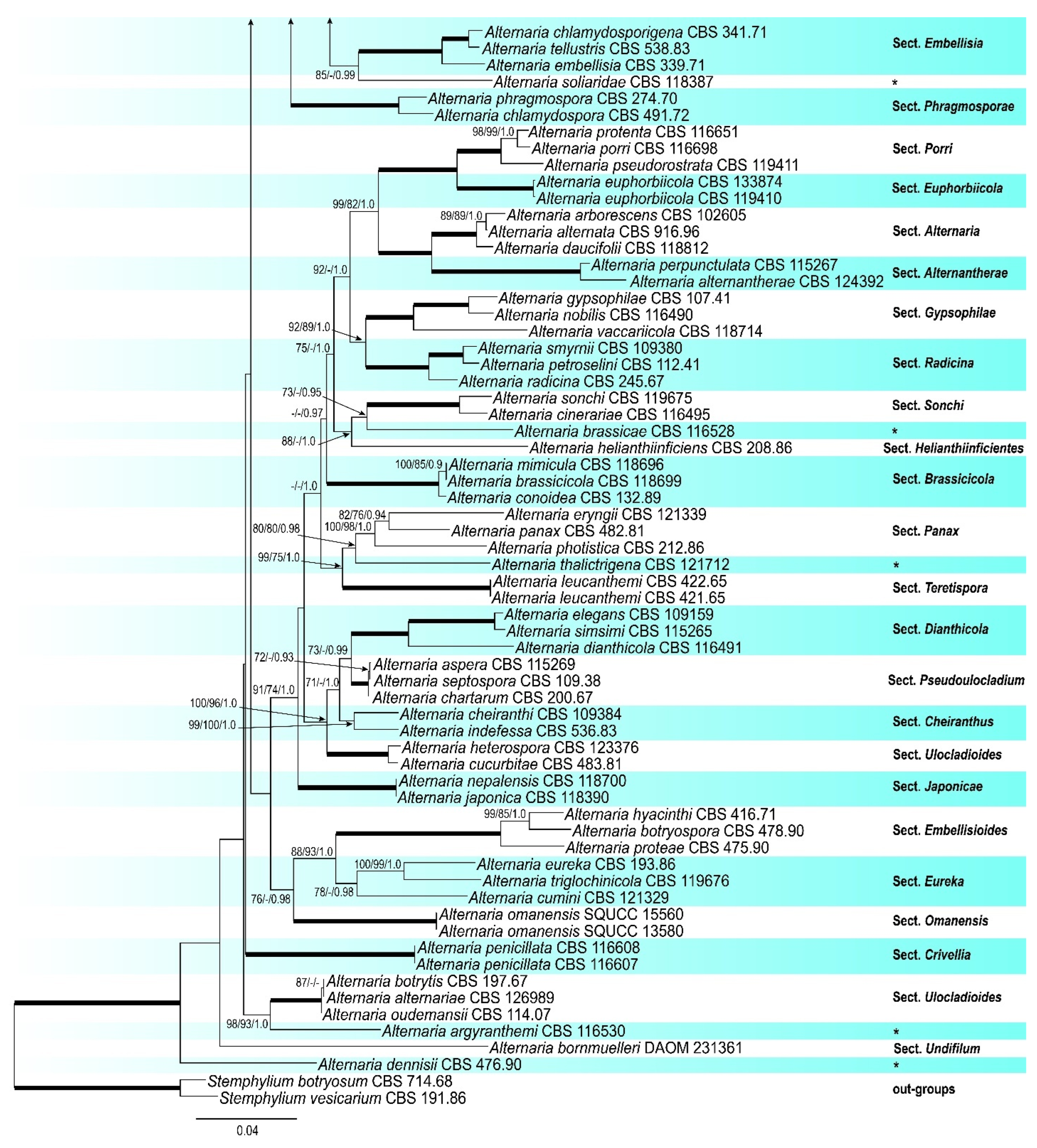
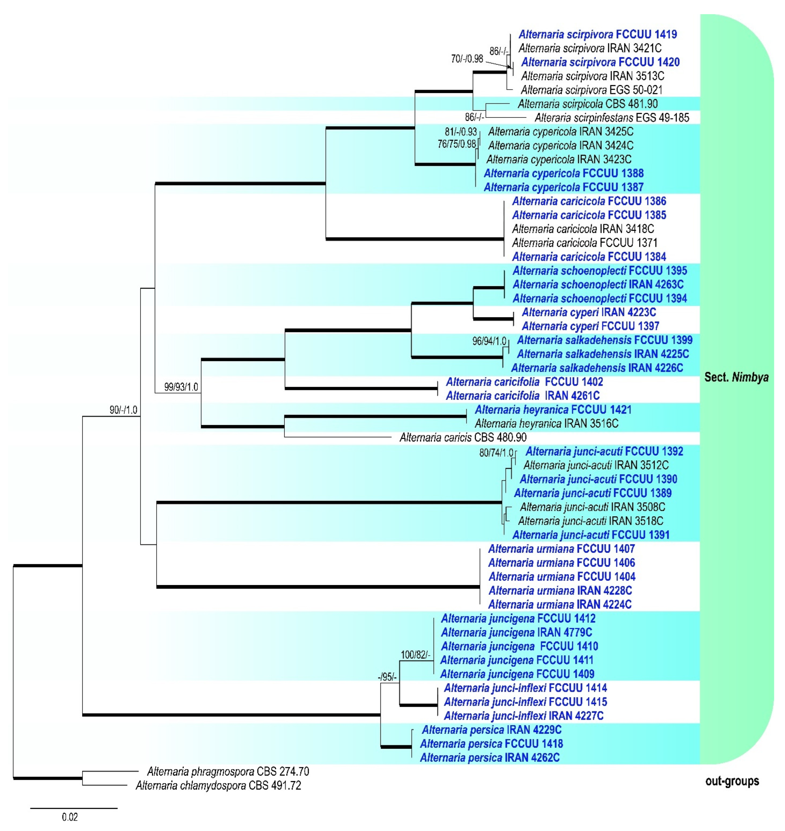
3.1. Phylogenetic Analyses

A summary of phylogenetic information and substitution models for each dataset is provided in Table 3. The topologies of individual gene trees were consistent, with no conflicts observed in species delimitation. In the large-scale phylogenetic tree, combined analyses using BI and ML/MP methods revealed that our isolates grouped with strong support (ML/MP/BI = 99/74/1.0) within the Alternaria section Nimbya, distributed in 13 lineages (Figure 2). The multi-locus datasets for large-scale analyses included a total of 2299 characters (1257 constant sites, 1042 variable sites, 106 parsimony-uninformative sites, and 936 parsimony-informative sites), including gaps (462 for ITS, 524 for GAPDH, 187 for TEF1, 690 for RPB2, and 436 for Alt a 1) (Table 3). For the small-scale analyses, which focused exclusively on Alternaria species within section Nimbya, a total of 2307 characters from 55 strains were used (1610 constant sites, 697 variable sites, 52 parsimony-uninformative sites, and 645 parsimony-informative sites), including gaps (469 for ITS, 514 for GAPDH, 177 for TEF1, 716 for RPB2, and 431 for Alt a 1) (Table 3). In the Alternaria section Nimbya phylogenetic tree, our isolates formed 13 distinct lineages with high support values, indicating the presence of 13 species; five have been previously described and eight are newly identified (Figure 2 and Figure 3).

3.2. Taxonomy

A total of 189 fungal isolates were obtained from Cyperaceae and Juncaceae plants. All isolates were examined based on morphology, and ISSR-PCR molecular marker banding patterns, and 38 representative isolates were selected from different plant hosts for phylogenetic analyses (Supplementary Figure S1). Based on phylogenetic analyses and morphological characteristics, the studied isolates were assigned to 13 species in the Alternaria section Nimbya, including eight new species (A. caricifolia, A. cyperi, A. juncigena, A. junci-inflexi, A. persica, A. schoenoplecti, A. salkadehensis, and A. urmiana). Detailed morphological descriptions and illustrations of these eight new species are provided, and their phylogenetic relationships with other species in the Alternaria section Nimbya are discussed below. The new host matrices for three previously known species are introduced (A. caricicola on Cyperus sp., A. cypericola on Eleocharis sp., and A. junci-acuti on Carex sp.), and a key to the recognized species in the Alternaria section Nimbya from Iran is provided.

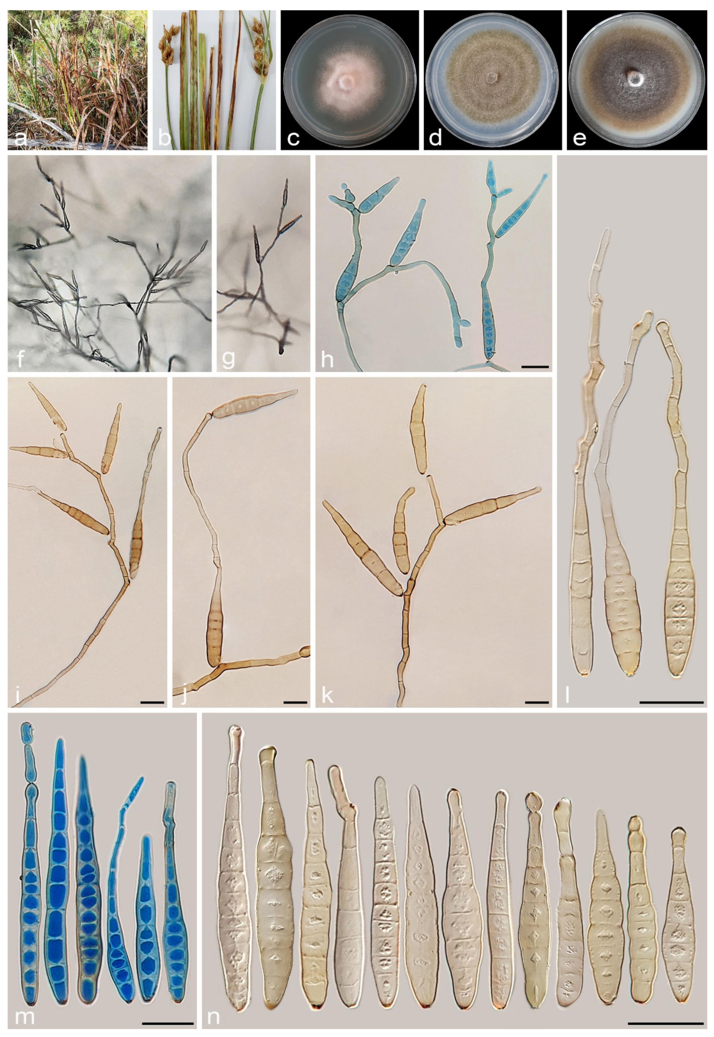
3.2.1. Alternaria caricifolia A. Ahmadpour, Y. Ghosta, Z. Alavi, F. Alavi and A. Poursafar, sp. nov. (Figure 4 and Figure 5)

MycoBank No. MB 857570
Etymology. Named after the host, Carex sp. from which this fungus was isolated.
Typification. Iran, West Azarbaijan province, Mahabad County, isolated from the leaves and culms of Carex sp. (Cyperaceae, Poales) with circular to fusiform lesions, light brown to gray at center, brown to dark brown at margins, 10 July 2020, A. Ahmadpour (holotype IRAN 18109F, ex-type culture IRAN 4261C = FCCUU 1401).
Description. Sexual morph on PCA medium: Ascomata pseudothecial, ovoid to subglobose, dark brown to black, relatively thick-walled with flattened base, scattered or rarely aggregated, 120–240 × 90–220 μm ( x ¯ = 180 × 170 μm, n = 30), formed on the surface of or embedded in culture medium. Pseudoparaphyses hyphoid, septate, 50–100 × 2–3 μm. Asci bitunicate, cylindrical to clavate, straight or slightly curved, with round apex, short pedicel, 57–80 × 15–20 μm ( x ¯ = 66 × 7 μm, n = 50), 8-spored, biseriate. Ascospores fusiform to ellipsoidal, hyaline to pale brown, smooth, 2–4 transverse septa, 1–2 longitudinal septa and rarely with one oblique septum, conspicuously constricted at transverse septa, 17–25 × 5–10 μm ( x ¯ = 21 × 7 μm, n = 50). Asexual morph on PCA medium: Hyphae branched, septate, smooth, light brown, 2–4 μm wide. Conidiophores macronematous, solitary, erect, simple, straight to slightly curved, septate, light brown to brown, with a single apical conidiogenous locus, or rarely 1–3 geniculate with 1–3 conidiogenous loci, 25–50 × 4–5 μm ( x ¯ = 35 × 4.5 μm, n = 50). Conidia mostly solitary or occasionally in chains of two conidia, straight or slightly curved, obclavate to ellipsoid, conidial bodies (35–)50–87(–100) × 5–9 μm ( x ¯ = 63 × 7 μm, n = 50), light brown to brown, surface smooth, (3–)6–8(–11) transverse distosepta, 1–2 eusepta, slightly constricted near eusepta, without longitudinal or oblique septum. The cell lumina are distinctly delimited and rectangular, rounded, hexagonal, or encompass the entire cell volume. True beaks are absent, but with an apical cell extension, septate, unbranched, hyaline to light brown, 12–45 × 2–3 μm, occasionally swollen at the apex. Chlamydospores not observed.
Culture characteristics. Colony on PCA flat, entire, floccose, white to rosy buff at center and hazel at margins, 58 mm diam after 7 days at 25 °C. Colony on PDA flat, entire, floccose, rosy buff, 50 mm diam. Colony on V-8A flat, entire, floccose, white to rosy buff, 56 mm diam. Sporulation abundant on PCA, and V-8A media, from the erect conidiophores that arise directly from the surface or the aerial hyphae. Sexual morphs were abundantly formed on PCA and V-8A after 30 days at 25 °C under fluorescent light with an 8/16 h light/dark cycle.
Additional specimen examined. Iran, West Azarbaijan province, Mahabad County, isolated from leaves and culms of Carex sp. (Cyperaceae, Poales), 10 July 2020, A. Ahmadpour (culture FCCUU 1402).
Notes. Based on the results of phylogenetic analyses (Figure 2 and Figure 3), two studied isolates of A. caricifolia clustered well in a separate lineage with 100% ML/MP bootstrap, and 1.0 BI posterior probabilities values, with a sister relationship to a clade consisting of A. cyperi, A. salkadehensis, and A. schoenoplecti. The PHI analysis confirms that A. caricifolia has no significant genetic recombination with closely related species (Φw = > 0.05, Figure 6). A comparison of nucleotide differences in ITS–rDNA, GAPDH, TEF1, RPB2, and Alt a 1 indicates that A. caricifolia type strain (IRAN 4261C) differs from A. cyperi type strain (IRAN 4223C) by 21/456 bp (4.60%) in ITS–rDNA, 31/495 bp (6.26%, with five gaps (1%)) in GAPDH, 17/170 bp (10%, with four gaps (2%)) in TEF1, 36/746 bp (4.82%) in RPB2 and 57/426 bp (13.38%, with two gaps (0%)) in Alt a 1, from A. salkadehensis type strain (IRAN 4225C) by 23/457 bp (5.03%, with one gap (0%)) in ITS–rDNA, 22/451 bp (4.87%, with three gaps (0%)) in GAPDH, 12/170 bp (7.05%, with four gaps (2%)) in TEF1, 37/748 bp (4.94%) in RPB2 and 53/429 bp (12.35%, with two gaps (2%)) in Alt a 1 and from A. schoenoplecti type strain (IRAN 4263C) by 20/456 bp (4.38%) in ITS–rDNA, 31/495 bp (6.26%, with five gaps (1%)) in GAPDH, 10/170 bp (5.88%, with four gaps (2%)) in TEF1, 34/748 bp (4.54%) in RPB2 and 53/426 bp (12.44%, with two gaps (0%)) in Alt a 1. Alternaria caricifolia can be differentiated of A. cyperi, A. salkadehensis and A. schoenoplecti by its shorter conidiophores (25–50 μm vs. 40–110 μm, (85–)150–300 μm, 40–90 μm, respectively, narrower conidia (5–9 μm vs. 10–12 μm, 10–13 μm, and 12–15(–18) μm, respectively) and presence of sexual morph.
Figure 4. Sexual morph of Alternaria caricifolia (IRAN 4261C). (ae) Ascomata formed on PCA medium after 30 days ((b) = 4×, (c) = 10×); (fj) Asci; (k) Ascospores; (l) Germinated ascospore. Scale bars: (d,e) = 100 μm; (fl) = 20 μm.
Jof 11 00225 g004
Figure 5. Asexual morph of Alternaria caricifolia (IRAN 4261C). (a,b) Symptoms on the leaves and culms of Carex sp.; (ce) Colony on PDA (c), PCA (d), and V-8A (e) after 7 days; (f) Sporulation pattern on PCA (40×); (gl) Conidiophores and conidia. Scale bars: (gl) = 20 μm.
Jof 11 00225 g005
Figure 6. The split diagram showing the results of the pairwise homoplasy index (PHI) test of Alternaria caricifolia with its most closely related species (Φw = 0.1625). PHI test results (Φw) < 0.05 indicate significant recombination within the dataset. The new taxa are shown in bold blue.
Jof 11 00225 g006

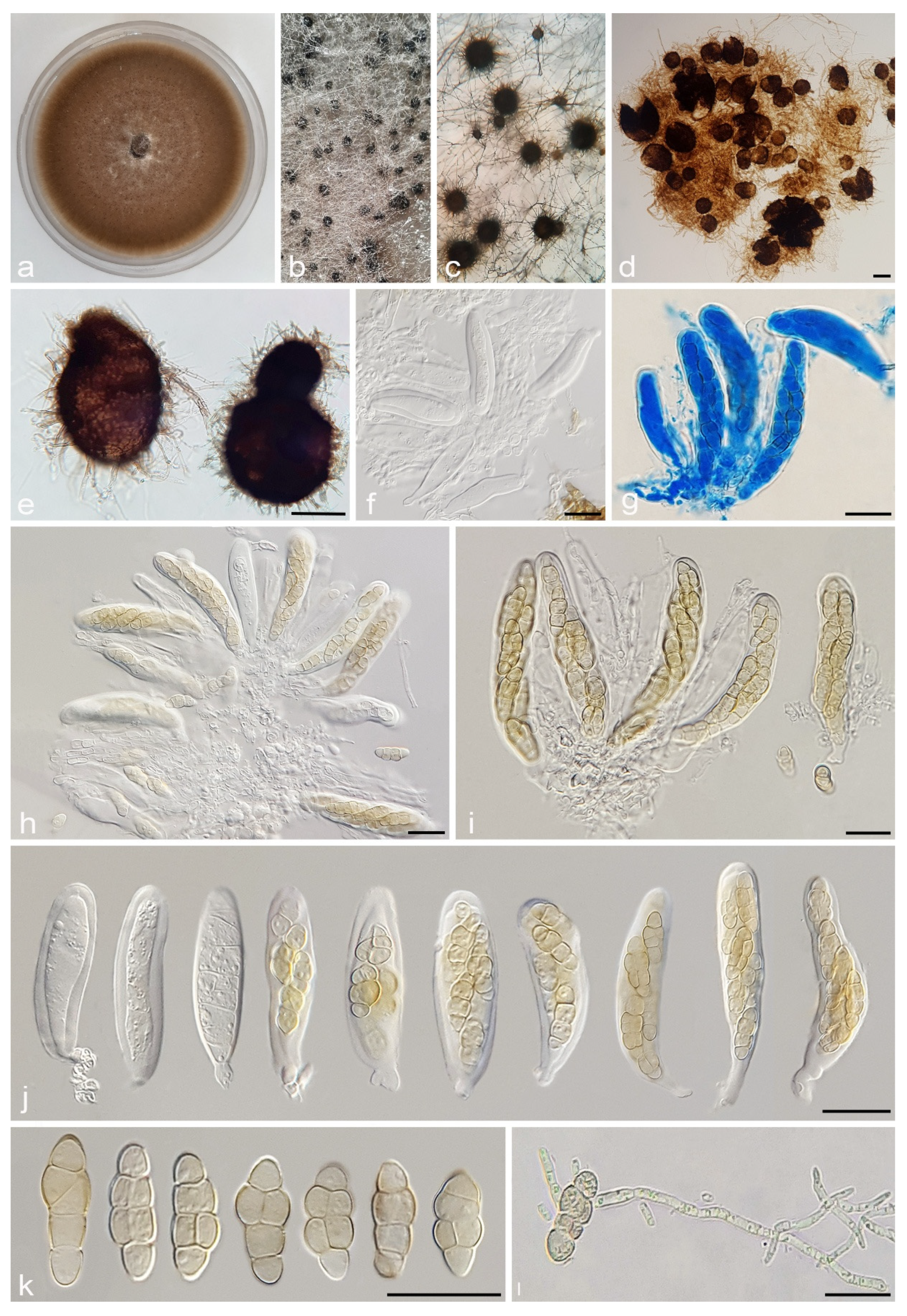
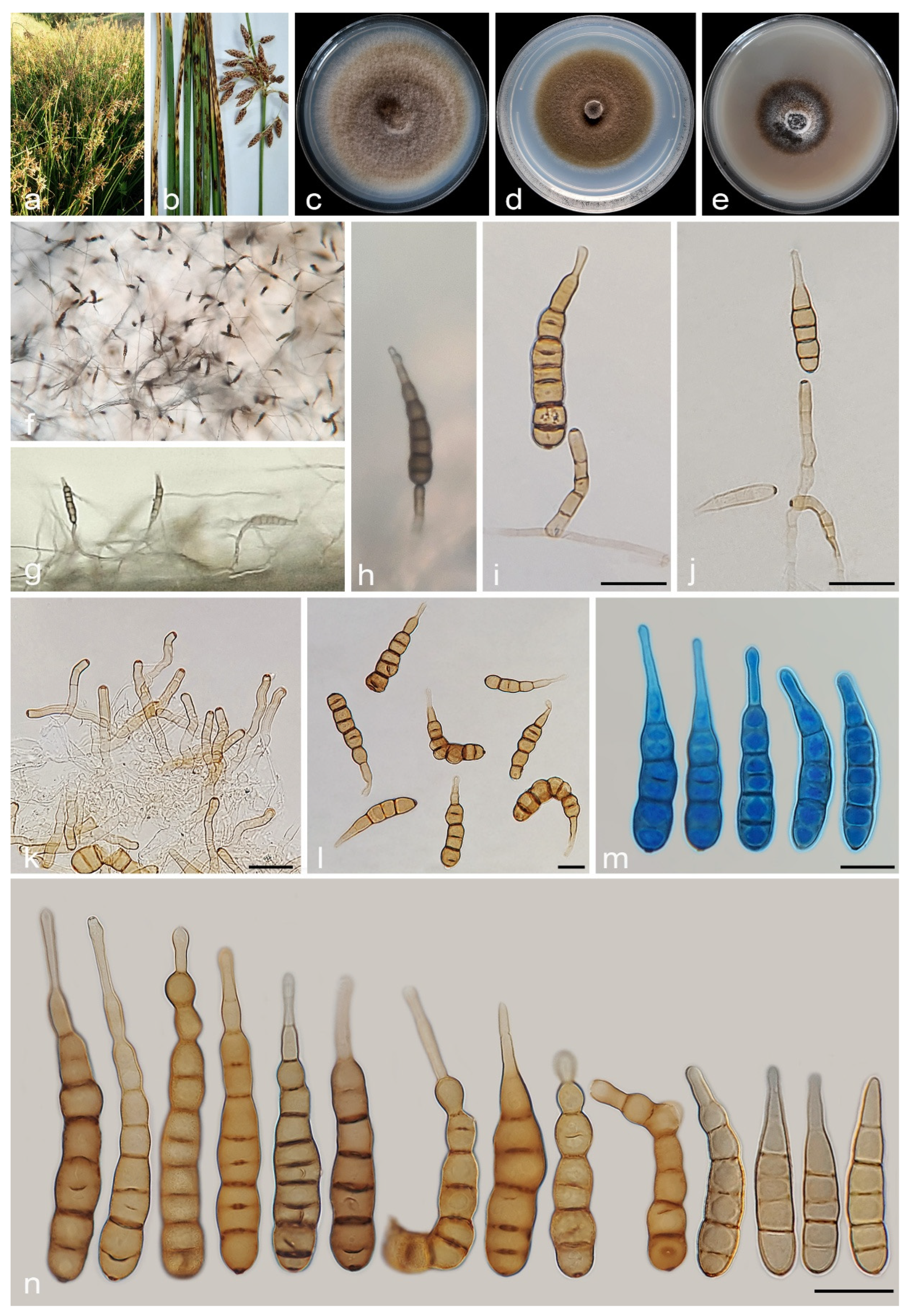
3.2.2. Alternaria cyperi A. Ahmadpour, Y. Ghosta, Z. Alavi, F. Alavi and A. Poursafar, sp. nov. (Figure 7)

MycoBank No. MB 857571
Etymology. Named after the host, Cyperus sp. from which this fungus was isolated.
Typification. Iran, East Azarbaijan province, Bonab County, isolated from leaves and culms of Cyperus sp. (Cyperaceae, Poales) with brown to black lesions and blight, 30 October 2019, A. Ahmadpour (holotype IRAN 18096F, ex-type culture IRAN 4223C = FCCUU 1396).
Description. Asexual morph on PCA medium: Hyphae branched, septate, smooth, light brown, 2–4 μm wide. Conidiophores macronematous, solitary, erect, simple, straight to slightly curved, unbranched, septate, light brown to brown, with a single apical conidiogenous locus, or 1–3 geniculate with 1–3 conidiogenous loci, 40–110 × 4–6 μm ( x ¯ = 70 × 5 μm, n = 50). Conidia mostly in branched chains of 2–4 conidia or occasionally solitary, straight or slightly curved, obclavate to ellipsoid, apically tapered, conidial bodies (55–)60–90(–100) × 10–12 μm ( x ¯ = 80 × 11 μm, n = 50), light brown to brown, surface smooth, (5–)6–9(–12) transverse distosepta, 1–5 eusepta, slightly constricted near eusepta, without longitudinal or oblique septa. True beaks are absent, but with an apical cell extension (secondary conidiophore), up to 15 µm long and 2–4 µm wide, with 1–4 geniculation and 1–4 conidiogenous loci. The cell lumina are distinctly delimited and rectangular, rounded, hexagonal, or encompass the entire cell volume. Chlamydospores and sexual morph were not observed.
Culture characteristics. Colony on PCA flat, entire, floccose, fawn with salmon aerial mycelia, 59 mm diam after 7 days at 25 °C. Colony on PDA flat, entire, floccose, white to rosy buff at center and olivaceous buff at margins, 50 mm diam. Colony on V-8A flat, entire, floccose, white at center and fulvus at margins, 25 mm diam. Sporulation is moderate to scarce on PCA, and V-8A media, from the erect conidiophores that arise directly from the surface or aerial hyphae.
Additional specimen examined. Iran, East Azarbaijan province, Bonab County, isolated from the leaves and culms of Cyperus sp. (Cyperaceae, Poales), 30 October 2019, A. Ahmadpour (culture FCCUU 1397).
Notes. Alternaria cyperi is phylogenetically closely related to A. schoenoplecti and A. salkadehensis (Figure 2 and Figure 3). The PHI analysis confirms that A. cyperi shows no significant genetic recombination with closely related species (Φw = > 0.05, Figure 6). A comparison of nucleotide differences in ITS–rDNA, GAPDH, TEF1, RPB2, and Alt a 1 indicates that A. cyperi type strain (IRAN 4223C) differs from A. salkadehensis type strain (IRAN 4225C) by 2/458 bp (0.43%, with one gap (0%)) in ITS–rDNA, 12/495 bp (2.42%) in GAPDH, 5/165 bp (3.03%) in TEF1, 22/746 bp (2.94%) in RPB2 and 43/425 bp (10.11%) in Alt a 1 and from A. schoenoplecti type strain (IRAN 4263C) by 2/457 bp (0.43%) in ITS–rDNA, 8/495 bp (1.61%) in GAPDH, 2/165 bp (1.21%) in TEF1, 14/746 bp (1.87%) in RPB2 and 10/425 bp (2.35%) in Alt a 1. Alternaria cyperi differs from A. salkadehensis based on shorter conidiophores (40–110 μm vs. (85–)150–300 μm) and shorter apical cell extension (up to 15 μm vs. up to 45 μm). Alternaria schoenoplecti morphologically differs from A. cyperi by having solitary and C-shaped conidia with strongly constricted at the septa.
Figure 7. Alternaria cyperi (IRAN 4223C). (a,b) Symptoms on the leaves and culms of Cyperus sp.; (ce) Colony on PDA (c), PCA (d), and V-8A (e) after 7 days; (f) Sporulation pattern on PCA (40×); (gn) Conidiophores and conidia. Scale bars: (gn) = 20 μm.
Jof 11 00225 g007

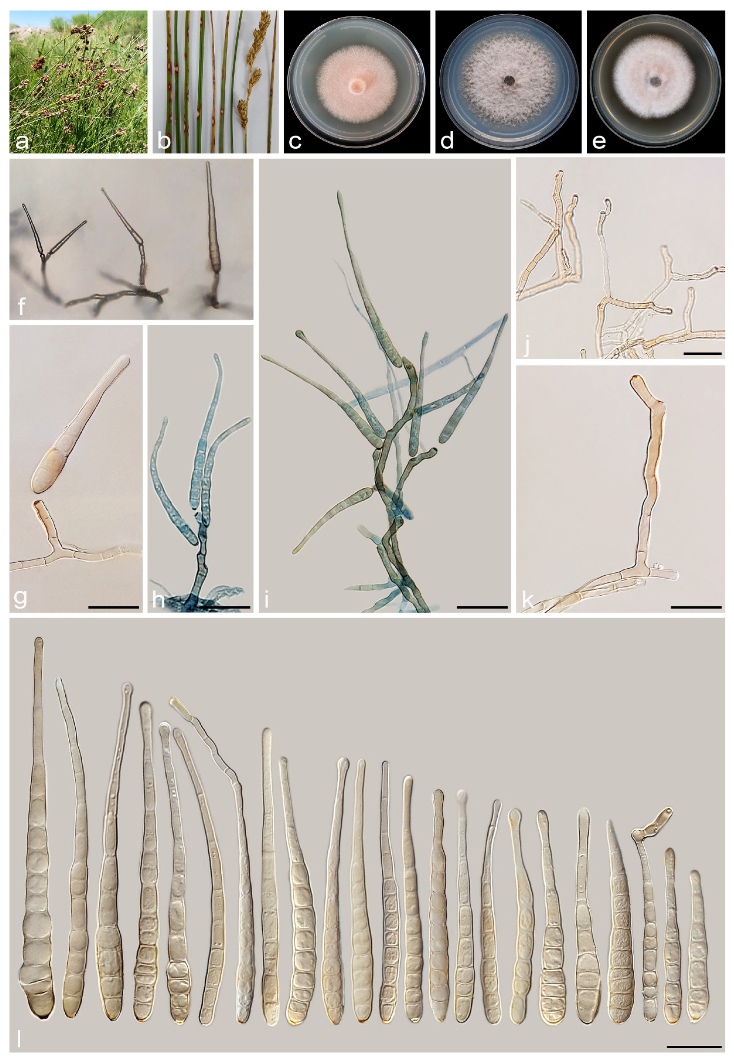
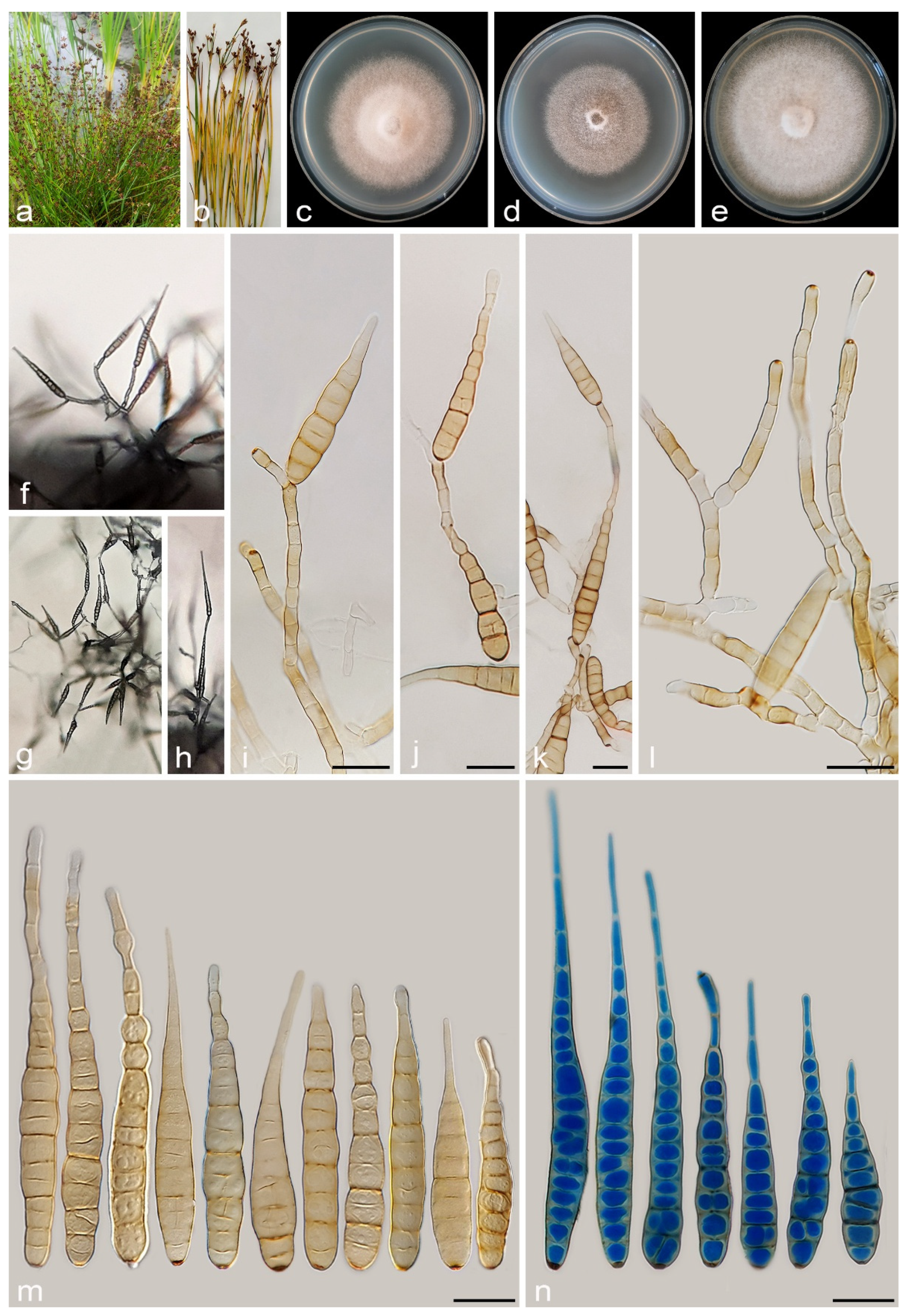
3.2.3. Alternaria juncigena A. Ahmadpour, Y. Ghosta, Z. Alavi, F. Alavi and A. Poursafar, sp. nov. (Figure 8)

MycoBank No. MB 857572
Etymology. Named after the host, Juncus sp. from which this fungus was isolated.
Typification. Iran, West Azarbaijan province, Khoy County, Salkadeh Village, isolated from the culms of Juncus sp. (Juncaceae, Poales), with light to dark brown irregular lesions, 25 September 2020, A. Ahmadpour (holotype IRAN 18211F, ex-type culture IRAN 4779C = FCCUU 1408).
Description. Asexual morph on PCA medium: Hyphae branched, septate, smooth, light brown, 2–4 μm wide. Conidiophores macronematous, solitary, erect, simple, septate, brown, 1–3 geniculate, with 1–3 conidiogenous loci, 40–85 × 5–6 μm ( x ¯ = 57 × 5.5 μm, n = 50). Conidia solitary or in chains of 2–3 conidia, straight or slightly curved, obclavate to ellipsoid, conidial bodies (40–)55–90(–110) × 10–14(–16) μm ( x ¯ = 70 × 12 μm, n = 50), light brown to brown, surface smooth, (5–)8–12(–15) transverse distosepta, 1–3 eusepta, slightly constricted near eusepta, with 1–3 longitudinal and 0–2 oblique septa. True beaks are absent but with apical cell extension, up to 50 µm in length and 2–3 µm wide, occasionally swollen at apex, rarely with 1–3 geniculate and 1–3 conidiogenous loci. The cell lumina are distinctly delimited and rectangular, rounded, hexagonal, or encompass the entire cell volume. Chlamydospores and sexual morph were not observed.
Culture characteristics. Colony on PCA flat, entire, velvety, pale vinaceous to vinaceous buff, with sparse aerial mycelia, 50 mm diam after 7 days at 25 °C. Colony on PDA flat, entire, floccose, white at center, and rosy buff at margins, 60 mm diam. Colony on V-8A flat, entire, floccose, white to pale violaceous, 68 mm diam. Sporulation moderate to scarce on PCA, and V-8A media, from the erect conidiophores that arise directly from the surface or the aerial hyphae.
Additional specimens examined. Iran, West Azarbaijan province, Khoy County, Salkadeh Village, isolated from the culms of Juncus sp., 25 September 2020, A. Ahmadpour (cultures FCCUU 1409, FCCUU 1410, FCCUU 1411, FCCUU 1412).
Notes. Phylogenetically, Alternaria juncigena forms a distinct lineage with high support values (ML/MP/BI= 97/88/0.99) (Figure 2), and is closely related to A. junci-inflexi and A. persica. A comparison of nucleotide differences in ITS–rDNA, GAPDH, TEF1, RPB2, and Alt a 1 indicates that A. juncigena type strain (IRAN 4779C) differs from A. junci-inflexi type strain (IRAN 4227C) by 2/449 bp (0.44%) in ITS–rDNA, 6/490 bp (1.22%) in GAPDH, 2/169 bp (1.18%) in TEF1, 10/690 bp (1.44%) in RPB2 and 8/428 bp (1.86%) in Alt a 1, and from A. persica type strain (IRAN 4262C) by 3/450 bp (0.66%, with one gap (0%)) in ITS–rDNA, 10/490 bp (2.04%) in GAPDH, 5/170 bp (2.94%, with one gap (0%)) in TEF1, 11/748 bp (1.47%) in RPB2 and 2/428 bp (0.46%) in Alt a 1. The PHI analysis confirms that A. juncigena has no significant genetic recombination with closely related species (Φw = > 0.05, Figure 9). Alternaria juncigena differs from A. junci-inflexi and A. persica by having shorter conidiophores (40–85 μm vs. (55–)100–175(–212) μm, 45–110 μm, respectively), smooth surface, longer and wider conidia ((40–)55–90(–110) × 10–16 μm vs. smooth to verrucose surface, ((21–)45–65(–80) × (8–)9–12 μm in A. junci-inflexi and smooth to verrucose surface, (40–)55–85 × 8–11 μm in A. persica) and longer apical cell extension (up to 50 μm vs. up to 15 μm in A. junci-inflexi).
Figure 8. Alternaria juncigena (IRAN 4779C). (a,b) Symptoms on Juncus sp.; (ce) Colony on PDA (c), PCA (d), and V-8A (e) after 7 days; (fh) Sporulation pattern on PCA (f = 40×, g,h = 20×); (in) Conidiophores and conidia. Scale bars: (in) = 20 μm.
Jof 11 00225 g008
Figure 9. The split diagram showing the results of the pairwise homoplasy index (PHI) test of Alternaria juncigena with its most closely related species (Φw = 0.2303). PHI test results (Φw) < 0.05 indicate significant recombination within the dataset. The new taxa are shown in bold blue.
Jof 11 00225 g009

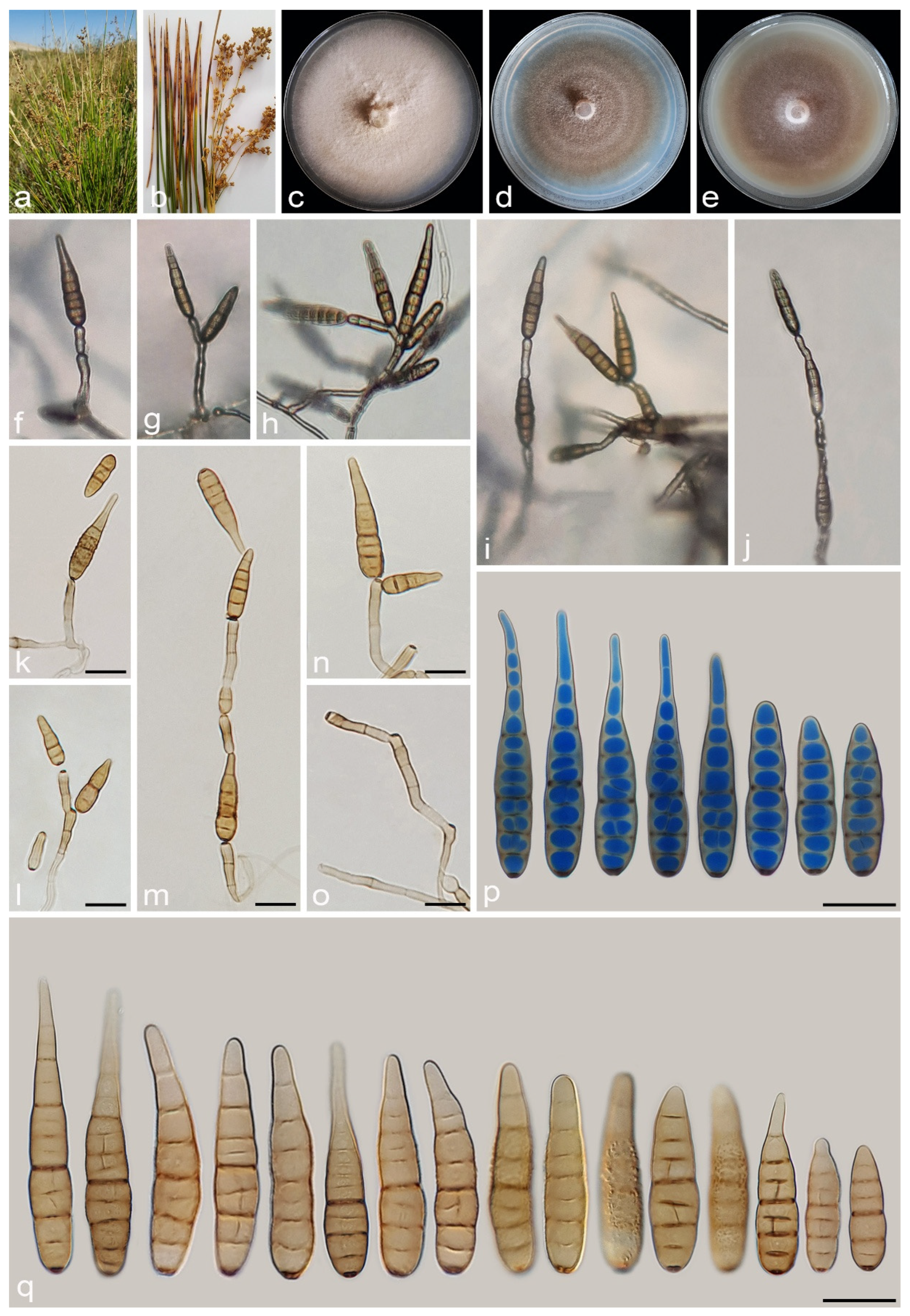
3.2.4. Alternaria junci-inflexi A. Ahmadpour, Y. Ghosta, Z. Alavi, F. Alavi and A. Poursafar, sp. nov. (Figure 10)

MycoBank No. MB 857573
Etymology. Named after the host, Juncus inflexus, from which the fungus was isolated.
Typification. Iran, West Azarbaijan province, Khoy County, Salkadeh Village, isolated from the culms of Juncus inflexus (Juncaceae, Poales) with light brown to dark brown irregular lesions, 25 Sept. 2020, A. Ahmadpour (holotype IRAN 18099F, ex-type culture IRAN 4227C = FCCUU 1413).
Description. Asexual morph on PCA: Hyphae branched, septate, light brown, smooth, 2–4 μm wide. Conidiophores macronematous, solitary, straight or slightly curved, simple, unbranched, septate, light brown to brown, mostly with a single apical conidiogenous locus, or 1–5 geniculate with 1–5 conidiogenous loci, (55–)100–175(–212) × 4–5 μm ( x ¯ = 135 × 4.5 μm, n = 50). Conidia mostly solitary, occasionally in chains of 2(–4) conidia, straight or slightly curved, mostly obclavate to narrowly ellipsoid or ovoid, conidial bodies (21–)45–65(–80) × (8–)9–12 μm ( x ¯ = 51 × 10.5 μm, n = 50), light brown to brown, smooth to verrucose, 2–9 (mostly 3–7) transverse distosepta, 1–3 transverse eusepta, with 1–3 longitudinal and 0–1 oblique distosepta, and slightly constricted at eusepta. True beaks are absent but with apical cell extension, light brown to brown, unbranched, 5–15 × 2–4 μm, occasionally without apical extension. The cell lumina are distinctly delimited and rectangular, rounded, hexagonal, or encompass the entire cell volume. Chlamydospores and sexual morph were not observed. Chlamydospores and sexual morph were not observed.
Culture characteristics. Colony on PCA flat, entire, velvety, fawn with off-white aerial mycelia, reaching 65 mm diam after 7 days at 25 °C. Colony on PDA flat, entire, felty, rosy buff with white aerial mycelia, 60 mm diam. Colony on V-8A flat, entire, felty, fawn with off-white aerial mycelia, 59 mm diam. Sporulation is abundant on PCA and V-8A media, from the erect conidiophores that arise directly from the surface or the aerial hyphae.
Additional specimens examined. Iran, West Azarbaijan province, Khoy County, Salkadeh Village, isolated from culms of Juncus inflexus, 25 Sept. 2020, A. Ahmadpour (culture FCCUU 1414).—Iran, East Azarbaijan province, Maraghe County, isolated from culms of Juncus inflexus, 20 Sept. 2019, A. Ahmadpour (culture FCCUU 1415).
Notes. Alternaria junci-inflexi is phylogenetically (Figure 2 and Figure 3) closely related to A. juncigena and A. persica, but can be distinguished by its shorter conidia ((21–)45–65(–80) μm vs. (40–)55–90(–110) μm in A. juncigena and (40–)55–85 μm in A. persica), fewer transverse distosepta (2–9 vs. (5–)8–12(–15) in A. juncigena and (4–)7–12(–15) in A. persica) and shorter apical cell extension (5–15 μm vs. up to 50 μm in A. juncigena and A. persica). The PHI analysis confirms that A. junci-inflexi has no significant genetic recombination with closely related species (Φw = > 0.05, Figure 9). A comparison of nucleotide differences in ITS–rDNA, GAPDH, TEF1, RPB2, and Alt a 1 indicates that A. junci-inflexi type strain (IRAN 4227C) differs from A. juncigena type strain (IRAN 4779C) by 2/449 bp (0.44%) in ITS–rDNA, 6/490 bp (1.22%) in GAPDH, 2/169 bp (1.18%) in TEF1, 10/690 bp (1.44%) in RPB2 and 8/428 bp (1.86%) in Alt a 1, and from A. persica type strain (IRAN 4262C) by 3/450 bp (0.66%, with one gap (0%)) in ITS–rDNA, 9/490 bp (1.83%) in GAPDH, 7/170 bp (4.11%, with one gap (0%)) in TEF1, 12/690 bp (1.73%) in RPB2 and 1/428 bp (0.23%) in Alt a 1.
Figure 10. Alternaria junci-inflexi (IRAN 4227C). (a,b) Symptoms on culms of Juncus inflexus; (ce) Colony on PDA (c), PCA (d), and V-8A (e) after 7 days; (fj) Sporulation pattern on PCA (40×); (kq) Conidiophores and conidia. Scale bars: (kq) = 20 μm.
Jof 11 00225 g010

3.2.5. Alternaria persica A. Ahmadpour, Y. Ghosta, Z. Alavi, F. Alavi and A. Poursafar, sp. nov. (Figure 11)

MycoBank No. MB 857574
Etymology. The name refers to the old name of Iran, Persia, where the fungus was collected.
Typification. Iran, West Azarbaijan province, Khoy County, Salkadeh Village, isolated from the culms of Juncus sp. (Juncaceae, Poales), with irregular brown lesions and blight, 25 September 2020, A. Ahmadpour (holotype IRAN 18110F, ex-type culture IRAN 4262C = FCCUU 1417).
Description. Asexual morph on PCA medium: Hyphae branched, septate, smooth, light brown, 2–4 μm wide. Conidiophores macronematous, solitary, erect, simple, septate, brown, 1–4 geniculate, with 1–4 conidiogenous loci, 45–110 × 4–5 μm ( x ¯ = 88 × 4.5 μm, n = 50). Conidia solitary or in chains of 2–3 conidia, straight or slightly curved, obclavate, ellipsoid to ovoid, surface smooth to verrucose, conidial bodies (40–)55–85 × 8–11 μm ( x ¯ = 65 × 9.5 μm, n = 50), light brown to brown, (4–)7–12(–15) transverse distosepta, 1–3 eusepta, slightly constricted near eusepta, with 0–3 longitudinal and 0–2 oblique septa. True beaks are absent but with apical cell extension, light brown to brown, septate, unbranched, 10–50 × 2–3 μm, occasionally swollen at the apex. The cell lumina are distinctly delimited and rectangular, rounded, hexagonal, or encompass the entire cell volume. Chlamydospores and sexual morph were not observed.
Culture characteristics. Colony on PCA flat, entire, floccose, vinaceous buff with off-white aerial mycelium, 60 mm diam after 7 days at 25 °C. Colony on PDA flat, entire, floccose, rosy buff, 64 mm diam. Colony on V-8A flat, entire, floccose, vinaceous buff with white aerial mycelium, 61 mm diam. Sporulation moderate to scarce on PCA, and V-8A media, from the erect conidiophores that arise directly from the surface or the aerial hyphae.
Additional specimens examined. Iran, West Azarbaijan province, Khoy County, Salkadeh Village, isolated from culms of Juncus sp., 25 September 2020, A. Ahmadpour (cultures IRAN 4229C = FCCUU 1416, FCCUU 1417).
Notes. Phylogenetically, A. persica isolates formed a monophyletic lineage with high support values (ML/MP/BI = 100/100/1.0) and is closely related to A. junci-inflexi and A. juncigena (Figure 2 and Figure 3). However, it can be distinguished from A. junci-inflexi based on the larger conidia ((40–)55–85 × 8–11 μm vs. (21–)45–65(–80) × (8–)9–12 in A. juci-inflexi), more transverse distosepta ((4–)7–12(–15) vs. 2–9 in A. junci-inflexi) and longer apical cell extension (10–50 μm vs. 5–15 μm in A. junci-inflexi) and from A. juncigena by having longer conidiophores (45–110 μm vs. 40–85 μm in A. juncigena) and smooth to verrucose surface and narrower conidia (8–11 μm vs. smooth surface, 10–16 μm in A. juncigena). Comparisons involving the nucleotide differences and PHI analysis (Φw = > 0.05, Figure 9) in these species are listed in the A. juncigena and A. junci-inflexi notes section.
Figure 11. Alternaria persica (IRAN 4262C). (a,b) Symptoms on Juncus sp.; (ce) Colony on PDA (c), PCA (d), and V-8A (e) after 7 days; (fh) Sporulation pattern on PCA (40×); (ip) Conidiophores and conidia. Scale bars: (ip) = 20 μm.
Jof 11 00225 g011

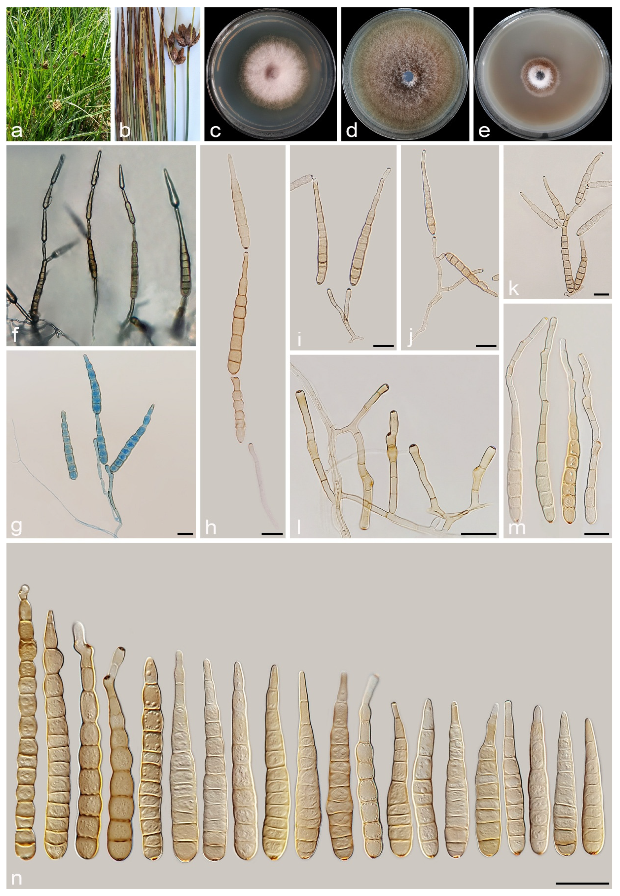
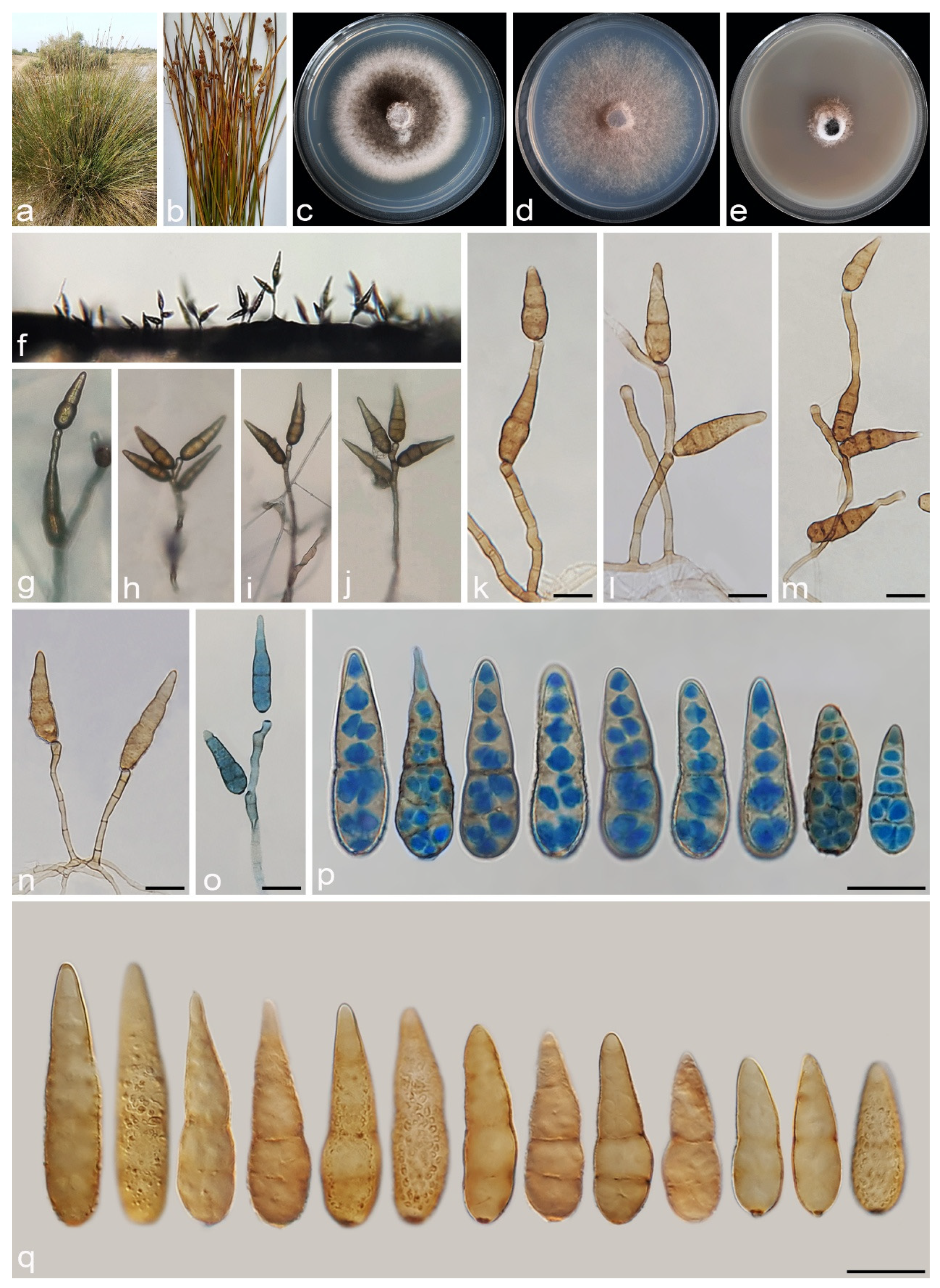
3.2.6. Alternaria salkadehensis A. Ahmadpour, Y. Ghosta, Z. Alavi, F. Alavi and A. Poursafar, sp. nov. (Figure 12)

MycoBank No. MB 857575
Etymology. Named after the location, Salkadeh Village, Khoy County, from where the type was collected.
Typification. Iran, West Azarbaijan province, Khoy, Salkadeh Village, from the leaves and culms of Cyperus sp. (Cyperaceae, Poales) with light brown to brown irregular lesions and blight, 10 Jul. 2019, A. Ahmadpour (holotype IRAN 18098F, ex-type culture IRAN 4225C = FCCUU 1400).
Description. Asexual morph on PCA medium: Hyphae branched, septate, smooth, light brown, 2–4 μm wide. Conidiophores macronematous, solitary, erect, simple, septate, brown, 1–5 geniculate, with 1–5 conidiogenous loci, (85–)150–300 × 5–6 μm ( x ¯ = 200 × 5.5 μm, n = 50). Conidia mostly chains of 2–4 conidia, occasionally solitary, straight or slightly curved, obclavate, ellipsoid to ovoid, conidial bodies (35–)55–90(–100) × 10–13 μm ( x ¯ = 70 × 11 μm, n = 50), light brown to brown, surface smooth, (4–)5–9(–13) transverse distosepta, 1–3 eusepta, slightly constricted near eusepta, without longitudinal or oblique septa. True beaks are absent, but with apical cell extension (secondary conidiophores), up to 45 µm long and 2–4 µm wide, with 3–4(–6) geniculate and 3–4(–6) conidiogenous loci, each bearing one conidium. The cell lumina are distinctly delimited and rectangular, rounded, hexagonal, or encompass the entire cell volume. Chlamydospores and sexual morph were not observed.
Culture characteristics. Colony on PCA flat, entire, velvety, hazel, with sparse aerial mycelium, 63 mm diam after 7 days at 25 °C. Colony on PDA flat, entire, floccose, rose buff with white aerial mycelium, 55 mm diam. Colony on V-8A flat, entire, velvety, dark brick at the center and ochreous at the margin, with sparse off-white aerial mycelium, 63 mm diam. Sporulation is abundant on PCA and V-8A media, from the erect conidiophores that arise directly from the surface or aerial hyphae.
Additional specimens examined. Iran, West Azarbaijan province, Khoy County, Salkadeh Village, from the leaves and culms of Cyperus sp. (Cyperaceae, Poales), 10 July 2019, A. Ahmadpour (culture FCCUU 1399).—ibid. on leaves and culms of Carex sp. (Cyperaceae, Poales), 10 July 2019, A. Ahmadpour (culture IRAN 4226C = FCCUU 1398).
Notes. Alternaria salkadehensis is phylogenetically closely related to A. cyperi and A. schoenoplecti (ML/MP/BI= 100/100/1.0) (Figure 2 and Figure 3), but it can be differentiated from A. cyperi by its longer conidiophores (85–)150–300 μm vs. 40–110 μm) and longer apical cell extension (up to 45 μm vs. up to 15 μm). Alternaria schoenoplecti morphologically differs from A. salkadehensis by having solitary and straight or curved conidia (C shape) vs. catenate, straight or slightly curved conidia. The PHI analysis confirms that A. salkadehensis has no significant genetic recombination with closely related species (Φw = > 0.05, Figure 6). A comparison of nucleotide differences in ITS–rDNA, GAPDH, TEF1, RPB2, and Alt a 1 indicates that A. salkadehensis type strain (IRAN 4225C) differs from A. cyperi type strain (IRAN 4223C) by 2/458 bp (0.43%, with one gap (0%)) in ITS–rDNA, 12/495 bp (2.42%) in GAPDH, 5/165 bp (3.03%) in TEF1, 22/746 bp (2.94%) in RPB2 and 43/425 bp (10.11%) in Alt a 1 and from A. schoenoplecti type strain (IRAN 4263C) by 4/458 bp (0.87%, with one gap (0%)) in ITS–rDNA, 12/495 bp (2.42%) in GAPDH, 4/167 bp (2.39%) in TEF1, 23/748 bp (3.07%) in RPB2 and 39/428 bp (9.11%) in Alt a 1.
Figure 12. Alternaria salkadehensis (IRAN 4225C). (a,b) Symptoms on the leaves and culms of Cyperus sp.; (ce) Colony on PDA (c), PCA (d), and V-8A (e) after 7 days; (f,g) Sporulation pattern on PCA (20×); (hn) Conidiophores and conidia. Scale bars: (hn) = 20 μm.
Jof 11 00225 g012

3.2.7. Alternaria schoenoplecti A. Ahmadpour, Y. Ghosta, Z. Alavi, F. Alavi and A. Poursafar, sp. nov. (Figure 13)

MycoBank No. MB 857576
Etymology. Named after the host, Schoenoplectus sp. from which the fungus was isolated.
Typification. Iran, West Azarbaijan province, Miyandoab County, from the culms of Schoenoplectus sp. (Cyperaceae, Poales), with brown to dark brown, ovoid to fusiform lesions, 10 June 2019, A. Ahmadpour (holotype IRAN 18111F, ex-type culture IRAN 4263C = FCCUU 1393).
Description. Asexual morph on PCA medium: Hyphae branched, septate, smooth, light brown, 2–4 μm wide. Conidiophores macronematous, solitary, erect, simple, straight to slightly curved, septate, light brown to brown, with a single apical conidiogenous locus, 40–90 × 5–7 μm ( x ¯ = 66 × 6 μm, n = 50). Conidia mostly solitary, rarely in chains of two conidia, straight or curved, obclavate to narrowly ellipsoid, conidial bodies (40–)50–70(–88) × 12–15(–18) μm ( x ¯ = 63 × 14 μm, n = 50), light brown to brown, smooth, (3–)4–8(–11) transverse distosepta, 1–3 eusepta, rarely with 1–2 longitudinal or oblique distosepta. Mature conidia are strongly constricted at most of their transverse septa, and in some conidia, the median cells are markedly swollen. True beaks are absent, but with an apical cell extension up to 22 μm long and 3–5 μm wide, occasionally swollen at the apex. The cell lumina are distinctly delimited and rectangular, rounded, hexagonal, or encompass the entire cell volume. Chlamydospores and sexual morph were not observed.
Culture characteristics. Colony on PCA flat, entire, velvety, sepia to cinnamon, with sparse aerial hyphae, 49 mm diam. after 7 days at 25 °C. Colony on PDA flat, entire, floccose, dark brick to cinnamon, 55 mm diam. Colony on V-8A flat, entire, velvety, sepia with white aerial mycelia, 30 mm diam. Sporulation is scarce on PCA, V-8A, or SNA media, from the erect conidiophores that arise directly from the surface or aerial hyphae.
Additional specimens examined. Iran, West Azarbaijan province, Miyandoab County, from culms of Schoenoplectus sp., 10 June 2019, A. Ahmadpour (cultures FCCUU 1394, FCCUU 1395).
Notes. Alternaria schoenoplecti is phylogenetically closely related to A. cyperi and A. salkadehensis (Figure 2 and Figure 3). Comparisons of nucleotide differences and PHI analysis (Φw = > 0.05, Figure 6) in these species are listed in the A. cyperi and A. salkadehensis notes section. Moreover, A. schoenoplecti differs from A. cyperi and A. salkadehensis morphologically in having solitary and straight or curved conidia (C shape) vs. catenate, straight or slightly curved conidia.
Figure 13. Alternaria schoenoplecti (IRAN 4263C). (a,b) Symptoms on culms of Schoenoplectus sp.; (ce) Colony on PDA (c), PCA (d), and V-8A (e) after 7 days; (fh) Sporulation pattern on PCA (f = 10×, g = 20×, h = 40×); (in) Conidiophores and conidia. Scale bars: (in) = 20 μm.
Jof 11 00225 g013

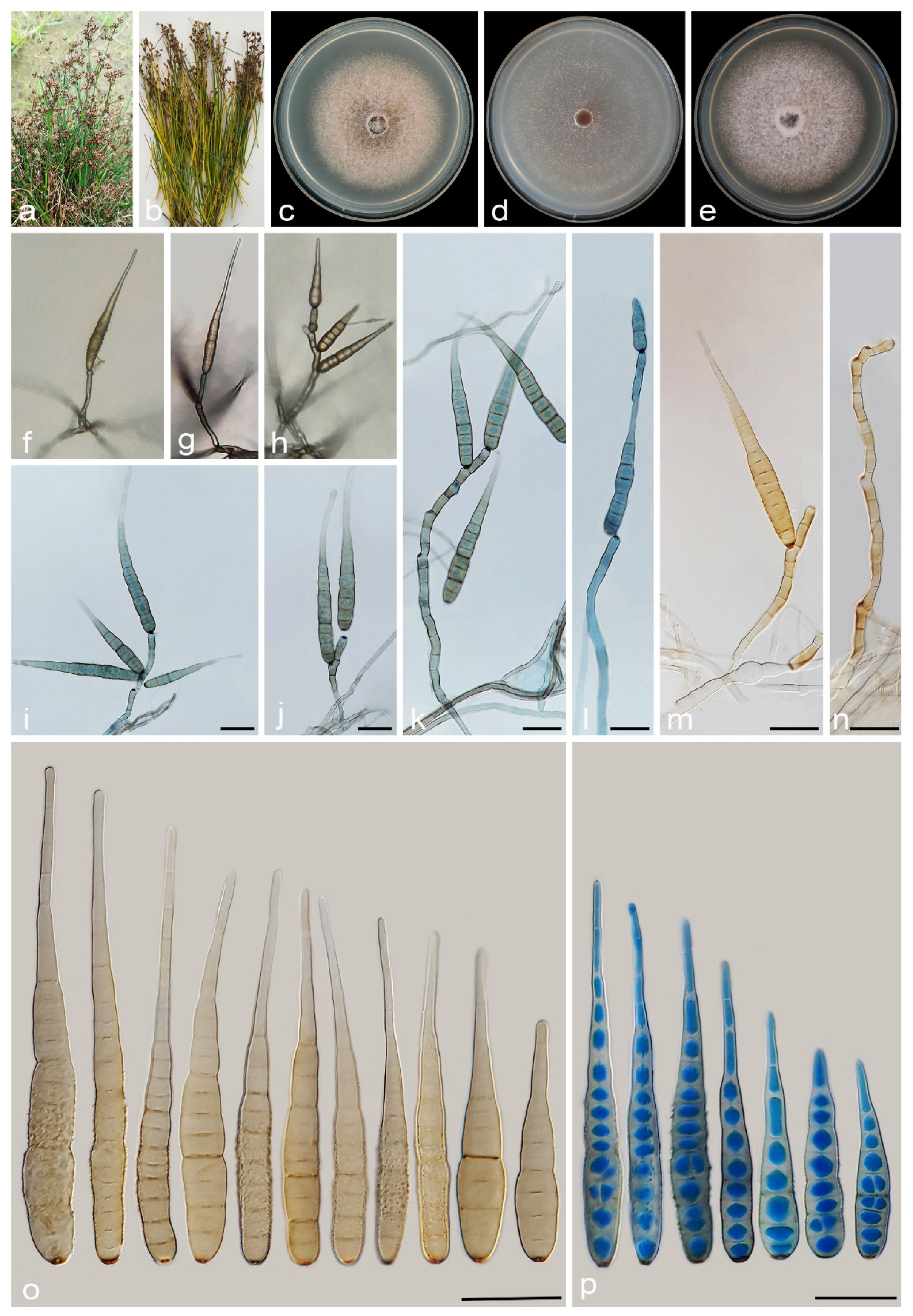
3.2.8. Alternaria urmiana A. Ahmadpour, Y. Ghosta, Z. Alavi, F. Alavi and A. Poursafar, sp. nov. (Figure 14)

MycoBank No. MB 857577
Etymology. The name refers to the Urmia County, where the holotype was collected.
Typification. Iran, West Azarbaijan province, Urmia County, isolated from the culms of Juncus acutus (Juncaceae, Poales) with brown lesions and blight symptoms, 20 September 2019, Y. Ghosta (holotype IRAN 18097F, ex-type culture IRAN 4224C = FCCUU 1405).
Description. Asexual morph on PCA: Hyphae branched, septate, light brown, smooth, 2–4 μm wide. Conidiophores macronematous, solitary, straight or slightly curved, simple, unbranched, septate, light brown to brown, mostly with a single apical conidiogenous locus, or 1–4 geniculate with 1–4 conidiogenous loci, (75–)100–150(–225) × 4–5 μm ( x ¯ = 136 × 4.5 μm, n = 50). Conidia mostly solitary, rarely in chains of two conidia, straight or slightly curved, mostly obclavate, ellipsoid to ovoid, conidial bodies (35–)40–51(–55) × 12–15 μm ( x ¯ = 46 × 13 μm, n = 50), light brown to brown, smooth to verrucose, 3–8 (mostly 3–6) transverse distosepta, 1–2 transverse eusepta, with 1–3 longitudinal and 0–2 oblique distosepta. Conidium body slightly constricted at eusepta, mostly without apical cell extension, occasionally with apical cell extension, light brown to brown, 4–10 × 2–4 μm. The cell lumina are distinctly delimited and rectangular, rounded, hexagonal, or encompass the entire cell volume. Chlamydospores and sexual morph were not observed.
Culture characteristics. Colony on PCA flat, entire, velvety, rosy buff with sparse, off-white aerial mycelium, reaching 51 mm diam. after 7 days at 25 °C. Colony on PDA flat, entire, floccose, dark birk to sepia at the center and white at the margin, with sparse aerial mycelium, 50 mm diam. Colony on V-8A flat, entire, floccose, white at center, fawn at margins, 25 mm diam. Sporulation abundant on PCA, and V-8A media, from the erect conidiophores that arise directly from the surface or the aerial hyphae.
Additional specimens examined. Iran, West Azarbaijan province, Urmia County, isolated from culms of Juncus acutus, 20 September 2019, Y. Ghosta (cultures FCCUU 1406, FCCUU 1407).—Iran, West Azarbaijan, Khoy County, Salkadeh Village, isolated from culms of Juncus inflexus, 25 September 2020, A. Ahmadpour (cultures IRAN 4228C, FCCUU 1404).
Notes. Phylogenetically, Alternaria urmiana clusters in a distinct subclade compared to all other species in Alternaria section Nimbya, with 100% ML/MP bootstrap, and 1.0 BI posterior probability values, with a sister relationship to a clade consisting A. junci-acuti (Figure 2 and Figure 3). A comparison of nucleotide differences in ITS–rDNA, GAPDH, TEF1, RPB2, and Alt a 1 indicates that A. urmiana type strain (IRAN 4224C) differs from A. junci-acuti type strain (IRAN 3512C) by 25/433 bp (5.77%, with three gaps (0%)) in ITS–rDNA, 30/490 bp (6.12%, with five gaps (1%)) in GAPDH, 17/157 bp (10.82%, with two gaps (1%)) in TEF1, 78/746 bp (10.45%) in RPB2 and 61/428 bp (14.25%, with two gaps (0%)) in Alt a 1. Morphologically, A. urmiana is similar to A. junci-inflexi but can be distinguished by its smaller and wider conidia ((35–)40–51(–55) × 12–15 μm vs. (21–)45–65(–80) × (8–)9–12 μm) with shorter apical cell extension (up to 10 μm vs. up to 15 μm).
Figure 14. Alternaria urmiana (IRAN 4224C). (a,b) Symptoms on culms of Juncus acutus; (ce) Colony on PDA (c), PCA (d), and V-8A (e) after 7 days; (fj) Sporulation pattern on PCA (f = 10×, g–j = 40×); (kq) Conidiophores and conidia. Scale bars: (kq) = 20 μm.
Jof 11 00225 g014

3.2.9. Alternaria caricicola Ahmadpour, Phytotaxa 405(2): 69. 2019 [19]

Description and Illustration. See Ahmadpour [19] and Ahmadpour et al. [20].
Habitat and Distribution. On culms of Carex sp. [19,20] and Cyperus sp. (Cyperaceae, Poales) (this study), Iran.
Materials examined. Iran, West Azarbaijan province, Mahabad County, on infected leaves and culms of Cyperus sp. (Cyperaceae, Poales), 10 July 2020, A. Ahmadpour (cultures FCCUU 1384, FCCUU 1385).—Iran, East Azarbaijan province, Bonab County, isolated from the leaves and culms of Cyperus sp., 30 October 2019, A. Ahmadpour (culture FCCUU 1386).
Notes. Alternaria caricicola was originally reported from the culms of Carex sp. with gray to brown lesions in West Azarbaijan province, Iran [19]. In this study, three isolates from Cyperus sp. (FCCUU 1384, FCCUU 1385, and FCCUU 1386) were identified as A. caricicola in a well-supported clade with ML/MP/BI = 100/100/1.0 (Figure 2 and Figure 3), so Cyperus spp. is reported as matrix nova for this species. Our isolates (FCCUU 1384, FCCUU 1385, and FCCUU 1386) had 100% sequence homology with A. caricicola type strain (IRAN 3418C), according to the gene regions (ITS–rDNA, GAPDH, TEF1, RPB2, and Alt a 1 genes). Alternaria caricicola is morphologically similar to A. cypericola, A. heteroschemos, A. juncicola, and A. scirpicola [1,9,19,20], but it can be differentiated from A. scirpicola and A. heteroschemos by the longer and narrower conidia (87.5–205 × 10–15 µm vs. 75–100 × 18–22 μm in A. heteroschemos and 100–120 × 15–20 μm in A. scirpicola) and more distosepta (8–15 vs. 6–9 in A. heteroschemos and 9–11 in A. scirpicola). Alternaria juncicola has shorter conidiophores (60–130 μm), more distosepta (up to 17 vs. up to 14), and eusepta (3–8 vs. 1–4) in A. caricicola [1,9,19,20]. Alternaria caricicola can be differentiated from A. cypericola based on longer conidia (87.5–205 µm vs. 87.5–155 µm in A. cypericola) and more transverse distosepta (7–14 vs. 6–12 in A. cypericola) [19,20].

3.2.10. Alternaria cypericola Ahmadpour, Poursafar and Ghosta, Mycologia 113: 1078. 2021 [20]

Description and Illustration. Ahmadpour et al. [20].
Habitat and Distribution. On infected leaves and culms of Cyperus sp. (Cyperaceae) [20], Cyperus sp. and Eleocharis sp. (Cyperaceae, Poales) (this study), Iran.
Materials examined. Iran, West Azarbaijan province, Khoy County, Salkadeh Village, from the leaves and culms of Cyperus sp. (Cyperaceae, Poales), 10 Aug. 2020, A. Ahmadpour (culture FCCUU 1387).—ibid. on the culms of Eleocharis sp. (Cyperaceae, Poales), 10 Aug. 2020, A. Ahmadpour (culture FCCUU 1388).
Notes. Alternaria cypericola was first reported from infected leaves and culms of Cyperus sp. (Cyperaceae) with light brown to brown lesions in Iran [20]. In this study, two isolates (FCCUU 1387 and FCCUU 1388) obtained from the leaves and culms of Cyperus sp. and Eleocharis sp. were identified as A. cypericola; thus, Eleocharis sp. is reported here as matrix nova for this species. Our isolates (FCCUU 1387 and FCCUU 1388) exhibited 100% sequence homology with the A. cypericola type strain (IRAN 3423C) in the ITS–rDNA, RPB2, and Alt a 1 genes. This species is phylogenetically closely related to, but distinct from A. scirpicola, A. scirpinfestans, and A. scirpivora (Figure 2 and Figure 3). Alternaria cypericola differs from A. scirpicola on the basis of longer and narrower conidia (87.5–155 × 10–15 μm vs. 100–120 × 15–20 μm in A. scirpicola) [1,9]. Also, it differs from A. scirpinfestans and A. scirpivora based on longer and wider conidia (90–130 × 6–8 μm in A. scirpinfestans and 90–120 × 7–12 μm in A. scirpivora) and more transverse distosepta (6–12 vs. 3–8 in A. scirpinfestans and 4–9 in A. scirpivora) [8,9].

3.2.11. Alternaria heyranica Ahmadpour, Poursafar and Ghosta, Mycologia 113: 1078. 2021 [20]

Description and Illustration. Ahmadpour et al. [20].
Material examined. Iran, Guilan province, Astara, Heyran Village, on infected leaves of Carex sp. (Cyperaceae, Poales), 10 October 2020, A. Ahmadpour (FCCUU 1421).
Habitat and Distribution. On infected leaves of Carex sp. from Iran ([9], this study).
Notes. Alternaria heyranica was first described on infected leaves of Carex sp. in Guilan province, Iran. In the phylogenetic tree, one studied isolate (FCCUU 1421) clustered with the ex-type of A. heyranica (IRAN 3516C) with 1.0 BI posterior probabilities and 100% ML/MP bootstrap values (Figure 1 and Figure 2). The isolate FCCUU 1421 had 100% sequence homology with A. heyranica type strain (IRAN 3516C), according to the gene regions (ITS–rDNA, GAPDH, TEF1, RPB2, and Alt a 1 genes). Alternaria heyranica is most closely related to A. caricis strain CBS 480.90, but is distinguished by having longer filiform true beaks (50–175 μm length), smaller conidia (50–75 × 10–12 μm vs. 65–95 × 10–16 μm in A. caricis) and formation of hyphal swelling [1,20].

3.2.12. Alternaria junci-acuti Ahmadpour, Poursafar and Ghosta, Mycologia 113: 1080. 2021 [20]

Description and Illustration. See Ahmadpour et al. [20].
Materials examined. Iran, West Azarbaijan province, Khoy County, on infected leaves and culms of Carex sp. (Cyperaceae, Poales), 20 October 2019, A. Ahmadpour (cultures FCCUU 1390, FCCUU 1392).—Iran, West Azarbaijan province, Miyandoab County, from the leaves and culms of Carex sp., 30 October 2019, A. Ahmadpour (culture FCCUU 1391).—Iran, Golestan province, Gorgarn County, from the leaves and culms of Carex sp., 10 July 2020, A. Ahmadpour (culture FCCUU 1389).
Habitat and Distribution. On infected leaves and culms of Juncus acutus [20] and Carex sp. (this study) from Iran.
Notes. Alternaria junci-acuti was first identified on symptomatic culms of Juncus acutus in West Azarbaijan province, Urmia County, Iran [20]. In this study, four isolates of this species were recovered from the leaves and culms of Carex sp., leading to the identification of Carex sp. as a new host (matrix nova) for A. Junci-acuti. Additionally, this study confirmed the presence of this species in various geographic regions across Iran, thereby expanding its known distribution range. Our isolates (FCCUU 1389, FCCUU 1390, FCCUU 1391, and FCCUU 1392) exhibited 99–100% sequence homology with the A. junci-acuti type strain (IRAN 3512C) based on the gene regions (ITS–rDNA, GAPDH, RPB2, and Alt a 1 genes). Alternaria junci-acuti is phylogenetically related to A. urmiana and forms a well-supported clade (ML/MP/BI= 100/100/1.0) (Figure 2 and Figure 3). It is morphologically similar to A. heyranica due to the formation of a long filiform true beak, but it can be distinguished based on the longer and narrower conidia in A. junci-acuti (50–87.5 × 7–9 μm) vs. A. heyranica (50–75 × 10–12 μm), and more transverse distosepta in conidia of A. junci-acuti (up to 14 vs. up to 9 in A. heyranica) [20]. Also, A. junci-acuti is readily distinguished from A. caricis concerning having a long filiform true beak, narrower conidia (10–16 μm in A. caricis), and more distosepta (up to 10 in A. caricis) [1,9].

3.2.13. Alternaria scirpivora (E.G. Simmons and D.A. Johnson) Woudenb. and Crous, Stud. Mycol. 75: 198. 2013 [17]

Basionym. Nimbya scirpivora E.G. Simmons and D.A. Johnson, Mycotaxon 84: 424. 2002 [8].
Synonym. Macrospora scirpivora E.G. Simmons and D.A. Johnson, Mycotaxon 84: 422. 2002 [8].
Description and Illustration. See Johnson et al. [8], Ahmadpour et al. [20], and Alavi et al. [55].
Habitat and Distribution. On infected culms of Scirpus acutus and S. validus (Cyperaceae) in the USA and on S. acutus in Iran [8,9,20,55,56].
Materials examined. Iran, Ardebil province, Hir County, Abbas Abad Village, on infected culms of Scirpus acutus (Cyperaceae), 10 July 2020, A. Ahmadpour (culture FCCUU 1419).—Iran, Mazandaran province, Larim County, on infected culms of S. acutus, 10 October 2021, A. Ahmadpour (culture FCCUU 1420).
Notes. This species was originally described from culm lesions of S. acutus in the Pacific Northwest and Minnesota, USA [8]. In Iran, it has been reported from several locations in West Azarbaijan province [20,55], and this study further extends its known range to include Ardebil and Mazandaran provinces, broadening its geographic distribution. Our isolates (FCCUU 1419 and FCCUU 1420) had 99–100% sequence homology with A. scirpivora type strain (EGS 50-021), according to the gene regions (ITS–rDNA, GAPDH, and Alt a 1 genes). Alternaria scirpivora is phylogenetically related to A. scirpicola and A. scirpinfestans (Figure 2 and Figure 3), but is distinguished concerning the sporulation pattern, size of conidia, and the number of transverse pseudosepta [8,20]. According to the phylogenetic tree, the two studied isolates were clustered well with A. scirpivora in a distinct subclade with 1.0 BI posterior probabilities and 100% ML/MP bootstrap values (Figure 2 and Figure 3). Alternaria scirpivora and A. scirpinfestans are pathogens of Scirpus spp. (S. acutus and S. validus) [8,20,55].
A key to recognized species in the Alternaria section Nimbya from Iran
  • 1 Conidia with true beaks ………………………………………………………………………………………………………… 2
  • 1′ Conidia without true beaks, but with apical cell extension ………………………………………………………………… 5
  • 2 Beak long, more than 100 µm, chlamydospores present ……………………….……….…………………………………… 3
  • 2′ Beak short, less than 100 µm, chlamydospores absent …………….………………………………………………………… 4
  • 3 Chlamydospores bulbous, hyaline to light brown …………………….….….………………………….………… A. heyranica
  • 3′ Chlamydospores not bulbous, brown to dark brown ……………………….………………………………….. A. junci-acuti
  • 4 Conidia 87.5–205 × 10–15 μm, 7–14 distosepta ……………………….……………………………………………. A. caricicola
  • 4′ Conidia 87.5–155 × 10–15 μm, 6–12 distosepta ……………….………………………….……….………………. A. cypericola
  • 5 Conidia strongly curved, C shaped, on Schoenoplectus ………….……….……………….…………………… A. schoenoplecti
  • 5′ Conidia straight or slightly curved, not C shape………………………………….………………………………………….. 6
  • 6 Secondary conidiophores long with 2–5 geniculations ………………………………….…………………………………… 7
  • 6′ Secondary conidiophores short, without geniculations ………………….…………………………………………………. 8
  • 7 Primary conidiophores long, up to 300 µm, longer apical cell extension (up to 45 μm) ……………………………………………………………………….………………………………………… A. salkadehensis
  • 7′ Primary conidiophores short, up to 110 µm, shorter apical cell extension (up to 15 μm) ……………………………………………………………….………………………………………………………… A. cyperi
  • 8 Conidia mostly solitary, rarely in chains of 2 conidia ………………………………………………………………………… 9
  • 8′ Conidia solitary or mostly in chains of 2–4(–8) conidia ………………….…….…………………………………………… 10
  • 9 Conidium body short and wide (35–55 × 12–15 µm), conidial surface smooth or verrucose, without ascomata ………………………………………………………………………………………….……………………… A. urmiana
  • 9′ Conidium body long and narrow (35–100 × 5–9 µm), conidial surface smooth, without longisepta and with ascomata ……………………………………………………………………………………….…………….………… A. caricifolia
  • 9″ Conidium body moderate size (21–80 × 8–12 µm), conidial surface smooth or verrucose, with 1–3 longisepta, without ascomata ……………………………………………….…….……………………………………………………… A. junci-inflexi
  • 10 Conidia mostly in chains of 2–8, conidium body small (20–60 × 5–8 µm) ……………………………………. A. scirpivora
  • 10′ Conidia mostly in chains of 2–3, conidium body medium (55–85 × 8–11 µm), conidial surface smooth or verrucose ………………………………………………………………….……………….……….…………….……… A. persica
  • 10″ Conidia mostly in chains of 2–4, conidium body large (40–110 × 10–16 µm), conidial surface smooth ……………………………………………………………………….………………………………………… A. junicigena

4. Discussion

This study recovered 189 fungal isolates with conidial characteristics typical of Alternaria section Nimbya from plant species in the families Cyperaceae and Juncaceae. The plant samples exhibiting blight, leaf, and culm lesions were collected from wetlands across six provinces in Iran (Table 4). The ISSR marker banding pattern effectively grouped the isolates into distinct categories, and a comparison of their morphological characteristics also aligned with the ISSR results. This finding supports our previous study, which demonstrated that ISSR markers can be used to group the isolates, with different banding patterns indicating distinct species [20]. Since evaluating morphological characteristics for a large number of isolates can be time-consuming and requires standardized and controlled cultural conditions (e.g., temperature, light, and growth medium), the ISSR marker offers a more efficient method, reducing the time required for analysis while providing clear species differentiation.
Species in the Alternaria section Nimbya belong to one of the seven Alternaria sections with reported sexual forms [8,17,57,58]. Lucas and Webster [59] successfully obtained the ascomata of A. scirpicola (≡Macrospora scirpicola) in mono-ascospore and mono-conidia cultures on sterilized Cyperus stems. Johnson et al. [8] also induced ascomata formation in A. scirpinfestans and A. scirpivora by inoculating autoclave-sterilized culm sections of host plants on 2% water agar/PCA, with ascomata maturing within three to four weeks. Notably, A. scirpivora exhibited faster maturation than A. scirpinfestans, confirming its homothallic nature. Despite attempts to induce ascomata formation in the isolates studied in this work, it was only observed in A. caricifolia, suggesting its homothallic nature. Although Alternaria has traditionally been considered an asexual genus, several species possess functional mating type genes (MAT loci) that resemble those of heterothallic fungi [60,61,62,63,64,65]. Fungi use various reproductive strategies (e.g., sexual, asexual, and parasexual) each with distinct benefits. Sexual reproduction enhances genetic diversity, promotes adaptation, eliminates harmful mutations, selects beneficial ones, and produces durable, resistant spores capable of long-term survival in unfavorable conditions. In most ascomycetes, sexual spores are dispersed by wind, facilitating gene flow over long distances [64,66]. Given the critical role of reproductive mode in shaping population genetics, evolution, and fungal pathogen management, future research should explore the distribution and function of mating-type genes in Alternaria section Nimbya isolates.
With molecular biology and technology advancements, new methods have become widely adopted in fungal taxonomy and systematics. Based on molecular biology, the phylogenetic species identification approach addresses some limitations of morphological species identification, offering a more scientific means of understanding fungal phylogenetic relationships. It also provides a reliable foundation and technical methodologies for rapid molecular detection and strain identification [65,67,68]. Recent studies indicate that the Alternaria section Nimbya comprises numerous species, including cryptic ones that can be distinguished through phylogenetic analysis [20]. In this study, a combination of morphological characteristics, phylogenetic analyses, and Pairwise Homoplasy Index (PHI) tests was used to identify new species within the Nimbya section. Phylogenetic analysis based on a five-gene dataset strongly confirmed the distinctiveness of the identified species, supported by robust monophyletic statistical values. In addition, the PHI test supported the results of morphological characteristics and phylogenetic analyses. The findings of this study highlight the high diversity of Alternaria species within the Nimbya section, associated with the plants in the families of Cyperaceae and Juncaceae (Table 4). Further studies in other regions could determine the exact diversity of Alternaria species in the section Nimbya from these plants, the geographical distribution status of the species, and a better understanding of the host relationships.
Cyperaceae and Juncaceae are two well-established families within the order Poales, playing major roles in ecological, economic, and ethnobotanical contexts. These families are particularly dominant in wetland ecosystems [25,69], where they provide essential food and habitat for numerous animal and fungal species with specialized interrelationships. The decline of each plant species within these families could disrupt the associated biota and other dependent organisms [70]. Among the key threats to their growth, reproduction, and survival are fungal pathogens. Understanding the diversity of fungi linked to these plants and their host specificity is essential for effective conservation efforts. Conversely, members of the Cyperaceae family have also been recognized as invasive weeds, posing significant challenges to natural ecosystems, agriculture, and forestry [26,71,72]. Weeds compete for vital resources such as water, nutrients, and sunlight, ultimately reducing both the quantity and quality of agricultural products. While chemical herbicides are widely used for weed control, their extensive application has led to issues like environmental pollution, negative impacts on human and animal health, harm to cultivated plants, and the development of herbicide-resistant weed populations. As a result, sustainable and effective weed management alternatives have become a major research focus in recent years. One promising approach is the use of plant-pathogenic fungi as biocontrol agents. Among these, more than ten species of Alternaria have been identified as potential candidates for biological weed control [73,74,75,76]. Given the specific host association between species in the Alternaria section Nimbya and Cyperaceae plants, further studies are needed to identify the most suitable and effective fungal species for biocontrol applications.

Supplementary Materials

The following supporting information can be downloaded at: https://www.mdpi.com/article/10.3390/jof11030225/s1, Figure S1: Banding patterns of different isolates of Alternaria section Nimbya species using ISSR-PCR with ISSR5 ((GA)5YC) primer. M: 1kb marker.

Author Contributions

A.A., Y.G. and A.P. designed and performed sampling, fungal isolation, experiments, writing and editing. A.A., Z.A. and F.A. took photography. A.A., F.A. and P.H.R. carried out phylogenetic analyses. All authors have read and agreed to the published version of the manuscript.

Funding

This work was financially supported by Iran National Science Foundation (INSF) (Grant No. 99033118), the Iranian Mycological Society (IMS), and the Research Deputy of Urmia University.

Institutional Review Board Statement

Not applicable.

Informed Consent Statement

Not applicable.

Data Availability Statement

The original contributions presented in this study are included in the article and Supplementary Materials. Further inquiries can be directed to the corresponding authors.

Conflicts of Interest

The authors declare no conflicts of interest.

References

  1. Simmons, E.G. Macrospora Fuckel (Pleosporales) and related anamorphs. Sydowia 1989, 41, 314–329. [Google Scholar]
  2. Simmons, E.G. Alternaria an Identification Manual; CBS Fungal Biodiversity Centre: Utrecht, The Netherlands, 2007. [Google Scholar]
  3. Simmons, E.G. Alternaria themes and variations (112–144). Mycotaxon 1995, 55, 55–163. [Google Scholar]
  4. Simmons, E.G. Alternaria themes and variations (151–223). Mycotaxon 1997, 65, 1–91. [Google Scholar]
  5. Simmons, E.G. Alternaria themes and variations (244–286) species on Solanaceae. Mycotaxon 2000, 75, 1–115. [Google Scholar]
  6. Simmons, E.G. Novel dematiaceous hyphomycetes. Stud. Mycol. 2004, 50, 109–118. [Google Scholar]
  7. Chen, W.Q.; Lin, X.F.; Zhang, T.Y. A new species of Nimbya. Mycosystema 1997, 16, 106–108. [Google Scholar]
  8. Johnson, D.A.; Simmons, E.G.; Miller, J.S.; Stewart, E.L. Taxonomy and pathology of Macrospora/Nimbya on some North American bulrushes (Scirpus spp.). Mycotaxon 2002, 84, 413–428. [Google Scholar]
  9. Zhao, G.Z.; Zhang, T.Y. Notes on dictyosporous hyphomycetes from China VII. The genus Nimbya. Fungal Divers. 2005, 19, 201–215. [Google Scholar]
  10. Pryor, B.M.; Bigelow, D.M. Molecular characterization of Embellisia and Nimbya species and their relationship to Alternaria, Ulocladium and Stemphylium. Mycologia 2003, 95, 1141–1154. [Google Scholar] [CrossRef]
  11. Wang, Y.; Geng, Y.; Ma, J.; Wang, Q.; Zhang, X.G. Sinomyces: A new genus of anamorphic Pleosporaceae. Fungal Biol. 2011, 115, 188–195. [Google Scholar] [CrossRef]
  12. Lawrence, D.P.; Park, M.S.; Pryor, B.M. Nimbya and Embellisia revisited, with nov. comb For Alternaria celosiae and A. perpunctulata. Mycol. Prog. 2012, 11, 799–815. [Google Scholar] [CrossRef]
  13. Simmons, E.G. Alternaria taxonomy: Current status, viewpoint, challenge. In Alternaria Biology, Plant Diseases and Metabolites; Chelkowski, J., Visconti, A., Eds.; Elsevier Science Publishers: Amsterdam, The Netherlands, 1992; pp. 1–35. [Google Scholar]
  14. Lawrence, D.P.; Gannibal, P.B.; Peever, T.L.; Pryor, B.M. The sections of Alternaria: Formalizing species-groups concepts. Mycologia 2013, 105, 530–546. [Google Scholar] [CrossRef] [PubMed]
  15. Neergaard, P. Danish Species of Alternaria and Stemphylium; Oxford University Press: London, UK, 1945. [Google Scholar]
  16. Joly, P. Le Genre Alternaria. Encyclopédie Mycologique XXXIII; P. Lechevalier: Paris, France, 1964. [Google Scholar]
  17. Woudenberg, J.H.C.; Groenewald, J.Z.; Binder, M.; Crous, P.W. Alternaria redefined. Stud. Mycol. 2013, 75, 171–212. [Google Scholar] [CrossRef] [PubMed]
  18. Gannibal, P.B. Distribution of Alternaria species among sections. 4. Species formerly assigned to genus Nimbya. Mycotaxon 2018, 133, 37–43. [Google Scholar] [CrossRef]
  19. Ahmadpour, A. Alternaria caricicola, a new species of Alternaria in the section Nimbya from Iran. Phytotaxa 2019, 405, 65–73. [Google Scholar] [CrossRef]
  20. Ahmadpour, A.; Ghosta, Y.; Poursafar, A. Novel species of Alternaria section Nimbya from Iran as revealed by morphological and molecular data. Mycologia 2021, 113, 1073–1088. [Google Scholar] [CrossRef]
  21. Bouchenak-Khelladi, Y.; Muaysa, A.M.; Linder, H.P. A revised evolutionary history of Poales: Origins and diversification. Bot. J. Linn. Soc. 2014, 175, 4–16. [Google Scholar] [CrossRef]
  22. Elliott, T.L.; Spalink, D.; Larridon, I.; Zuntini, A.R.; Escudero, M.; Hackel, J.; Barrett, R.L.; Martín-Bravo, S.; Márquez-Corro, J.I.; Mendoza, C.G.; et al. Global analysis of Poales diversification—Parallel evolution in space and time into open and closed habitats. New Phytol. 2023, 242, 727–743. [Google Scholar] [CrossRef]
  23. Larridon, I. A linear classification of Cyperaceae. Kew Bull. 2022, 77, 309–315. [Google Scholar] [CrossRef]
  24. Mishra, S.; Tripathi, A.; Tripathi, D.K.; Chauhan, D.K. Role of sedges (Cyperaceae) in wetlands, environmental cleaning and as food material: Possibilities and future perspectives. In Plant-Environment Interaction: Responses and Approaches to Mitigate Stress; Azooz, M.M., Ahmad, P., Eds.; John Wiley & Sons, Ltd.: Bognor Regis, UK, 2016; pp. 327–338. [Google Scholar]
  25. Rameshkumar, K.B.; Sruthy, B.; Viji, A.R.; Dhruvan, T. Diversity of Cyperaceae Plants in South India: Phytochemical Perspective; KSCSTE-Jawaharlal Nehru Tropical Botanic Garden and Research Institute: Palode, India, 2022. [Google Scholar]
  26. Bryson, C.T.; Carter, R. The significance of Cyperaceae as weeds. In Sedges: Uses, Diversity, and Systematics of the Cyperaceae; Naczi, R.C.F., Ford, B.A., Eds.; Missouri Botanical Garden Press: St. Louis, MO, USA, 2008; pp. 15–101. [Google Scholar]
  27. Rodrigues, M.J.; Gangadhar, K.N.; Zengin, G.; Mollica, A.; Varela, J.; Barreira, L.; Custódio, L. Juncaceae species as sources of innovative bioactive compounds for the food industry: In vitro antioxidant activity, neuroprotective properties and in silico studies. Food Chem. Toxicol. 2017, 107, 590–596. [Google Scholar] [CrossRef]
  28. Park, S.H.; Kang, J.W.; Park, G.H.; Jo, S.J.; Min, K.W.; Lee, K.S.; Park, H.B. Isolation and identification of antifungal metabolites from Juncus torreyi. Nat. Prod. Sci. 2024, 30, 161–166. [Google Scholar] [CrossRef]
  29. Nirenberg, H.I. Untersuchungen über die morphologische und biologische Differenzierung in der Fusarium-Section Liseola. Mitt. Biol. Bundesanst. Land-Und Forstwirtsch. Berl.-Dahl. 1976, 169, 1–117. [Google Scholar]
  30. Rayner, R.W. A Mycological Colour Chart; Commonwealth Mycological Institute: Kew, UK, 1970. [Google Scholar]
  31. Crous, P.W.; Gams, W.; Stalpers, J.A.; Robert, V.; Stegehuis, G. MycoBank: An online initiative to launch mycology into the 21st century. Stud. Mycol. 2004, 50, 19–22. [Google Scholar]
  32. White, T.J.; Bruns, T.; Lee, S.; Taylor, J. Amplification and direct sequencing of fungal ribosomal RNA genes for phylogenetics. In PCR Protocols: A Guide to Methods and Applications; Innis, M.A., Gelfand, D.H., Shinsky, J.J., White, T.J., Eds.; Elsevier: Berlin/Heidelberg, Germany, 1990; pp. 315–322. [Google Scholar]
  33. Berbee, M.L.; Pirseyedi, M.; Hubbard, S. Cochliobolus phylogenetics and the origin of known, highly virulent pathogens, inferred from ITS and glyceraldehyde-3-phosphate dehydrogenase gene sequences. Mycologia 1999, 91, 964–977. [Google Scholar] [CrossRef]
  34. Carbone, I.; Kohn, L.M. A method for designing primer sets for speciation studies in filamentous ascomycetes. Mycologia 1999, 91, 553–556. [Google Scholar] [CrossRef]
  35. Sung, G.H.; Sung, J.M.; Hywel-Jones, N.L.; Spatafora, J.W. A multi-gene phylogeny of Clavicipitaceae (Ascomycota, Fungi): Identification of localized incongruence using a combinational bootstrap approach. Mol. Phylogenet. Evol. 2007, 44, 1204–1223. [Google Scholar] [CrossRef]
  36. Liu, Y.J.; Whelen, S.; Hall, B.D. Phylogenetic relationships among ascomycetes: Evidence from an RNA polymerase II subunit. Mol. Biol. Evol. 1999, 16, 1799–1808. [Google Scholar] [CrossRef]
  37. Hong, S.G.; Cramer, R.A.; Lawrence, C.B.; Pryor, B.M. Alt a 1 allergen homologs from Alternaria and related taxa: Analysis of phylogenetic content and secondary structure. Fungal Genet. Biol. 2005, 42, 119–129. [Google Scholar] [CrossRef]
  38. Tamura, K.; Stecher, G.; Peterson, D.; Filipski, A.; Kumar, S. MEGA6: Molecular evolutionary genetics analysis version 6.0. Mol. Biol. Evol. 2013, 30, 2725–2729. [Google Scholar] [CrossRef]
  39. Woudenberg, J.H.C.; Truter, M.; Groenewald, J.Z.; Crous, P.W. Large–spored Alternaria pathogens in section Porri disentangled. Stud. Mycol. 2014, 79, 1–47. [Google Scholar] [CrossRef]
  40. Woudenberg, J.H.C.; Seidl, M.F.; Groenewald, J.Z.; de Vries, M.; Stielow, J.B.; Thomma, B.P.H.J.; Crous, P.W. Alternaria section Alternaria: Species, formae speciales or pathotypes? Stud. Mycol. 2015, 82, 1–21. [Google Scholar] [CrossRef] [PubMed]
  41. Al Ghafri, A.A.; Maharachchikumbura, S.S.N.; Hyde, K.D.; Al-Saady, N.A.; Al-Sadi, A.M. A new section and a new species of Alternaria from Oman. Phytotaxa 2019, 405, 279–289. [Google Scholar] [CrossRef]
  42. Nishikawa, J.; Nakashima, C. Japanese species of Alternaria and their species boundaries based on host range. Fungal Syst. Evol. 2020, 5, 197–281. [Google Scholar] [CrossRef] [PubMed]
  43. Gannibal, P.B.; Orina, A.S.; Gasich, E.L. A new section for Alternaria helianthiinficiens found on sunflower and new asteraceous hosts in Russia. Mycol. Prog. 2022, 21, 34. [Google Scholar] [CrossRef]
  44. Katoh, K.; Rozewicki, J.; Yamada, K.D. MAFFT online service: Multiple sequence alignment, interactive sequence choice and visualization. Brief. Bioinform. 2019, 108, 1160–1166. [Google Scholar] [CrossRef]
  45. Maddison, W.P.; Maddison, D.R. Mesquite: A Modular System for Evolutionary Analysis. Version 3.61. 2019. Available online: http://www.mesquiteproject.org (accessed on 20 February 2025).
  46. Ronquist, F.; Teslenko, M.; van der Mark, P.; Ayres, D.L.; Darling, A.; Höhna, S.; Larget, B.; Liu, L.; Suchard, M.A.; Huelsenbeck, J.P. MrBayes 3: Efficient Bayesian phylogenetic inference and model choice across a large model space. Syst. Biol. 2012, 61, 539–542. [Google Scholar] [CrossRef]
  47. Nylander, J.A.A. MrModeltest v2.0. Program Distributed by the Author; Evolutionary Biology Centre, Uppsala University: Uppsala, Sweden, 2004. [Google Scholar]
  48. Stamatakis, A. RAxML version 8: A tool for phylogenetic analysis and post-analysis of large phylogenies. Bioinformatics 2014, 30, 1312–1313. [Google Scholar] [CrossRef]
  49. Miller, M.A.; Pfeiffer, W.; Schwartz, T. The CIPRES Science Gateway: Enabling High-Impact Science for Phylogenetics Researchers with Limited Resources. In Proceedings of the 1st Conference of the Extreme Science and Engineering Discovery Environment: Bridging from the Extreme to the Campus and Beyond (ACM), Chicago, IL, USA, 16–20 July 2012. [Google Scholar]
  50. Swofford, D.L. Paup: Phylogenetic Analysis Using Parsimony (and Other Methods) 4.0. B5. Version 4.0b10; Sinauer Associates: Sunderland, MA, USA, 2002. [Google Scholar]
  51. Rambaut, A. FigTree, a Graphical Viewer of Phylogenetic Trees. 2019. Available online: http://tree.bio.ed.ac.uk/software/figtree (accessed on 20 February 2025).
  52. Quaedvlieg, W.; Binder, M.; Groenewald, J.Z.; Summerell, B.A.; Carnegie, A.J.; Burgess, T.I.; Crous, P.W. Introducing the consolidated species concept to resolve species in the Teratosphaeriaceae. Persoonia 2014, 33, 1–40. [Google Scholar] [CrossRef]
  53. Bruen, T.C.; Philippe, H.; Bryant, D. A simple and robust statistical test for detecting the presence of recombination. Genetics 2006, 172, 2665–2681. [Google Scholar] [CrossRef]
  54. Huson, D.H.; Bryant, D. Application of phylogenetic networks in evolutionary studies. Mol. Biol. Evol. 2006, 23, 254–267. [Google Scholar] [CrossRef]
  55. Alavi, Z.; Ahmadpour, A.; Ghosta, Y. Sexual morph of Alternaria scirpivora (Alternaria section Nimbya) from Iran. Mycol. Iran. 2024, 11, 73–81. [Google Scholar]
  56. Farr, D.F.; Rossman, A.Y.; Castlebury, L.A. United States National Fungus Collections Fungus-Host Dataset. Available online: https://fungi.ars.usda.gov/ (accessed on 20 February 2025).
  57. Hashemlou, E.; Ghosta, Y.; Poursafar, A.; Azizi, R. Morphological and molecular identification of Alternaria hedjaroudei sp. nov., a new species in section Panax from Iran. Phytotaxa 2020, 438, 130–140. [Google Scholar] [CrossRef]
  58. Li, J.; Jiang, H.; Jeewon, R.; Hongsanan, S.; Bhat, D.J.; Tang, S.M.; Lumyong, S.; Mortimer, P.E.; Xu, J.C.; Camporesi, E.; et al. Alternaria: Update on species limits, evolution, multi-locus phylogeny, and classification. Stud. Fungi 2023, 7, 23. [Google Scholar] [CrossRef]
  59. Lucas, M.T.; Webster, J. Conidia of Pleospora scirpicola and P. valesiaca. Trans. Br. Mycol. Soc. 1964, 47, 247–256. [Google Scholar] [CrossRef]
  60. Arie, T.; Christiansen, S.K.; Yoder, O.C.; Turgeon, B.G. Efficient cloning of Ascomycete mating type genes by PCR amplification of the conserved MAT HMG Box. Fungal Genet. Biol. 1997, 21, 118–130. [Google Scholar] [CrossRef]
  61. Arie, T.; Kaneko, I.; Yoshida, T.; Noguchi, M.; Nomura, Y.; Yamaguchi, I. Mating-type genes from asexual phytopathogenic ascomycetes Fusarium oxysporum and Alternaria alternata. Mol. Plant Microb. Interact. 2000, 13, 1330–1339. [Google Scholar] [CrossRef]
  62. Berbee, M.L.; Payne, B.P.; Zhang, G.; Roberts, R.G.; Turgeon, B.G. Shared ITS DNA substitutions in isolates of opposite mating type reveal a recombining history for three presumed asexual species in the filamentous ascomycete genus Alternaria. Mycol. Res. 2003, 107, 169–182. [Google Scholar] [CrossRef]
  63. Meng, J.W.; Zhu, W.; He, M.H.; Wu, E.J.; Duan, G.H.; Xie, Y.K.; Jin, Y.J.; Yang, L.N.; Shang, L.P.; Zhan, J. Population genetic analysis reveals cryptic sex in the phytopathogenic fungus Alternaria alternata. Sci. Rep. 2015, 5, 18250. [Google Scholar] [CrossRef]
  64. Stewart, J.E.; Kawabe, M.; Abdo, Z.; Arie, T.; Peever, T.L. Contrasting codon usage patterns and purifying selection at the mating locus in putatively asexual Alternaria fungal species. PLoS ONE 2011, 6, e20083. [Google Scholar] [CrossRef]
  65. Gannibal, P.B.; Gomzhina, M.M. Revision of Alternaria sections Pseudoulocladium and Ulocladioides: Assessment of species boundaries, determination of mating-type loci, and identification of Russian strains. Mycologia 2024, 116, 744–763. [Google Scholar] [CrossRef]
  66. Sun, S.; Heitman, J. From two to one: Unipolar sexual reproduction. Fungal Biol. Rev. 2015, 29, 118–125. [Google Scholar] [CrossRef]
  67. Aung, S.L.L.; Liu, F.Y.; Gou, Y.N.; New, Z.M.; Yu, Z.H.; Deng, J.X. Morphological and phylogenetic analyses reveal two new Alternaria species (Pleosporales, Pleosporaceae) in Alternaria section from Cucurbitaceae plants in China. MycoKeys 2024, 107, 125–139. [Google Scholar] [CrossRef] [PubMed]
  68. He, J.; Li, D.W.; Cui, W.L.; Huang, L. Seven new species of Alternaria (Pleosporales, Pleosporaceae) associated with Chinese fir, based on morphological and molecular evidence. MycoKeys 2024, 101, 1–44. [Google Scholar] [CrossRef] [PubMed]
  69. Simpson, D.A.; Yesson, C.; Culham, A.; Couch, C.A.; Musaya, A.M. Climate change and Cyperaceae. In Climate Change, Ecology and Systematics; Hodkinson, T., Jones, M., Waldren, S., Parnell, J., Eds.; Cambridge University Press: Cambridge, UK, 2011; pp. 439–456. [Google Scholar]
  70. Léveillé-Bourret, É.; Eggertson, Q.; Hambleton, S.; Starr, J.R. Cryptic diversity and significant cophylogenetic signal detected by DNA barcoding the rust fungi (Pucciniaceae) of Cyperaceae-Juncaceae. J. Syst. Evol. 2021, 59, 833–851. [Google Scholar] [CrossRef]
  71. Mannaf, M.N.H.A.; Juraimi, A.S.; Motmainna, M.; Ya, N.N.C.; Su, A.S.M.; Roslim, M.H.M.; Ahmad, A.; Noor, N.M. Detection of sedge weeds infestation in wetland rice cultivation using hyperspectral images and artificial intelligence: A review. Pertanika J. Sci. Technol. 2024, 32, 1317–1334. [Google Scholar] [CrossRef]
  72. Shi, B.; Osunkoya, O.O.; Chadha, A.; Florentine, S.K.; Dhileepan, K. Biology, ecology and management of the invasive navua sedge (Cyperus aromaticus)—A global review. Plants 2021, 10, 1851. [Google Scholar] [CrossRef]
  73. Cai, X.; Gu, M. Bio-herbicides in organic horticulture. Horticulturae 2016, 2, 3. [Google Scholar] [CrossRef]
  74. Feng, L.; Hu, L.; Bo, J.; Ji, M.; Ze, S.; Ding, Y.; Yang, B.; Zhao, N. Identification and biological characteristics of Alternaria gossypina as a promising biocontrol agent for the control of Mikania micrantha. J. Fungi 2024, 10, 691. [Google Scholar] [CrossRef]
  75. Hoagland, R.E.; Boyette, C.D. Effects of the fungal bioherbicide, Alternaria cassia on peroxidase, pectinolytic and proteolytic activities in sicklepod seedlings. J. Fungi 2021, 7, 1032. [Google Scholar] [CrossRef]
  76. Pantović, J.G.; Sečanski, M.; Gordanić, S.; Todosijević, Š.L. Weed biological control with fungi-based bioherbicides. Acta Agric. Serb. 2023, 28, 23–37. [Google Scholar] [CrossRef]
Figure 1. Wetlands sampled in this research. (a) Qezeljeh Dam wetland in Khoy County, West Azarbaijan; (b) Gori Lake (Qurugöl) wetland in Bostanabad County, East Azarbaijan province; (c) Neor Lake in Ardebil County, Ardebil province; (d) Norouzlu Dam wetland in Miyandoab County, West Azarbaijan; (e) Kani Barazan wetland in Mahabad County, West Azarbaijan province.
Jof 11 00225 g001
Figure 2. Large-scale phylogenetic tree inferred using Bayesian Inference (BI) based on a combined dataset of ITS, GAPDH, TEF1, RPB2, and Alt a 1 of Alternaria species. Maximum Likelihood (ML), Maximum Parsimony (MP) bootstrap support (BS) values > 70%, and Bayesian posterior probabilities (PP) > 0.90 are indicated at the nodes. The tree is rooted with Stemphylium botryosum (CBS 714.68) and S. vesicarium (CBS 191.86), and newly identified strains are highlighted in blue. Thickened branches represent the statistical support values of ML/MP/BI analyses equal to 100/100/1.0. The scale bar indicates the number of nucleotide substitutions. The monotypic lineages are indicated by a black asterisk.
Jof 11 00225 g002aJof 11 00225 g002b
Figure 3. Small-scale phylogenetic tree of Alternaria section Nimbya species, derived from Bayesian analysis of the combined ITS, GAPDH, TEF1, RPB2, and Alt a 1 sequence alignment. Maximum likelihood (ML), Maximum Parsimony (MP) bootstrap support values > 70%, and Bayesian posterior probabilities (PP > 0.90) are shown at the nodes. Thickened branches represent the statistical support values of ML/MP/BI equal to 100/100/1.0. Taxonomic novelties are highlighted in bold and blue. The tree is rooted with Alternaria chlamydospora (CBS 491.72) and A. phragmospora (CBS 274.70).
Jof 11 00225 g003
Table 1. Primer sets used for PCR amplifications in this study, with sequences and references.
LociPrimer NamePrimer Sequence (5′–3′)DirectionReference
ITSITS1TCCGTAGGTGAACCTGCGGForward[32]
ITS4TCCTCCGCTTATTGATATGCReverse
GAPDHgpd1CAACGGCTTCGGTCGCATTGForward[33]
gpd2GCCAAGCAGTTGGTTGTGReverse
TEF1EF1-728FCATCGAGAAGTTCGAGAAGGForward[34]
EF1-986RTACTTGAAGGAACCCTTACCReverse
RPB2RPB2-5F2GGGGWGAYCAGAAGAAGGCForward[35]
RPB2-7cRCCCATRGCTTGTYYRCCCATReverse[36]
Alt a 1Alt-forATGCAGTTCAC CACCATCGCForward[37]
Alt-revACGAGGGTGAY GTAGGCGTCReverse
ISSRISSR5(GA)5YC--
Table 2. Lists of Alternaria species and sections used for phylogenetic analyses, with details about host/substrate, country, and GenBank accession numbers. Newly generated sequences are in bold.
Species NameSectionCollection No.CountryHost/SubstrateGenBank Accession Numbers
ITSGAPDHTEF1RPB2Alt a 1
Alternaria abundansChalastosporaCBS 534.83New ZealandFragaria sp.JN383485KC584154KC584707KC584448JN383503
A. alternantheraeAlthernantheraeCBS 124392ChinaSolanum melongenaKC584179KC584096KC584633KC584374KP123846
A. alternariaeUlocladiumCBS 126989USADaucus carotaAF229485AY278815KC584730KC584470AY563316
A. alternataAlternariaCBS 916.96IndiaArachis hypogaeaAF347031AY278808KC584634KC584375AY563301
A. arborescensAlternariaCBS 102605USASolanum lycopersicumAF347033AY278810KC584636KC584377AY563303
A. argyranthemi-CBS 116530New ZealandArgyranthemum sp.KC584181KC584098KC584637KC584378-
A. arrhenatheriPseudoalternariaLEP 140372USAArrhenatherum elatiusJQ693677JQ693635---
A. asperaPseudoulocladiumCBS 115269JapanPistacia veraKC584242KC584166KC584734KC584474KF533899
A. bornmuelleriUndifilumDAOM 231361AustriaSecurigera variaFJ357317FJ357305KC584751KC584491JN383516
A. botryosporaEmbellisioidesCBS_478.90New ZealandLeptinella dioicaAY278844AY278831KC584720KC584461AY563324
A. botrytisUlocladiumCBS 197.67USAContaminantKC584243KC584168KC584736KC584476-
A. brassicae-CBS 116528USABrassica oleraceaKC584185KC584102KC584641KC584382-
A. brassicicolaBrassicicolaCBS 118699USABrassica oleraceaJX499031KC584103KC584642KC584383-
A. brassicifolii-CNU 111118KoreaBrassica rapa L. subsp. pekinensisJQ317188KM821537---
A. breviramosaChalastosporaCBS 121331AustraliaTriticum sp.FJ839608KC584148KC584700KC584442-
A. caricicolaNimbyaIRAN 3418CIranCarex sp.MK508871MK505392MT187265MT187279MT187233
A. caricicolaNimbyaFCCUU 1371IranCarex sp.MK508872MK505393MT187266MT187280MT187234
A. caricicolaNimbyaFCCUU 1384IranCyperus sp.OM349100OM640668OM640735OM640719OM616893
A. caricicolaNimbyaFCCUU 1385IranCyperus sp.OM349115OM640669OM640734OM640718OM616894
A. caricicolaNimbyaFCCUU 1386IranCyperus sp.OM349116OM640670OM640736OM640720OM616895
A. caricifoliaNimbyaIRAN 4261CIranCyperus sp.OM349113OM640666OM640748OM640714OM616920
A. caricifoliaNimbyaFCCUU 1402IranCyperus sp.OM349114OM640667OM640749OM640715OM616921
A. caricisNimbyaCBS 480.90USACarex hoodiiAY278839AY278826KC584726KC584467AY563321
A. ceteraChalastosporaCBS 121340AustraliaElymus scabrusJN383482AY562398KC584699KC584441AY563278
A. chartarumPseudoulocladiumCBS 200.67CanadaPopulus sp.AF229488KC584172KC584741KC584481AY563319
A. cheiranthiCheiranthusCBS 109384ItalyCheiranthus cheiriAF229457KC584107KC584646KC584387AY563290
A. chlamydosporaPhragmosporaeCBS 491.72EgyptSoilKC584189KC584108KC584647KC584388-
A. chlamydosporigenaEmbellisiaCBS 341.71USAAirKC584231KC584156KC584710KC584451-
A. cinerariaeSonchiCBS 116495USALigularia sp.KC584190KC584109KC584648KC584389-
A. conjunctaInfectoriaeCBS 196.86SwitzerlandPastinaca sativaFJ266475AY562401KC584649KC584390AY563281
A. conoideaBrassicicolaCBS 132.89Saudi ArabiaRicinus communisAF348226FJ348227KC584711KC584452FJ348228
A. cucurbitaeUlocladioidesCBS 483.81New ZealandCucumis sativusFJ266483AY562418KC584743KC584483AY563315
A. cuminiEurekaCBS 121329IndiaCuminum cyminumKC584191KC584110KC584650KC584391-
A. cyperiNimbyaIRAN 4223CIranCyperus sp.OM349106OM640676OM640745OM640712OM616918
A. cyperiNimbyaFCCUU 1397IranCyperus sp.OM349107OM640677OM640746OM640713OM616919
A. cypericolaNimbyaIRAN 3423CIranCyperus sp.MT176120MT187250MT187262MT187276MT187235
A. cypericolaNimbyaIRAN 3424CIranCyperus sp.MT176122MT187251MT187263MT187277MT187236
A. cypericolaNimbyaFCCUU 1387IranCyperus sp.OM349101--OM640716OM616926
A. cypericolaNimbyaFCCUU 1388IranEleocharis sp.OM349102--OM640717OM616927
A. cypericolaNimbyaIRAN 3425CIranCyperus sp.MT176121MT187252MT187264MT187278MT187237
A. daucifoliiAlternataCBS 118812USADaucus carotaKC584193KC584112KC584652KC584393KP123905
A. dennisii-CBS 476.90Isle of ManSenecio jacobaeaJN383488JN383469KC584713KC584454JN383505
A. dianthicolaDianthicolaCBS 116491New ZealandDianthus × allwoodiiKC584194KC584113KC584653KC584394-
A. elegansDianthicolaCBS 109159Burkina FasoLycopersicon esculentumKC584195KC584114KC584654KC584395-
A. embellisiaEmbellisiaCBS 339.71USAAllium sativumKC584230KC584155KC584708KC584449-
A. ershadiiPseudoalternariaIRAN 3275CIranTriticum aestivumMK829647MK829645---
A. eryngiiPanaxCBS 121339-Eryngium sp.JQ693661AY562416KC584656KC584397AY563313
A. euphorbiicolaEuphorbiicolaCBS 133874USAEuphorbia hyssopifoliaKJ718174KJ718019KJ718522KJ718347-
A. euphorbiicolaEuphorbiicolaCBS 119410USAEuphorbia pulcherrimaKJ718173KJ718018KJ718521KJ718346-
A. eurekaEurekaCBS 193.86AustraliaMedicago rugosaJN383490JN383471KC584715KC584456JN383507
A. gypsophilaeGypsophilaeCBS 107.41NetherlandsGypsophila elegansKC584199KC584118KC584660KC584401KJ718688
A. helianthiinficienshelianthiinficiensCBS 208.86USAHelianthus annuusJX101649KC584120EU130548KC584403-
A. heterosporaUlocladioidesCBS 123376ChinaLycopersicon esculentumKC584248KC584176KC584748KC584488EU855805
A. heyranicaNimbyaIRAN 3516CIranCarex sp.MT176114MT187244MT187256MT187270MT187238
A. heyranicaNimbyaFCCUU 1421IranCarex sp.OM535905OM640671OM640758OM640726OM616930
A. hyacinthiEmbellisioidesCBS 416.71NetherlandsHyacinthus orientalisKC584233KC584158KC584716KC584457-
A. indefessaCheiranthusCBS 536.83USASoilKC584234KC584159KC584717KC584458AY563323
A. infectoriaInfectoriaeCBS 210.86UKTriticum aestivumDQ323697AY278793KC584662KC584404FJ266502
A. japonicaJaponicaeCBS 118390USABrassica chinensisKC584201KC584121KC584663KC584405-
A. junci-acutiNimbyaIRAN 3508CIranJuncus acutusMT176111MT187241MT187253MT187267MT187230
A. junci-acutiNimbyaIRAN 3518CIranJuncus acutusMT176112MT187242MT187254MT187268MT187232
A. junci-acutiNimbyaIRAN 3512CIranJuncus acutusMT176113MT187243MT187255MT187269MT187231
A. junci-acutiNimbyaFCCUU 1389IranCarex sp.OM349091OM640674-OM640704OM616923
A. junci-acutiNimbyaFCCUU 1390IranCarex sp.OM349092OM640672-OM640702OM616922
A. junci-acutiNimbyaFCCUU 1391IranCarex sp.OM349093OM640675-OM640705OM616924
A. junci-acutiNimbyaFCCUU 1392IranCarex sp.OM349090OM640673-OM640703OM616925
A. juncigenaNimbyaIRAN 4779CIranJuncus sp.OM349085OM640661OM640740OM640697OM616904
A. juncigenaNimbyaFCCUU 1409IranJuncus sp.OM349086OM640662OM640741OM640698OM616905
A. juncigenaNimbyaFCCUU 1410IranJuncus sp.OM349087OM640663OM640742OM640699OM616906
A. juncigenaNimbyaFCCUU 1411IranJuncus sp.OM349088OM640664OM640743OM640700OM616907
A. juncigenaNimbyaFCCUU 1412IranJuncus sp.OM349089OM640665OM640744OM640701OM616908
A. junci-inflexiNimbyaIRAN 4227CIranJuncus inflexusOM349097OM640660OM640752OM640696OM616912
A. junci-inflexiNimbyaFCCUU 1414IranJuncus inflexusOM349098OM640659OM640751OM640695OM616914
A. junci-inflexiNimbyaFCCUU 1415IranJuncus inflexusOM349099OM640658OM640750OM640694OM616913
A. kordkuyanaPseudoalternariaIRAN 2764CIranTriticum aestivumMF033843MF033826---
A. kulundiiSodaCBS 137525RussiaSoilKJ443262KJ649618-KJ443176-
A. leucanthemiTeretisporaCBS 421.65NetherlandsChrysanthemum maximumKC584240KC584164KC584732KC584472-
A. leucanthemiTeretisporaCBS 422.65USAChrysanthemum maximumKC584241KC584165KC584733KC584473-
A. mimiculaBrassicicolaCBS 118696USALycopersicon esculentumFJ266477AY562415KC584669KC584411AY563310
A. nepalensisJaponicaeCBS 118700NepalBrassica sp.KC584207KC584126KC584672KC584414-
A. nobilisGypsophilaeCBS 116490New ZealandDianthus caryophyllusKC584208KC584127KC584673KC584415JQ646385
A. omanensisOmanensisSQUCC 15560Omandead woodMK878563MK880900MK880897MK880894-
A. omanensisOmanensisSQUCC 13580Omandead woodMK878562MK880899MK880896MK880893-
A. oregonensisInfectoriaeCBS 542.94USATriticum aestivumFJ266478FJ266491KC584674KC584416FJ266503
A. oudemansiiUlocladiumCBS 114.07--FJ266488KC584175KC584746KC584486FJ266514
A. panaxPanaxCBS 482.81USAAralia racemosaKC584209KC584128KC584675KC584417JQ646382
A. penicillataCrivelliaCBS 116607AustriaPapaver rhoeasKC584229KC584153KC584706KC584447-
A. penicillataCrivelliaCBS 116608AustriaPapaver rhoeasFJ357311FJ357299KC584698KC584440JN383502
A. perpunctulataAlthernantheraeCBS 115267USAAlternanthera philoxeroidesKC584210KC584129KC584676KC584418JQ905111
A. persicaNimbyaIRAN 4229CIranJuncus sp.OM349082OM640655OM640737OM640691OM616901
A. persicaNimbyaIRAN 4262CIranJuncus sp.OM349083OM640656OM640738OM640692OM616902
A. persicaNimbyaFCCUU 1418IranJuncus sp.OM349084OM640657OM640739OM640693OM616903
A. petroseliniRadicinaCBS 112.41-Petroselinum sativumKC584211KC584130KC584677KC584419AY563288
A. petuchovskiiSodaCBS 137517RussiaSoilKJ443254KJ649616-KJ443170-
A. photisticaPanaxCBS 212.86UKDigitalis purpureaKC584212KC584131KC584678KC584420AY563282
A. phragmosporaPhragmosporaeCBS 274.70NetherlandsSoilJN383493JN383474KC584721KC584462JN383509
A. porriPorriCBS 116699USAAllium cepaKJ718218KJ718053KJ718564KJ718391KJ718727
A. proteaeEmbellisioidesCBS 475.90AustraliaProtea sp.AY278842KC584161KC584723KC584464-
A. protentaPorriCBS 116651USASolanum tuberosumKC584217KC584139KC584688KC584430GQ180097
A. pseudorostrataPorriCBS 119411USAEuphorbia pulcherrimaJN383483AY562406KC584680KC584422AY563295
A. radicinaRadicinaCBS 245.67USADaucus carotaKC584213KC584133KC584681KC584423FN689405
A. rosaePseudoalternariaCBS 121341New ZealandRosa rubiginosaJQ693639JQ646279--JQ646370
A. salkadehensisNimbyaIRAN 4226CIranCarex sp.OM349094OM640690OM640747OM640706OM616911
A. salkadehensisNimbyaFCCUU 1399IranCyperus sp.OM349095OM640688OM640732OM640707OM616909
A. salkadehensisNimbyaIRAN 4225CIranCyperus sp.OM349096OM640689OM640733OM640708OM616910
A. schoenoplectiNimbyaIRAN 4263CIranSchoenoplectu sp.OM349103OM640678OM640729OM640709OM616915
A. schoenoplectiNimbyaFCCUU 1394IranSchoenoplectu sp.OM349104OM640679OM640730OM640710OM616916
A. schoenoplectiNimbyaFCCUU 1395IranSchoenoplectu sp.OM349105OM640680OM640731OM640711OM616917
A. scirpicolaNimbyaCBS 481.90UKScirpus sp.KC584237KC584163KC584728KC584469-
A. scirpinfestansNimbyaEGS 49-185USAScirpus acutusJN383499JN383480--JN383514
A. scirpivoraNimbyaEGS 50-021USAScirpus acutusJN383500JN383481--JN383515
A. scirpivoraNimbyaFCCUU 1419IranScirpus acutusOM535903OM640681OM640759OM640727OM616928
A. scirpivoraNimbyaFCCUU 1420IranScirpus acutusOM535904OM640682OM640760OM640728OM616929
A. scirpivoraNimbyaIRAN 3421CIranScirpus acutusMT176118MT187248MT187260MT187274MT187240
A. scirpivoraNimbyaIRAN 3419CIranScirpus acutusMT176119MT187249MT187261MT187275-
A. septosporaPseudoulocladiumCBS 109.38ItalyWoodFJ266489FJ266500KC584747KC584487FJ266515
A. shukurtuziiSodaCBS 137520RussiaSoilKJ443257KJ649620-KJ443172-
A. simsimiDianthicolaCBS 115265ArgentinaSesamum indicumJF780937KC584137KC584686KC584428-
A. smyrniiRadicinaCBS 109380UKSmyrnium olusatrumAF229456KC584138KC584687KC584429AY563289
A. soliaridae-CBS 118387USASoilKC584218KC584140KC584689KC584431-
A. sonchiSonchiCBS 119675CanadaSonchus asperKC584220KC584142KC584691KC584433-
A. tellustrisEmbellisiaCBS 538.83USASoilFJ357316AY562419KC584724KC584465AY563325
A. thalictrigena-CBS 121712GermanyThalictrum sp.EU040211KC584144KC584694KC584436-
A. triglochinicolaEurekaCBS 119676AustraliaTriglochin proceraKC584222KC584145KC584695KC584437-
A. urmianaNimbyaIRAN 4228CIranJuncus inflexusOM349108OM640683OM640754OM640721OM616896
A. urmianaNimbyaFCCUU 1404IranJuncus inflexusOM349109OM640684OM640755OM640722OM616897
A. urmianaNimbyaIRAN 4224CIranJuncus acutusOM349110OM640685OM640753OM640723OM616898
A. urmianaNimbyaFCCUU 1406IranJuncus acutusOM349111OM640686OM640756OM640724OM616899
A. urmianaNimbyaFCCUU 1407IranJuncus acutusOM349112OM640687OM640757OM640725OM616900
A. vaccariicolaGypsophilaeCBS 118714USAVaccaria hispanicaKC584224KC584147KC584697KC584439JQ646384
Stemphylium botryosum-CBS 714.68CanadaMedicago sativaKC584238AF443881KC584729AF107804-
S. vesicarium-CBS 191.86IndiaMedicago sativaKC584239AF443884KC584731KC584471-
Table 3. Phylogenetic information of individual and combined sequence datasets used in phylogenetic analyses.
GeneParameter
Number of TaxaTotal CharactersConstant SitesVariable SitesParsimony Informative SitesParsimony Uninformative SitesAIC Substitution Model *Lset Nst, Rates−lnL
Section NimbyaITS5546938881765SYM + I + G6, invgamma1393.2854
GAPDH535143681461388GTR + I6, propinv2019.9746
TEF14617711364604K80 + G2, gamma758.6035
RPB25371652619017812GTR + G6, gamma2521.9597
Alt a 15043121521619323GTR + I + G6, invgamma2506.1685
Combined55230716106976455210,258.9258
All
Alternaria sections
ITS13246233312911118GTR + I + G6, invgamma2978.5835
GAPDH13052431121319221GTR + I + G6, invgamma5840.7612
TEF11071875912710918GTR + G6, gamma2732.1633
RPB21256904372532476SYM + I + G6, invgamma7530.5337
Alt a 18743611732027743HKY + I + G6, invgamma7424.5391
Combined13222991257104293610628,937.1276
* Akaike information criterion substitution models implemented in Bayesian inference.
Table 4. A comparative analysis of the 13 species identified in this study based on host association, morphological features, and cultural characteristics on PCA medium at 25 °C after 7 days of growth. Measurements for conidial characteristics are given in ranges (minimum–maximum).
SpeciesHost Family (Genus or Species)Conidial CharacteristicsColony Characteristics (on PCA)Sexual MorphDistribution in Iran (province)
A. caricicolaCyperaceae (Cyperus sp. and Carex sp.)87–205 × 10–15 μm; 7–14 transverse septa; smooth; Beaks 30–100Flat, entire, olivaceous green to grey olivaceous; 60–70 mm/7dNot observedEast Azarbaijan; West Azarbaijan
A. caricifoliaCyperaceae (Carex sp.)35–100 × 5–9 μm; 3–11 transverse septa; smooth, apical cell extension up to 45 μmFlat, floccose, white to rosy buff center, hazel margins; 58 mm/7dPresent; ascomata 120–240 × 90–220 μmWest Azarbaijan
A. cyperiCyperaceae (Cyperus sp.)55–100 × 10–12 μm; 5–12 transverse septa; smooth; apical cell extension up to 15 μmFawn with salmon tints; velvety; 59 mm/7dNot observedEast Azarbaijan
A. cypericolaCyperaceae (Cyperus sp. and Eleocharis sp.)87–155 × 10–15 μm; 6–12 transverse septa; smooth; Beaks 37.5–137Flat, entire, dark green to olivaceous brown; 60–70 mm/7dNot observedWest Azarbaijan
A. heyranicaCyperaceae (Carex sp.)50–75 × 10–12 μm; 4–9 transverse septa; smooth, Beaks 50–175Flat, entire, olivaceous brown; 25–30 mm/7dNot observedGuilan
A. junci–acutiCyperaceae (Carex sp.); Juncaceae (Juncus acutus)50–87 × 7–9 μm; 5–14 transverse septa; smooth, Beaks 20–200Flat, entire, dark green to olivaceous brown; 70–80 mm/7dNot observedGolestan; West Azarbaijan
A. juncigena sp. nov.Juncaceae (Juncus sp.)40–110 × 10–16 μm; 5–15 transverse septa; smooth; apical cell extension up to 50 μmFlat, entire, velvety, pale vinaceous to vinaceous buff; 50 mm/7dNot observedWest Azarbaijan
A. junci–inflexiJuncaceae (Juncus inflexus)21–80 × 8–12 μm; 2–9 transverse septa; smooth to verrucose; apical cell extension up to 15 μmFlat, entire, velvety, fawn; 65 mm/7dNot observedEast Azarbaijan; West Azarbaijan
A. persicaJuncaceae (Juncus sp.)40–85 × 8–11 μm; 4–15 transverse septa; smooth to verrucose; apical cell extension up to 50 μmFloccose, vinaceous buff; 60 mm/7dNot observedWest Azarbaijan
A. salkadehensisCyperaceae (Cyperus sp. and Carex sp.)35–100 × 10–13 μm; 4–13 transverse septa, smooth; apical cell extension up to 45 μmFlat, entire, velvety, hazel; 63 mm/7dNot observedWest Azarbaijan
A. schoenoplectiCyperaceae (Schoenoplectus sp.)40–88 × 12–18 μm; 3–11 transverse septa; smooth; apical cell extension up to 22 μmFlat, entire, velvety, sepia to cinnamon; 49 mm/7dNot observedWest Azarbaijan
A. scirpivoraCyperaceae (Scirpus acutus)30–60 × 5–8 μm; 3–10 transverse septa; smooth; apical cell extension up to 50 μmFlat, entire, dark green to olivaceous brown; 60–70 mm/7dPresent; ascomata 280–500 × 250–450 μmMultiple provinces
A. urmianaJuncaceae (Juncus acutus and Juncus inflexus)35–55 × 12–15 μm; 3–8 transverse septa; smooth to verrucose; apical cell extension up to 10 μmFlat, entire, velvety, rosy buff; 51 mm/7dNot observedWest Azarbaijan
Disclaimer/Publisher’s Note: The statements, opinions and data contained in all publications are solely those of the individual author(s) and contributor(s) and not of MDPI and/or the editor(s). MDPI and/or the editor(s) disclaim responsibility for any injury to people or property resulting from any ideas, methods, instructions or products referred to in the content.

