Article

The Histone Acetyltransferase CgHat1 Regulates Growth, Development, and Pathogenicity of Colletotrichum gloeosporioides

1
Institute of Cotton and Sericulture, Hunan Academy of Agricultural Sciences, Changsha 410127, China
2
Yuelushan Laboratory, Changsha 410128, China
*
Authors to whom correspondence should be addressed.
J. Fungi 2025, 11(11), 768; https://doi.org/10.3390/jof11110768 (registering DOI)
Submission received: 15 September 2025 / Revised: 15 October 2025 / Accepted: 17 October 2025 / Published: 24 October 2025

Abstract

Colletotrichum gloeosporioides causes anthracnose on a wide range of plants, resulting in serious economic losses worldwide. However, the molecular mechanisms underlying its pathogenicity still remain largely unknown. In the past 20 years, the importance of acetylation/deacetylation modification in the pathogenicity of phytopathogens has already been highlighted, but how it functions in C. gloeosporioides is obscure. Here, we identified and functionally characterized a histone acetyltransferase CgHat1 in C. gloeosporioides. As suspected, CgHat1 is localized to the nucleus and regulates the acetylation levels of histone H4K5 and H4K12. Targeted gene deletion revealed that CgHat1 plays crucial roles in growth, colony pigmentation, and conidiation. Furthermore, we provided evidence showing that ΔCghat1 mutant is defective in conidial germination, appressorial formation, and response to endoplasmic reticulum (ER) stress. These combined effects lead to the decreased pathogenicity of ΔCghat1 mutant. Our studies not only firstly shed light on the pleiotropic roles of histone acetyltransferase in C. gloeosporioides, but also offer a potential fungicide target for anthracnose control.

1. Introduction

Colletotrichum spp. is one of the top 10 pathogens in plant-pathogenic fungi and almost every crop grown in the world is susceptible to one or more species of Colletotrichum [1,2]. Among them, C. gloeosporioides is one of the most common and widely distributed species in the world, which causes anthracnose on a wide range of plants, including Gossypium hirsutum, Morus alba, Malus pumila, and Capsicum annuum, resulting in economic losses of 30–60% or even more [3]. Apart from its economic importance, C. gloeosporioides has also emerged as a model for scientists to study fungal pathogenicity. For example, the actin-bundling protein CgFim1-organized F-actin dynamics during appressorium development regulates the pathogenicity of C. gloeosporioides in Hevea brasiliensis [4]. The conserved MAPK kinase CgMkk1 phosphorylates the transcriptional factor CgCrzA to respond to the CFW-induced stress and underpin the virulence of C. gloeosporioides in Cunninghamia lanceolate [5]. The secreted protein CgCsa-mediated Fe3+ homeostasis regulates the pathogenicity of C. gloeosporioides in Capsicum spp. [6]. Although much progress has been made in the study of its pathogenesis, the molecular mechanisms underlying pathogenicity still remain largely unknown.
The transcriptome analyses revealed that Colletotrichum spp. reprograms fungal gene expression to infect host plants [2]. The acetylation and deacetylation of histones are pivotal epigenetic mechanisms for reprograming gene transcription [7]. Histone acetylation promotes the opening of chromatin and the further binding of transcription factors, thus promoting gene transcription. Conversely, histone deacetylation reduces the accessibility of transcription factors by forming a closed chromatin conformation, thus inhibiting gene transcription [7,8]. The histone acetyltransferase Hat1 is one of the first identified histone acetyltransferases. It mainly promotes gene transcription by promoting the acetylation of lysine K5 and K12 in histone H4 [9,10].
Although the mechanism of Hat1 regulating histone acetylation is highly conserved, its biological function is species-specific. The ScHat1 in Saccharomyces cerevisiae is dispensable for growth, while the absence of HAT1 gene in Candida albicans could cause elongated cells [11,12]. There are a few reports about the important roles of Hat1 on virulence in pathogenic fungi, but its pathogenic mechanism is different in different fungi. In the plant pathogenic fungus Magnaporthe oryzae, MoHat1 governs the pathogenicity mainly by regulating the turgor pressure of appressorium, while in the animal pathogenic fungus Candida albicans, CaHat1 affects the pathogenicity mainly by regulating the formation of infection structure [12,13]. These raise the question of how CgHat1 functions in C. gloeosporioides.
In the presented study, we identified the histone acetyltransferase CgHat1 in C. gloeosporioides and characterized its function. We revealed that nucleus-locating protein CgHat1 plays crucial roles in the acetylation of H4K5 and H4K12. Furthermore, we found that CgHat1 is involved in growth, colony pigmentation, and conidiation. Importantly, we provided evidence demonstrating the importance of this protein in responses to ER stress, appressorium development, and pathogenicity of C. gloeosporioides.

2. Materials and Methods

2.1. Strains and Culture Conditions

The C. gloeosporioides strain, isolated from M. alba leaves and identified by molecular and morphological analysis, was used as WT in this study and was preserved in Yuelushan Laboratory at −80 °C. All strains were cultured on CM, MM or PDA agar plates in darkness at 28 °C. Liquid CM was used to culture strains for extracting total proteins and harvesting conidia.

2.2. Targeted Gene Deletion, Complementation and Subcellular Localization

Targeted gene deletion was carried out by one-step gene replacement strategy [5]. First, two sequences of around 1.0 kb flanking the CgHAT1 were amplified by PCR with primer pairs (Table S1). Then, the upstream and downstream flanking sequences were overlapped with the flanks of hygromycin resistance cassette (HPH), respectively. Finally, the resulting 3.4 kb fragments were introduced into the protoplasts of WT strain for gene deletion. For complementation assays, the CgHAT1 gene and its native promoter regions were amplified by PCR with primer pairs (Table S1) using high-fidelity DNA polymerase and inserted into pYF11 vector. After sequencing, the construct was introduced into the protoplasts of ΔCghat1 mutant as described previously [14]. For the subcellular localization of CgHat1, the mycelia and conidia of the strains co-expressing CgHat1-GFP and H1-RFP were observed and visualized under a fluorescent microscope. The merged GFP fluorescence and RFP fluorescence indicated their co-localization in nucleus.

2.3. Growth and Stress Response Assays

To test hyphal growth, small agar blocks of the strains were cut from the periphery of the colonies and cultured on fresh CM, MM, and PDA agar plates at 28 °C in darkness. After 3 days incubation, the colony diameters were measured. To test stress response, the strains were cultured on fresh CM or CM supplemented with ER stress (2.5 mM DTT), osmotic stresses (1 M NaCl or 1 M KCl), cell wall integrity stresses (0.01% SDS or 400 μg/mL Congo Red), oxidative stress (5 mM H2O2), and rapamycin stress (25 nM rapamycin).

2.4. Conidiation, Appressorial Formation and Pathogenicity Assays

To test conidiation, the strains were first cultured in 200 mL liquid CM for 3 days and then the mycelia were filtered by three layers of lens paper, harvesting the conidia in filtrates for statistical analysis. The harvested conidia were also further washed by ddH2O twice and were resuspended to a concentration of 2 × 10⁵ spores per milliliter. Finally, resuspended conidia were inoculated on cover slips (hydrophobic) for conidial germination and appressorial formation. To test pathogenicity, the strains were inoculated onto the M. alba leaves in the high-humidity plates. After incubation in darkness at 28 °C for 3 days, the lesions were observed, photographed and assessed by ImageJ software [15].

2.5. Protein Extraction and Acetylation Analysis

The strains were cultured in liquid CM for 36 h and then the mycelia of each strain were harvested. Next, the mycelia were ground into fine powder in a mortar containing liquid nitrogen and resuspended with 1 mL RIPA (radio immunoprecipitation assay) buffer with protease inhibitors. The lysates were placed on the ice for 30 min and vortex-mixed once 10 min for protein lysing, followed by centrifugation at 13,400× g for 20 min at 4 °C to remove the cell debris. The supernatant lysates were collected as extracted proteins and were analyzed by 10% SDS-PAGE followed by Western blotting with anti-H4K5Ac (PTM BIO, Hangzhou, Zhejiang, China, PTM-119, 1:2000), anti-H4K12Ac (PTM BIO, Hangzhou, Zhejiang, China, PTM-121, 1:2000), anti-H4 (EpiZyme, Shanghai, Shanghai, China, P010074, 1:1000), and anti-Tubulin (Engibody, Dover, DE, USA, AT0003, 1:10,000) for detection.

2.6. Bioinformatics and Statistical Analysis

The phylogenetic tree of Hat1 proteins was constructed by MEGA5.05 using a neighbor-joining method with 1000 bootstrap replicates. The sequences of Hat1 proteins were collected from the NCBI database (https://www.ncbi.nlm.nih.gov/, accessed on 12 August 2025). The domain of CgHat1 were predicted using the SMART website (http://smart.embl-heidelberg.de/, accessed on 12 August 2025). For statistical analysis, all experiments were performed with at least three technical replicates and repeated three times, which showed consistent results. Data were assessed for normality and then analyzed by Student’s t-test with SPSS 20.0 software. p < 0.05 was considered as a significant difference.

3. Results

3.1. Identification and Bioinformatic Analysis of CgHat1

Using S. cerevisiae Hat1 sequence as a query, we acquired its homolog by a BLAST_P search in the C. gloeosporioides genome database and named it CgHat1. Phylogenetic analysis of CgHat1 and its homologs from other fungi revealed that Hat1 proteins are highly conserved, with MoHat1 being most homologous to Cnhat1 in C. noveboracense (98% identify and 99% similarity) and most distant to yeast Hat1p (still 30% identify and 47% similarity) (Figure 1A). CgHat1 encodes a 481-amino acid (aa) polypeptide and is predicted with a conserved N-terminal HAT domain (7–163 aa) by the SMART website (Figure 1B).

3.2. CgHat1 Is Localized to the Nucleus

To observe the subcellular localization of CgHat1, a green fluorescent protein (GFP) tag was fused to the C-terminus of CgHat1. Spot green fluorescent signals were observed under fluorescent microscope. To test whether the spot signals indicate nucleus, a nucleus marker H1-RFP was introduced into the CgHat1-GFP expressed strains. We found that all the green fluorescence was co-localized with green fluorescence in mid and tip regions of the vegetative hyphae (Figure 2A). Furthermore, we also found that CgHat1-GFP was co-localized well with H1-RFP in conidia (Figure 2B). These results indicate that CgHat1 is localized to the nucleus in both vegetative hyphae and conidia.

3.3. CgHat1 Regulates the Acetylation of H4K5 and H4K12

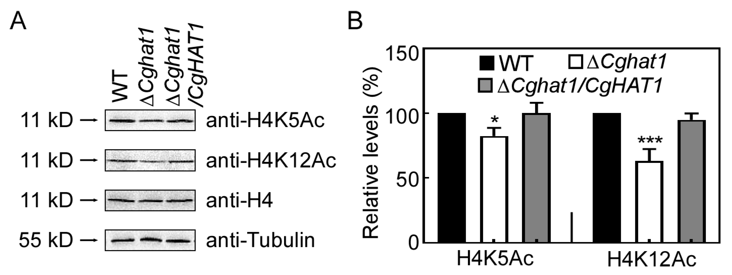
To test the roles of CgHat1 in nucleus, we obtained the CgHAT1 gene deletion mutant ΔCghat1 according to the homologous recombination principle (Figure S1A,B). Since two mutant strains were indistinguishable in general phenotypes, we randomly selected ΔCghat1#15 for further study. The yeast ScHat1 acetylates the histone H4K5 and H4K12 [9,10]. Thus, we examined the acetylation levels of H4K5 and H4K12. Compared to wide-type (WT) and complemented strain ΔCghat1/CgHAT1, ΔCghat1 showed significantly decreased acetylation levels of both H4K5 and H4K12 (Figure 3). These results suggest that CgHat1 regulates the acetylation of H4K5 and H4K12.

3.4. CgHat1 Is Important for the Growth on PDA Media and for the Colony Pigmentation

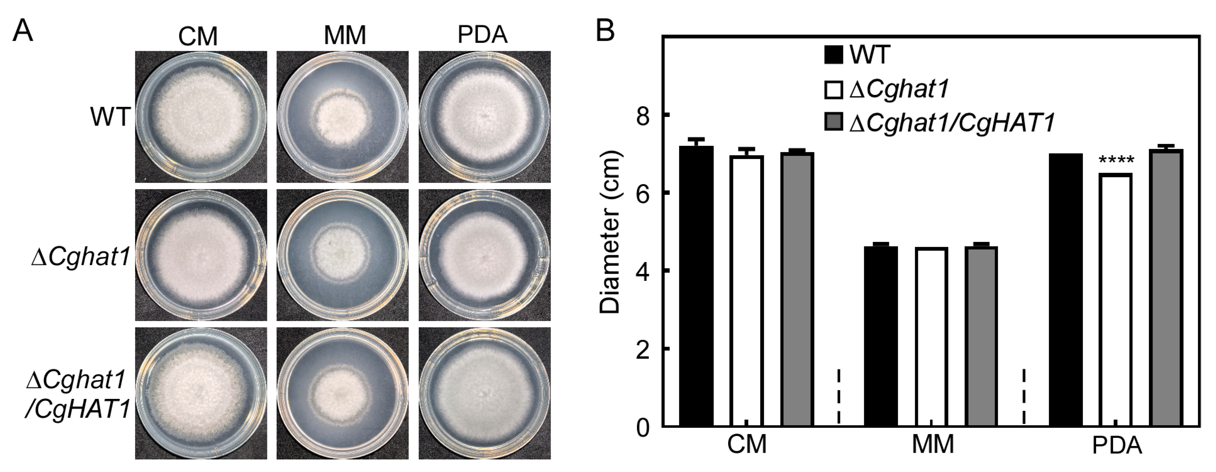
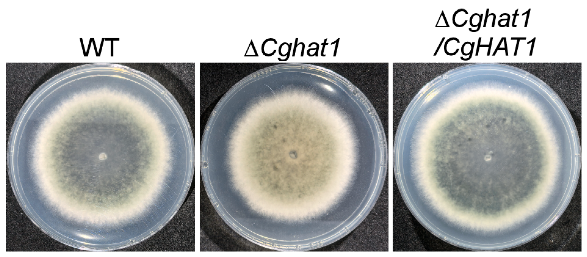
To address the role of CgHat1 in growth, the WT, ΔCghat1, and ΔCghat1/CgHAT1 strains were cultured on CM, MM, and PDA agar plates. The results showed that all the strains exhibit similar growth levels on CM and MM agar plates, whereas the ΔCghat1 mutant showed significantly decreased growth rates compared to WT and ΔCghat1/CgHAT1 on PDA agar plates (Figure 4). Additionally, compared to the colony pigmentation in WT and ΔCghat1/CgHAT1, the ΔCghat1 mutant showed absolutely no pigmentation (Figure 5). These results suggest that CgHat1 plays important in growth on PDA media and in colony pigmentation.

3.5. CgHat1 Is Critical for Asexual Development

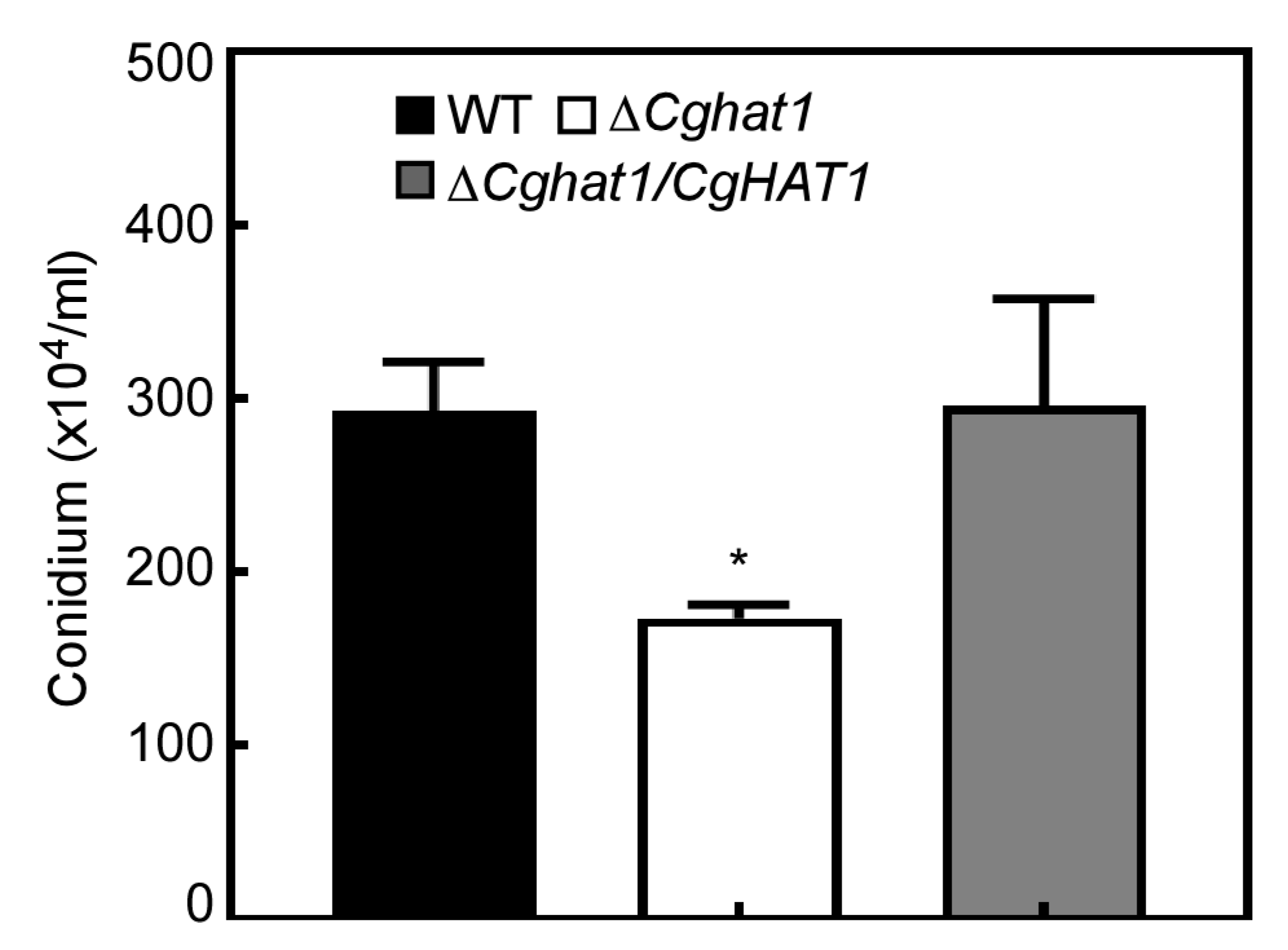
As asexual conidia play an important role in the disease cycle and infection of Colletotrichum [16,17], the conidiation production of the WT, ΔCghat1, and ΔCghat1/CgHAT1 strains were collected, quantified, and analyzed. After 3 days of incubation, the WT and ΔCghat1/CgHAT1 strains produced about 300 × 104 conidia per milliliter, compared to no more than 200 × 104 conidia/mL produced by ΔCghat1 mutant (Figure 6). This result indicates that CgHat1 is critical for asexual development.

3.6. CgHat1 Is Required for Full Virulence

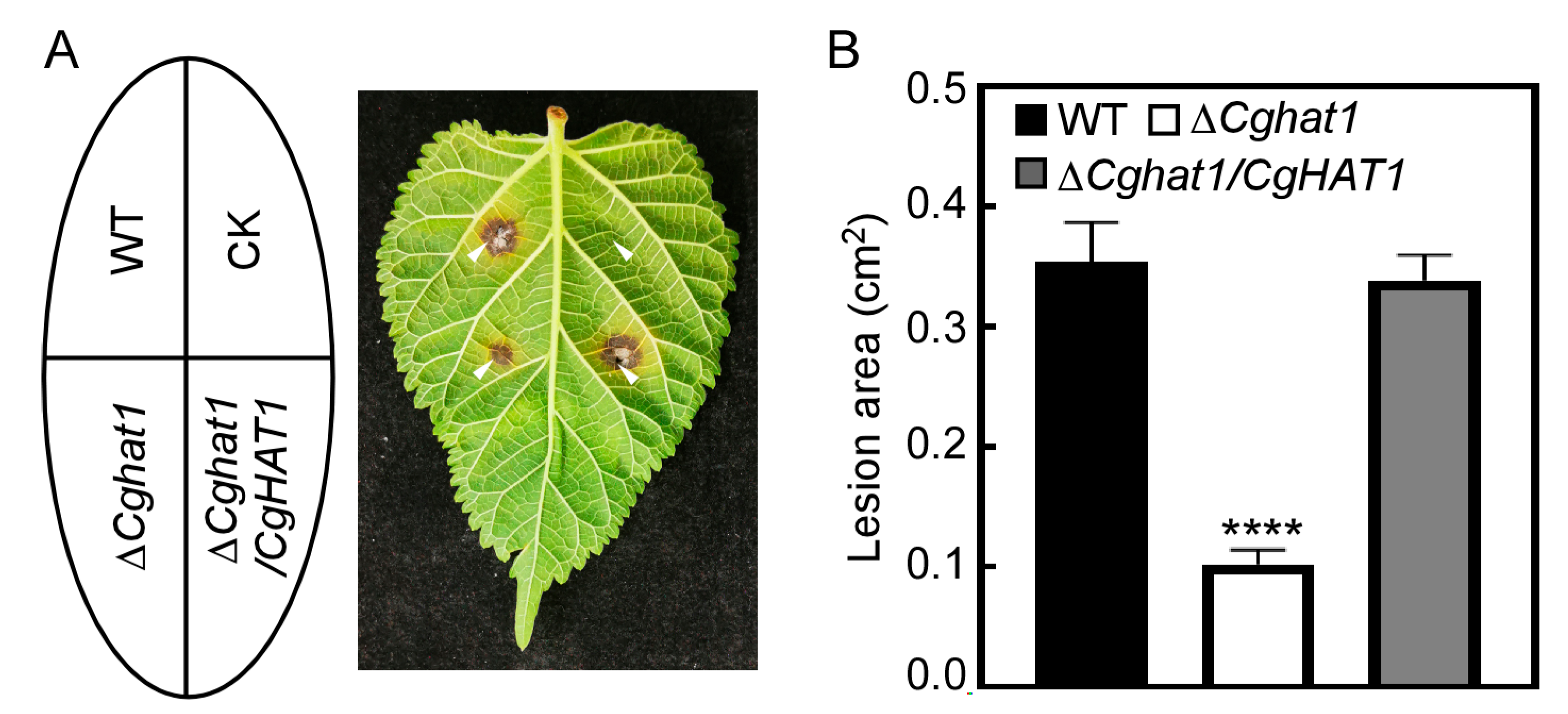
As a phytopathogen, we focus more of our interest on examining the roles of CgHat1 in virulence. The WT, ΔCghat1, and ΔCghat1/CgHAT1 strains were inoculated onto the M. alba leaves. After 4 days incubation, the lesions caused by WT and ΔCghat1/CgHAT1 strains showed obviously larger than that caused by ΔCghat1 mutant (Figure 7A). Furthermore, the statistical analysis revealed that the WT and ΔCghat1/CgHAT1 caused more than 0.3 cm2 lesion areas on M. alba leaves, compared to about 0.1 cm2 lesion areas caused by ΔCghat1 mutant (Figure 7B). This result indicates that CgHat1 is required for full virulence of C. gloeosporioides.

3.7. CgHat1 Plays Important Roles in Conidial Germination and Appressorial Formation

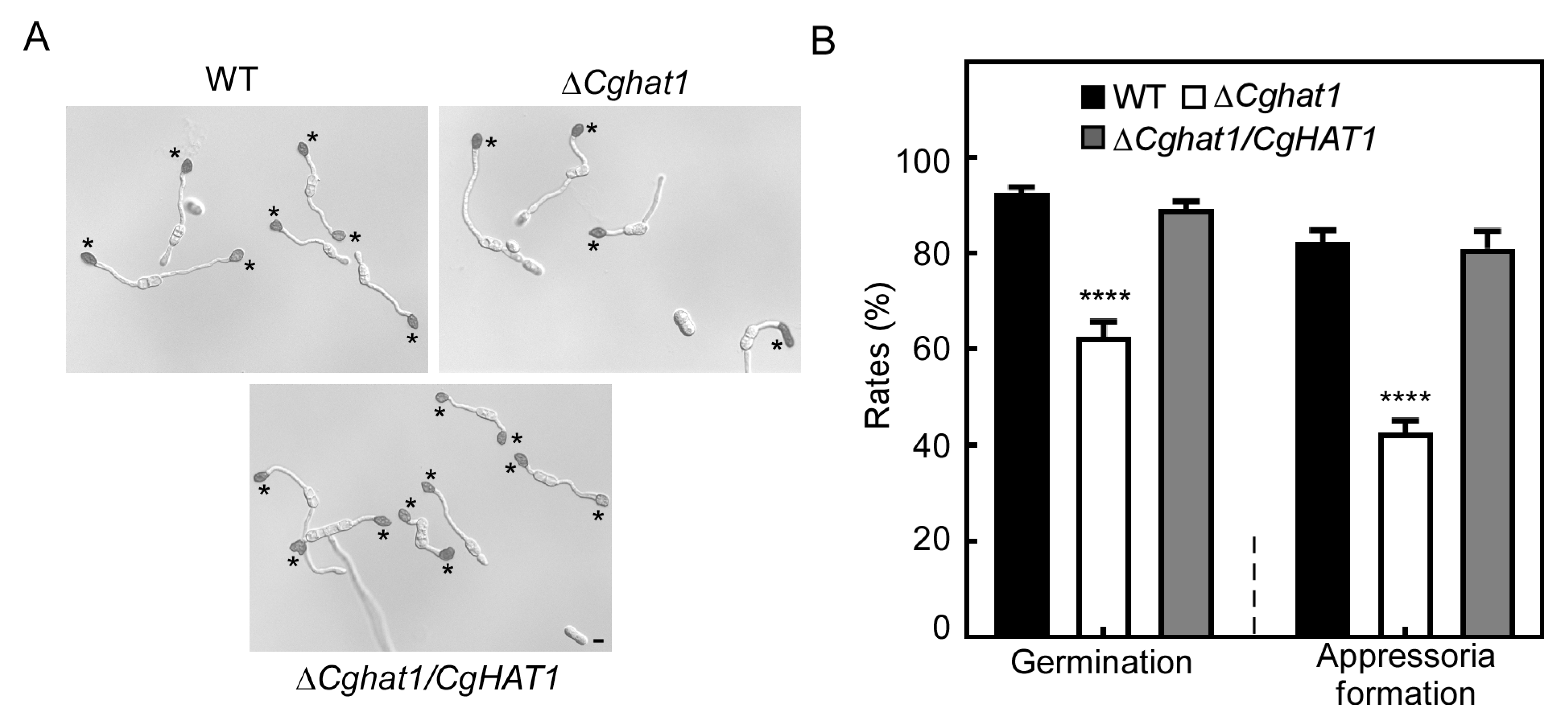
To investigate the possible reasons for the decreased virulence in ΔCghat1 mutant, we tested the appressorial development. The conidia of WT, ΔCghat1, and ΔCghat1/CgHAT1 strains were incubated on artificial hydrophobic surfaces. The percentages of conidial germination were more than 85% in WT and ΔCghat1/CgHAT1, compared to about 60% of that in the ΔCghat1 mutant (Figure 8). Similar decreased rates of ΔCghat1 mutant were also observed in appressorial formation, compared to WT and ΔCghat1/CgHAT1 strains (Figure 8). This result indicates that CgHat1 plays important roles in conidial germination and appressorial formation.

3.8. CgHat1 Regulates the Response to ER Stress

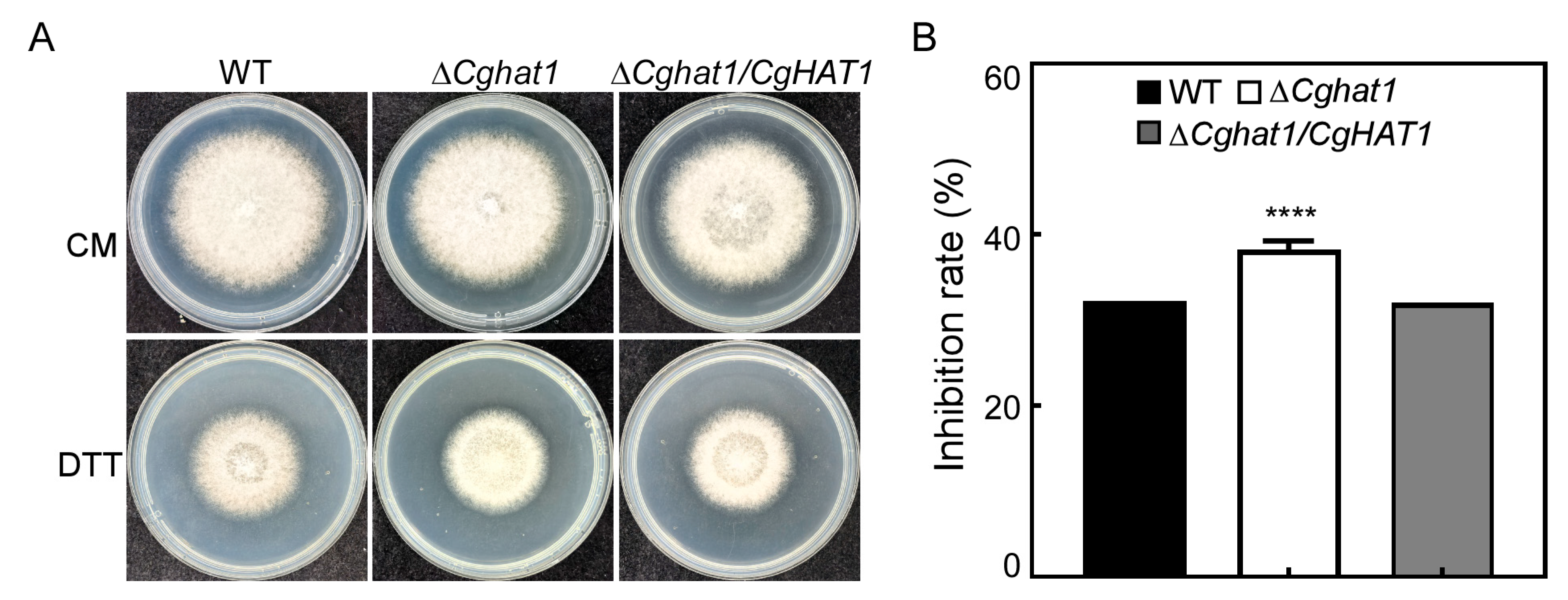
In order to grow normally and infect plants, pathogenic fungi must overcome many stresses [18,19]. Therefore, we tested whether CgHat1 may also associate with the response to environmental stresses. We found that the ΔCghat1 mutant showed significantly higher inhibition rates to endoplasmic reticulum (ER) stress (DTT) than that in WT and ΔCghat1/CgHAT1 strains (Figure 9). Meanwhile, we also found that the ΔCghat1 mutant showed similar sensitivity with WT and ΔCghat1/CgHAT1 strains to osmotic stresses (NaCl and KCl), cell wall integrity stresses (SDS and CR), oxidative stress (H2O2) and rapamycin stress (Figure S2). These results suggest that CgHat1 regulates the response to ER stress.

4. Discussion

In the long-term and complicated arms race with host plants, pathogens, including Colletotrichum spp., successfully infect host plants through various strategies such as gene transcription rearrangement [2]. HATs are necessary co-activators for transcriptional activation by modifying histones [20,21]. In the past 20 years, the importance of HATs in the pathogenicity of fungi have already been highlighted [22,23,24]. The Hat1 protein is one of the first identified histone acetyltransferases and mainly promotes gene transcription by promoting the acetylation of lysine K5 and K12 in histone H4 [9,10], which is consistent with our study showing that CgHat1 is localized in nucleus to regulate the H4K5Ac and H4K12Ac in C. gloeosporioides. Furthermore, the phylogenetic analysis revealed that the sequence of Hat1 proteins is highly similar in fungi, especially in Colletotrichum species. The conserved role of Hat1 in acetylation and sequence similarity of such proteins might forecast its conservative biological functions in different organisms.
Actually, CgHat1 plays important roles in conidiation, which is consistent with its homologues in Pestalotiopsis microspora [25]. Meanwhile, its biological function might also be species-specific. Our results revealed that CgHat1 is dispensable for growth in synthetic medium and positively regulates growth in semisynthetic medium. However, the yeast ScHat1 is not involved in growth, while the absence of the HAT1 gene in C. albicans could cause elongated cells [11,12]. Furthermore, although both of the Hat1 proteins in M. oryzae and C. albicans participate in the pathogenic process, their pathogenic mechanisms vary across different species. To achieve successful infection, M. oryzae employs Hat1 to regulate the turgor pressure of appressorium, whereas C. albicans primarily utilizes Hat1 to influence pathogenicity by controlling the formation of its infection structures [12,13]. These raise the question of how CgHat1 functions in the pathogenicity of C. gloeosporioides.
The significantly decreased virulence of ΔCghat1 revealed that CgHat1 is required for pathogenicity. We reasoned that the defect in virulence of ΔCghat1 mutant was directly due to the decreased conidial germination and appressorial formation, which is essential for the penetration and host-colonization of Colletotrichum spp. [16,17]. Moreover, the studies in M. oryzae showed that the host-derived ER stress is a common barrier for the successful infection of pathogens [26,27]. Therefore, the susceptibility of ΔCghat1 mutant to ER stress might also be a reason for its pathogenicity defect. Additionally, the melanin layer is essential for the penetration and pathogenicity of M. oryzae and Alternaria alternata [28,29,30]. The reduced pigmentation in ΔCghat1 mutant might be another reason for its reduced pathogenicity. However, it is unclear whether there is a direct correlation between pigmentation and pathogenicity in Colletotrichum and further studies are highly warranted.

5. Conclusions

Taken together, we identified and functional characterized the histone acetyltransferase CgHat1 in C. gloeosporioides. Our findings showed that CgHat1 is localized in the nucleus and regulates the H4K5Ac and H4K12Ac. The ΔCghat1 mutant is pleiotropic in defects, ranging from growth to conidiation and responses to ER stress. Our studies provide new insight for exploring such a protein as a potential fungicide target for anthracnose control. However, the genes regulated by CgHat1 are not yet clear in Colletotrichum. Further studies will focus on the interactive analysis between transcriptome and acetylomics.

Supplementary Materials

The following supporting information can be downloaded at https://www.mdpi.com/article/10.3390/jof11110768/s1: Figure S1: Targeted gene deletion of CgHAT1 gene in C. gloeosporioides; Figure S2: CgHat1 is dispensable for the response to multiple stresses; Table S1: Primers used in this study.

Author Contributions

Conceptualization, C.X. and J.A.; methodology, B.J.; software, B.J.; validation, B.J., C.X. and J.A.; formal analysis, B.J., C.X. and J.A.; investigation, B.J. and Q.Z.; resources, Y.L., M.L., Z.C. and J.A.; data curation, B.J.; writing—original draft preparation, B.J.; writing—review and editing, B.J., C.X. and J.A.; visualization, B.J.; supervision, B.J., C.X. and J.A.; project administration, C.Z. and J.A.; funding acquisition, J.A. All authors have read and agreed to the published version of the manuscript.

Funding

This research was funded by Modern Agricultural Industry Technology System Special Project, the earmarked fund for CARS-18.

Institutional Review Board Statement

Not applicable.

Informed Consent Statement

Not applicable.

Data Availability Statement

The original contributions presented in this study are included in the article/Supplementary Materials. Further inquiries can be directed to the corresponding authors.

Conflicts of Interest

The authors declare no conflicts of interest. The funders had no role in the design of the study; in the collection, analyses, or interpretation of data; in the writing of the manuscript; or in the decision to publish the results.

Abbreviations

The following abbreviations are used in this manuscript:
CMComplete medium
CRCongo red
DTTDimethyl sulfoxide
EREndoplasmic reticulum
HATHistone acetyltransferase
MMMinimal medium
PCRPolymerase Chain Reaction
PDAPotato Dextrose Agar

References

  1. Dean, R.; Van Kan, J.A.; Pretorius, Z.A.; Hammond-Kosack, K.E.; Di Pietro, A.; Spanu, P.D.; Rudd, J.J.; Dickman, M.; Kahmann, R.; Ellis, J.; et al. The Top 10 fungal pathogens in molecular plant pathology. Mol. Plant Pathol. 2012, 13, 414–430. [Google Scholar] [CrossRef]
  2. O’Connell, R.J.; Thon, M.R.; Hacquard, S.; Amyotte, S.G.; Kleemann, J.; Torres, M.F.; Damm, U.; Buiate, E.A.; Epstein, L.; Alkan, N.; et al. Lifestyle transitions in plant pathogenic Colletotrichum fungi deciphered by genome and transcriptome analyses. Nat. Genet. 2012, 44, 1060–1065. [Google Scholar] [CrossRef]
  3. Weir, B.S.; Johnston, P.R.; Damm, U. The Colletotrichum gloeosporioides species complex. Stud. Mycol. 2012, 73, 115–180. [Google Scholar] [CrossRef]
  4. Zhang, Y.; An, B.; Wang, W.; Zhang, B.; He, C.; Luo, H.; Wang, Q. Actin-bundling protein fimbrin regulates pathogenicity via organizing F-actin dynamics during appressorium development in Colletotrichum gloeosporioides. Mol. Plant Pathol. 2022, 23, 1472–1486. [Google Scholar] [CrossRef]
  5. Yang, J.; Li, B.; Pan, Y.T.; Wang, P.; Sun, M.L.; Kim, K.T.; Sun, H.; Ye, J.R.; Jiao, Z.; Lee, Y.H.; et al. Phospho-code of a conserved transcriptional factor underpins fungal virulence. BMC Biol. 2024, 22, 179. [Google Scholar] [CrossRef]
  6. Liu, S.; Bu, Z.; Zhang, X.; Chen, Y.; Sun, Q.; Wu, F.; Guo, S.; Zhu, Y.; Tan, X. The new CFEM protein CgCsa required for Fe(3+) homeostasis regulates the growth, development, and pathogenicity of Colletotrichum gloeosporioides. Int. J. Biol. Macromol. 2024, 274, 133216. [Google Scholar] [CrossRef]
  7. Bannister, A.J.; Kouzarides, T. Regulation of chromatin by histone modifications. Cell Res. 2011, 21, 381–395. [Google Scholar] [CrossRef] [PubMed]
  8. Kurdistani, S.K.; Grunstein, M. Histone acetylation and deacetylation in yeast. Nat. Rev. Mol. Cell Biol. 2003, 4, 276–284. [Google Scholar] [CrossRef]
  9. Suter, B.; Pogoutse, O.; Guo, X.; Krogan, N.; Lewis, P.; Greenblatt, J.F.; Rine, J.; Emili, A. Association with the origin recognition complex suggests a novel role for histone acetyltransferase Hat1p/Hat2p. BMC Biol. 2007, 5, 38. [Google Scholar] [CrossRef] [PubMed]
  10. Wu, H.; Moshkina, N.; Min, J.; Zeng, H.; Joshua, J.; Zhou, M.M.; Plotnikov, A.N. Structural basis for substrate specificity and catalysis of human histone acetyltransferase 1. Proc. Natl. Acad. Sci. USA 2012, 109, 8925–8930. [Google Scholar] [CrossRef] [PubMed]
  11. Qin, S.; Parthun, M.R. Histone H3 and the histone acetyltransferase Hat1p contribute to DNA double-strand break repair. Mol. Cell Biol. 2002, 22, 8353–8365. [Google Scholar] [CrossRef]
  12. Tscherner, M.; Stappler, E.; Hnisz, D.; Kuchler, K. The histone acetyltransferase Hat1 facilitates DNA damage repair and morphogenesis in Candida albicans. Mol. Microbiol. 2012, 86, 1197–1214. [Google Scholar] [CrossRef]
  13. Yin, Z.; Chen, C.; Yang, J.; Feng, W.; Liu, X.; Zuo, R.; Wang, J.; Yang, L.; Zhong, K.; Gao, C.; et al. Histone acetyltransferase MoHat1 acetylates autophagy-related proteins MoAtg3 and MoAtg9 to orchestrate functional appressorium formation and pathogenicity in Magnaporthe oryzae. Autophagy 2019, 15, 1234–1257. [Google Scholar] [CrossRef]
  14. Bruno, K.S.; Tenjo, F.; Li, L.; Hamer, J.E.; Xu, J.R. Cellular localization and role of kinase activity of PMK1 in Magnaporthe grisea. Eukaryot. Cell 2004, 3, 1525–1532. [Google Scholar] [CrossRef]
  15. Schneider, C.A.; Rasband, W.S.; Eliceiri, K.W. NIH Image to ImageJ: 25 years of image analysis. Nat. Methods 2012, 9, 671–675. [Google Scholar] [CrossRef] [PubMed]
  16. Fukada, F.; Kodama, S.; Nishiuchi, T.; Kajikawa, N.; Kubo, Y. Plant pathogenic fungi Colletotrichum and Magnaporthe share a common G(1) phase monitoring strategy for proper appressorium development. New Phytol. 2019, 222, 1909–1923. [Google Scholar] [CrossRef] [PubMed]
  17. Fukada, F.; Kubo, Y. Colletotrichum orbiculare Regulates Cell Cycle G1/S Progression via a Two-Component GAP and a GTPase to Establish Plant Infection. Plant Cell 2015, 27, 2530–2544. [Google Scholar] [CrossRef]
  18. Yin, Z.; Zhang, X.; Wang, J.; Yang, L.; Feng, W.; Chen, C.; Gao, C.; Zhang, H.; Zheng, X.; Wang, P.; et al. MoMip11, a MoRgs7-interacting protein, functions as a scaffolding protein to regulate cAMP signaling and pathogenicity in the rice blast fungus Magnaporthe oryzae. Environ. Microbiol. 2018, 20, 3168–3185. [Google Scholar] [CrossRef]
  19. Cheng, Y.T.; Zhang, L.; He, S.Y. Plant-microbe interactions facing environmental challenge. Cell Host Microbe 2019, 26, 183–192. [Google Scholar] [CrossRef] [PubMed]
  20. Guo, R.; Chen, J.; Mitchell, D.L.; Johnson, D.G. GCN5 and E2F1 stimulate nucleotide excision repair by promoting H3K9 acetylation at sites of damage. Nucleic Acids Res. 2011, 39, 1390–1397. [Google Scholar] [CrossRef]
  21. Strenkert, D.; Schmollinger, S.; Sommer, F.; Schulz-Raffelt, M.; Schroda, M. Transcription factor-dependent chromatin remodeling at heat shock and copper-responsive promoters in Chlamydomonas reinhardtii. Plant Cell 2011, 23, 2285–2301. [Google Scholar] [CrossRef] [PubMed]
  22. Jeon, J.; Kwon, S.; Lee, Y.H. Histone acetylation in fungal pathogens of plants. Plant Pathol. J. 2014, 30, 1–9. [Google Scholar] [CrossRef]
  23. Zhang, S.; Liang, M.; Naqvi, N.I.; Lin, C.; Qian, W.; Zhang, L.H.; Deng, Y.Z. Phototrophy and starvation-based induction of autophagy upon removal of Gcn5-catalyzed acetylation of Atg7 in Magnaporthe oryzae. Autophagy 2017, 13, 1318–1330. [Google Scholar] [CrossRef]
  24. Qian, H.; Song, L.; Wang, L.; Yang, Q.; Wu, R.; Du, J.; Zheng, B.; Liang, W. FolIws1-driven nuclear translocation of deacetylated FolTFIIS ensures conidiation of Fusarium oxysporum. Cell Rep. 2024, 43, 114588. [Google Scholar] [CrossRef] [PubMed]
  25. Zhang, Q.; Chen, L.; Yu, X.; Liu, H.; Akhberdi, O.; Pan, J.; Zhu, X. A B-type histone acetyltransferase Hat1 regulates secondary metabolism, conidiation, and cell wall integrity in the taxol-producing fungus Pestalotiopsis microspora. J. Basic Microbiol. 2016, 56, 1380–1391. [Google Scholar] [CrossRef]
  26. Tang, W.; Jiang, H.; Aron, O.; Wang, M.; Wang, X.; Chen, J.; Lin, B.; Chen, X.; Zheng, Q.; Gao, X.; et al. Endoplasmic reticulum-associated degradation mediated by MoHrd1 and MoDer1 is pivotal for appressorium development and pathogenicity of Magnaporthe oryzae. Environ. Microbiol. 2020, 22, 4953–4973. [Google Scholar] [CrossRef]
  27. Yin, Z.; Feng, W.; Chen, C.; Xu, J.; Li, Y.; Yang, L.; Wang, J.; Liu, X.; Wang, W.; Gao, C.; et al. Shedding light on autophagy coordinating with cell wall integrity signaling to govern pathogenicity of Magnaporthe oryzae. Autophagy 2020, 16, 900–916. [Google Scholar] [CrossRef]
  28. Kawamura, C.; Moriwaki, J.; Kimura, N.; Fujita, Y.; Fuji, S.; Hirano, T.; Koizumi, S.; Tsuge, T. The melanin biosynthesis genes of Alternaria alternata can restore pathogenicity of the melanin-deficient mutants of Magnaporthe grisea. Mol. Plant Microbe Interact. 1997, 10, 446–453. [Google Scholar] [CrossRef]
  29. Talbot, N.J. On the trail of a cereal killer: Exploring the biology of Magnaporthe grisea. Annu. Rev. Microbiol. 2003, 57, 177–202. [Google Scholar] [CrossRef]
  30. Qi, Z.; Liu, M.; Dong, Y.; Zhu, Q.; Li, L.; Li, B.; Yang, J.; Li, Y.; Ru, Y.; Zhang, H.; et al. The syntaxin protein (MoSyn8) mediates intracellular trafficking to regulate conidiogenesis and pathogenicity of rice blast fungus. New Phytol. 2016, 209, 1655–1667. [Google Scholar] [CrossRef] [PubMed]
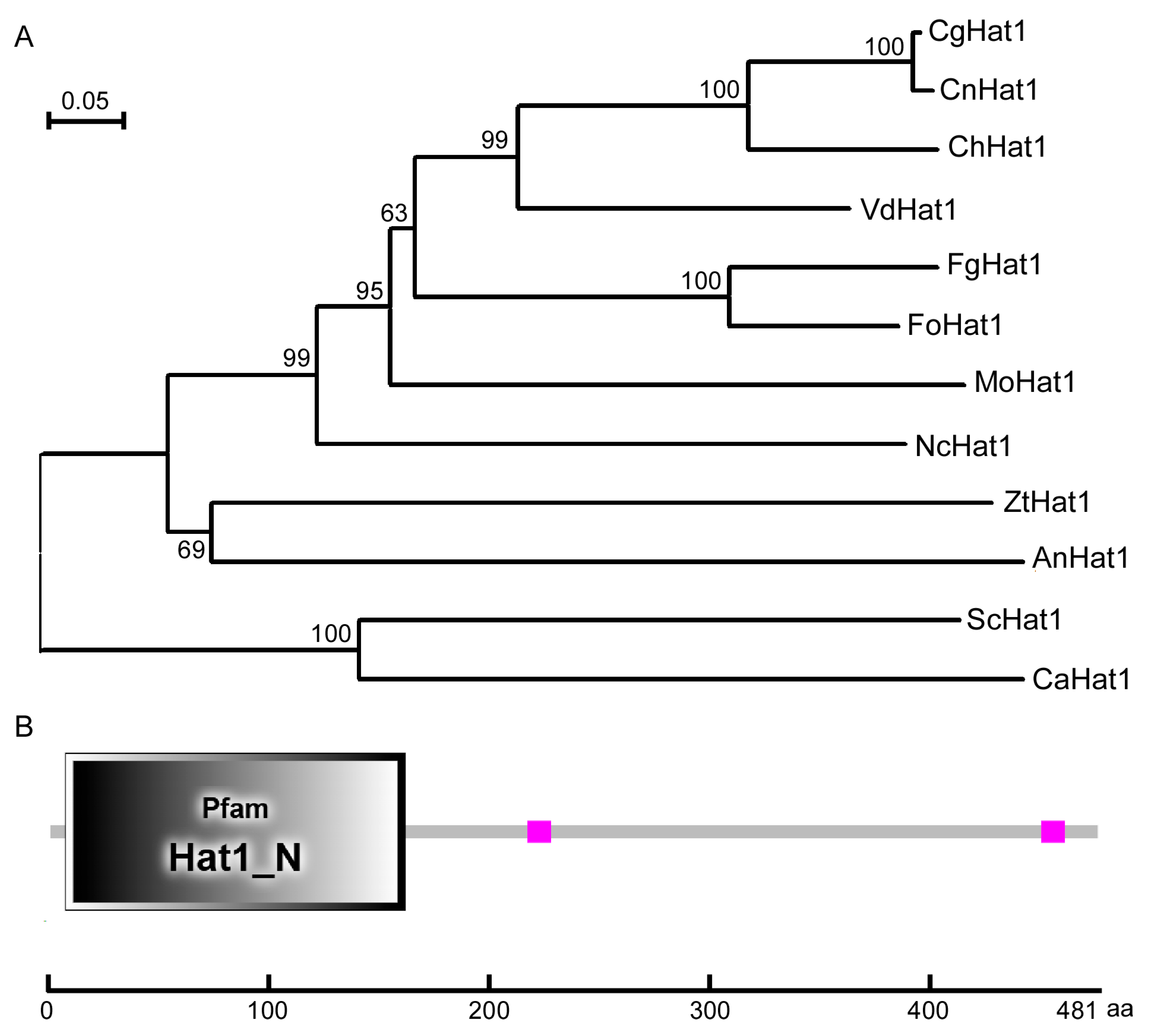
Figure 1. Phylogenetic analysis and domain prediction of CgHat1. (A). The phylogenetic tree of Hat1 proteins in diverse fungi constructed by MEGA5.05 using neighbor-joining method with 1000 bootstrap replicates. The GenBank accession numbers and the related organisms are as follows: CgHat1 (EQB43824.1, Colletotrichum gloeosporioides), CnHat1 (KAJ0285795.1, Colletotrichum noveboracense), ChHat1 (XP_018162470.1, Colletotrichum higginsianum), VdHat1 (KAF3354044.1, Verticillium dahliae), FgHat1 (XP_011326992.1, Fusarium graminearum), FoHat1 (KAI7768789.1, Fusarium oxysporum), MoHat1 (XP_003717199.1, Magnaporthe oryzae), NcHat1 (XP_957363.1, Neurospora crassa), ZtHat1 (XP_003854292.1, Zymoseptoria tritici), AnHat1 (XP_663818.1, Aspergillus nidulans), ScHat1 (NP_015324.1, Saccharomyces cerevisiae), and CaHat1 (KHC75880.1, Candida albicans). (B). The domain prediction of CfAda3 by the SMART website. The gray quadrilateral represents the Hat1 domain.
Jof 11 00768 g001
Figure 2. CgHat1 is localized to the nucleus. (A). CgHat1 is localized to the nucleus in hyphae. The hyphae of strains co-expressed CgHat1-GFP and H1-RFP observed under fluorescent microscope. The merged area of GFP and RFP signal showed that CgHat1-GFP localized in the nucleus. (B). CgHat1 is localized to the nucleus in conidia. The merged area showed that CgHat1-GFP localized in the nucleus. Bar = 5 μm.
Jof 11 00768 g002
Figure 3. CgHat1 regulates the acetylation of H4K5 and H4K12. (A). Western blot analysis of the H4K5 and H4K12 acetylation with anti-H4K5, anti-H4K12, anti-H4, and anti-Tubulin antibodies. (B). The relative levels of acetylation were calculated as the anti-H4K5Ac/anti-H4 and anti-H4K12Ac/anti-H4 ratios by integrated signal density using ImageJ 1.48v software. The ratio of WT was normalized as 1 (* p < 0.05, *** p < 0.001).
Jof 11 00768 g003
Figure 4. CgHat1 is involved in the growth on PDA media. (A). Colony morphology of the WT, ΔCghat1, and ΔCghat1/CgHAT1 strains on the top of CM, MM, and PDA agar plates. (B). Statistical analysis of the colony diameters of strains on CM, MM, and PDA media. Error bars represent SD and asterisks denote statistical significances (**** p < 0.0001).
Jof 11 00768 g004
Figure 5. CgHat1 is important for colony pigmentation. Colony morphology of the WT, ΔCghat1, and ΔCghat1/CgHAT1 strains on the bottom of PDA agar plates.
Jof 11 00768 g005
Figure 6. CgHat1 is critical for asexual development. The conidia produced by WT, ΔCghat1, and ΔCghat1/CgHAT1 strains were collected, quantified, and analyzed (* p < 0.05). Three biological experiments were performed for each strain with three technical replicates per biological experiment.
Jof 11 00768 g006
Figure 7. CgHat1 is required for full virulence. (A). The leaves of M. alba inoculated with WT, ΔCghat1, and ΔCghat1/CgHAT1 strains. Photographs taken at 3 days post-inoculation (dpi). The arrowheads denote infected sites. (B). Statistical analysis of lesion areas calculated by Image J on M. alba leaves. Error bars represent SD and asterisks denote significant differences (**** p < 0.0001).
Jof 11 00768 g007
Figure 8. CgHat1 plays important roles in conidial germination and appressorial formation. (A) The conidia of WT, ΔCghat1, and ΔCghat1/CgHAT1 strains incubated on artificial hydrophobic surfaces, and observed under microscopy. Asterisks indicate appressoria. Bar = 5 μm. (B). Statistical analysis of the conidial germination and appressorial formation rates. Error bars represent SD and asterisks denote significant differences (**** p < 0.0001).
Jof 11 00768 g008
Figure 9. CgHat1 regulates the response to ER stress. (A). Colony morphology of the WT, ΔCghat1, and ΔCghat1/CgHAT1 strains on the CM and CM supplemented with 2.5 mM DTT agar plates. (B). Statistical analysis of inhibition rates of the strains to DTT stress. Error bars represent SD and asterisks denote statistical significances (**** p < 0.0001).
Jof 11 00768 g009
Disclaimer/Publisher’s Note: The statements, opinions and data contained in all publications are solely those of the individual author(s) and contributor(s) and not of MDPI and/or the editor(s). MDPI and/or the editor(s) disclaim responsibility for any injury to people or property resulting from any ideas, methods, instructions or products referred to in the content.