Article

Identification and Aggressiveness of Fusarium Species Associated with Onion Bulb (Allium cepa L.) during Storage

by
Roderic Gilles Claret Diabankana
1,*,
Mikhail Frolov
1,
Bakhtiyar Islamov
1,
Elena Shulga
1,
Maria Nikolaevna Filimonova
2,
Daniel Mawuena Afordoanyi
1 and
Shamil Validov
1
1
Laboratory of Molecular Genetics and Microbiology Methods, Kazan Scientific Center of the Russian Academy of Sciences, 420111 Kazan, Russia
2
Academic and Research Centre, Institute of Fundamental Medicine and Biology, Kazan Federal University, 420008 Kazan, Russia
*
Author to whom correspondence should be addressed.
J. Fungi 2024, 10(2), 161; https://doi.org/10.3390/jof10020161
Submission received: 25 December 2023 / Revised: 16 February 2024 / Accepted: 17 February 2024 / Published: 19 February 2024

Abstract

:
Plant pathogens present a major challenge to crop production, leading to decreased yield and quality during growth and storage. During long-term storage, healthy onions can develop diseases from latent pathogen infections. This poses a challenge for onion growers because infected bulbs without visible symptoms can lead to significant crop losses during the growing season. In this study, we aimed to isolate and identify Fusarium species from yellow onion bulbs (Allium cepa L.) that developed disease symptoms during storage. The aggressiveness of these strains against onion bulbs and seedlings was also evaluated. The isolated strains were further subjected to morphological and molecular differentiation. The results revealed that all 16 isolated strains belonged to the Fusarium complex species incarnatum-equiseti and Fusarium fujikuroi, namely, F. proliferatum (98%), F. oxysporum (1%), and Fusarium sp. (1%). Koch’s postulate analysis of isolated strains revealed varying aggressiveness on onion bulbs and plants depending on fungal species. Disease symptoms developed more slowly on plants than on onion bulb plants according to Koch’s postulates. Moreover, the results revealed that Fusarium strains that can infect onion plants were less pathogenic to onion bulbs and vice versa. In addition, three isolates were found to be non-pathogenic to onions. Furthermore, the in vitro control of Fusarium species through Bacillus velezensis KS04-AU and Streptomyces albidoflavus MGMM6 showed high potential for controlling the growth of these pathogenic fungi. These results may contribute to the development of environmentally friendly approaches for controlling onion spoilage caused by pathogens during storage.

1. Introduction

Technological advancements in agriculture have greatly contributed to the development of global crop production. However, despite the progress made in production, postharvest loss during storage remains a significant challenge. The spoilage of crops caused by microorganisms, including many bacterial and fungal species, has emerged as a critical issue in modern agriculture, resulting in substantial economic losses and threats to worldwide food security [1]. According to the Food Safety Department of the World Health Organization, foodborne illness affects nearly 1 out of 10 individuals worldwide, resulting in approximately 420,000 deaths each year and a significant loss of 33 million years of healthy life. The onion bulb (Allium cepa L.) is an important seasonal crop with a high commercial value, and its yield is affected by several factors during long-term storage. Onion bulbs are susceptible to numerous postharvest diseases caused by several phytopathogens from genera such as Aspergillus, Fusarium, Rhizopus, Penicillium, Urocystis, and Alternaria [1,2]. The postharvest losses of onions can reach as high as 40–60% [3,4,5]. Among these losses, 10–40% can be attributed to diseases caused by fungi, bacteria, nematodes, or viruses [6,7]. Among phytopathogenic microorganisms, Fusarium is the most persistent soilborne fungal plant pathogen [8,9]. Species of this genus are prevalent in soils across diverse climate regions and are associated with a wide variety of plants, causing considerable damage to crops [10,11]. Fusarium, a major causal agent of bulb basal rot, has been reported in several studies [12,13,14]. Fusarium pathogens, known for their devastating effects on plant health, can pose significant risks to human health [15]. These pathogens not only infect plants but also produce toxic secondary metabolites called mycotoxins, which can contaminate food and feed products. The consumption of such contaminated food can lead to severe health issues in humans, including acute poisoning, chronic diseases, and even cancer [16]. Furthermore, Fusarium species can also cause opportunistic infections in immunocompromised individuals, such as those with compromised immune systems or those recovering from surgery [17]. These infections can manifest in various forms, including skin infections, keratitis (eye infections), and invasive disseminated diseases [18]. Control of postharvest onion diseases is essential to ensure the long-term storage and marketability of onion bulbs. Several methods such as sanitation and hygiene practices, temperature and humidity management, as well as chemical treatments, have been used to control the development of disease during long-term onion bulb storage [19,20,21]. These methods offer effective disease management, however, they are also supplied with challenges related to their cost, potentially raising concerns regarding food safety and environmental impact, as well as the risk of resistance development [22]. Onion bulbs affected by phytopathogens may not show symptoms of infection during visual inspection (a method that is usually used at onion packaging enterprises). However, over time, these pathogens may cause severe damage during storage [23]. In addition, these infected onion bulbs without obvious symptoms could reach markets nationwide. This concern is exacerbated by the presence of known mycotoxins released by these pathogens, which may negatively affect human health [24,25]. Hence, identifying phytopathogenic microorganisms responsible for onion crop spoilage and determining their sources are crucial for making decisions about plant protection measures. Biological control agents (BCAs) are a promising alternative for controlling plant pathogens, offering environmentally friendly and sustainable methods of suppressing diseases [26]. Several BCAs have shown efficacy against phytopathogenic microorganisms belonging to the following genera Fusarium, Rhizopus, Penicillium, Alternaria Pantoea, Pseudomonas, Burkholderia, Xanthomonas, and Erwinia [27,28]. These BCAs act by competing with pathogens for nutrients and niches, as well as, by producing antifungal compounds [29,30]. In this study, we focused on identifying and characterizing spoilage causal agents, specifically Fusarium, that affect the most common variety of yellow onions (Allium cepa L.) sold in various commercial marketplaces in Kazan (Tatarstan, Russia), as well as on the development of an environmentally friendly approach to control these causal agents.

2. Materials and Methods

2.1. Isolation and Identification of Pathogens from Diseased Onion Bulbs

The onion bulbs (10 pieces of bulbs) variety “golden semko” (Semko-Junior LLC, Moscow, Russia) used in this study were purchased from different commercial marketplaces located in Kazan (Tatarstan, Russia,) and stored at room temperature and in the absence of light. During storage, onion bulbs with rotting parts were randomly removed and used to identify the causal pathogens of Fusarium species (Figure 1).
For this purpose, the tissues were cut into small segments, surface disinfected with a 70% ethanol solution, and rinsed with sterile water. The segments were then immersed for 1 min in a 2% sodium hypochlorite solution (containing 0.5% sodium dodecyl sulfate (SDS)), followed by washing with distilled water. The segments were plated on potato dextrose agar (HIMEDIA, Moscow, Russia) supplemented with rifampicin to a final concentration of 100 µg/mL and incubated at 27 ± 1 °C in the dark. The grown fungi were preselected based on their morphology and replated onto potato dextrose agar (PDA). Furthermore, to obtain a pure fungal colony, the single-spore subculture method was performed according to Leslie and Summerell [31]. After serial dilution, aliquots of 100 µL from the last three dilutions (5-fold dilution) were plated on Sabouraud Dextrose Agar (HIMEDIA, Moscow, Russia) and incubated at 27 ± 1 °C. To distinguish the isolated fungal species, the grown colonies were fingerprinted using BOX-PCR. The selected strains were further subjected to molecular sequence-based identification.

2.2. Molecular Analysis

2.2.1. DNA Isolation

Chromosomal DNA was extracted from 5-day-old fungi grown on PDA agar plates using a phenol–chloroform extraction method according to Green et al. [32]. The quantification of isolated DNA was analyzed using a NanoDrop spectrophotometer and gel electrophoresis [33].

2.2.2. DNA Fingerprinting Analysis (BOX-PCR)

BOX-PCR was performed in a 25 µL volume containing 2.5 µL of 10× PCR buffer, 0.4 µL of dNTP mixture (10 µM), 1.25 µL of 10 µM BOXAIR (5′-CTACGGCAAGGCGACGCTGACG-3′), 5.0 µL of DNA template (50 ng), 1.0 µL of Taq DNA polymerase (5 U), 50 ng of template DNA, and free nuclease-free water. Amplification was performed using a T100 thermocycler (Bio-Rad, Hercules, CA, USA) under the following conditions: initial denaturation at 95 °C for 2 min; 30 cycles of 94 °C for 30 s and 58 °C for 30 s; and 72 °C for 8 min. The final cycle was extended to 72 °C for 10 min. The DNA fragments were detected via 1.5% agarose gel electrophoresis and visualized on Gel Doc EZ Imager with Image Lab 6.0 software (Bio-Rad, Hercules, CA, USA). Dendrogram analysis was performed using GelJ software v.3.0 (Java Application, Logroño, Spain). Hierarchical clustering analysis of the BOX DNA profiles of the isolated fungi was performed using the Jaccard similarity coefficient and UPGMA clustering analysis methods, with the matching band tolerance set at 1%.

2.2.3. Molecular Fungal Species Identification and Phylogenetic Relationships

The preselected fungal strains were molecularly identified via PCR amplification and fragmentation sequencing analysis of the targeting translation elongation factor-1 alpha (tef-1α) gene and the universal internal transcribed spacer (ITS). The polymerase chain reaction amplification was performed using QuantStudio 5 (Thermo Fisher Scientific, Cleveland, OH, USA) in a 50 µL reaction mixture containing 1 µL of each (100 µM) primer, 10 mM DNTP mixture, 10 µL of PCR master mix (Evrogen, Moscow, Russia), 3 µL of template DNA (50 ng/mL), and water-free nuclease. For tef-1α (F-5′-ATGGGTAAGGAAGACAAGAC-3′; R-5′-GGAAGTACCAGTGATCATGTT-3′) [34]. PCR amplification was performed under the following conditions: 95 °C for 2 min; 30 cycles of 94 °C for 30 s, 47 °C for 20 s, and 72 °C for 30 s. The final PCR cycle was followed by a cycle at 4°C for 10 min. The universal primer (ITS1-F-5′-TCCGTAGGTGAACCTGCGG-3′; ITS4-R-5′-GCTGCGTTCTCCATCGATGC-3′) was used to amplify the entire ITS region [35]. The amplification was performed under the following conditions: denaturation at 95 °C for 5 min; 35 cycles of 95 °C for 50 s, 56 °C for 50 s, and 72 °C for 1 min; and a final cycle of 72 °C for 10 min. The obtained PCR products were fragmented by electrophoresis in 1.5% agarose gel and visualized on Gel Doc EZ Imager with Image Lab 6.0 software (Bio-Rad, Hercules, CA, USA). The target fragment was purified using a DNA cleanup kit (Evrogen, Moscow, Russian) according to the manufacturer’s protocol. After purification, the DNA fragment was sequenced by the Sanger method using an automated sequencer 3500×L Dx Genetic Analyzer (Applied Biosystems, Waltham, MA, USA). The chromatograms were further analyzed using Snapgene software v. 7.0 (GSL BioTech LLC, San Diego, CA, USA).
The generated partial fragments were further compared with fungal DNA sequences using an online approach against the Fusarium multilocus sequence (MLST) database (https://fusarium.mycobank.org/page/Fusarium_identification; 20 September 2023). Furthermore, the obtained consensus sequences were subjected to BLAST searches for homology against the GenBank database using the BLASTN tool [36]. More than 40 related trains (with less than 96% similarity) were downloaded from NCBI and used to construct a phylogenetic tree. The bootstrap consensus tree inferred from 1000 replicates was used to represent the evolutionary history of the taxa analyzed [37]. Branches corresponding to partitions reproduced in less than 50% of the bootstrap clustering threshold were generated. Evolutionary distances were computed using the maximum composite likelihood method [38] and are expressed in units of the number of base substitutions per site. All ambiguous positions were removed for each sequence pair (pairwise deletion option). Evolutionary analyses were performed using MEGA11 [39].

2.3. Test of Pathogenicity

2.3.1. Preparation of Conidial Suspension of Isolated Fungal Strains

The conidial suspension of each isolated fungal strain was prepared from fungal isolated cultures grown up to seven days in Sabouraud broth (SDA; Merck, Darmstadt, Germany) at 25 ± 1 °C with an agitation rate of 150 rpm. After sporulation, the culture was passed through sterile cotton wool for spore filtration. The colony forming unit (CFU) was measured using a hemocytometer.

2.3.2. Pathogenicity Assay of Onion Seeds

The pathogenicity of the isolated fungus was tested in a pot containing sterile sand supplemented with a plant nutrient solution. For this purpose, the onion seeds variety “golden semko” (Semko-Junior LLC, Moscow, Russia) was sterilized according to Simons et al. [40] and incubated overnight at 4 °C for emergency germination. The pre-germinate seeds were inoculated for 10 min in a fungal spore suspension (spore diluted in 1% carboxymethyl cellulose to 103 CFU/mL, prepared as previously described by Afordoanyi et al. [41]) of each isolated strain and planted in pots (34.0 × 16.0 × 13.0 cm) containing sand amended with plant nutrient solution (PNS) [42]. The control group was prepared under the same conditions but without pathogens. Pots were incubated in a climate-controlled chamber under the following conditions: light intensity, 90%; day–night cycles, 16:8; humidity, 70%; and temperature, 26 ± 1 °C. For statistical analysis, 50 seeds inoculated with each Fusarium strain suspension were planted in a pot (a sample group). Each sample group was maintained in three replicates. For statistical analysis, the experiment was repeated in three independent tests. After 30 days of incubation, the plants were examined. The pathogenicity score (P.S.) of each isolated fungal strain was calculated using the following formula [43]:
P . S = a b N K × 100
where ∑ ab represents the total obtained by multiplying the number of diseased plants by their corresponding degree of damage. “N” is the total number of plants analyzed, representing the highest grade on the scale. This scale ranges from 0 to 4, where 0 indicates asymptomatic plants, 1 represents plants with small lesions (<2 mm), 2 indicates moderate damage without severe harm to the plant, 3 represents severe damage or developed lesions, and 4 indicates dead plants.

2.3.3. Pathogenicity Assay of Onion Bulbs

Koch’s postulates were used to confirm the pathogenicity of the isolated Fusarium species. For this purpose, onion bulbs were injected with 25 μL of Fusarium spore suspension (spore diluted in distilled water to 103 CFU/mL) using a sterile syringe. For the control group, onion bulbs were injected with sterile distilled water. Onion bulbs were placed in a container (34.0 × 16.0 × 13.0 cm) and incubated in the dark for up to 3 weeks at 24 ± 1 °C. For each treatment, 10 onion bulbs were maintained in 3 independent replicates over time. After incubation, onion bulbs were cut with a scalpel, and the severity of symptoms of Fusarium was evaluated as follows: no visible symptoms (0), slight damage (1), moderate damage (2), severe damage (3), high damage and bulb death (4) according to Tirado-Ramirez et al. [44]. After 4 weeks, onion bulbs were examined using the following formula, as described in Section 2.3.1.

2.3.4. In Vitro Control of Fusarium Isolates Using Bacillus velezensis KS04-AU and Streptomyces albidoflavus MGMM6

The antagonistic effects of B. velezensis KS04-AU (bacterial strain isolated from Senna occidentalis [45], microbial collection of FRC Kazan Scientific Center) and S. albidoflavus MGMM6 (bacterial strain isolated from the rhizosphere soil of spring wheat (Triticum aestivum L.) [46], microbial collection of FRC Kazan Scientific Center) against isolated Fusarium strains were assayed in dual cultures on PDA media. For this purpose, 10 µL of the highly virulent fungal strain suspension prepared in Section 2.3.1 was inoculated in the center of agar plates, after which 5 µL of the cell suspension of the KS04-AU and MGMM6 bacterial strains was co-inoculated. The plates were incubated at 28 ± 1 °C for up to 10 days.

2.4. In Planta Control of Fusarium Isolates through Bacillus velezensis KS04-AU and Streptomyces albidoflavus MGMM6

The in planta control of Fusarium isolates associated with onion bulbs through S. albidoflavus MGMM6 was evaluated as described in Section 2.3.1. For this purpose, non-sterile seeds were inoculated in the bacterial suspension of S. albidoflavus MGMM6 for 15 min and dried in a Laminar hood. Seeds were then planted in pots filled with sand mixed with a suspension of fungal spores (a consortium of pathogenic isolated Fusarium strains) diluted in PNS to a final concentration of 103 CFU/mL. For the control group, seeds were treated with water. For statistical analysis, 50 seeds inoculated with each Fusarium strain suspension were planted in a pot (sample group). Each sample group was maintained in three replicates. After 4 weeks, the ability of B. velezensis KS04-AU and S. albidoflavus MGMM6 to suppress disease-caused Fusarium strains was examined as prescribed in Section 2.3.1.

2.5. Statistical Analysis

Statistical data analysis was performed using the statistical program OriginLab Pro SR1 b9.5.1.195 (OriginLab Corp., Northampton, MA, USA). Disease development between groups was analyzed using one-way ANOVA and post hoc Tukey’s honestly significant difference test (p < 0.05). Pearson correlation analysis was used to evaluate the aggressiveness of the isolates towards onion plants and bulbs.

3. Results

3.1. Characterization and Identification of Fungal Isolates

The isolated fungal strains were subsequently grown on Sabouraud dextrose agar and examined under a microscope to assess their genus distribution based on colony and spore morphology. A total of 20 fungal strains were pre-isolated from onion bulbs, namely, Fo1 to Fo20. Visualization via microscopy (Figure S1) revealed that among the isolated strains, 16 belong to the large genus of filamentous fungi Fusarium (Figure 2).

3.2. BOX PCR for Genetic Diversity Analyses

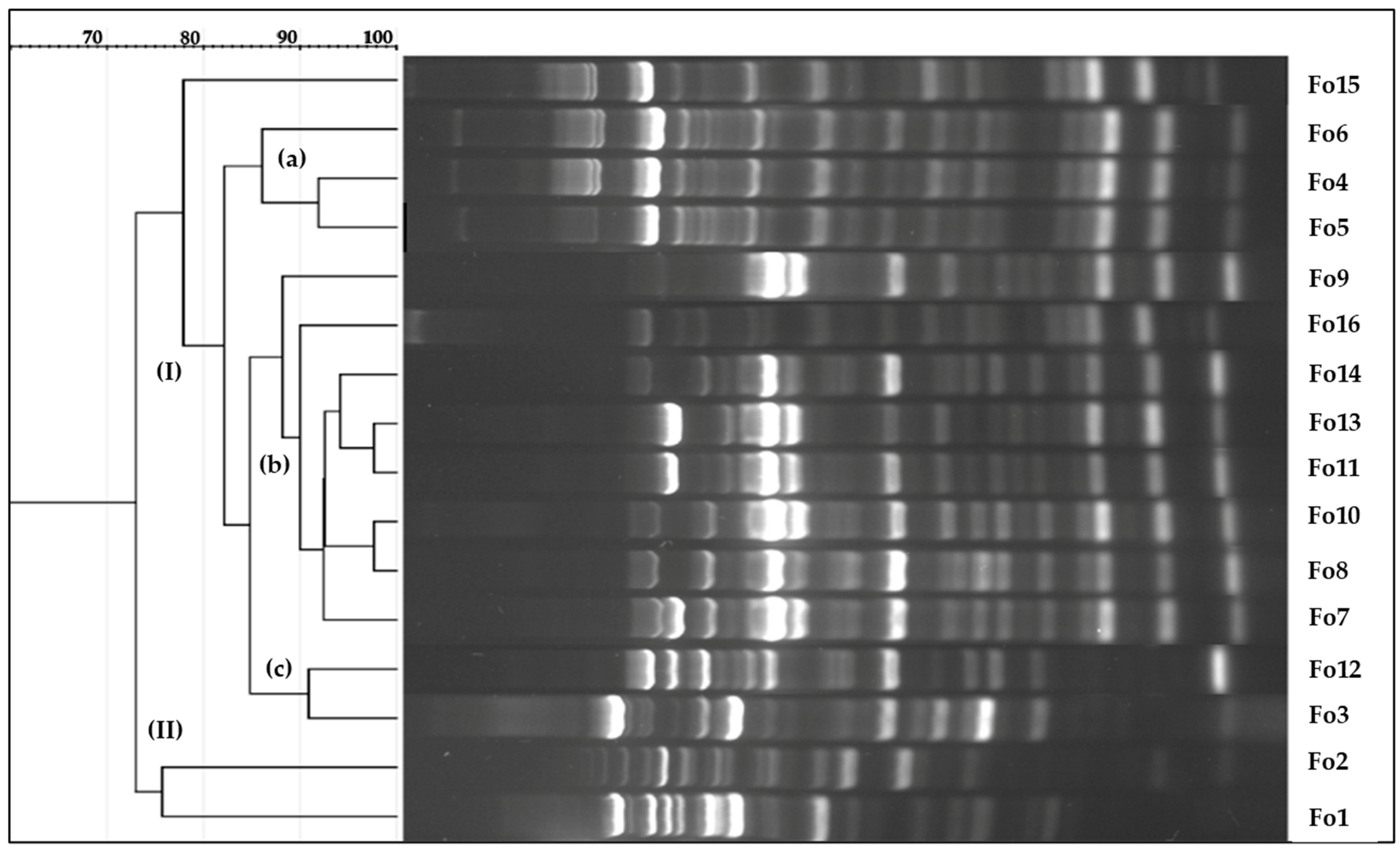
Furthermore, to investigate the genetic diversity and discriminate the preselected strains, we performed BOX-PCR, and the results were analyzed using UPGMA. The results are shown in Figure 3. A dendrogram based on BOX elements revealed that the isolated strains could be classified into major and minor clusters (I and II) with a similarity coefficient of 90%. The major cluster included almost all the isolates, except for Fo1 and Fo2, which were included in a minor cluster. The major cluster comprises three subclusters (a, b, c) with a similarity coefficient of more than 90%. In addition, an out subcluster was formed among isolates Fo8 and Fo10, Fo4 and Fo5, and Fo11 and Fo13, all of which exhibited up to 97% coefficient similarities, indicating a high level of genetic homogeneity.

3.3. Molecular Identification

The molecular identification of Fusarium isolates at the strain level was confirmed using ITS and tef-1α. BLAST search for similar ITS partial genes and those present in the NCBI GenBank database showed that all the isolated strains belonged to the Fusarium genus, with similarity percentages ranging from 93% to 98% (Table 1). The obtained tef-1α sequences were analyzed using the Fusarium-ID database. The isolated strain Fo1 was identified as F. oxysporum with 96.95% similarity. The isolated strain Fo2, which belongs to the Fusarium complex group Fusarium incarnatum-equiseti, was identified as a Fusarium sp. with 96.60% similarity (Table 1). The isolated Fo6, Fo7, and Fo16 strains, which belong to the F. fujikuroi species complex (GFSC), were identified as F. proliferatum Fo3, Fo4, Fo5, and other isolated strains belonging to Fusarium fujikuroi were identified as F. proliferatum, with similarity percentages ranging from 93.02% to 97.67%. The amplicons of ITS gene fragments of the isolated Fusarium species were further deposited in the NCBI GenBank database.
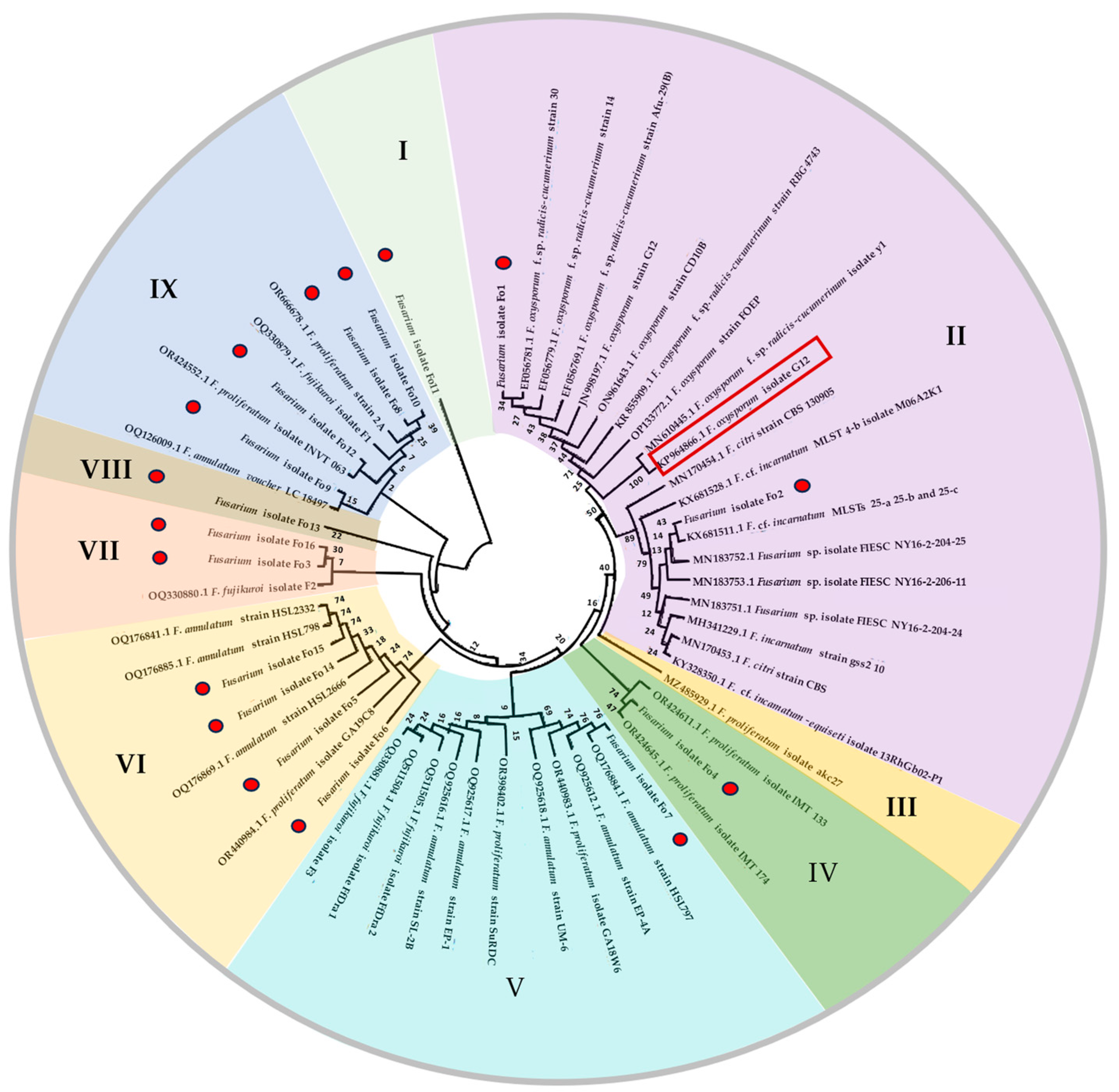
A phylogenetic tree analysis based on the neighbor-joining (NJ) method was performed to identify the phylogenetic relationships of the isolated Fusarium isolates. The F. oxysporum isolate G12 was used as the outgroup for rooting the gene tree. The analysis showed that the isolated fungal strains could be grouped into nine major clusters with significant variability in bootstrap frequency (Figure 4). The analysis revealed the wide genetic diversity of the isolated strains. Fusarium oxysporum Fo1 and Fusarium sp. Fo2 were grouped in cluster II and found to be closely related to F. oxysporum. The isolated fungal strains Fo4, Fo6, Fo14, and Fo15, which were grouped in cluster IV, were found to be most closely related to the species F. proliferatum. Fusarium strain Fo4, which was grouped in cluster IV, was found to be the closest related to F. proliferatum. Fusarium strain Fo7 formed cluster V with F. annulatum strain HSL797, whereas strains Fo6, Fo5, Fo14, and Fo15 were grouped in cluster VI with other strains of F. annulatum and F. proliferatum. Fusarium strains Fo3 and Fo16 formed cluster VII with F. fujikuroi isolate F2. The isolated fungal strains Fo13 and Fo11, grouped in clusters VIII and I, respectively, were found to constitute an outgroup cluster. The isolated fungal strains Fo10, Fo8, Fo12, and Fo9 identified as F. proliferatum were grouped in cluster IX with other strains of F. annulatum voucher LC 18497, F. fujikuroi isolate F1, F. proliferatum strain 2A, and F. proliferatum isolate INVT 063.

3.4. Pathogenicity Assay of Onion Seeds and Planta Control Using Bacillus velezensis KS04-AU

3.4.1. Pathogenicity Assay of Onion Seeds

Pathogenicity tests were also conducted using a subset of the genetic diversity generated by BOX-PCR (Figure 3). The obtained results are shown in Figure 5. Nearly all the isolated fungal strains were found to be pathogenic to onions. However, exceptions were observed for F. oxysporum Fo1, F. proliferatum Fo11, and Fo16, which did not cause any disease symptoms, such as necrosis in the radicle or leaf curling on plant onion seedlings. The aggressiveness of the isolated strains could be classified into three categories: high, intermediate, and low aggressive fungi. The isolates Fo3, Fo4, and Fo14 were found to be highly pathogenic to onion plants. The aggressive indices of the two groups were calculated to be 69.37 ± 8.81%, 64.97 ± 7.42%, and 62.61 ± 7.25%, respectively (Figure 3). In addition, no significant difference between these isolates was observed at p < 0.05.
Pretreated plants with Fusarium sp. Fo2 and F. proliferatum Fo8, Fo9, Fo12, or Fo15 exhibited intermediate aggressiveness, with no significant difference (p-value < 0.05). The aggressive indices were 38.51 ± 6.39%, 48.23 ± 5.59%, 47.77 ± 5.71%, 39.20 ± 6.91%, and 38.01 ± 7.97%, respectively. Finally, F. proliferatum Fo5, Fo6, Fo7, Fo10, and Fo13 exhibited up to 3.5-fold lower disease indices than did the highly virulent fungal isolates Fo3, Fo4, and Fo14. In line with this, no significant difference in aggressivity was observed between isolates Fo7 and Fo10. Additionally, strains Fo1, Fo11, and Fo16 were found to be aggressive toward seedling onions.

3.4.2. Pathogenicity Assay of Onion Bulbs

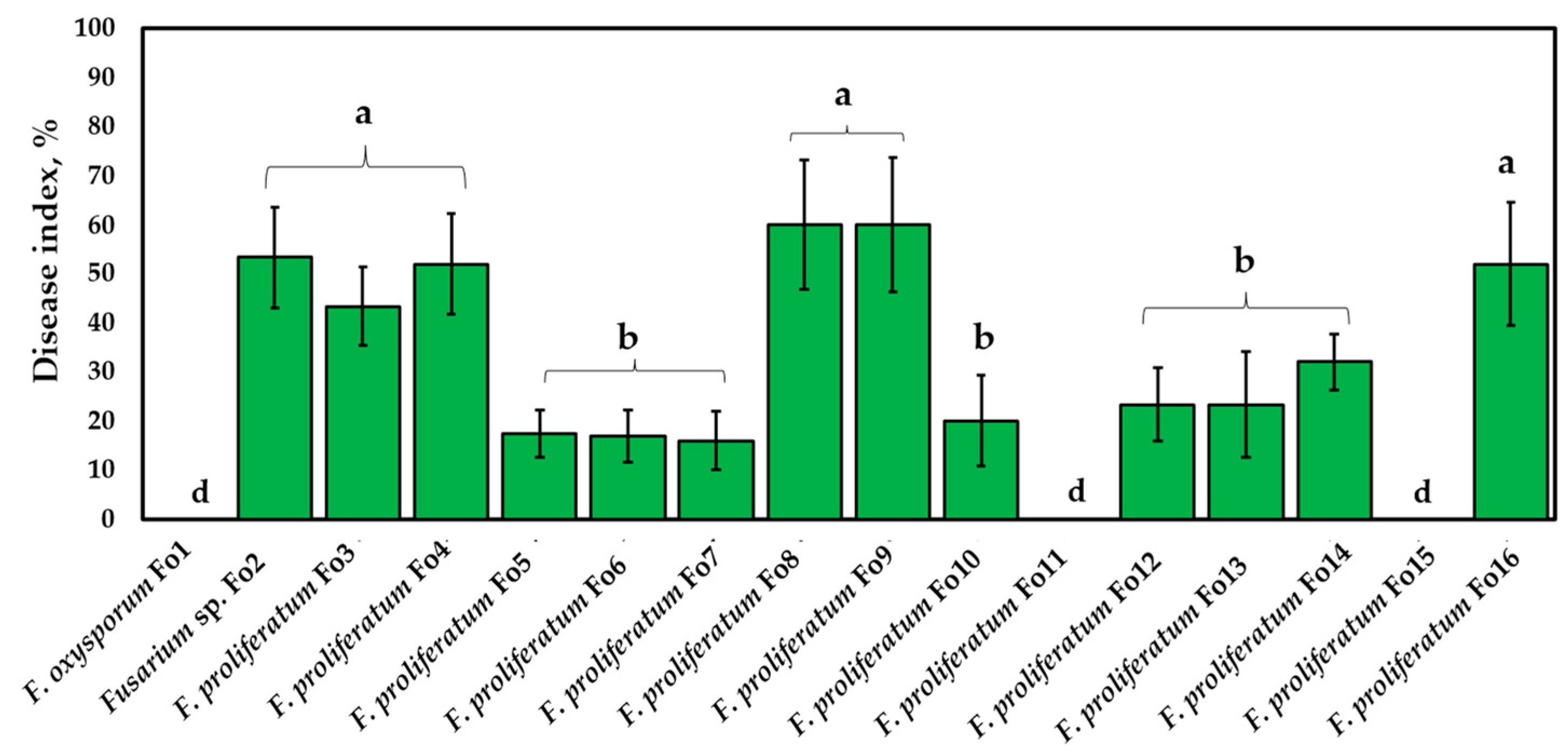
The results revealed that the isolated Fusarium strains could be classified into two categories according to their aggressiveness (intermediate and low-virulence fungi) (Figure 6).
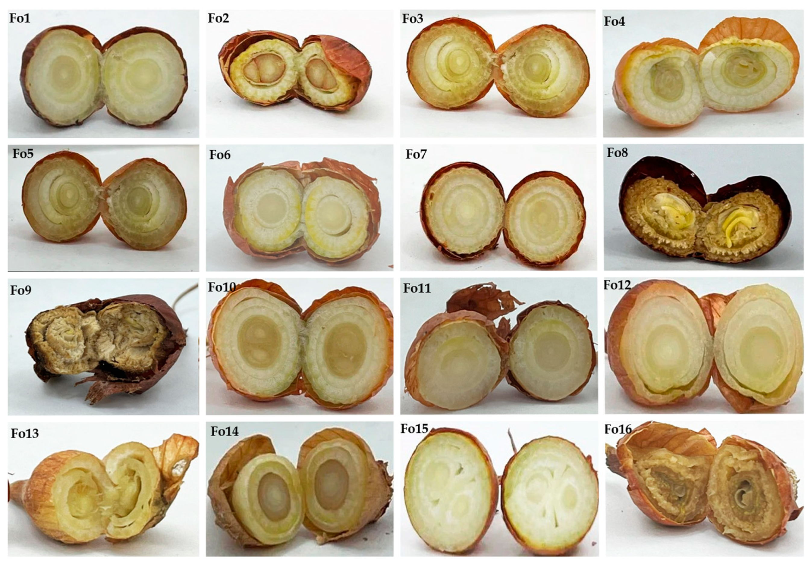
According to Koch’s postulate, the Fo2, Fo3, Fo4, Fo8, Fo9, and Fo16 strains exhibited high aggression toward onion bulbs, but the differences were not significant (p < 0.05). These strains cause typical symptoms of Fusarium infection in onions, such as rotting of bulbs and whitish mycelium on the bulb surface (Figure 7). The disease indices were 53.33 ± 10.32%, 43.41 ± 8.04%, 52.04 ± 9.87%, 60.15 ± 10.53%, 60.24 ± 11.08%, and 52.04 ± 12.43%, respectively. In contrast, the Fusarium strains Fo5, Fo6, Fo7, Fo10, Fo12, Fo13, and Fo14 exhibited low aggressiveness, ranging between 16% and 32%, without statistical significance (p-value < 0.05). Their aggressivities were 17.5 ± 4.8%, 16.9 ± 5.23%, 16.01 ± 5.87%, 20.05 ± 9.21%, 23.36 ± 10.73%, 23.36 ± 9.73%, and 32.06 ± 5.78%, respectively. Moreover, F. oxysporum Fo1, Fo11, and Fo15 were found to be unaggressive toward the onion bulbs used in this study.

3.5. In Vitro Control of Fusarium Isolates Using Bacillus velezensis KS04-AU and S. albidoflavus MGMM6

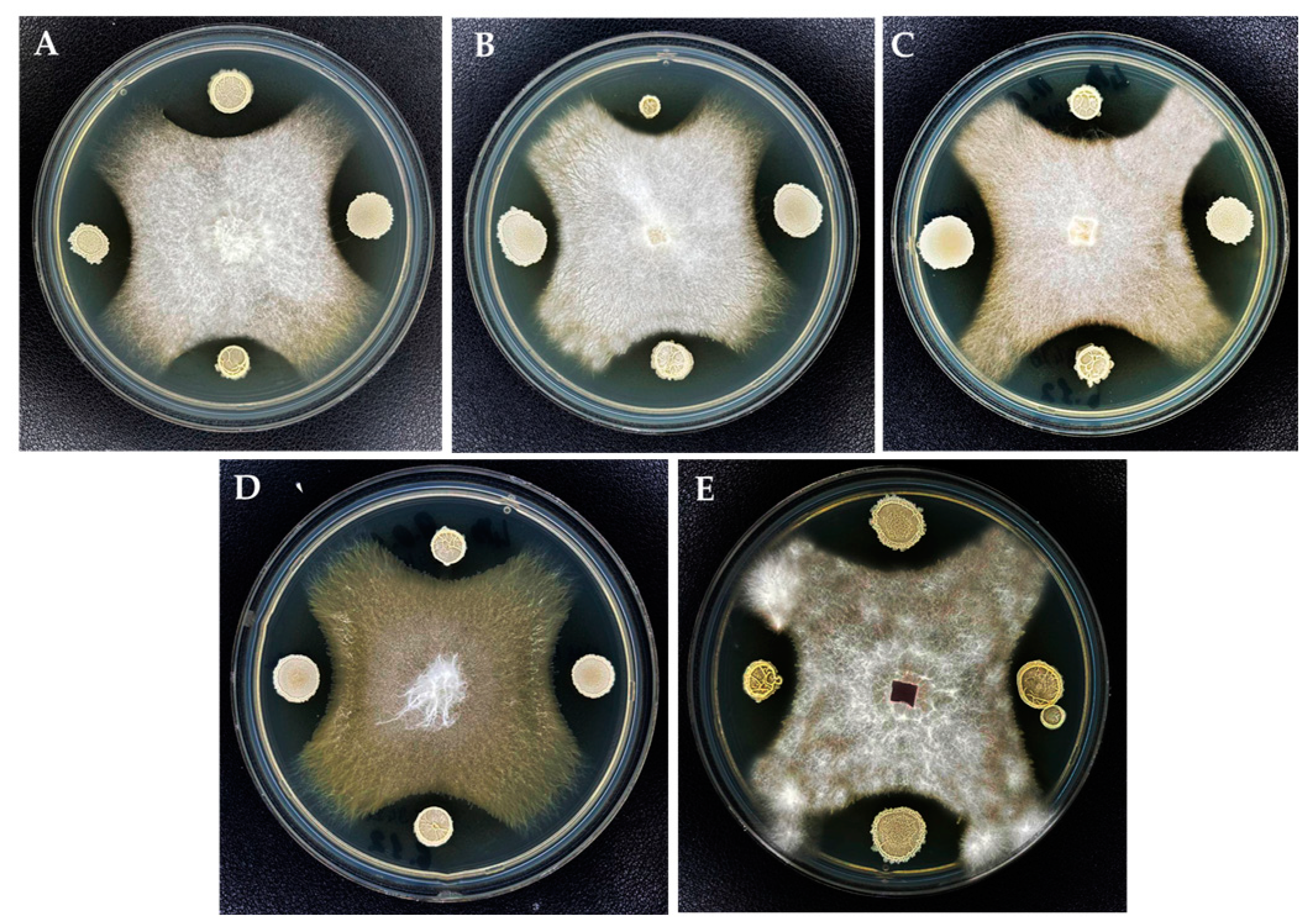
For qualitative antagonistic analysis, the strains were selected based on their aggressiveness towards plants and onion bulbs. Five strains were further chosen: one non-aggressive strain, two highly aggressive strains, and two low aggressive Fusarium strains. The antagonistic effect of B. velezensis KS04-AU and S. albidoflavus MGMM6 against Fusarium strains is shown in Figure 8. Both B. velezensis KS04-AU and S. albidoflavus MGMM6 exhibited moderate inhibitory activity against the tested phytopathogenic fungi F. oxysporum Fo1 (A), Fusarium sp. Fo2 (B), F. proliferatum Fo3 (C), F. proliferatum Fo9 (D), and Fusarium proliferatum Fo13 (E). In addition, among the tested phytopathogenic fungi F. oxysporum Fo1, Fusarium sp. Fo2, F. proliferatum Fo3, and F. proliferatum Fo9 were strongly suppressed by KS04-AU.

3.6. In Planta Control of Fusarium Isolates through Bacillus velezensis KS04-AU and Streptomyces albidoflavus MGMM6

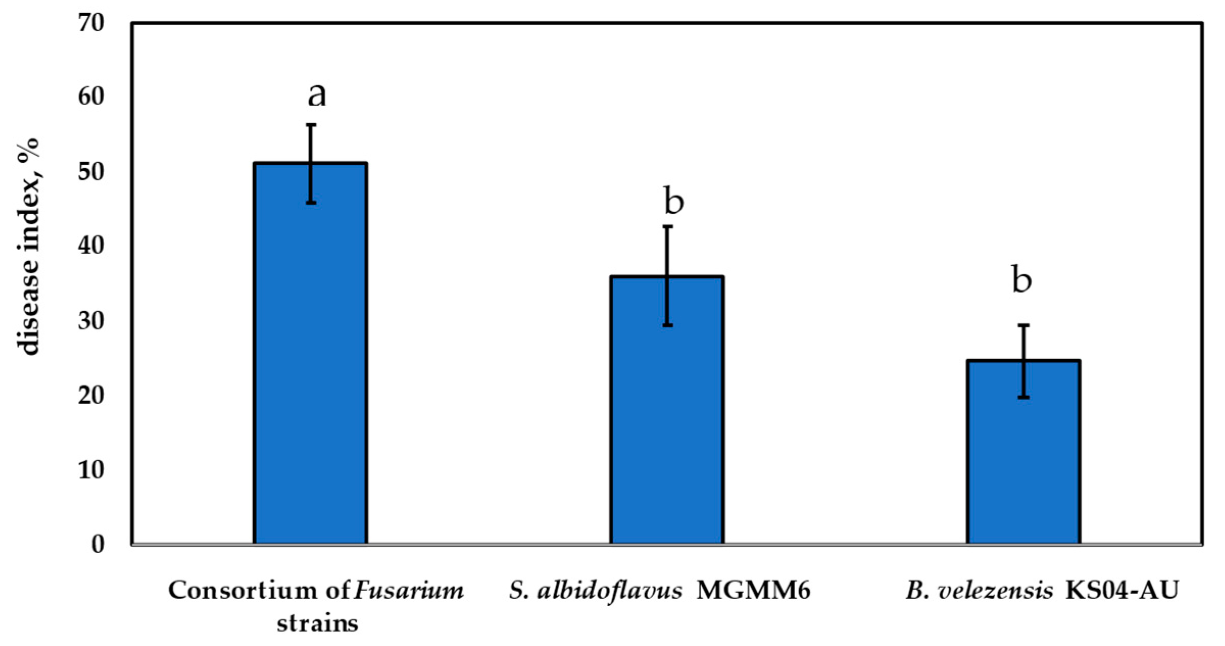
The ability of S. albidoflavus MGMM6 and B. velezensis KS04-AU in planta to protect onion plants and inhibit the growth of a consortium of Fusarium strains isolated in this study is shown in Figure 9. The most observed symptoms were necrosis in the radicle, apical strangulation, and leaf curling. The results demonstrated a significant decrease in disease development at a p-value of p < 0.05. The disease index in the group of plants pretreated with S. albidoflavus MGMM6 and B. velezensis KS04-AU was up to 12.64 ± 3.45% and 18.65 ± 4.26%, respectively, less than those without treatment (treated with a consortium of pathogenic Fusarium strains), which was assessed as 51.17 ± 5.22%. The effectiveness of S. albidoflavus MGMM6 and B. velezensis KS04-AU against these phytopathogens was measured as 24.73 ± 4.86% and 36.06 ± 6.61%, respectively.
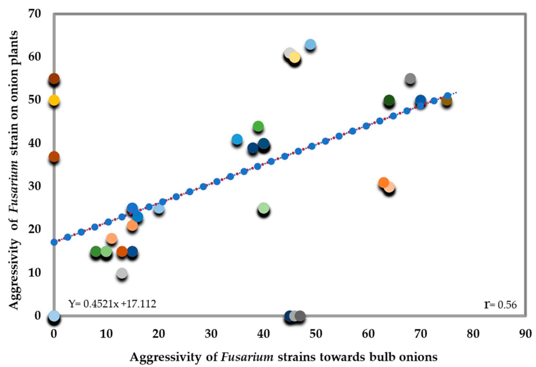
In terms of their aggressiveness towards onion bulbs and plants, a moderate positive correlation was observed (R = 0.566, p < 0.01) (Figure 10). This suggests that as the aggressiveness of strains increases, there is a moderate tendency to increase damage to both onion bulbs and plants, indicating a constant but not ideal relationship between the aggressiveness of strains and the harm they cause to onion bulbs and plants.

4. Discussion

Fusarium, a prominent genus of filamentous fungi, encompasses numerous plant pathogenic species that cause significant agricultural yield losses, reaching up to 14% annually [47,48,49,50,51]. Therefore, studying the diversity of these organisms is crucial for understanding their interactions in ecosystems and developing effective strategies for disease control, crop protection, and ecosystem conservation. Plant pathogens exhibit rapid and continuous evolutionary changes in response to various environmental pressures, including climate change and disease control measures. These stresses on pathogens necessitate constant monitoring and understanding to effectively manage and mitigate their impact on plants and ecosystems [52,53,54,55,56]. In this study, we characterized the diversity of fungal strains that cause spoilage of onion bulbs. We found that this spoilage was primarily caused by Fusarium species. To confirm the genetic diversity of the isolated fungal species, we used BOX-PCR, a well-known molecular method that allows differentiation at the species level [57,58,59]. Molecular analysis of the Fusarium isolates using the tef gene revealed the strong presence of F. proliferatum (98%), F. oxysporum (1%), and Fusarium sp. (1%). Most of the isolated strains belonged to the Fusarium fujikuroi species complex, with 96% similarity and coverage. This complex is renowned for its ability to cause a range of destructive diseases in various crops, including onion [60,61,62,63,64,65,66]. Pathogenicity assessment is necessary to identify potential risks associated with the introduction of virulent species or strains into new geographical regions or crops. Koch’s postulate, based on measuring the pathogenicity of isolated pathogenic strains via inoculation, followed by monitoring seedling emergence, survival, and health [67,68,69,70,71,72], is a crucial method for evaluating and developing effective disease control strategies, as well as resistance to these pathogens, in various plant varieties. Koch’s postulates on onion seeds and bulbs confirmed that the isolates developed different disease indices, reaching up to 96% with wilt symptoms such as discoloration, wilting, stunted growth, and spoilage of the onion bulbs. The diversity of pathogenicity of these isolated strains in infecting onion seedlings and bulbs may be related to their lifestyle, such as the secretion of pathogenicity-related genes, infection mechanisms, and interactions with plant hosts [73,74]. Interestingly, the nonaggressive Fusarium strains isolated in this study belonged to a subgroup phylogenetically distinct from highly and moderately aggressive pathogenic strains. These findings suggested that these isolates are forma specialis to onions, as previously reported by Dissanayake et al. [75] and Sasaki et al. [76]. The genus Fusarium contains a variety of complex species, some of which are non-pathogenic and pathogenic depending on the host plant. Non-pathogenic Fusarium species such as F. oxysporum have been identified as biocontrol agents and endophytic agents [77,78]. Moreover, these non-pathogenic Fusarium strains that have been isolated from onion may proliferate and become pathogenic to other plant species through crop rotation, making them difficult to control once they have contaminated crops. This difficulty arises because of the lack of reliable morphological features in the Fusarium complex, leading to the indistinguishability of pathogenic and non-pathogenic isolates. Therefore, the determination of pathogenicity still relies on tests conducted on host plants.
According to Koch’s postulation of seedlings, the development of disease symptoms in isolated Fusarium strains tends to be slower than that in onion bulbs. Moreover, Fusarium strains that can infect onion plants were found to be less pathogenic to onion bulbs and vice versa. Similar results have been previously reported [79,80,81,82]. For example, a study conducted by Carrieri et al. [83] showed that F. tricinctum is harmful to onion plants, but Koch’s postulates did not cause any symptoms of disease in onion plants. The current methods of wilt disease control have several limitations. One promising alternative is the use of microorganisms to reduce disease development through biocontrol. Considering the pathogenic virulence diversity of the isolated strains, we wanted to develop an eco-friendly approach that could suppress the growth of these Fusarium strains, causing spoilage of onion bulbs during storage. Biocontrol analysis using S. albidoflavus MGMM6 and B. velezensis KS04-AU could control the growth of these pathogenic fungi in vitro. The use of a microbial agent to control the growth of phytopathogenic fungi during crop storage is considered a promising method for crop protection [84,85]. The effectiveness of B. velezensis and S. albidoflavus in inhibiting the growth of phytopathogens such as Fusarium species has been well documented [86,87]. Therefore, our results suggest that S. albidoflavus MGMM6 and B. velezensis KS04-AU are effective bioagents for suppressing the growth of isolated Fusarium strains. Several studies have demonstrated the ability of S. albidoflavus and B. velezensis isolates to suppress the growth of phytopathogenic fungi in planta [85,86,87,88]. In this study, we found that S. albidoflavus MGMM6 is less effective against a consortium of phytopathogenic Fusarium strains isolated from onion bulbs. This may be due to the strong pathogenicity induced by synergistic interactions of isolated Fusarium strains that contribute to their enhanced virulence when applied in a consortium, as reported by Sidharthan [89]. The synergistic interactions between these pathogenic strains may contribute to their collective virulence.

5. Conclusions

Phytopathogenic microorganisms can lead to significant crop losses, which comprise both qualitative damage that reduces the economic value of the crop and makes it unsuitable for human consumption. This study successfully identified and evaluated the aggressiveness of Fusarium species associated with onion bulbs during storage, shedding light on the potential threats to onion production and storage. The morphological and molecular differentiation of the isolated strains revealed their taxonomic identity belonging to Fusarium complex species incarnatum-equiseti and Fusarium fujikuroi. Koch’s postulate analysis demonstrated the varying aggressiveness of the isolated strains on onion bulbs and plants, with disease symptoms developing more slowly on plants according to the postulates. Importantly, this study highlighted the presence of Fusarium strains that could infect onion plants less aggressively than onion bulbs, or vice versa. In addition, some strains were found to be non-aggressive, which indicated the ability of each strain to occupy its ecological niche and did not suppress the viability of other fungi belonging to the same genus Fusarium. Moreover, we found that the development of isolated fungi can be controlled using a biological approach. This indicates the potential for developing an environmentally friendly alternative to controlling the growth of phytopathogens, which could lead to a reduction in onion losses during storage. This finding could serve as an instrument in the development of biocontrol strategies for onion crop protection against phytopathogenic microorganisms during storage. However, studies in field conditions with different onion varieties are needed to elucidate their potential effectiveness.

Supplementary Materials

The following supporting information can be downloaded at: https://www.mdpi.com/article/10.3390/jof10020161/s1, Figure S1: Microscopic fungal spores’ visualization under light microscopy. Sequence data for the tef-1α gene partial from 16 Fusarium strains isolated from onion bulb (Allium cepa L.).

Author Contributions

Conceptualization, R.G.C.D.; methodology, R.G.C.D.; software, R.G.C.D.; validation, R.G.C.D.; formal analysis, R.G.C.D. and M.F.; investigation, R.G.C.D., B.I., E.S. and M.F.; resources, S.V.; data curation, R.G.C.D.; writing—original draft preparation, R.G.C.D.; writing—review and editing, R.G.C.D., D.M.A., M.N.F. and S.V.; supervision, R.G.C.D.; project administration, S.V.; funding acquisition, S.V. All authors have read and agreed to the published version of the manuscript.

Funding

This study was conducted with financial support provided by the Ministry of Education and Science of the Russian Federation, grant # RF-1930.61321X0001/15.IP.21.0020, and from the government assignment for the FRC Kazan Scientific Center of RAS.

Institutional Review Board Statement

Not applicable.

Informed Consent Statement

Not applicable.

Data Availability Statement

Data are contained within the article and supplementary materials.

Conflicts of Interest

The authors declare no conflict of interest.

References

  1. Tournas, V.H. Spoilage of vegetable crops by bacteria and fungi and related health hazards. Crit. Rev. Microbiol. 2005, 31, 33–44. [Google Scholar] [CrossRef] [PubMed]
  2. Kumar, V.; Sharma, S.; Neeraj, B.; Narashans, A.S. Postharvest management of fungal diseases in onion—A review. Int. J. Curr. Microbiol. Appl. Sci. 2015, 4, 737–752. [Google Scholar]
  3. Sakhi, S.S.; Sohi, S.S.; Singh, D.P.; Joshi, M.C. Source of resistance to basal rot of onion caused by Fusarium oxysporum. Indian J. Mycol. Plant Pathol. 1973, 4, 214–215. [Google Scholar] [CrossRef]
  4. Boyhan, G.E.; Torrance, R.L. Vidalia onions—Sweet onion production in southeastern Georgia. HortTechnology 2002, 12, 196–202. [Google Scholar] [CrossRef]
  5. Anbukkarasi, V.; Paramaguru, P.; Pugalendhi, L.; Ragupathi, N.; Jeyakumar, P. Studies on pre- and postharvest treatments for extending shelf life in onion–A review. Agric. Rev. 2013, 34, 256–268. [Google Scholar] [CrossRef]
  6. Gupta, R.P.; Verma, L.R. Problem of diseases during storage in onion and garlic and their strategic management. In Implication of Plant Diseases on Produce Quality; Kalyani Publishers: Ludhiana, Indian, 2002. [Google Scholar]
  7. Oerke, E.C. Crop losses to pests. J. Agric. Sci. 2006, 144, 31–43. [Google Scholar] [CrossRef]
  8. Saremi, H.; Okhovvat, S.M.; Ashrafi, S.J. Fusarium diseases as the main soil-borne fungal pathogen on plants and their control management with soil solarization in Iran. Afr. J. Biotechnol. 2011, 10, 18391–18398. [Google Scholar] [CrossRef]
  9. Arie, T. Fusarium diseases of cultivated plants, control, diagnosis, and molecular and genetic studies. J. Pestic. Sci. 2019, 44, 275–281. [Google Scholar] [CrossRef]
  10. Summerell, B.A. Resolving Fusarium: Current status of the genus. Annu. Rev. Phytopathol. 2019, 57, 323–339. [Google Scholar] [CrossRef]
  11. Smith, S.N. An overview of ecological and habitat aspects in the genus Fusarium with special emphasis on the soil-borne pathogenic forms. Plant Pathol. Bull. 2007, 16, 97–120. [Google Scholar]
  12. Caligiore Gei, P.F.; Valdez, J.G.; Piccolo, R.J.; Galmarini, C.R. Influence of Fusarium spp. isolate and inoculum density on resistance screening tests in onion. Trop. Plant Pathol. 2014, 39, 19–27. [Google Scholar] [CrossRef]
  13. Schwartz, H.F.; Mohan, S.K. Compendium of Onion and Garlic Diseases and Pests, 2nd ed.; American Phytopathological Society: St. Paul, MN, USA, 2008; pp. 11–15. [Google Scholar]
  14. Kamle, M.; Mahato, D.K.; Devi, S.; Lee, K.E.; Kang, S.G.; Kumar, P. Fumonisins: Impact on agriculture, food, and human health and their management strategies. Toxins 2019, 11, 328. [Google Scholar] [CrossRef] [PubMed]
  15. Rather, I.A.; Koh, W.Y.; Paek, W.K.; Lim, J. The sources of chemical contaminants in food and their health implications. Front. Pharmacol. 2017, 8, 830. [Google Scholar] [CrossRef]
  16. Walsh, T.J.; Groll, A.; Hiemenz, J.; Fleming, R.; Roilides, E.; Anaissie, E. Infections due to emerging and uncommon medically important fungal pathogens. Clin. Microbiol. Infect. 2004, 10, 48–66. [Google Scholar] [CrossRef] [PubMed]
  17. Nucci, M.; Anaissie, E. Fusarium infections in immunocompromised patients. Clin. Microbiol. Rev. 2007, 20, 695–704. [Google Scholar] [CrossRef] [PubMed]
  18. Vahling-Armstrong, C.; Dung, J.K.S.; Humann, J.L.; Schroeder, B.K. Effects of postharvest onion curing parameters on bulb rot caused by Pantoea agglomerans, Pantoea ananatis and Pantoea allii in storage. Plant Pathol. 2016, 65, 536–544. [Google Scholar] [CrossRef]
  19. Galván, G.A.; Koning-Boucoiran, C.F.; Koopman, W.J.; Burger-Meijer, K.; González, P.H.; Waalwijk, C.; Kik, C.; Scholten, O.E. Genetic variation among Fusarium isolates from onion, and resistance to Fusarium basal rot in related Allium species. Eur. J. Plant Pathol. 2008, 121, 499–512. [Google Scholar] [CrossRef]
  20. Ji, S.H.; Kim, T.K.; Keum, Y.S.; Chun, S.C. The major postharvest disease of onion and its control with thymol fumigation during low-temperature storage. Mycobiology 2018, 46, 242–253. [Google Scholar] [CrossRef]
  21. Kadam, D.M.; Nangare, D.D.; Oberoi, H.S. Influence of pre-treatments on microbial load of stored dehydrated onion slices. Int. J. Food Sci.Technol. 2009, 44, 1902–1908. [Google Scholar] [CrossRef]
  22. Brent, K.J.; Hollomon, D.W. Fungicide Resistance: The Assessment of Risk; Global Crop Protection Federation: Brussels, Belgium, 1998; Volume 2. [Google Scholar]
  23. Nelson, P.E.; Dignani, M.C.; Anaissie, E.J. Taxonomy, biology, and clinical aspects of Fusarium species. Clin. Microbiol. Rev. 1994, 7, 479–504. [Google Scholar] [CrossRef]
  24. Missmer, S.A.; Suarez, L.; Felkner, M.; Wang, E.; Merrill, A.H., Jr.; Rothman, K.J.; Hendricks, K.A. Exposure to fumonisins and the occurrence of neural tube defects along the Texas–Mexico border. Environ. Health Perspect. 2006, 114, 237–241. [Google Scholar] [CrossRef] [PubMed]
  25. Antonissen, G.; Martel, A.; Pasmans, F.; Ducatelle, R.; Verbrugghe, E.; Vandenbroucke, V.; Croubels, S. The impact of Fusarium mycotoxins on human and animal host susceptibility to infectious diseases. Toxins 2014, 6, 430–452. [Google Scholar] [CrossRef] [PubMed]
  26. Lahlali, R.; Ezrari, S.; Radouane, N.; Kenfaoui, J.; Esmaeel, Q.; El Hamss, H.; Belabess, Z.; Barka, E.A. Biological Control of Plant Pathogens: A Global Perspective. Microorganisms 2022, 10, 596. [Google Scholar] [CrossRef] [PubMed]
  27. Narayanasamy, P. Biological Management of Diseases of Crops; Springer: Dordrecht, The Netherlands, 2013; p. 673. [Google Scholar] [CrossRef]
  28. Fenta, L.; Mekonnen, H.; Kabtimer, N. The Exploitation of Microbial Antagonists against Postharvest Plant Pathogens. Microorganisms 2023, 11, 1044. [Google Scholar] [CrossRef]
  29. Köhl, J.; Kolnaar, R.; Ravensberg, W.J. Ravensberg. Mode of action of microbial biological control agents against plant diseases: Relevance beyond efficacy. Front. Plant Sci. 2019, 10, 845. [Google Scholar] [CrossRef]
  30. Compant, S.; Duffy, B.; Nowak, J.; Clément, C.; Barka, E.A. Use of plant growth-promoting bacteria for biocontrol of plant diseases: Principles, mechanisms of action, and future prospects. Appl. Environ. Microbiol. 2005, 71, 4951–4959. [Google Scholar] [CrossRef]
  31. Leslie, J.F.; Summerell, B.A. The Fusarium Laboratory Manual; John Wiley & Sons: Hoboken, NJ, USA, 2008. [Google Scholar]
  32. Green, M.R.; Sambrook, J. Isolation of High-Molecular-Weight DNA Using Organic Solvents. Cold Spring Harb Protoc. 2017, 2017, pdb.prot093450. [Google Scholar] [CrossRef]
  33. Desjardins, P.; Conklin, D. NanoDrop microvolume quantitation of nucleic acids. JoVE 2010, 22, e2565. [Google Scholar] [CrossRef]
  34. Altinok, H.H. Fusarium species isolated from common weeds in eggplant fields and symptomless hosts of Fusarium oxysporum f. sp. melongenae in Turkey. J. Phytopathol. 2013, 161, 335–340. [Google Scholar] [CrossRef]
  35. Zarrin, M.; Ganj, F.; Faramarzi, S. Analysis of the rDNA internal transcribed spacer region of the Fusarium species by polymerase chain reaction-restriction fragment length polymorphism. Biomed. Rep. 2016, 4, 471–474. [Google Scholar] [CrossRef]
  36. Rzhetsky, A.; Nei, M. A simple method for estimating and testing minimum evolutionary trees. Mol. Biol. Evol. 1992, 9, 945–967. [Google Scholar] [CrossRef]
  37. Felsenstein, J. Confidence limits on phylogenies: An approach using the bootstrap. Evolution 1985, 39, 783–791. [Google Scholar] [CrossRef]
  38. Tamura, K.; Nei, M.; Kumar, S. Prospects for inferring very large phylogenies by using the neighbor-joining method. Proc. Natl. Acad. Sci. USA 2004, 101, 11030–11035. [Google Scholar] [CrossRef] [PubMed]
  39. Tamura, K.; Stecher, G.; Kumar, S. MEGA11: Molecular evolutionary genetics analysis version 11. Mol. Biol. Evol. 2021, 38, 3022–3027. [Google Scholar] [CrossRef] [PubMed]
  40. Simon, M.; van der Bij, A.J.; Brand, J.; de Weger, L.A.; Wijffelman, C.A.; Lugtenberg, B.J.J. Gnotobiotic system for studying rhizosphere colonization by plant growth-promoting Pseudomonas bacteria. Mol. Plant Microbe Interact 1996, 9, 600–607. [Google Scholar] [CrossRef] [PubMed]
  41. Afordoanyi, D.M.; Diabankana, R.G.C.; Akosah, Y.A.; Validov, S.Z. Are formae speciales pathogens truly host specific? A broadened host specificity in Fusarium oxysporum f.sp. radicis-cucumerinum. Braz. J. Microbiol. 2022, 53, 1745–1759. [Google Scholar] [CrossRef]
  42. Diabankana, R.G.C.; Afordoanyi, D.M.; Safin, R.I.; Nizamov, R.M.; Karimova, L.Z.; Validov, S.Z. Antifungal properties, abiotic stress resistance, and biocontrol ability of Bacillus mojavensis PS17. Curr. Microbiol. 2021, 78, 3124–3132. [Google Scholar] [CrossRef]
  43. Koyshibayev, M.; Muminjanov, H. Guidelines for Monitoring Diseases, Pest and Weeds in Cereal Crops; Food and Agriculture Organization of the United Nations: Ankara, Turkey, 2016; p. 8. Available online: http://www.fao.org/3/a-i5550e (accessed on 1 February 2016).
  44. Tirado-Ramirez, A.; López-Urquídez, G.A.; Amarillas-Bueno, L.A.; Retes-Manjarrez, J.E.; Vega-Gutiérrez, T.A.; López Avendaño, J.E.; López-Orona, C.A. Identification and virulence of Fusarium falciforme and Fusarium brachygibbosum as causal agents of basal rot on onion in Mexico. Can. J. Plant Pathol. 2021, 43, 722–733. [Google Scholar] [CrossRef]
  45. Diabankana, R.G.C.; Shulga, E.U.; Validov, S.Z.; Afordoanyi, D.M. Genetic Characteristics and Enzymatic Activities of Bacillus velezensis KS04AU as a Stable Biocontrol Agent against Phytopathogens. Int. J. Plant Biol. 2022, 13, 201–222. [Google Scholar] [CrossRef]
  46. Diabankana, R.G.C.; Frolov, M.; Keremli, S.; Validov, S.Z.; Afordoanyi, D.M. Genomic Insights into the Microbial Agent Streptomyces albidoflavus MGMM6 for Various Biotechnology Applications. Microorganisms 2023, 11, 2872. [Google Scholar] [CrossRef] [PubMed]
  47. Heras, J.; Domínguez, C.; Mata, E.; Pascual, V.; Lozano, C.; Torres, C.; Zarazaga, M. GelJ–a tool for analyzing DNA fingerprint gel images. BMC Bioinform. 2015, 16, 270. [Google Scholar] [CrossRef] [PubMed]
  48. Waller, J.M.; Brayford, D. Fusarium diseases in the tropics. Int. J. Pest Manag. 1990, 36, 181–194. [Google Scholar] [CrossRef]
  49. Anand, G.; Rajeshkumar, K.C. Challenges and Threats Posed by Plant Pathogenic Fungi on Agricultural Productivity and Economy. In Fungal Diversity, Ecology and Control Management; Fungal Biology; Rajpal, V.R., Singh, I., Navi, S.S., Eds.; Springer: Singapore, 2022. [Google Scholar] [CrossRef]
  50. Oldenburg, E.; Höppner, F.; Ellner, F.; Weinert, J. Fusarium diseases of maize associated with mycotoxin contamination of agricultural products intended to be used for food and feed. Mycotoxin Res. 2017, 33, 167–182. [Google Scholar] [CrossRef] [PubMed]
  51. Rojo, F.G.; Reynoso, M.M.; Ferez, M.; Chulze, S.N.; Torres, A.M. Biological control by Trichoderma species of Fusarium solani causing peanut brown root rot under field conditions. Crop Prot. 2007, 26, 549–555. [Google Scholar] [CrossRef]
  52. Kintega, K.R.; Zida, P.E.; Tarpaga, V.W.; Sankara, P.; Sereme, P. Identification of Fusarium species associated with onion (Allium cepa L.) plants in field in Burkina Faso. Adv. Biosci. Biotechnol. 2020, 11, 94–110. [Google Scholar] [CrossRef]
  53. Zhan, J.; Mundt, C.C.; Hoffer, M.E.; McDonald, B.A. Local adaptation and effect of host genotype on the rate of pathogen evolution: An experimental test in a plant pathosystem. J. Evol. Biol. 2002, 15, 634–647. [Google Scholar] [CrossRef]
  54. Zhan, J.; McDonald, B.A. Thermal adaptation in the fungal pathogen Mycosphaerella graminicola. Mol. Ecol. 2011, 20, 1689–1701. [Google Scholar] [CrossRef] [PubMed]
  55. Wu, E.J.; Wang, Y.P.; Yahuza, L.; He, M.H.; Sun, D.L.; Huang, Y.M.; Liu, Y.C.; Yang, L.N.; Zhu, W.; Zhan, J. Rapid adaptation of the Irish potato famine pathogen Phytophthora infestans to changing temperature. Evol. Appl. 2020, 13, 769–781. [Google Scholar] [CrossRef]
  56. Yang, L.N.; Nkurikiyimfura, O.; Pan, Z.C.; Wang, Y.P.; Waheed, A.; Chen, R.S.; Burdon, J.J.; Sui, Q.J.; Zhan, J. Plantdiversity ameliorates the evolutionary development of fungicide resistance in an agricultural ecosystem. J. Appl. Ecol. 2021, 58, 2566–2578. [Google Scholar] [CrossRef]
  57. Marques, A.S.; Marchaison, A.; Gardan, L.; Samson, R. BOX-PCR-based identification of bacterial species belonging to Pseudomonas syringae: P. viridiflava group. Genet. Mol. Biol. 2008, 31, 106–115. [Google Scholar] [CrossRef]
  58. Tacão, M.; Alves, A.; Saavedra, M.J.; Correia, A. BOX-PCR is an adequate tool for typing Aeromonas spp. Antonie Leeuwenhoek 2005, 88, 173–179. [Google Scholar] [CrossRef]
  59. Proctor, R.H.; Desjardins, A.E.; Moretti, A. Biological and chemical complexity of Fusarium proliferatum. In The Role of Plant Pathology in Food Safety; Strange, R.N., Gullino, M.L., Eds.; Springer: Berlin, Germany, 2009; pp. 97–111. [Google Scholar] [CrossRef]
  60. Niehaus, E.M.; Münsterkötter, M.; Proctor, R.H.; Brown, D.W.; Sharon, A.; Idan, Y.; Tudzynski, B. Comparative “omics” of the Fusarium fujikuroi species complex highlights differences in genetic potential and metabolite synthesis. Genome Biol. Evol. 2016, 8, 3574–3599. [Google Scholar] [CrossRef]
  61. Dong, T.; Qiao, S.; Xu, J.; Shi, J.; Qiu, J.; Ma, G. Effect of Abiotic Conditions on Growth, Mycotoxin Production, and Gene Expression by Fusarium fujikuroi Species Complex Strains from Maize. Toxins 2023, 15, 260. [Google Scholar] [CrossRef]
  62. O’Donnell, K.; Cigelnik, E.; Nirenberg, H.I. Molecular systematics and phylogeography of the Gibberella fujikuroi species complex. Mycologia 1998, 90, 465–493. [Google Scholar] [CrossRef]
  63. Huss, M.J.; Campbell, C.L.; Jennings, D.B.; Leslie, J.F. Isozyme variation among biological species in the Gibberella fujikuroi species complex (Fusarium section Liseola). Appl. Environ. Microbiol. 1996, 62, 3750–3756. [Google Scholar] [CrossRef]
  64. Stankovic, S.; Levic, J.; Petrovic, T.; Logrieco, A.; Moretti, A. Pathogenicity and mycotoxin production by Fusarium proliferatum isolated from onion and garlic in Serbia. Eur. J. Plant Pathol. 2007, 118, 165–172. [Google Scholar] [CrossRef]
  65. Heidarian, Z.; Javan-Nikkhah, M.; Fotouhifar, K.B.; Ahmadpour, A. Morphological and phylogenetic investigation on selected Fusarium species belong to Gibberella fujikuroi species complex in Iran. Rostaniha 2013, 14, 163–174. [Google Scholar] [CrossRef]
  66. Dissanayake, M.L.M.C.; Kashima, R.; Tanaka, S.; and Ito, S.I. Pathogenic variation and molecular characterization of Fusarium species isolated from wilted Welsh onion in Japan. Plant Pathol. J. 2009, 75, 37–45. [Google Scholar] [CrossRef]
  67. Haapalainen, M.; Latvala, S.; Kuivainen, E.; Qiu, Y.; Segerstedt, M.; Hannukkala, A.O. Fusarium oxysporum, F. proliferatum and F. redolens associated with basal rot of onion in Finland. Plant Pathol. 2016, 65, 1310–1320. [Google Scholar] [CrossRef]
  68. Taylor, A.; Vagany, V.; Barbara, D.J.; Thomas, B.; Pink, D.A.C.; Jones, J.E.; Clarkson, J.P. Identification of differential resistance to six Fusarium oxysporum f. sp. cepae isolates in commercial onion cultivars through the development of a rapid seedling assay. Plant Pathol. 2013, 62, 103–111. [Google Scholar] [CrossRef]
  69. Özer, N.; Köycü, N.D.; Chilosi, G.; Magro, P. Resistance to Fusarium basal rot of onion in greenhouse and field and associated expression of antifungal compounds. Phytoparasitica 2004, 32, 388–394. [Google Scholar] [CrossRef]
  70. Singer, M. Pathogen-pathogen interaction: A syndemic model of complex biosocial processes in disease. Virulence 2010, 1, 10–18. [Google Scholar] [CrossRef] [PubMed]
  71. Braga, R.M.; Dourado, M.N.; Araújo, W.L. Microbial interactions: Ecology in a molecular perspective. Braz. J. Microbiol. 2016, 47, 86–98. [Google Scholar] [CrossRef] [PubMed]
  72. Bektas, I.; Kusek, M. Phylogenetic and morphological characterization of Fusarium oxysporum f. sp. cepae the causal agent of basal rot on onion isolated from Turkey. Fresenius Environ. Bull. 2019, 28, 1733–1742. [Google Scholar]
  73. Beck, K.D.; Reyes-Corral, C.; Rodriguez-Rodriguez, M.; May, C.; Barnett, R.; Thornton, M.K.; Schroeder, B.K. First report of Fusarium proliferatum causing necrotic leaf lesions and bulb rot on storage onion (Allium cepa) in southwestern Idaho. Plant Dis. 2021, 105, 494. [Google Scholar] [CrossRef]
  74. Abdullah, A.S.; Moffat, C.S.; Lopez-Ruiz, F.J.; Gibberd, M.R.; Hamblin, J.; Zerihun, A. Host–multipathogen warfare: Pathogen interactions in coinfected plants. Front. Plant Sci. 2017, 8, 1806. [Google Scholar] [CrossRef]
  75. Dissanayake, M.; Kashima, R.; Tanaka, S.; Ito, S.I. Genetic diversity and pathogenicity of Fusarium oxysporum isolated from wilted Welsh onion in Japan. J. Gen. Plant Pathol. 2009, 75, 125–130. [Google Scholar] [CrossRef]
  76. Sasaki, K.; Nakahara, K.; Tanaka, S.; Shigyo, M.; Ito, S.I. Genetic and pathogenic variability of Fusarium oxysporum f. sp. cepae isolated from onion and Welsh onion in Japan. Phytopathology 2015, 105, 525–532. [Google Scholar] [CrossRef]
  77. Alabouvette, C.; Olivain, C.; Migheli, Q.; Steinberg, C. Microbiological control of soil-borne phytopathogenic fungi with special emphasis on wilt-inducing Fusarium oxysporum. New Phytol. 2009, 184, 529–544. [Google Scholar] [CrossRef]
  78. De Lamo, F.J.; Takken, F.L. Biocontrol by Fusarium oxysporum using endophyte-mediated resistance. Front. Plant Sci. 2020, 11, 37. [Google Scholar] [CrossRef]
  79. Gärber, U.; Grosch, R.; Goßmann, M.; Büttner, C. Auftreten und Nachweis von Fusarium oxysporum und F. proliferatum an Steck-und Saatzwiebeln. Gesunde Pflanzen 2012, 64, 1–10. [Google Scholar] [CrossRef]
  80. Labanska, M.; Van Amsterdam, S.; Jenkins, S.; Clarkson, J.P.; Covington, J.A. Preliminary studies on detection of Fusarium basal rot infection in onions and shallots using electronic nose. Sensors 2022, 22, 5453. [Google Scholar] [CrossRef] [PubMed]
  81. Lager, S. Survey of Fusarium Species on Yellow Onion (Allium cepa) on Öland; Uppsala Swedish University of Agricultural Science: Uppsala, Sweden, 2011. [Google Scholar]
  82. Tsitsigiannis, D.I.; Dimakopoulou, M.; Antoniou, P.P.; Tjamos, E.C. Biological control strategies of mycotoxigenic fungi and associated mycotoxins in Mediterranean basin crops. Phytopathol. Mediterr. 2012, 51, 158–174. [Google Scholar]
  83. Carrieri, R.; Raimo, F.; Pentangelo, A.; Lahoz, E. Fusarium proliferatum and Fusarium tricinctum as causal agents of pink rot of onion bulbs and the effect of soil solarization combined with compost amendment in controlling their infections in field. Crop Prot. 2013, 43, 31–37. [Google Scholar] [CrossRef]
  84. Chernin, L.; Chet, I. Microbial enzymes in biocontrol of plant pathogens and pests. In Enzymes in the Environment: Activity, Ecology, and Applications; Marcel Dekker, Inc.: New Yoer, NY, USA, 2002; pp. 171–225. [Google Scholar]
  85. Du, Y.; Wang, T.; Jiang, J.; Wang, Y.; Lv, C.; Sun, K.; Huang, L. Biological control and plant growth promotion properties of Streptomyces albidoflavus St-220 isolated from Salvia miltiorrhiza rhizosphere. Front. Plant Sci. 2022, 13, 976813. [Google Scholar] [CrossRef]
  86. Carlucci, A.; Raimondo, M.L.; Colucci, D.; Lops, F. Streptomyces albidoflavus strain CARA17 as a biocontrol agent against fungal soil-borne pathogens of fennel plants. Plants 2022, 11, 1420. [Google Scholar] [CrossRef]
  87. Rabbee, M.F.; Hwang, B.S.; Baek, K.-H. Bacillus velezensis: A Beneficial Biocontrol Agent or Facultative Phytopathogen for Sustainable Agriculture. Agronomy 2023, 13, 840. [Google Scholar] [CrossRef]
  88. Djebaili, R.; Pellegrini, M.; Ercole, C.; Farda, B.; Kitouni, M.; Del Gallo, M. Biocontrol of soil-borne pathogens of Solanum lycopersicum L. and Daucus carota L. by plant growth-promoting actinomycetes: In vitro and in planta antagonistic activity. Pathogens 2021, 10, 1305. [Google Scholar] [CrossRef]
  89. Sidharthan, V.K.; Pothi-raj, G.; Suryaprakash, V.; Singh, A.K.; Aggarwal, R.; Shanmugam, V. A synergic and compatible microbial-based consortium for biocontrol of Fusarium wilt of tomato. Phytopathol. Mediterr. 2023, 62, 183–197. [Google Scholar] [CrossRef]
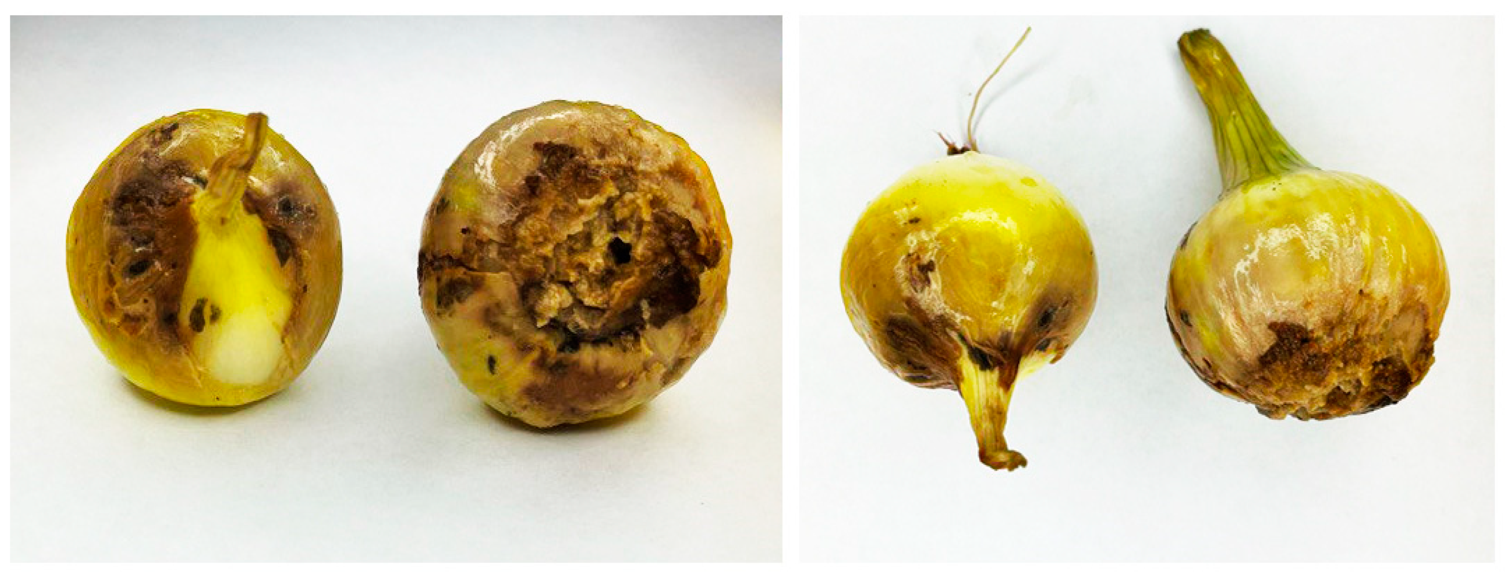
Figure 1. The infected yellow onion bulbs were used in this study.
Jof 10 00161 g001
Figure 2. Phenotypical characteristics of Fusarium strains isolated from onion bulbs (Allium cepa L.). (AP)—Fusarium isolated strains Fo1–Fo16, respectively. The isolated fungal strains were subsequently grown on Sabouraud media supplemented with rifampicin to a final concentration of 100 µg/µL.
Jof 10 00161 g002
Figure 3. Dendrogram of BOX PCR constructed based on DNA profiles of fungal isolates using GelJ software version 3.0 [47]. Dendrograms were generated using the Jaccard similarity coefficient and UPGMA cluster analysis with a matching band tolerance set at 5%; (I) and (II) are major and minor clusters, respectively; (ac) are out of subclusters.
Jof 10 00161 g003
Figure 4. Maximum likelihood tree of 16 isolated Fusarium species based on the tef-α genes partial (Supplementary Material and Data). Bootstrap values (1000 replicates) are indicated on the branches. Fusarium oxysporum isolate G12 used as the outgroup for rooting the generated tree is highlighted with a red box. The fungal strains isolated in this study are marked with a red dot. I–IX as the number of generated clusters.
Jof 10 00161 g004
Figure 5. Pathogenicity of Fusarium strain on onion seeds. Different letters above the bars indicate a significant difference at p < 0.05 according to ANOVA and Tukey’s post hoc tests.
Jof 10 00161 g005
Figure 6. Pathogenicity of isolated Fusarium strains in bulb onions (Allium cepa L.). Koch’s postulates were applied in three replicates, and the test was repeated twice. Different letters above the bars indicate a significant difference at p < 0.05 according to ANOVA and Tukey’s post hoc tests.
Jof 10 00161 g006
Figure 7. Koch postulates analysis of isolated Fusarium strains.
Jof 10 00161 g007
Figure 8. Antagonistic activity of B. velezensis KS04-AU (bacterial strain inoculated at the left and right of the growing fungus) and S. albidoflavus MGMM6 (bacterial strain inoculated above and below the growing fungus) against Fusarium strains isolated from onion bulbs (Allium cepa L.). F. oxysporum Fo1 (A), Fusarium sp. Fo2 (B), F. proliferatum Fo3 (C), F. proliferatum Fo9 (D), and Fusarium proliferatum Fo13 (E).
Jof 10 00161 g008
Figure 9. Biocontrol ability of B. velezensis KS04-AU and S. albidoflavus MGMM6 against a consortium of isolated Fusarium strains. Different letters above the bars indicate a significant difference at p < 0.05 according to ANOVA and Tukey’s post hoc tests.
Jof 10 00161 g009
Figure 10. Correlation between the aggressivity of isolated Fusarium strains towards onion bulbs and on plants. Each data point represents a relationship between aggressivity of isolated Fusarium strains.
Jof 10 00161 g010
Table 1. Molecular identification of fungal strains isolated from onion bulbs based on tef-1α and ITS genes.
IsolatesMarkerSpeciesAccession Number Deposited in NCBI GenbankSimilaritySpecies ComplexReference Accession Number
Fo1tef-1αF. oxysporumPP14039196.95%F. oxysporumFJ985420
ITSF. oxysporum98.51%F. oxysporumDQ790536
Fo2tef-1αFusarium sp.PP14039296.60%F. incarnatum-equisetiJF740715
ITS98.50%F. oxysporumDQ790536
Fo3tef-1αF. proliferatumPP14039397.67%F. fujikuroiMH582346
ITS98.00%F. oxysporumDQ790536
Fo4tef-1αF. proliferatumPP14039497.66%F. fujikuroiMH582346
ITS99.04%Gibberella fujikuroiU34569
Fo5tef-1αF. proliferatumPP14039597.30%F. fujikuroiMH582346
ITS98.57%Gibberella fujikuroiU34569
Fo6tef-1αF. proliferatumPP14039698.07%F. fujikuroiMH582347
ITS99.04%Gibberella fujikuroiU34569
Fo7tef-1αF. proliferatumPP14039795.80%F. fujikuroiMH582347
ITS98.56%Gibberella fujikuroiU34569
Fo8tef-1αF. proliferatumPP14039896.91%F. fujikuroiMH582346
ITS99.04%Gibberella fujikuroiU34569
Fo9tef-1αF. proliferatumPP14039994.02%F. fujikuroiMH582346
ITS97.00%F. incarnatum-equisetiDQ790541
Fo10tef-1αF. proliferatumPP14040095.74%F. fujikuroiMH582346
ITS99.04%Gibberella fujikuroiU34569
Fo11tef-1αF. proliferatumPP14040197.67%F. fujikuroiMH582346
ITS99.04%Gibberella fujikuroiU34569
Fo12tef-1αF. proliferatumPP14040295.38%F. fujikuroiMH582346
ITS98.56%Gibberella fujikuroiU34569
Fo13tef-1αF. proliferatumPP14040395.38%F. fujikuroiMH582346
ITS97.52%Gibberella fujikuroiU34569
Fo14tef-1αF. proliferatumPP14040495.38%F. fujikuroiMH582346
ITS98.61%Gibberella fujikuroiU34569
Fo15tef-1αF. proliferatumPP14040598.05%F. fujikuroiMH582346
ITS97.02%Gibberella fujikuroiU34569
Fo16tef-1αF. proliferatumPP14040698.07%F. fujikuroiMH582347
ITS98.54%Gibberella fujikuroiU34569
Disclaimer/Publisher’s Note: The statements, opinions and data contained in all publications are solely those of the individual author(s) and contributor(s) and not of MDPI and/or the editor(s). MDPI and/or the editor(s) disclaim responsibility for any injury to people or property resulting from any ideas, methods, instructions or products referred to in the content.