Article

Influence of Salinity on the Extracellular Enzymatic Activities of Marine Pelagic Fungi

by
Katherine Salazar-Alekseyeva
1,2,*,
Gerhard J. Herndl
1,3 and
Federico Baltar
1,*
1
Bio-Oceanography and Marine Biology Unit, Department of Functional and Evolutionary Ecology, University of Vienna, 1030 Vienna, Austria
2
Bioprocess Engineering Group, Department of Agrotechnology and Food Sciences, Wageningen University and Research, 6708 WG Wageningen, The Netherlands
3
Department of Marine Microbiology and Biogeochemistry, Royal Netherlands Institute for Sea Research (NIOZ), University of Utrecht, 1790 AB Texel, The Netherlands
*
Authors to whom correspondence should be addressed.
J. Fungi 2024, 10(2), 152; https://doi.org/10.3390/jof10020152
Submission received: 27 September 2023 / Revised: 13 December 2023 / Accepted: 15 December 2023 / Published: 13 February 2024
(This article belongs to the Special Issue Halotolerant and Halophilic Fungi)

Abstract

:
Even though fungi are ubiquitous in the biosphere, the ecological knowledge of marine fungi remains rather rudimentary. Also, little is known about their tolerance to salinity and how it influences their activities. Extracellular enzymatic activities (EEAs) are widely used to determine heterotrophic microbes’ enzymatic capabilities and substrate preferences. Five marine fungal species belonging to the most abundant pelagic phyla (Ascomycota and Basidiomycota) were grown under non-saline and saline conditions (0 g/L and 35 g/L, respectively). Due to their sensitivity and specificity, fluorogenic substrate analogues were used to determine hydrolytic activity on carbohydrates (β-glucosidase, β-xylosidase, and N-acetyl-β-D-glucosaminidase); peptides (leucine aminopeptidase and trypsin); lipids (lipase); organic phosphorus (alkaline phosphatase), and sulfur compounds (sulfatase). Afterwards, kinetic parameters such as maximum velocity (Vmax) and half-saturation constant (Km) were calculated. All fungal species investigated cleaved these substrates, but some species were more efficient than others. Moreover, most enzymatic activities were reduced in the saline medium, with some exceptions like sulfatase. In non-saline conditions, the average Vmax ranged between 208.5 to 0.02 μmol/g biomass/h, and in saline conditions, 88.4 to 0.02 μmol/g biomass/h. The average Km ranged between 1553.2 and 0.02 μM with no clear influence of salinity. Taken together, our results highlight a potential tolerance of marine fungi to freshwater conditions and indicate that changes in salinity (due to freshwater input or evaporation) might impact their enzymatic activities spectrum and, therefore, their contribution to the oceanic elemental cycles.

1. Introduction

In marine environments, fungi can live obligatorily or facultatively [1]. Obligate marine fungi can only grow in marine environments, whereas the facultative ones are species of terrestrial origin that can also grow in them [2]. Compared to their terrestrial and freshwater counterparts, marine fungi need to tolerate high salinity and limited substrate availability [3,4,5]. Therefore, marine fungi might have developed unique strategies to survive and thrive in this environment.
High salinity can cause osmotic and ionic stress to cells [6], hence, reduced survival [7]; however, fungi can perform several strategies to overcome this. One strategy is the “salt in” [8], which consists of equalizing internal and external salt concentrations, usually with KCl, but a special adaptation of the intracellular systems is needed [9,10]. Another strategy is the “compatible solute” [11], based on polyols, which are organic molecules that do not interfere with vital cellular functions [3,12]. A conserved pathway, known as the high-osmolarity glycerol (HOG), accumulates these polyols intracellularly to balance the osmotic pressure; hence, internal salt concentrations are preserved below toxic levels without any special adaptation of the intracellular systems [3,9,11,12,13,14]. Other strategies have also been reported in fungi, such as Na+/H+ antiporters that expel sodium ions from the cell and allow a turgor pressure within the extracellular environment [9]. Additionally, the cell wall is a crucial structure that protects them from environmental stresses [15], preserving the integrity and function of the stressed cells [11].
The biochemical versatility of fungi might be related not only to physiological adaptations, but also to their capability to use resources that suddenly become available [16]. Fungi as osmotrophs, synthesize different enzymes to break down high molecular weight polymers into smaller monomers that can then be absorbed by the cell wall [17,18,19]. The spectrum of secreted enzymes determines physiological needs [20], as well as adaptation to nutrition sources [18,21,22,23]. Thus, by releasing the necessary enzymes for the hydrolysis of polymers present in their respective environments [16], fungi are capable of changing between different lifestyles [18].
In land–sea transition zones, the role of marine fungi is similar to their terrestrial counterparts acting as decomposers, pathogens, and mutualistic symbionts [3,24]. The contribution of these fungi is unknown, but they might be involved in the recycling of organic carbon, nitrogen, and phosphorus [25,26]. However, the role of fungi in the open ocean is less known. Recent studies suggest that open-water fungi play an active role in marine biogeochemical cycles [27] and that they degrade a variety of carbohydrates [28]. Moreover, Gutiérrez, Pantoja [29] suggested that fungi are significant components of the marine microbial community involved in the breakdown of organic matter.
In the ocean, microbial enzymes are crucial for processing organic matter [30,31]. Since the enzymatic capabilities and substrate preferences differ between organisms [25], the study of extracellular enzymatic activities (EEAs) can be used as a functional trait to investigate potential functional diversity [32]. Fungi are also a group present in marine environments [29,33,34,35,36], but limited information is available on their ecological role and what factors affect their activities. Here, we used five marine fungi species, two belonging to the phylum Ascomycota and three to Basidiomycota, the two dominant pelagic fungal phyla [27,37,38], to study their enzymatic capabilities. As the number of substrates used can be suggestive of the functional metabolic potential and diversity [39], we used eight substrates as representatives of the main molecules found in the oceans [40,41,42]. Due to their sensitivity and specificity [43], we used fluorogenic substrate analogues widely used in aquatic environments [26,30,31,44,45]. Furthermore, for a greater understanding of the factors potentially influencing their activities and response to changing environmental conditions, we investigated the effect of salinity, as this has been reported as an important environmental variable influencing the enzymatic activities of marine bacteria [30].
The desalination of the oceans, also known as ocean freshening, is a freshwater input from ice melting, riverine, and precipitation that exceeds the evaporation, hence influencing the salinity of the oceans [21,46,47,48,49]. Moreover, this freshwater input can intensify stratification, which reduces the vertical mixing of the water column [46,47,50]. Drastic salinity changes lead to osmotic and ionic stress on the organism [3]. Even though most marine fungi are adapted to tolerate high salinity [1], they might not be halophilic [3]. Thus, how marine fungi might be influenced by salinity changes remains enigmatic.

2. Materials and Methods

2.1. Culture of Fungal Species

Strains of Blastobotrys parvus (HA 1620), Metschnikowia australis (HA 635), Rhodotorula sphaerocarpa (HB 738), and Sakaguchia dacryoidea (HB 877) were obtained from the Austrian Center of Biological Resources (ACBR). These species were originally isolated from the Antarctic Ocean at salinities of 34.2 [51], 32.5 to 36.4 [52], N/A [53], and 34.7 [54] g/L, respectively. The species Rhodotorula mucilaginosa was isolated from the Atlantic Ocean during the Poseidon Cruise in March 2019 at a salinity of 36.9 g/L. These pure isolates were maintained on yeast malt extract agar [55,56] at room temperature and were renewed monthly. The mentioned species were chosen as representatives of the most abundant pelagic phyla reported (Ascomycota dominate, Basidiomycota follow) [27,37,38].
Two media were prepared, both containing 2 g/L of glucose, malt extract, peptone, and yeast extract. To compare the effects of salinity, 35 g/L of artificial sea salts (S9883 Sigma-Aldrich, Vienna, Austria) were added to one medium to represent the average salinity of the global ocean. To the other medium, no salts were added (0 g/L) to represent freshwater. For both media, the pH was adjusted to 8.0. Both media were autoclaved, and afterward, 0.5 g/L of chloramphenicol was added to each medium to avoid bacterial contamination that could lead to bias in the enzymatic activities. In a vertical laminar airflow cabinet (Steril Bio Ban 72), an arbitrary amount of the pure isolates cultured on yeast malt extract agar for one week was taken with a sterile loop and diluted in artificial seawater (35 g/L sea salts S9883 Sigma-Aldrich) to achieve an OD660 ≈ 1 [57]. The optical density at 660 nm wavelength (OD660) was used as it minimizes the effect of the variation in cell sizes [58]. This was measured with a UV-1800 Shimadzu spectrophotometer, and 1 mL of the diluted fungal culture was added to 100 mL of autoclaved medium. Afterward, 150 mL of this medium containing fungi were filled into Schott bottles and incubated in triplicate at 5 °C on a rotary shaker (Jeio Tech ISS-7100 Incubated Shaker, Daejeon, South Chungcheong, Republic of Korea). Approximately 500 μL of the liquid culture was used to track the daily growth of the cultures via OD660, and once the exponential phase was reached, samples with similar OD660 values were chosen in triplicates for further analyses (EEAs and biomass).

2.2. Measuring Extracellular Enzymatic Activity and Fungal Biomass Determination

The hydrolysis of the fluorogenic substrate analogues, such as 4-methylumbelliferyl β-D-glucopyranoside (M3633 Sigma-Aldrich), 4-methylumbelliferyl β-D-xylopyranoside (M7008 Sigma-Aldrich), and 4-methylumbelliferyl N-acetyl-β-D-glucosaminide (M2133 Sigma-Aldrich) was used to determine the potential activity of the enzymes β-glucosidase (BGL), β-xylosidase (BXY), and N-acetyl-β-D-glucosaminidase (NAG) respectively (Table 1). As these enzymes are capable of hydrolyzing part of the cellulose [59], chitin, and xylan [60,61,62,63], respectively, they were used as potential indicators of carbohydrate cleavage. For the potential enzymatic activity of lipase (OLE), alkaline phosphatase (APA), and sulfatase (SUL), the fluorogenic substrate analogues 4-methylumbelliferyl-oleate (75164 Sigma-Aldrich), 4-methylumbelliferyl phosphate (M8883 Sigma-Aldrich), and 4-methylumbelliferyl sulfate potassium salt (M7133 Sigma-Aldrich), respectively were used. OLE mediates the cleavage of fatty acids [64,65], whereas APA and SUL are indicative of the cleavage of phosphate and sulfate esters in molecules [45], respectively. The hydrolysis of N-succinyl-Ala-Ala-Pro-Phe-7-amido-4-methylcoumarin (L2145 Sigma-Aldrich) and t-butyloxycarbonyl-L-phenylalanyl-L-seryl-L-arginine-7-amido-4-methylcoumarin (3107-v PeptaNova) was used to identify the potential enzymatic activity of leucine aminopeptidase (LAP), and trypsin (TRY), both involved in the cleavage of proteins and peptides [45,66]. Finally, the fluorophores methylcoumaryl amide (MCA) (A9891 Sigma-Aldrich) and methylumbelliferone (MUF) (M1381 Sigma-Aldrich) were used to standardize the hydrolytic activity.
For the enzymatic assays, sterile 96-well microplates with F bottom and low protein binding (XT64.1, Carl Roth Karlsruhe, Baden-Wurtemberg, Germany) were used. The protocol was based on Hoppe [45] and Salazar Alekseyeva, Herndl, and Baltar [67]. The MCA fluorophore was dissolved in 2-methoxyethanol to a final concentration of 100 μM, 50 μM, 10 μM, and 1 μM, whereas MUF was diluted to a final concentration of 2000 μM, 1000 μM, 100 μM, and 50 μM. Both MCA and MUF served as fluorescent standards to normalize the fluorescence obtained in the incubations of the fluorogenic substrates. The liquid culture that contained the fungi that reached the exponential phase was used in biological triplicates. As standards, 15 μL of the respective fluorophore was added to 285 μL of the mentioned liquid culture, and as blanks, 300 μL of only this liquid culture was added. For the determination of the enzymatic activity, 30 μL of the respective fluorogenic substrate was added to 270 μL of liquid culture. This was serially diluted with the liquid culture to obtain 12 final substrate concentrations ranging from 1000 to 0.5 μM, except trypsin, which ranged from 500 to 0.2 μM. The volume was completed with an additional 150 μL of liquid culture, all also in biological triplicates. The emitted fluorescence was measured with FluoroLog® Horiba at an excitation wavelength of 365 nm and an emission wavelength of 445 nm. An initial measurement (T0) was performed, followed by hourly measurements (T1, T2, and T3) over 3 h. The microplates were incubated in the dark at 5 °C between measurements.
To determine fungal biomass, 40 mL of the individual fungal cultures that reached the exponential phase were filtered gently onto pre-weighed and combusted (450 °C for 6 h) Whatman GF/F filters (WHA1825047 Sigma-Aldrich, 47 mm diameter). The filters with the collected fungal biomass were dried at 80 °C for 3 d. Thereafter, the samples were weighed again, and the biomass was determined.
To determine the cell-specific biomass, 1.5 mL of sample that reached the exponential phase were filtered with a pluriStrainer Mini (43-10040-50 pluriSelect, 40 µm mesh size) to obtain a single-cell suspension. The sample was fixed in the dark with 0.5% (final concentration) glutaraldehyde for 10 minutes and subsequently frozen at −80 °C until further processing. Due to the multicellular structures of the filamentous species Blastobotrys parvus, its cell abundance could not be determined. For the other species, depending on the optical density, 10 to 40 μL of the sample was diluted with TE to obtain a final volume of 500 μL which was later stained with 5 μL SYBR® Green 100× (S9430, Sigma-Aldrich). The cell abundance was determined using a BD Accuri™ C6 Plus Flow Cytometry set at “Run with limits” of 10,000 events and “Medium” and the cell abundance was obtained with the BD Accuri C6 Software version 264.

2.3. Determination of the Enzyme Kinetics

The increased fluorescence over time in the samples with the fluorogenic substrate added was converted into a hydrolysis rate (μmol L−1 h−1) using the standard calibration with MCA and MUF. The obtained hydrolysis rates were fitted directly to the Michaelis–Menten equation using nonlinear least-squares regression analysis with R software version 4.3.2. [68]. The kinetic parameters Vmax and Km were calculated. Vmax represents the maximum reaction value, which is independent of the substrate concentration, whereas Km is the enzyme’s affinity to the substrate [69]. For the biomass-specific activity, the Vmax was normalized to the dry weight (μmol/g biomass/h). The cell-specific activity was obtained by normalizing the hydrolysis rate to the cell abundance and is given in amol/cell/h.

2.4. Statistical Analysis

Shapiro–Wilk test was used to evaluate distribution. For the kinetic parameters Vmax and Km, one-way Analysis of Variance (ANOVA) was used to determine whether the differences found between fungal species were significant. Tukey’s honestly significant difference (Tukey’s HSD) was performed to identify significance at the species level in the respective salinity after variances were tested (Levene’s test).

3. Results

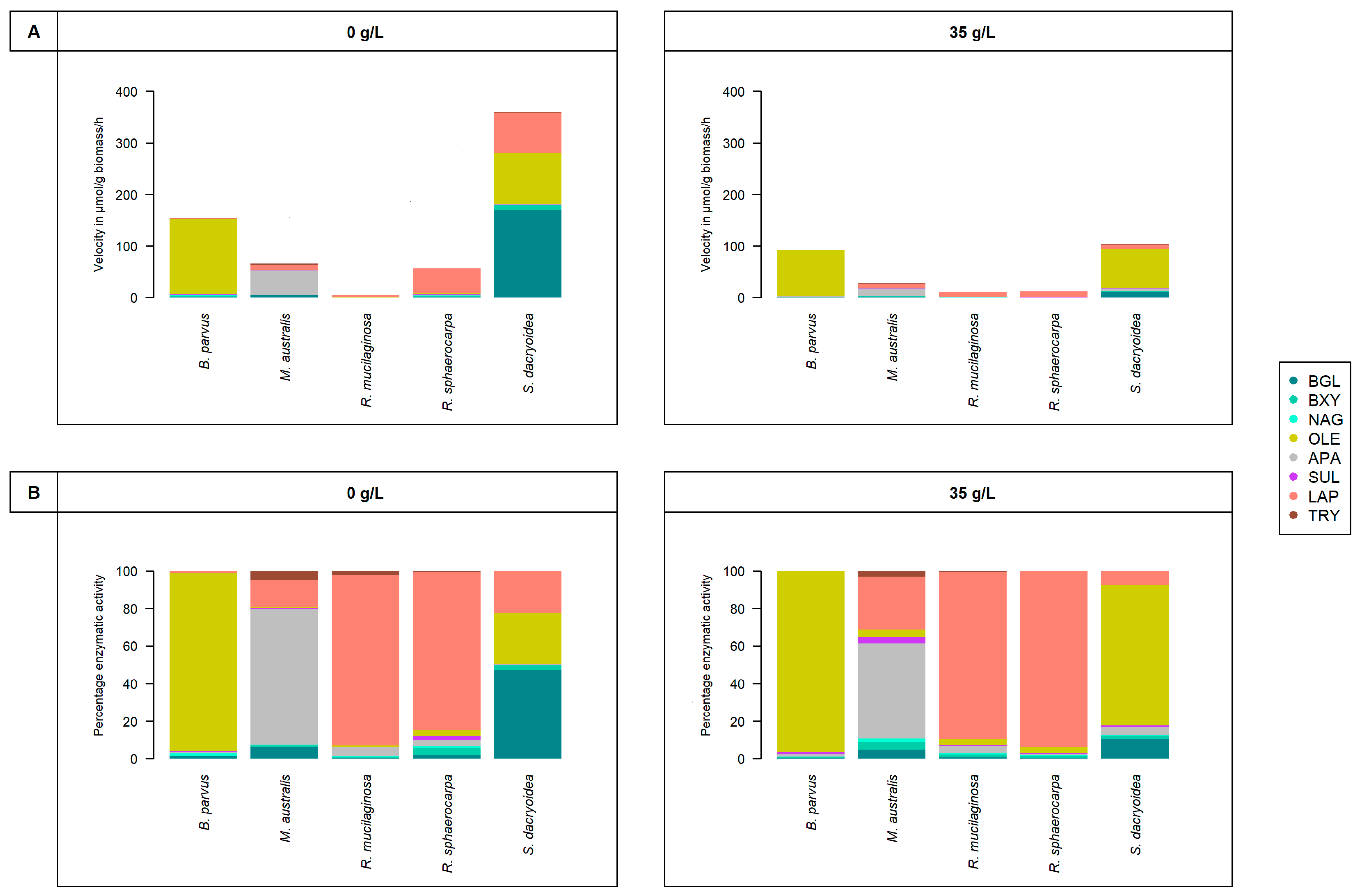
All the fungal strains used in this study were able to grow in non-saline (0 g/L) and saline (35 g/L) media (Figure 1) and hydrolyze all the fluorogenic substrates targeting carbohydrates (Figure 2), lipids, phosphorus, and sulfur moieties (Figure 3), and peptides (Figure 4). The results obtained suggested that the substrate concentrations needed to achieve half of the maximum velocity were variable among the different fungal strains (Figure 5). Generally, the extracellular enzymatic activities (EEAs) were higher in the non-saline than in the saline medium (Figure 6).

3.1. Carbohydrate Cleavage

3.1.1. β-Glucosidase (BGL)

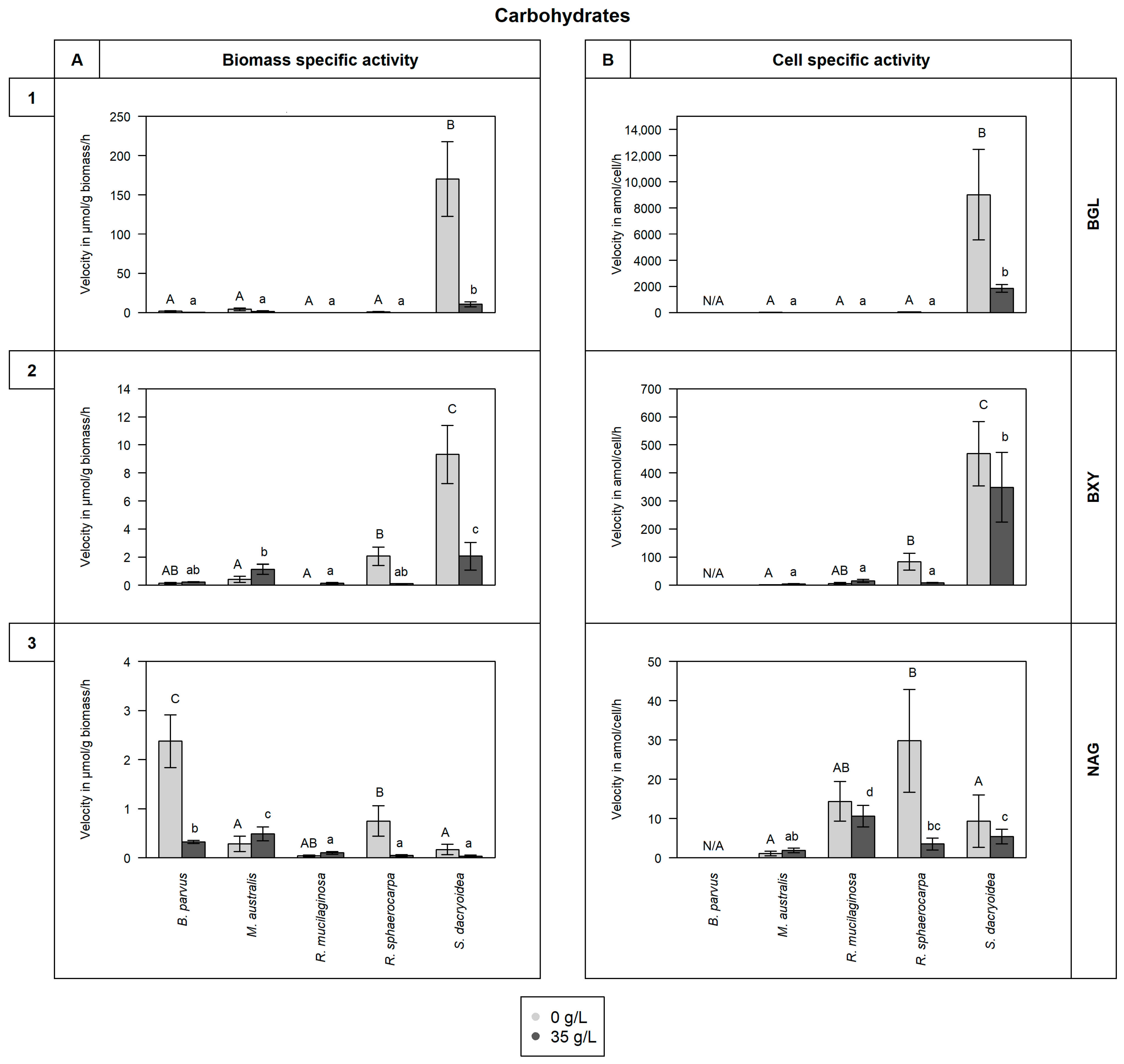
Compared to the other fungal species, S. dacryoidea exhibited a significantly higher Vmax for BGL (p < 0.001; Figure 2(1). This was significantly higher at a salinity of 0 g/L (170.1 ± 47.7 μmol/g biomass/h and 9015.0 ± 3453.3 amol/cell/h) than at 35 g/L (10.7 ± 3.2 μmol/g biomass/h and 1853.4 ± 283.6 amol/cell/h) (p < 0.001). Even though the other fungal species exhibited a generally low BGL hydrolysis rate, this was significantly higher at a salinity of 0 g/L (p < 0.001), except for R. mucilaginosa. Interestingly, this was the only species that exhibited a higher BGL activity at a salinity of 35 g/L (0.07 ± 0.02 μmol/g biomass/h and 7.6 ± 2.4 amol/cell/h) compared to 0 g/L (0.02 ± 0.00 μmol/g biomass/h and 7.0 ± 1.3 amol/cell/h) (p < 0.001).
Sakaguchia dacryoidea exhibited a higher BGL activity and a higher Km (767.3 ± 251.3 μM) at a salinity of 0 g/L than all the other fungal strains tested (p < 0.001; Figure 5A). The Km values of the other Basidiomycota species (R. mucilaginosa and R. sphaerocarpa) were also higher in the non-saline than in the saline medium (p = 0.001). In contrast, there was no clear influence of salinity on the Km of the Ascomycota strains B. parvus and M. australis (p = 0.2).

3.1.2. β-Xylosidase (BXY)

For BXY, S. dacryoidea also exhibited a significantly higher Vmax than the other fungal strains (p < 0.001; Figure 2(2)). The Vmax of this species was significantly higher in the non-saline (9.3 ± 2.1 μmol/g biomass/h and 468.9 ± 114.8 amol/cell/h) than in the saline medium (2.1 ± 1.0 μmol/g biomass/h and 348.3 ± 124.4 amol/cell/h) (p = 0.01). Similarly, R. sphaerocarpa exhibited a significantly higher Vmax at a salinity of 0 g/L (p < 0.001) than at 35 g/L. The two Ascomycota species B. parvus and M. australis, and the Basidiomycota species R. mucilaginosa showed low BXY activity.
Blastobotrys parvus, R. mucilaginosa, and S. dacryoidea, displayed a significantly higher Km at a salinity of 35 g/L than at 0 g/L (p= 0.004; Figure 5B). S. dacryoidea exhibited the highest Km value (1036.3 ± 397.9 μM) at 35 g/L of all the fungal strains tested, followed by B. parvus also at 35 g/L (631.8 ± 382.4 μM). In the non-saline medium, the Km for S. dacryoidea was only about half (521.9 ± 165.7 μM) of the one determined in the saline medium. Interestingly, M. australis presented the lowest Km in both media (10.9 ± 4.3 μM).

3.1.3. N-Acetyl-β-D-glucosaminidase (NAG)

Even though all the fungal strains were capable of using the fluorogenic substrate for NAG, the overall activity rates were low (Figure 2(3). The Vmax of B. parvus at a salinity of 0 g/L (2.4 ± 0.5 μmol/g biomass/h) was significantly higher than the Vmax of the other fungal species (p < 0.001). Also, the species of the Basidiomycota phylum, R. mucilaginosa, R. sphaerocarpa, and S. dacryoidea exhibited a higher Vmax in the non-saline medium (p = 0.001), whereas M. australis exhibited a higher Vmax in the saline medium (p = 0.009).
In the non-saline medium, B. parvus exhibited the highest NAG activity and with 477.4 ± 150.6 μM also the highest Km (Figure 5C), so it was significantly higher than the Km for NAG of the other fungal strains (p = 0.004). The Km of the other fungal strains varied between 46.7 and 0.01 μM.

3.2. Hydrolytic Activity on Organic Compounds Containing Lipids, Phosphorus and Sulfur Moieties

3.2.1. Lipase (OLE)

When normalizing OLE activity to biomass, B. parvus and S. dacryoidea exhibited significantly higher Vmax values than the other species (p < 0.001; Figure 3(1)). Nevertheless, when OLE activity was normalized to cell-specific activity, S. dacryoidea exhibited the highest Vmax of all the species (p < 0.001), specially in the saline medium (13,159.9 ± 4080.3 amol/cell/h). M. australis, R. mucilaginosa, and R. sphaerocarpa were also capable of cleaving lipids, but at much lower rates ranging from 2.9 to 0.03 μmol/g biomass/h and 127.6 to 0.5 amol/cell/h, and there was not a clear pattern on the effect of salinity (p = 0.1).
Generally, in all fungal strains, the Km was significantly higher in the saline than in the non-saline medium except in R. sphaerocarpa and B. parvus (p < 0.001; Figure 5D), where the Km was significantly higher in the non-saline medium (p = 0.01) and was 478.3 ± 284.7 μM. In all the other fungal strains, the Km varied between 172.8 and 0.6 μM in the non-saline medium and from 700.7 to 29.1 μM in the saline medium.

3.2.2. Alkaline Phosphatase (APA)

Normalizing APA activity to biomass revealed that the Vmax of M. australis in the non-saline medium was significantly higher than the Vmax of the other species (p < 0.001; Figure 3(2)). When the Vmax was normalized by the cell abundance, S. dacryoidea exhibited the highest Vmax in the saline medium (757.2 ± 201.1 amol/cell/h) compared to the other fungal strains (p < 0.001), followed by M. australis with a Vmax of 182.4 ± 43.1 amol/cell/h in the non-saline medium. Similarly, R. mucilaginosa and R. sphaerocarpa showed higher Vmax in the non-saline medium (p < 0.001) than the other fungal strains except M. australis, whereas B. parvus exhibited a higher Vmax in the saline medium (p = 0.02).
Under non-saline conditions, R. mucilaginosa exhibited a significantly lower Vmax than the other fungal strains (p < 0.001; Figure 3(2)), while its Km (506.2 ± 103.8 μM) was the highest (p < 0.001; Figure 5E). While in M. australis and S. dacryoidea the Km was lower in saline conditions than in the other conditions (p < 0.001). The Km values of B. parvus and R. sphaerocarpa were not significantly different between both conditions (p = 0.2).

3.2.3. Sulfatase (SUL)

All fungal strains studied were capable of cleaving sulfate esters, however, at low rates (Figure 3(3)). All the species, except R. sphaerocarpa, showed a significantly higher Vmax in saline than in the non-saline medium (p = 0.05). The highest Vmax in the saline medium was detected in S. dacryoidea (0.8 ± 0.4 μmol/g biomass/h and 124.1 ± 45.9 amol/cell/h) while in R. sphaerocarpa the Vmax was higher in the non-saline than in the other medium (p < 0.001).
However, as shown in Figure 5F, there was no clear effect of salinity on the Km of sulfatase (p = 0.3). Significantly higher values were found for both salinities in B. parvus than in the other strains, with Km values of 524.7 ± 220.6 μM in the non-saline and 493.3 ± 160.6 μM in the saline medium.

3.3. Proteins Cleavage

3.3.1. Leucine Aminopeptidase (LAP)

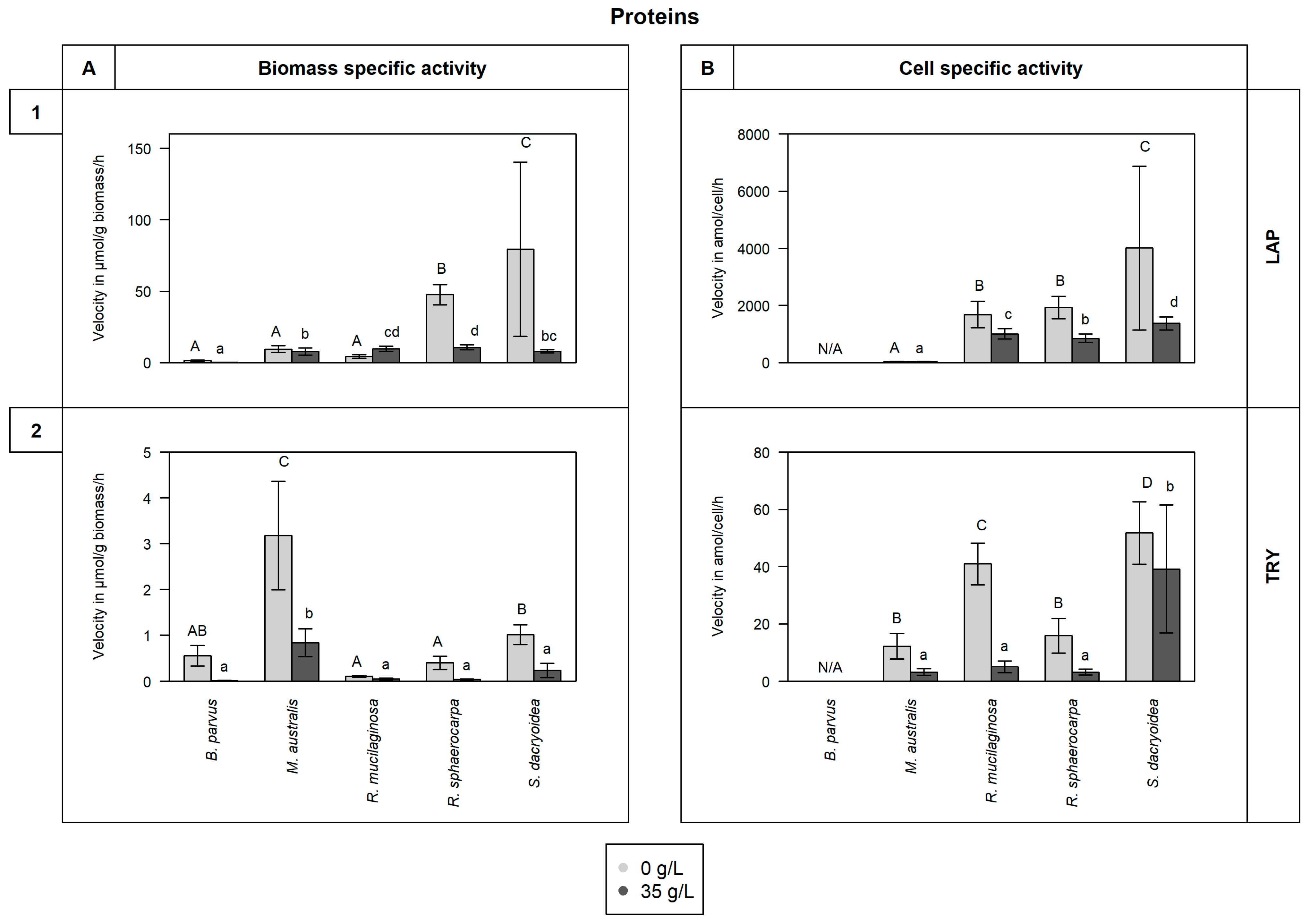
For the species of the phylum Basidiomycota, R. sphaerocarpa, and S. dacryoidea, the Vmax for LAP was significantly higher in the non-saline than in the saline medium (p = 0.004; Figure 4(1)). The Vmax values of the Basidiomycota species ranged from 208.5 to 2.9 μmol/g biomass/h, and between 10,101.1 to 604.4 amol/cell/h.
The Km was significantly higher in the Basidiomycota (except R. mucilaginosa) than in the Ascomycota strains (p < 0.001; Figure 5G). Compared to the other enzymes, the LAP Km values were the highest. Even though there was no clear influence of the salinity on the Km for all the species except R. sphaerocarpa (p = 0.5), S. dacryoidea exhibited high Km values (1536.7 ± 832.8 μM in the non-saline and 1415.4 ± 615.3 μM in the saline medium).

3.3.2. Trypsin (TRY)

Similar to LAP, the Vmax values for TRY in all the fungal strains were significantly higher in the non-saline than in the saline medium (p < 0.001; Figure 4(2)). Comparing the biomass-specific TRY activity and the cell-specific activity, the overall pattern did not change. However, when calculating the Vmax based on the biomass-specific TRY activity, M. australis exhibited a significantly higher Vmax (3.2 ± 1.2 μmol/g biomass/h) in the non-saline medium (p < 0.001) than all the other strains, while the Vmax calculated from the cell-specific activity, was higher in S. dacryoidea in the non-saline medium (51.8 ± 10.9 amol/cell/h; p < 0.001) than in all the other fungal strains.
For all the fungal strains except B. parvus, Km values were significantly higher in the saline medium (p < 0.001; Figure 5H). For this species, there was no clear influence of the salinity on the Km (p = 1.0). The highest Km detected corresponded to M. australis and R. mucilaginosa in the saline medium (471.7 ± 72.7 μM and 407.0 ± 136.0 μM, respectively).

4. Discussion

The use of fluorogenic substrate analogues allowed the determination of potential extracellular enzymatic activities of marine pelagic fungal isolates. The fungal strains studied, B. parvus, M. australis, R. mucilaginosa, R. sphaerocarpa, and S. dacryoidea exhibited a broad spectrum of extracellular enzymes in saline and non-saline media. They were able to enzymatically hydrolyze part of carbohydrates like cellulose, chitin, and xylan, and proteins such as peptide chains and leucine residues. Moreover, they expressed enzymes to hydrolytically cleave lipids, phosphate-containing compounds, and sulfate esters. Since these measurements were performed during the exponential phase, fungi were allowed to adapt to the new media. Finally, a short incubation period (3 h) and the addition of chloramphenicol avoided the potential growth of other microorganisms that could cause bias in these measurements.

4.1. Extracellular Enzymatic Activities of Pelagic Fungal Strains

4.1.1. Release of Enzymes Hydrolytically Cleaving Carbohydrates

Cellulose is the most common natural polysaccharide [70], so its degradation is crucial in the carbon cycle [71]. Several microorganisms, specially fungi, perform cellulolytic activities with three major classes of enzymes such as endoglucanases, exoglucanases, and β-glucosidases [72,73,74,75,76]. The hydrolysis of cellulose has been reported by a wide variety of marine fungi [67,75,77,78,79,80,81,82,83,84], but their capability to decompose this complex compound depends on their enzymatic machinery [85]. Vaz, Rosa [86] and Gonçalves, Paço [87] showed that 76% and 68%, respectively, of the marine fungi they studied were able to release enzymes related to cellulolytic activity. However, compared to other cellulolytic enzymes, β-glucosidase hydrolyzes the products of those enzymes to glucose [88]. In the soil, a widely distributed species, Aspergillus niger, exhibited a Vmax of 11.7 μmol/g/h, and other fungal species, had values between 14.8 and 6.7 μmol/g/h [89]. All our fungal isolates were able to hydrolytically cleave cellulose, with S. dacryoidea dominating this cellulolytic activity among all the strains tested (Figure 2(1)). Moreover, all the species exhibited Km values > 97.0 μM, except the two species belonging to the genus Rhodotorula under saline conditions (Figure 5A). Thus, even though the studied species can use this type of cellulolytic enzyme, the Km values obtained indicate that relatively high cellulose concentrations are preferred. In marine environments, cellulose is present in mangroves and associated with hemicellulose and lignin [90,91], but it is also present in some algal species [92]. As the organic matter of terrestrial origin is more available in coastal areas and less abundant in the open ocean [93,94,95], only specific microorganisms might be able to use cellulose [96] and dominate its decay in the open ocean.
After cellulose, chitin is the second most abundant polysaccharide [70,97,98,99] and is composed of monomers of N-acetyl-D-glucosamine (GlcNAc) linked by β-glycosidic bond [100,101,102,103]. Hexosaminidases are enzymes catalyzing the cleavage of these glycosidic bonds [97,104]. As chitin is abundant in marine environments [17], several marine fungal species are efficient chitin degraders [67,84,87,105,106], and together with bacteria, they are the main chitin degraders [102]. Fungi can also hydrolyze chitin, but unlike bacteria, fungi can use it as a building block for new chitin synthesis [107] or as a carbon and nitrogen source [102]. Nonetheless, dissimilarities between hyphae-like fungi and yeast chitinolytic enzymes have been reported [108]. The amount of chitinases released by fungi is related to their chitin content, so it is also dependent on the growth mode [109]. As hyphae-like fungal cell walls contain 10 to 20% of chitin [99], these fungi typically have 10 to 30 chitinases [110,111]. In contrast, yeast cell walls have a chitin content as low as 0.5 to 5% [112]. A hyphae-like fungi species, Fusarium oxysporum, exhibited a Vmax of 20.7 μmol/g/h, and for its isoenzymes, 4.3 μmol/g/h [113]. B. parvus, also a filamentous fungus, had the highest chitinase Vmax detected, under non-saline conditions (Figure 2(3)), and also presented the highest Km (Figure 5C). In filamentous fungi, chitinolytic enzymes are important for cell wall regeneration and hyphae formation [114]. For the species not forming hyphae, their low Km indicate a high affinity of the enzyme to the substrate at low concentrations [22]. Hence, all the studied species can use chitin, even when present at low concentrations, but hyphae-like species (B. parvus) might have an advantage over yeasts.
Due to xylan heterogeneity and complexity, several enzymes with diverse specificities are required to cleave it [115,116,117,118]. Thus, the xylanolytic activities are performed by xylanases, β-xylosidases, α-arabinosidases, α-glucuronidases, and esterases [118,119]. Widely distributed fungal species such as Trichoderma viride and Emericella nidulans exhibit β-xylosidase activity [120]. Biely, Vršanská, and Krátký [121] reported low β-xylosidase activity for Cryptococcus albidus but high xylanase activity, so 1,4 β-xylanase was highlighted as the main “xylan-attacking enzyme”. For the first enzyme, these authors obtained Vmax values as low as 0.06 μmol/g/h. In the case of pelagic marine fungi, Salazar Alekseyeva, Herndl, and Baltar [67] also reported low β-xylosidase activity which is consistent with the present study, where the two species of the phylum Ascomycota, B. parvus and M. australis, and one species of Basidiomycota, R. mucilaginosa, also exhibited generally low β-xylosidase activity (Figure 2(2)). R. sphaerocarpa and S. dacryoidea, however, exhibited higher enzymatic activity in a non-saline than in the saline medium. As shown in Figure 5B, in the saline medium, B. parvus, R. mucilaginosa, and S. dacryoidea exhibited a high Km, indicative of a low affinity to the substrate [68] and inefficient organisms to obtain resources present at low concentrations [22]. In Antarctic environments, where the majority of the studied fungi were initially isolated [51,52,53,54], xylan is limited, but it can be found in the cell walls of green and red algae [122]. Thus, the low Km or high affinity to the substrate of the only endemic species, M. australis [53,123], might be an advantage over the other species at the time of cleaving xylan.

4.1.2. Release of Enzymes Cleaving Lipids, Phosphorus, and Sulfur Moieties

Lipase is the third largest enzyme group that can catalyze both hydrolysis and synthesis of esters and fatty acids [124,125]. These enzymes have been widely reported in marine fungi like Hortaea werneckii, Trimmatostroma salinum [126], Rhodotorula glutinis [127], Yarrowia lipolytica [128,129], Leucosporidium scottii [130], and in some Antarctic Ocean [131] and China Sea [132] strains. A species also included in the present study, R. mucilaginosa was suggested to be a promising fungal strain for lipase production [133], similar to Wang, Chi [132]. Contrary, we found that other species, such as B. parvus and S. dacryoidea had a higher OLE activity than R. mucilaginosa (Figure 3(1)), but the substrate dissociates easily from the enzyme (Figure 5F).
Inorganic phosphate (Pi, PO43−) is the preferred phosphorus source by microorganisms [134,135]. In surface waters, it is readily used by bacteria and phytoplankton, leading to low PO43− concentrations in vast areas of the global ocean [135,136,137,138]. To overcome this P-limitation, microorganisms release alkaline phosphatase to use a range of available dissolved organic phosphorus (DOP) [136,139,140]. This release might also be related to sporadic pulses of organic matter [141,142]. APA has been reported in marine fungi species like Debaryomyces hansenii, exhibiting a Vmax of 0.83 μmol/g/h [143], and some species of Ascomycota and Basidiomycota phylum [67]. At a sporadic high organic matter concentration, a high Km might be advantageous by allowing higher hydrolysis rates, for instance, for species like B. parvus (Figure 3(2) and Figure 5E). Nonetheless, a low Km, such as for S. dacryoidea, might be more suitable to overcome the P-limitation most of the time when organic matter is low.
In contrast to terrestrial polysaccharides, numerous marine polysaccharides are highly sulfated [144,145,146]. For marine organisms, sulfated polysaccharides might be a physiological adaptation to the high ionic strength of their environment [146,147,148,149]. Nonetheless, microorganisms need to remove the sulfate groups to gain access to the carbohydrates [150,151]. The terrestrial fungus Fusarium proliferatum [152] and the marine fungus Paradendryphiella salina [84] were reported to assimilate sulfates from brown algae. In the present study, all the marine fungal species, except R. sphaerocarpa, exhibited a higher sulfatase activity in the saline medium (Figure 3(3)). The Km was low for the species of the genus Rhodotorula (Figure 5F), but for the other examined species, the Km was high. This might indicate an advantage for the species R. mucilaginosa and R. sphaerocarpa, as fewer enzymes and substrates would be needed to speed up this reaction in saline conditions, which would also allow the use of sulfated polysaccharides, common in marine environments.

4.1.3. Release of Enzymes Hydrolytically Cleaving Proteins

Leucine aminopeptidase (LAP) is a ubiquitous enzyme involved in the hydrolysis of peptides [153,154,155]. In marine environments, the microbial production of LAP might indicate the use of nitrogen from suspended and sinking organic matter [30,44,156], so LAP is a key player in both carbon and nitrogen recycling [154]. Gutiérrez, Pantoja [29] associated an increase in LAP and BGL activities in surface waters with the availability of new organic matter during periods of high phytoplankton biomass production. Thus, microorganisms might respond to changing substrate availability by producing LAP [156]. In the present study, due to higher Vmax values detected in the non-saline medium, it seems that the fungal strains produce less LAP in marine environments than in freshwater environments (Figure 4(1)). In the study of Caruso and Zaccone [30], changes in LAP activity of marine microbial communities were related to physical (salinity and temperature), chemical (nutrient composition), and microbiological variables. In our study, Basidiomycota species expressed a higher LAP activity than the Ascomycota species. As the substrate concentration needed to achieve half the maximum velocity is considerably high (Figure 5G), in pelagic environments where the organic matter is readily used within the water column [157] and decreases with depth [158], the activity of LAP by marine fungi might be preferably associated to environments with relatively high organic matter content.
Trypsin is an enzyme that cleaves proteins, specifically at the carboxyl end of lysine and arginine [159]. Comparable to leucine aminopeptidase, the production of this enzyme also depends on the nitrogen source [160,161], so it is important in the carbon and nitrogen cycles [162]. Aspergillus ustus, a deep-sea isolate from the Central Indian Basin, was a high producer of serine protease [163]. In the present study, we found that R. mucilaginosa had a high trypsin activity, but also other species, such as S. dacryoidea, specially in the non-saline medium (Figure 4(2)). Nonetheless, only R. sphaerocarpa presented relatively low Km values (Figure 5H). As trypsin might be used under nitrogen deficiency to acquire this element [164], only a high affinity to the substrate might be beneficial to convert the substrate efficiently into products that can be used for this purpose [165].

4.2. Influence of Salinity on Extracellular Enzymatic Activity

Some fungi do not require salts to grow [6], but as reported by Jones and Jennings [166], salinity might not affect remarkably the growth of fungi in marine environments compared with freshwater and terrestrial environments. Similar results were found by Arfi, Chevret [167]. However, we found that even though salinity does not remarkably influence the growth of some fungi (Figure 1), it might exert an important effect on the kinetics of certain enzymes (Figure 2, Figure 3, Figure 4 and Figure 5).
In marine environments, Caruso and Zaccone [30] stated that salinity was an important variable affecting microbial enzymatic activities. In soil studies, a salinity increase leads to the exponential decrease of some microbial enzymatic activities such as alkaline phosphatase and β-glucosidase [168]. For the halotolerant species Debaryomyces hansenii, a high salinity resulted in low alkaline phosphatase activity [143]. In the present study, we also found that the majority of extracellular enzymatic activities were reduced at higher salinity (Figure 6). The alkaline phosphatase, β-glucosidase, and N-acetyl-β-D-glucosaminidase activities were lower for all the species, except S. dacryoidea for alkaline phosphatase, R. mucilaginosa for β-glucosidase, and M. australis for N-acetyl-β-D-glucosaminidase (Figure 2 and Figure 3(1)). Also, the hydrolysis of proteins and peptides by leucine-aminopeptidase and trypsin [45,66] was lower in the saline medium for all the studied species (Figure 4). For S.dacryoidea and R. mucilaginosa, the normalization of these values by the cell abundance allowed us to identify higher activities. As these species had lower cell abundance values, the cell-specific activity increased.
Proteins are tightly associated with water as they tend to bind to the hydrophobic groups in the protein surface [169,170,171]. Water is incorporated inside the protein, which makes it an important part of its native structure, hence its proper function [170,172,173]. Nevertheless, salt ions tend to sequester water into other hydrated ionic structures, which diminishes intermolecular hydrogen bonds and also the availability of free water molecules [174,175]. As stated by Sinha and Khare [176], the salinity effect depends on the salt concentration along with the chemical composition of the protein. Based on our results, we suggest that salinity influenced differently the kinetic parameters Vmax and Km (Figure 2, Figure 3, Figure 4 and Figure 5). The majority of Vmax (APA, BGL, BXY, LAP, and TRY) were reduced in the saline conditions, whereas some enzymes (BXY and TRY) exhibited a lower affinity under saline conditions. For the remaining enzymes, there was no detectable clear influence of the salinity on the Km. According to Blomberg and Adler [13], under osmotic stress, the capacity for substrate uptake limits growth. This might lead to a decrease in activation energy, and hence, to increase and maximize the reaction rate [177].
Some enzymes, however, are salt-tolerant or halophilic [178]. Compared to other enzymes, the halophilic ones have a surface with more amino acid residues that tend to bind to hydrated cations [179], resulting in a substantially larger multi-layered hydration shell [172]. This reduces the enzyme hydrophobicity, prevents aggregation [172,180], and also allows the halophilic enzyme to adopt a conformational structure that is flexible [176,181,182,183]. In this study, we detected potential halophilic enzymes, which were APA and OLE for S. dacryoidea, and SUL for all species except R. sphaerocarpa (Figure 3). Interestingly, R. mucilaginosa was the only species that did not exhibit a sulfatase activity in the non-saline medium but displayed one in the saline medium. As mentioned above, as sulfate esters are less common in terrestrial plants [184], sulfated polysaccharides might be a physiological adaptation to the high ionic strength of the marine environment [146,147,148,149]. Even though the overall rates of sulfatase were low compared to other enzymes, the studied marine fungi used it, which might be an advantage in marine environments to access monosaccharides from sulfated polysaccharides. Similarly, Gladfelter, James, and Amend [3] and Gonçalves, Hilário [185] supported that as fungal nutritional sources are at high environmental osmolyte concentrations, fungi might have evolved enzymatic activities to compete for its uptake. In addition, we did not find differences in the Km between the saline and non-saline media (Figure 5F). Therefore, the affinity to sulfate esters might depend on the species rather than on salinity.
The results obtained in this study suggest that the salinity effect on the enzyme kinetics of marine fungi depends on the species as well as on the enzyme type, specially for Km (Figure 5). For Vmax, the studied marine fungi seem to be adapted to low and high levels of salinity, but generally, extracellular enzymatic activities were higher in the non-saline medium (Figure 6). Similar results were found for cellulase and xylanase produced in the absence of salts by the mangrove species Pestalotiopsis sp. [167]. When these were added to a saline medium, they exhibited lower activities. As suggested by Zaccai [171], each protein seems to have evolved a stable form for a particular environment. This was also proposed for another mangrove fungal species that secreted two enzymes in the presence of salts and another one in the absence of salt [186]. Thus, the release of isoenzymes might be an adaptation mechanism to salinity [167]. As all the studied fungal species displayed enzymatic activities in both media, we suggest that different enzymes might catalyze the same reaction (isoenzymes) and allow them to perform their activities at a wide range of salinities. Nonetheless, it is important to highlight that the degradation of complex compounds also requires a wide range of enzymes [72].

4.3. Life Strategies

In the study of Solis, Draeger, and Cruz [187], some marine fungi that released algal degrading enzymes, such as cellulase, were related to the development of ice–ice disease in red seaweed. Other enzymes, such as proteases, were also involved in normal and pathological processes [188]. In terrestrial environments, trypsin-like enzymes are characteristic of plant pathogens [16,23,66,189]. Therefore, the spectrum of secreted enzymes for the hydrolysis of polysaccharides and proteins by some pathogenic fungal strains has been taken as indicative of less specialized nutritional requirements [16]. The present study found that some species are more specialized in cleaving certain substrates, while others appear to be more generalists (Figure 6). S. dacryoidea exhibited the highest extracellular enzymatic activity of all the tested fungal strains. Allen, Quayyum [190] found that while the percentage of the total yeast community present on healthy leaves of bentgrass was <9.9%, their biomass increased to 32.0–44.7% on infected leaves. This biomass included yeast species such as S. dacryoidea. Therefore, the outstanding combination of cellulase, xylosidase, and trypsin-like activities of S. dacryoidea in the non-saline and saline medium suggests less specialized nutritional requirements and potential pathogenicity that could affect plants in terrestrial environments as well as algae in aquatic systems.
Chitinases and trypsin inhibitors have been highlighted as antifungal proteins that are released by the host to prevent or fight against fungal infection [97,191]. In the case of chitinases, fungi can produce them for nutritional, symbiotic, or parasitic purposes [103,111,192], while bacteria can use chitin as a carbon and energy source [193]. Unlike bacteria, many fungi can penetrate solid substrates [2], specially filamentous ones [161], so hyphal growth is an important feature in colonizing substrates [126]. In the present study, B. parvus was the only species that had a filamentous structure. Even though the overall rate of NAG was low when compared to other enzymes, B. parvus had the highest NAG (Figure 2(3)). Some fungi live on the surface of other organisms as ectobionts, or live within them [2]. Our results suggest that the high release of chitinases by the filamentous species B. parvus might be an advantage in penetrating and using this solid substrate. Moreover, the hydrolysis of chitin by the other studied marine fungi might indicate the capability to degrade organic matter of animal origin and other fungi, even if it is performed at low rates.
Some marine fungi species are obligatory marine, whereas others were introduced by river runoff, wind, or other organisms animals [24,131]. Thus, facultative marine fungi are frequent [2]. A comparative study using Paradendryphiella salina and its terrestrial relative Septoria lycopersic showed that the latter had more genes for cellulose and hemicellulose degradation, but both harbored an equal amount of genes for chitin hydrolysis [84]. Nonetheless, P. salina was capable of degrading most components of brown algae, seagrasses, and terrestrial plant material [84]. Hence, this species has a broad and saprobic lifestyle and also colonizes terrestrial and marine environments [84]. S. dacryoidea has a worldwide distribution and has been reported from the coast of Portugal [194], New Zeeland [195], and grasslands in Georgia, USA [190], but was originally isolated from the Antarctic Peninsula [54]. This wide distribution, together with our results suggests that the high enzymatic activity of S. dacryoidea in both the saline and non-saline media is related to potential facultative marine species that are adapted to different salinities (Figure 6). Moreover, it seems that S. dacryoidea conserves enzymes to degrade carbohydrates of terrestrial origin, such as cellulose and xylose, which might be an advantage over obligatory marine species like M. australis [53,123].

4.4. Potential Environmental Implications

As shown in Figure 1, even though the growth was similar for the majority of species (R. mucilaginosa, R. sphaerocarpa, and M. australis) at 0 g/L and 35 g/L salinities, the array of enzymes secreted was different. Our results suggest that the release of certain enzymes might be favored in freshwater. Similar results were reported by Caruso and Zaccone [30], where higher microbial LAP enzymatic activities were found in the upper layers of marine waters associated with freshwater input. During the second half of the 20th century, more than 80% of the Antarctic Peninsula glaciers have retreated [196], so freshwater input has lowered the salinity of coastal waters [197]. Consistently, salinity variations have been reported in the Arctic [198]. Rojas-Jimenez, Rieck [199] stated that salinity variation influences the fungal community composition. Our results indicate that the studied marine fungal species might be adapted to different salinities. However, their extracellular enzymatic activities were generally higher under freshwater conditions (Figure 6). Hence, in regions where the ocean is freshening, the species with broader enzymatic capabilities at lower salinities might thrive, whereas others might disfavored. The potential stratification of the water column might also influence this activity, as the critical transport of deep-water nutrients might be affected [47,50]. This might lead to the expression of different enzymes, which might influence the role of marine fungi in the biogeochemical cycles of the surface ocean.

5. Conclusions

The presented results broaden the current ecological knowledge of marine fungi. Together with similar results on freshwater and terrestrial fungi support the great biochemical versatility of this group of organisms. Generally, the fungal growth was not significantly influenced by the presence or absence of salts. However, using eight fluorogenic substrates as representatives of the main molecules found in the oceans, together with kinetics, allowed us to identify that their extracellular enzymatic activities might be affected differently depending on the species and the enzyme type. Although the studied marine fungi were able to degrade these complex molecules under both conditions, we suggest that this might be generally enhanced in non-saline conditions, with some exceptions like sulfatase. Therefore, we suggest that changes in salinities due to natural and human processes might influence the array of enzymes released by marine fungi and potentially also their role in the biogeochemical cycles.

Author Contributions

Conceptualization, K.S.-A. and F.B.; data curation, K.S.-A.; formal analysis, K.S.-A.; funding acquisition, F.B.; investigation, K.S.-A.; methodology, K.S.-A. and F.B.; project administration, F.B.; resources, G.J.H. and F.B.; software, K.S.-A.; supervision, F.B.; validation, K.S.-A.; visualization, K.S.-A.; writing—original draft preparation, K.S.-A.; writing—review and editing writing, K.S.-A., G.J.H. and F.B. All authors have read and agreed to the published version of the manuscript.

Funding

F.B. was supported by the Austrian Science Fund (FWF) projects OCEANIDES (P34304-B), ENIGMA (TAI534), EXEBIO (P35248), and OCEANBIOPLAST (P35619). Open Access Funding by the Austrian Science Fund (FWF). G.J.H. was supported by the FWF projects ARTEMIS (P28781-B21) and DECOMB (I 4978-B).

Institutional Review Board Statement

Not applicable.

Informed Consent Statement

Not applicable.

Data Availability Statement

The raw data supporting the conclusions of this article will be made available by the authors without undue reservation to any qualified researcher.

Acknowledgments

We would like to thank Katarína Tamášová for her valuable help in the laboratory work and Eduard Fadeev for his help in the visualization of the results on the BD Accuri C6 Software (version 264).

Conflicts of Interest

The authors declare no conflicts of interest.

References

  1. Jennings, D. Some aspects of the physiology and biochemistry of marine fungi. Biol. Rev. 1983, 58, 423–459. [Google Scholar] [CrossRef]
  2. Raghukumar, S. Fungi in Coastal and Oceanic Marine Ecosystems; Springer: Cham, Switzerland, 2017. [Google Scholar]
  3. Gladfelter, A.S.; James, T.Y.; Amend, A.S. Marine fungi. Curr. Biol. 2019, 29, 191–195. [Google Scholar] [CrossRef] [PubMed]
  4. Ruisi, S.; Barreca, D.; Selbmann, L.; Zucconi, L.; Onofri, S. Fungi in Antarctica. Rev. Environ. Sci. Bio/Technol. 2007, 6, 127–141. [Google Scholar] [CrossRef]
  5. Shivaji, S.; Prasad, G. Antarctic yeasts: Biodiversity and potential applications. In Yeast Biotechnology: Diversity and Applications; Satyanarayana, T., Kunze, G., Eds.; Springer: Dordrecht, The Netherlands, 2009; pp. 3–18. [Google Scholar]
  6. Plemenitaš, A.; Lenassi, M.; Konte, T.; Kejžar, A.; Zajc, J.; Gostinčar, C.; Gunde-Cimerman, N. Adaptation to high salt concentrations in halotolerant/halophilic fungi: A molecular perspective. Front. Microbiol. 2014, 5, 199. [Google Scholar] [CrossRef] [PubMed]
  7. Śliżewska, W.; Struszczyk-Świta, K.; Marchut-Mikołajczyk, O. Metabolic potential of halophilic filamentous fungi-current perspective. Int. J. Mol. Sci. 2022, 23, 4189. [Google Scholar] [CrossRef] [PubMed]
  8. Norkrans, B.; Kylin, A. Regulation of the potassium to sodium ratio and of the osmotic potential in relation to salt tolerance in yeasts. J. Bacteriol. 1969, 100, 836–845. [Google Scholar] [CrossRef] [PubMed]
  9. Oren, A. Bioenergetic aspects of halophilism. Microbiol. Mol. Biol. Rev. 1999, 63, 334–348. [Google Scholar] [CrossRef]
  10. Madern, D.; Ebel, C.; Zaccai, G. Halophilic adaptation of enzymes. Extremophiles 2000, 4, 91–98. [Google Scholar] [CrossRef]
  11. Gostinčar, C.; Lenassi, M.; Gunde-Cimerman, N.; Plemenitaš, A. Fungal adaptation to extremely high salt concentrations. In Advances in Applied Microbiology; Laskin, A., Sariaslani, S., Gadd, G., Eds.; Academic Press: Cambridge, MA, USA, 2011; Volume 77, pp. 71–96. [Google Scholar]
  12. Luard, E.J. Activity of isocitrate dehydrogenase from three filamentous fungi in relation to osmotic and solute effects. Arch. Microbiol. 1983, 134, 233–237. [Google Scholar] [CrossRef]
  13. Blomberg, A.; Adler, L. Physiology of osmotolerance in fungi. Adv. Microb. Physiol. 1992, 33, 145–212. [Google Scholar]
  14. Dihazi, H.; Kessler, R.; Eschrich, K. High osmolarity glycerol (HOG) pathway-induced phosphorylation and activation of 6-phosphofructo-2-kinase are essential for glycerol accumulation and yeast cell proliferation under hyperosmotic stress. J. Biol. Chem. 2004, 279, 23961–23968. [Google Scholar] [CrossRef]
  15. Velez, P.; Alejandri-Ramírez, N.D.; González, M.C.; Estrada, K.J.; Sanchez-Flores, A.; Dinkova, T.D. Comparative transcriptome analysis of the cosmopolitan marine fungus Corollospora maritima under two physiological conditions. G3 Genes Genomes Genet. 2015, 5, 1805–1814. [Google Scholar] [CrossRef]
  16. Leger, R.J.S.; Joshi, L.; Roberts, D.W. Adaptation of proteases and carbohydrates of saprophytic, phytopathogenic and entomopathogenic fungi to the requirements of their ecological niches. Microbiology 1997, 143, 1983–1992. [Google Scholar] [CrossRef] [PubMed]
  17. Arnosti, C. Microbial extracellular enzymes and the marine carbon cycle. Annu. Rev. Mar. Sci. 2011, 3, 401–425. [Google Scholar] [CrossRef] [PubMed]
  18. Muszewska, A.; Stepniewska-Dziubinska, M.M.; Steczkiewicz, K.; Pawlowska, J.; Dziedzic, A.; Ginalski, K. Fungal lifestyle reflected in serine protease repertoire. Sci. Rep. 2017, 7, 9147. [Google Scholar] [CrossRef] [PubMed]
  19. Richards, T.A.; Jones, M.D.; Leonard, G.; Bass, D. Marine fungi: Their ecology and molecular diversity. Annu. Rev. Mar. Sci. 2012, 4, 495–522. [Google Scholar] [CrossRef] [PubMed]
  20. Cooper, G.M. The central role of enzymes as biological catalysts. In The Cell: A Molecular Approach; Cooper, G.M., Ed.; Sinauer Associates: Sunderland, MA, USA, 2000; Volume 1, pp. 1–5. [Google Scholar]
  21. Kohler, T.J.; Peter, H.; Fodelianakis, S.; Pramateftaki, P.; Styllas, M.; Tolosano, M.; De Staercke, V.; Schön, M.; Busi, S.B.; Wilmes, P. Patterns and drivers of extracellular enzyme activity in New Zealand glacier-fed streams. Front. Microbiol. 2020, 11, 591465. [Google Scholar] [CrossRef] [PubMed]
  22. Mulder, C.; Hendriks, A.J. Half-saturation constants in functional responses. Glob. Ecol. Conserv. 2014, 2, 161–169. [Google Scholar] [CrossRef]
  23. Semenova, T.A.; Dunaevsky, Y.E.; Beljakova, G.A.; Borisov, B.A.; Shamraichuk, I.L.; Belozersky, M.A. Extracellular peptidases as possible markers of fungal ecology. Appl. Soil Ecol. 2017, 113, 591465. [Google Scholar] [CrossRef]
  24. Boddy, L. Fungi, ecosystems, and global change. In The Fungi; Watkinson, S.C., Boddy, L., Money, N.P., Eds.; Academic Press: Cambridge, MA, USA, 2016; Volume 3, pp. 361–400. [Google Scholar]
  25. Arnosti, C.; Bell, C.; Moorhead, D.L.; Sinsabaugh, R.L.; Steen, A.D.; Stromberger, M.; Wallenstein, M.; Weintraub, M.N. Extracellular enzymes in terrestrial, freshwater, and marine environments: Perspectives on system variability and common research needs. Biogeochemistry 2014, 117, 5–21. [Google Scholar] [CrossRef]
  26. Caruso, G. Leucine aminopeptidase, β-glucosidase and alkaline phosphatase activity rates and their significance in nutrient cycles in some coastal Mediterranean sites. Mar. Drugs 2010, 8, 916–940. [Google Scholar] [CrossRef] [PubMed]
  27. Morales, S.E.; Biswas, A.; Herndl, G.J.; Baltar, F. Global structuring of phylogenetic and functional diversity of pelagic fungi by depth and temperature. Front. Mar. Sci. 2019, 6, 131. [Google Scholar] [CrossRef]
  28. Baltar, F.; Zhao, Z.; Herndl, G.J. Potential and expression of carbohydrate utilization by marine fungi in the global ocean. Microbiome 2021, 9, 106. [Google Scholar] [CrossRef] [PubMed]
  29. Gutiérrez, M.H.; Pantoja, S.; Tejos, E.; Quiñones, R.A. The role of fungi in processing marine organic matter in the upwelling ecosystem off Chile. Mar. Biol. 2011, 158, 205–219. [Google Scholar] [CrossRef]
  30. Caruso, G.; Zaccone, R. Estimates of leucine aminopeptidase activity in different marine and brackish environments. J. Appl. Microbiol. 2000, 89, 951–959. [Google Scholar] [CrossRef] [PubMed]
  31. Urvoy, M.; Labry, C.; Delmas, D.; Creac’h, L.; l’Helguen, S. Microbial enzymatic assays in environmental water samples: Impact of Inner Filter Effect and substrate concentrations. Limnol. Oceanogr. Methods 2020, 18, 725–738. [Google Scholar] [CrossRef]
  32. Cullings, K.; Courty, P.-E. Saprotrophic capabilities as functional traits to study functional diversity and resilience of ectomycorrhizal community. Oecologia 2009, 161, 661–664. [Google Scholar] [CrossRef]
  33. Orsi, W.; Biddle, J.F.; Edgcomb, V. Deep sequencing of subseafloor eukaryotic rRNA reveals active fungi across marine subsurface provinces. PLoS ONE 2013, 8, e56335. [Google Scholar] [CrossRef]
  34. Bochdansky, A.B.; Clouse, M.A.; Herndl, G.J. Eukaryotic microbes, principally fungi and labyrinthulomycetes, dominate biomass on bathypelagic marine snow. ISME J. 2017, 11, 362–373. [Google Scholar] [CrossRef]
  35. Picard, K.T. Coastal marine habitats harbor novel early-diverging fungal diversity. Fungal Ecol. 2017, 25, 1–13. [Google Scholar] [CrossRef]
  36. Ueda, M.; Nomura, Y.; Doi, K.; Nakajima, M.; Honda, D. Seasonal dynamics of culturable thraustochytrids (Labyrinthulomycetes, Stramenopiles) in estuarine and coastal waters. Aquat. Microb. Ecol. 2015, 74, 187–204. [Google Scholar] [CrossRef]
  37. Amend, A.; Burgaud, G.; Cunliffe, M.; Edgcomb, V.P.; Ettinger, C.L.; Gutiérrez, M.H.; Heitman, J.; Hom, E.F.Y.; Ianiri, G.; Jones, A.C.; et al. Fungi in the marine environment: Open questions and unsolved problems. mBio 2019, 10, 1–18. [Google Scholar] [CrossRef]
  38. Taylor, J.D.; Cunliffe, M. Multi-year assessment of coastal planktonic fungi reveals environmental drivers of diversity and abundance. ISME J. 2016, 10, 2118–2128. [Google Scholar] [CrossRef]
  39. Martínez-García, S.; Bunse, C.; Pontiller, B.; Baltar, F.; Israelsson, S.; Fridolfsson, E.; Lindh, M.; Lundin, D.; Legrand, C.; Pinhassi, J. Seasonal dynamics in carbon cycling of marine bacterioplankton are lifestyle dependent. Front. Microbiol. 2022, 13, 834675. [Google Scholar] [CrossRef]
  40. Benner, R. Chemical composition and reactivity. Biogeochem. Mar. Dissolved Org. Matter 2002, 1, 59–90. [Google Scholar]
  41. Benner, R. Molecular indicators of the bioavailability of dissolved organic matter. In Aquatic Ecosystems; Findlay, S.E.G., Sinsabaugh, R.L., Eds.; Academic Press: Cambridge, MA, USA, 2003; Volume 1, pp. 121–137. [Google Scholar]
  42. Benner, R.; Pakulski, J.D.; McCarthy, M.; Hedges, J.I.; Hatcher, P.G. Bulk chemical characteristics of dissolved organic matter in the ocean. Science 1992, 255, 1561–1564. [Google Scholar] [CrossRef] [PubMed]
  43. Chróst, R.J. Microbial ectoenzymes in aquatic environments. In Aquatic Microbial Ecology; Overbeck, J., Chróst, R.J., Eds.; Springer: New York, NY, USA, 1990; Volume 1, pp. 47–78. [Google Scholar]
  44. Baltar, F.; Aristegui, J. Fronts at the surface ocean can shape distinct regions of microbial activity and community assemblages down to the bathypelagic zone: The Azores Front as a case study. Front. Mar. Sci. 2017, 4, 252. [Google Scholar] [CrossRef]
  45. Hoppe, H.-G. Significance of exoenzymatic activities in the ecology of brackish water: Measurements by means of methylumbelliferyl-substrates. Mar. Ecol. Prog. Ser. 1983, 11, 299–308. [Google Scholar] [CrossRef]
  46. Reid, P.C.; Fischer, A.C.; Lewis-Brown, E.; Meredith, M.P.; Sparrow, M.; Andersson, A.J.; Antia, A.; Bates, N.R.; Bathmann, U.; Beaugrand, G. Impacts of the oceans on climate change. Adv. Mar. Biol. 2009, 56, 1–150. [Google Scholar] [PubMed]
  47. Hutchins, D.A.; Fu, F. Microorganisms and ocean global change. Nat. Microbiol. 2017, 2, 17058. [Google Scholar] [CrossRef] [PubMed]
  48. Munk, W. Ocean freshening, sea level rising. Science 2003, 300, 2041–2043. [Google Scholar] [CrossRef]
  49. Myers, R.A.; Akenhead, S.A.; Drinkwater, K. The influence of Hudson Bay runoff and ice-melt on the salinity of the inner Newfoundland Shelf. Atmos.-Ocean 1990, 28, 241–256. [Google Scholar] [CrossRef]
  50. Balaguru, K.; Foltz, G.R.; Leung, L.R.; Emanuel, K.A. Global warming-induced upper-ocean freshening and the intensification of super typhoons. Nat. Commun. 2016, 7, 13670. [Google Scholar] [CrossRef]
  51. Fell, J.W.; Statzell, A.C. Sympodiomyces gen. n., a yeast-like organism from southern marine waters. Antonie Van Leeuwenhoek 1971, 37, 359–367. [Google Scholar] [CrossRef]
  52. Newell, S.Y.; Fell, J.W. The perfect form of a marine-occurring yeast of the genus Rhodotorula. Mycologia 1970, 62, 272–281. [Google Scholar] [CrossRef]
  53. Fell, J.W.; Hunter, I.L. Isolation of heterothallic yeast strains of Metschnikowia Kamienski and their mating reactions with Chlamydozyma Wickerham spp. Antonie Van Leeuwenhoek 1968, 34, 365–376. [Google Scholar] [CrossRef] [PubMed]
  54. Fell, J.W.; Hunter, I.L.; Tallman, A.S. Marine basidiomycetous yeasts (Rhodosporidium spp. n.) with tetrapolar and multiple allelic bipolar mating systems. Can. J. Microbiol. 1973, 19, 643–657. [Google Scholar] [CrossRef] [PubMed]
  55. Wickerham, L.J. A taxonomic study of Monilia albicans with special emphasis on morphology and morphological variation. J. Trop. Med. Hyg. 1939, 42, 174–179. [Google Scholar]
  56. Wickerham, L.J. Taxonomy of Yeasts; US Department of Agriculture: Washington, DC, USA, 1951.
  57. Salazar Alekseyeva, K.; Mähnert, B.; Berthiller, F.; Breyer, E.; Herndl, G.J.; Baltar, F. Adapting an ergosterol extraction method with marine yeasts for the quantification of oceanic fungal biomass. J. Fungi 2021, 7, 690. [Google Scholar] [CrossRef]
  58. Morris, S.; Nicholls, J. An evaluation of optical density to estimate fungal spore concentrations in water suspensions. Strain 1978, 1, 1240–1242. [Google Scholar] [CrossRef]
  59. Pointing, S.B.; Vrijmoed, L.L.P.; Jones, E.B.G. A qualitative assessment of lignocellulose degrading enzyme activity in marine fungi. Bot. Mar. 1998, 41, 293–298. [Google Scholar] [CrossRef]
  60. Sinsabaugh, R.; Moorhead, D. Resource allocation to extracellular enzyme production: A model for nitrogen and phosphorus control of litter decomposition. Soil Biol. Biochem. 1994, 26, 1305–1311. [Google Scholar] [CrossRef]
  61. Collins, T.; Gerday, C.; Feller, G. Xylanases, xylanase families and extremophilic xylanases. FEMS Microbiol. Rev. 2005, 29, 3–23. [Google Scholar] [CrossRef] [PubMed]
  62. dos Santos, J.A.; Vieira, J.M.F.; Videira, A.; Meirelles, L.A.; Rodrigues, A.; Taniwaki, M.H.; Sette, L.D. Marine-derived fungus Aspergillus cf. tubingensis LAMAI 31: A new genetic resource for xylanase production. AMB Express 2016, 6, 25. [Google Scholar] [CrossRef] [PubMed]
  63. Ravn, J.L.; Engqvist, M.K.; Larsbrink, J.; Geijer, C. CAZyme prediction in ascomycetous yeast genomes guides discovery of novel xylanolytic species with diverse capacities for hemicellulose hydrolysis. Biotechnol. Biofuels 2021, 14, 150. [Google Scholar] [CrossRef] [PubMed]
  64. Joo, Y.C.; Seo, E.S.; Kim, Y.S.; Kim, K.R.; Park, J.B.; Oh, D.K. Production of 10-hydroxystearic acid from oleic acid by whole cells of recombinant Escherichia coli containing oleate hydratase from Stenotrophomonas maltophilia. J. Biotechnol. 2012, 158, 17–23. [Google Scholar] [CrossRef] [PubMed]
  65. Prem, S.; Helmer, C.P.; Dimos, N.; Himpich, S.; Brück, T.; Garbe, D.; Loll, B. Towards an understanding of oleate hydratases and their application in industrial processes. Microb. Cell Factories 2022, 21, 58. [Google Scholar] [CrossRef]
  66. Dubovenko, A.G.; Dunaevsky, Y.E.; Belozersky, M.A.; Oppert, B.; Lord, J.C.; Elpidina, E.N. Trypsin-like proteins of the fungi as possible markers of pathogenicity. Fungal Biol. 2010, 114, 151–159. [Google Scholar] [CrossRef]
  67. Salazar Alekseyeva, K.; Herndl, G.J.; Baltar, F. Extracellular enzymatic activities of oceanic pelagic fungal strains and the influence of temperature. J. Fungi 2022, 8, 571. [Google Scholar] [CrossRef]
  68. Huitema, C.; Horsman, G. Analyzing enzyme kinetic data using the powerful statistical capabilities of R. bioRxiv 2018. [Google Scholar] [CrossRef]
  69. Johnson, K.A.; Goody, R.S. The original Michaelis constant: Translation of the 1913 Michaelis–Menten paper. Biochemistry 2011, 50, 8264–8269. [Google Scholar] [CrossRef]
  70. Brown, H.E.; Esher, S.K.; Alspaugh, J.A. Chitin: A “hidden figure” in the fungal cell wall. Fungal Cell Wall 2019, 1, 83–111. [Google Scholar]
  71. Norkrans, B. Degradation of cellulose. Annu. Rev. Phytopathol. 1963, 1, 325–350. [Google Scholar] [CrossRef]
  72. Metreveli, E.; Khardziani, T.; Elisashvili, V. The carbon source controls the secretion and yield of polysaccharide-hydrolyzing enzymes of Basidiomycetes. Biomolecules 2021, 11, 1341. [Google Scholar] [CrossRef] [PubMed]
  73. Kuhad, R.C.; Gupta, R.; Singh, A. Microbial cellulases and their industrial applications. Enzym. Res. 2011, 2011, 280696. [Google Scholar] [CrossRef] [PubMed]
  74. Lindenmuth, B.E.; McDonald, K.A. Production and characterization of Acidothermus cellulolyticus endoglucanase in Pichia pastoris. Protein Expr. Purif. 2011, 77, 153–158. [Google Scholar] [CrossRef] [PubMed]
  75. Pointing, S.; Buswell, J.; Jones, E.G.; Vrijmoed, L. Extracellular cellulolytic enzyme profiles of five lignicolous mangrove fungi. Mycol. Res. 1999, 103, 696–700. [Google Scholar] [CrossRef]
  76. van den Brink, J.; de Vries, R.P. Fungal enzyme sets for plant polysaccharide degradation. Appl. Microbiol. Biotechnol. 2011, 91, 1477–1492. [Google Scholar] [CrossRef]
  77. Ab Wahab, A.F.F.; Karim, N.A.A.; Ling, J.G.; Hasan, N.S.; Yong, H.Y.; Bharudin, I.; Kamaruddin, S.; Bakar, F.D.A.; Murad, A.M.A. Functional characterisation of cellobiohydrolase I (Cbh1) from Trichoderma virens UKM1 expressed in Aspergillus niger. Protein Expr. Purif. 2019, 154, 52–61. [Google Scholar] [CrossRef]
  78. Baraldo Junior, A.; Borges, D.G.; Tardioli, P.W.; Farinas, C.S. Characterization of β-glucosidase produced by Aspergillus niger under solid-state fermentation and partially purified using MANAE-Agarose. Biotechnol. Res. Int. 2014, 2014, 317092. [Google Scholar] [CrossRef]
  79. Elyas, K.; Mathew, A.; Sukumaran, R.K.; Ali, P.M.; Sapna, K.; Kumar, S.R.; Mol, K.R. Production optimization and properties of beta glucosidases from a marine fungus Aspergillus-SA 58. New Biotechnol. 2010, 27, 347–351. [Google Scholar] [CrossRef]
  80. Hong, J.-H.; Jang, S.; Heo, Y.M.; Min, M.; Lee, H.; Lee, Y.M.; Lee, H.; Kim, J.-J. Investigation of marine-derived fungal diversity and their exploitable biological activities. Mar. Drugs 2015, 13, 4137–4155. [Google Scholar] [CrossRef]
  81. Lee, S.; Park, M.S.; Lee, H.; Kim, J.-J.; Eimes, J.A.; Lim, Y.W. Fungal diversity and enzyme activity associated with the macroalgae, Agarum clathratum. Mycobiology 2019, 47, 50–58. [Google Scholar] [CrossRef]
  82. MacDonald, M.J.; Hartley, D.L.; Speedie, M.K. Location of cellulolytic enzyme activity in the marine fungus Trichocladium achrasporum. Can. J. Microbiol. 1985, 31, 145–148. [Google Scholar] [CrossRef]
  83. Meyers, S.; Scott, E. Cellulose degradation by Lulworthia floridana and other lignicolous marine fungi. Mar. Biol. 1968, 2, 41–46. [Google Scholar] [CrossRef]
  84. Pilgaard, B.; Wilkens, C.; Herbst, F.-A.; Vuillemin, M.; Rhein-Knudsen, N.; Meyer, A.S.; Lange, L. Proteomic enzyme analysis of the marine fungus Paradendryphiella salina reveals alginate lyase as a minimal adaptation strategy for brown algae degradation. Sci. Rep. 2019, 9, 12338. [Google Scholar] [CrossRef] [PubMed]
  85. Hudson, H.J. Fungal Saprophytism; Hodder Education: London, UK, 1980; Volume 1. [Google Scholar]
  86. Vaz, A.B.M.; Rosa, L.H.; Vieira, M.L.A.; de Garcia, V.; Brandão, L.R.; Teixeira, L.C.R.S.; Moliné, M.; Libkind, D.; van Broock, M.; Rosa, C.A. The diversity, extracellular enzymatic activities and photoprotective compounds of yeasts isolated in Antarctica. Braz. J. Microbiol. 2011, 42, 937–947. [Google Scholar] [CrossRef] [PubMed]
  87. Gonçalves, M.F.; Paço, A.; Escada, L.F.; Albuquerque, M.S.; Pinto, C.A.; Saraiva, J.; Duarte, A.S.; Rocha-Santos, T.A.; Esteves, A.C.; Alves, A. Unveiling biological activities of marine fungi: The effect of sea salt. Appl. Sci. 2021, 11, 6008. [Google Scholar] [CrossRef]
  88. Singh, G.; Verma, A.; Kumar, V. Catalytic properties, functional attributes and industrial applications of β-glucosidases. 3 Biotech 2016, 6, 1–14. [Google Scholar] [CrossRef] [PubMed]
  89. Nishimura, T.; Ishihara, M. Action of fungal β-glucosidase on the mono-O-methylated p-nitrophenyl β-d-glucopyranoside. Holzforschung 2007, 63, 47–51. [Google Scholar] [CrossRef]
  90. Behera, B.; Sethi, B.; Mishra, R.; Dutta, S.; Thatoi, H. Microbial cellulases–diversity & biotechnology with reference to mangrove environment: A review. J. Genet. Eng. Biotechnol. 2017, 15, 197–210. [Google Scholar] [PubMed]
  91. Bucher, V.; Hyde, K.; Pointing, S.; Reddy, C. Production of wood decay enzymes, mass loss and lignin solubilization in wood by marine ascomycetes and their anamorphs. Fungal Divers. 2004, 15, 14. [Google Scholar]
  92. Domozych, D.; Ciancia, M.; Fangel, J.; Mikkelsen, M.; Ulvskov, P.; Willats, W. The cell walls of green algae: A journey through evolution and diversity. Front. Plant Sci. 2012, 3, 82. [Google Scholar] [CrossRef] [PubMed]
  93. Hernes, P.J.; Benner, R. Terrigenous organic matter sources and reactivity in the North Atlantic Ocean and a comparison to the Arctic and Pacific oceans. Mar. Chem. 2006, 100, 66–79. [Google Scholar] [CrossRef]
  94. Opsahl, S.; Benner, R. Distribution and cycling of terrigenous dissolved organic matter in the ocean. Nature 1997, 386, 480–482. [Google Scholar] [CrossRef]
  95. Pakulski, J.D.; Benner, R. Abundance and distribution of carbohydrates in the ocean. Limnol. Oceanogr. 1994, 39, 930–940. [Google Scholar] [CrossRef]
  96. Arora, D.K. Handbook of Applied Mycology: Soil and Plants; CRC Press: Boca Ratón, FL, USA, 1991; Volume 1. [Google Scholar]
  97. Duo-Chuan, L. Review of fungal chitinases. Mycopathologia 2006, 161, 345–360. [Google Scholar] [CrossRef]
  98. Numata, K.; Kaplan, D.L. Biologically derived scaffolds. In Advanced Wound Repair Therapies; Farrar, D., Ed.; Woodhead Publishing: Cambridge, UK, 2011; Volume 1, pp. 524–551. [Google Scholar]
  99. Ruiz-Herrera, J. Fungal Cell Wall: Structure, Synthesis, and Assembly; CRC Press: Boca Ratón, FL, USA, 1991; Volume 1. [Google Scholar]
  100. Suresh, P.; Anil Kumar, P. Enhanced degradation of α-chitin materials prepared from shrimp processing byproduct and production of N-acetyl-D-glucosamine by thermoactive chitinases from soil mesophilic fungi. Biodegradation 2012, 23, 597–607. [Google Scholar] [CrossRef]
  101. Chen, J.-K.; Shen, C.-R.; Yeh, C.-H.; Fang, B.-S.; Huang, T.-L.; Liu, C.-L. N-acetyl glucosamine obtained from chitin by chitin degrading factors in Chitinbacter tainanesis. Int. J. Mol. Sci. 2011, 12, 1187–1195. [Google Scholar] [CrossRef] [PubMed]
  102. Gaderer, R.; Seidl-Seiboth, V.; de Vries, R.P.; Seiboth, B.; Kappel, L. N-acetylglucosamine, the building block of chitin, inhibits growth of Neurospora crassa. Fungal Genet. Biol. 2017, 107, 1–11. [Google Scholar] [CrossRef] [PubMed]
  103. Slámová, K.; Bojarová, P.; Petrásková, L.; Křen, V. β-N-acetylhexosaminidase: What’s in a name…? Biotechnol. Adv. 2010, 28, 682–693. [Google Scholar] [CrossRef] [PubMed]
  104. Zhang, R.; Yip, V.L.Y.; Withers, S.G. Mechanisms of enzymatic glycosyl transfer. In Comprehensive Natural Products II; Hung-Wen, L., Mander, L., Eds.; Elsevier: Oxford, UK, 2010; pp. 385–422. [Google Scholar]
  105. Fenice, M. The psychrotolerant Antarctic fungus Lecanicillium muscarium CCFEE 5003: A powerful producer of cold-tolerant chitinolytic enzymes. Molecules 2016, 21, 447. [Google Scholar] [CrossRef] [PubMed]
  106. Fenice, M.; Selbmann, L.; Di Giambattista, R.; Federici, F. Chitinolytic activity at low temperature of an Antarctic strain (A3) of Verticillium lecanii. Res. Microbiol. 1998, 149, 289–300. [Google Scholar] [CrossRef] [PubMed]
  107. Edson, C.; Brody, S. Biochemical and genetic studies on galactosamine metabolism in Neurospora crassa. J. Bacteriol. 1976, 126, 799–805. [Google Scholar] [CrossRef] [PubMed]
  108. Plíhal, O.; Sklenář, J.; Hofbauerová, K.; Novák, P.; Man, P.; Pompach, P.; Kavan, D.; Ryšlavá, H.; Weignerová, L.; Charvátová-Pišvejcová, A. Large propeptides of fungal β-N-acetylhexosaminidases are novel enzyme regulators that must be intracellularly processed to control activity, dimerization, and secretion into the extracellular environment. Biochemistry 2007, 46, 2719–2734. [Google Scholar] [CrossRef] [PubMed]
  109. Hartl, L.; Zach, S.; Seidl-Seiboth, V. Fungal chitinases: Diversity, mechanistic properties and biotechnological potential. Appl. Microbiol. Biotechnol. 2012, 93, 533–543. [Google Scholar] [CrossRef]
  110. Kubicek, C.P.; Herrera-Estrella, A.; Seidl-Seiboth, V.; Martinez, D.A.; Druzhinina, I.S.; Thon, M.; Zeilinger, S.; Casas-Flores, S.; Horwitz, B.A.; Mukherjee, P.K. Comparative genome sequence analysis underscores mycoparasitism as the ancestral life style of Trichoderma. Genome Biol. 2011, 12, R40. [Google Scholar] [CrossRef]
  111. Seidl, V. Chitinases of filamentous fungi: A large group of diverse proteins with multiple physiological functions. Fungal Biol. Rev. 2008, 22, 36–42. [Google Scholar] [CrossRef]
  112. Garcia-Rubio, R.; de Oliveira, H.C.; Rivera, J.; Trevijano-Contador, N. The fungal cell wall: Candida, Cryptococcus, and Aspergillus species. Front. Microbiol. 2020, 10, 2993. [Google Scholar] [CrossRef]
  113. Gkargkas, K.; Mamma, D.; Nedev, G.; Topakas, E.; Christakopoulos, P.; Kekos, D.; Macris, B.J. Studies on a N-acetyl-β-D-glucosaminidase produced by Fusarium oxysporum F3 grown in solid-state fermentation. Process Biochem. 2004, 39, 1599–1605. [Google Scholar] [CrossRef]
  114. Reyes, F.; Calatayud, J.; Vazquez, C.; Martínez, M.J. β-N-acetylglucosaminidase from Aspergillus nidulans which degrades chitin oligomers during autolysis. FEMS Microbiol. Lett. 1989, 65, 83–87. [Google Scholar] [CrossRef]
  115. Biely, P. Microbial xylanolytic systems. Trends Biotechnol. 1985, 3, 286–290. [Google Scholar] [CrossRef]
  116. Beg, Q.; Kapoor, M.; Mahajan, L.; Hoondal, G. Microbial xylanases and their industrial applications: A review. Appl. Microbiol. Biotechnol. 2001, 56, 326–338. [Google Scholar] [CrossRef] [PubMed]
  117. Knob, A.; Terrasan, C.F.; Carmona, E. β-xylosidases from filamentous fungi: An overview. World J. Microbiol. Biotechnol. 2010, 26, 389–407. [Google Scholar] [CrossRef]
  118. Sunna, A.; Antranikian, G. Xylanolytic enzymes from fungi and bacteria. Crit. Rev. Biotechnol. 1997, 17, 39–67. [Google Scholar] [CrossRef]
  119. Bajpai, P. Microbial xylanolytic enzyme system: Properties and applications. In Advances in Applied Microbiology; Neidleman, S.L., Laskin, A.I., Eds.; Academic Press: Cambridge, MA, USA, 1997; Volume 43, pp. 141–194. [Google Scholar]
  120. Matsuo, M.; Yasui, T. β-xylosidases of several fungi. In Methods in Enzymology; Packer, L., Glazer, A.N., Eds.; Academic Press: Cambridge, MA, USA, 1988; Volume 160, pp. 684–695. [Google Scholar]
  121. Biely, P.; Vršanská, M.; Krátký, Z. Xylan-degrading enzymes of the yeast Cryptococcus albidus: Identification and cellular localization. Eur. J. Biochem. 1980, 108, 313–321. [Google Scholar] [CrossRef]
  122. Turkiewicz, M.; Kalinowska, H.; Zielińska, M.; Bielecki, S. Purification and characterization of two endo-1, 4-β-xylanases from Antarctic krill, Euphausia superba Dana. Comp. Biochem. Physiol. 2000, 127, 325–335. [Google Scholar] [CrossRef]
  123. Fell, J.W.; Pitt, J.I. Taxonomy of the yeast genus Metschnikowia: A correction and a new variety. J. Bacteriol. 1969, 98, 853–854. [Google Scholar] [CrossRef]
  124. Geoffry, K.; Achur, R.N. A novel halophilic extracellular lipase with both hydrolytic and synthetic activities. Biocatal. Agric. Biotechnol. 2017, 12, 125–130. [Google Scholar] [CrossRef]
  125. Basheer, S.M.; Chellappan, S.; Beena, P.; Sukumaran, R.K.; Elyas, K.; Chandrasekaran, M. Lipase from marine Aspergillus awamori BTMFW032: Production, partial purification and application in oil effluent treatment. New Biotechnol. 2011, 28, 627–638. [Google Scholar] [CrossRef] [PubMed]
  126. Zalar, P.; Kocuvan, M.A.; Plemenitaš, A.; Gunde-Cimerman, N. Halophilic black yeasts colonize wood immersed in hypersaline water. Bot. Mar. 2005, 48, 323–326. [Google Scholar] [CrossRef]
  127. Papaparaskevas, D.; Christakopoulos, P.; Kekos, D.; Macris, B.J. Optimizing production of extracellular lipase fromRhodotorula glutinis. Biotechnol. Lett. 1992, 14, 397–402. [Google Scholar] [CrossRef]
  128. Papanikolaou, S.; Chevalot, I.; Galiotou-Panayotou, M.; Komaitis, M.; Marc, I.; Aggelis, G. Industrial derivative of tallow: A promising renewable substrate for microbial lipid, single-cell protein and lipase production by Yarrowia lipolytica. Electron. J. Biotechnol. 2007, 10, 425–435. [Google Scholar] [CrossRef]
  129. Louhasakul, Y.; Cheirsilp, B.; Prasertsan, P. Valorization of palm oil mill effluent into lipid and cell-bound lipase by marine yeast Yarrowia lipolytica and their application in biodiesel production. Waste Biomass Valoriz. 2016, 7, 417–426. [Google Scholar] [CrossRef]
  130. Duarte, A.W.F.; Bonugli-Santos, R.C.; Duarte, A.L.F.; Gomes, E.; Sette, L.D. Statistical experimental design applied to extracellular lipase production by the marine Antarctic yeast Leucosporidium scottii CRM 728. Biocatal. Agric. Biotechnol. 2021, 32, 101954. [Google Scholar] [CrossRef]
  131. Duarte, A.; Dayo-Owoyemi, I.; Nobre, F.; Pagnocca, F.C.; Chaud, L.C.S.; Pessoa, A.; Felipe, M.d.G.d.A.; Sette, L. Taxonomic assessment and enzymes production by yeasts isolated from marine and terrestrial Antarctic samples. Extremophiles 2013, 17, 1023–1035. [Google Scholar] [CrossRef] [PubMed]
  132. Wang, L.; Chi, Z.; Wang, X.; Liu, Z.; Li, J. Diversity of lipase-producing yeasts from marine environments and oil hydrolysis by their crude enzymes. Ann. Microbiol. 2007, 57, 495–501. [Google Scholar] [CrossRef]
  133. Potumarthi, R.; Subhakar, C.; Vanajakshi, J.; Jetty, A. Effect of aeration and agitation regimes on lipase production by newly isolated Rhodotorula mucilaginosa–MTCC 8737 in stirred tank reactor using molasses as sole production medium. Appl. Biochem. Biotechnol. 2008, 151, 700–710. [Google Scholar] [CrossRef] [PubMed]
  134. Chrost, R.J.; Siuda, W.; HALEMEjko, G.Z. Longterm studies on alkaline phosphatase activity (APA) in a lake with fish-aquaculture in relation to lake eutrophication and phosphorus cycle. Arch. Für Hydrobiol. 1984, 70, 1–32. [Google Scholar]
  135. Karl, D.M. Phosphorus, the staff of life. Nature 2000, 406, 31–33. [Google Scholar] [CrossRef]
  136. Colman, A.S.; Blake, R.E.; Karl, D.M.; Fogel, M.L.; Turekian, K.K. Marine phosphate oxygen isotopes and organic matter remineralization in the oceans. Proc. Natl. Acad. Sci. USA 2005, 102, 13023–13028. [Google Scholar] [CrossRef] [PubMed]
  137. Paytan, A.; McLaughlin, K. The oceanic phosphorus cycle. Chem. Rev. 2007, 107, 563–576. [Google Scholar] [CrossRef]
  138. Brembu, T.; Mühlroth, A.; Alipanah, L.; Bones, A.M. The effects of phosphorus limitation on carbon metabolism in diatoms. Philos. Trans. R. Soc. London Ser. B Biol. Sci. 2017, 372, 20160406. [Google Scholar] [CrossRef]
  139. Hassan, H.; Pratt, D. Biochemical and physiological properties of alkaline phosphatases in five isolates of marine bacteria. J. Bacteriol. 1977, 129, 1607–1612. [Google Scholar] [CrossRef] [PubMed]
  140. Srivastava, A.; Saavedra, D.E.M.; Thomson, B.; García, J.A.L.; Zhao, Z.; Patrick, W.M.; Herndl, G.J.; Baltar, F. Enzyme promiscuity in natural environments: Alkaline phosphatase in the ocean. ISME J. 2021, 15, 3375–3383. [Google Scholar] [CrossRef] [PubMed]
  141. Deng, J.j.; Huang, X.f.; Hu, J.w.; Li, C.x.; Yi, Y.; Long, J. Distribution of several microorganisms and activity of alkaline phosphatase in sediments from Baihua Lake. Asia-Pac. J. Chem. Eng. 2009, 4, 711–716. [Google Scholar] [CrossRef]
  142. Baltar, F.; Lundin, D.; Palovaara, J.; Lekunberri, I.; Reinthaler, T.; Herndl, G.J.; Pinhassi, J. Prokaryotic responses to ammonium and organic carbon reveal alternative CO2 fixation pathways and importance of alkaline phosphatase in the mesopelagic North Atlantic. Front. Microbiol. 2016, 7, 1670. [Google Scholar] [CrossRef]
  143. Adler, L. Properties of alkaline phosphatase of the halotolerant yeast Debaryomyces hansenii. Biochim. Biophys. Acta (BBA)-Enzymol. 1978, 522, 113–121. [Google Scholar] [CrossRef]
  144. Wang, Y.; Barth, D.; Tamminen, A.; Wiebe, M.G. Growth of marine fungi on polymeric substrates. BMC Biotechnol. 2016, 16, 1–9. [Google Scholar] [CrossRef]
  145. Helbert, W. Marine polysaccharide sulfatases. Front. Mar. Sci. 2017, 4, 3. [Google Scholar] [CrossRef]
  146. Kloareg, B.; Quatrano, R. Structure of the cell walls of marine algae and ecophysiological functions of the matrix polysaccharides. Oceanogr. Mar. Biol. Annu. Rev. 1988, 26, 259–315. [Google Scholar]
  147. Aquino, R.S.; Grativol, C.; Mourão, P.A. Rising from the sea: Correlations between sulfated polysaccharides and salinity in plants. PLoS ONE 2011, 6, e18862. [Google Scholar] [CrossRef]
  148. Ciancia, M.; Fernández, P.V.; Leliaert, F. Diversity of sulfated polysaccharides from cell walls of coenocytic green algae and their structural relationships in view of green algal evolution. Front. Plant Sci. 2020, 11, 554585. [Google Scholar] [CrossRef] [PubMed]
  149. Aquino, R.S.; Landeira-Fernandez, A.M.; Valente, A.P.; Andrade, L.R.; Mourao, P.A. Occurrence of sulfated galactans in marine angiosperms: Evolutionary implications. Glycobiology 2005, 15, 11–20. [Google Scholar] [CrossRef]
  150. Hettle, A.G.; Vickers, C.J.; Boraston, A.B. Sulfatases: Critical enzymes for algal polysaccharide processing. Front. Plant Sci. 2022, 13, 837636. [Google Scholar] [CrossRef] [PubMed]
  151. Schultz-Johansen, M.; Bech, P.K.; Hennessy, R.C.; Glaring, M.A.; Barbeyron, T.; Czjzek, M.; Stougaard, P. A novel enzyme portfolio for red algal polysaccharide degradation in the marine bacterium Paraglaciecola hydrolytica S66T encoded in a sizeable polysaccharide utilization locus. Front. Microbiol. 2018, 9, 839. [Google Scholar] [CrossRef]
  152. Shvetsova, S.V.; Zhurishkina, E.V.; Bobrov, K.S.; Ronzhina, N.L.; Lapina, I.M.; Ivanen, D.R.; Gagkaeva, T.Y.; Kulminskaya, A.A. The novel strain Fusarium proliferatum LE1 (RCAM02409) produces α-l-fucosidase and arylsulfatase during the growth on fucoidan. J. Basic Microbiol. 2015, 55, 471–479. [Google Scholar] [CrossRef]
  153. Burley, S.K.; David, P.R.; Taylor, A.; Lipscomb, W.N. Molecular structure of leucine aminopeptidase at 2.7-A resolution. Proc. Natl. Acad. Sci. USA 1990, 87, 6878–6882. [Google Scholar] [CrossRef]
  154. Matsui, M.; Fowler, J.H.; Walling, L.L. Leucine aminopeptidases: Diversity in structure and function. Biol. Chem. 2006, 387, 1535–1544. [Google Scholar] [CrossRef] [PubMed]
  155. Wahid, M.I.; Bitoon, S.R.; Fukunaga, T.; Yoshikawa, T.; Sakata, T. Comparative study of leucine aminopeptidases from marine labyrinthulid and thraustochytrid strains. Mem Fac Fish 2008, 1, 26–33. [Google Scholar]
  156. Huston, A.; Deming, J. Relationships between microbial extracellular enzymatic activity and suspended and sinking particulate organic matter: Seasonal transformations in the North Water. Deep Sea Res. Part II Top. Stud. Oceanogr. 2002, 49, 5211–5225. [Google Scholar] [CrossRef]
  157. Middelburg, J.J.; Vlug, T.; Jaco, F.; Van der Nat, W. Organic matter mineralization in marine systems. Glob. Planet. Chang. 1993, 8, 47–58. [Google Scholar] [CrossRef]
  158. Druffel, E.R.; Williams, P.M.; Bauer, J.E.; Ertel, J.R. Cycling of dissolved and particulate organic matter in the open ocean. J. Geophys. Res. Ocean. 1992, 97, 15639–15659. [Google Scholar] [CrossRef]
  159. Olsen, J.V.; Ong, S.-E.; Mann, M. Trypsin cleaves exclusively C-terminal to arginine and lysine residues. Mol. Cell. Proteom. 2004, 3, 608–614. [Google Scholar] [CrossRef] [PubMed]
  160. Kumar, A. Aspergillus nidulans: A potential resource of the production of the native and heterologous enzymes for industrial applications. Int. J. Microbiol. 2020, 2020, 8894215. [Google Scholar] [CrossRef] [PubMed]
  161. Souza, P.M.d.; Bittencourt, M.L.d.A.; Caprara, C.C.; Freitas, M.d.; Almeida, R.P.C.d.; Silveira, D.; Fonseca, Y.M.; Ferreira Filho, E.X.; Pessoa Junior, A.; Magalhães, P.O. A biotechnology perspective of fungal proteases. Braz. J. Microbiol. 2015, 46, 337–346. [Google Scholar] [CrossRef] [PubMed]
  162. Bong, C.W.; Obayashi, Y.; Suzuki, S. Succession of protease activity in seawater and bacterial isolates during starvation in a mesocosm experiment. Aquat. Microb. Ecol. 2013, 69, 33–46. [Google Scholar] [CrossRef]
  163. Damare, S.; Raghukumar, C.; Muraleedharan, U.D.; Raghukumar, S. Deep-sea fungi as a source of alkaline and cold-tolerant proteases. Enzym. Microb. Technol. 2006, 39, 172–181. [Google Scholar] [CrossRef]
  164. You, Y.; Sun, X.; Ma, M.; He, J.; Li, L.; Porto, F.W.; Lin, S. Trypsin is a coordinate regulator of N and P nutrients in marine phytoplankton. Nat. Commun. 2022, 13, 4022. [Google Scholar] [CrossRef]
  165. Roussel, M.R. A Life Scientist’s Guide to Physical Chemistry; Cambridge University Press: Cambridge, UK, 2012; Volume 1. [Google Scholar]
  166. Jones, E.G.; Jennings, D. The effect of salinity on the growth of marine fungi in comparison with non-marine species. Trans. Br. Mycol. Soc. 1964, 47, 619–625. [Google Scholar] [CrossRef]
  167. Arfi, Y.; Chevret, D.; Henrissat, B.; Berrin, J.-G.; Levasseur, A.; Record, E. Characterization of salt-adapted secreted lignocellulolytic enzymes from the mangrove fungus Pestalotiopsis sp. Nat. Commun. 2013, 4, 1810. [Google Scholar] [CrossRef] [PubMed]
  168. Rietz, D.; Haynes, R. Effects of irrigation-induced salinity and sodicity on soil microbial activity. Soil Biol. Biochem. 2003, 35, 845–854. [Google Scholar] [CrossRef]
  169. Kornblatt, J.; Kornblatt, M. Water as it applies to the function of enzymes. Int. Rev. Cytol. 2002, 215, 49–73. [Google Scholar] [PubMed]
  170. Saenger, W. Structure and dynamics of water surrounding biomolecules. Annu. Rev. Biophys. Biophys. Chem. 1987, 16, 93–114. [Google Scholar] [CrossRef]
  171. Zaccai, G. The effect of water on protein dynamics. Philos. Trans. R. Soc. London Ser. B Biol. Sci. 2004, 359, 1269–1275. [Google Scholar] [CrossRef]
  172. Karan, R.; Capes, M.D.; DasSarma, S. Function and biotechnology of extremophilic enzymes in low water activity. Aquat. Biosyst. 2012, 8, 4. [Google Scholar] [CrossRef]
  173. Kuntz, I.D., Jr. Hydration of macromolecules. IV. Polypeptide conformation in frozen solutions. J. Am. Chem. Soc. 1971, 93, 516–518. [Google Scholar] [CrossRef] [PubMed]
  174. Danson, M.J.; Hough, D.W. The structural basis of protein halophilicity. Comp. Biochem. Physiol. 1997, 117, 307–312. [Google Scholar] [CrossRef]
  175. Mountain, R.D.; Thirumalai, D. Alterations in water structure induced by guanidinium and sodium ions. J. Phys. Chem. B 2004, 108, 19711–19716. [Google Scholar] [CrossRef]
  176. Sinha, R.; Khare, S.K. Protective role of salt in catalysis and maintaining structure of halophilic proteins against denaturation. Front. Microbiol. 2014, 5, 165. [Google Scholar] [CrossRef]
  177. Feller, G.; Gerday, C. Psychrophilic enzymes: Molecular basis of cold adaptation. Cell. Mol. Life Sci. 1997, 53, 830–841. [Google Scholar] [CrossRef]
  178. Larsen, H. Biochemical aspects of extreme halophilism. In Advances in Microbial Physiology; Rose, A.H., Wilkinson, J.F., Eds.; Academic Press: Cambridge, MA, USA, 1967; Volume 1, pp. 97–132. [Google Scholar]
  179. Lanyi, J.K. Salt-dependent properties of proteins from extremely halophilic bacteria. Bacteriol. Rev. 1974, 38, 272–290. [Google Scholar] [CrossRef]
  180. DasSarma, S.; DasSarma, P. Halophiles and their enzymes: Negativity put to good use. Curr. Opin. Microbiol. 2015, 25, 120–126. [Google Scholar] [CrossRef]
  181. Baxter, R. An interpretation of the effects of salts on the lactic dehydrogenase of Halobacterium salinarium. Can. J. Microbiol. 1959, 5, 47–57. [Google Scholar] [CrossRef] [PubMed]
  182. Hutcheon, G.W.; Vasisht, N.; Bolhuis, A. Characterisation of a highly stable α-amylase from the halophilic archaeon Haloarcula hispanica. Extremophiles 2005, 9, 487–495. [Google Scholar] [CrossRef]
  183. Karan, R.; Mathew, S.; Muhammad, R.; Bautista, D.B.; Vogler, M.; Eppinger, J.; Oliva, R.; Cavallo, L.; Arold, S.T.; Rueping, M. Understanding high-salt and cold adaptation of a polyextremophilic enzyme. Microorganisms 2020, 8, 1594. [Google Scholar] [CrossRef] [PubMed]
  184. Anderson, N.; Dolan, T.; Rees, D. Evidence for a common structural pattern in the polysaccharide sulphates of the Rhodophyceae. Nature 1965, 205, 1060–1062. [Google Scholar] [CrossRef] [PubMed]
  185. Gonçalves, M.F.; Hilário, S.; Van de Peer, Y.; Esteves, A.C.; Alves, A. Genomic and metabolomic analyses of the marine fungus Emericellopsis cladophorae: Insights into saltwater adaptability mechanisms and its biosynthetic potential. J. Fungi 2021, 8, 31. [Google Scholar] [CrossRef]
  186. Li, X.; Kondo, R.; Sakai, K. Studies on hypersaline-tolerant white-rot fungi L: Screening of lignin-degrading fungi in hypersaline conditions. J. Wood Sci. 2002, 48, 147–152. [Google Scholar] [CrossRef]
  187. Solis, M.J.L.; Draeger, S.; Cruz, T.E.E.d. Marine-derived fungi from Kappaphycus alvarezii and K. striatum as potential causative agents of ice-ice disease in farmed seaweeds. Bot. Mar. 2010, 53, 587–594. [Google Scholar] [CrossRef]
  188. Razzaq, A.; Shamsi, S.; Ali, A.; Ali, Q.; Sajjad, M.; Malik, A.; Ashraf, M. Microbial proteases applications. Front. Bioeng. Biotechnol. 2019, 7, 110. [Google Scholar] [CrossRef]
  189. Gzogyan, L.; Proskuryakov, M.; Ievleva, E.; Valueva, T. Trypsin-like proteinases and trypsin inhibitors in fruiting bodies of higher fungi. Appl. Biochem. Microbiol. 2005, 41, 538–541. [Google Scholar] [CrossRef]
  190. Allen, T.W.; Quayyum, H.A.; Burpee, L.L.; Buck, J.W. Effect of foliar disease on the epiphytic yeast communities of creeping bentgrass and tall fescue. Can. J. Microbiol. 2004, 50, 853–860. [Google Scholar] [CrossRef]
  191. Mauch, F.; Mauch-Mani, B.; Boller, T. Antifungal hydrolases in pea tissue: II. Inhibition of fungal growth by combinations of chitinase and β-1,3-glucanase. Plant Physiol. 1988, 88, 936–942. [Google Scholar] [CrossRef]
  192. Gooday, G.W.; Zhu, W.-Y.; O’Donnell, R.W. What are the roles of chitinases in the growing fungus? FEMS Microbiol. Lett. 1992, 100, 387–391. [Google Scholar] [CrossRef]
  193. Park, J.K.; Morita, K.; Fukumoto, I.; Yamasaki, Y.; Nakagawa, T.; Kawamukai, M.; Matsuda, H. Purification and characterization of the chitinase (ChiA) from Enterobacter sp. Gl. Biosci. Biotechnol. Biochem. 1997, 61, 684–689. [Google Scholar] [CrossRef]
  194. Gadanho, M.; Almeida, J.M.; Sampaio, J.P. Assessment of yeast diversity in a marine environment in the south of Portugal by microsatellite-primed PCR. Antonie Van Leeuwenhoek 2003, 84, 217–227. [Google Scholar] [CrossRef] [PubMed]
  195. Francis, M.; Webb, V.; Zuccarello, G. Marine yeast biodiversity on seaweeds in New Zealand waters. N. Z. J. Bot. 2016, 54, 30–47. [Google Scholar] [CrossRef]
  196. Cook, A.; Fox, A.; Vaughan, D.; Ferrigno, J. Retreating glacier fronts on the Antarctic Peninsula over the past half-century. Science 2005, 308, 541–544. [Google Scholar] [CrossRef] [PubMed]
  197. Meredith, M.P.; Venables, H.J.; Clarke, A.; Ducklow, H.W.; Erickson, M.; Leng, M.J.; Lenaerts, J.T.; van den Broeke, M.R. The freshwater system west of the Antarctic Peninsula: Spatial and temporal changes. J. Clim. 2013, 26, 1669–1684. [Google Scholar] [CrossRef]
  198. Dickson, R.R.; Meincke, J.; Malmberg, S.-A.; Lee, A.J. The “great salinity anomaly” in the northern North Atlantic 1968–1982. Prog. Oceanogr. 1988, 20, 103–151. [Google Scholar] [CrossRef]
  199. Rojas-Jimenez, K.; Rieck, A.; Wurzbacher, C.; Jürgens, K.; Labrenz, M.; Grossart, H.-P. A salinity threshold separating fungal communities in the Baltic Sea. Front. Microbiol. 2019, 10, 680. [Google Scholar] [CrossRef] [PubMed]
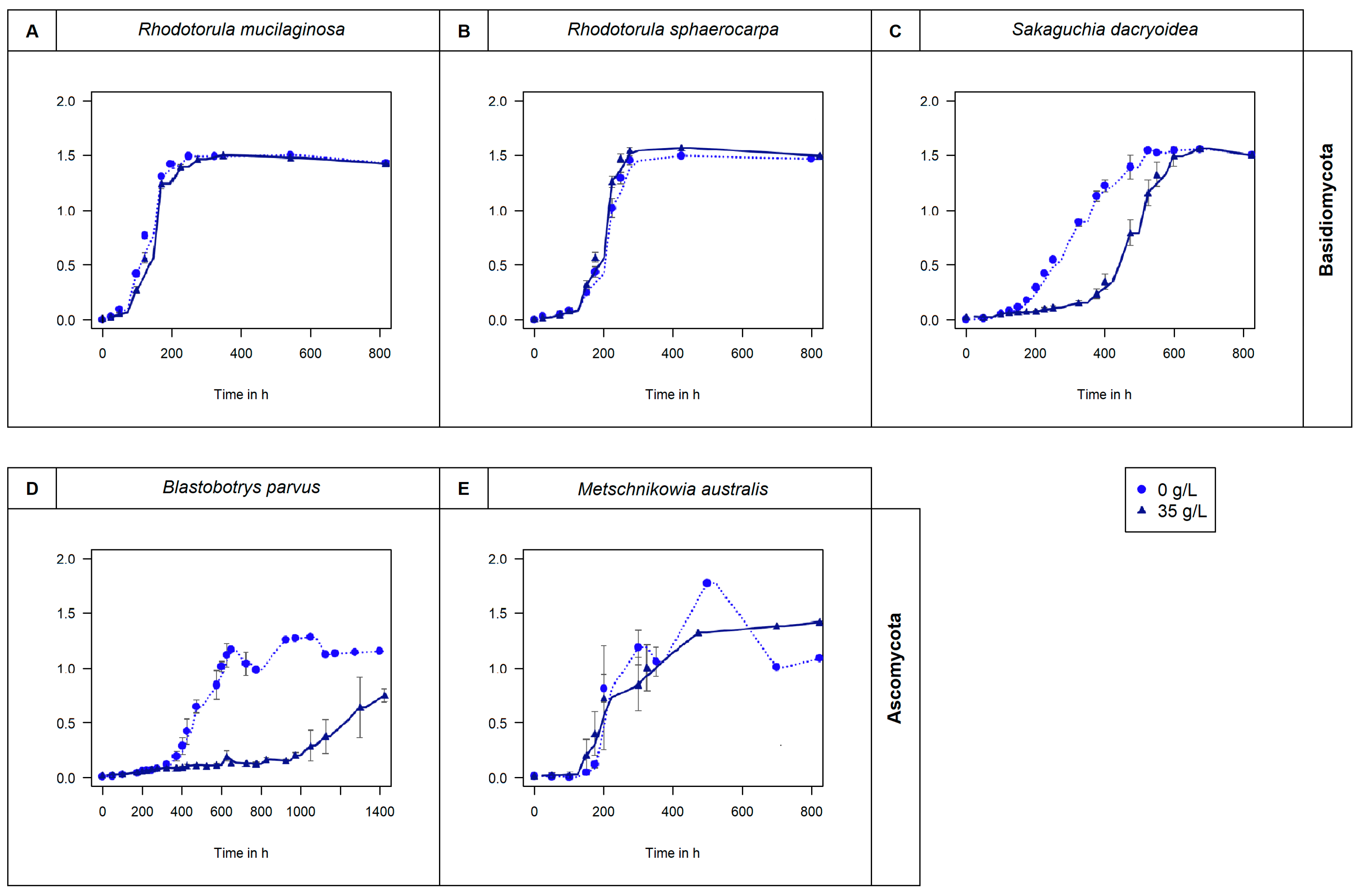
Figure 1. Fungal growth in non-saline (0 g/L) and saline (35 g/L) media measured by optical density (OD660) per hour. Basidiomycota species (A) R. mucilaginosa, (B) R. sphaerocarpa, and (C) S. Dacryoidea. Ascomycota species (D) B. parvus, and (E) M. australis.
Jof 10 00152 g001
Figure 2. Vmax in (A) μmol/g biomass/h and in (B) amol/cell/h obtained from the total enzymatic activity of the fungal strains B. parvus, M. australis, R. mucilaginosa, R. sphaerocarpa, and S. dacryoidea grown in non-saline (0 g/L) and saline (35 g/L) media. The substrates used represented the hydrolysis of carbohydrates by (1) β-glucosidase (BGL), (2) β-xylosidase (BXY), and (3) N-acetyl-β-D-glucosaminidase (NAG). However, due to the filamentous structure of B. parvus, its cell abundance was not pssible to calculate. Tukey’s HSD was performed to test for significance (p < 0.05) between the extracellular enzymatic activity in the non-saline (light grey and uppercase) and saline (dark grey and lowercase) media, where letters represent the different groups obtained. Bars not sharing any letter (A, a, B, b, C, c, and d) were significantly different, and bars sharing a letter (AB, ab, and bc) were not significantly different.
Jof 10 00152 g002
Figure 3. Vmax in (A) μmol/g biomass/h and in (B) amol/cell/h obtained from the total enzymatic activity of the fungal strains B. parvus, M. australis, R. mucilaginosa, R. sphaerocarpa, and S. dacryoidea in non-saline (0 g/L) and saline (35 g/L) media. The substrates used represented the hydrolysis of lipids, phosphorus, and sulfur moieties by (1) lipase (OLE), (2) alkaline phosphatase (APA), and (3) sulfatase (SUL), respectively. However, due to the filamentous structure of B. parvus, its cell abundance was not possible to calculate. R. mucilaginosa did not exhibit any SUL activity under non-saline conditions, indicated by “N/A”. Tukey’s HSD was performed to test for significance (p < 0.05) between the extracellular enzymatic activity in the non-saline (light grey and uppercase) and saline (dark grey and lowercase) media where letters represent the different groups obtained. Bars not sharing any letter (A, a, B, b, C, c, and D) were significantly different, and bars sharing a letter (AB, ab, BC, and bc) were not significantly different.
Jof 10 00152 g003
Figure 4. Vmax in (A) μmol/g biomass/h and in (B) amol/cell/h obtained from the total enzymatic activity of the fungal strains B. parvus, M. australis, R. mucilaginosa, R. sphaerocarpa, and S. dacryoidea grown in non-saline (0 g/L) and saline (35 g/L) media. The substrates used represented the hydrolysis of proteins by (1) leucine aminopeptidase (LAP) and (2) trypsin (TRY). However, due to the filamentous structure of B. parvus, its cell abundance was not possible to calculate. Tukey’s HSD was performed to test for significance (p < 0.05) between the extracellular enzymatic activity in the non-saline (light grey and uppercase) and saline (dark grey and lowercase) media where letters represent the different groups obtained. Bars not sharing any letter (A, a, B, b, C, c, D, and d) were significantly different, and bars sharing a letter (AB, bc, and cd) were not significantly different.
Jof 10 00152 g004
Figure 5. Km in µM obtained from the total enzymatic activity of five marine fungal isolates B. parvus, M. australis, R. mucilaginosa, R. sphaerocarpa, and S. dacryoidea grown in non-saline (0 g/L) and saline (35 g/L) media, for substrates representatives of carbohydrates: (A) β-glucosidase (BGL), (B) β-xylosidase (BXY), and (C) N-acetyl-β-D-glucosaminidase (NAG); lipids, phosphorus and sulfur moieties: (D) lipase (OLE), (E) alkaline phosphatase (APA), and (F) sulfatase (SUL), respectively; and proteins: (G) leucine aminopeptidase (LAP), and (H) trypsin (TRY). Tukey’s HSD was performed to test for significance (p < 0.05) between the extracellular enzymatic activity in the non-saline (light grey and uppercase) and saline (dark grey and lowercase) media where letters represent the different groups obtained. Bars not sharing any letter (A, a, B, b, C, and c) were significantly different (p < 0.05), and bars sharing a letter (AB, ab, bc, and abc) were not significantly different.
Jof 10 00152 g005
Figure 6. Individual enzymatic contribution of the fungal strains B. parvus, M. australis, R. mucilaginosa, R. sphaerocarpa, and S. dacryoidea in (A) μmol/g biomass/h and in (B) percentage for the enzymes β-glucosidase (BGL), β-xylosidase (BXY), N-acetyl-β-D-glucosaminidase (NAG), lipase (OLE), alkaline phosphatase (APA), sulfatase (SUL), leucine aminopeptidase (LAP), and trypsin (TRY).
Jof 10 00152 g006
Table 1. Targeted enzymes with fluorogenic substrate analogues and their respective fluorophore standards (MUF—methylumbelliferyl, MCA—methylcoumarylamide).
TypeCodeNameStandard
CarbohydratesBGLβ-glucosidaseMUF
BXYβ-xylosidaseMUF
NAGN-acetyl-β-D-glucosaminidaseMUF
Lipids, phosphorus, and sulfur moietiesOLELipaseMUF
APAAlkaline phosphataseMUF
SULSulfataseMUF
ProteinsLAPLeucine aminopeptidaseMCA
TRYTrypsinMCA
Disclaimer/Publisher’s Note: The statements, opinions and data contained in all publications are solely those of the individual author(s) and contributor(s) and not of MDPI and/or the editor(s). MDPI and/or the editor(s) disclaim responsibility for any injury to people or property resulting from any ideas, methods, instructions or products referred to in the content.