Article

Rambellisea gigliensis and Rambellisea halocynthiae, gen. et spp. nov. (Lulworthiaceae) from the Marine Tunicate Halocynthia papillosa

by
Martina Braconcini
1,
Susanna Gorrasi
1,
Massimiliano Fenice
1,2,
Paolo Barghini
1 and
Marcella Pasqualetti
1,3,*
1
Department of Ecological and Biological Sciences (DEB), University of Tuscia, 01100 Viterbo, Italy
2
Laboratory of Applied Marine Microbiology, CoNISMa, Department of Ecological and Biological Sciences, University of Tuscia, 01100 Viterbo, Italy
3
Laboratory of Ecology of Marine Fungi, CoNISMa, Department of Ecological and Biological Sciences, University of Tuscia, 01100 Viterbo, Italy
*
Author to whom correspondence should be addressed.
J. Fungi 2024, 10(2), 127; https://doi.org/10.3390/jof10020127
Submission received: 20 November 2023 / Revised: 24 January 2024 / Accepted: 1 February 2024 / Published: 3 February 2024
(This article belongs to the Special Issue Fungal Diversity in Various Environments, 2nd Edition)

Abstract

:
In this study, 15 Lulworthiales strains isolated from the marine tunicate Halocynthia papillosa collected in the central Tyrrhenian Sea were characterized using a polyphasic approach (morpho-physiological, molecular, and phylogenetic analyses). Based on multi-locus phylogenetic inference and morphological characters, a new genus, Rambellisea, and two new species, R. halocynthiae and R. gigliensis (Lulworthiales), were proposed. Multi-locus phylogenetic analyses using the nuclear ribosomal regions of DNA (nrITS1-nr5.8S-nrITS2, nrLSU, and nrSSU) sequence data strongly supported the new taxa. Phylogenetic inference, estimated using Maximum Likelihood and Bayesian Inference, clearly indicates that Rambellisea gen. nov. forms a distinct clade within the order Lulworthiales. Moreover, the two new species were separated into distinct subclades, solidly supported by the analyses. This is the first report of Lulworthiales species isolated from animals.

1. Introduction

Marine habitats cover more than 70% of the planet’s surface and host a large amount of unknown biological and chemical diversity [1,2,3,4]. The sea represents a limitless resource of unexploited substrata and new microorganisms [5,6,7,8]. Over the last decades, researchers have paid close attention to marine microbiology, investigating new environments and/or substrata [6,9,10]. In particular, scientists have spent much effort to expand our understanding of fungal biodiversity and highlight fungi’s importance in several ecosystem services [6,11,12,13,14].
The total number of fungi from marine environments, reported on the “Marine Fungi” specialized website [15], accounts for 1947 species (30 October 2023). However, it has been estimated that this is only a small fraction (<0.2%) of the total marine fungal diversity [11]. To date, up to 90% of marine described species belong to the phyla Ascomycota and Basidiomycota [15]. With more than 940 species and 385 genera, Ascomycota is the most common taxon, and the greatest number of species are found in the classes Dothideomycetes, Eurotiomycetes, and Sordariomycetes [16]. Some orders of Sordariomycetes are exclusively or preferentially marine: Koralionastetales and Lulworthiales host only marine species [17], whereas the Halosphaeriaceae family includes both freshwater and marine species, even though the marine ones are the most numerous [18,19].
The order Lulworthiales, with the single family Lulworthiaceae, was established by Kohlmeyer et al. [20] based on phylogenetic analyses and morphological characters to accommodate the genera Lulworthia and Lindra, previously included in the order Halosphaeriales. Over the past few years, several new marine fungi have been described in the family (Lulworthia atlantica, and L. fundyensis), including the recently established genus Paralulworthia, in 2020. This genus was established to accommodate five new species that were discovered in Posidonia oceanica (P. gigaspora, P. posidoniae, P. candida, P. elbensis, and P. mediterranea) [21,22,23,24,25]. The species P. candida, P. elbensis, and P. mediterranea, which do not produce reproductive structures, were established solely based on phylogenetic analyses [23]. It should be noted that several marine fungal strains exhibit only mycelia sterilia, and they could be identified exclusively by molecular approach [5,26,27]. This approach, although significantly different from the traditional taxonomy based on reproductive characterization, is now widely accepted by the scientific community [28]. In the last few years, several new species and genera were established in the absence of sexual or asexual structures [29,30,31,32].
The Lulworthiaceae family is characterized by filamentous ascospores [20]; nevertheless, for some recently described species, reproductive structures have not been observed. The family comprises 80 species and 16 genera (https://www.indexfungorum.org/, accessed on 17 September 2023; https://www.mycobank.org/, accessed on 17 September 2023) [33]. Members of the family have a cosmopolitan distribution and live in a wide range of habitats, including drifts, submerged woods, algae, and seagrasses [20,24,34,35,36,37,38,39,40,41]. Some species have also been reported from polluted water, such as those from oil-spilled areas [42]. To the best of our knowledge, members of the order Lulworthiales have not yet been isolated from marine animals, despite some metabarcoding studies revealing the presence of some genera in association with various coral species. The species L. calcicola has been described from coral rock [43,44,45,46].
During a survey carried out in the central Tyrrhenian Sea to study epizoic fungi, some new strains belonging to Lulworthiales were isolated from the tunicate Halocynthia papillosa. Halocynthia papillosa is a common ascidian species inhabiting the Mediterranean Sea [47,48], and it presents a tunic composed of cellulose, acid mucopolysaccharides, proteins, and sulfated glycans [49]. Some of these compounds, such as cellulose, are extremely rare biomolecules in animals [50].
In this study, 15 Lulworthiales strains isolated from H. papillosa were characterized using a polyphasic approach (morpho-physiological, molecular, and phylogenetic analyses). Based on multi-locus phylogenetic inference and morphological characters, a new genus, Rambellisea, and two new species, Rambellisea halocynthiae and Rambellisea gigliensis, are here proposed.

2. Materials and Methods

2.1. Fungal Isolation

Five specimens of H. papillosa were collected near the “Punta Gabbianara” cape (42°21′50″ N–10°55′24″ E), Giglio Island (Tuscan Archipelago, North Tyrrhenian Sea) at 23–28 m depth in March 2022. The samples were placed in sterile containers and maintained at 4 °C. Isolations were carried out within 24 h as follows: Samples were washed in sterilized artificial seawater (SW; Sea Salts, 35 g dissolved in 1 L, Sigma-Aldrich, St. Louis, MO, USA) to eliminate debris and any potential transient propagules. For each animal, the tunic (T) was separated from the inner tissues (I) to evaluate mycobiota differences related to animal districts. For fungal isolation, the following two different techniques were used:
(i)
Direct plating: tunic was cut into pieces of about 1 cm3 and directly plated (5 pieces for each plate) onto Petri dishes (90 mm) containing Malt Extract Agar seawater (MEAsw; 50 g MEA—Sigma-Aldrich dissolved in 1 L of seawater) and Corn Meal Agar seawater (CMAsw; 17 g CMA–Fluka analytical, Buchs, Switzerland, dissolved in 1 L of seawater).
(ii)
Homogenization: 5 g of each district (T, I) was homogenized in 10 mL of sterile seawater using a sterile device (ULTRA-TURRAX, IKA, Staufen, Germany). A total of 500 μL of each suspension was plated onto Petri dishes (90 mm) containing MEAsw and CMAsw.
To avoid bacterial growth, all media were supplemented with antibiotics (Streptomycin Sulfate, 0.2 g/L; Penicillin G 0.07 g/L; Chloramphenicol, 0.05 g/L). All plates were incubated at 25 °C in the dark and checked daily for four weeks. Strains were isolated in axenic culture on CMAsw and cryogenically maintained at −40 °C in the culture collection of microorganisms of the “Laboratory of Ecology of Marine Fungi” (DEB, University of Tuscia, Viterbo, Italy). Samples of each species were also preserved at the Mycotheca Universitatis Taurinensis (MUT) culture collection.
The fungal strains analyzed in this study were HPa3, HPa15, HPa16, HPa50, HPa51, HPa52, HPa53, HPa54, HPa58, HPa59, HPa60, HPa61, HPa62, HPa63, and HPa64.

2.2. Morphology and Growth Studies on Different Media

Morphological analyses were carried out on plates utilizing different cultural media: Potato Dextrose Agar seawater (PDAsw; 39 g PDA—Sigma-Aldrich dissolved in 1 L of filtered seawater), Malt Extract Agar seawater (MEAsw), Corn Meal Agar seawater (CMAsw), and Oatmeal Agar seawater (OAsw; 30 g oatmeal powder, 20 g agar dissolved in 1 L of seawater).
The plates (5 cm or 9 cm Ø) were inoculated with a single agar disc (2 mm2) cut from the actively growing margin of 14 d strain cultures on PDAsw and incubated at 25 °C in sealed plastic boxes. These were humidified by a small beaker of distilled water to prevent evaporation and salt precipitation. Growth was monitored for 28 days, and the macroscopic and microscopic features were annotated.
To promote reproduction, fungal strains were inoculated on different natural substrata, such as bark (Quercus cerris), wood (Pinus pinaster), and tunic of H. papillosa (substrate of isolation). All substrata were sterilized, cut into small pieces (3 × 1 cm), and transferred to the surface of PDAsw well-developed colonies (21 days old). The plates were incubated for 4 weeks at 25 °C to allow natural substrata colonization. Following that, some of the inoculated fragments were transferred into tubes containing 20 mL of sterile seawater to simulate natural conditions, while others were transferred to moist chambers and further incubated for 4 months. All inoculated fragments were checked regularly.
The strains’ growth preference in relation to salinity was also investigated: each strain was inoculated, as mentioned above, on PDA plates (5 cm Ø) supplemented with different amounts of NaCl (0, 30, 50, 70, 80, and 100‰). The growth diameter was measured daily for 21 days. All experiments were carried out in triplicate.

2.3. DNA Extraction, PCR Amplification, and Data Assembling

Genomic DNA was extracted from fresh mycelium (about 100 mg) using the ZR Fungal/Bacterial DNA MiniPrep Kit (Zymo Research, Irvine, CA, USA), according to the manufacturer’s directions. The extracted DNA was spectrophotometrically quantified (Qubit, Thermo Fisher Scientific, Waltham, MA, USA) and stored at −20 °C.
For each fungal strain, the ITS1-5.8S-ITS2, LSU, and SSU of rDNA regions were amplified using the primer pairs ITS5/ITS4 [51], LR0R/LR7 [52], and NS1/NS4 [51], respectively. Amplifications were run in a 2720 Thermal Cycler (Applied Biosystem, Waltham, MA, USA) programmed as described in Table 1.
Polymerase chain reactions (PCR) were performed in a volume of 25 μL mixture containing 0.5 μL of each primer (10 μM), 2.5 μL of MgCl2 (25 mM), 1.5 μL of 5× buffer, 0.5 μL of dNTPs (10 mM), 0.2 μL of Go-Taq Polymerase (Promega, Madison, WI, USA), and 2 μL of genomic DNA; the final volume (25 μL) was reached by adding ultrapure water. The PCR products were purified (E.Z.N.A. Cycle Pure kit Omega Bio-tek, Norcross, GA, USA) and sent to Eurofins Genomics (Ebersberg, Germany) for sequencing. The sequences obtained were checked and trimmed with the Chromas Lite 2.1 program and then compared with those deposited in GenBank NCBI (National Center for Biotechnology Information, Bethesda, MD, USA). Newly generated sequences were deposited in GenBank (Table 2).

2.4. Sequence Alignment and Phylogenetic Analyses

For the phylogenetic analyses, a concatenated dataset of nrSSU, nrITS, and nrLSU sequences (Table 2) based on BLASTn results including the most representative species of the Lulworthiales genera according to the literature was used [21,22,23,24,25]. The single gene sequence datasets were aligned with the Clustal X 2.1 software [53] using the default parameters for gap opening and gap extension. Alignments were checked and edited using BioEdit Alignment Editor 7.2.5 [54] and manually adjusted in MEGA 10.2.6 when necessary. Positions where one or more species had a long mutation, as well as ambiguously aligned regions, were excluded from the subsequent phylogenetic analyses. The datasets were concatenated with MEGA X. Phylogenetic inference was estimated using Maximum Likelihood (ML) and Bayesian Inference (BI).
Maximum Likelihood analyses including 1000 bootstrap (BS) replicates were run using the IQ-TREE web server under different models for each dataset in the concatenated matrix [55]. ModelFinder on the IQ-TREE web server was used to determine the best nucleotide substitution model for each partition. TNe+G4 is the best-fit model for nrLSU, nr5.8S, and nrITS2, TIM2e+G4 for nrITS1, and TN+F+G4 for nrSSU [56]. The best scoring tree, with final likelihood values of −19719.747, was visualized using FigTree v.1.4 (http://tree.bio.ed.ac.uk/software/figtree/, accessed on 17 September 2023). The Bayesian Inference was performed with Mr Bayes 3.2.7 [57] under different models for each partition of the matrix as evaluated by jModelTest 2 [58] using Bayesian Information Criterion (TPM2+I+G for nrSSU part1 and TrN+G for nrSSUpart2; TIM1ef+G for nrITS1; TrNef+G for nr5.8S; TrN+G for nrITS2 and nrLSU). Substitution rates, gamma distribution shape parameter, and proportion of invariable sites were reported for each partition in Supplementary Materials (Table S1). The alignment was run for 1 million generations in two independent runs, each with four Markov Chains Monte Carlo (MCMC) and sampling every 100 iterations. As a “burn-in” measure, the first 25% of generated trees were discarded. MrBayes’ “sumt” function was used to generate a consensus tree, and Bayesian posterior probabilities (BYPP) were calculated.
Sequence alignment and phylogenetic tree were deposited in TreeBASE (www.treebase.org, accessed on 8 October 2023) (submission number: 30823). The new taxonomical names were recorded in Mycobank (MB850303, MB850305, MB850306).

3. Results

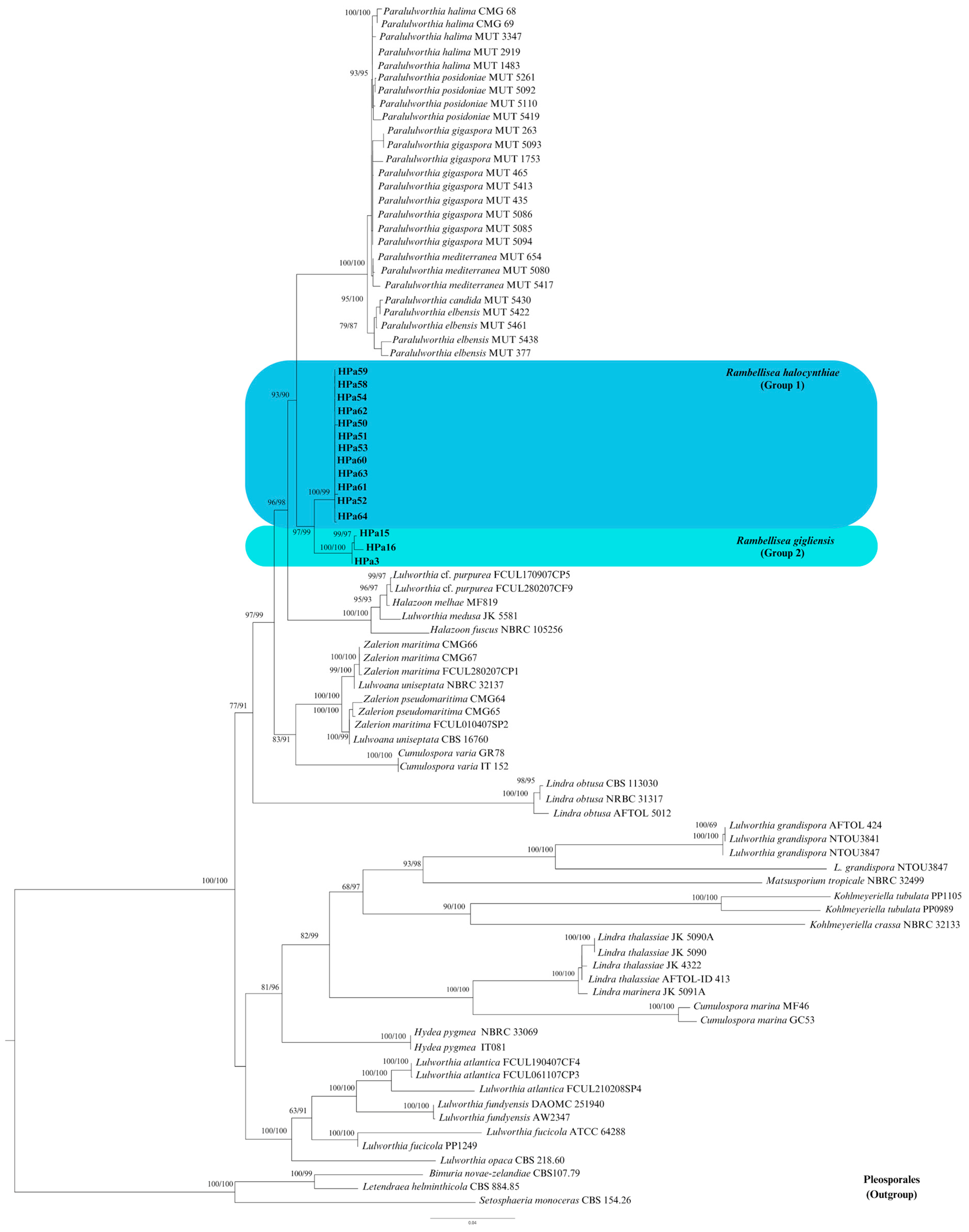
3.1. Phylogenetic Inference
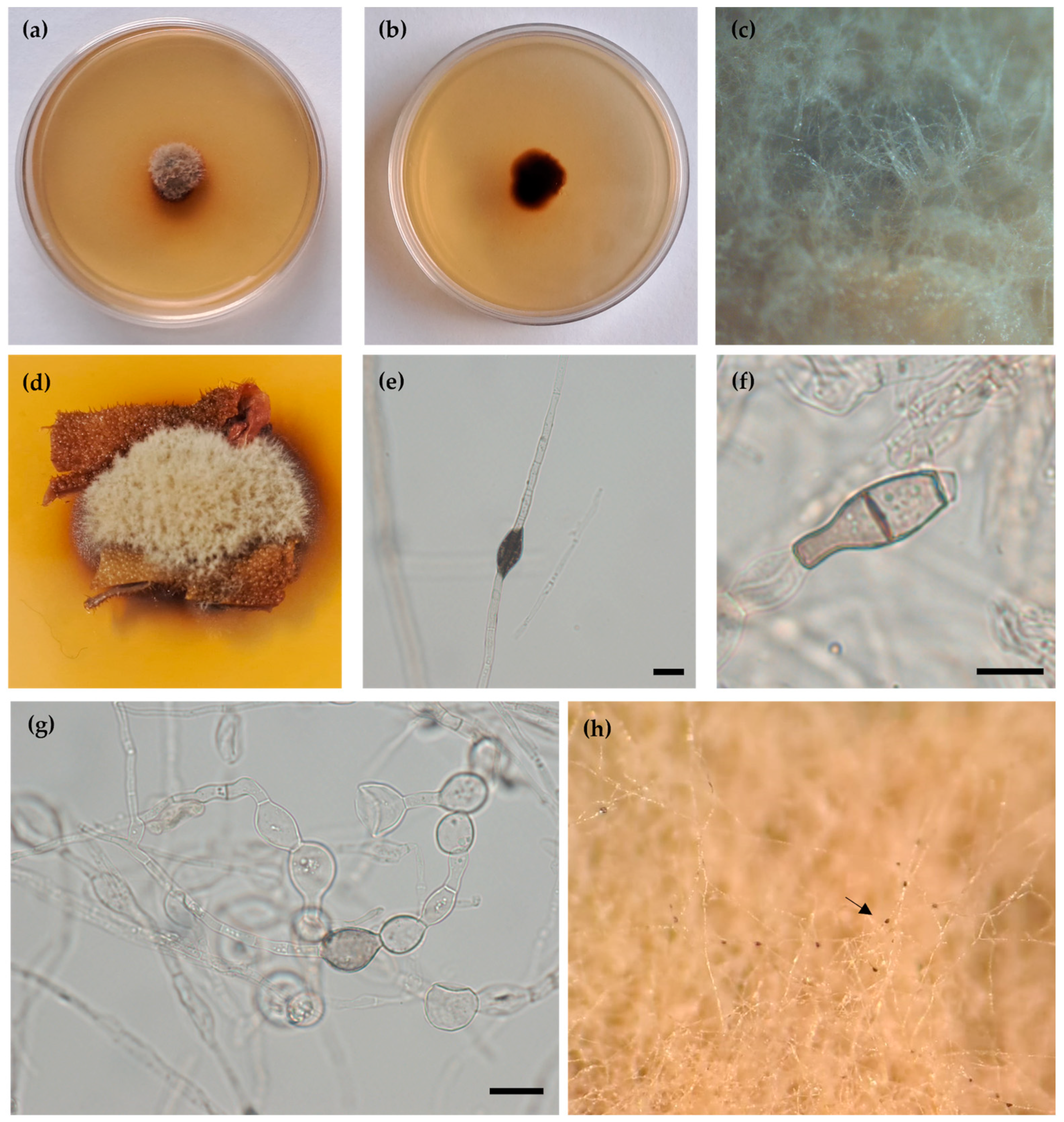
A preliminary phylogenetic analysis was carried out individually for nrITS, nrLSU, and nrSSU. Since no incongruences were observed among the single-loci phylogenetic trees, a multi-locus analysis was performed thereafter. The dataset includes 84 strains, 29 species, and 11 genera belonging to the family Lulworthiaceae, with 3 pleosporelean species, Bimuria novae-zelandiae, Setosphaeria monoceras, and Letendraea helminthicola, as outgroup taxa. Globally, 26 sequences (15 nrITS, 5 nrSSU, and 6 nrLSU) were newly generated, whereas 186 were obtained from GenBank (Table 2).
The aligned concatenate dataset has 3346 characters, including gaps (1329 for SSU, 181 for ITS1, 150 for 5.8S, 310 for ITS2, and 1367 for LSU). Among them, 1598 distinct patterns, with 36.2% undetermined characters or gaps, 914 parsimony-informative sites, 434 singleton sites, and 1998 constant sites, were observed. Estimated base frequencies were A = 24.69%, T = 21.66%, C = 24.86%, and G = 28.80%.
ML analysis yielded a best-scoring three with a final optimization likelihood value of −19719.747. The ML and BI analyses resulted in generally congruent topologies, which were also in line with previous works [5,24]. Given the topological similarity of the two resulting trees, only the ML tree with BS and BYPP values was reported (Figure 1).
The 15 isolates under investigation formed a well-supported clade (BS = 97%; BYPP = 99%), constituting a new monophyletic lineage within the order Lulworthiales, with Paralulworthia species as their closest relatives (Figure 1). Within the new lineage, two groups can be distinguished, group 1: strains HPa50, HPa51, HPa52, HPa53, HPa54, HPa58, HPa59, HPa60, HPa61, HPa62, HPa63, and HPa64, and group 2: strains HPa3, HPa15, and HPa16. Both groups were strongly supported, with BS and BYPP values exceeding 99% (Group 1: BS = 100%; BYPP = 99%; Group 2: BS = 100%; BYPP = 100%).
The phylogenetic analysis appeared to support the conclusion that the fifteen strains isolated from H. papillosa belong to two novel species within a new genus in the Lulworthiaceae family (Figure 1).
The new genus Rambellisea is herein proposed, with the description of the following two new species: Rambellisea halocynthiae sp. nov. and Rambellisea gigliensis sp. nov.
Nucleotide divergence between the novel species and the closest was annotated for each locus when it occurred and reported as Supplementary Materials (Tables S2–S4).

3.2. Taxonomy

Rambellisea Pasqualetti & Braconcini, gen. nov.
MycoBank no.: MB850303
Etymology. The prefix “Rambelli-”. In honor of the Italian Mycologist Angelo Rambelli, and the name “Rambellisea” refers to the genus habitat “sea”.
Diagnosis. Differs from the genus Paralulworthia to which it appears phylogenetically most closely related in the absence of sexual features and conidiogenous structures.
Phylogenetic placement. Lulworthiaceae, Lulworthiales, and Sordariomycetes. The genus Rambellisea gen. nov. clusters together with the genus Paralulworthia (Figure 1).
Type species. Rambellisea gigliensis.
Rambellisea gigliensis Pasqualetti & Braconcini sp. nov. (Figure 2).
MycoBank no.: MB 850306.
Etymology. Referred to the sample collection site “Giglio Island”.
Type. Italy, Tuscany, Mediterranean Sea, Giglio Island (Grosseto), Punta Gabbianara, 42°21′50″ N, 10°55′24″ E, 25 m depth. Isolated from the tunic of Halocynthia papillosa, March 2022, Martina Braconcini. Holotype MUT 6843 (strain HPa3), living culture permanently preserved in a metabolically inactive state at MUT.
Diagnosis. R. gigliensis is an epizoic marine fungus. R. gigliensis (MUT 6843) differs from its closest phylogenetic neighbor R. halocynthiae (MUT 6851) by genetic characters in nrITS, nrLSU, and nSSU sequences (Tables S2–S4) and in the production of characteristically enlarged hyphae and chlamydospore production.
Description. Growing on H. papillosa tunic, Q. cerris bark, and P. pinaster wood.
Hyphae 3.0–4.6 μm wide, septate, sub-hyaline sometimes lightly pigmented, assuming a toruloid aspect mainly in submerged mycelium. In old cultures, dark concretions like small droplets were observed on hyphae (Figure 2e). Chlamydospores 10.5–20.0 μm, subhyaline to light brown, globose, sub-globose, monocellular, sometimes one septate, and pyriform (Figure 2f,g). Sexual and asexual structures not observed.
Colony description. Colonies on PDAsw, reaching 8 mm diameter after 28 days at 25 °C, dome-like, surface flocculose, smoke-grey to brown; aerial mycelium, whitish to light brown; margins regular, reverse brown. Soluble pigment is yellowish to orange, or absent exudates are absent (Figure 2a,b). Colonies on MEAsw, reaching 14 mm of diameter after 28 days at 25 °C, umbonate, surface floccose, beige to brown; aerial mycelium abundant, light brown; margins regular, reverse brown (Figure S1); soluble pigment absent; exudates present black in small droplets on aerial hyphae (Figure 2h). Colonies on CMAsw reached 32 mm in diameter after 28 days at 25 °C, plane slightly umbonate, surface velutinous, olive-grey to pale brown, margin regular submerged, aerial mycelium, whitish to light brown, mainly in the central area, reverse brown. Soluble pigment and exudates are not produced (Figure S1).
Notes. Based on a Megablast search on the NCBI nucleotide database, the closest hits of R. gigliensis (OR367423) using the nrITS are R. halocynthiae (GenBank accession no. OR36748; identities 492/541 (91%), 20 gaps), Lulworthiales sp. (GenBank accession no. LC544102; identities 468/549 (85%), 22 gaps), and Zalerion sp. (GenBank accession no. FJ430722; identities 411/468 (88%), 18 gaps). The closest hits using the nrLSU sequences are R. halocynthiae (GenBank accession no. OR371457; identities 1099/1115 (99%), 4 gaps), P. posidoniae (GenBank accession no. MZ357739; identities 1076/1107 (97%), 4 gaps), and P. halima (GenBank accession no. MZ357750; identities 1073/1103 (97%), 4 gaps). The closest hits using the nrSSU sequences are R. halocynthiae GenBank accession no. OR371485; identities 1039/1049 (99%), 0 gaps), Lulworthia uniseptata (GenBank accession no. AY879034; identities 1039/1050 (99%), 0 gaps), and Z. maritima (GenBank accession no. NG_078728; identities 1038/1050 (99%), 0 gaps). R. gigliensis isolates can be collected from the tunic and the internal tissues of H. papillosa and can be cultured on media with and without sea salt; the best growth was observed at the sea salinity on Corn Meal Agar (CMAsw).
Additional material examined. Italy, Tuscany, Mediterranean Sea, Giglio Island (Grosseto), Punta Gabbianara, 42°21′50″ N, 10°55′24″ E, 28 m depth. Isolated from the internal tissues of H. papillosa, March 2022, Martina Braconcini, living culture HPa15. Italy, Tuscany, Mediterranean Sea, Giglio Island (Grosseto), Punta Gabbianara, 42°21′50″ N, 10°55′24″ E, 23 m depth. Isolated from the internal tissues of H. papillosa, March 2022, Martina Braconcini, living culture HPa16.
Rambellisea halocynthiae Pasqualetti & Braconcini, sp. nov. (Figure 3).
MycoBank no.: MB850305.
Etymology. Referred to the substrate of isolation.
Type. Italy, Tuscany, Mediterranean Sea, Giglio Island (Grosseto), Punta Gabbianara, 42°21′50″ N, 10°55′24″ E, 25 m depth. Isolated from the tunic of H. papillosa, March 2022, Marcella Pasqualetti. Holotype MUT 6851 = HPa52, living culture permanently preserved in a metabolically inactive state at MUT.
Diagnosis. R. halocynthiae is an epizoic marine fungus. R. halocynthiae (MUT 6851) differs from its closest phylogenetic neighbor R. gigliensis (MUT 6843) by genetic characters in nrITS, nrLSU, and nSSU sequences (Tables S2–S4).
Description. Growing on H. papillosa tunic, Q. cerris bark, and P. pinaster wood.
Hyphae 2.2–4.4 μm wide, septate, sub-hyaline to slightly pigmented. Sexual and asexual structures are not observed.
Colony description. Colonies on PDAsw, reaching 13.5 mm in diameter after 28 days at 25 °C, plane centrally umbonate, surface velutinous to feltrose, smoke-grey to pale brown with a light brown marginal area; aerial mycelium sparse, whitish to light brown, mainly in the central area; margins regular, moderately deep, reverse brown. Soluble pigment is yellowish to orange or absent; no exudates were observed (Figure 3). Colonies on MEAsw (Figure S2), reaching 22.5 mm in diameter after 28 days at 25 °C, are morphologically similar to PDAsw. Colonies on CMAsw (Figure S2) reaching 47.3 mm in diameter after 28 days at 25 °C, plane slightly umbonate, surface velutinous, olive-grey to pale brown with a large submerged peripheric area up to 10 mm, aerial mycelium, whitish to light brown, mainly in the central area, reverse brown. Soluble pigment and exudates not produced.
Notes. Based on a Megablast search on the NCBI nucleotide database, the closest hits of nrITS of R. halocynthiae (OR367549) are R. gigliensis (GenBank accession no. OR367423; identities 489/538 (91%), 20 gaps), Lulworthia sp. (GenBank accession no. KU214534; identities 464/532 (87%), 29 gaps), and P. gigaspora (GenBank accession no. MN649244; identities 467/536 (87%), 33 gaps). The closest hits using the nrLSU sequences are R. gigliensis (GenBank accession no. OR369725; identities 900/914 (98%), 4 gaps), P. halima (GenBank accession no. MT235754; identities 888/910 (98%), 0 gaps), and P. posidoniae (GenBank accession no. MZ357739; identities 887/909 (98%), 0 gaps). The closest hits using the nrSSU sequences are L. uniseptata (GenBank accession no. AY879034; identities 1039/1051 (99%), 0 gaps), R. gigliensis (GenBank accession no. OR371466; identities 1033/1043 (99%), 0 gaps), and Z. maritima (GenBank accession no. MT235710; identities 1038/1051 (99%), 0 gaps).
R. halocynthiae isolates can be collected from the tunic and the internal tissues of H. papillosa and can be cultured on media with and without sea salt; the best growth was observed at the sea salinity on Corn Meal Agar (CMAsw).
Additional material examined. Italy, Tuscany, Mediterranean Sea, Giglio Island (Grosseto), Punta Gabbianara, 42°21′50″ N, 10°55′24″ E, 25 m depth. Isolated from tunic or internal tissues of H. papillosa, March 2022, Marcella Pasqualetti, living culture HPa50, HPa54. Italy, Tuscany, Mediterranean Sea, Giglio Island (Grosseto), Punta Gabbianara, 42°21′50″ N, 10°55′24″ E, 28 m depth. Isolated from the tunic and internal tissues of H. papillosa, March 2022, Marcella Pasqualetti, living cultures HPa51, HPa53, HPa58, HPa59, HPa60, HPa61, HPa62, HPa63, and HPa64.

4. Discussion

Fungi are key players in terrestrial and marine environments and represent a substantial proportion of the microbial diversity on Earth [15]. Even if the role of marine fungi in several basic ecosystem functions, such as their contribution to aquatic carbon pump efficiency or regulation of phytoplankton composition, is largely recognized, the diversity of marine fungi seems to be largely unexplored. It was estimated that up to 90% of marine species have not been described yet [14]. Considering this gap, the exploration of habitats and substrates that have never been studied by mycologists appears to be an essential issue to enhance our knowledge of marine fungal biodiversity. Indeed, the new taxa proposed in this study were isolated from H. papillosa, a substratum that has never been previously studied from a mycological point of view.
The fifteen new isolates, obtained from the external tunic and internal tissues of the studied tunicate, developed only sterile mycelia. According to the literature, all strains were cultivated on different substrates, including artificial media (PDAsw, MEAsw, CMAsw, and OAsw), and natural matrices (bark, wood, and tunic of H. papillosa). To promote reproduction and the possible development of reproductive structures, the inoculated matrices were placed in both humid chambers and submerged in seawater during incubation [22,59,60,61]. Fungi development occurred in all studied conditions; nevertheless, sexual reproductive structures or asexual conidia have never been observed. Asexual chlamydospores were observed in 28-day-old cultures of R. gigliensis in all studied conditions, while R. halocynthiae produced vegetative mycelium only. Mycelia sterilia are not unusual among marine fungi [62,63], according to Damare and co-workers [64], it is possible that many marine fungi have evolved hyphal fragmentation as the preferential dispersion system. This would explain the broad presence of the toruloid mycelium observed in R. gigliensis; similar mycelia were reported for other Lulworthiales too [24].
Considering the absence of reproductive structures, except for the mentioned chlamydospores (propagules primarily devoted to perennation, not dissemination) in R. gigliensis, a molecular taxonomical approach was carried out for the taxonomical characterization of the identified strains. A preliminary analysis of the universal barcode for fungi (nrITS region) revealed similarity values inferior to 88% with all sequences deposited in the NCBI nucleotide database. This low identity clearly indicates that these strains were new taxa. Nevertheless, the ITS analyses indicated that all strains belonged to the order Lulworthiales, and the multi-locus molecular analyses, based on ribosomal genes (nrLSU, nrITS, and nrSSU), were performed to infer their phylogeny according to recent literature [21,22,23,24,25]. The phylogenetic tree clearly showed that our strains formed a well-supported clade that did not encompass any known fungus, indicating the presence of a new lineage inside the family Lulworthiaceae (Figure 1).
The order Lulworthiales includes only strictly marine species [20], commonly found in association with wood, seagrass, and algae. To the best of our knowledge, members of the order Lulworthiales have not been isolated from ascidians yet or from other marine animals [65,66,67,68]. The newly studied strains are epizoic, facultative halophytes. They can grow in media devoid of seawater, even if the optimal growth was observed at Mediterranean Sea salinity (38‰).

5. Conclusions

The present paper provides a morphological and phylogenetic study of fifteen strains obtained from the marine tunicate Halocynthia papillosa collected in the central Tyrrhenian Sea; this tunicate has never been studied for its mycobiota. The strains form a novel lineage within the family Lulworthiaceae. In light of this, the new genus Rambellisea has been established, including the two new species, Rambellisea halocynthiae sp. nov. and Rambellisea gigliensis sp. nov. The identification of fungi belonging to Lulworthiales significantly contributes to the advancement of knowledge about this order of marine species, confirming that the marine ecosystem constitutes an extensive repository of biodiversity, largely unexplored, in particular for its microbial components.

Supplementary Materials

The following supporting information can be downloaded at https://www.mdpi.com/article/10.3390/jof10020127/s1, Table S1: Substitution rates, gamma distribution shape parameter and proportion of invariable sites for each partition, Table S2: The variable sites detected in the nrITS region among Rambellisea halocynthiae, R. gigliensis and its neighbor species belonging to the genera Paralulworthia, Table S3: The variable sites detected in the nrLSU region among Rambellisea halocynthiae, R. gigliensis and its neighbor species belonging to the genera Paralulworthia, Table S4: The variable sites detected in the nrSSU region among Rambellisea halocynthiae, R. gigliensis and its neighbor species belonging to the genera Paralulworthia, Figure S1: Rambellisea gigliensis sp. nov. HPa3 (MUT 6843). (a) 28-day-old colony: colony texture on MEAws at 25 °C; (b) 28-day-old colony: colony texture on CMAws at 25 °C, Figure S2: Rambellisea halocynthiae sp. nov. HPa52 (MUT 6851). (a) 28-day-old colony on MEAws (Ø 9 cm) at 25 °C (b) and reverse; (c) 28-day-old colony on CMAws (Ø 9 cm) at 25 °C (d) and reverse.

Author Contributions

Conceptualization, M.P. and M.B.; methodology, M.P., M.B., M.F., S.G., and P.B.; software, M.P., S.G., and M.B.; validation, M.P., M.B., S.G., and M.F.; formal analysis, M.P.; investigation, M.P., M.B., P.B., and S.G.; resources, M.P. and M.F.; data curation, M.P. and M.B.; writing—original draft preparation, M.P., M.B., and M.F.; writing—review, and editing, M.P., M.B., M.F., and S.G.; visualization, M.P, M.B., and S.G.; supervision, M.P.; project administration, M.P.; funding acquisition, M.P. All authors have read and agreed to the published version of the manuscript.

Funding

The work was partially supported by the Programma Operativo Nazionale (PON—DOT1335703) Ricerca e Innovazione of the Italian Ministry of University and Research (MUR) and by the “Progetto di Ricerca di Interesse Nazionale”(PRIN)—Production and characterization of new bioactive molecules against emerging and/or multidrug-resistant pathogens by neglected poly-extremophilic marine fungi (MYCOSEAS, no. 2022MPTT35).

Institutional Review Board Statement

Not applicable.

Informed Consent Statement

Not applicable.

Data Availability Statement

All data generated or analyzed during this study are included in this published article and its supplementary information files. All sequences were deposited in GenBank (https://www.ncbi.nlm.nih.gov/nuccore, accessed on 1 August 2023) and alignments were deposited at TreeBase (https://www.treebase.org/treebase-web/search/studySearch.html, accessed on 8 October 2023).

Acknowledgments

The authors wish to thank Edoardo Casoli and Gianluca Mancini for their kind support for the sampling.

Conflicts of Interest

The authors declare no conflicts of interest.

References

  1. Giovannini, V.; Barghini, P.; Gorrasi, S.; Massimiliano, F.; Pasqualetti, M. Marine fungi: A potential source of novel enzymes for environmental and biotechnological applications. J. Environ. Prot. Ecol. 2019, 20, 1214–1222. [Google Scholar]
  2. Pasqualetti, M.; Barghini, P.; Giovannini, V.; Fenice, M. High production of chitinolytic activity in halophilic conditions by a new marine strain of Clonostachys rosea. Molecules 2019, 24, 101880. [Google Scholar] [CrossRef] [PubMed]
  3. Botta, L.; Saladino, R.; Barghini, P.; Fenice, M.; Pasqualetti, M. Production and identification of two antifungal terpenoids from the Posidonia oceanica epiphytic Ascomycota Mariannaea humicola IG100. Microb. Cell Fact. 2020, 19, 184. [Google Scholar] [CrossRef] [PubMed]
  4. Pasqualetti, M.; Gorrasi, S.; Giovannini, V.; Braconcini, M.; Fenice, M. Polyextremophilic chitinolytic activity by a marine strain (IG119) of Clonostachys rosea. Molecules 2022, 27, 688. [Google Scholar] [CrossRef] [PubMed]
  5. Poli, A.; Bovio, E.; Ranieri, L.; Varese, G.C.; Prigione, V. News from the sea: A new genus and seven new species in the Pleosporalean families Roussoellaceae and Thyridariaceae. Diversity 2020, 12, 144. [Google Scholar] [CrossRef]
  6. Pasqualetti, M.; Giovannini, V.; Barghini, P.; Gorrasi, S.; Fenice, M. Diversity and ecology of culturable marine fungi associated with Posidonia oceanica leaves and their epiphytic algae Dictyota dichotoma and Sphaerococcus coronopifolius. Fungal Ecol. 2020, 44, 100906. [Google Scholar] [CrossRef]
  7. Poli, A.; Bovio, E.; Perugini, I.; Varese, G.C.; Prigione, V. Corollospora mediterranea: A novel species complex in the Mediterranean Sea. Appl. Sci. 2021, 11, 5452. [Google Scholar] [CrossRef]
  8. Gorrasi, S.; Franzetti, A.; Ambrosini, R.; Pittino, F.; Pasqualetti, M.; Fenice, M. Spatio-temporal variation of the bacterial communities along a salinity gradient within a thalassohaline environment (Saline di Tarquinia salterns Italy). Molecules 2021, 26, 1338. [Google Scholar] [CrossRef]
  9. Gorrasi, S.; Pesciaroli, C.; Barghini, P.; Pasqualetti, M.; Fenice, M. Structure and diversity of the bacterial community of an Arctic estuarine system (Kandalaksha Bay) subject to intense tidal currents. J. Mar. Syst. 2019, 196, 77–85. [Google Scholar] [CrossRef]
  10. Gorrasi, S.; Franzetti, A.; Brandt, A.; Minzlaff, U.; Pasqualetti, M.; Fenice, M. Insights into the prokaryotic communities of the abyssal-hadal benthic-boundary layer of the Kuril Kamchatka Trench. Environ. Microbiome 2023, 18, 67. [Google Scholar] [CrossRef]
  11. Amend, A.; Burgaud, G.; Cunliffe, M.; Edgcomb, V.P.; Ettinger, C.L.; Gutiérrez, M.H.; Heitman, J.; Hom, E.F.Y.; Ianiri, G.; Jones, A.; et al. Fungi in the marine environment: Open questions and unsolved problems. mBio 2019, 10, e01189-18. [Google Scholar] [CrossRef]
  12. Gladfelter, A.S.; James, T.Y.; Amend, A.S. Marine fungi. Curr. Biol. 2019, 29, R191–R195. [Google Scholar] [CrossRef] [PubMed]
  13. Grossart, H.P.; Van den Wyngaert, S.; Kagami, M.; Wurzbacher, C.; Cunliffe, M.; Rojas-Jimenez, K. Fungi in aquatic ecosystems. Nat. Rev. Microbiol. 2019, 17, 339–354. [Google Scholar] [CrossRef]
  14. Gonçalves, M.F.; Esteves, A.C.; Alves, A. Marine Fungi: Opportunities and Challenges. Encyclopedia 2022, 2, 559–577. [Google Scholar] [CrossRef]
  15. Jones, E.B.G.; Pang, K.L.; Abdel-Wahab, M.A.; Scholz, B.; Hyde, K.D.; Boekhout, T.; Ebel, R.; Rateb, M.E.; Henderson, L.; Sakayaroj, J.; et al. An online resource for marine fungi. Fungal Divers. 2019, 96, 347–433. [Google Scholar] [CrossRef]
  16. Jensen, P.R. Secondary metabolites from marine fungi. In Fungi in Marine Environments; Hyde, K.D., Ed.; Fungal Diversity Research Series; Fungal Diversity Press: Hong Kong, China, 2002; Volume 7, pp. 293–315. [Google Scholar]
  17. Overy, D.P.; Rämä, T.; Oosterhuis, R.; Walker, A.K.; Pang, K.L. The neglected marine fungi sensu stricto and their isolation for natural products’ discovery. Mar. Drugs 2019, 17, 42. [Google Scholar] [CrossRef] [PubMed]
  18. Campbell, J.; Inderbitzin, P.; Kohlmeyer, J.; Volkmann-Kohlmeyer, B. Koralionastetales a new order of marine Ascomycota in the Sordariomycetes. Mycol. Res. 2009, 113, 373–380. [Google Scholar] [CrossRef]
  19. Sakayaroj, J.; Pang, K.L.; Jones, E.G. Multi-gene phylogeny of the Halosphaeriaceae: Its ordinal status relationships between genera and morphological character evolution. Fungal Divers. 2011, 46, 87–109. [Google Scholar] [CrossRef]
  20. Kohlmeyer, J.; Spatafora, J.W.; Volkmann-Kohlmeyer, B. Lulworthiales a new order of marine Ascomycota. Mycologia 2000, 92, 453–458. [Google Scholar] [CrossRef]
  21. Azevedo, E.; Barata, M.; Marques, M.I.; Caeiro, M.F. Lulworthia atlantica: A new species supported by molecular phylogeny and morphological analysis. Mycologia 2017, 109, 287–295. [Google Scholar] [CrossRef]
  22. Poli, A.; Bovio, E.; Ranieri, L.; Varese, G.C.; Prigione, V. Fungal Diversity in the Neptune Forest: Comparison of the Mycobiota of Posidonia oceanica; Flabellia petiolata and Padina pavonica. Front. Microbiol. 2020, 11, 933. [Google Scholar] [CrossRef]
  23. Poli, A.; Prigione, V.; Bovio, E.; Perugini, I.; Varese, G.C. Insights on Lulworthiales inhabiting the Mediterranean Sea and description of three novel species of the genus Paralulworthia. J. Fungi 2021, 7, 940. [Google Scholar] [CrossRef]
  24. Goncalves, M.F.; Abreu, A.C.; Hilario, S.; Alves, A. Diversity of marine fungi associated with wood baits in the estuary Ria de Aveiro with descriptions of Paralulworthia halima comb. nov. Remispora submersa sp. nov. and Zalerion pseudomaritima sp. nov. Mycologia 2021, 113, 664–683. [Google Scholar] [CrossRef] [PubMed]
  25. Crous, P.W.; Boers, J.; Holdom, D.; Steinrucken, T.V.; Tan, Y.P.; Vitelli, J.S.; Shivas, R.G.; Barret, M.; Boxshall, A.G.; Broadbridge, J.; et al. Fungal Planet description sheets: 1383–1435. Persoonia 2022, 48, 261–371. [Google Scholar] [CrossRef] [PubMed]
  26. Jones, E.B.G.; Pang, K.L. Introduction marine fungi. In Marine Fungi and Fungal-Like Organisms; Walter de Gruyter: Berlin, Germany, 2012; pp. 1–13. [Google Scholar]
  27. Abdel-Wahab, M.A.; Hodhod, M.S.; Bahkali, A.H.; Jones, E.G. Marine fungi of Saudi Arabia. Bot. Mar. 2014, 57, 323–335. [Google Scholar] [CrossRef]
  28. Aime, M.C.; Miller, A.N.; Aoki, T.; Bensch, K.; Cai, L.; Crous, P.W.; Hawksworth, D.L.; Hyde, K.D.; Kirk, P.M. How to publish a new fungal species or name version 3.0. IMA Fungus 2021, 12, 11. [Google Scholar] [CrossRef] [PubMed]
  29. Knapp, D.G.; Kovács, G.M.; Zajta, E.; Groenewald, J.Z.; Crous, P.W. Dark septate endophytic pleosporalean genera from semiarid areas. Persoonia 2015, 35, 87–100. [Google Scholar] [CrossRef] [PubMed]
  30. Koch, R.A.; Jean Lodge, D.; Sourell, S.; Nakasone, K.; McCoy, A.G.; Aime, M.C. Tying up loose threads: Revised taxonomy and phylogeny of an avian-dispersed Neotropical rhizomorph-forming fungus. Mycol. Prog. 2018, 17, 989–998. [Google Scholar] [CrossRef]
  31. Crous, P.W.; Carnegie, A.J.; Wingfield, M.J.; Sharma, R.; Mughini, G.; Noordeloos, M.E.; Santini, A.; Shouche, Y.S.; Bezerra, J.D.P.; Dima, B.; et al. Fungal Planet description sheets: 868–950. Persoonia 2019, 42, 291. [Google Scholar] [CrossRef] [PubMed]
  32. Noumeur, S.R.; Teponno, R.B.; Helaly, S.E.; Wang, X.W.; Harzallah, D.; Houbraken, J.; Crous, P.W.; Stadler, M. Diketopiperazines from Batnamyces globulariicola gen. sp. nov. (Chaetomiaceae) a fungus associated with roots of the medicinal plant Globularia alypum in Algeria. Mycol. Prog. 2020, 19, 589–603. [Google Scholar] [CrossRef]
  33. Hyde, K.D.; Norphanphoun, C.; Maharachchikumbura, S.S.N.; Bhat, D.J.; Jones, E.; Bundhun, D.; Chen, Y.J.; Bao, D.F.; Boonmee, S.; Calabon, M.S.; et al. Refined families of Sordariomyetes. Mycosphere 2020, 11, 305–1059. [Google Scholar] [CrossRef]
  34. Zuccaro, A.; Schoch, C.L.; Spatafora, J.W.; Kohlmeyer, J.; Draeger, S.; Mitchell, J.I. Detection and identification of fungi intimately associated with the brown seaweed Fucus serratus. Appl. Environ. Microbiol. 2008, 74, 931–941. [Google Scholar] [CrossRef] [PubMed]
  35. Rämä, T.; Nordén, J.; Davey, M.L.; Mathiassen, G.H.; Spatafora, J.W.; Kauserud, H. Fungi ahoy! Diversity on marine wooden substrata in the high North. Fungal Ecol. 2014, 8, 46–58. [Google Scholar] [CrossRef]
  36. Garzoli, L.; Gnavi, G.; Varese, G.C.; Picco, A.M. Mycobiota associated with the rhodophyte alien species Asparagopsis taxiformis (Delile) Trevisan de Saint-Léon in the Mediterranean Sea. Mar. Ecol. 2015, 36, 959–968. [Google Scholar] [CrossRef]
  37. Calado, M.D.L.; Carvalho, L.; Barata, M.; Pang, K.L. Potential roles of marine fungi in the decomposition process of standing stems and leaves of Spartina maritima. Mycologia 2019, 111, 371–383. [Google Scholar] [CrossRef] [PubMed]
  38. Hagestad, O.C.; Andersen, J.H.; Altermark, B.; Hansen, E.; Rämä, T. Cultivable marine fungi from the Arctic Archipelago of Svalbard and their antibacterial activity. Mycology 2019, 11, 230–242. [Google Scholar] [CrossRef] [PubMed]
  39. Cha, H.J.; Chiang, M.W.; Guo, S.Y.; Lin, S.M.; Pang, K.L. Culturable fungal community of Pterocladiella capillacea in Keelung Taiwan: Effects of surface sterilization method and isolation medium. J. Fungi 2021, 7, 651. [Google Scholar] [CrossRef] [PubMed]
  40. Torta, L.; Burruano, S.; Giambra, S.; Conigliaro, G.; Piazza, G.; Mirabile, G.; Pirrotta, M.; Calvo, R.; Bellissimo, G.; Calvo, S.; et al. Cultivable fungal endophytes in roots rhizomes and leaves of Posidonia oceanica (L.) Delile along the coast of Sicily Italy. Plants 2022, 11, 1139. [Google Scholar] [CrossRef] [PubMed]
  41. Vohník, M. Are lulworthioid fungi dark septate endophytes of the dominant Mediterranean seagrass Posidonia oceanica? Plant Biol. 2022, 24, 127–133. [Google Scholar] [CrossRef]
  42. Bovio, E.; Gnavi, G.; Prigione, V.; Spina, F.; Denaro, R.; Yakimov, M.; Calogero, R.; Crisafi, F.; Varese, G.C. The culturable mycobiota of a Mediterranean marine site after an oil spill: Isolation identification and potential application in bioremediation. Sci. Total Environ. 2017, 576, 310–318. [Google Scholar] [CrossRef]
  43. Kohlmeyer, J.; Volkmann-Kohlmeyer, B. A new Lulworthia (Ascomycotina) from corals. Mycologia 1989, 81, 289–292. [Google Scholar] [CrossRef]
  44. Amend, A.S.; Barshis, D.J.; Oliver, T.A. Coral-associated marine fungi form novel lineages and heterogeneous assemblages. ISME J. 2012, 6, 1291–1301. [Google Scholar] [CrossRef]
  45. Bonthond, G.; Merselis, D.G.; Dougan, K.E.; Graff, T.; Todd, W.; Fourqurean, J.W.; Rodriguez-Lanetty, M. Inter-domain microbial diversity within the coral holobiont Siderastrea siderea from two depth habitats. PeerJ 2018, 6, e4323. [Google Scholar] [CrossRef] [PubMed]
  46. Góes-Neto, A.; Marcelino, V.R.; Verbruggen, H.; da Silva, F.F.; Badotti, F. Biodiversity of endolithic fungi in coral skeletons and other reef substrates revealed with 18S rDNA metabarcoding. Coral Reefs 2020, 39, 229–238. [Google Scholar] [CrossRef]
  47. Luna-Pérez, B.; Valle, C.; Fernández, T.V.; Sanchez-Lizaso, J.L.; Ramos-Espla, A.A. Halocynthia papillosa (Linnaeus 1767) as an indicator of SCUBA diving impact. Ecol. Ind. 2010, 10, 1017–1024. [Google Scholar] [CrossRef]
  48. Coppari, M.; Gori, A.; Rossi, S. Size spatial and bathymetrical distribution of the ascidian Halocynthia papillosa in Mediterranean coastal bottoms: Benthic–pelagic coupling implications. Mar. Biol. 2014, 161, 2079–2095. [Google Scholar] [CrossRef]
  49. Lübbering-Sommer, B.; Compère, P.; Goffinet, G. Cytochemical investigations on tunic morphogenesis in the sea peach Halocynthia papillosa (Tunicata Ascidiacea) 1: Demonstration of polysaccharides. Tissue Cell 1996, 28, 621–630. [Google Scholar] [CrossRef] [PubMed]
  50. Song, G.; Delroisse, J.; Schoenaers, D.; Kim, H.; Nguyen, T.C.; Horbelt, N.; Leclère, P.; Hwang, D.S.; Harrington, M.J.; Flammang, P. Structure and composition of the tunic in the sea pineapple Halocynthia roretzi: A complex cellulosic composite biomaterial. Acta Biomater. 2020, 111, 290–301. [Google Scholar] [CrossRef] [PubMed]
  51. White, T.J.; Bruns, T.; Lee, S.; Taylor, J. Amplification and direct sequencing of fungal ribosomal RNA genes for phylogenetics. In PCR Protocols; Elsevier: Amsterdam, The Netherlands; Academic Press: New York, NY, USA, 1990; pp. 315–322. [Google Scholar]
  52. Vilgalys, R.; Hester, M. Rapid genetic identification and mapping of enzymatically amplified ribosomal DNA from several Cryptococcus species. J. Bacteriol. 1990, 172, 4238–4246. [Google Scholar] [CrossRef] [PubMed]
  53. Thompson, J.D.; Gibson, T.J.; Plewniak, F.; Jeanmougin, F.; Higgins, D.G. The CLUSTAL_X windows interface: Flexible strategies for multiple sequence alignment aided by quality analysis tools. Nucleic Acids Res. 1997, 25, 4876–4882. [Google Scholar] [CrossRef]
  54. Hall, T.A. BioEdit: A user-friendly biological sequence alignment editor and analysis program for windows 95/98/NT. Nucleic Acids Sym. 1990, 41, 95–98. [Google Scholar]
  55. Trifinopoulos, J.; Nguyen, L.T.; von Haeseler, A.; Minh, B.Q. W-IQ-TREE: A fast online phylogenetic tool for maximum likelihood analysis. Nucleic Acids Res. 2016, 44, W232–W235. [Google Scholar] [CrossRef]
  56. Kalyaanamoorthy, S.; Minh, B.Q.; Wong, T.K.F.; von Haeseler, A.; Jermiin, L.S. ModelFinder: Fast model selection for accurate phylogenetic estimates. Nat. Methods 2017, 14, 587–589. [Google Scholar] [CrossRef]
  57. Ronquist, F.; Teslenko, M.; Van Der Mark, P.; Ayres, D.L.; Darling, A.; Höhna, S.; Larget, B.; Liu, L.; Suchard, M.A.; Huelsenbeck, J.P. MrBayes 32: Efficient Bayesian phylogenetic inference and model choice across a large model space. Syst. Biol. 2012, 61, 539–542. [Google Scholar] [CrossRef]
  58. Posada, D. jModelTest: Phylogenetic model averaging. Mol. Biol. Evol. 2008, 25, 1253–1256. [Google Scholar] [CrossRef] [PubMed]
  59. Panebianco, C.; Tam, W.Y.; Jones, E.B.G. The effect of pre-inoculation of balsa wood by selected marine fungi and their effect on subsequent colonisation in the sea. Fungal Divers. 2002, 10, 77–88. [Google Scholar]
  60. Su, Y.Y.; Qi, Y.L.; Cai, L. Induction of sporulation in plant pathogenic fungi. Mycology 2012, 3, 195–200. [Google Scholar] [CrossRef]
  61. Senanayake, I.C.; Rathnayaka, A.R.; Marasinghe, D.S.; Calabon, M.S.; Gentekaki, E.; Lee, H.B.; Hurdeal, V.G.; Pem, D.; Dissanayake, L.S.; Wijesinghe, S.N.; et al. Morphological approaches in studying fungi: Collection examination isolation sporulation and preservation. Mycosphere 2020, 11, 2678–2754. [Google Scholar] [CrossRef]
  62. Raghukumar, S. The Role of Fungi in Marine Detrital Processes. In Marine Microbiology: Facets and Opportunities; Ramaiah, N., Ed.; National Institute of Oceanography: Goa, India, 2004; pp. 91–101. [Google Scholar]
  63. Morrison-Gardiner, S. Dominant fungi from Australian coral reefs. Fungal Divers. 2002, 9, 105–121. [Google Scholar]
  64. Damare, S.; Raghukumar, C.; Raghukumar, S. Fungi in deep-sea sediments of the Central Indian Basin. Deep Sea Res. Part I Oceanogr. Res. Pap. 2006, 53, 14–27. [Google Scholar] [CrossRef]
  65. Menezes, C.B.; Bonugli-Santos, R.C.; Miqueletto, P.B.; Passarini, M.R.; Silva, C.H.; Justo, M.R.; Leal, R.R.; Fantinatti-Garboggini, F.; Oliveira, V.M.; Berlinck, R.G.S.; et al. Microbial diversity associated with algae ascidians and sponges from the north coast of São Paulo state Brazil. Microbiol. Res. 2010, 165, 466–482. [Google Scholar] [CrossRef]
  66. López-Legentil, S.; Erwin, P.M.; Turon, M.; Yarden, O. Diversity of fungi isolated from three temperate ascidians. Symbiosis 2015, 66, 99–106. [Google Scholar] [CrossRef]
  67. Vacondio, B.; Birolli, W.G.; Ferreira, I.M.; Seleghim, M.H.; Gonçalves, S.; Vasconcellos, S.P.; Porto, A.L. Biodegradation of pentachlorophenol by marine-derived fungus Trichoderma harzianum CBMAI 1677 isolated from ascidian Didemnun ligulum. Biocatal. Agric. Biotechnol. 2015, 4, 266–275. [Google Scholar] [CrossRef]
  68. Godinho, V.M.; de Paula, M.T.R.; Silva, D.A.S.; Paresque, K.; Martins, A.P.; Colepicolo, P.; Rosa, C.A.; Rosa, L.H. Diversity and distribution of hidden cultivable fungi associated with marine animals of Antarctica. Fungal Biol. 2019, 123, 507–516. [Google Scholar] [CrossRef] [PubMed]
Figure 1. Phylogenetic inference based on combined nrITS, nrSSU, and nrLSU sequence dataset inferred using the Maximum Likelihood method. The tree is rooted to species of Pleosporales (Bimuria novae-zelandiae, Setosphaeria monoceras, and Letendraea helminthicola). Branch numbers indicate BS and BYPP values. Bar = expected changes per site (0.04). The strains resulting from the current study are in bold and the strains of each new species are distinguished by various colors.
Jof 10 00127 g001
Figure 2. Rambellisea gigliensis sp. nov. HPa3 (MUT 6843) (a) 28-day-old colony on PDAsw (Ø 5 cm) at 25 °C (b) and reverse; (c) colony texture; (d) growth on H. papillosa tunic; (e) dark concretions on hyphae (MEAsw); (f,g) Chlamydospores; (h) exudates (arrow) produced on MEAsw. Scale bars: 10 μm (e,f), 20 μm (g).
Jof 10 00127 g002
Figure 3. Rambellisea halocynthiae sp. Nov. Hpa52 (MUT 6851). (a) A 28-day-old colony on PDAsw (Ø 5 cm) at 25 °C (b) and reverse; (c,d) growth on Halocynthia papillosa tunic.
Jof 10 00127 g003
Table 1. Details of PCR programs for the different markers used.
PCR Steps nrITSnrLSUnrSSU
ITS5/ITS4LR0R/LR7NS1/NS4
Initial denaturation 94 °C for 2′95 °C for 10′95 °C for 10′
PCR cycledenaturation94 °C for 20″95 °C for 1′95 °C for 1′
annealing56 °C for 30″50 °C for 50″50 °C for 50″
elongation72 °C for 45″72 °C for 1.5′72 °C for 1.5′
Final elongation 72 °C for 10′72 °C for 10′72 °C for 10′
Number of cycles 354040
Legend: nrITS = nuclear ribosomal Internal Transcribed Spacer; nrLSU = nuclear ribosomal Large ribosomal SubUnit; nrSSU = nuclear ribosomal Small ribosomal SubUnit.
Table 2. Taxa used for the phylogenetic analyses and GenBank accession number. Newly generated sequences are indicated in bold.
SpeciesStrainSubstratesnrITSnrSSU nrLSU
Lulworthiales
Cumulospora marinaMF46Submerged wood-GU252136GU252135
GC53Submerged wood-GU256625GU256626
Cumulospora variaGR78Submerged wood-EU848593EU848578
IT 152Wood-EU848579-
Halazoon melhaeMF819Submerged wood-GU252144GU252143
Halazoon fuscusNBRC 105256Driftwood-GU252148GU252147
Hydea pygmeaNBRC 33069Driftwood-GU252134GU252133
IT081Driftwood-GU256632GU256633
Kohlmeyeriella crassaNBRC 32133DriftwoodLC146741-LC146742
Kohlmeyeriella tubulataPP1105Sea foam-AY878998AF491265
PP0989Marine environment-AY878997AF491264
Lindra marineraJK 5091AMarine environment-AY879000AY878958
Lindra obtuseNRBC 31317Sea foamLC146744AY879002AY878960
AFTOL 5012Marine environment-FJ176847FJ176902
CBS 113030--AY879001AY878959
Lindra thalassiaeJK 5090AMarine environment-U46874U46891
AFTOL-ID 413Marine environmentDQ491508DQ470994DQ470947
JK 5090Marine environment-AF195634AF195635
JK 4322Thalassia testudinum-AF195632AF195633
Lulwoana uniseptateNBRC 32137Submerged woodLC146746LC146746LC146746
CBS 16760Driftwood-AY879034AY878991
Lulworthia atlanticaFCUL210208SP4Sea waterKT347205KT347193JN886843
FCUL190407CF4Sea waterKT347207KT347198JN886809
FCUL061107CP3Sea waterKT347208KT347196JN886825
Lulworthia fucicolaATCC 64288Intertidal wood-AY879007AY878965
PP1249Marine environment-AY879008AY878966
Lulworthia fundyensisDAOMC 251940Marine woodNR_178138--
AW2347Marine woodMH465123MH465136MH458750
Lulworthia grandisporaAFTOL 424Dead Rhizophora sp. -DQ522855DQ522856
NTOU3841Driftwood-KY026044KY026048
NTOU3847Mangrove wood-KY026046KY026049
NTOU3849Mangrove wood-KY026047KY026050
Lulworthia medusaJK 5581Spartina sp. -AF195636AF195637
Lulworthia opacaCBS 218.60Driftwood -AY879003AY878961
Lulworthia cf. purpureaFCUL170907CP5SeawaterKT347219KT347201JN886824
FCUL280207CF9SeawaterKT347218KT347202JN886808
Matsusporium tropicaleNBRC 32499Submerged wood-GU252142GU252141
Paralulworthia candidaMUT 5430P. oceanicaMZ357724MZ357767MZ357746
Paralulworthia elbensisMUT 377P. oceanicaMZ357710MZ357753MZ357732
MUT 5422P. oceanicaMZ357723MZ357766MZ357745
MUT 5438P. oceanicaMZ357712MZ357755MZ357734
MUT 5461P. oceanicaMZ357725MZ357768MZ357747
Paralulworthia gigasporaMUT 435P. oceanicaMN649242MN649246MN649250
MUT 5413P. oceanicaMN649243MN649247MN649251
MUT 263SeawaterMZ357729MZ357772MZ357751
MUT 465P. oceanicaMZ357726MZ357769MZ357748
MUT 1753SeawaterMZ357730MZ357773MZ357752
MUT 5085P. oceanicaMZ357715MZ357758MZ357737
MUT 5086P. oceanicaMZ357716MZ357759MZ357738
MUT 5093P. oceanicaMZ357718MZ357761MZ357740
MUT 5094P. oceanicaMZ357719MZ357762MZ357741
Paralulworthia halimaCMG 68Submerged woodMT235736MT235712MT235753
CMG 69Submerged woodMT235737MT235713MT235754
MUT 1483Submerged woodMZ357727MZ357770MZ357749
MUT 2919Submerged woodMZ357713MZ357756MZ357735
MUT 3347Submerged woodMZ357728MZ357771MZ357750
Paralulworthia mediterraneaMUT 654P. oceanicaMZ357711MZ357754MZ357733
MUT 5080P. oceanicaMZ357714MZ357757MZ357736
MUT 5417P. oceanicaMZ357721MZ357764MZ357743
Paralulworthia posidoniaeMUT 5261P. oceanicaMN649245MN649249MN649253
MUT 5092P. oceanicaMZ357717MZ357760MZ357739
MUT 5110P. oceanicaMZ357720MZ357763MZ357742
MUT 5419P. oceanicaMZ357722MZ357765MZ357744
Rambellisea halocynthiaeHPa50H. papillosaOR367481OR371485OR371457
HPa51H. papillosaOR367548OR371484OR371461
HPa52H. papillosaOR367549-OR371460
HPa53H. papillosaOR367614--
HPa54H. papillosaOR378535--
HPa58H. papillosaOR367660--
HPa59H. papillosaOR367678--
HPa60H. papillosaOR367679--
HPa61H. papillosaOR367717--
HPa62H. papillosaOR378536--
HPa63H. papillosaOR378537--
HPa64H. papillosaOR378538--
Rambellisea gigliensisHPa3H. papillosaOR367423OR371466OR369726
HPa15H. papillosaOR367447OR371482OR369725
HPa16H. papillosaOR367450OR371483OR371456
Zalerion maritimaFCUL280207CP1SeawaterKT347216KT347203JN886806
FCUL010407SP2SeawaterKT347217KT347204JN886805
CM66Submerged woodMT235734MT235710MT235751
CM67Submerged woodMT235735MT235711MT235752
Zalerion pseudomaritimaCMG64Submerged woodMT235732MT235708MT235749
CMG65Submerged woodMT235733MT235709MT235750
Pleosporales
Bimuria novae-zelandiaeCBS107.79soilMH861181AY016338MH872950
Setosphaeria monocerasCBS 154.26-DQ337380DQ238603AY016368
Letendraea helminthicolaCBS 884.85Yerba mateMK404145AY016345AY016362
Disclaimer/Publisher’s Note: The statements, opinions and data contained in all publications are solely those of the individual author(s) and contributor(s) and not of MDPI and/or the editor(s). MDPI and/or the editor(s) disclaim responsibility for any injury to people or property resulting from any ideas, methods, instructions or products referred to in the content.

