Article

Multilocus Phylogeny and Characterization of Five Undescribed Aquatic Carnivorous Fungi (Orbiliomycetes)

1
Institute of Eastern-Himalaya Biodiversity Research, Dali University, Dali 671003, China
2
Center of Excellence in Fungal Research, Mae Fah Luang University, Chiang Rai 57100, Thailand
3
School of Science, Mae Fah Luang University, Chiang Rai 57100, Thailand
4
The Provincial Innovation Team of Biodiversity Conservation and Utility of the Three Parallel Rivers Region, Dali University, Dali 671003, China
5
Yunling Back-and-White Snub-Nosed Monkey Observation and Research Station of Yunnan Province, Dali 671003, China
*
Authors to whom correspondence should be addressed.
J. Fungi 2024, 10(1), 81; https://doi.org/10.3390/jof10010081
Submission received: 15 December 2023 / Revised: 18 January 2024 / Accepted: 18 January 2024 / Published: 20 January 2024
(This article belongs to the Special Issue Freshwater Fungal Diversity)

Abstract

:
The diversity of nematode-trapping fungi (NTF) holds significant theoretical and practical implications in the study of adaptive evolution and the bio-control of harmful nematodes. However, compared to terrestrial ecosystems, research on aquatic NTF is still in its early stages. During a survey of NTF in six watersheds in Yunnan Province, China, we isolated 10 taxa from freshwater sediment. Subsequent identification based on morphological and multigene (ITS, TEF1-α, and RPB2) phylogenetic analyses inferred they belong to five new species within Arthrobotrys. This paper provides a detailed description of these five novel species (Arthrobotrys cibiensis, A. heihuiensis, A. jinshaensis, A. yangbiensis, and A. yangjiangensis), contributing novel insights for further research into the diversity of NTF and providing new material for the biological control of aquatic harmful nematodes. Additionally, future research directions concerning aquatic NTF are also discussed.

1. Introduction

Nematode-trapping fungi (NTF) are a group of fungi that possess a unique trapping structure to capture nematodes for nutrition [1,2,3,4]. NTF in Orbiliomycetes are considered the core representatives of NTF due to their rich species diversity, and intricate and diverse trapping structures, as well as their important role in maintaining ecological balance and their potential value in the bio-control of harmful nematodes [4,5,6,7]. Currently, this group of fungi includes 125 species from three genera: Arthrobotrys (73 species) which captures nematodes using adhesive networks; Dactylellina (35 species), the genus that captures nematodes with adhesive branches, non-constricting rings, and adhesive knobs; and Drechslerella (17 species) which catches nematodes using constricting rings [4,7,8].
These fungi are widely distributed in various habitats because of their unique survival strategy. They are commonly found in the soils from farmlands, forests, and even heavy metal-contaminated areas [4,9,10,11], as well as in sediments from marine, freshwater, and even hot springs [12,13,14]. But compared to the well-studied terrestrial ecosystems, the diversity of NTF in freshwater habitats remains insufficiently studied [13,15,16]. Previous studies have confirmed the existence of a rich diversity of NTF in freshwater ecosystems, which is reasonable given the abundance of nematodes in aquatic environments [13,17]. Meanwhile, the diverse array of nematodes in aquatic habitats includes parasitic species that pose threats to aquatic crops and fisheries [18,19]. So, studying aquatic NTF resources is an important part of NTF diversity research and bio-control of harmful aquatic nematodes. Additionally, the study on aquatic NTF also provides a valuable entry point for investigating fungal adaptive evolution, as aquatic NTF originate from their terrestrial counterparts.
In the past 10 years, we have investigated the NTF in the six major watersheds in Yunnan Province and successfully isolated 10 strains, which were identified as five novel members of Arthrobotrys. This paper provides a comprehensive account of these species, offering new material for the bio-control research of harmful nematodes and the study of fungal aquatic adaptive evolution.

2. Materials and Methods

2.1. Samples Collection

All freshwater sediment samples involved in this study were collected using a Peterson bottom sampler (HL-CN, Wuhan Hengling Technology Company, Limited, Wuhan, China). The samples were placed into plastic zip-lock bags to preserve moisture. Collecting sites, date, and collector were recorded (Table 1). The samples were stored at 4 °C and processed within a week.

2.2. Fungal Isolation

Nematodes (Panagrellus redivivus Goodey, free-living nematodes) cultured on oatmeal medium [4] were isolated using the Baermann funnel method [20] and the concentration of the nematodes was adjusted with sterile water to 3000–5000 nematodes per milliliter. The soil sprinkling technique was used to disperse the sediment sample onto the surface of corn meal agar plates (CMA) [4] and 1 mL of nematode suspension was added to promote the germination of NTF. The plates were incubated at room temperature (14–28 °C) for about three weeks and a stereomicroscope was used to observed the plates to search for the NTF spores. The single-spore isolation method was used for the isolation and purification of the NTF [4].

2.3. Morphological Observation

The isolates were inoculated onto potato dextrose agar (PDA) [4] plates and cultured at 26 °C for colony observation. The isolate was transferred to CMA observation plates (creating an observation well by removing a 2 × 2 cm piece of agar from the center of the CMA plate and obliquely inserting a sterile cover glass into the surface of the medium) and incubated at 26 °C [21]. After the observation well was covered by the mycelium, about 1000 nematodes (P. redivivus) were added as baits to induce the production of traps. The types of traps were checked and photographed using an Olympus BX53 microscope (Olympus Corporation, Tokyo, Japan). When the mycelium had spread over the cover glass, the cover glass was removed with tweezers and a temporary slide made with sterile water [22]. An Olympus BX53 microscope (Olympus Corporation, Tokyo, Japan) was used to photograph and measure the morphological characteristics such as conidia, conidiophores, and chlamydospores.

2.4. Collection of DNA Molecular Data

The mycelium grown on PDA plates was used to extract genomic DNA, as described by Zhang et al. [15]. The primer pairs ITS4-ITS5 [23], 526F-1567R [24], and 6F-7R [25] were used to amplify the ITS, TEF1-α, and RPB2 regions, respectively, under the reaction system and conditions described in the previous study [21]. The PCR products were sent to BioSune Biotech Company Limited (Shanghai, China) for purification and sequencing (TEF1-α genes were sequenced using the 247F-609R [7] primer pair and ITS and RPB2 regions were sequenced with PCR primers).
The generated sequences were carefully examined, edited, and assembled using SeqMan v. 7.0 [26]. All sequences obtained in this study have been submitted to the GenBank database (NCBI; https://www.ncbi.nlm.nih.gov/; accessed on 29 November 2023) for deposition.

2.5. Phylogenetic Analysis

The sequences generated in this study were compared with the GenBank database using BLASTn (https://blast.ncbi.nlm.nih.gov/; accessed on 9 November 2023). Our five species were placed within Arthrobotrys according to the BLASTn search and their trapping structures [4]. Consequently, relevant publications [3,4,7,15,21,27] and the BLASTn search results were used to retrieve all reliable ITS, TEF1-α, and RPB2 sequences of Arthrobotrys taxa from the GenBank database (Table S1). Three genes were aligned via the online program MAFFT v. 7 (http://mafft.cbrc.jp/alignment/server/; accessed on 14 November 2023) [28]. MEGA6.0 [29] was used to adjust and link the three alignments.
The best-fit optimal substitution models for ITS (GTR + I + G), TEF1-α (SYM + I + G), and RPB2 (SYM + I + G) were calculated using jModelTest v. 2.1.10 [30].
Two Vermispora species (V. fusarina (YXJ02-13-5) and V. leguminacea (CGMCC 6.0291)) were set as outgroups. IQ-Tree v. 1.6.5 [31] and MrBayes v. 3.2.6 [32] were used to infer the phylogenetic trees using maximum likelihood (ML) and Bayesian inference (BI) methods. The related parameter settings are the same as in the previous study [21].
The trees were visualized via FigTree v. 1.3.1 [33] and edited using Microsoft PowerPoint v. 2016 (Microsoft, Redmond, WA, USA) and Adobe Photoshop CS6 software (Adobe Systems, San Jose, CA, USA).

3. Results

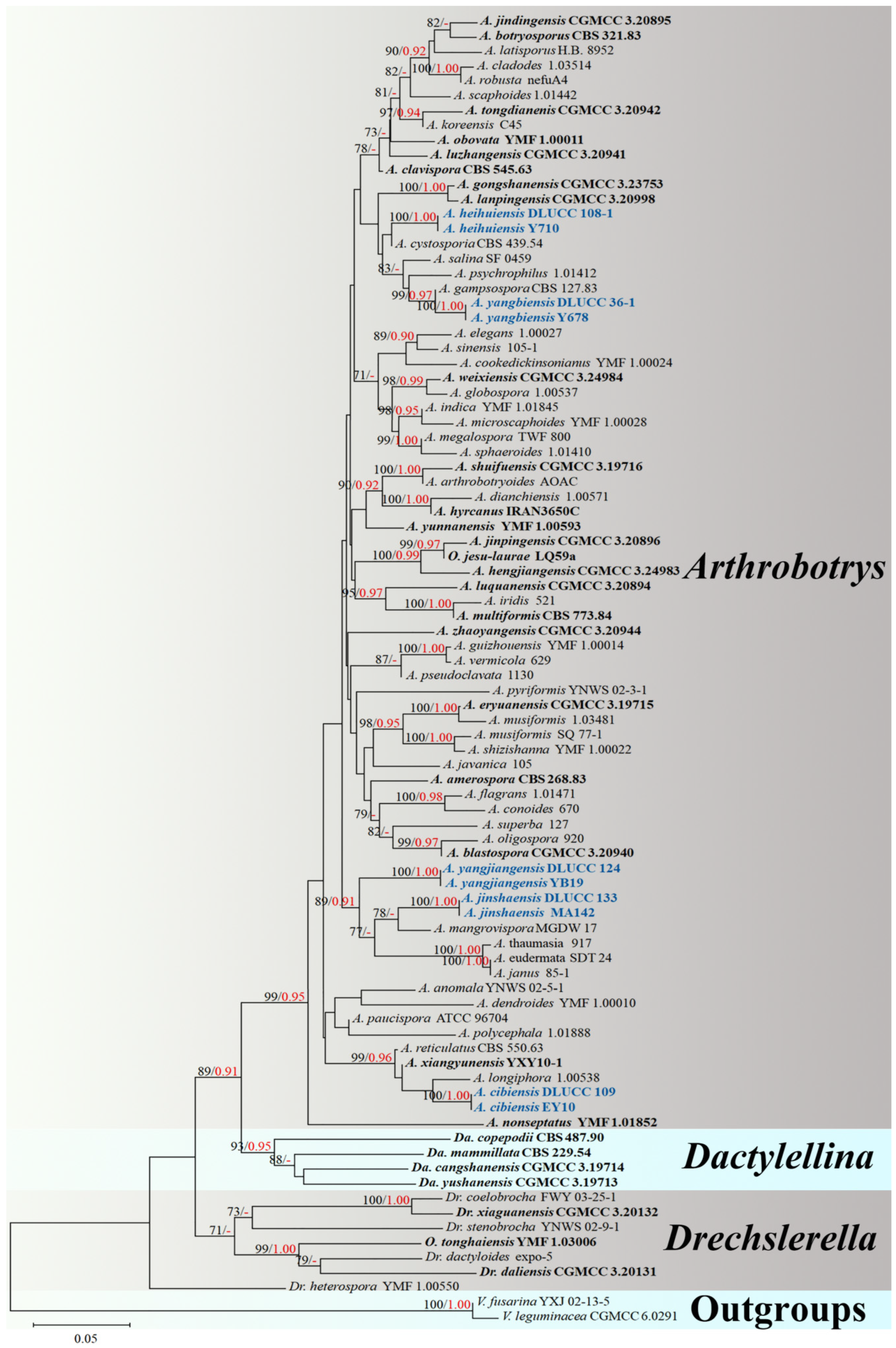
3.1. Phylogenetic Analysis

The combined ITS, TEF1-α, and RPB2 alignment dataset consisted of 88 sequences of ITS, 62 sequences of TEF1-α, and 64 sequences of RPB2 from Arthrobotrys 75 taxa, representing 69 valid species (plus our five new species), other related taxa in Orbiliomycetes (Dactylellina four taxa and Drechslerella seven taxa), and two outgroup taxa. The final dataset comprised 2000 characters (585 for ITS, 8321 for RPB2, and 583 for TEF1-α), among which 900 base pair (bp) are constant, 1087 bp are variable, and 886 bp are parsimony-informative.
The best-scoring ML tree was generated with a final ML optimization likelihood value of −6817.314758. Bayesian analysis (BI) was used to evaluate the Bayesian posterior probabilities with a final average standard deviation of the split frequency of 0.009092. Both ML and BI trees consistently grouped all tested nematode-trapping fungi into three major clades and five new species exhibited distinct divergence from known species. Therefore, the ML tree was chosen for presentation (Figure 1).
The phylogenetic tree inferred from the ITS, TEF1-α, and RPB2 combined dataset placed five new species in Arthrobotrys. The phylogenetic position of A. heihuiensis is uncertain but clearly diverges from known species. The two isolates of A. yangbiensis formed a distinct lineage basal to A. gampsospora with 99% MLBS and 0.97 BYPP support. Furthermore, A. yangjiangensis and A. jinshaensis clustered together with A. mangrovispora, A. thaumasia, A. eudermata, and A. janus with 89% MLBS and 0.91 BYPP support. A. cibiensis formed a distinct lineage basal to A. longiphora, A. xiangyunensis, and A. reticulatus with 99% MLBS and 0.96 BYPP support (Figure 1).

3.2. Taxonomy

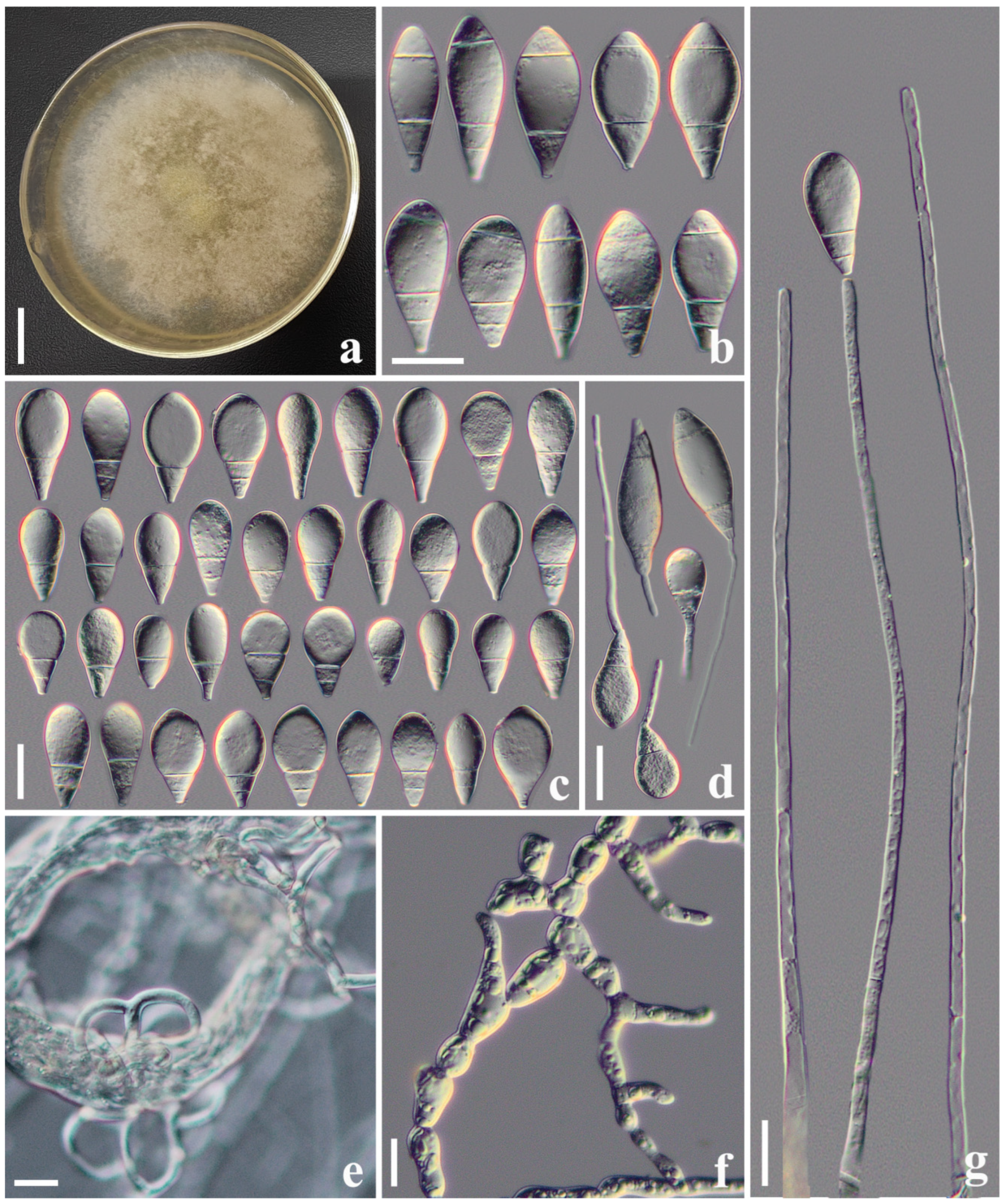
3.2.1. Arthrobotrys cibiensis F. Zhang, S. Boonmee, and X.Y. Yang sp. nov. (Figure 2)

Index Fungorum number: IF901486; Facesoffungi number: FOF14174
Etymology: The species name “cibiensis” refers to the name of the sample collection site: Cibi Lake, Eryuan County, Dali City, Yunnan Province, China.
Material examined: China, Yunnan Province, Dali City, Eryuan County, Cibi Lake, 26°9′7.14″ N, 99°56′32.72″ E, from freshwater sediment, 4 June 2013, F. Zhang. Holotype CGMCC 3.20970, preserved in the China General Microbiological Culture Collection Center. Ex-type culture DLUCC 109, preserved in the Dali University Culture Collection. Additional specimen examined: China, Yunnan Province, Dali City, Eryuan County, Cibi Lake, 26°9′7.14″ N, 99°56′32.72″ E, from freshwater sediment, 4 June 2013, F. Zhang. Living culture EY10, preserved in germplasm resources center of Institute of Eastern-Himalaya Biodiversity Research, Dali University.
Culture characteristics: Colonies on PDA white, cottony, reaching 60 mm diameter after 10 days at 26 °C. Hypha composed of septate, branched, smooth, and hyaline. Conidiophores erect, septate, hyaline, unbranched, bearing a single conidium at the apex, 145–315.5 µm long ( X ¯ = 234.4 µm, n = 50), 4.5–7.5 µm ( X ¯ = 5.6 µm, n = 50) wide at the base, and 2–4 µm ( X ¯ = 3.1 µm, n = 50) wide at the apex. Conidia smooth-faced and hyaline, rounded at the apex and truncated at the base, 26.5–46 × 13.5–23 µm ( X ¯ = 37.1 × 17.7 μm, n = 50), immature having drop-shaped, obovate, with a super cell (the cell in the conidia significantly larger than other cells) at the apex and one to three septa (mostly two-septate) at the base of the conidia; mature conidia subfusiform, two- to three-septate (mostly three-septate, one septum at the apex and two septa at the base), with a super cell at the middle of the conidia. Conidia germinate from the small cells at both ends and the super cells never germinate. Catching nematodes with adhesive networks. Chlamydospores 9–35.5 × 6.5–13 µm ( X ¯ = 16.2 × 10.1 μm, n = 50), smooth-faced and hyaline, cylindrical, globose or ellipsoidal, hyaline, and in chains when present.
Notes: The phylogenetic analyses revealed that A. cibiensis are grouped within a clade of A. longiphora, A. reticulatus, and A. xiangyunensis with 99% MLBS and 0.96 BYPP support (Figure 1). A comparison of the ITS nucleotides indicates that A. cibiensis differs from A. longiphora (11.8% (56/473 bp)), A. reticulatus (3.0% (25/834 bp)), and A. xiangyunensis (3.1% (24/768 bp)), respectively. However, A. cibiensis produces conidia with no more than three septa, while A. longiphora, A. xiangyunensis, and A. reticulatus produce four- or five-septate conidia. In addition, the conidia of A. cibiensis are significantly smaller than these three species (A. cibiensis, 26.5–46 (37.1) × 13.5–23 (17.7) µm versus A. longiphora, 40–90 (54) × 15–27.5 (18) µm versus A. xiangyunensis, 27–72 (55.8) × 14.5–28.5 (21.9) µm versus A. reticulatus, 50–65 × 20–25 µm) [4,12,34].
Figure 2. Arthrobotrys cibiensis (CGMCC 3.20970). (a) Colony. (b) Mature conidia. (c) Immature conidia. (d) Germinating conidia. (e) Adhesive networks. (f) Chlamydospores. (g) Conidiophores. Scale bars: (a) = 1 cm, (bg) = 20 μm.
Jof 10 00081 g002

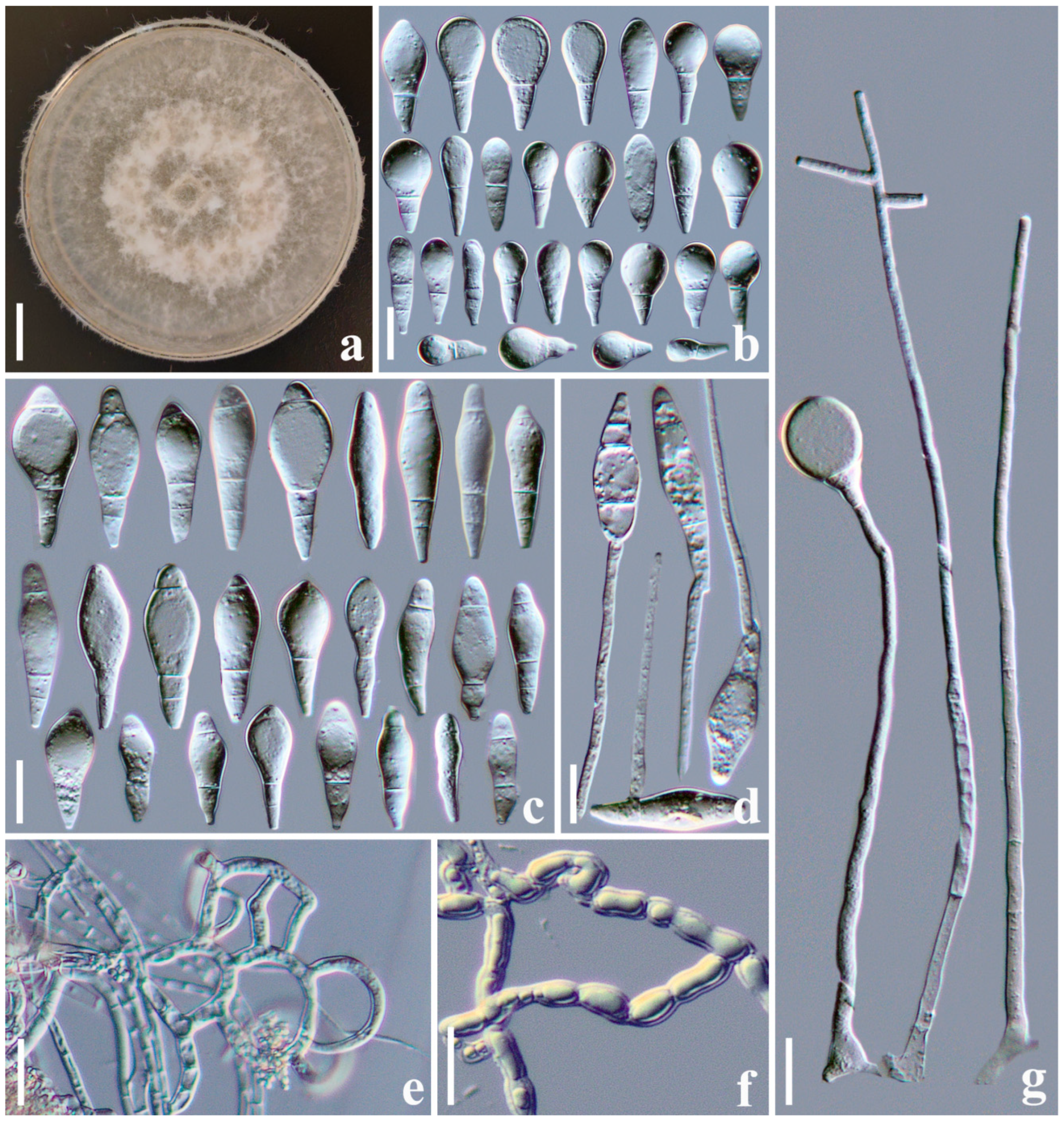
3.2.2. Arthrobotrys heihuiensis F. Zhang, S. Boonmee, and X.Y. Yang sp. nov. (Figure 3)

Index Fungorum number: IF901485; Facesoffungi number: FOF14175
Etymology: The species name “heihuiensis” refers to the Heihui River, the alias of the Yangbi River, where the species was first collected.
Material examined: China, Yunnan Province, Dali City, Yangbi County, Heihui River, 25°37′4.13″ N, 100°1′52.06″ E, from freshwater sediment, 6 April 2018, F. Zhang. Holotype CGMCC 3.20967, preserved in the China General Microbiological Culture Collection Center. Ex-type culture DLUCC 108-1, preserved in the Dali University Culture Collection. Additional specimen examined: China, Yunnan Province, Dali City, Yangbi County, Heihui River, 25°37′4.13″ N, 100°1′52.06″ E, from freshwater sediment, 6 April 2018, F. Zhang. Living culture Y710, preserved in germplasm resources center of Institute of Eastern-Himalaya Biodiversity Research, Dali University.
Culture characteristics: Colonies on PDA white, cottony, reaching 55 mm diameter after 10 days at 26 °C. Hypha composed of septate, branched, smooth, hyaline. Conidiophores erect, septate, unbranched or occasionally producing a long branch, each branch produces several clusters of short denticles (1–3 denticles, mostly 1) by repeated elongation, each short denticle bear a single conidium, 360–720 µm long ( X ¯ = 561.3 µm, n = 50), 3.5–6.5 µm ( X ¯ = 5 µm, n = 50) wide at the base, 1.5–3.5 µm ( X ¯ = 2.3 µm, n = 50) wide at the apex. Conidia two types: macroconidia 31–56 × 6.5–14.5 µm ( X ¯ = 45.4 × 11.5 μm, n = 50), hyaline, clavate or subfusiform, rounded at the apex, constricted and truncated at the base, two- to five-septate (mostly three-septate, one septum at the apex and two septa at the base), with a super cell at the middle. Microconidia 18.5–26.5 × 5–9.5 µm ( X ¯ = 20.2 × 6.7 μm, n = 50), hyaline, smooth-faced, clavate, drop-shaped or lagenate, rounded at the apex and truncated at the base, zero- to two-septate (mostly zero- or one-septate), producing with micro-cycle conidiation. Macroconidia germinate from the small cells at both ends, and the super cells never germinate. Catching nematodes with adhesive networks. Chlamydospores 7–30 × 6–12.5 µm ( X ¯ = 16.2 × 9.3 μm, n = 50), hyaline, smooth-faced, cylindrical, globose or ellipsoidal, in chains when present.
Notes: Phylogenetically, A. heihuiensis is sister to A. cystosporia but lacking in statistical support (Figure 1). Of all Arthrobotrys species, the morphological characteristics of A. heihuiensis are relatively unique and only A. scaphoides may be confused with A. heihuiensis but there are several obvious differences between them: (1) the conidia of A. heihuiensis usually scattered on the short denticles of the conidiophores, while the conidia of A. scaphoides are usually clustered on the node of the short branch produced by conidiophores; and (2) A. scaphoides produces five- or six-septate conidia, while the conidia of A. heihuiensis have no more than four septa. Additionally, the conidia of A. heihuiensis are obviously smaller than that of A. scaphoides (A. heihuiensis, 31–56 (45.4) × 6.5–14.5 (11.5) µm versus A. scaphoides, 36.6–79.3 (57) × 11–17.5 (14) µm); (3) A. heihuiensis produces clavate, drop-shaped or lagenate, zero- to two-septate microconidia, while A. scaphoides does not produce microconidia [4,35,36].
Figure 3. Arthrobotrys heihuiensis (CGMCC 3.20967). (a) Colony. (b) Microconidia. (c) Macroconidia. (d) Adhesive networks. (e) Chlamydospores. (f) Germinating conidia. (g) Conidiophores. Scale bars: (a) = 1 cm, (b) = 10 µm, (cg) = 20 µm.
Jof 10 00081 g003

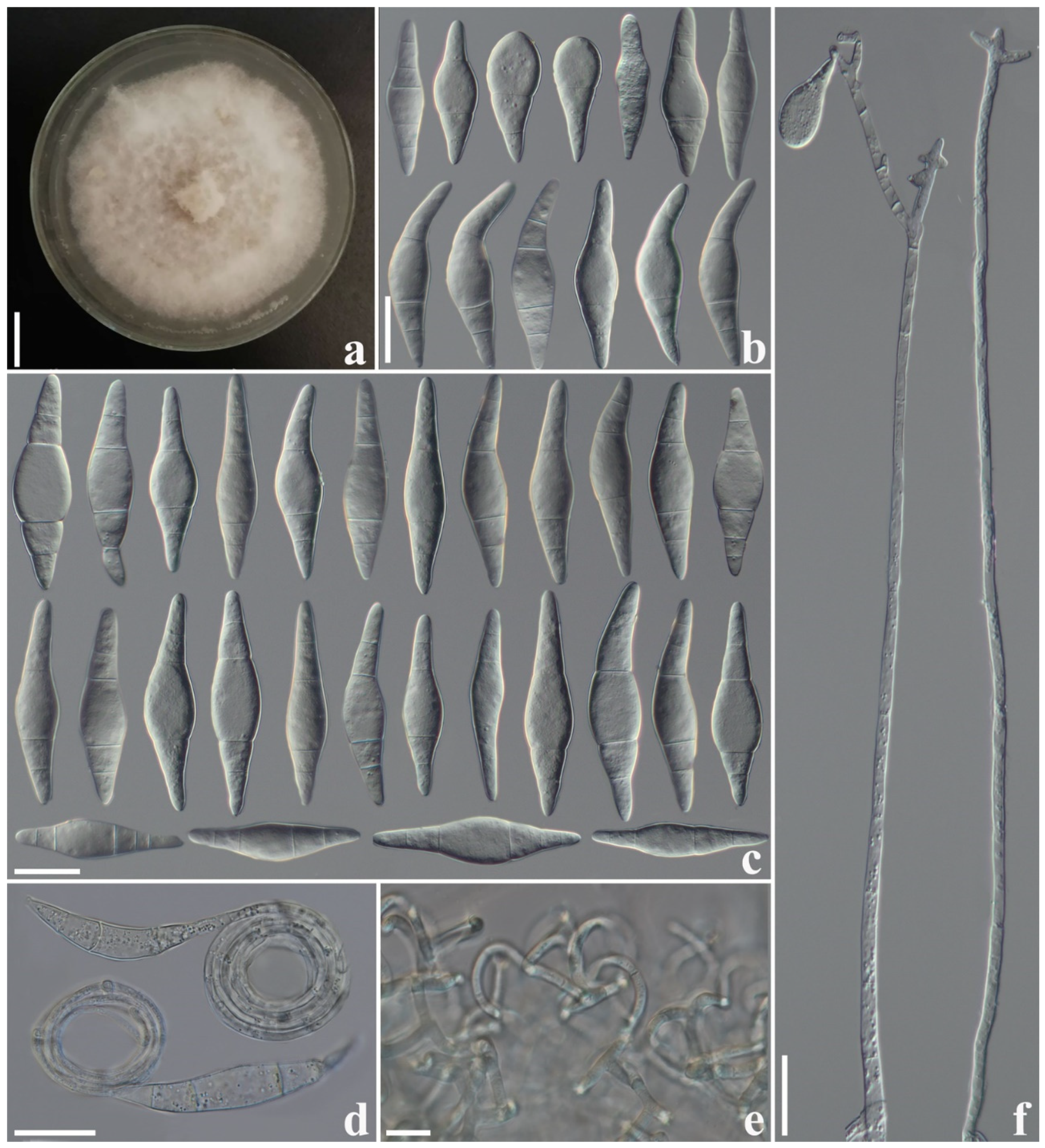
3.2.3. Arthrobotrys jinshaensis F. Zhang, S. Boonmee and X.Y. Yang sp. nov. (Figure 4)

Index Fungorum number: IF901489; Facesoffungi number: FOF14176
Etymology: The species name “jinshaensis” refers to the name of sample collection site: Jinsha River, Jinjiang Town, Shangri-La City, Yunnan Province, China.
Material examined: China, Yunnan Province, Shangri-La City, Jinjiang Town, Jinsha River, 27°8′50.56″ N, 99°49′39.43″ E, from freshwater sediment, 9 July 2014, F. Zhang. Holotype CGMCC 3.20969, preserved in the China General Microbiological Culture Collection Center. Ex-type culture DLUCC 133, preserved in the Dali University Culture Collection. Additional specimen examined: China, Yunnan Province, Shangri-La City, Jinjiang Town, 27°8′50.56″ N, 99°49′39.43″ E, from freshwater sediment, 9 July 2014, F. Zhang. Living culture MA142, preserved in germplasm resources center of Institute of Eastern-Himalaya Biodiversity Research, Dali University.
Culture characteristics: Colonies on PDA white, cottony, reaching 55 mm diameter after 10 days at 26 °C. Hypha composed of septate, branched, smooth, hyaline. Conidiophores erect, septate, hyaline, unbranched or occasionally producing one to four short branches near the apex and bearing one to four conidia, 130–357 µm long ( X ¯ = 246.3 µm, n = 50), 3.5–6 µm ( X ¯ = 4.8 µm, n = 50) wide at the base, 2–4 µm ( X ¯ = 3.1 µm, n = 50) wide at the apex. Conidia smooth-faced and hyaline, rounded at the apex and truncated at the base, 14.5–57 × 7.5–23 µm ( X ¯ = 36.6 × 14.7 μm, n = 50), immature conidia drop-shaped, obovate, clavate, with a super cell at the apex and one to two septa (mostly two-septate) at the base; mature conidia subfusiform, clavate, two- to four-septate (mostly three-septate, one septum located at the apex and two septa at the base of the conidia), with a super cell at the middle of the conidia. Conidia germinate from the small cells at both ends, and the super cells never germinate. Catching nematodes with adhesive networks. Chlamydospores 9.5–21.5 × 5.5–9 µm ( X ¯ = 14.8 × 7.2 μm, n = 50), smooth-faced and hyaline, cylindrical, globose or ellipsoidal, in chains when present.
Notes: Phylogenetically, A. jinshaensis clusters together with A. yangjiangensis, A. mangrovispora, A. thaumasia, A. eudermata, and A. janus with 89% MLBS and 0.91 BYPP support (Figure 1). There are 7.9% (49/622 bp), 7.3% (43/586 bp), 7.6% (46/607 bp), 8.2% (50/611 bp), and 8% (46/577 bp) differences between A. jinshaensis and A. yangjiangensis, A. mangrovispora, A. thaumasia, A. eudermata, and A. janus in ITS sequences, respectively. Morphologically, A. jinshaensis, A. mangrovispora, A. megalospora, A. microscaphoides, A. obovata, A. oudemansii, and A. psychrophila all produced conidiophores with short branches at the apex. However, A. jinshaensis can be easily distinguished from A. megalospora, A. microscaphoides, A. obovata, A. oudemansii, and A. psychrophila by its clavate, subfusiform, irregularly constricted, two- to four-septate conidia [4]. In comparison, A. jinshaensis is more similar to A. mangrovispora in its variable conidia. The main difference between A. jinshaensis and A. mangrovispora is that the conidia of A. jinshaensis are one- to four-septate and mostly three-septate, while the conidia of A. mangrovispora are one- to three-septate and mostly two-septate. Furthermore, a single conidiophores of A. mangrovispora may bear one to six conidia, while the conidiophore of A. jinshaensis bear no more than four conidia [4,37].
Figure 4. Arthrobotrys jinshaensis (CGMCC 3.20969). (a) Colony. (b) Immature conidia. (c) Mature conidia. (d) Germinating conidia. (e) Adhesive networks. (f) Chlamydospores. (g) Conidiophores. Scale bars: (a) = 1 cm, (bg) = 20 µm.
Jof 10 00081 g004

3.2.4. Arthrobotrys yangbiensis F. Zhang, S. Boonmee, and X.Y. Yang sp. nov. (Figure 5)

Index Fungorum number: IF901487; Facesoffungi number: FOF14177
Etymology: The species name “yangbiensis” refers to the name of sample collection site: Yangbi County, Dali City, Yunnan Province, China.
Material examined: China, Yunnan Province, Dali City, Yangbi County, Yangbi River, 25°42′37.94″ N, 99°54′52.15″ E, from freshwater sediment, 4 April 2018, F. Zhang. Holotype CGMCC 3.24985, preserved in the China General Microbiological Culture Collection Center. Ex-type culture DLUCC 36-1, preserved in the Dali University Culture Collection. Additional specimen examined: China, Yunnan Province, Dali City, Yangbi County, Yangbi River, 25°42′37.94″ N, 99°54′52.15″ E, from freshwater sediment, 4 April 2018, F. Zhang. Living culture Y678, preserved in germplasm resources center of Institute of Eastern-Himalaya Biodiversity Research, Dali University.
Culture characteristics: Colonies on PDA white, cottony, reaching 40 mm diameter after 10 days at 26 °C. Hypha composed of septate, branched, smooth, and hyaline. Conidiophores erect, septate, hyaline, unbranched or sometimes branched at the upper half part, producing a cluster of short denticles (2–5) at the apex or producing several clusters of short denticles by repeated elongation, 210–365 µm long ( X ¯ = 284.7 µm, n = 50), 3–5.5 µm ( X ¯ = 3.9 µm, n = 50) wide at the base, and 1.5–3.5 µm ( X ¯ = 2.4 µm, n = 50) wide at the apex. Conidia 40.5–73 × 8.5–18 µm ( X ¯ = 55.4 × 13.6 μm, n = 50), elongate–fusiform or clavate, some conidia curved, hyaline, smooth-faced, two- to five-septate (mostly three- or four-septate), with a super cell at the middle or apex of the conidia, and some conidia produce coiled filamentous appendages. Conidia germinate from the small cells at both ends and the super cells never germinate. They capture nematodes with adhesive networks. Chlamydospore not observed.
Notes: The phylogenetic analyses revealed that A. yangbiensis is sister to A. gampsospora with 99% MLBS and 0.97 BYPP support (Figure 1). A. yangbiensis is 14.7% (118/798 bp) different from A. gampsospora in ITS sequences. In morphology, A. yangbiensis shares some morphological features of short denticle conidiophores and sub-fusiform, curved conidia with A. gampsospora but they differ in shape and size of microconidia. In addition, the conidiophores of A. yangbiensis produce several clusters of short denticles by repeated elongation or geniculate branches of conidiophores, and bear several clusters of conidia, while A. gampsospora bears a single cluster of conidia at the apex of conidiophores [4,38].
Figure 5. Arthrobotrys yangbiensis (CGMCC 3.20943). (a) Colony. (b,c) Conidia. (d) Conidia with filamentous appendages. (e) Adhesive networks. (f) Conidiophores. Scale bars: (a) = 1 cm, (bf) = 20 µm.
Jof 10 00081 g005

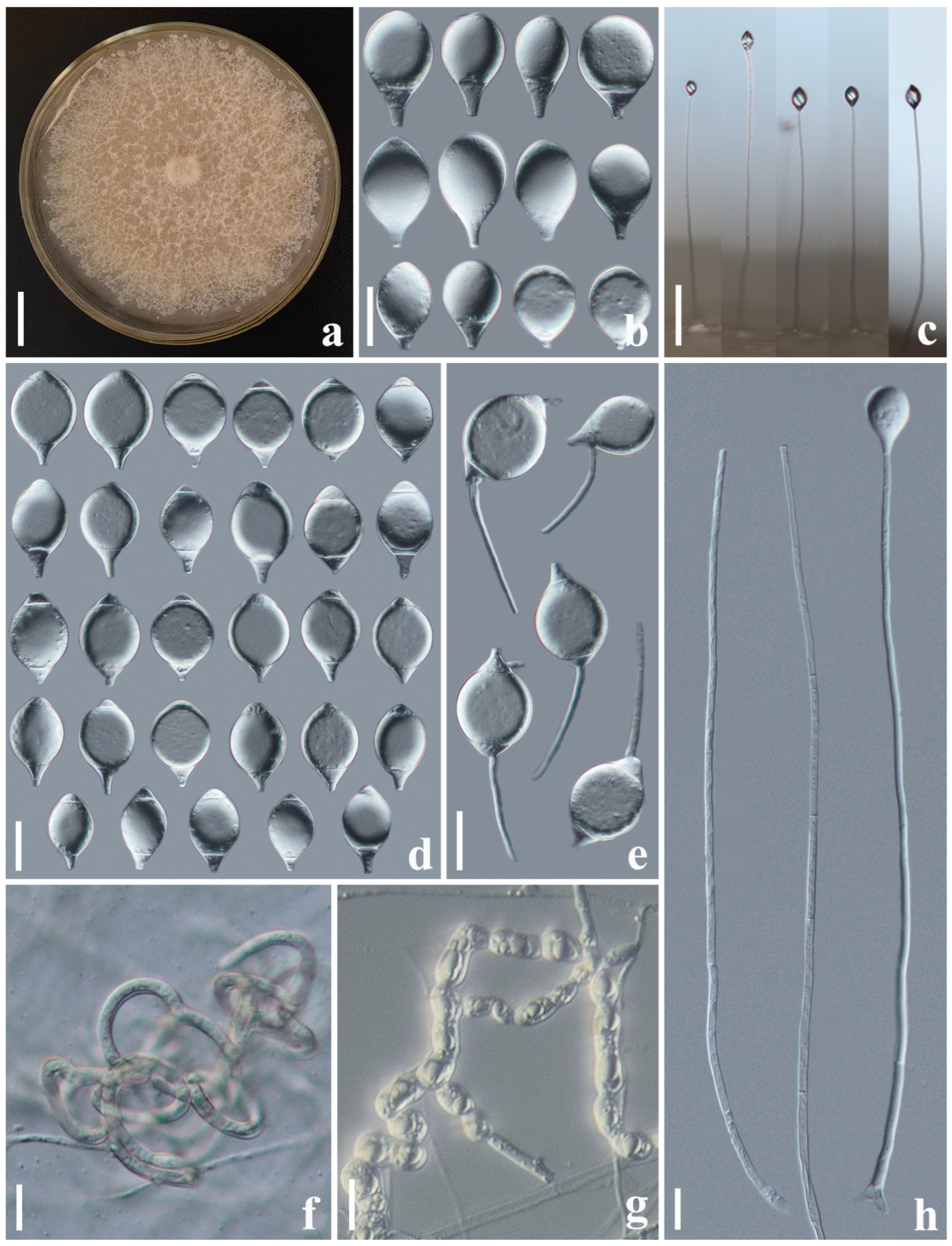
3.2.5. Arthrobotrys yangjiangensis F. Zhang, S. Boonmee, and X.Y. Yang sp. nov. (Figure 6)

Index Fungorum number: IF901488; Facesoffungi number: FOF14178
Etymology: The species name “yangjiangensis” refers to the name of the sample collection site: Yangjiang Town, Yangbi County, Dali City, Yunnan Province, China.
Material examined: China, Yunnan Province, Dali City, Yangbi County, Yangjiang Town, Yangbi River, 25°45′52.11″ N, 99°54′46.43″ E, from freshwater sediment, 14 May 2018, F. Zhang. Holotype CGMCC 3.20968, preserved in the China General Microbiological Culture Collection Center. Ex-type culture DLUCC 124, preserved in the Dali University Culture Collection. Additional specimen examined: China, Yunnan Province, Dali City, Yangbi County, Yangjiang Town, Yangbi River, 25°45′52.11″ N, 99°54′46.43″ E, from freshwater sediment, 14 May 2018, F. Zhang. Living culture YB19, preserved in germplasm resources center of Institute of Eastern-Himalaya Biodiversity Research, Dali University.
Culture characteristics: Colonies on PDA white, cottony, reaching 55 mm diameter after 10 days in the incubator at 26 °C. Hypha composed of septate, branched, smooth, and hyaline. Conidiophores erect, septate, hyaline, unbranched, bearing a single conidium at the apex, 198–537 µm long ( X ¯ = 409.4 µm, n = 50), 3–5.5 µm ( X ¯ = 4.7 µm, n = 50) wide at the base, and 2–3.5 µm ( X ¯ = 2.6 µm, n = 50) wide at the apex. Conidia smooth-faced and hyaline, rounded at the apex and truncated at the base, 24.5–47 × 14.5–29.5 µm ( X ¯ = 36.2 × 23.2 μm, n = 50), immature conidia drop-shaped, obovate, with a super cell at the apex and one to two septa (mostly two-septate) at the base; mature conidia broad fusiform, two- to three-septate (mostly three-septate, one septum at the apex and two septa at the base), with a super cell at the middle of the conidia. Conidia germinate from the only small cells at both ends, and the super cells never germinate. Catching nematodes with adhesive networks. Chlamydospores 8.5–24 × 7–13.5 µm ( X ¯ = 14 × 10 μm, n = 50), smooth-faced and hyaline, cylindrical, globose or ellipsoidal, hyaline, and in chains when present.
Notes: Phylogenetically, A. yangjiangensis formed a basal lineage with A. jinshaensis (another new species reported in this study), A. mangrovispora, A. thaumasia, A. eudermata, and A. janus with 89% MLBS and 0.91 BYPP support (Figure 1). A comparison of ITS nucleotides reveled A. yangjiangensis was 7.9% (49/622 bp), 12.2% (92/754 bp), 7% (42/604 bp), 9.4% (75/795 bp), and 6.3% (36/573 bp) different from A. jinshaensis, A. mangrovispora, A. thaumasia, A. eudermata, and A. janus, respectively. Morphologically, A. yangjiangensis can be easily distinguished from A. jinshaensis and A. janus by its conidial shape with drop-shaped, broad fusiform, two- to three-septate conidia [4,39]. The distinguishing characteristics between A. yangjiangensis, A. mangrovispora, and A. thaumasia is that the conidiophores of A. yangjiangensis bear only one single conidium at the apex, while the conidiophores of the latter two species produce several short denticles at the apex and bear multiple conidia [4,37,40]. By contrast, A. yangjiangensis is more similar to A. eudermata in its simple conidiophores and broad fusiform conidia. However, the conidia of A. yangjiangensis are noticeably smaller than those of A. eudermata (A. yangjiangensis, 2.5–4 (36.2) × 14.5–29.5 (23.2) µm versus A. eudermata, 37–55 (49) × 17.5–35 (28) µm). Furthermore, A. eudermata produces ellipsoid, aseptate microconidia, whereas A. yangjiangensis does not [4,41].
Figure 6. Arthrobotrys yangjiangensis (CGMCC 3.20968). (a) Colony. (b) Immature conidia. (d) Mature conidia. (e) Germinating conidia (f) Adhesive networks. (g) Chlamydospores. (c,h) Conidiophores. Scale bars: (a) = 1 cm, (b,dh) = 20 µm, (c) = 100 µm.
Jof 10 00081 g006

4. Discussion

Among the 130 species (plus our five new species) of Orbiliomycetes nematode-trapping fungi (NTF), 61 species have distribution records in freshwater habitats. Among these species, 17 species were first isolated from freshwater habitats. To date, A. hyrcanus, A. blastospora, A. dainchiensis, A. eryuanensis, A. hengjiangensis and five new species reported in this study were exclusively found in freshwater environments [12,13,14,15,16,17,34,35,42]. These findings collectively emphasize that freshwater NTF are an important component of NTF diversity. Hence, future studies on NTF diversity should fully consider the significance of freshwater habitats.
The five newly identified species presented were all derived from sediment samples in water depths of less than 2 m. Previous investigations have demonstrated the absence of NTF beyond a water depth of 4 m [13]. However, abundant aquatic nematodes still exist in deeper waters [43,44]. At such depths, there must be other nematode regulators to perform the function of aquatic nematode population regulation instead of NTF. Accordingly, it is speculated that more novel nematophagous microorganisms may be discovered in deeper waters employing efficacious research methodologies. The exploration and examination of these enigmatic nematophagous microorganisms hold the potential to provide valuable insights into the origins and evolutionary processes of carnivorous microorganisms, while also offering promising prospects for the biological control of detrimental nematodes.
It is generally believed that aquatic NTF originate from terrestrial ecosystems. However, unlike terrestrial environments, NTF face numerous challenges in aquatic habitats. For instance, the interaction between NTF and nematodes relies on processes such as host recognition, generation of trapping structures, invasion and digestion of nematode, etc. These processes are more or less dependent on the transmission of extracellular signaling factors [45,46,47]. However, in aquatic environments, these extracellular signaling molecules are likely to be diluted and lose their corresponding functions. Consequently, how aquatic NTF prey on nematodes in water, how they maintain osmotic balance in water, and how they reproduce and spread in water remain unresolved questions in the field. Investigating these questions can not only deepen our understanding of fungal adaptive evolution but also constitutes an important aspect of the study on the origin and evolution of these extraordinary organisms.

Supplementary Materials

The following supporting information can be downloaded at: https://www.mdpi.com/article/10.3390/jof10010081/s1, Table S1: GenBank accession numbers involved in this study.

Author Contributions

X.-Y.Y. and W.X. supervised the data collection and manuscript writing; F.Z., Y.-Q.Y. and F.-P.Z. collected relevant data; F.Z. drafted the manuscript; S.B. and X.-Y.Y. revised the manuscript. All authors have read and agreed to the published version of the manuscript.

Funding

This research was funded by the National Natural Science Foundation Program of P.R. China, grant number 32360002, and the Project for Talent and Platform of Science and Technology in Yunnan Province Science and Technology Department, grant number 202205AM070007.

Institutional Review Board Statement

Not applicable.

Informed Consent Statement

Not applicable.

Data Availability Statement

The data that support the finding of this study are contained within the article.

Acknowledgments

We sincerely grateful Yu Ze-Fen of Yunnan University for all her help and suggestions during the writing process.

Conflicts of Interest

The authors declare no conflicts of interest.

References

  1. Barron, G.L. The Nematode-Destroying Fungi; Canadian Biological Publications Ltd.: Guelph, ON, Canada, 1977; p. 45. [Google Scholar]
  2. Swe, A.; Li, J.; Zhang, K.Q.; Pointing, S.B.; Jeewon, R.; Hyde, K.D. Nematode-trapping fungi. Curr. Res. Environ. Appl. Mycol. 2011, 1, 1–26. [Google Scholar]
  3. Zhang, K.Q.; Mo, M.H. Flora Fungorum Sinicorum (Vol. 33): Arthrobotrys et Gengra Cetera Cognata; Science Press: Beijing, China, 2006. [Google Scholar]
  4. Zhang, K.Q.; Hyde, K.D. Nematode-Trapping Fungi; Springer Science & Business: Berlin, Germany, 2014. [Google Scholar] [CrossRef]
  5. Jiang, X.; Xiang, M.; Liu, X. Nematode-trapping fungi. Microbiol. Spectr. 2017, 5, 1. [Google Scholar] [CrossRef] [PubMed]
  6. Soliman, M.S.; El-Deriny, M.M.; Ibrahim, D.S.S.; Zakaria, H.; Ahmed, Y. Suppression of root-knot nematode Meloidogyne incognita on tomato plants using the nematode trapping fungus Arthrobotrys oligospora Fresenius. J. Appl. Microbiol. 2021, 131, 2402–2415. [Google Scholar] [CrossRef] [PubMed]
  7. Yang, Y.; Yang, E.; An, Z.; Liu, X.Z. Evolution of nematode-trapping cells of predatory fungi of the Orbiliaceae based on evidence from rRNA-encoding DNA and multiprotein sequences. Proc. Natl. Acad. Sci. USA 2007, 104, 8379–8384. [Google Scholar] [CrossRef] [PubMed]
  8. Ahrén, D.; Ursing, B.M.; Tunlid, A. Phylogeny of nematode-trapping fungi based on 18S rDNA sequences. FEMS Microbiol. Lett. 1998, 158, 179–184. [Google Scholar] [CrossRef] [PubMed]
  9. Mo, M.H.; Chen, W.M.; Yang, H.R.; Zhang, K.Q. Diversity and metal tolerance of nematode-trapping fungi in Pb-polluted soils. J. Microbiol. 2008, 46, 16–22. [Google Scholar] [CrossRef] [PubMed]
  10. Singh, R.K.; Pandey, S.K.; Chattopadhyay, A. Biodiversity and periodical/seasonal distribution of nematode trapping fungi from different habitats. J. Pure Appl. Microbiol. 2014, 9, 767–776. [Google Scholar]
  11. Zhang, Y.; Li, S.; Li, H.; Wang, R.; Zhang, K.Q.; Xu, J. Fungi–nematode interactions: Diversity, ecology, and biocontrol prospects in agriculture. J. Fungi 2020, 6, 206. [Google Scholar] [CrossRef]
  12. Liu, S.; Su, H.; Su, X.; Zhang, F.; Liao, G.; Yang, X. Arthrobotrys xiangyunensis, a novel nematode-trapping taxon from a hot-spring in Yunnan Province, China. Phytotaxa 2014, 174, 89–96. [Google Scholar] [CrossRef]
  13. Hao, Y.; Mo, M.; Su, H.; Zhang, K.Q. Ecology of aquatic nematode-trapping hyphomycetes in southwestern China. Aquat. Microb. Ecol. 2005, 40, 175–181. [Google Scholar] [CrossRef]
  14. Swe, A.; Jeewon, R.; Pointing, S.B.; Hyde, K.D. Diversity and abundance of nematode-trapping fungi from decaying litter in terrestrial, freshwater and mangrove habitats. Biodivers. Conserv. 2009, 18, 1695–1714. [Google Scholar] [CrossRef]
  15. Zhang, F.; Boonmee, S.; Bhat, J.D.; Xiao, W.; Yang, X.Y. New Arthrobotrys nematode-trapping species (Orbiliaceae) from terrestrial soils and freshwater sediments in China. J. Fungi 2022, 8, 671. [Google Scholar] [CrossRef] [PubMed]
  16. Tarigan, W.E.; Munir, E.; Hastuti, L.D.S.; Hartanto, A. Occurrence of nematophagous fungi in freshwater samples of Toba Lake, North Sumatra, Indonesia. J. Phys. Conf. Ser. 2020, 1462, 012052. [Google Scholar] [CrossRef]
  17. Hao, Y.E.; Luo, J.; Zhang, K.Q. A new aquatic nematode trapping hyphomycete. Mycotaxon 2004, 89, 235–239. [Google Scholar]
  18. González-Solís, D.; Jiménez-García, M.I. Parasitic nematodes of freshwater fishes from two Nicaraguan crater lakes. Comp. Parasitol. 2006, 73, 188–192. [Google Scholar] [CrossRef]
  19. Tahseen, Q. Nematodes in aquatic environments: Adaptations and survival strategies. Biodivers. J. 2012, 3, 13–40. [Google Scholar]
  20. Staniland, L.N. A modification of the Baermann funnel technique for the collection of nematodes from plant material. J. Helminthol. 1954, 28, 115–117. [Google Scholar] [CrossRef]
  21. Zhang, F.; Boonmee, S.; Yang, Y.Q.; Zhou, F.P.; Xiao, W.; Yang, X.Y. Arthrobotrys blastospora sp. nov. (Orbiliomycetes): A Living Fossil Displaying Morphological Traits of Mesozoic Carnivorous Fungi. J. Fungi 2023, 9, 451. [Google Scholar] [CrossRef]
  22. Su, H.Y.; Li, M.; Liu, X.X. Improvement of the slide making method of nematode trapping fungi. J. Dali Univ. 2006, 6, 25–26+30. [Google Scholar]
  23. White, T.J.; Bruns, T.; Lee, S.; Taylor, J. Amplification and direct sequencing of fungal ribosomal RNA genes for phylogenetics. PCR Protoc. A Guide Methods Appl. 1990, 18, 315–322. [Google Scholar] [CrossRef]
  24. O’Donnell, K.; Kistler, H.C.; Cigelnik, E.; Ploetz, R.C. Multiple evolutionary origins of the fungus causing Panama disease of banana: Concordant evidence from nuclear and mitochondrial gene genealogies. Proc. Natl. Acad. Sci. USA 1998, 95, 2044–2049. [Google Scholar] [CrossRef]
  25. Liu, Y.J.; Whelen, S.; Hall, B.D. Phylogenetic relationships among ascomycetes: Evidence from an RNA polymerse II subunit. Mol. Biol. Evol. 1999, 16, 1799–1808. [Google Scholar] [CrossRef] [PubMed]
  26. Swindell, S.R.; Plasterer, T.N. Seqman. In Sequence Data Analysis Guidebook; Swindell, S.R., Ed.; Springer: Totowa, NJ, USA, 1997; pp. 75–89. [Google Scholar]
  27. Masigol, H.; Rezakhani, F.; Pourmoghaddam, M.J.; Khodaparast, S.A.; Grossart, H.P. The Introduction of Two New Species of Aquatic Fungi from Anzali Lagoon, Northern Iran. Diversity 2022, 14, 889. [Google Scholar] [CrossRef]
  28. Katoh, K.; Standley, D.M. MAFFT Multiple Sequence Alignment Software Version 7: Improvements in Performance and Usability. Mol. Biol. Evol. 2013, 30, 772–780. [Google Scholar] [CrossRef] [PubMed]
  29. Tamura, K.; Stecher, G.; Peterson, D.; Filipski, A.; Kumar, S. MEGA6: Molecular Evolutionary Genetics Analysis Version 6.0. Mol. Biol. Evol. 2013, 30, 2725–2729. [Google Scholar] [CrossRef]
  30. Posada, D. jModelTest: Phylogenetic Model Averaging. Mol. Biol. Evol. 2008, 25, 1253–1256. [Google Scholar] [CrossRef]
  31. Nguyen, L.T.; Schmidt, H.A.; Von Haeseler, A.; Minh, B.Q. IQ-TREE: A Fast and Effective Stochastic Algorithm for Estimating Maximum-Likelihood Phylogenies. Mol. Biol. Evol. 2015, 32, 268–274. [Google Scholar] [CrossRef]
  32. Huelsenbeck, J.P.; Ronquist, F. MRBAYES: Bayesian inference of phylogenetic trees. Bioinformatics 2001, 17, 754–755. [Google Scholar] [CrossRef]
  33. Rambaut, A. FigTree v1. 3.1. 2010. Available online: http://tree.bio.ed.ac.uk/software/figtree/ (accessed on 16 March 2012).
  34. Peach, M. Aquatic predacious fungi. Trans. Br. Mycol. Soc. 1950, 33, 148–153. [Google Scholar] [CrossRef]
  35. Peach, M. Aquatic predacious fungi. II. Trans. Br. Mycol. Soc. 1952, 35, 19–23. [Google Scholar] [CrossRef]
  36. Zhang, Y.; Qiao, M.; Weber, E.; Baral, H.O.; Hagedorn, G.; Yu, Z.F. Arthrobotrys scaphoides from China and Europe with a phylogenetic analysis including the type strain. Mycotaxon 2010, 111, 291. [Google Scholar] [CrossRef]
  37. Swe, A.; Jeewon, R.; Pointing, S.B.; Hyde, K.D. Taxonomy and molecular phylogeny of Arthrobotrys mangrovispora, a new marine nematode-trapping fungal species. Bot. Mar. 2008, 51, 331–338. [Google Scholar] [CrossRef]
  38. Drechsler, C. Some clampless hyphomycetes predacious on nematodes and rhizopods. Sydowi Annal. Mycol. Ser. II 1962, 15, 9–24. [Google Scholar]
  39. Li, S.D.; Miao, Z.Q.; Zhang, Y.H.; Liu, X.Z. Monacrosporium janus sp. nov., a new nematode-trapping hyphomycete parasitizing sclerotia and hyphae of Sclerotinia sclerotiorum. Mycol. Res. 2003, 107, 888–894. [Google Scholar] [CrossRef] [PubMed]
  40. Drechsler, C. Some hyphomycetes that prey on free-living terricolous nematodes. Mycologia 1937, 29, 447–552. [Google Scholar] [CrossRef]
  41. Drechsler, C. Several species of Dactylella and Dactylaria that capture free-living nematodes. Mycologia 1950, 42, 1–79. [Google Scholar] [CrossRef]
  42. Deng, W.; Zhang, F.; Li, Y.P.; Zhang, X.; Fornacca, D.; Yang, X.Y.; Wen, X. Uncovering the biogeographic pattern of the widespread nematode-trapping fungi Arthrobotrys oligospora: Watershed is the key. Front. Microbiol. 2023, 14, 1152751. [Google Scholar] [CrossRef]
  43. de Jesús-Navarrete, A.; Armenteros, M.; Espositos, A.V.; Rocha-Olivares, A. Size spectra, biomass, and trophic groups of free-living marine nematodes along a water-depth gradient in the northwestern Gulf of Mexico. Mar. Ecol. 2022, 43, e12723. [Google Scholar] [CrossRef]
  44. Ptatscheck, C.; Traunspurger, W. The ability to get everywhere: Dispersal modes of free-living, aquatic nematodes. Hydrobiologia 2020, 847, 3519–3547. [Google Scholar] [CrossRef]
  45. Hsueh, Y.P.; Mahanti, P.; Schroeder, F.C.; Sternberg, P.W. Nematode-trapping fungi eavesdrop on nematode pheromones. Curr. Biol. 2013, 23, 83–86. [Google Scholar] [CrossRef]
  46. Kuo, T.H.; Yang, C.T.; Chang, H.Y.; Hsueh, Y.P.; Hsu, C.C. Nematode-trapping fungi produce diverse metabolites during predator–prey interaction. Metabolites 2020, 10, 117. [Google Scholar] [CrossRef] [PubMed]
  47. Tunlid, A.; Ahrén, D. Molecular mechanisms of the interaction between nematode-trapping fungi and nematodes: Lessons from genomics. In Biological Control of Plant-Parasitic Nematodes: Building Coherence between Microbial Ecology and Molecular Mechanisms; Springer: Dordrecht, The Netherlands, 2011; pp. 145–169. [Google Scholar] [CrossRef]
Figure 1. The maximum likelihood tree inferred from a combined ITS, TEF1-α, and RPB2 dataset. The black and red numbers in front of the node indicate Bootstrap support values for maximum likelihood equal or greater than 70% and Bayesian posterior probabilities values equal or greater than 0.90, respectively. Our new isolates are in blue and the type strains are in bold.
Jof 10 00081 g001
Table 1. Samples information involved in this study.
Sample SourceSampling LocationSampling DateNumber of Samples
Cibi Lake26°9′7.14″ N, 99°56’32.72″ E4 June 201325
Heihui River25°37′4.13″ N, 100°1′52.06″ E6 April 201810
Jinsha River27°8′50.56″ N, 99°49′39.43″ E9 July 201410
Yangbi River25°42′37.94″ N, 99°54′52.15″ E4 April 201810
Yangjiang River25°45′52.11″ N, 99°54′46.43″ E14 May 201810
Disclaimer/Publisher’s Note: The statements, opinions and data contained in all publications are solely those of the individual author(s) and contributor(s) and not of MDPI and/or the editor(s). MDPI and/or the editor(s) disclaim responsibility for any injury to people or property resulting from any ideas, methods, instructions or products referred to in the content.

