Article

Foliar Lesions Induced by Pestalotiopsis arengae in Oil Palm (O × G) in the Colombian Southwest Palm Zone

by
William Fabian Betancourt-Ortiz
,
Hector Camilo Medina-Cardenas
,
Jose Luis Padilla-Agudelo
,
Francia Helena Varon
,
Yuri Adriana Mestizo-Garzón
,
Anuar Morales-Rodríguez
and
Greicy Andrea Sarria-Villa
*
Pest and Disease Program—Phytopathology Area, Colombian Oil Palm Research Center (CENIPALMA), Centro Empresarial Pontevedra, Calle 98 N° 70-91, Piso 14, Bogotá 111211, Colombia
*
Author to whom correspondence should be addressed.
J. Fungi 2024, 10(1), 24; https://doi.org/10.3390/jof10010024
Submission received: 15 September 2023 / Revised: 12 December 2023 / Accepted: 26 December 2023 / Published: 29 December 2023
(This article belongs to the Section Fungal Pathogenesis and Disease Control)

Abstract

:
In Colombia, plantings with the oil palm hybrid between Elaeis oleifera × Elaeis guineensis, known as O × G hybrid, have increased due to its tolerance to bud rot. Despite this, different degrees of foliar necrosis, chlorosis, and leaf blight have been reported in some cultivars; therefore, this work aimed to diagnose this problem. We visited plantation plots with palms exhibiting the mentioned symptoms and collected 21 samples of affected tissues in different disease states. The affected tissues were examined and seeded in a culture medium. Pathogenicity tests were performed and the isolates were characterized by culture and morphological and molecular features. Curvularia, Colletotrichum, Phoma, and 25 Pestalotiopsis-like fungi were isolated from the foliar lesions. In the pathogenicity tests, the symptoms observed in the field were reproduced with MFTU01-1, MFTU12, and MFTU21 isolates, which were identified at the species level through a sequence analysis of three genes (ITS, TUB2, and TEF1-α) as Pestalotiopsis arengae with an identical level of 99% based on the results of BLAST and phylogenetic tree analyses. The remaining 22 Pestalotiopsis-like non-pathogenic isolates were identified as species of Neopestalotiopsis and Pseudopestalotiopsis. The direct association of P. arengae with the disease was confirmed via molecular detection in affected tissues in 15 of 21 samples collected for this evaluation. This is the first report of P. arengae as the causal agent of foliar lesions in O × G hybrid oil palm in Colombia.

1. Introduction

Among oil crops, oil palm registers the highest yields per hectare [1]. Colombia is currently the fourth largest palm oil producer in the world after Indonesia, Malaysia, and Thailand [2]. The palm industry in Colombia generates approximately 177,400 direct and indirect jobs and contributes to 6% of the country’s GDP [3]. Oil palm is affected by biotic and abiotic agents. Bud rot (BR) disease caused by the oomycete Phytophthora palmivora is one of Colombia’s most limiting phytosanitary problems [4]. In the southwestern Colombian palm-growing region (Tumaco, Nariño), this disease reached epidemic proportions in the first decade of the 2000s and devastated the 36,000 hectares planted with Elaeis guineensis cultivars [5]. As a result of the BR epidemic in the Colombian Southwest Palm Zone, renewals have been made with hybrid cultivars of Elaeis oleifera × Elaeis guineensis (O × G) since 2008. The genetic solution seems promising: incorporating the resistance of the American oil palm (E. oleifera) into the African oil palm (E. guineensis) generates the O × G interspecific hybrid that is tolerant to diseases such as BR [6,7,8,9,10].
Some plantations in the zone have reported emerging problems in the different hybrids, such as the drying of lower leaves, chlorosis of the canopy, and leaf blight, with the latter having the highest incidence. These symptoms have been associated in E. guineensis with the disease known as Pestalotiopsis or leaf blight, which involves a complex of fungi, including Pestalotiopsis palmarum and P. glandicola, and other genera like Helminthosporium sp., Curvularia sp., Colletotrichum sp., Phyllosticta sp., and Macrophoma palmarum [11,12,13]. Traditionally, the development of the disease has been linked to primary damage caused by insects with sucking, chewing, and scraping habits [13,14,15,16,17,18]. However, there are few studies where isolations and pathogenicity tests have been performed to verify the direct association of microorganisms with the development of foliar lesions.
The taxonomic classification at the species level within the Pestalotiopsis genus has traditionally been made according to the host from which fungi were isolated, the conidia morphology, the color intensity of the conidia median cells, and molecular analyses based on rDNA sequences [19,20]. However, recent studies by Maharachchikumbura et al. [21] and Ayoubi and Soleimani [22], supported by the molecular analysis of multiple genes (which included the internal transcribed spacer of ribosomal DNA (ITS) [23], beta-tubulin (TUB2) [24], and translation elongation factor 1-alpha (TEF1-α) [25]) have contributed to improving the traditional classification system of the species, indicating that the trait of median cell coloration is not a reliable taxonomic trait in the discrimination of species.
Several reports have indicated Pestalotiopsis species as plant pathogens in tropical and subtropical countries [26,27]. In Colombia, Solarte [28] studied the genetic diversity of the genera Pestalotiopsis and Neopestalotiopsis related to the Scab disease of Guava (Psidium guajava L.). In Italy, Ismail [29] reported that P. uvicola and P. clavispora caused gray leaf spots on mangoes (Mangifera indica L.). Keith [30] reported that P. clavispora, P. microspora, and P. disseminata caused guava scab (Psidium guajava L.) in Hawaii. Maharachchikumbura et al. [31] reported that P. semarangensis caused fruit rot of Pomarrosa (Syzygium samarangense) in Thailand. Maharachchikumbura [32] reported that Neopestalotiopsis, Pestalotiopsis, and Truncatella species are associated with trunk disease in grapevine (Vitis vinifera L.) in France. Additionally, Maharachchikumbura et al. [33] associated Pseudopestalotiopsis ignota and Ps. camelliae with grey blight disease of tea (Camellia sinensis) in China.
In oil palm crops in Colombia, some studies by Jimenéz O. [34] have associated Pestalotiopsis species with leaf blight. For example, on the cultivar E. guineensis, a complex of fungi was identified in association with leaf blight or añublo foliar. However, there are no studies on the interspecific hybrid O × G. In China, Shen et al. [35] reported Pestalotiopsis microspora causing foliar lesions in African oil palm; in Thailand, P. theae has been reported to cause foliar lesions in E. guineensis [36]; and in Queensland, Australia, Fröhlich [37] associated P. phoenicis, P. elastica, P. theae, P. adjusta, and P. palmarum with the foliar lesions presented in the coconut tree (Cocos nucifera). In Colombia, leaf blight of oil palm has been registered as a disease of economic importance. It generally affects palms older than two years [34]. Jiménez and Reyes [12] compared the production of slightly affected palms (19–24% defoliation) with severely affected palms (55–66% defoliation) and reported losses in bunch production close to 36% per year, equivalent to 5.2 tons of bunches/hectare per year and the depressive effects persisted until 33 months after having carried out control of the disease. The problems at the leaf level observed in hybrid cultivars in the Colombian Southwest Palm Zone and the scarce information on the association of the microorganisms involved in this pathology have motivated this research, with the purposes of (i) identifying and describing the symptomatology of this foliar disease; (ii) performing pathogenicity tests with the obtained microorganisms with similar characteristics to Pestalotiopsis; and (iii) characterizing the causal agent. These are all indispensable steps for the management practices that will be necessary to mitigate the incidence and severity of the disease.

2. Materials and Methods

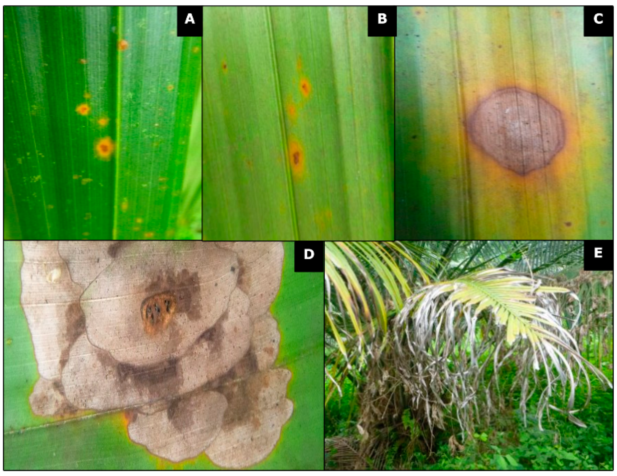
2.1. Description of Symptoms and Foliar Tissue Sampling

This research was carried out in plantations in the Colombian Southwest Palm Zone located in the Department of Nariño, specifically in the Municipality of Tumaco (1°48′0″ N, 78°45′0″ W); according to the Meteorological Station located in the El Mira Research Center of Agrosavia (observation window of the last 20 years), Tumaco has an average annual precipitation of 2910 mm, distributed mainly in a single wet season between the months of January to June. Likewise, the average annual temperature is 26.6 °C with a maximum of 27.8 °C and a minimum of 25.6 °C (Figure 1).
Under field conditions, initial observations were made to select hybrid palms to be evaluated, identifying the leaflet lesions, describing the initial symptoms, and marking the progress toward the most affected leaves. Additionally, photographic records of the symptoms were taken at different stages of the development of the disease. Affected tissues were collected at different stages of infection from the oil palms sampled at the study site, which were planted between 2007 and 2010. The collected samples were transported under refrigeration to Cenipalma’s plant pathology laboratory for immediate processing.

2.2. Isolation and Purification of Microorganisms

To identify the fungal microorganisms present in the foliar tissue, imprints of the field-collected material were created. To induce sporulation, the tissue was placed in a moist chamber consisting of 28 oz aluminum trays. Each tray contained a paper towel moistened with sterile, distilled water and was covered with a piece of tulle to prevent direct contact between the leaves and the water. The trays were then placed inside Ziploc© bags. Finally, part of the tissue was processed following the traditional method for fungal isolation, which consists of washing the sample with tap water, drying it, and selecting affected tissue from the area of disease progression in 0.3 to 0.5 cm long pieces. Subsequently, the samples were disinfected with 70% alcohol for 30 s and 1% sodium hypochlorite for 1 min and rinsed three times with sterile distilled water. The samples were then dried and seeded in several culture media: potato dextrose agar (PDA), vegetable juice (V8) agar, water agar, corn meal agar, malt extract agar, and oatmeal agar [38]. The samples were incubated at 24 °C (±2 °C) for 24 h under white light and 24 h in darkness. When the reproductive structures of the fungi were formed, monosporic cultures were obtained by directly transferring the conidia to Petri plates with PDA medium [39].

2.3. Culture and Morphological Characterization

The culture and morphological characterization for Pestalotiopsis-like isolates were performed using the methods used to identify fungal genera of agricultural importance [38,40,41]. Regarding the culture characterization, the development of the microorganism in the PDA culture media was described according to the colony’s color, shape, margin elevation or surface edge, texture, and conidiomata production [30].
All the isolates were identified using the morphological basis of the reproductive structures: the length and width of the conidia, lengths of apical and basal appendages, the number of basal and apical appendages, the numbers of cells and septa present in the conidia, and finally the color exhibited by the median cells [21,42].
The conidia were taken from the conidial suspension and observed under an Olympus BX40 light microscope with 40× magnification. Photographic records were taken using an Olympus DP73 camera (Olympus Corporation, Tokyo, Japan). Biometric variables were estimated with the CellSens® software cellSens V1.14 (https://www.olympus-lifescience.com.cn/en/sofware/cellsens/, accessed on 10 July 2021). The 95% confidence interval was found for each variable, and the maximum and minimum values and the mean values of 30 measurements were estimated. The measurements of the structures were compared using an analysis of variance (ANOVA) and the means were separated according to Tukey’s test (p < 0.05) using the statistical software SAS 9.0.

2.4. Molecular Identification and Phylogenetic Analysis

The biomass development of 25 Pestalotiopsis-like isolates was produced in potato dextrose broth (Potato Dextrose Broth, DIFCO) with shaking at 110 rpm for seven days. From fresh fungal mycelia, genomic DNA was extracted using the QIAGEN DNeasy Plant Mini kit (Hilden, Germany), following the manufacturer’s instructions. The final concentration of the DNA was 100–200 ng/μL. The amplified gene regions included the internal transcribed spacer of ribosomal DNA (ITS) using primers ITS4 and ITS5 [23], beta-tubulin (TUB2) with primers Bt2a and Bt2b [24], and translation elongation factor 1-alpha (TEF1-α) with primers EF1-526F and EF1-1567R [25]. The reactions were incubated in a T3 thermocycler (Biometra, Göttingen, Germany). For each reaction, a total volume of 25 µL was used containing 12.5 µL of the kit’s GoTaq® Green Master Mix (Promega, Madison, WI, USA), 9.5 µL of nuclease-free water, 0.3 µM of each primer, and 30 ng of genomic DNA. The reaction conditions included an initial denaturation at 95 °C (5 min), followed by 35 cycles of denaturation at 94 °C for 1 min, binding at 52, 55, and 54 °C for ITS, TUB2, and TEF1-α, respectively, for 30 s, and extension at 72 °C for 1 min with a final extension at 72 °C for 10 min. The PCR products were run on a 1% agarose gel. After electrophoresis, the gels were visualized under UV light. The bands at the expected sizes were cut directly from the gel, purified using the QIAquick Gel Extraction kit (Qiagen, Hilden, Germany), and sent to Universidad Nacional de Colombia for sequencing with the primers used in the PCR analysis.
The sequences obtained with each primer were edited using the BioEdit 7.0.5.3 program [43], and the consensus sequence was constructed with the CAP contig assembly accessory for each isolate. Once the sequences were obtained, their identity was confirmed by comparing them with the GenBank database based on ITS, TUB2, and TEF1-α. The sequences generated in this study were supplemented with additional sequences obtained from other related strains of Pestalotiopsis, Neopestalotiopsis, and Pseudopestalotiopsis species from GenBank based on the literature reported for phylogenetic analyses (Supplementary Materials, Table S1). Subsequently, sequence alignment was carried out using the MUSCLE algorithm included in the MEGAX program [44]. The statistical method was the neighbor-joining method of the consensus sequences of the ITS gene, and evolutionary distances were computed using the Kimura 2-parameter method. This analysis involved 103 nucleotide sequences. There were a total of 464 positions in the final dataset. The support of the nodes was determined using the bootstrap method with 1000 repetitions. Our consensus sequences are available under the accession numbers for the ITS region, TUB2, and TEF1-α in Section 3.5. Also, we developed a phylogenetic tree for each genus (Supplementary Materials, Figures S1–S3).

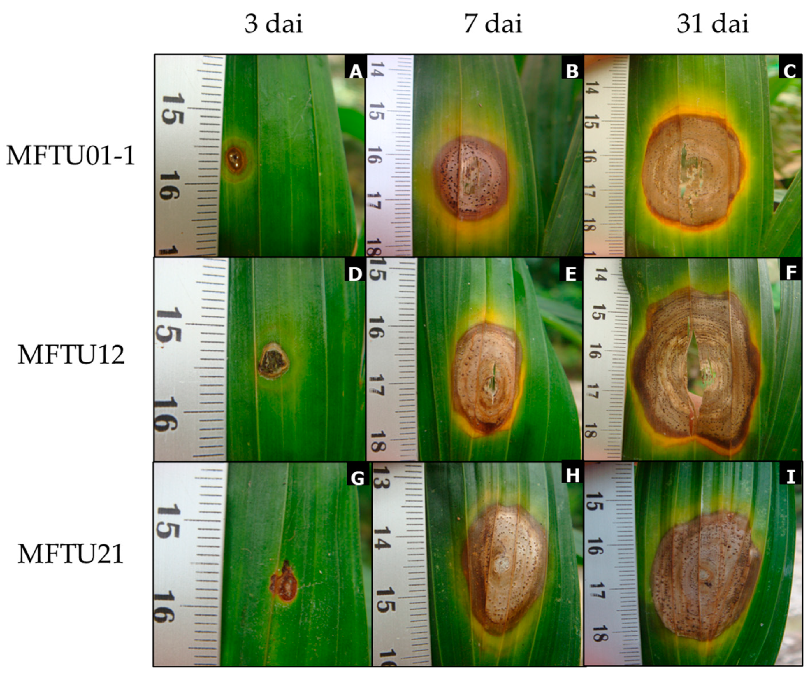
2.5. Pathogenicity Tests

For the pathogenicity tests, we used 7-month-old O × G hybrid cultivar Sinú (cerete) × Deli Yangambí. The tests included microorganisms isolated from symptomatic tissue isolates such as Pestalotiopsis-like sp. (25 isolates), Curvularia sp. (1 isolate), Colletotrichum sp. (1 isolate), and Phoma sp. (1 isolate). Preliminary tests were conducted by using different inoculation methods to select the most effective one in reproducing the symptoms [35,45,46]. A wound was necessary to develop lesions, acting as a facilitator for the entry of the microorganism.
The inoculation method used a 5 mm mycelial agar disc from the edge of a colony of the different isolates from an approximately 10-day-old culture. A sterile entomological pin (0.40 mm diameter by 37 mm long) was used to create the wound in the leaf tissue before placing the plug. The same procedure was used with pathogen-free PDA discs for the control treatments. The plants were kept under moist chamber conditions for 48 h in both cases. Subsequently, they were maintained in semi-controlled conditions under a shade house (70% dark) at an average temperature of 28–30 °C combined with a misting system to promote infection. The development of the infection over time was analyzed using ImageJ® program developed by Wayne Rasband (National Institutes of Health, Bethesda, MD, USA, https://www.nih.gov/, accessed on 10 July 2021). Ten repetitions were performed for every isolate, and one palm with two lanceolate and inoculated leaves was a repetition. The size of the lesion (mm2) was measured at 3, 7, and 31 days after inoculation (dai) and analyzed utilizing a repeated measures analysis (RM-ANOVA). The averages were analyzed using Tukey’s test (p < 0.05) using the statistical software SAS 9.0.

2.6. Primer Design and Molecular Detection of Pestalotiopsis Arengae in Foliar Tissues

To confirm the presence and direct association of P. arengae with foliar lesions from the field, the specific primers used were based on the comparison of the ITS region of P. arengae isolates and different sequences of Pestalotiopsis, Neopestalotiopsis, and Pseudopestalotiopsis deposited in GenBank and obtained in our study. The primers Parenga155ITSF (5′-TGTAGCGCCCTACCCTGGAACGA-3′) and Parenga534ITSR (5′-GGCTAAGGACGCTGCAACTCCAGT-3′) were designed with the AmplifX tool (https://inp.univ-amu.fr/en/amplifx, accessed on 10 June 2021) to amplify 380 bp PCR products. The specificity of the primers was evaluated at an in silico level, and the homology of the amplicon of the expected size was compared against other species reported in the NCBI databases using the BLASTn tool (BLAST+ 2.12.0, https://blast.ncbi.nlm.nih.gov/Blast.cgi, accessed 12 September 2021). The reaction conditions included an initial denaturation at 95 °C (5 min), 40 cycles of denaturation at 94 °C for 30 s, binding at 56 °C for 30 s, and extension at 72 °C for 1 min, and a final extension at 72 °C for 10 min. For this evaluation, at least 1 sample from each of the 16 plantations visited was used, and 21 samples were processed for the molecular detection of the pathogen. The methodology for the extraction of total genetic material was the same as that carried out for the fungal isolates and followed the manufacturer’s instructions. The PCR products obtained were sequenced, and their identity was confirmed by comparison with the GenBank database.

3. Results

3.1. Description of Symptoms in Naturally Infected Farms

Lesions were frequently distributed on both sides of the leaf sheet (top and underside), with oval or elongated shapes, an aqueous and translucent appearance, and surrounded by an undefined chlorotic halo. As the lesions progressed, they turned irregular and reddish brown, delimited by a dark brown halo and accompanied by a chlorotic halo. In advanced stages of the infection, lesions coalesced and formed concentric zones with brown coloration and shades of grey; these lesions always exhibited a yellowish orange halo. When the lesions coalesced, the leaflets became chlorotic, and finally, the leaves presented large necrotic areas with greyish drying and loss of foliar tissue (Figure 2).
Forty-seven samples of diseased plant material from 16 plantations in the three regions (subzones (Figure 1)) that comprise the oil palm area of southwestern Colombia were collected. These regions contain different E. oleifera × E. guineensis (O × G) genotypes under different crop management and edaphoclimatic conditions.

3.2. Isolation of Microorganisms

A total of 25 Pestalotiopsis-like isolates were obtained from symptomatic plants from different oil palm hybrid cultivars. In addition, other fungi such as Curvularia sp. (one isolate), Colletotrichum sp. (one isolate), and Phoma sp. (one isolate) were isolated from the symptomatic plants. Some of these microorganisms have been associated with foliar diseases in oil palms. The information on the Pestalotiopsis isolates is available in Table 1.

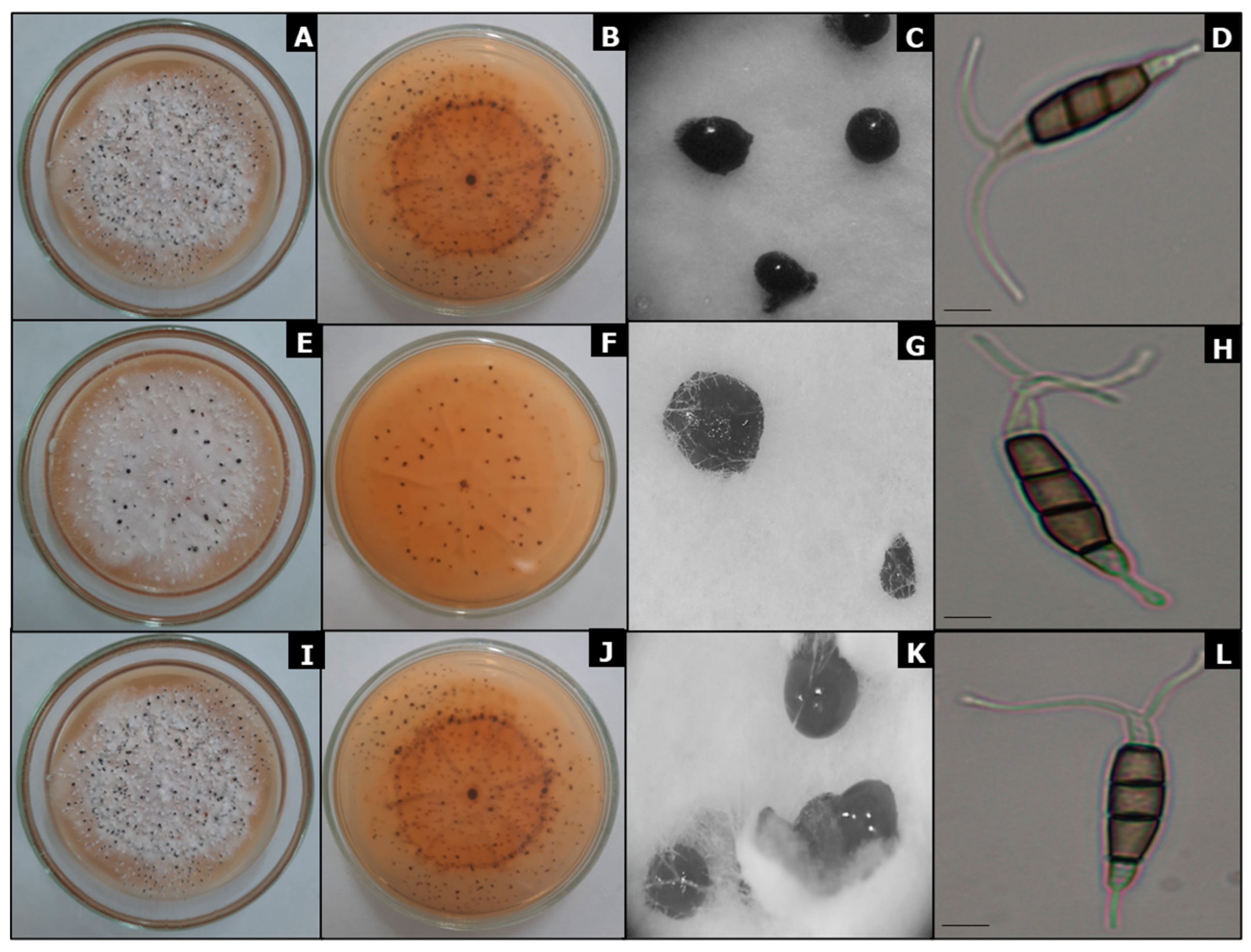
3.3. Cultural Characterization

The 25 isolates with ten days of growth in PDA were distributed in six morphotypes. Morphotype 1 had 10 isolates (40%) with a cotton texture, irregular edges, high and limited appearance, white-grey (above) and white-salmon (reverse) colony, and development of surface conidiomata. Morphotype 2 had five isolates (20%) with a cotton texture, irregular edges, high and limited appearance, white-cream (above) and white-salmon (reverse) colony, and development of surface conidiomata. Morphotype 3 had four isolates (16%) with a cotton texture, irregular edges, fat, and extended appearance, white-cream (above) and white-cream–red-orange (reverse) colony, and development of surface conidiomata. Morphotype 4 had three isolates (12%) with a cotton texture, irregular edges, high and limited appearance, white-grey (above) and white-grey (reverse) colony, and development of surface conidiomata. Morphotype 5 had two isolates (8%) with a cotton texture, irregular edges, fat, and extended appearance, white-cream (above) and white to salmon (reverse) colony, and development of surface conidiomata. Morphotype 6 had one isolate (4%) with a cotton texture, irregular edges, high and limited appearance, white-grey (above) and brown-reddish (reverse) colony, and development of surface conidiomata (Table 2).

3.4. Morphological Characterization

Based on the initial morphological characterization, the isolates belonged to two genera: Neopestalotiopsis (19 isolates) and Pestalotiopsis (six isolates); this classification was assigned based on the number of cells, the number of septa, and the color exhibited by the median cells of the conidia. Additionally, the length and width of the conidia, apical and basal appendages length, median cell length, and the number of basal and apical appendages were determined and are available in Table 2. Furthermore, a molecular analysis (see Section 3.5) enabled the identification of a third genus, known as Pseudopestalotiopsis (comprising three isolates that were initially classified under the genus Pestalotiopsis based solely on their morphological characteristics).
There were significant differences between genera for each biometric variable evaluated (p < 0.05). The measurements of the conidia are shown in Table 2. The isolates belonging to the genus Neopestalotiopsis showed fixed, solitary to slightly gregarious conidiomata, which were semi-immersive and brown to black in color. The conidial exudate was dark brown and globose. The conidia had a fusiform, ellipsoid shape and were straight to slightly curved, formed by five cells, and divided into four septa. Measurements of the length and width of the conidia ranged from (17.7) 23.7 to 24.3 (48.5) x ¯ ± SD = 24.0 ± 4.0 μm and (4.5) 6.3 to 6.4 (10.2) x ¯ ± SD = 6.4 ± 0.8 μm. The basal and apical cells were hyaline. The length of basal and apical appendage measurements ranged from (2.5) 4.9 to 5.0 (9.2) x ¯ ± SD = 4.9 ± 1.2 μm and (3.0) 16.9 to 17.6 (34.1) x ¯ ± SD = 17.2 ± 4.5 μm, respectively. The median cells were dark brown, clear brown, and olive-brown colored; they had 1–4 unbranched apical appendages and a single basal, tubular, unbranched appendage. The length of medium cell measurements ranged from (11.3) 15.6 to 16.1 (23.7) x ¯ ± SD = 15.8 ± 2.6 μm.
The isolates belonging to the genus Pestalotiopsis presented globose, clavate, solitary, or aggregated conidiomata, and a semi-immersed, dark brown to black mucilaginous appearance. The conidia presented an ellipsoid shape and were straight to slightly curved and slightly contracted at septa, with values that ranged between (23.2) 27.8 to 28.7 (33.1) x ¯ ± SD = 28.3 ± 2.1 μm length and (5.8) 6.6 to 6.8 (7.9) x ¯ ± SD = 6.7 ± 0.5 μm width. The conidia were made up of five cells and were divided into four septa: the apical and basal cells were hyaline with a cylindrical shape, and the median cells were concolorous brown, with septa darker than the rest of the cells ranging from (15.5) 17.9 to 18.5 (21.5) x ¯ ± SD = 18.2 ± 1.3 μm length; 2–3 unbranched filiform apical appendages ranging from (9.0) 14.7 to 15.7 (19.8) x ¯ ± SD = 15.2 ± 2.4 μm length; and one basal, simple, tubular, unbranched apical appendage that was (2.7) 4.4–4.9 (8.4) x ¯ ± SD = 4.6 ± 1.2 μm in length (Figure 3).
The conidiomata of Pseudopestalotiopsis isolates were immersed in a viscous, globose, and glossy mass. Morphologically, the conidia were fusiform, ellipsoid, straight to slightly curved, with four septa that were (22.7) 27.7–29.0 (40.5) x ¯ ± SD = 28.3 ± 3.2 μm in length × (4.1) 6.5–6.9 (8.8) x ¯ ± SD = 6.7 ± 1.0 μm in width; apical and basal hyaline cells; median cells that were concolorous pale brown and (16.7) 18.9–19.5 (23.8) x ¯ ± SD = 19.2 ± 1.5 μm in length with 2–4 tubular apical appendages (mainly three) that were unbranched and (14.8) 21.1–22.7 (31.5) x ¯ ± SD = 21.9 ± 3.9 μm in length; and a single basal appendage that was tubular, unbranched, centered, and (3.2) 4.8–5.4 (8.4) x ¯ ± SD = 5.1 ± 1.3 μm in length.

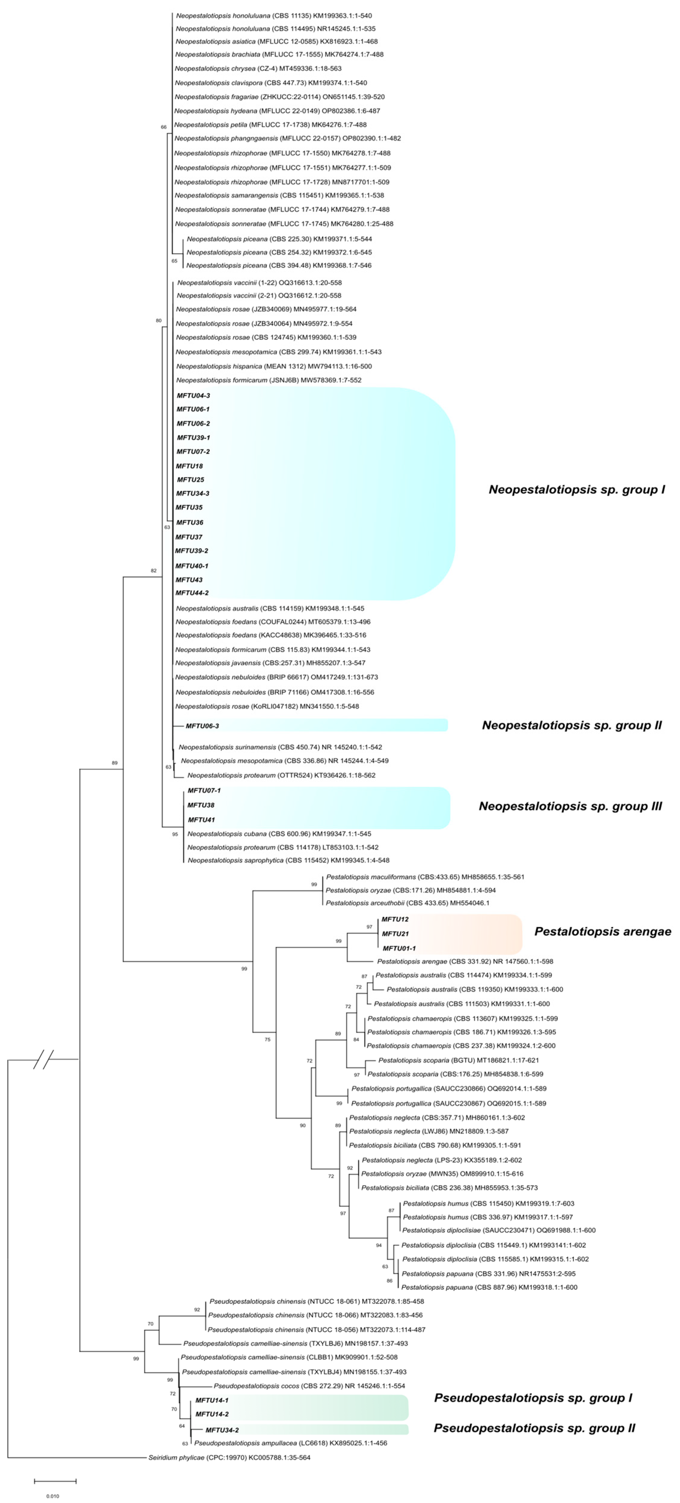
3.5. Molecular Characterization

The PCR for the 25 isolates produced amplicons for rDNA between 530 and 630 bp in size for ITS, 440–470 bp for TUB2, and 850–1030 bp for TEF1. Based on the initial comparison of our sequences against GenBank using BLAST, it was revealed that all isolates in this study belonged to three genera: there were three isolates of Pestalotiopsis (MFTU01-1, MFTU12, and MFTU21), three of Pseudopestalotiopsis (MFTU14-1, MFT014-2, and MFTU34-2), and 19 isolates of Neopestalotiopsis (MFTU04-3, MFTU06-1, MFTU06-2, MFTU06-3, MFTU07-1, MFTU07-2, MFTU18, MFTU25, MFTU34-3, MFTU35, MFTU36, MFTU37, MFTU38, MFTU39-1, MFTU39-2, MFTU40-1, MFTU41, MFTU43, and MFTU44-2) (Table 3). To establish the relationship between species within each genus, we utilized a concatenated dataset comprising 27 taxa for Pestalotiopsis, 8 taxa for Pseudopestalotiopsis, and 41 taxa for Neopestalotiopsis. The type strain of Seiridium phylicae (KC005788) served as the outgroup taxa (Figure 4).
The phylogenetic analysis allowed the grouping of three isolates corresponding to Pestalotiopsis (MFTU01-1, MFTU12, and MFTU21), with a relative support on the branches of 99% with the holotype strain of Pestalotiopsis arengae (CBS 331.92) that was reported by Maharachchikumbura (Supplementary Materials, Figure S1) [47]. For the Pseudopestalotiopsis group, the isolate MFTU34-2 formed one clade with Pseudopestalotiopsis ampullacea (NKT0P03) that was reported by Liu [48]. The isolates denominated MFTU14-1 and MFTU14-2 were grouped with Pseudopestalotiopsis cocos (CBS 272.29) that was obtained from Cocos nucifera in Indonesia and reported by Maharachchikumbura [47] (2014) (Supplementary Materials, Figure S2).
In the case of the 19 remaining isolates, they were distributed into three main clades with reliability support on the branch above 85% with different species of the genus Neopestalotiopsis. The total isolates of this group were clustered into three clades defined in this study as Neopestalotiopsis sp. I, Neopestalotiopsis sp. II, and Neopestalotiopsis sp. III. The isolates MFTU07-1, MFTU38, and MFTU41 were grouped with reference sequences like Neopestalotiopsis cubana (CBS 600.96), Neopestalotiopsis protearum (CBS 114178), and Neopestalotiopsis saprophytica (CBS 115452), which were previously published by Maharachchikumbura [47] and Monclova-Santana [49], respectively. In the phylogenetic tree for each genus, we found that MFTU07-1 formed a clade with the strain Neopestalotiopsis sp. 10 (CBS 110.20) [47]. The isolates MFTU06-3 clustered in a clade (II) with Neopestalotiopsis surinamensis (CBS 450.74). The isolates denominated MFTU04-3 and MFTU06-1 were grouped in the sub-clade (I) recently published by Solarte et al. (2018) [28] with Neopestalotiopsis sp. 14 (strain VR1ep) obtained from guava fruit in the Department of Valle del Cauca (Colombia) (Supplementary Materials, Figure S3). Also, MFTU41 was grouped with two published strains, VTman6 and SVsnp3, based on sequences from the same study.

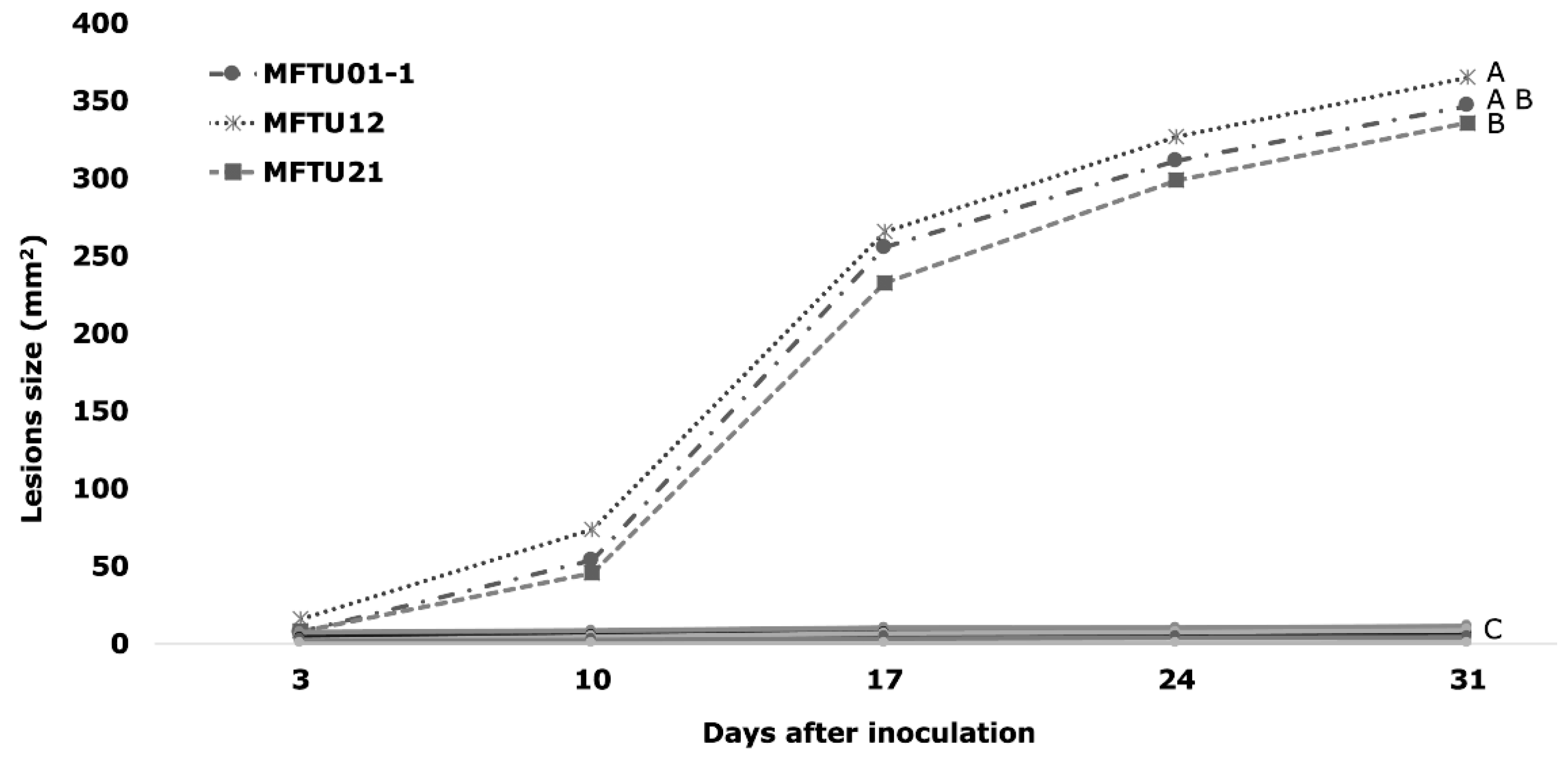
3.6. Pathogenicity Tests

To confirm pathogenicity, seedlings of the cultivar Sinú (cerete) × Deli Yangambí were inoculated with a mycelial PDA disc on wounded tissue. Cuvularia sp. caused small circular lesions that were initially chlorotic and then turned brown but always retained their circular development. The leaves inoculated with Colletotrichum sp. presented elongated brown necrotic lesions with irregular growth. Finally, Phoma sp. presented small yellowish-brown circular lesions with a reddish border [50]. The symptoms produced by these three pathogens were different from the symptoms observed in the leaf spot in this work (Supplementary Materials, Figure S4).
The 25 Pestalotiopsis isolates showed different degrees of aggressiveness, measured as lesion size (mm2). Three days after inoculation (dai), the first lesions were oval and brown, surrounded by a chlorotic halo like the symptoms in the field. The development of lesions beyond the inoculation site was observed only for three isolates (MFTU01-1, MFTU12, and MFTU21) (Figure 5), and the characteristics of the lesions were like those reported for the genus Pestalotiopsis by Suwannarach [36]. Only one isolate (MFTU04-3) induced small lesions but did not show development over time, reaching only 2% of the affected leaf surface. In addition, the cultivars Brazil × 7 pollen africans, Coarí × La Mé, and Manaos × Compacta were evaluated (Supplementary Materials, Figures S5–S8).
The isolates MFTU01-1, MFTU12, and MFTU21 initially induced circular or oval brown lesions surrounded by an irregular chlorotic halo. The lesions developed until 31 dai when they turned dark brown at the edge with grey tones at the center; in more advanced stages, the lesions presented a necrotic area in the center, as well as tissue loss and the development of concentric black-dot-like conidiomata; the chlorotic irregular halo was present in all stages of the diseases (Figure 5). There were significant differences in the size of the lesions (p < 0.05) compared with the other isolates. The average lesion sizes at the end of the test were 365, 346, and 336 mm2 for MFTU12, MFTU01-1, and MFTU21 isolates, respectively (Figure 6). Control plants did not show any development lesions. Re-isolation of the microorganism was obtained from the lesions, fulfilling Koch’s postulates.

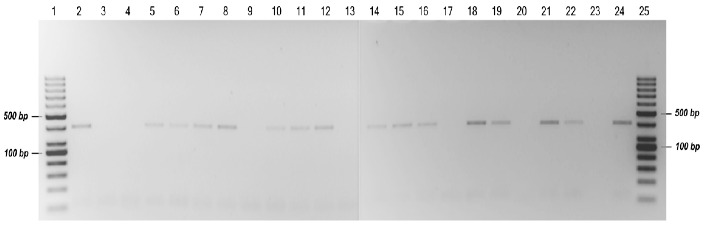
3.7. Molecular Detection of Pestalotiopsis arengae in Foliar Tissues

The specific primers designed in this study detected the presence of P. arengae in naturally infected leaf tissues from the field via PCR. The expected specific size of the PCR products was visible on an agarose gel for 15 of the 21 newly collected and evaluated samples (Figure 7). The comparative analysis of the sequenced samples against the GenBank database confirmed with an identity of 100% that P. arengae (E-value: 0.0) is the causal agent of leaf blight in the oil palm hybrid O × G in Colombia. The sequences of the amplicons obtained with the specific primers were deposited in GenBank under accession numbers MZ045516–MZ045530.

4. Discussion

This study revealed two important aspects of the disease known as leaf blight in the cultivation of oil palms in Colombia. The first one was the pathogenic association of the genus Pestalotiopsis with the development of foliar lesions in the interspecific O × G hybrid of oil palm. Secondly, not all fungal microorganisms obtained from foliar lesions such as blight are etiologically related to the direct development of the disease.
In Colombia, there is little information about the presence, prevalence, and etiology of the main foliar diseases that affect O × G hybrid cultivars. In this study, the genotypes planted in Tumaco, Colombian Southwest Palm Zone, had lesions that caused drying and necrosis of leaves, mainly in the lower leaves of the palms. In E. guineensis, several microorganisms have been associated with leaf spots, with a greater prevalence of Pestalotiopsis, which is considered the causal agent of leaf blight. In this sense, Jiménez and Reyes [12] conducted a study in the zone of Puerto Wilches (Santander, Colombia) and found that some Pestalotiopsis species in association with the insect Leptopharsa gibbicarina (Hemiptera: Tingidae) [15] can induce necrosis, drying, and defoliation because the insect generates the wounds necessary for the penetration and colonization of the pathogen. On the other hand, Martínez and Plata [51] reported the Pestalotiopsis genus as the cause of leaf blight. They associated it with the damage caused by several lepidopteran insects commonly found on E. guineensis in some plantations of Colombia. Although these authors pointed out the role of lepidopteran insects in the development of the disease, isolation and pathogenicity tests of Pestalotiopsis were not performed; thus, a direct association of the microorganism with the disease cannot be defined.
In our study, several fungal organisms were observed and isolated from the samples taken from the different hybrid cultivars in Tumaco, including Curvularia, Colletotrichum, Phoma, and Pestalotiopsis-like fungi. In Colombia, some references indicate the association of these fungal microorganisms with leaf spots in E. guineensis [12,52,53], but there is no information on their pathogenicity or aggressiveness in hybrid O × G cultivars. This association described in E. guineensis with Curvularia, Colletotrichum, and Phoma was based on observing initial lesions that did not continue a progressive and infective process over time (Jiménez and Reyes, 1977). In the pathogenicity test carried out in this research, placing the agar plugs with mycelial growth and conidiomata on the wound made in the tissue was an effective method for reproducing symptoms. This finding coincides with that reported by Elliott [54], Keith [30], and Solarte [28], indicating that Pestalotiopsis requires a wound to penetrate the plant, which is a necessary factor for the development and severity of the disease; for this reason, the microorganism is considered weak because it requires a facilitator in its infectious process [13,14,34,55].
Different Pestalotiopsis species have been reported to affect palms of E. guineensis in different countries. Shen [35] reported for the first time that P. microspora causes leaf spot diseases in China. Likewise, Suwannarach [36] reported P. theae as the causal agent of this foliar disease in Thailand. In this study, 3 isolates, denominated MFTU01-1, MFTU12, and MFTU21, out of 25 samples, were identified within the genus Pestalotiopsis. These isolates exhibited morphological characteristics consistent with Pestalotiopsis arengae and were further confirmed through molecular tools [47]. For the species delimitation, a comparison of the ITS sequence of our isolates with P. arengae (CBS 331.92) revealed that 590/593 base pairs are similar with only two gaps between them and a query cover up to 98% [56]. Our isolates initiated the infective process and developed lesions in the inoculated plants. The species of P. arengae in the O × G hybrid oil palm constitutes a new record for this host and fungi species as they have yet to be reported earlier on this crop. Due to the low number of pure isolates, it was necessary to implement other methodologies, such as PCR, to detect the presence or absence of this microorganism. The low isolation rate is attributed to the presence of other secondary and opportunistic microorganisms in the affected tissues. These microorganisms were able to grow in the culture media used in our study because it was not selective for the isolation of P. arengae. The direct association of the presence of P. arengae in affected leaf tissue from the field was confirmed via molecular detection using PCR with species-specific primers designed in this study. The species registered as the causal agent of leaf blight in Colombia has been previously reported in Singapore as Arenga undulatifolia by Maharachchikumbura [47], and it is closely related to P. anacardiacearum isolated from mango in China [46] and P. hawaiiensis isolated from Leucospermum sp. in Hawaii [46].
The differentiation of Pestalotiopsis species with morphological characteristics is complicated due to the overlapping ranges of values. However, our pathogenic morphotypes obtained from the O × G hybrid oil palm present similar characteristics to those obtained and reported in another palm species, Arenga undulatifolia [47]. In the PDA culture medium, the colonies of MFTU01-1, MFTU12, and MFTU21 (Figure 3) exhibited cream and pale white tones on the top and the bottom of the plate; the margins were wavy with slightly aerial mycelium and gregarious conidiomata; and the conidia had a fusiform, ellipsoid, straight to slightly curved shape and were formed by five cells and four septa. The apical and basal hyaline cells were cylindrical, and the median cells were concolorous. The conidia had 2–3 unbranched filiform apical appendages and one simple, tubular, unbranched basal appendage. In contrast, differences in conidia width and the apical and basal appendage length were observed. Maharachchikumbura [47] reported that P. arengae conidia were 7–9.5 (10) μm wide, x ¯ ± SD = 8 ± 0.4 μm, while the reproductive structures reported in this work were slightly thinner at (5.8) 6.6–6.8 (7.9) x ¯ ± SD = 6.7 ± 0.5 μm. The apical appendages observed in the isolates MFTU01-1, MFTU12, and MFTU21 were (9.0) 14.7–15.7 (19.8) x ¯ ± SD = 15.2 ± 2.4 μm in length, which are longer than those reported in A. undulatifolia ((4) 4.5–11 (12) μm long, x ¯ ± SD = 7.3 ± 1.3 μm). The length of the basal appendage ((2.7) 4.4–4.9 (8.4) x ¯ ± SD = 4.6± 1.2 μm) is longer than that reported for the morphotype distributed in Singapore (1.5–3 μm). The conidia length ((23.2) 27.8–28.7 (33.1) x ¯ ± SD = 28.3 ± 2.1 μm) and long media cells ((15.5) 17.9–18.5 (21.5) x ¯ ± SD = 18.2 ± 1.3 μm) were similar to those recorded by Maharachchikumbura [47] for P. arengae ((24) 25–32 (33) x ¯ ± SD = 27.6 ± 2 μm for the conidia length and (17) 17.5–21.5 (22) μm long, x ¯ ± SD = 19 ± 1.3 μm, for the median cells).
Through the combined sequence analysis of ITS, TUB2, and TEF1-α genes, it was possible to resolve the identity of a few species of the Neopestalotiopsis genus obtained in this study; however, some isolates were not grouped with any previously reported sequences. These isolates formed clades denominated in our study as Neopestalotiopsis sp. I and III and could be defined as new species; similar results were obtained in Colombia by Solarte [28] and in Mexico by Gerardo-Lugo [57]. For the other isolates grouped in the genus Pseudopestalotiopsis, it was possible to resolve their taxonomic identification by combining the analyses of the three genes and considering their phylogenetic grouping with two previously reported species.
This is the first report of Pestalotiopsis arengae causing foliar lesions in the O × G hybrid in Colombia or elsewhere. In fact, this species has not yet been associated with damage or leaf spots in other crops of agricultural importance in Colombia. In this study, we also described the use of PCR to detect and confirm the presence of P. arengae from affected foliar tissues. Likewise, we showed the diversity of species of the genus Neopestalotiopsis and Pseudopestalotiopsis without any detrimental effect on the leaves of the O × G hybrid in Colombia. In future studies, we expect to analyze the association of the Neopestalotiopsis and Pseudopestalotiopsis isolates with the development of lesions in joint inoculations with the isolates of Pestalotiopsis to continue studying their biology, epidemiology, and the economic impact on the oil palm crops.

Supplementary Materials

The following supporting information can be downloaded at: https://www.mdpi.com/article/10.3390/jof10010024/s1.

Author Contributions

Conceptualization, W.F.B.-O. and G.A.S.-V.; methodology, G.A.S.-V.; software validation, F.H.V.; formal analysis, G.A.S.-V. and J.L.P.-A.; investigation, W.F.B.-O., H.C.M.-C. and J.L.P.-A.; data curation, W.F.B.-O., G.A.S.-V., H.C.M.-C. and J.L.P.-A.; writing—original draft preparation, W.F.B.-O., J.L.P.-A., H.C.M.-C., Y.A.M.-G., A.M.-R. and G.A.S.-V.; writing—review and editing, J.L.P.-A. and G.A.S.-V.; supervision, project administration, G.A.S.-V.; funding acquisition, G.A.S.-V. and A.M.-R. All authors have read and agreed to the published version of the manuscript.

Funding

This research was funded by the Oil Palm Promotion Fund (FFP) administrated by Fedepalma, Colombia. Grant number 2020203.

Institutional Review Board Statement

Not applicable.

Informed Consent Statement

Not applicable.

Data Availability Statement

Data are contained within the article and supplementary materials.

Acknowledgments

We thank Tumaco’s plantation managers, engineers, and field staff for contributing to this research’s development. In addition, we thank Eloina Mesa and Andres Tupaz for their collaboration and contribution to the analysis. Finally, we thank Sajeewa Maharachchikumbura for the corrections and revisions to the article.

Conflicts of Interest

The authors declare that they have no conflicts of interest. The founders had no role in the design of the study; in data acquisition, analyses, or interpretation of the data; in the writing of the manuscript; or in the decision to publish the results.

References

  1. Corley, R.; Tinker, B. Origin and development of oil palm. In The Oil Palm, 4th ed.; Bath Press: Bath, UK; Blackwell Science Ltd.: Chichester, UK, 2003; p. 604. [Google Scholar]
  2. Potter, L. Colombia’s oil palm development in times of war and ‘peace’: Myths, enablers and the disparate realities of land control. J. Rural. Stud. 2020, 78, 491–502. [Google Scholar] [CrossRef]
  3. Federación Nacional de Cultivadores de Palma de Aceite, F Minianuario estadístico 2022: Principales Cifras de la Agroindustria de Palma de Aceite en Colombia. Colombia. 19 May 2022. pp. 2344–8482. Available online: https://repositorio.fedepalma.org/handle/123456789/141446 (accessed on 10 March 2023).
  4. Sarria, G.; Torres, G.; Velez, D.; Rodríguez, J.; Noreña, C.; Varón, F.; Coffey, M.D.; Elliott, M.L.; Martínez, G.; Castro, B.L. Caracterización morfológica y molecular de Phytophthora palmivora agente causal de las lesiones iniciales de la pudrición del cogollo (PC) de la palma de aceite en Colombia. Fitopatol Colomb. 2008, 32, 39–44. [Google Scholar]
  5. Martínez, G.; Arango, M.; Torres, G.; Sarria, G.; Vélez, D.; Rodríguez, J.; Mestizo, Y.; Aya, H.; Noreña, C.; Varón, F.; et al. Advances in the Research of the Two Most Important Diseases on Oil Palm in Colombia: Bud Rot and Lethal Wilt. Palmas 2013, 34, 39–47. [Google Scholar]
  6. Meunier, J. Una posible solucion genetica para el control de la pudricion de cogollo en la palma aceitera. Hibrido interespecífico Elaeis oleifera × Elaeis guineensis. Palmas 1991, 12, 39–42. [Google Scholar]
  7. Bastidas, S.; Peña, E.; Reyes, R. Avances sobre el comportamiento de los híbridos de primera generación de retrocruzamiento entre palma Nolí (Elaeis oleifera) y palma africana (Elaeis guineensis). Corporación Colomb. Investig. Agropecu. CORPOICA Rev. Reg. Noved. Técnicas 2003, 3, 32–36. [Google Scholar]
  8. Torres, V.; Rey, B.; Gelves, R.; Santacruz, A.L. Evaluación del comportamiento de los híbridos interespecíficos elaeis oleífera × Elaeis guineensis, en la plantación de Guaicaramo S. A. Rev. Palmas 2004, 25, 350–357. [Google Scholar]
  9. Zambrano, J.E. Los híbridos interespecíficos elaeis oleífera HBK. x Elaeis guineensis Jacq.: Una alternativa de renovación para la Zona Oriental de Colombia. Rev. Palmas 2004, 25, 339–349. [Google Scholar]
  10. Chinchilla, C.; Alvarado, A.; Albertazzi, H.; Torres, R. Tolerancia y resistencia a las pudriciones del cogollo en fuentes de diferente origen de Elaeis guineensis. Rev. Palmas 2007, 28, 273–284. [Google Scholar]
  11. Sánchez, A. Estado fitosanitario de los cultivos de coco y palma africana en los litorales del Atlántica y Pacifico (Colombia). Inst. Fom. Algodón. Programa Fitopatol. 1962, 55. [Google Scholar]
  12. Jiménez, O.D.; Reyes, A. Estudio de una necrosis foliar que afecta varias plantaciones de palma de aceite (Elaeis guineensis Jacq.) en Colombia. Rev. Fitopatol. Colomb. 1977, 6, 15–32. [Google Scholar]
  13. Genty, P.; Garzon, A.; Garcia, R. Daños y control del complejo Leptopharsa-Pestalotiopsis en la palma africana. Rev. Palmas 1984, 5, 9–15. [Google Scholar]
  14. Reyes, A. Añublo foliar de la palma africana (Elaeis guineensis Jacq) en Colombia: Importancia económica, etiología y control. Rev. Palmas 1988, 9, 33–39. [Google Scholar]
  15. Guzmán, L.; Calvache, H.; Aldana, J.; Méndez, A. Manejo de Leptopharsa gibbicarina Froeschner (Hemíptera: Tingidae) con la hormiga Crematogaster sp. en una plantación de palma de aceite. Rev Palmas 1997, 18, 19–26. [Google Scholar]
  16. Méndez, A. Manejo integrado de la Pestalotiopsis en una plantación comercial de palma de aceite. Rev. Palmas 2000, 21, 1–5. [Google Scholar]
  17. Aldana de La Torre, R.C.; Aldana de La Torre, J.A. Reconocimiento y manejo de insectos defoliadores y asociados a la pestalotiopsis. Tecnologías para la agroindustria de la palma de aceite: Guía para facilitadores. Palmas 2011, 180, 71–111. [Google Scholar]
  18. Barrios, C.; Bustillo-Pardey, A.E.; Ocampo, K.; Reina, M.A.; Alvarado, H.L. Eficacia de hongos entomopatógenos en el control de L. gibbicarina (Hemipetra: Tingidae) en palma de aceite. Rev. Colomb. Entomol. 2016, 42, 22–27. [Google Scholar] [CrossRef]
  19. Jeewon, R.; Liew, E.C.Y.; Simpson, J.A.; Hodgkiss, J.H.K. Phylogenetic significance of morphological characters in the taxonomy of Pestalotiopsis species. Mol. Phylogenet. Evol. 2003, 27, 372–383. [Google Scholar] [CrossRef]
  20. Jeewon, R.; Liew, E.C.Y.; Hyde, K.D. Phylogenetic evaluation of species nomenclature of Pestalotiopsis in relation to host association. Fungal. Divers. 2004, 17, 39–55. [Google Scholar]
  21. Maharachchikumbura, S.S.N.; Guo, L.D.; Cai, L.; Chukeatirote, E.; Wu, W.P.; Sun, X.; Crous, P.W.; Bhat, D.J.; McKenzie, E.H.C.; Bahkali, A.H.; et al. A multi-locus backbone tree for Pestalotiopsis, with a polyphasic characterization of 14 new species. Fungal Divers. 2012, 56, 95–129. [Google Scholar] [CrossRef]
  22. Ayoubi, N.; Soleimani, M.J. Strawberry Fruit Rot Caused by Neopestalotiopsis iranensis sp. nov., and N. mesopotamica. Curr. Microbiol. 2015, 72, 329–336. [Google Scholar] [CrossRef]
  23. White, T.J.; Bruns, T.; Lee, S.; Taylor, J. Amplification and Direct Sequencing of Fungal Ribosomal RNA Genes for Phylogenetics. In PCR Protocols; Academic Press: Cambridge, MA, USA, 1990; pp. 315–322. [Google Scholar]
  24. Glass, N.L.; Donaldson, G.C. Development of primer sets designed for use with the PCR to amplify conserved genes from filamentous ascomycetes. Appl. Environ. Microbiol. 1995, 61, 1323–1330. [Google Scholar] [CrossRef]
  25. Carbone, I.; Kohn, L.M. A method for designing primer sets for speciation studies in filamentous ascomycetes. Mycologia 1999, 91, 553–556. [Google Scholar] [CrossRef]
  26. Bate-Smith, E. Leuco-anthocyanins. 3. The nature and systematic distribution of tannin in dicotyledonous plants. Bot. J. 1957, 55, 669–705. [Google Scholar] [CrossRef]
  27. Das, R.; Chutia, M.; Das, K.; Jha, D.K. Factors affecting sporulation of Pestalotiopsis disseminata causing grey blight disease of Persea bombycina Kost., the primary food plant of muga silkworm. Crop. Prot. 2010, 29, 963–968. [Google Scholar] [CrossRef]
  28. Solarte, F.; Muñoz, C.G.; Maharachchikumbura, S.S.N.; Álvarez, E. Diversity of neopestalotiopsis and pestalotiopsis spp., causal agents of guava scab in Colombia. Plant Dis. 2018, 102, 49–59. [Google Scholar] [CrossRef]
  29. Ismail, A.M.; Cirvilleri, G.; Polizzi, G. Characterisation and pathogenicity of Pestalotiopsis uvicola and Pestalotiopsis clavispora causing grey leaf spot of mango (Mangifera indica L.) in Italy. Eur. J. Plant. Pathol. 2013, 135, 619–625. [Google Scholar] [CrossRef]
  30. Keith, L.M.; Velasquez, M.E.; Zee, F.T.; Plant, T.; Resource, G.; Unit, M. Identification and Characterization of Pestalotiopsis spp. Causing Scab Disease of Guava, Psidium guajava, in Hawaii. Plant Dis. 2006, 90, 16–23. [Google Scholar] [CrossRef]
  31. Maharachchikumbura, S.S.N.; Guo, L.; Chukeatirote, E.; Mckenzie, E.H.C.; Hyde, K.D. A destructive new disease of Syzygium samarangense in Thailand caused by the new species Pestalotiopsis samarangensis. Trop. Plant. Pathol. 2013, 38, 227–235. [Google Scholar] [CrossRef]
  32. Maharachchikumbura, S.S.N.; Larignon, P.; Hyde, K.D.; Al-sadi, A.M.; Liu, Z.Y. Characterization of Neopestalotiopsis, Pestalotiopsis and Truncatella species associated with grapevine trunk diseases in France. Phytopathol. Mediterr. 2016, 55, 380–390. [Google Scholar]
  33. Maharachchikumbura, S.; Guo, L.; Liu, Z.; Hyde, K. Pseudopestalotiopsis ignota and Ps. camelliae spp. nov. associated with grey blight disease of tea in China. Mycol. Prog. 2016, 15, 1–7. [Google Scholar] [CrossRef]
  34. Jimenéz, O. El Añublo Foliar de la Palma Africana en Colombia. Palmas 1984, 5, 89–92. [Google Scholar]
  35. Shen, H.; Zhang, J.; Lin, B.; Pu, X. First Report of Pestalotiopsis microspore Causing Leaf Spot of Oil Palm (Elaeis guineensis) in China. Am. Phytopathol. Soc. 2014, 98, 1429. [Google Scholar]
  36. Suwannarach, N.; Sujarit, K.; Kumla, J.; Bussaban, B.; Lumyong, S. First report of leaf spot disease on oil palm caused by Pestalotiopsis theae in Thailand. J. Gen. Plant. Pathol. 2013, 79, 277–279. [Google Scholar] [CrossRef]
  37. Fröhlich, J.; Hyde, D.K.; Guest, D.I. Fungi associated with leaf spots of palms in north Queensland, Australia. Mycol. Res. 1997, 101, 721–732. [Google Scholar] [CrossRef]
  38. Loredo, J.; Mena, J.; Ríos, J. Manual De Prácticas Del Laboratorio De Fitopatología; Universidad Autónoma De Sinaloa Unidad Regional Norte: Los Mochis, Mexico, 2009; p. 81. [Google Scholar]
  39. Khadidja, K. Isolation, Characterization of Phytopathogenic Fungi Alternaria sp., and Physico-Chemical Study; Faculty of Natural and Life Science Departement of Biologie: Mostaghanem, Algeria, 2020. [Google Scholar]
  40. Barnett, H.L.; Hunter, B.B. Illustrated Genera de Imperfect Fungi, 4th ed.; The American Phytopathological Society: St. Paul. MN, USA, 1988; 218p. [Google Scholar]
  41. Agrios, G.N. Plant Pathology; Elsevier Academia Press: Cambridge, MA, USA, 2005; p. 922. [Google Scholar]
  42. Steyaert, R. Contribution a l’etude monographique de Pestalotia de Not. et Monochaetia Sacc. (Truncatella gen. nov. et Pestalotiopsis gen. nov.). Plant Dis. 1949, 19, 285–354. [Google Scholar] [CrossRef]
  43. Hall, T.A. BioEdit: A user friendly biological seque. In Nucleid Acids Symposium Series; Oxford University Press: Oxford, UK, 1999; pp. 95–98. [Google Scholar]
  44. Tamura, K.; Stecher, G.; Peterson, D.; Filipski, A.; Kumar, S. MEGA6: Molecular evolutionary genetics analysis version 6.0. Mol. Biol. Evol. 2013, 30, 2725–2729. [Google Scholar] [CrossRef]
  45. Sosa, N.T.; Alvarez, R.; Cabrera, M. Ocurrencia de Pestalotia sp. causando lesiones necróticas en plantas de Jazmín del Cabo (Gardenia Augusta); Universidad Nacional del Nordeste Comunicación es Científicas y Tecnológicas: Corrientes, Argentina, 2003; pp. 1–3. Available online: https://www.unne.edu.ar/unnevieja/Web/cyt/cyt/2003/comunicaciones/05-Agrarias/A-019.pdf (accessed on 22 February 2018).
  46. Jayawardena, R.S.; Zhang, W.; Liu, M.; Maharachchikumbura, S.S.N.; Zhou, Y.; Huang, J.; Nilthong, S.; Wang, Z.; Li, X.; Yan, J.; et al. Identification and characterization of Pestalotiopsis-like fungi related to grapevine diseases in China. Fungal. Biol. 2015, 119, 348–361. [Google Scholar] [CrossRef]
  47. Maharachchikumbura, S.S.N.; Hyde, K.D.; Groenewald, J.; Xu, Z.; Crous, P.W. Pestalotiopsis revisited. Stud Mycol. 2014, 79, 121–186. [Google Scholar] [CrossRef]
  48. Liu, F.; Hou, L.; Raza, M.; Cai, L. Pestalotiopsis and allied genera from Camellia, with description of 11 new species from China. Sci. Rep. 2017, 7, 866. [Google Scholar] [CrossRef]
  49. Monclova-Santana, C. Pathogenic Threats of ten Endangered Plant Species of the Karst Region of Puerto Rico; University of Puerto Rico, Mayagüez Campus: Mayagüez, Puerto Rico, 2015. [Google Scholar]
  50. Pineda, B.; Martínez, G. Reconocimiento de enfermedades en la palma de aceite. Tecnologías para la agroindustria de la palma de aceite: Guía para facilitadores. Rev. Cenipalma. 2013, 138. [Google Scholar]
  51. Martínez, L.; Plata, A. Lepidoptera vectors of Pestalotiopsis fungal disease: First record in oil palm plantations from Colombia [Internet]. Trop. Insect Sci. 2013, 33, 239–246. Available online: https://aplicaciones.msp.gob.ec/upload/upload/00000403_2011_00000403.PDF (accessed on 14 November 2022). [CrossRef]
  52. Turner, P. Oil Palm Disease and Disorders; Oxford University Press: Kuala Lumpur, Malaysia, 1981; p. 280. [Google Scholar]
  53. Sánchez, A. Enfermedades de la palma de aceite en América Latina. Palmas 1990, 1, 5–38. [Google Scholar]
  54. Elliott, M.L. Pestalotiopsis (Pestalotia) Diseases of Palm; University of Florida, Institute of Food and Agricultural Sciences: Gainesville, FL, USA, 2019; Volume 2006, pp. 2–4. [Google Scholar]
  55. Maharachchikumbura, S.S.N.; Guo, L.-D.; Chukeatirote, E.; Bahkali, A.H.; Hyde, K.D. Pestalotiopsis—Morphology, phylogeny, biochemistry and diversity. Fungal. Divers. 2011, 50, 173–175. [Google Scholar] [CrossRef]
  56. Jeewon, R.; Hyde, K.D. Establishing species boundaries and new taxa among fungi: Recommendations to resolve taxonomic ambiguities. Mycosphere 2016, 7, 1669–1677. [Google Scholar] [CrossRef]
  57. Gerardo-Lugo, S.S.; Tovar-Pedraza, J.M.; Maharachchikumbura, S.S.N.; Apodaca-Sánchez, M.A.; Correia, K.C.; Sauceda-Acosta, C.P.; Camacho-Tapia, M.; Hyde, K.D.; Marraiki, N.; Elgorban, A.M.; et al. Characterization of neopestalotiopsis species associated with mango grey leaf spot disease in Sinaloa, Mexico. Pathogens 2020, 9, 788. [Google Scholar] [CrossRef]
Figure 1. Location of oil palm plantations containing the interspecific hybrid O × G sampled for Pestalotiopsis or leaf blight.
Jof 10 00024 g001
Figure 2. Development of foliar lesions in hybrid oil palm. (A) (top) and (B) (underside): Oval or elongated brown lesions surrounded by irregular chlorotic halo. (C): Progressive lesion delimited by a dark brown halo and accompanied by a chlorotic halo. (D): Coalesced irregular lesions forming concentric zones. (E): Chlorosis of leaflets followed by drying.
Jof 10 00024 g002
Figure 3. Pestalotiopsis arengae. From left to right, sporulation in Petri dishes containing potato dextrose agar (front and back, respectively), conidiomata in PDA, and conidia. Scale bars = 5 µm. Isolates: MFTU01-1 (AD); MFTU12 (EH); and MFTU21 (IL). (AJ): View of colony growth in Petri dish; upper- and underside. (CK): Conidiomata. (DL): Conidia.
Jof 10 00024 g003
Figure 4. Phylogenetic tree obtained using the neighbor-joining method of the consensus of the ITS region of some species of the genera Pestalotiopsis, Pseudopestalotiopsis, and Neopestalotiopsis. The evolutionary distances were computed using the Kimura 2-parameter method. This analysis involved 103 nucleotide sequences. There were a total of 464 positions in the final dataset. Evolutionary analyses were conducted in MEGA X. The isolates corresponding to this work are indicated in bold. The species Seiridium phylicae (CPC:19970) was included as an outgroup.
Jof 10 00024 g004
Figure 5. Pathogenicity test of Pestalotiopsis isolates. Leaf spot after inoculation, from left to right, 3, 17, and 31 days after inoculation. Isolates: MFTU01-1 (AC); MFTU12 (DF); and MFTU21 (GI).
Jof 10 00024 g005
Figure 6. Development of symptoms in plants of the genotype Cereté × Deli after inoculation with Pestalotiopsis isolates. Different letters show significant differences according to the Tukey test (p < 0.05).
Jof 10 00024 g006
Figure 7. Agarose gel electrophoresis of PCR-amplified products using the specific primers Parenga155ITSF and Parenga534ITSR. Lanes 1 and 25–100 bp DNA ladder marker; lanes 2–22—amplified results with DNA samples from affected foliar tissues from the field; lane 23—negative control (healthy foliar tissue); and lane 24—positive control (isolate MFTU01-1).
Jof 10 00024 g007
Table 1. Information on Pestalotiopsis, Neopestalotiopsis, and Pseudopestalotiopsis isolates obtained from the leaf blight of the oil palm hybrid (O × G).
Isolate CodeGenusHost A Source Geographic Origin
MFTU01-1 Pestalotiopsis sp. Sinú (cerete) × Deli Yangambí Leaf Tumaco, Nariño, Colombia
MFTU04-3 Neopestalotiopsis sp. Brazil × 7 polen africans Leaf Tumaco, Nariño, Colombia
MFTU06-1 Neopestalotiopsis sp. Brazil × 7 polen africans Leaf Tumaco, Nariño, Colombia
MFTU06-2 Neopestalotiopsis sp. Brazil × 7 polen africans Leaf Tumaco, Nariño, Colombia
MFTU06-3 Neopestalotiopsis sp. Brazil × 7 polen africans Leaf Tumaco, Nariño, Colombia
MFTU07-1 Neopestalotiopsis sp. Brazil × 7 polen africans Leaf Tumaco, Nariño, Colombia
MFTU07-2 Neopestalotiopsis sp. Brazil × 7 polen africans Leaf Tumaco, Nariño, Colombia
MFTU12 Pestalotiopsis sp. Sinú (cerete) × Deli Yangambí Leaf Tumaco, Nariño, Colombia
MFTU14-1 Pseudopestalotiopsis sp. Coarí × La Mé Leaf Tumaco, Nariño, Colombia
MFTU14-2 Pseudopestalotiopsis sp. Coarí × La Mé Leaf Tumaco, Nariño, Colombia
MFTU18 Neopestalotiopsis sp. Manaos × Compacta Leaf Tumaco, Nariño, Colombia
MFTU21 Pestalotiopsis sp. Coarí × La Mé Leaf Tumaco, Nariño, Colombia
MFTU25 Neopestalotiopsis sp. Coarí × La Mé Leaf Tumaco, Nariño, Colombia
MFTU34-2 Pseudopestalotiopsis sp. Brazil × 7 polen africans Leaf Tumaco, Nariño, Colombia
MFTU34-3 Neopestalotiopsis sp. Brazil × 7 polen africans Leaf Tumaco, Nariño, Colombia
MFTU35 Neopestalotiopsis sp. Coarí × La Mé Leaf Tumaco, Nariño, Colombia
MFTU36 Neopestalotiopsis sp. Coarí × La Mé Leaf Tumaco, Nariño, Colombia
MFTU37 Neopestalotiopsis sp. Coarí × La Mé Leaf Tumaco, Nariño, Colombia
MFTU38 Neopestalotiopsis sp. Coarí × La Mé Leaf Tumaco, Nariño, Colombia
MFTU39-1 Neopestalotiopsis sp. Coarí × La Mé Leaf Tumaco, Nariño, Colombia
MFTU39-2 Neopestalotiopsis sp. Coarí × La Mé Leaf Tumaco, Nariño, Colombia
MFTU40-1 Neopestalotiopsis sp. Coarí × La Mé Leaf Tumaco, Nariño, Colombia
MFTU41 Neopestalotiopsis sp. Coarí × La Mé Leaf Tumaco, Nariño, Colombia
MFTU43 Neopestalotiopsis sp. Coarí × La Mé Leaf Tumaco, Nariño, Colombia
MFTU44-2 Neopestalotiopsis sp. Coarí × La Mé Leaf Tumaco, Nariño, Colombia
A Oil palm genotype where infected foliar tissue was collected.
Table 2. Cultural and morphological characteristics of 19 Neopestalotiopsis, 3 Pestalotiopsis, and 3 Pseudopestalotiopsis isolates obtained from the leaf blight of the oil palm hybrid.
Isolate CodeGenus/SpeciesColony Type CConidia Length (µm)Conidia Width (µm)Length of Basal Appendage (µm)Median Cell Length (μm)No. of Apical Appendages (Range)Length of Apical Appendages (µm)Color of Middle Cells
MFTU01-1Pestalotiopsis sp.328.1 ± 1.6 Ba D6.6 ± 0.5a5.6 ± 1.3b18.2 ± 1.2b3–215.1 ± 2.6cConcolorous
MFTU04-3Neopestalotiopsis sp.524.4 ± 1.9b6.3 ± 0.6b5.4 ± 1.1ab16.3 ± 1.3c3–219.8 ± 3.5bVersicolorous
MFTU06-1Neopestalotiopsis sp.324.4 ± 1.1b6.5 ± 0.4b6.2 ± 0.9ab16.0 ± 1.3c3–223.3 ± 2.9bVersicolorous
MFTU06-2Neopestalotiopsis sp.228.8 ± 2.8b7.1 ± 0.5b5.1 ± 1.0ab18.7 ± 1.3c3–112.7 ± 2.7bVersicolorous
MFTU06-3Neopestalotiopsis sp.121.7 ± 1.8b5.9 ± 0.4b5.7 ± 1.1ab13.9 ± 1.2c3–217.0 ± 2.5bVersicolorous
MFTU07-1Neopestalotiopsis sp.222.0 ± 1.7b5.7 ± 0.4b4.6 ± 0.7ab14.8 ± 1.2c3–216.0 ± 2.2bVersicolorous
MFTU07-2Neopestalotiopsis sp.223.2 ± 1.5b5.7 ± 0.3b5.1 ± 0.9ab15.1 ± 1.1c3–218.0 ± 2.8bVersicolorous
MFTU12Pestalotiopsis sp.328.1 ± 2.3a6.5 ± 0.4a3.7 ± 0.6b17.8 ± 1.3b3–214.7 ± 2.4cConcolorous
MFTU14-1Pseudopestalotiopsis sp.426.1 ± 1.5a7.6 ± 0.5a4.2 ± 0.3a18.9 ± 1.0a4–219.2 ± 3.0aConcolorous
MFTU14-2Pseudopestalotiopsis sp.427.1 ± 1.4a6.7 ± 0.4a4.5 ± 0.5a18.8 ± 1.1a3–22.5 ± 3.8aConcolorous
MFTU18Neopestalotiopsis sp.520.9 ± 1.4b6.2 ± 0.3b4.9 ± 0.8ab13.7 ± 0.9c4–214.6 ± 2.5bVersicolorous
MFTU21Pestalotiopsis sp.328.6 ± 2.4a6.9 ± 0.4a4.7 ± 0.5b18.5 ± 1.2b3–215.7 ± 1.9cConcolorous
MFTU25Neopestalotiopsis sp.121.1 ± 1.3b6.4 ± 0.4b3.7 ± 0.5ab14.1 ± 0.9c3–220.1 ± 4.5bVersicolorous
MFTU34-2Pseudopestalotiopsis sp.431.7 ± 3.2a5.6 ± 0.5a6.6 ± 1.3a19.9 ± 1.9a3–222.9 ± 3.6aConcolorous
MFTU34-3Neopestalotiopsis sp.221.8 ± 1.7b5.8 ± 0.5b5.5 ± 1.0ab14.0 ± 0.9c3–221.7 ± 2.9bVersicolorous
MFTU35Neopestalotiopsis sp.122.9 ± 1.2b6.7 ± 0.5b5.1 ± 0.7ab14.9 ± 0.8c3–222.2 ± 5.0bVersicolorous
MFTU36Neopestalotiopsis sp.121.4 ± 1.2b6.5 ± 0.4b4.9 ± 0.6ab14.2 ± 0.8c3–221.3 ± 2.3bVersicolorous
MFTU37Neopestalotiopsis sp.119.8 ± 1.8b6.3 ± 0.4b5.4 ± 0.8ab13.3 ± 1.3c4–215.3 ± 2.8bVersicolorous
MFTU38Neopestalotiopsis sp.120.6 ± 1.1b5.9 ± 0.5b5.6 ± 0.9ab13.6 ± 0.9c4–221.3 ± 3.6bVersicolorous
MFTU39-1Neopestalotiopsis sp.121.9 ± 2.2b5.9 ± 0.5b5.7 ± 1.1ab14.5 ± 1.3c3–218.1 ± 2.9bVersicolorous
MFTU39-2Neopestalotiopsis sp.121.7 ± 1.7b5.3 ± 0.4b5.6 ± 0.7ab13.8 ± 1.2c3–215.9 ± 1.8bVersicolorous
MFTU40-1Neopestalotiopsis sp.627.8 ± 1.8b7.2 ± 0.7b3.5 ± 0.4ab19.1 ± 1.3c3–212.7 ± 1.9bVersicolorous
MFTU41Neopestalotiopsis sp.223.5 ± 1.9b7.1 ± 0.5b3.4 ± 0.5ab15.9 ± 1.1c3–113.7 ± 1.8bVersicolorous
MFTU43Neopestalotiopsis sp.121.5 ± 1.3b6.1 ± 0.3b4.9 ± 0.6ab14.1 ± 0.8c3–219.8 ± 2.7bVersicolorous
MFTU44-2Neopestalotiopsis sp.121.9 ± 1.8b6.1 ± 0.4b5.9 ± 0.9ab14.4 ± 1.0c3–219.5 ± 2.9bVersicolorous
a–c: The averages with the same letter are not significantly different. B Each value is the mean ± SE from measurements of 30 conidia. C Six colony morphotypes were as follows: 1 = cotton texture, irregular edges, high and limited appearance, white-grey (above) and white-salmon (reverse) colony, and development of surface conidiomata; 2 = cotton texture, irregular edges, high and limited appearance, white-cream (above) and white-salmon (reverse) colony, and development of surface conidiomata; 3 = cotton texture, irregular edges, flat and extended appearance, white-cream (above) and white-cream–red-orange (reverse) colony, and development of surface conidiomata; 4 = cotton texture, irregular edges, high and limited appearance, white-grey (above) and white-grey (reverse) colony, and development of surface conidiomata; 5 = cotton texture, irregular edges, flat and extended appearance, white-cream (above) and white-salmon (reverse) colony, and development of surface conidiomata; 6 = cotton texture, irregular edges, high and limited appearance, white-grey (above) and brown-reddish (reverse) colony, and development of surface conidiomata. D The average difference between each group is significant at 0.05 level; values in the same column with the same letters do not differ significantly according to Tukey’s test.
Table 3. Details of isolates representing species in the phylogenetic clades of Pestalotiopsis, Neopestalotiopsis, and Pseudopestalotiopsis obtained in this study.
Isolate CodeGenus/SpeciesHostGeographic OriginGenBank Accession Number
ITSBTUB 2TEF1-α
MFTU01-1Pestalotiopsis arengaeSinú (cerete) × Deli YangambíTumaco-Nariño, ColombiaMT952577MT957907MT957932
MFTU04-3Neopestalotiopsis sp.Brazil × 7 polen africansTumaco-Nariño, ColombiaMT952578MT957908MT957933
MFTU06-1Neopestalotiopsis sp.Brazil × 7 polen africansTumaco-Nariño, ColombiaMT952579MT957909MT957934
MFTU06-2Neopestalotiopsis sp.Brazil × 7 polen africansTumaco-Nariño, ColombiaMT952580MT957910MT957935
MFTU06-3N. surinamensisBrazil × 7 polen africansTumaco-Nariño, ColombiaMT952581MT957911MT957936
MFTU07-1Neopestalotiopsis sp.Brazil × 7 polen africansTumaco-Nariño, ColombiaMT952582MT957912MT957937
MFTU07-2Neopestalotiopsis sp.Brazil × 7 polen africansTumaco-Nariño, ColombiaMT952583MT957913MT957938
MFTU12P. arengaeSinú (cerete) × Deli YangambíTumaco-Nariño, ColombiaMT952584MT957914MT957939
MFTU14-1Pseudopestalotiopsis cocosCoarí × La MéTumaco-Nariño, ColombiaMT952585MT957915MT957940
MFTU14-2Ps. CocosCoarí × La MéTumaco-Nariño, ColombiaMT952586MT957916MT957941
MFTU18Neopestalotiopsis sp.Manaos × CompactaTumaco-Nariño, ColombiaMT952587MT957917MT957942
MFTU21P. arengaeCoarí × La MéTumaco-Nariño, ColombiaMT952588MT957918MT957943
MFTU25Neopestalotiopsis sp.Coarí × La MéTumaco-Nariño, ColombiaMT952589MT957919MT957944
MFTU34-2Ps. ampullaceaBrazil × 7 polen africansTumaco-Nariño, ColombiaMT952590MT957920MT957945
MFTU34-3Neopestalotiopsis sp.Brazil × 7 polen africansTumaco-Nariño, ColombiaMT952591MT957921MT957946
MFTU35Neopestalotiopsis sp.Coarí × La MéTumaco-Nariño, ColombiaMT952592MT957922MT957947
MFTU36Neopestalotiopsis sp.Coarí × La MéTumaco-Nariño, ColombiaMT952593MT957923MT957948
MFTU37Neopestalotiopsis sp.Coarí × La MéTumaco-Nariño, ColombiaMT952594MT957924MT957949
MFTU38Neopestalotiopsis sp.Coarí × La MéTumaco-Nariño, ColombiaMT952595MT957925MT957950
MFTU39-1Neopestalotiopsis sp.Coarí × La MéTumaco-Nariño, ColombiaMT952596MT957926MT957951
MFTU39-2Neopestalotiopsis sp.Coarí × La MéTumaco-Nariño, ColombiaMT952597MT957927MT957952
MFTU40-1Neopestalotiopsis sp.Coarí × La MéTumaco-Nariño, ColombiaMT952598MT957928MT957953
MFTU41Neopestalotiopsis sp.Coarí × La MéTumaco-Nariño, ColombiaMT952599MT957929MT957954
MFTU43Neopestalotiopsis sp.Coarí × La MéTumaco-Nariño, ColombiaMT952600MT957930MT957955
MFTU44-2Neopestalotiopsis sp.Coarí × La MéTumaco-Nariño, ColombiaMT952601MT957931MT957956
Disclaimer/Publisher’s Note: The statements, opinions and data contained in all publications are solely those of the individual author(s) and contributor(s) and not of MDPI and/or the editor(s). MDPI and/or the editor(s) disclaim responsibility for any injury to people or property resulting from any ideas, methods, instructions or products referred to in the content.

