Non-Invasive Imaging in Diabetic Cardiomyopathy


Abstract
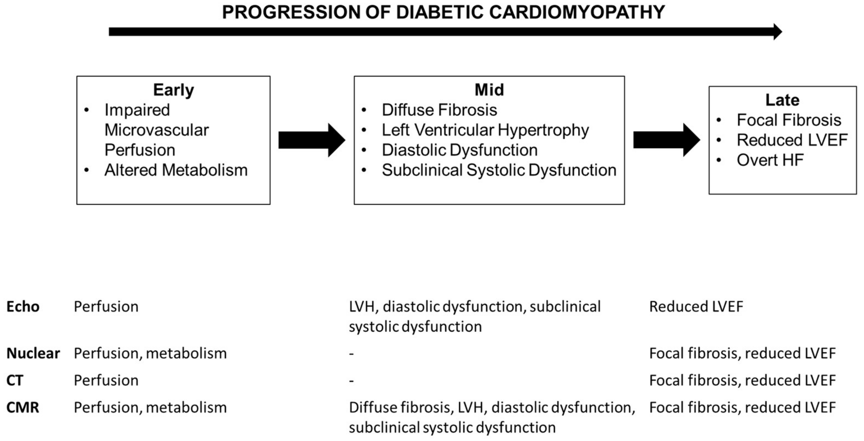
1. Introduction
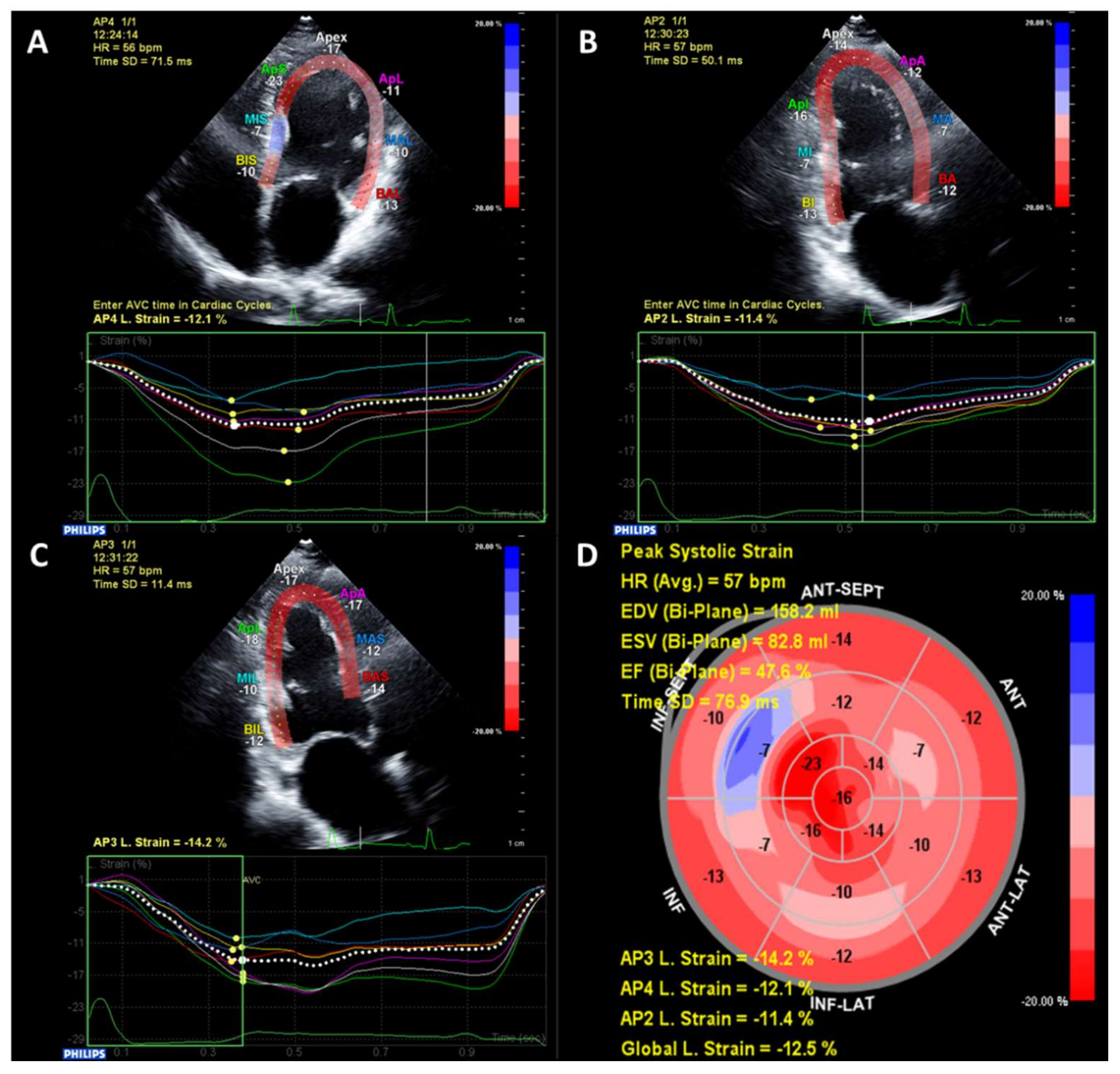
2. Echocardiography
3. Nuclear Imaging
4. Cardiovascular Computed Tomography
5. Cardiovascular Magnetic Resonance
6. Future Directions
7. Conclusions
Funding
Conflicts of Interest
References
- Iribarren, C.; Karter, A.J.; Go, A.S.; Ferrara, A.; Liu, J.Y.; Sidney, S.; Selby, J.V. Glycemic control and heart failure among adult patients with diabetes. Circulation 2001, 103, 2668–2673. [Google Scholar] [CrossRef] [PubMed]
- McMurray, J.J.; Gerstein, H.C.; Holman, R.R.; Pfeffer, M.A. Heart failure: A cardiovascular outcome in diabetes that can no longer be ignored. Lancet Diabetes Endocrinol. 2014, 2, 843–851. [Google Scholar] [CrossRef]
- Lee, M.M.Y.; McMurray, J.J.V.; Lorenzo-Almoros, A.; Kristensen, S.L.; Sattar, N.; Jhund, P.S.; Petrie, M.C. Diabetic cardiomyopathy. Heart 2018. [Google Scholar] [CrossRef]
- Jia, G.; Hill, M.A.; Sowers, J.R. Diabetic Cardiomyopathy: An Update of Mechanisms Contributing to This Clinical Entity. Circ. Res. 2018, 122, 624–638. [Google Scholar] [CrossRef] [PubMed]
- McKee, P.A.; Castelli, W.P.; McNamara, P.M.; Kannel, W.B. The natural history of congestive heart failure: The Framingham study. New Engl. J. Med. 1971, 285, 1441–1446. [Google Scholar] [CrossRef] [PubMed]
- Kannel, W.B.; Hjortland, M.; Castelli, W.P. Role of diabetes in congestive heart failure: The Framingham study. Am. J. Cardiol. 1974, 34, 29–34. [Google Scholar] [CrossRef]
- Ehl, N.F.; Kuhne, M.; Brinkert, M.; Muller-Brand, J.; Zellweger, M.J. Diabetes reduces left ventricular ejection fraction-irrespective of presence and extent of coronary artery disease. Eur. J. Endocrinol. 2011, 165, 945–951. [Google Scholar] [CrossRef]
- Devereux, R.B.; Roman, M.J.; Paranicas, M.; O’Grady, M.J.; Lee, E.T.; Welty, T.K.; Fabsitz, R.R.; Robbins, D.; Rhoades, E.R.; Howard, B.V. Impact of diabetes on cardiac structure and function: The strong heart study. Circulation 2000, 101, 2271–2276. [Google Scholar] [CrossRef]
- Eguchi, K.; Boden-Albala, B.; Jin, Z.; Rundek, T.; Sacco, R.L.; Homma, S.; di Tullio, M.R. Association between diabetes mellitus and left ventricular hypertrophy in a multiethnic population. Am. J. Cardiol. 2008, 101, 1787–1791. [Google Scholar] [CrossRef]
- Lee, M.; Gardin, J.M.; Lynch, J.C.; Smith, V.E.; Tracy, R.P.; Savage, P.J.; Szklo, M.; Ward, B.J. Diabetes mellitus and echocardiographic left ventricular function in free-living elderly men and women: The Cardiovascular Health Study. Am. Heart J. 1997, 133, 36–43. [Google Scholar] [CrossRef]
- Stewart, M.H.; Lavie, C.J.; Shah, S.; Englert, J.; Gilliland, Y.; Qamruddin, S.; Dinshaw, H.; Cash, M.; Ventura, H.; Milani, R. Prognostic Implications of Left Ventricular Hypertrophy. Prog. Cardiovasc. Dis. 2018, 61, 446–455. [Google Scholar] [CrossRef] [PubMed]
- Vakili, B.A.; Okin, P.M.; Devereux, R.B. Prognostic implications of left ventricular hypertrophy. Am. Heart J. 2001, 141, 334–341. [Google Scholar] [CrossRef]
- Lieb, W.; Xanthakis, V.; Sullivan, L.M.; Aragam, J.; Pencina, M.J.; Larson, M.G.; Benjamin, E.J.; Vasan, R.S. Longitudinal tracking of left ventricular mass over the adult life course: clinical correlates of short- and long-term change in the framingham offspring study. Circulation 2009, 119, 3085–3092. [Google Scholar] [CrossRef] [PubMed]
- Markus, M.R.; Stritzke, J.; Wellmann, J.; Duderstadt, S.; Siewert, U.; Lieb, W.; Luchner, A.; Doring, A.; Keil, U.; Schunkert, H.; et al. Implications of prevalent and incident diabetes mellitus on left ventricular geometry and function in the ageing heart: The MONICA/KORA Augsburg cohort study. Nutr. Metab Cardiovasc. Dis. 2011, 21, 189–196. [Google Scholar] [CrossRef]
- Felicio, J.S.; Ferreira, S.R.; Plavnik, F.L.; Moises, V.; Kohlmann, O., Jr.; Ribeiro, A.B.; Zanella, M.T. Effect of blood glucose on left ventricular mass in patients with hypertension and type 2 diabetes mellitus. Am. J. Hypertens. 2000, 13, 1149–1154. [Google Scholar] [CrossRef][Green Version]
- Lonnebakken, M.T.; Izzo, R.; Mancusi, C.; Gerdts, E.; Losi, M.A.; Canciello, G.; Giugliano, G.; de Luca, N.; Trimarco, B.; de Simone, G. Left Ventricular Hypertrophy Regression During Antihypertensive Treatment in an Outpatient Clinic (the Campania Salute Network). J. Am. Heart Assoc. 2017, 6. [Google Scholar] [CrossRef]
- Diamond, J.A.; Phillips, R.A. Regression of left ventricular hypertrophy: Are there preferred drugs? Curr. Hypertens. Rep. 2003, 5, 368–371. [Google Scholar] [CrossRef] [PubMed]
- Poirier, P.; Bogaty, P.; Garneau, C.; Marois, L.; Dumesnil, J.G. Diastolic dysfunction in normotensive men with well-controlled type 2 diabetes: Importance of maneuvers in echocardiographic screening for preclinical diabetic cardiomyopathy. Diabetes Care 2001, 24, 5–10. [Google Scholar] [CrossRef] [PubMed]
- From, A.M.; Scott, C.G.; Chen, H.H. The development of heart failure in patients with diabetes mellitus and pre-clinical diastolic dysfunction a population-based study. J. Am. Coll. Cardiol. 2010, 55, 300–305. [Google Scholar] [CrossRef]
- Blomstrand, P.; Engvall, M.; Festin, K.; Lindstrom, T.; Lanne, T.; Maret, E.; Nystrom, F.H.; Maret-Ouda, J.; Ostgren, C.J.; Engvall, J. Left ventricular diastolic function, assessed by echocardiography and tissue Doppler imaging, is a strong predictor of cardiovascular events, superior to global left ventricular longitudinal strain, in patients with type 2 diabetes. Eur. Heart J.-Card. Img. 2015, 16, 1000–1007. [Google Scholar] [CrossRef] [PubMed]
- Ernande, L.; Bergerot, C.; Rietzschel, E.R.; de Buyzere, M.L.; Thibault, H.; Pignonblanc, P.G.; Croisille, P.; Ovize, M.; Groisne, L.; Moulin, P.; et al. Diastolic dysfunction in patients with type 2 diabetes mellitus: Is it really the first marker of diabetic cardiomyopathy? J. Am. Soc. Echocardiog. 2011, 24, 1268–1275 e1. [Google Scholar] [CrossRef] [PubMed]
- Potter, E.; Marwick, T.H. Assessment of Left Ventricular Function by Echocardiography: The Case for Routinely Adding Global Longitudinal Strain to Ejection Fraction. Jacc Cardiovasc. Imag. 2018, 11, 260–274. [Google Scholar] [CrossRef] [PubMed]
- Collier, P.; Phelan, D.; Klein, A. A Test in Context: Myocardial Strain Measured by Speckle-Tracking Echocardiography. J. Am. Coll. Cardiol. 2017, 69, 1043–1056. [Google Scholar] [CrossRef] [PubMed]
- Stanton, T.; Leano, R.; Marwick, T.H. Prediction of all-cause mortality from global longitudinal speckle strain: comparison with ejection fraction and wall motion scoring. Circ. Cardiovasc. Imag. 2009, 2, 356–364. [Google Scholar] [CrossRef]
- Ernande, L.; Bergerot, C.; Girerd, N.; Thibault, H.; Davidsen, E.S.; Gautier Pignon-Blanc, P.; Amaz, C.; Croisille, P.; de Buyzere, M.L.; Rietzschel, E.R.; et al. Longitudinal myocardial strain alteration is associated with left ventricular remodeling in asymptomatic patients with type 2 diabetes mellitus. J. Am. Soc. Echocardiog. 2014, 27, 479–488. [Google Scholar] [CrossRef] [PubMed]
- Holland, D.J.; Marwick, T.H.; Haluska, B.A.; Leano, R.; Hordern, M.D.; Hare, J.L.; Fang, Z.Y.; Prins, J.B.; Stanton, T. Subclinical LV dysfunction and 10-year outcomes in type 2 diabetes mellitus. Heart 2015, 101, 1061–1066. [Google Scholar] [CrossRef]
- Liu, J.H.; Chen, Y.; Yuen, M.; Zhen, Z.; Chan, C.W.; Lam, K.S.; Tse, H.F.; Yiu, K.H. Incremental prognostic value of global longitudinal strain in patients with type 2 diabetes mellitus. Cardiovasc. Diabetol. 2016, 15, 22. [Google Scholar] [CrossRef]
- Mondillo, S.; Cameli, M.; Caputo, M.L.; Lisi, M.; Palmerini, E.; Padeletti, M.; Ballo, P. Early detection of left atrial strain abnormalities by speckle-tracking in hypertensive and diabetic patients with normal left atrial size. J. Am. Soc. Echocardiog. 2011, 24, 898–908. [Google Scholar] [CrossRef]
- Kadappu, K.K.; Boyd, A.; Eshoo, S.; Haluska, B.; Yeo, A.E.; Marwick, T.H.; Thomas, L. Changes in left atrial volume in diabetes mellitus: More than diastolic dysfunction? Eur. Heart J.-Card Img. 2012, 13, 1016–1023. [Google Scholar] [CrossRef] [PubMed]
- Tadic, M.; Celic, V.; Cuspidi, C.; Ilic, S.; Pencic, B.; Radojkovic, J.; Ivanovic, B.; Stanisavljevic, D.; Kocabay, G.; Marjanovic, T. Right heart mechanics in untreated normotensive patients with prediabetes and type 2 diabetes mellitus: A two- and three-dimensional echocardiographic study. J. Am. Soc. Echocardiog. 2015, 28, 317–327. [Google Scholar] [CrossRef] [PubMed]
- Hamada-Harimura, Y.; Seo, Y.; Ishizu, T.; Nishi, I.; Machino-Ohtsuka, T.; Yamamoto, M.; Sugano, A.; Sato, K.; Sai, S.; Obara, K.; et al. Incremental Prognostic Value of Right Ventricular Strain in Patients With Acute Decompensated Heart Failure. Circ. Cardiovasc Imag. 2018, 11, e007249. [Google Scholar] [CrossRef]
- Motoki, H.; Borowski, A.G.; Shrestha, K.; Hu, B.; Kusunose, K.; Troughton, R.W.; Tang, W.H.; Klein, A.L. Right ventricular global longitudinal strain provides prognostic value incremental to left ventricular ejection fraction in patients with heart failure. J Am Soc Echocardiogr. 2014, 27, 726–732. [Google Scholar] [CrossRef]
- Cameli, M.; Lisi, M.; Focardi, M.; Reccia, R.; Natali, B.M.; Sparla, S.; Mondillo, S. Left atrial deformation analysis by speckle tracking echocardiography for prediction of cardiovascular outcomes. Am. J. Cardiol. 2012, 110, 264–269. [Google Scholar] [CrossRef] [PubMed]
- Marwick, T.H.; Case, C.; Sawada, S.; Vasey, C.; Short, L.; Lauer, M. Use of stress echocardiography to predict mortality in patients with diabetes and known or suspected coronary artery disease. Diabetes Care 2002, 25, 1042–1048. [Google Scholar] [CrossRef] [PubMed][Green Version]
- Albers, A.R.; Krichavsky, M.Z.; Balady, G.J. Stress testing in patients with diabetes mellitus: Diagnostic and prognostic value. Circulation 2006, 113, 583–592. [Google Scholar] [CrossRef]
- Fang, Z.Y.; Najos-Valencia, O.; Leano, R.; Marwick, T.H. Patients with early diabetic heart disease demonstrate a normal myocardial response to dobutamine. J. Am. Coll. Cardiol. 2003, 42, 446–453. [Google Scholar] [CrossRef]
- Ha, J.W.; Lee, H.C.; Kang, E.S.; Ahn, C.M.; Kim, J.M.; Ahn, J.A.; Lee, S.W.; Choi, E.Y.; Rim, S.J.; Oh, J.K.; et al. Abnormal left ventricular longitudinal functional reserve in patients with diabetes mellitus: Implication for detecting subclinical myocardial dysfunction using exercise tissue Doppler echocardiography. Heart 2007, 93, 1571–1576. [Google Scholar] [CrossRef]
- Philouze, C.; Obert, P.; Nottin, S.; Benamor, A.; Barthez, O.; Aboukhoudir, F. Dobutamine Stress Echocardiography Unmasks Early Left Ventricular Dysfunction in Asymptomatic Patients with Uncomplicated Type 2 Diabetes: A Comprehensive Two-Dimensional Speckle-Tracking Imaging Study. J. Am. Soc. Echocardiog. 2018, 31, 587–597. [Google Scholar] [CrossRef]
- Cortigiani, L.; Rigo, F.; Gherardi, S.; Galderisi, M.; Bovenzi, F.; Sicari, R. Prognostic meaning of coronary microvascular disease in type 2 diabetes mellitus: A transthoracic Doppler echocardiographic study. J. Am. Soc. Echocardiog. 2014, 27, 742–748. [Google Scholar] [CrossRef]
- Cortigiani, L.; Gherardi, S.; Faggioni, M.; Bovenzi, F.; Picano, E.; Petersen, C.; Molinaro, S.; Sicari, R. Dual-Imaging Stress Echocardiography for Prognostic Assessment of High-Risk Asymptomatic Patients with Diabetes Mellitus. J. Am. Soc. Echocardiog. 2017, 30, 149–158. [Google Scholar] [CrossRef]
- Kang, X.; Berman, D.S.; Lewin, H.; Miranda, R.; Erel, J.; Friedman, J.D.; Amanullah, A.M. Comparative ability of myocardial perfusion single-photon emission computed tomography to detect coronary artery disease in patients with and without diabetes mellitus. Am. Heart J. 1999, 137, 949–957. [Google Scholar] [CrossRef]
- Kang, X.; Berman, D.S.; Lewin, H.C.; Cohen, I.; Friedman, J.D.; Germano, G.; Hachamovitch, R.; Shaw, L.J. Incremental prognostic value of myocardial perfusion single photon emission computed tomography in patients with diabetes mellitus. Am. Heart J. 1999, 138, 1025–1032. [Google Scholar] [CrossRef]
- Bourque, J.M.; Patel, C.A.; Ali, M.M.; Perez, M.; Watson, D.D.; Beller, G.A. Prevalence and predictors of ischemia and outcomes in outpatients with diabetes mellitus referred for single-photon emission computed tomography myocardial perfusion imaging. Circ.-Cardiovasc. Imag. 2013, 6, 466–477. [Google Scholar] [CrossRef] [PubMed]
- Rajagopalan, N.; Miller, T.D.; Hodge, D.O.; Frye, R.L.; Gibbons, R.J. Identifying high-risk asymptomatic diabetic patients who are candidates for screening stress single-photon emission computed tomography imaging. J. Am. Coll. Cardiol. 2005, 45, 43–49. [Google Scholar] [CrossRef]
- Vanzetto, G.; Halimi, S.; Hammoud, T.; Fagret, D.; Benhamou, P.Y.; Cordonnier, D.; Denis, B.; Machecourt, J. Prediction of cardiovascular events in clinically selected high-risk NIDDM patients. Prognostic value of exercise stress test and thallium-201 single-photon emission computed tomography. Diabetes Care 1999, 22, 19–26. [Google Scholar] [CrossRef]
- De Lorenzo, A.; Lima, R.S.; Siqueira-Filho, A.G.; Pantoja, M.R. Prevalence and prognostic value of perfusion defects detected by stress technetium-99m sestamibi myocardial perfusion single-photon emission computed tomography in asymptomatic patients with diabetes mellitus and no known coronary artery disease. Am. J. Cardiol. 2002, 90, 827–832. [Google Scholar] [CrossRef]
- Storto, G.; Pellegrino, T.; Sorrentino, A.R.; Luongo, L.; Petretta, M.; Cuocolo, A. Estimation of coronary flow reserve by sestamibi imaging in type 2 diabetic patients with normal coronary arteries. J. Nucl. Cardiol. 2007, 14, 194–199. [Google Scholar] [CrossRef] [PubMed]
- Yokoyama, I.; Momomura, S.; Ohtake, T.; Yonekura, K.; Nishikawa, J.; Sasaki, Y.; Omata, M. Reduced myocardial flow reserve in non-insulin-dependent diabetes mellitus. J. Am. Coll. Cardiol. 1997, 30, 1472–1477. [Google Scholar] [CrossRef]
- Potier, L.; Chequer, R.; Roussel, R.; Mohammedi, K.; Sismail, S.; Hartemann, A.; Amouyal, C.; Marre, M.; Le Guludec, D.; Hyafil, F. Relationship between cardiac microvascular dysfunction measured with 82Rubidium-PET and albuminuria in patients with diabetes mellitus. Cardiovasc. Diabetol. 2018, 17, 11. [Google Scholar] [CrossRef]
- Murthy, V.L.; Naya, M.; Foster, C.R.; Gaber, M.; Hainer, J.; Klein, J.; Dorbala, S.; Blankstein, R.; di Carli, M.F. Association between coronary vascular dysfunction and cardiac mortality in patients with and without diabetes mellitus. Circulation 2012, 126, 1858–1868. [Google Scholar] [CrossRef] [PubMed]
- Rijzewijk, L.J.; van der Meer, R.W.; Lamb, H.J.; de Jong, H.W.; Lubberink, M.; Romijn, J.A.; Bax, J.J.; de Roos, A.; Twisk, J.W.; Heine, R.J.; et al. Altered myocardial substrate metabolism and decreased diastolic function in nonischemic human diabetic cardiomyopathy: studies with cardiac positron emission tomography and magnetic resonance imaging. J. Am. Coll. Cardiol. 2009, 54, 1524–1532. [Google Scholar] [CrossRef] [PubMed]
- Hu, L.; Qiu, C.; Wang, X.; Xu, M.; Shao, X.; Wang, Y. The association between diabetes mellitus and reduction in myocardial glucose uptake: A population-based (18)F-FDG PET/CT study. Cardiovasc. Disord. 2018, 18, 203. [Google Scholar] [CrossRef]
- Beller, E.; Meinel, F.G.; Schoeppe, F.; Kunz, W.G.; Thierfelder, K.M.; Hausleiter, J.; Bamberg, F.; Schoepf, U.J.; Hoffmann, V.S. Predictive value of coronary computed tomography angiography in asymptomatic individuals with diabetes mellitus: Systematic review and meta-analysis. J. Cardiovasc. Comput. Tomogr. 2018, 12, 320–328. [Google Scholar] [CrossRef]
- Malik, S.; Zhao, Y.; Budoff, M.; Nasir, K.; Blumenthal, R.S.; Bertoni, A.G.; Wong, N.D. Coronary Artery Calcium Score for Long-term Risk Classification in Individuals With Type 2 Diabetes and Metabolic Syndrome From the Multi-Ethnic Study of Atherosclerosis. JAMA Cardio. 2017, 2, 1332–1340. [Google Scholar] [CrossRef] [PubMed]
- Levine, A.; Hecht, H.S. Cardiac CT Angiography in Congestive Heart Failure. J. Nucl. Med. 2015, 56 (Suppl. 4), 46S–51S. [Google Scholar] [CrossRef]
- Vliegenthart, R.; De Cecco, C.N.; Wichmann, J.L.; Meinel, F.G.; Pelgrim, G.J.; Tesche, C.; Ebersberger, U.; Pugliese, F.; Bamberg, F.; Choe, Y.H.; et al. Dynamic CT myocardial perfusion imaging identifies early perfusion abnormalities in diabetes and hypertension: Insights from a multicenter registry. J. Cardiovasc. Comput. Tomogr. 2016, 10, 301–308. [Google Scholar] [CrossRef] [PubMed]
- Tomizawa, N.; Fujino, Y.; Kamitani, M.; Chou, S.; Yamamoto, K.; Inoh, S.; Nojo, T.; Nakamura, S. Longer diabetes duration reduces myocardial blood flow in remote myocardium assessed by dynamic myocardial CT perfusion. J. Diabetes Complicat. 2018, 32, 609–615. [Google Scholar] [CrossRef] [PubMed]
- Kim, R.J.; Wu, E.; Rafael, A.; Chen, E.L.; Parker, M.A.; Simonetti, O.; Klocke, F.J.; Bonow, R.O.; Judd, R.M. The use of contrast-enhanced magnetic resonance imaging to identify reversible myocardial dysfunction. New Engl. J. Med. 2000, 343, 1445–1453. [Google Scholar] [CrossRef]
- Kwong, R.Y.; Sattar, H.; Wu, H.; Vorobiof, G.; Gandla, V.; Steel, K.; Siu, S.; Brown, K.A. Incidence and prognostic implication of unrecognized myocardial scar characterized by cardiac magnetic resonance in diabetic patients without clinical evidence of myocardial infarction. Circulation 2008, 118, 1011–1020. [Google Scholar] [CrossRef] [PubMed]
- Jellis, C.L.; Kwon, D.H. Myocardial T1 mapping: modalities and clinical applications. Cardiovasc. Diagn. 2014, 4, 126–137. [Google Scholar]
- Kammerlander, A.A.; Marzluf, B.A.; Zotter-Tufaro, C.; Aschauer, S.; Duca, F.; Bachmann, A.; Knechtelsdorfer, K.; Wiesinger, M.; Pfaffenberger, S.; Greiser, A.; et al. T1 Mapping by CMR Imaging: From Histological Validation to Clinical Implication. JACC: Cardiovasc. Imag. 2016, 9, 14–23. [Google Scholar]
- Puntmann, V.O.; Carr-White, G.; Jabbour, A.; Yu, C.Y.; Gebker, R.; Kelle, S.; Hinojar, R.; Doltra, A.; Varma, N.; Child, N.; et al. T1-Mapping and Outcome in Nonischemic Cardiomyopathy: All-Cause Mortality and Heart Failure. JACC: Cardiovasc. Imag. 2016, 9, 40–50. [Google Scholar]
- Mordi, I.R.; Singh, S.; Rudd, A.; Srinivasan, J.; Frenneaux, M.; Tzemos, N.; Dawson, D.K. Comprehensive Echocardiographic and Cardiac Magnetic Resonance Evaluation Differentiates Among Heart Failure With Preserved Ejection Fraction Patients, Hypertensive Patients, and Healthy Control Subjects. JACC: Cardiovasc. Imag. 2018, 11, 577–585. [Google Scholar] [CrossRef]
- Ng, A.C.; Auger, D.; Delgado, V.; van Elderen, S.G.; Bertini, M.; Siebelink, H.M.; van der Geest, R.J.; Bonetti, C.; van der Velde, E.T.; de Roos, A.; et al. Association between diffuse myocardial fibrosis by cardiac magnetic resonance contrast-enhanced T(1) mapping and subclinical myocardial dysfunction in diabetic patients: A pilot study. Circ.-Cardiovasc Imag. 2012, 5, 51–59. [Google Scholar] [CrossRef]
- Swoboda, P.P.; McDiarmid, A.K.; Erhayiem, B.; Ripley, D.P.; Dobson, L.E.; Garg, P.; Musa, T.A.; Witte, K.K.; Kearney, M.T.; Barth, J.H.; et al. Diabetes Mellitus, Microalbuminuria, and Subclinical Cardiac Disease: Identification and Monitoring of Individuals at Risk of Heart Failure. J. Am. Heart Assoc. 2017, 6. [Google Scholar] [CrossRef]
- McGavock, J.M.; Lingvay, I.; Zib, I.; Tillery, T.; Salas, N.; Unger, R.; Levine, B.D.; Raskin, P.; Victor, R.G.; Szczepaniak, L.S. Cardiac steatosis in diabetes mellitus: A 1H-magnetic resonance spectroscopy study. Circulation 2007, 116, 1170–1175. [Google Scholar] [CrossRef]
- Levelt, E.; Mahmod, M.; Piechnik, S.K.; Ariga, R.; Francis, J.M.; Rodgers, C.T.; Clarke, W.T.; Sabharwal, N.; Schneider, J.E.; Karamitsos, T.D.; et al. Relationship Between Left Ventricular Structural and Metabolic Remodeling in Type 2 Diabetes. Diabetes 2016, 65, 44–52. [Google Scholar]
- Levelt, E.; Rodgers, C.T.; Clarke, W.T.; Mahmod, M.; Ariga, R.; Francis, J.M.; Liu, A.; Wijesurendra, R.S.; Dass, S.; Sabharwal, N.; et al. Cardiac energetics, oxygenation, and perfusion during increased workload in patients with type 2 diabetes mellitus. Eur. Heart J. 2016, 37, 3461–3469. [Google Scholar] [CrossRef]
- Levelt, E.; Pavlides, M.; Banerjee, R.; Mahmod, M.; Kelly, C.; Sellwood, J.; Ariga, R.; Thomas, S.; Francis, J.; Rodgers, C.; et al. Ectopic and Visceral Fat Deposition in Lean and Obese Patients With Type 2 Diabetes. J. Am. Coll. Cardiol. 2016, 68, 53–63. [Google Scholar] [CrossRef]
- Al-Talabany, S.; Mordi, I.; Graeme Houston, J.; Colhoun, H.M.; Weir-McCall, J.R.; Matthew, S.Z.; Looker, H.C.; Levin, D.; Belch, J.J.F.; Dove, F.; et al. Epicardial adipose tissue is related to arterial stiffness and inflammation in patients with cardiovascular disease and type 2 diabetes. BMC Cardiovasc. Disor. 2018, 18, 31. [Google Scholar] [CrossRef]
- Heydari, B.; Juan, Y.H.; Liu, H.; Abbasi, S.; Shah, R.; Blankstein, R.; Steigner, M.; Jerosch-Herold, M.; Kwong, R.Y. Stress Perfusion Cardiac Magnetic Resonance Imaging Effectively Risk Stratifies Diabetic Patients With Suspected Myocardial Ischemia. Circ. Cardiovasc. Imag. 2016, 9, e004136. [Google Scholar] [CrossRef] [PubMed]
- Levelt, E.; Piechnik, S.K.; Liu, A.; Wijesurendra, R.S.; Mahmod, M.; Ariga, R.; Francis, J.M.; Greiser, A.; Clarke, K.; Neubauer, S.; et al. Correction to: Adenosine stress CMR T1-mapping detects early microvascular dysfunction in patients with type 2 diabetes mellitus without obstructive coronary artery disease. J. Cardiovasc. Magn. Reson. 2017, 19, 99. [Google Scholar] [CrossRef]
- Ernande, L.; Audureau, E.; Jellis, C.L.; Bergerot, C.; Henegar, C.; Sawaki, D.; Czibik, G.; Volpi, C.; Canoui-Poitrine, F.; Thibault, H.; et al. Clinical Implications of Echocardiographic Phenotypes of Patients With Diabetes Mellitus. J. Am. Coll. Cardiol. 2017, 70, 1704–1716. [Google Scholar] [CrossRef] [PubMed]
- Lorenzo-Almoros, A.; Tunon, J.; Orejas, M.; Cortes, M.; Egido, J.; Lorenzo, O. Diagnostic approaches for diabetic cardiomyopathy. Cardiovasc. Diabetol. 2017, 16, 28. [Google Scholar] [CrossRef] [PubMed]
- Budoff, M.J.; Raggi, P.; Beller, G.A.; Berman, D.S.; Druz, R.S.; Malik, S.; Rigolin, V.H.; Weigold, W.G.; Soman, P. Noninvasive Cardiovascular Risk Assessment of the Asymptomatic Diabetic Patient: The Imaging Council of the American College of Cardiology. JACC: Cardiovasc. Imag. 2016, 9, 176–192. [Google Scholar]


© 2019 by the author. Licensee MDPI, Basel, Switzerland. This article is an open access article distributed under the terms and conditions of the Creative Commons Attribution (CC BY) license (http://creativecommons.org/licenses/by/4.0/).
Share and Cite
Mordi, I.R. Non-Invasive Imaging in Diabetic Cardiomyopathy. J. Cardiovasc. Dev. Dis. 2019, 6, 18. https://doi.org/10.3390/jcdd6020018
Mordi IR. Non-Invasive Imaging in Diabetic Cardiomyopathy. Journal of Cardiovascular Development and Disease. 2019; 6(2):18. https://doi.org/10.3390/jcdd6020018
Chicago/Turabian StyleMordi, Ify R. 2019. "Non-Invasive Imaging in Diabetic Cardiomyopathy" Journal of Cardiovascular Development and Disease 6, no. 2: 18. https://doi.org/10.3390/jcdd6020018
APA StyleMordi, I. R. (2019). Non-Invasive Imaging in Diabetic Cardiomyopathy. Journal of Cardiovascular Development and Disease, 6(2), 18. https://doi.org/10.3390/jcdd6020018