Article

RNA-Binding Protein Trim71 Controls Epicardial Cell Migration

by
Juan Manuel Castillo-Casas
1,
Carlos García-Padilla
1,
Rita Carmona
2,
Estefanía Lozano-Velasco
1 and
Diego Franco
1,*
1
Cardiovascular Development Group, Department of Experimental Biology, Faculty of Experimental Sciences, University of Jaen, 23071 Jaen, Spain
2
Department of Human Anatomy, Legal Medicine and History of Science, Faculty of Medicine, University of Málaga, 29071 Málaga, Spain
*
Author to whom correspondence should be addressed.
J. Cardiovasc. Dev. Dis. 2026, 13(6), 237; https://doi.org/10.3390/jcdd13060237
Submission received: 17 February 2026 / Revised: 25 May 2026 / Accepted: 29 May 2026 / Published: 31 May 2026

Abstract

The epicardium is an embryonic tissue layer essential for heart morphogenesis, providing progenitor cells and regulatory signals that support myocardial growth and coronary vessel formation. Epicardial cells arise from the proepicardium (PE) and spread over the myocardium to form the embryonic epicardium (EE), a transition that requires tight coordination between proliferation, migration, and lineage priming. However, the molecular mechanisms controlling this developmental timing remain incompletely understood. Here, we identify Trim71 as a key regulator of epicardial cell behaviour during the PE-to-EE transition. Trim71 is enriched in the PE and subsequently downregulated as cells acquire migratory competence. Functional analyses show that loss of Trim71 function decreases proliferation while promoting migration, as well as inducing the expression of epicardial commitment markers, suggesting that Trim71 is a controller of a progenitor-like state. We further demonstrate that Trim71 is necessary for these processes through a reciprocal feedback loop with the microRNAs let-7c and miR-30c. Our findings establish Trim71 as a temporal gatekeeper that coordinates the balance between progenitor maintenance and migration during early epicardial development. This Trim71-miRNAs axis constitutes a novel post-transcriptional layer of regulation that ensures the correct timing of epicardium development during cardiogenesis.

1. Introduction

The embryonic epicardium (EE) originates from an extracardiac primordium known as the proepicardium (PE) [1]. In the mouse, the PE emerges at embryonic day (E) 9.5 as a cluster of mesothelial cells located between the sinus venosus and the hepatic primordium, being a highly conserved structure among vertebrates [1,2,3,4,5,6,7]. Following its formation, PE cells migrate to the surface of the naked heart, which is only composed of myocardium and endocardium at this stage, through cellular bridges or via cellular vesicles, progressively covering the heart and giving rise to the third cardiac layer, the EE, by E10.5 [1]. Once established, a subset of epicardial cells undergoes an epithelial-to-mesenchymal transition (EMT) and invades the myocardium, giving rise to epicardial-derived cells (EPDCs). In mice, this process occurs around E11.5 and is characterized by the loss of epithelial features and the acquisition of migratory and invasive properties similar to those of multipotent mesenchymal cells [8,9]. EPDCs constitute a major progenitor source for several cardiac cell types, including cardiac fibroblasts (CFs), smooth muscle cells (SMCs), and pericytes (PCs) [10,11]. Beyond its role as a progenitor cell source, the EE functions as a crucial signaling hub capable of inducing cardiomyocyte (CM) cell cycle re-entry, promoting angio-vasculogenesis, recruiting macrophages, and contributing to extracellular matrix deposition. These functions are essential for proper myocardial support and morphogenetic processes such as trabeculation and myocardial compaction [12,13,14]. Moreover, the epicardium can be reactivated after cardiac injury [12]. Consistent with its central role, defects in epicardial development result in a wide range of cardiac abnormalities, including valve malformations, myocardial non-compaction, fibrosis-associated arrhythmias, and cardiomyopathies, highlighting the functional importance of the epicardium [15,16,17,18].
Mammalian heart development is a highly coordinated process requiring precise regulation of cell differentiation, migration, and proliferation across multiple cardiac lineages. These events are tightly controlled by transcriptional programs governed by several transcription factors (TFs) [19]. In the epicardium, TFs such as Wt1 and Tbx18 have been identified as key regulators of essential processes, including EMT and EPDC differentiation [20,21,22,23]. However, transcriptional regulation alone cannot fully account for the dynamic changes in gene expression that accompany epicardial development. Beneath this layer of control lies post-transcriptional regulation, which is mediated in part by microRNAs (miRNAs). While a transcriptional network provides the framework for epicardial identity, post-transcriptional mechanisms act as essential fine-tuners that ensure the precise timing of these cellular transitions. miRNAs are small non-coding RNAs of approximately 22 nucleotides that bind to the 3′ untranslated regions (3′UTRs) of target mRNAs, leading to translational repression or mRNA degradation [24]. miRNA biogenesis begins in the nucleus and is completed in the cytoplasm, where mature miRNAs associate with the RNA-induced silencing complex (RISC) and Argonaute (Ago) proteins to guide them to their target transcripts [25,26,27,28]. Despite their known roles in multiple developmental contexts, post-transcriptional regulatory mechanisms in epicardial development remain poorly explored. Notably, epicardial deletion of Dicer, an enzyme essential for miRNA maturation, results in postnatal lethality, underscoring the functional importance of miRNAs in epicardial biology [29].
Although considerable progress has been made in characterizing the cellular transition from the PE to the EE, the molecular mechanisms governing this process remain poorly understood. To gain further insights, we previously performed RNA-seq analyses of the PE and EE, identifying distinct sets of differentially expressed mRNAs and miRNAs between these two developmental stages [30]. Among the transcripts enriched in the PE, Trim71 emerged as a prominent candidate. Trim71 is an RNA-binding protein and translational repressor known to promote cell proliferation, inhibit differentiation, and regulate cell cycle progression during early embryonic and neural development [31]. In addition, Trim71 has been shown to modulate miRNA biogenesis, further supporting its role as a post-transcriptional regulator [32]. However, its function in epicardial development has not yet been investigated. Of note, among the miRNAs upregulated in the EE, let-7c and miR-30c were identified as potential regulators of Trim71, since there are binding sites for both of them within the Trim71 3′UTR. These findings suggest the existence of an miRNA-dependent regulatory axis controlling Trim71 expression during the PE-to-EE transition. In this study, we investigate the functional relevance of the let-7c/miR-30c–Trim71 regulatory axis during epicardial development. Our data demonstrate that coordinated regulation of Trim71 by let-7c and miR-30c modulates embryonic epicardial cell migration, linking microRNA-mediated post-transcriptional control to a key cellular process required for epicardial maturation. Together, these findings identify Trim71 as a previously unrecognized regulator of epicardial biology and highlight the importance of miRNA-driven regulatory mechanisms in heart development.

2. Materials and Methods

2.1. Mouse Lines and Tissue Collection

All experimental procedures were conducted in accordance with the guidelines and approved consent of the Ethics Committee of the University of Jaén and Andalusian Regional Government (14/03/2022/038). Time-pregnant CD1 female mice were euthanized to collect embryos at developmental stages E9.5 to E10.5. For E9.5 samples, the proepicardium was manually dissected, pooled, and stored in a lysis buffer at −80 °C for subsequent RNA isolation. Embryonic epicardial cells were harvested from ventricular explants as previously described by Castillo-Casas et al. [30]. Additionally, the adult epicardium (180 days; n = 6 experimental replicates) was manually dissected from adult CD1 female mice and stored in buffer lysis at −80 °C until further processing.

2.2. Cell Lines

Two cell lines, the mouse embryonic epicardial cell line MEC1 (Sigma-Aldrich, St. Louis, MO, USA, SCC187) and 3T3 cells (Merck, Darmstadt, Germany, 93061524), were used in this study. Each cell line was cultured following the manufacturer’s recommendations for 24 h at 37 °C in a humidified atmosphere of 5% CO2.

2.3. microRNA, siRNA, and Plasmid Transfections

For experimental procedures, cells were seeded 24 h before transfection at a density of 4 × 10⁴ cells per well in 24-well plates or 104 cells in glass-bottom dishes for confocal analysis as previously described with lipofectamine 2000 (Thermo Fisher, Waltham, MA, USA, 11668019) [33]. For miRNA overexpression, cells were transfected with 50 nM of pre-miRNAs (Ambion, Austin, TX, USA). For Trim71 silencing, a concentration range of 60–80 nM of siRNA was initially tested, with the most efficient dose selected for subsequent experiments. Detailed siRNA sequences are provided in Supplementary Table S1. A scramble siRNA was used as a negative control to confirm that siRNA transfection itself did not affect gene expression (Supplementary Figure S1). For the luciferase assay, 200 ng/mL of the pmiR-Report-pGLuc plasmid was co-transfected with 50 nM of the corresponding pre-miRNA let7-c and miR-30c, respectively. Transfection efficiencies were validated by RT-qPCR in preliminary experiments, adjusting the doses for each condition (Supplementary Figure S2A). Then, 24 h post-transfection, cells were processed either for RT-qPCR or immunofluorescence (IFC) analyses. Negative controls treated with Lipofectamine 2000 were run in parallel. To perform IFC analyses, the cells were fixed in 4% PFA for 15 min at room temperature, rinsed two times in PBS for 5 min, and stored in PBS at 4 °C. For RT-qPCR analysis, cells and explant epicardial outgrowths were collected and stored at −80 °C until further processing.

2.4. RNA Isolation and qPCR

All RT-qPCR experiments were performed in accordance with the MIQE guidelines [34]. Briefly, total RNA isolation was performed with the ReliaPrep™ RNA Miniprep Systems kit (Promega, Madison, WI, USA) according to the manufacturer’s instructions. For mRNA expression measurements, 500 ng of total RNA was used for retro-transcription with PrimeScript™ RT Master Mix (Takara, Kyoto, Japan), and the resulting cDNA was diluted 1:5. For microRNA expression analyses, 20 ng of total RNA was used for retro-transcription with miRCURY LNA RT Kit (Qiagen, Venlo, The Netherlands) and the resulting cDNA was diluted 1:40. Non-reverse transcribed controls (no-RT) were performed for each sample to exclude genomic contamination, yielding no signal amplification in all cases. Quantitative Real-time PCR experiments were performed using 2 μL of cDNA, GoTaq qPCR Master Mix (Promega, Madison, WI, USA) with specific primer sets as described in Supplementary Table S1. All qPCRs were performed using a CFX384TM thermocycler (Bio-Rad, Hercules, CA, USA) following the manufacturer’s recommendations. The mRNA thermal cycling conditions consisted of 95 °C for 30 s (initial denaturation), followed by 40 cycles of 95 °C for 5 s (denaturation); 60 °C for 10 s (annealing); and 75 °C for 7 s (extension). For microRNAs, the protocol included a cycle of 95 °C for 10 min (initial denaturation), followed by 40 cycles of 95 °C for 5 s (denaturation) and 60 °C for 1 min (annealing and extension). Melting curve analysis for both cases was determined by an initial step of 95 °C for 5 s, followed by 0.5 °C increments for 7 s from 65 °C to 95 °C. The relative expression levels of each gene were calculated using Livak & Schmittgen [35] with Gapdh as the internal control for mRNA expression analyses and 5S for microRNA expression analyses. Each qPCR reaction was performed in technical triplicate and repeated for at least three distinct biological samples to obtain representative means.

2.5. Cell Migration Assay

Cell migration was analyzed using a wound-healing (scratch) assay, as previously described by Ascione et al. [36]. MEC1 epicardial cells were plated on 24-well plates at a density of 5 × 10⁴ cells per well and cultured until reaching 90–100% confluence. After 24 h of transfection, cell monolayers were manually scratched using a sterile p200 pipette tip to create a linear wound. After scratching, cells were washed twice with PBS to remove cellular debris and replaced with a fresh medium. Wound healing was monitored by capturing images at 0 h, 6 h, 12 h, and 24 h using an inverted microscope (10 wells per condition; n = 3 experimental replicates). The scratches were quantified using Image J software (version V2.9.0), and the migration rate was expressed as the percentage of wound closure relative to the initial area at 0 h.

2.6. Luciferase Assay

The murine Trim71 3′UTR sequence was retrieved from the Ensembl Genome Browser database, and mature miRNA sequences (let-7c-5p and miR-30c-5p) were obtained from miRBase. Sequence analysis and visualization were performed in R using the packages ggplot2, tibble, and dplyr, and putative miRNA binding sites were identified based on canonical seed-sequence complementarity and corroborated with TargetScan predictions. The target fragment was amplified using the iPROOF High-Fidelity PCR kit (Bio-Rad 1725330) and cloned into the pmiR-Report-pGLuc report vector. For the reporter assays, 3T3 cells were co-transfected with the pmiR-Trim71-3′UTR-pGLuc construct, the corresponding miRNA precursors, let-7c and miR-30c mimics, and pCLuc control vector for internal normalization in 3T3 cells. Luciferase activity was measured 24 h after transfection. Gaussia luciferase activity was quantified by the Pierce Gaussia Luciferase Flash Assay Kit (Thermo Fisher Scientific, Waltham, MA, USA, 16159), while normalization was performed by measuring Cypridina luciferase activity with the Pierce Cypridina Luciferase Flash Assay Kit (Thermo Fisher Scientific 16169). In all cases, transfections were carried out in triplicate (n = 3 experimental replicates). Atf6 and Raldh2 3′UTRs were used in parallel as positive and negative controls, respectively, for the luciferase assay (Supplementary Figure S2B) [37,38].

2.7. Immunofluorescence and Confocal Scanning Laser Microscopy Analyses

Immunofluorescence analyses were performed as previously reported by Bonet et al. [39]. Briefly, control and experimental cell cultures were collected after the corresponding treatment, rinsed in PBS for 5 min at room temperature, and fixed with 4% PFA at room temperature for 15 min. After fixation, the samples were rinsed three times in PBS (10 min each) at room temperature and then permeabilized with 0.02% Triton X-100, 50 nM NH4Cl, and PBS solution for 10 min at room temperature. Non-specific binding sites were blocked with 0.2% gelatin solution (Sigma-Aldrich), which was applied twice for 10 min. As the primary antibody, anti-phospho-histone H3 (pHH3, Abcam, Cambridge, United Kingdom, AB5176), anti-Wt1 (Abcam, ab89901), and anti-Nkx2.5 (Santa Cruz Biotechnology, Dallas, TX, USA, N19) in blocking solution were applied overnight at 4 °C. Cell cultures were rinsed 3 times in PBS for 10 min and incubated with secondary antibody donkey anti-rabbit Alexa Fluor 594 (Thermo A2107) at a 1:200 dilution for 30 min at room temperature. Nuclei were counterstained with DAPI at 1:1000 dilution (Sigma #10236276001) for 15 min at room temperature and rinsed twice in PBS for 5 min each. Each immunofluorescence assay was performed in technical triplicate and repeated at least six times on distinct biological samples to obtain representative means. The percentage of pHH3+ cells was calculated in relation to total DAPI+ cells in the corresponding region of interest (ROI). For cell size assay and morphology analysis, F-actin filaments were stained using Phalloidin-iFluor 488 Reagent (Abcam ab176753) according to the manufacturer’s instructions. A total of 80 cells per condition were measured with imageJ software using the freehand tool to mark the shape of the cell and adjusting the measurement with the scale bar. Fluorescent histograms for Nkx2.5 and Wt1 signal were measured using the raw integrated density of each antibody and normalized per DAPI raw integrated density of each picture (i.e., ROI). Cells were stored in PBS in the dark at 4 °C until imaging. Confocal images were acquired using a Leica TCS SP5 II confocal scanning laser microscope (Leica Microsystems, Wetzlar, Germany).

2.8. Statistical Analysis

Statistical analyses of datasets were performed using GraphPad Prism software 11.0.0 (GraphPad Software, Inc., San Diego, CA, USA). For comparison between two groups, an unpaired Student’s t-test was used. Significance levels or p-values are stated in each corresponding figure legend. p-value < 0.05 was considered statistically significant.

3. Results

3.1. Trim71 Sustains Cell Proliferation While Restraining Migration in Epicardial Cells

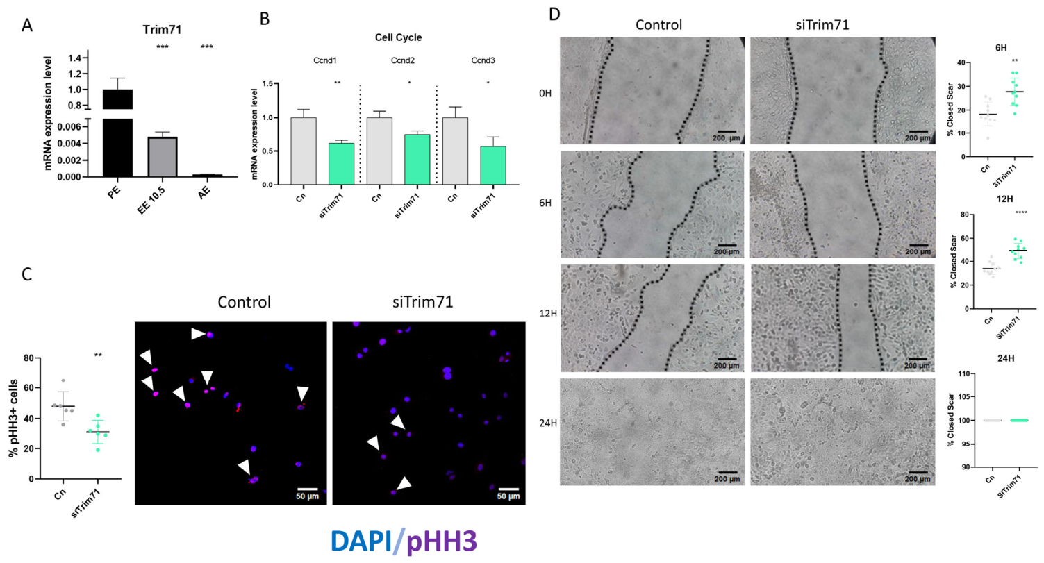
The TRIM-NHL family of E3 ubiquitin ligases is evolutionarily conserved in mammals, and it has been described as a regulator of development and differentiation, acting primarily as a translational repressor [31]. For the first time in the context of epicardial development, our research group identified Trim71 as a differentially expressed gene between the PE and EE at E10.5 through RNA-seq analysis [30]. RT-qPCR analysis confirmed that Trim71 is highly expressed in the PE, while it is decreased in embryonic epicardial cells at E10.5 (EE 10.5) and becomes undetectable in adult epicardial cells (AE) (Figure 1A). Importantly, embryonic and adult epicardial cells displayed an enrichment of epicardial markers Upk3b, Upk1b, Msln, and Pdpn, as expected (Supplementary Figure S3A), although it is important to note that residual contamination for the underlying myocardium cannot be fully discarded.
Trim71 has been previously described as a cell proliferation triggering factor [40]. Consistent with this role, we observed that Trim71 loss of function in the Mec1 epicardial cell line resulted in the reduced expression of Ccnd1, Ccnd2, and Ccnd3 cell cycle markers (Figure 1B). These RT-qPCR analyses were further supported by IFC of pHH3 showing a significant decrease in the percentage of proliferative cells after Trim71 inhibition (Figure 1C). These findings indicate that Trim71 is required to maintain the proliferative state of epicardial cells. Since cell migration is also a key process during the transition from the PE to the EE, we investigated whether Trim71 enrichment in the PE could restrain epicardial cell migration while promoting proliferation. To test this, we performed scratch wound-healing assays in Mec1 cells. Interestingly, Trim71 inhibition triggered an early migratory response, with increased cell movement observed as early as 6 h after treatment and maintained also 12 h later, indicating that Trim71 restrains epicardial cell migration (Figure 1D). Moreover, Trim71 loss of function led to an increase in cell size without significant changes in Phalloidin intensity, suggesting morphological rearrangements that may facilitate the migratory process (Supplementary Figure S4A).
Together, these results identify Trim71 as a PE-enriched gene that sustains epicardial cell proliferation while restraining cell migration. The downregulation of Trim71 between E9.5 and E10.5, therefore, is consistent with the cellular switch from a highly proliferative state in the PE to a migratory phenotype required for epicardial expansion and maturation.

3.2. Trim71 Loss of Function Promotes Epicardial Lineage Gene Expression While Attenuating EMT and Vascular Programs

One of the main characteristics of the EE is its ability to invade the myocardium via EMT, subsequently serving as a source of cellular diversity by differentiating into CFs, SMCs, and ECs while depositing the extracellular matrix (ECM) required for proper cardiac development [7,41]. Given the observed effects of Trim71 on epicardial proliferation and migration, we next investigated whether Trim71 also influences epicardial lineage specification through loss-of-function experiments in Mec1 cells.
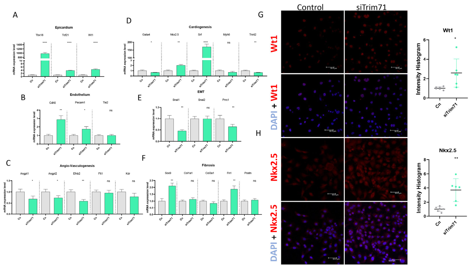
Trim71 inhibition resulted in increased expression of key epicardial markers, including transcription factors Tbx18, Tcf21, and Wt1, all of which are essential for proper epicardial formation and development (Figure 2A). We next examined the effect of Trim71 loss of function on the expression of endothelial markers, including Pecam1 and Tie2. Among these, Pecam1 expression was specifically increased following Trim71 inhibition (Figure 2B). The epicardium also plays a pivotal role in coronary vessel formation. In this context, Trim71 inhibition negatively affected this process, resulting in reduced expression of the vascular-associated markers Angpt1, Angpt22, and Efnb2, while no significant effect was observed in Flt1 and Kdr expression (Figure 2C).
We also evaluated the effect of Trim71 loss of function on the expression of cardiogenic markers. Trim71 inhibition resulted in increased expression of early cardiogenic markers such as Nkx2.5 and Srf, key players in cardiomyocyte differentiation. However, a negative effect was observed on the expression of Gata4 and Tnnt2, with no effect observed in Myh6 (Figure 2D).
Furthermore, we also analyzed the expression of EMT markers such as Snai1 and Prrx1, and both of them were downregulated upon Trim71 inhibition; however, no significant effect was observed in Snai2 expression. In contrast, Cdh5, an endothelial-enriched cadherin, displayed increased expression similarly to Pecam (Figure 2E–B). These results suggest that, in addition to regulating epicardial lineage identity, Trim71 may be involved in maintaining epicardial cells in a state that restricts premature commitment toward an endothelial-like lineage. Finally, we assessed the expression of fibrosis-associated genes and observed that Sox9 and Fn1 were upregulated following Trim71 inhibition (Figure 2F). Fn1 encodes an ECM protein required for cell migration, whereas Sox9 is a known regulator of fibrogenic processes. In contrast, no changes in the expression of Col1a1, Col3a1, and Postn were observed (Figure 2F). These data were validated at protein levels by immunofluorescent assays for Wt1 (Figure 2G) and Nkx2.5 (Figure 2H), observing an increased expression after the loss of function of Trim71.
Together, these results indicate that Trim71 loss of function promotes the expression of epicardial determination genes while attenuating EMT and vascular-associated programs. This transcriptional profile supports a role for Trim71 in maintaining epicardial cells in a progenitor-like state and limiting premature lineage commitment during epicardial development.

3.3. Reciprocal Regulation Between Trim71 and Let-7c/miR-30c During the PE to EE Transition

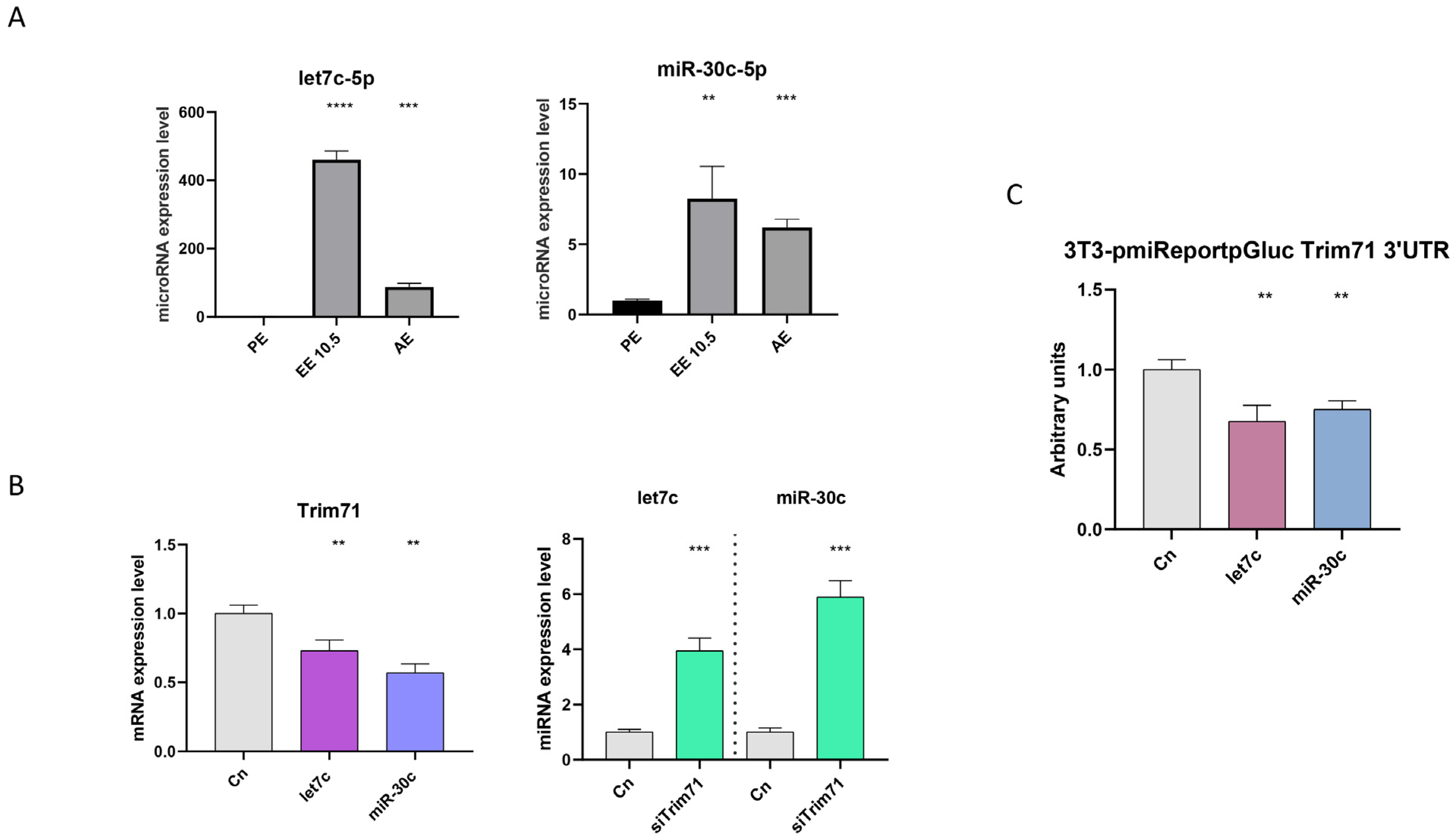
Post-transcriptional regulation is fundamental for proper heart development, enabling the modulation of gene expression in response to developmental cues. Among post-transcriptional regulators, miRNAs play a central role by binding to the 3′UTRs of target mRNAs, thereby repressing their translation or promoting their degradation [42]. Our group has previously shown that the loss of function of let-7c and miR-30c leads to increased Trim71 expression in both Mec1 cells and in primary epicardial cells isolated from E10.5 ventricles [30]. Both let-7c and miR-30c display differential upregulation in the EE at E10.5 compared to the PE, and they maintain detectable expression levels in the adult epicardium (Figure 3A). Additionally, Trim71 expression is significantly reduced after the gain of function of either let-7c or miR-30c. Conversely, Trim71 knockdown by siRNA resulted in the increased expression of both miRNAs, indicating the existence of a reciprocal negative feedback mechanism between Trim71 and these miRNAs (Figure 3B).
To determine whether let-7c and miR-30c directly target Trim71, we analyzed the Trim71 3′UTR sequence and identified multiple conserved binding sites for the seed sequences of both miRNAs (Supplementary Figure S2C). This interaction was subsequently validated using a luciferase reporter assay in 3T3 cells, given that these cell types have basal let-7c or miR-30c expression levels. The overexpression of let-7c or miR-30c resulted in a significant reduction in luciferase activity, confirming that both miRNAs directly target the Trim71 3′UTR (Figure 3C).
Together, these results demonstrate that Trim71 is directly regulated by let-7c and miR-30c through a post-transcriptional feedback loop. This regulatory axis provides a robust mechanism to fine-tune Trim71 expression during epicardial development, supporting a dynamic balance between progenitor maintenance and differentiation-associated processes.

3.4. Let-7c and miR-30c Gain-of-Function Mimic Key Aspects of Trim71 Loss of Function in Epicardial Cells

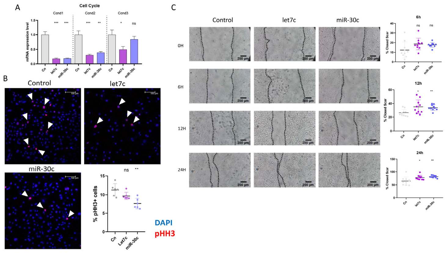
Based on our previously obtained results, we next sought to evaluate whether these miRNAs could recapitulate the phenotypes observed upon Trim71 loss of function. Notably, the gain of function of both let-7c and miR-30c led to a marked reduction in the expression of the cell cycle markers Ccnd1 and Ccnd2, whereas Ccnd3 expression was specifically decreased upon let-7c overexpression (Figure 4A). In addition, overexpression of miR-30c resulted in a reduced number of proliferative cells; conversely, the gain of function of let 7c did not produce statistically significant effects (Figure 4B), further supporting a decrease in proliferative activity. These effects are fully consistent with the impaired proliferative capacity observed following Trim71 inhibition.
In line with our previous observations demonstrating a role for let-7c in the regulation of epicardial cell migration ex vivo [31], the overexpression of let-7c, as well as miR-30c, significantly promoted epicardial cell migration in vitro at 12 and 24 h (Figure 4C). We further analyzed whether the effect of let-7c and miR-30c gain of function could recapitulate the increased cell size observed after Trim71 inhibition in Mec1 cells. While no significant changes in cell area were found, a significant reduction in phalloidin staining intensity was detected after let-7c overexpression, indicating a decrease in actin polymerization or a reorganization of the actin cytoskeleton (Supplementary Figure S4B). This migratory response in Mec1 cells closely resembles the phenotype observed upon Trim71 loss of function, further supporting a functional connection between these miRNAs and Trim71 in the regulation of epicardial cell behavior.
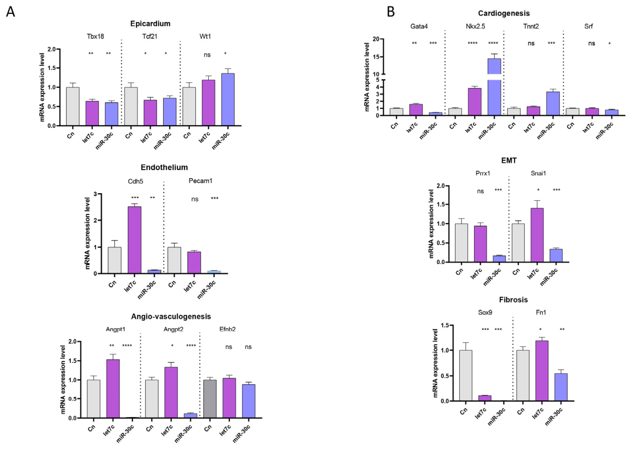
We next assessed whether the gain of function of let-7c and miR-30c was sufficient to recapitulate the effects of Trim71 inhibition on epicardial lineage markers. While Wt1 expression remained unchanged after let-7c modulation, it was significantly increased following miR-30c overexpression. In contrast, the expression of Tbx18 and Tcf21 was reduced after the gain of function of both miRNAs (Figure 5A). Regarding endothelial markers, Cdh5 mRNA expression was increased after let-7c gain of function, in contrast with the negative effect observed in miR-30c treatment. Moreover, let-7c overexpression had no effect on Pecam1 expression, whereas miR-30c gain of function resulted in a reduction of Pecam1 levels (Figure 5A). A similar negative effect on the angio-vasculogenesis markers Anggpt1 and Anggpt2 was observed after miR-30c overexpression, resembling the response seen after Trim71 inhibition. In contrast, these markers were increased following let-7c overexpression. Moreover, overexpression of both microRNAs has no effect on the expression of Efnb2 (Figure 5A).
We subsequently analyzed the impact of miRNA overexpression on cardiogenic-related genes, including Gata4, Nkx2.5, Tnnt2, and Srf. A differential regulation of Gata4 expression was observed, with upregulation after let-7c gain of function and downregulation following miR-30c overexpression, similarly to the effect observed after Trim71 inhibition. Nkx2.5 expression was increased upon gain of function of both miRNAs, consistent with Trim71 loss of function, whereas Tnnt2 expression was unaffected by let-7c but upregulated following miR-30c overexpression. In addition, Srf displays no significant modulation after let-7c gain of function, whereas overexpression of miR-30c decreases Srf expression (Figure 5B).
Concerning EMT-associated markers, let-7c overexpression positively regulated the expression of Snai1 and Cdh5, while Prrx1 levels remained unchanged. Conversely, miR-30c appeared to negatively regulate the EMT process, as indicated by reduced expression of Snai1 and Prrx1, mirroring the effects observed upon Trim71 inhibition (Figure 5B). Finally, fibrosis-associated markers, Sox9 and Fn1, that were upregulated following Trim71 inhibition displayed a differential modulation in response to miRNA overexpression. While Sox9 expression was reduced in both miRNA gain-of-function conditions, Fn1 expression was increased after let-7c overexpression and decreased following miR-30c treatment (Figure 5B).
Taken together, these results indicate that let-7c and miR-30c modulate key aspects of epicardial cell behavior that overlap with, but do not fully recapitulate, the effects observed after Trim71 loss of function. Both miRNAs regulate epicardial proliferation and migration in a manner consistent with Trim71 repression, supporting their functional interaction within a shared regulatory axis. However, their differential and sometimes opposing effects on epicardial, cardiogenic, endothelial, and EMT-associated markers suggest that miRNA-mediated control of Trim71 represents only one layer of a broader regulatory network. These findings highlight the complexity of post-transcriptional regulation during epicardial maturation and point out Trim71 as an integrative node for which its activity is fine-tuned by distinct miRNAs to coordinate multiple developmental programs. In summary, our results identify Trim71 as a key regulator of epicardial cell behavior during the PE to EE transition. Trim71 maintains epicardial cells in a proliferative, progenitor-like state while limiting migration and premature lineage commitment. This function is dynamically regulated by the miRNAs let-7c and miR-30c through a reciprocal feedback mechanism.

4. Discussion

Epicardial development is a highly dynamic and tightly regulated process that is essential for proper heart morphogenesis. Beyond serving as a progenitor source of cells, the embryonic epicardium functions as a signaling hub that orchestrates myocardial growth, coronary vessel formation, extracellular matrix deposition, and immune cell recruitment [8,15]. Consequently, precise temporal coordination of epicardial proliferation, migration, EMT, and lineage commitment is critical, and disruptions in these processes are known to result in congenital heart defects and adult cardiac pathologies [8,15,43].
In this study, we identify Trim71 as a key regulator of the transition between the PE and the EE. Trim71 is highly expressed in the PE, where epicardial progenitors expand, and it is downregulated as cells acquire migratory competence and mature into the EE. Functionally, inhibition of Trim71 decreases epicardial cell proliferation while promoting migration, consistent with a role in maintaining a progenitor-like state. This behavior mirrors the Trim71 functions described in other developmental contexts, particularly during early neurogenesis, where Trim71 sustains progenitor proliferation and prevents premature differentiation [31,44,45]. Consistent with these observations, the decline in Trim71 expression during epicardial development coincides with the acquisition of the migratory capacity required for epicardial expansion [7].
Beyond its effects on proliferation and migration, Trim71 also influences epicardial lineage-associated gene markers. Loss of Trim71 resulted in increased expression of core epicardial transcription factors, including Tbx18, Tcf21, and Wt1 while attenuating EMT- and vascular-associated gene expression. These findings suggest that Trim71 contributes to maintaining epicardial cells in a progenitor-like state, preventing premature lineage commitment. Notably, although some cardiogenic and endothelial markers were modulated upon Trim71 loss of function, the overall transcriptional profile indicates selective and context-dependent regulation. This reinforces the idea that Trim71 does not impose a specific fate but instead modulates the preparation of epicardial cells to respond to developmental signals.
A central mechanism underlying these effects is the reciprocal regulation between Trim71 and specific microRNAs. Members of the let-7 family are well-established developmental regulators that promote differentiation and cell cycle progression in different contexts [46,47,48]. Trim71 was originally identified as a conserved let-7 target, forming a regulatory loop that balances progenitor maintenance and differentiation [49]. Moreover, Trim71 has been shown to actively regulate miRNA pathways by promoting pre-let-7 degradation through interactions with LIN28 and TUT4, repressing mature let-7 activity via RNA-dependent interactions with RISC components, and controlling the biogenesis of other miRNAs such as miR-29a [32,50]. These findings position Trim71 as a broad modulator of post-transcriptional gene regulation rather than a simple miRNA target. In the epicardial context, we show that let-7c and miR-30c are upregulated in the EE compared to the PE, coinciding with Trim71 downregulation. The gain of function of these miRNAs reduced Trim71 expression and recapitulated key aspects of the Trim71 loss-of-function phenotype, including reduced proliferation and enhanced migration. In addition, Trim71 loss-of-function led to increased expression of both miRNAs, revealing a negative feedback loop. This reciprocal regulation provides a robust post-transcriptional mechanism to lower Trim71 levels during the PE to EE transition, ensuring a gradual and coordinated shift from a proliferative to a migratory epicardial state. Notably, miRNA overexpression only partially mimicked Trim71 loss of function regarding epicardial identity markers. While proliferation and migration were strongly affected, modulation of EMT, endothelial-associated, and extracellular matrix-related genes was more limited and, in some cases, divergent between let-7c and miR-30c. Current evidence shows that in other biological contexts, let-7c controls cell proliferation and migration by targeting HoxB7, Hoxa1, and E2f5 [48,49,51]. Similarly, miR-30c has been reported as a regulator of Sox9, Bcl2, and Mapk1 expression, thereby controlling cell migration and proliferation in different oncogenic settings [52,53,54]. This partial mimicry suggests that miRNA-mediated repression of Trim71 controls epicardial maturation rather than acting as an on/off switch and that Trim71 likely integrates additional regulatory inputs to coordinate multiple aspects of PE to EE progression. Such multilayered regulation may be particularly important during early epicardial development, where cells must maintain plasticity while responding to rapidly changing spatial and temporal cues.
It is important to acknowledge the limitations of this study. Our conclusions are primarily based on in vitro analyses using Mec1 epicardial cell lines. While this model enables precise manipulation of gene expression and dissection of cell-autonomous mechanisms governing PE- to EE-like transitions, it cannot fully recapitulate the complex tissue interactions and biomechanical forces present in vivo. Nevertheless, the consistency of our findings with established roles of Trim71 and miRNA-mediated regulation in other developmental systems supports their physiological relevance. Future in vivo studies will be required to determine how the Trim71–miRNA axis integrates with myocardial-derived signals to control epicardial development and cardiac morphogenesis.
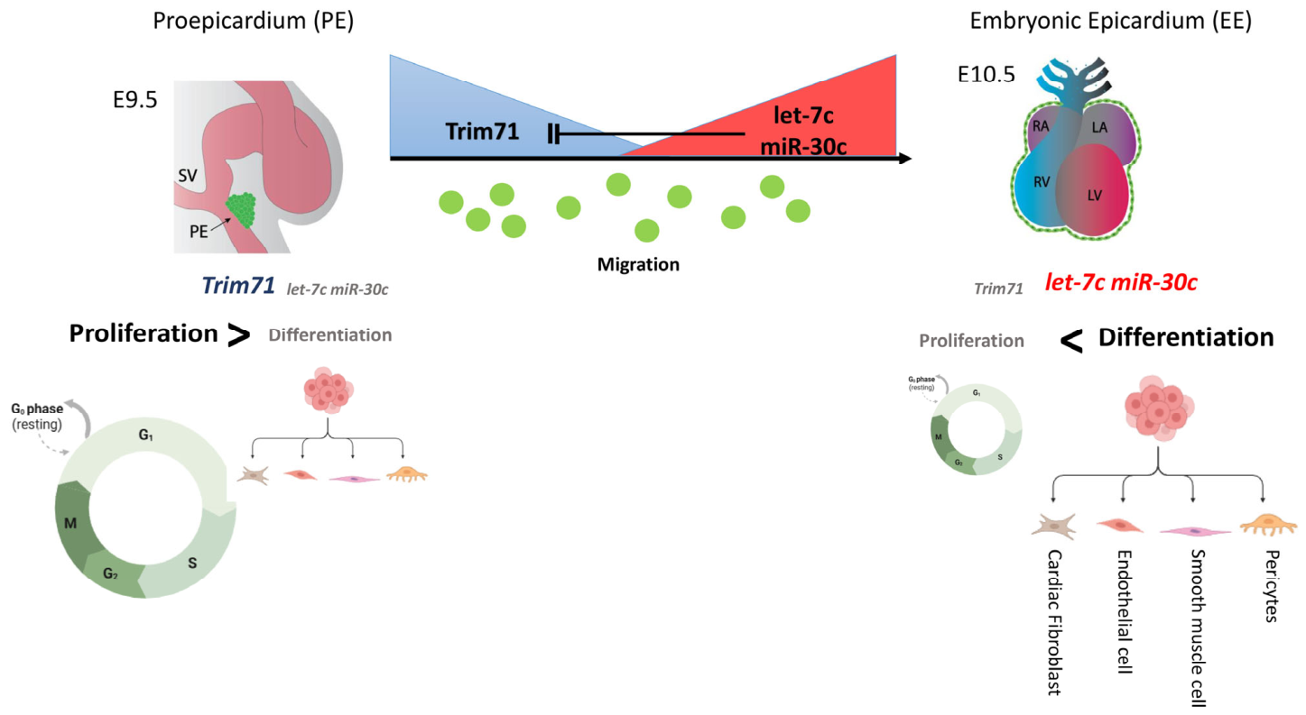
In conclusion, our results support a model in which Trim71 functions as a temporal regulator of epicardial maturation during the PE (E9.5) to EE (E10.5) transition. High Trim71 expression in the PE sustains progenitor expansion and limits premature migration by restraining epicardial cell commitment observed in the regulation of epicardial gene markers. The repression of Trim71 by let-7c and miR-30c induces migratory competence, which is essential for proper epicardial development. This regulatory axis ensures that epicardial cells reach the myocardial surface at the appropriate developmental stage, preserving both progenitor pool size and functional flexibility (Figure 6). Nonetheless, it is important to highlight that the usage of Mec1 epicardial cells, a cell line derived from E13.5 mouse ventricular epicardial explants, might limit the extrapolation of these results in vivo, since it likely models EE cells. Therefore, additional experiments will be required in vivo to validate this regulatory axis.

Supplementary Materials

The following supporting information can be downloaded at https://www.mdpi.com/article/10.3390/jcdd13060237/s1.

Author Contributions

Conceptualization, J.M.C.-C.; methodology, E.L.-V.; formal analysis, J.M.C.-C. and C.G.-P.; resources, R.C.; data curation, J.M.C.-C., C.G.-P. and E.L.-V.; writing—original draft preparation, J.M.C.-C., E.L.-V. and D.F.; writing—review and editing, R.C., E.L.-V. and D.F.; funding acquisition, D.F. All authors have read and agreed to the published version of this manuscript.

Funding

This research was funded by Agencia Estatal de Investigación, Ministerio de Innovación y Ciencia of the Spanish Government (grant number PID2022-138163OB-C32) and Consejería de Universidad, Investigación e Innovación of the Junta de Andalucia Regional Council (grant number ProyExcel_00409).

Institutional Review Board Statement

All experimental procedures were conducted in accordance with the guidelines and approved consent of the Ethics Committee of the University of Jaén and Andalusian Regional Government (14/03/2022/038, 14 March 2022).

Data Availability Statement

The original contributions presented in this study are included in this article/Supplementary Materials. Further inquiries can be directed to the corresponding author.

Conflicts of Interest

The authors declare no conflicts of interest.

References

  1. Quijada, P.; Trembley, M.A.; Small, E.M. The Role of the Epicardium during Heart Development and Repair. Circ. Res. 2020, 126, 377–394. [Google Scholar] [CrossRef] [Scilit]
  2. Masters, M.; Riley, P.R. The epicardium signals the way towards heart regeneration. Stem Cell Res. 2014, 13, 683–692. [Google Scholar] [CrossRef] [Scilit] [PubMed]
  3. Limana, F.; Capogrossi, M.C.; Germani, A. The epicardium in cardiac repair: From the stem cell view. Pharmacol. Ther. 2011, 129, 82–96. [Google Scholar] [CrossRef] [Scilit]
  4. Serluca, F.C. Development of the proepicardial organ in the zebrafish. Dev. Biol. 2008, 315, 18–27. [Google Scholar] [CrossRef] [Scilit] [PubMed]
  5. Jahr, M.; Schlueter, J.; Brand, T.; Männer, J. Development of the proepicardium in Xenopus laevis. Dev. Dyn. 2008, 237, 3088–3096. [Google Scholar] [CrossRef] [Scilit]
  6. Fransen, M.E.; Lemanski, L.F. Epicardial development in the axolotl, Ambystoma mexicanum. Anat. Rec. 1990, 226, 228–236. [Google Scholar] [CrossRef] [Scilit]
  7. Cao, Y.; Duca, S.; Cao, J. Epicardium in Heart Development. Cold Spring Harb. Perspect. Biol. 2020, 12, a037192. [Google Scholar] [CrossRef] [Scilit]
  8. Carmona, R.; Guadix, J.A.; Cano, E.; Ruiz-Villalba, A.; Portillo-Sánchez, V.; Pérez-Pomares, J.M.; Muñoz-Chápuli, R. The embryonic epicardium: An essential element of cardiac development. J. Cell Mol. Med. 2010, 14, 2066–2072. [Google Scholar] [CrossRef] [Scilit]
  9. Lamouille, S.; Xu, J.; Derynck, R. Molecular mechanisms of epithelial–mesenchymal transition. Nat. Rev. Mol. Cell Biol. 2014, 15, 178–196. [Google Scholar] [CrossRef] [Scilit] [PubMed]
  10. Pérez-Pomares, J.M.; Carmona, R.; González-Iriarte, M.; Atencia, G.; Wessels, A.; Muñoz-Chápuli, R. Origin of coronary endothelial cells from epicardial mesothelium in avian embryos. Int. J. Dev. Biol. 2002, 46, 1005–1013. [Google Scholar] [CrossRef] [Scilit] [PubMed]
  11. Mikawa, T.; Gourdie, R.G. Pericardial Mesoderm Generates a Population of Coronary Smooth Muscle Cells Migrating into the Heart along with Ingrowth of the Epicardial Organ. Dev. Biol. 1996, 174, 221–232. [Google Scholar] [CrossRef] [Scilit] [PubMed]
  12. Cao, J.; Poss, K.D. The epicardium as a hub for heart regeneration. Nat. Rev. Cardiol. 2018, 15, 631–647. [Google Scholar] [CrossRef] [Scilit] [PubMed]
  13. Simões, F.C.; Riley, P.R. The ontogeny, activation and function of the epicardium during heart development and regeneration. Development 2018, 145, dev155994. [Google Scholar] [CrossRef] [Scilit]
  14. Tian, X.; Pu, W.T.; Zhou, B. Cellular Origin and Developmental Program of Coronary Angiogenesis. Circ. Res. 2015, 116, 515–530. [Google Scholar] [CrossRef] [Scilit]
  15. Lie-Venema, H.; van den Akker, N.M.S.; Bax, N.A.M.; Winter, E.M.; Maas, S.; Kekarainen, T.; Hoeben, R.C.; deRuiter, M.C.; Poelmann, R.E.; Gittenberger-de Groot, A.C. Origin, Fate, and Function of Epicardium-Derived Cells (EPDCs) in Normal and Abnormal Cardiac Development. Sci. World J. 2007, 7, 1777–1798. [Google Scholar] [CrossRef] [Scilit] [PubMed]
  16. Gittenberger-de Groot, A.C.; Winter, E.M.; Goumans, M.J.; Bartelings, M.M.; Poelmann, R.E. The Arterial Epicardium: A Developmental Approach to Cardiac Disease and Repair. In Etiology and Morphogenesis of Congenital Heart Disease; Springer: Tokyo, Japan, 2016; pp. 8–11. [Google Scholar] [CrossRef] [Scilit]
  17. Briggs, L.E.; Kakarla, J.; Wessels, A. The pathogenesis of atrial and atrioventricular septal defects with special emphasis on the role of the dorsal mesenchymal protrusion. Differentiation 2012, 84, 117–130. [Google Scholar] [CrossRef] [Scilit]
  18. Gittenberger-de Groot, A.C.; Vrancken Peeters, M.P.F.M.; Bergwerff, M.; Mentink, M.M.T.; Poelmann, R.E. Epicardial Outgrowth Inhibition Leads to Compensatory Mesothelial Outflow Tract Collar and Abnormal Cardiac Septation and Coronary Formation. Circ. Res. 2000, 87, 969–971. [Google Scholar] [CrossRef] [Scilit]
  19. Kathiriya, I.S.; Nora, E.P.; Bruneau, B.G. Investigating the transcriptional control of cardiovascular development. Circ. Res. 2015, 116, 700–714. [Google Scholar] [CrossRef] [Scilit] [PubMed]
  20. Greulich, F.; Farin, H.F.; Schuster-Gossler, K.; Kispert, A. Tbx18 function in epicardial development. Cardiovasc. Res. 2012, 96, 476–483. [Google Scholar] [CrossRef] [Scilit]
  21. von Gise, A.; Zhou, B.; Honor, L.B.; Ma, Q.; Petryk, A.; Pu, W.T. WT1 regulates epicardial epithelial to mesenchymal transition through β-catenin and retinoic acid signaling pathways. Dev. Biol. 2011, 356, 421–431. [Google Scholar] [CrossRef] [Scilit]
  22. Grieskamp, T.; Rudat, C.; Lüdtke, T.H.W.; Norden, J.; Kispert, A. Notch Signaling Regulates Smooth Muscle Differentiation of Epicardium-Derived Cells. Circ. Res. 2011, 108, 813–823. [Google Scholar] [CrossRef] [Scilit]
  23. Leung, O.; Zhou, B.; Lui, K. Vascular Development and Regeneration in the Mammalian Heart. Dev. Dis. 2016, 3, 23. [Google Scholar] [CrossRef] [Scilit]
  24. Sayed, D.; Abdellatif, M. MicroRNAs in Development and Disease. Physiol. Rev. 2011, 91, 827–887. [Google Scholar] [CrossRef] [Scilit]
  25. Jonas, S.; Izaurralde, E. Towards a molecular understanding of microRNA-mediated gene silencing. Nat. Rev. Genet. 2015, 16, 421–433. [Google Scholar] [CrossRef] [Scilit] [PubMed]
  26. Treiber, T.; Treiber, N.; Meister, G. Regulation of microRNA biogenesis and function. Thromb. Haemost. 2012, 107, 605–610. [Google Scholar] [CrossRef] [Scilit] [PubMed]
  27. Krol, J.; Loedige, I.; Filipowicz, W. The widespread regulation of microRNA biogenesis, function and decay. Nat. Rev. Genet. 2010, 11, 597–610. [Google Scholar] [CrossRef] [Scilit]
  28. Zilberman, D.; Cao, X.; Jacobsen, S.E. ARGONAUTE4 Control of Locus-Specific siRNA Accumulation and DNA and Histone Methylation. Science 2003, 299, 716–719. [Google Scholar] [CrossRef] [Scilit] [PubMed]
  29. Singh, M.K.; Lu, M.M.; Massera, D.; Epstein, J.A. MicroRNA-processing Enzyme Dicer Is Required in Epicardium for Coronary Vasculature Development. J. Biol. Chem. 2011, 286, 41036–41045. [Google Scholar] [CrossRef] [Scilit]
  30. Castillo-Casas, J.M.; Dueñas, Á.; Hernández-Torres, F.; Carmona, R.; Muñoz-Chápuli, R.; Dopazo, A.; Álvarez, R.; de Luis, E.V.; Aranega, A.E.; Franco, D.; et al. Foxf1-mediated co-regulation of miR-495 and let-7c modulates epicardial cell migration and myocardial specification. Cell. Mol. Life Sci. 2025, 82, 254. [Google Scholar] [CrossRef] [Scilit]
  31. Loedige, I.; Gaidatzis, D.; Sack, R.; Meister, G.; Filipowicz, W. The mammalian TRIM-NHL protein TRIM71/LIN-41 is a repressor of mRNA function. Nucleic Acids Res. 2013, 41, 518–532. [Google Scholar] [CrossRef] [Scilit]
  32. Treiber, T.; Treiber, N.; Plessmann, U.; Harlander, S.; Daiß, J.L.; Eichner, N.; Lehmann, G.; Schall, K.; Urlaub, H.; Meister, G. A Compendium of RNA-Binding Proteins that Regulate MicroRNA Biogenesis. Mol. Cell 2017, 66, 270–284.e13. [Google Scholar] [CrossRef] [Scilit]
  33. Lozano-Velasco, E.; Vallejo, D.; Esteban, F.J.; Doherty, C.; Hernández-Torres, F.; Franco, D.; Aránega, A.E. A Pitx2-MicroRNA Pathway Modulates Cell Proliferation in Myoblasts and Skeletal-Muscle Satellite Cells and Promotes Their Commitment to a Myogenic Cell Fate. Mol. Cell Biol. 2015, 35, 2892–2909. [Google Scholar] [CrossRef] [Scilit]
  34. Bustin, S.A.; Benes, V.; Garson, J.A.; Hellemans, J.; Huggett, J.; Kubista, M.; Mueller, R.; Nolan, T.; Pfaffl, M.W.; Shipley, G.L.; et al. The MIQE Guidelines: Minimum Information for Publication of Quantitative Real-Time PCR Experiments. Clin. Chem. 2009, 55, 611–622. [Google Scholar] [CrossRef] [Scilit] [PubMed]
  35. Livak, K.J.; Schmittgen, T.D. Analysis of Relative Gene Expression Data Using Real-Time Quantitative PCR and the 2−ΔΔCT Method. Methods 2001, 25, 402–408. [Google Scholar] [CrossRef] [Scilit] [PubMed]
  36. Ascione, F.; Vasaturo, A.; Caserta, S.; D’Esposito, V.; Formisano, P.; Guido, S. Comparison between fibroblast wound healing and cell random migration assays in vitro. Exp. Cell Res. 2016, 347, 123–132. [Google Scholar] [CrossRef] [Scilit]
  37. Toro, R.; Pérez-Serra, A.; Mangas, A.; Campuzano, O.; Sarquella-Brugada, G.; Quezada-Feijoo, M.; Ramos, M.; Alcalá, M.; Carrera, E.; García-Padilla, C.; et al. miR-16-5p Suppression Protects Human Cardiomyocytes against Endoplasmic Reticulum and Oxidative Stress-Induced Injury. Int. J. Mol. Sci. 2022, 23, 1036. [Google Scholar] [CrossRef] [Scilit] [PubMed]
  38. García-Padilla, C.; Lozano-Velasco, E.; García-López, V.; Aránega, A.; Franco, D.; García-Martínez, V.; López-Sánchez, C. miR-1 as a Key Epigenetic Regulator in Early Differentiation of Cardiac Sinoatrial Region. Int. J. Mol. Sci. 2024, 25, 6608. [Google Scholar] [CrossRef] [Scilit]
  39. Bonet, F.; Dueñas, Á.; López-Sánchez, C.; García-Martínez, V.; Aránega, A.E.; Franco, D. MiR-23b and miR-199a impair epithelial-to-mesenchymal transition during atrioventricular endocardial cushion formation. Dev. Dyn. 2015, 244, 1259–1275. [Google Scholar] [CrossRef] [Scilit]
  40. Torres-Fernández, L.A.; Mitschka, S.; Ulas, T.; Weise, S.; Dahm, K.; Becker, M.; Händler, K.; Beyer, M.; Windhausen, J.; Schultze, J.L.; et al. The stem cell–specific protein TRIM71 inhibits maturation and activity of the prodifferentiation miRNA let-7 via two independent molecular mechanisms. RNA 2021, 27, 805–828. [Google Scholar] [CrossRef] [Scilit]
  41. Carmona, R.; Barrena, S.; López Gambero, A.J.; Rojas, A.; Muñoz-Chápuli, R. Epicardial cell lineages and the origin of the coronary endothelium. FASEB J. 2020, 34, 5223–5239. [Google Scholar] [CrossRef] [Scilit]
  42. Lozano-Velasco, E.; Garcia-Padilla, C.; del Mar Muñoz-Gallardo, M.; Martinez-Amaro, F.J.; Caño-Carrillo, S.; Castillo-Casas, J.M.; Sanchez-Fernandez, C.; Aranega, A.E.; Franco, D. Post-Transcriptional Regulation of Molecular Determinants during Cardiogenesis. Int. J. Mol. Sci. 2022, 23, 2839. [Google Scholar] [CrossRef] [Scilit]
  43. Sanchez-Fernandez, C.; Rodriguez-Outeiriño, L.; Matias-Valiente, L.; Ramírez de Acuña, F.; Franco, D.; Aránega, A.E. Understanding Epicardial Cell Heterogeneity during Cardiogenesis and Heart Regeneration. J. Cardiovasc. Dev. Dis. 2023, 10, 376. [Google Scholar] [CrossRef] [Scilit] [PubMed]
  44. Duy, P.Q.; Jux, B.; Zhao, S.; Mekbib, K.Y.; Dennis, E.; Dong, W.; Nelson-Williams, C.; Mehta, N.H.; Shohfi, J.P.; Juusola, J.; et al. TRIM71 mutations cause a neurodevelopmental syndrome featuring ventriculomegaly and hydrocephalus. Brain 2024, 147, 4292–4305. [Google Scholar] [CrossRef] [Scilit]
  45. Duy, P.Q.; Furey, C.G.; Kahle, K.T. Trim71/lin-41 Links an Ancient miRNA Pathway to Human Congenital Hydrocephalus. Trends Mol. Med. 2019, 25, 467–469. [Google Scholar] [CrossRef] [Scilit] [PubMed]
  46. Liu, H.Y.; Huang, C.M.; Hung, Y.F.; Hsueh, Y.P. The microRNAs Let7c and miR21 are recognized by neuronal Toll-like receptor 7 to restrict dendritic growth of neurons. Exp. Neurol. 2015, 269, 202–212. [Google Scholar] [CrossRef] [Scilit]
  47. Cai, L.; Wang, Z.; Zheng, H.; Xu, L. The let-7c/HoxB7 axis regulates the cell proliferation, migration and apoptosis in hepatocellular carcinoma. Anti-Cancer Drugs 2020, 31, 6–18. [Google Scholar] [CrossRef] [Scilit]
  48. Zhan, M.; Qu, Q.; Wang, G.; Liu, Y.Z.; Tan, S.L.; Lou, X.Y.; Yu, J.; Zhou, H.H. Let-7c Inhibits NSCLC Cell Proliferation by Targeting HOXA1. Asian Pac. J. Cancer Prev. 2013, 14, 387–392. [Google Scholar] [CrossRef] [Scilit]
  49. Liu, Q.; Chen, X.; Novak, M.K.; Zhang, S.; Hu, W. Repressing Ago2 mRNA translation by Trim71 maintains pluripotency through inhibiting let-7 microRNAs. eLife 2021, 10, e66288. [Google Scholar] [CrossRef] [Scilit] [PubMed]
  50. Yin, J.; Kim, T.H.; Park, N.; Shin, D.; Choi, H.I.; Cho, S.; Park, J.B.; Kim, J.H. TRIM71 suppresses tumorigenesis via modulation of Lin28B-let-7-HMGA2 signaling. Oncotarget 2016, 7, 79854–79868. [Google Scholar] [CrossRef] [Scilit]
  51. Huang, M.; Gong, X. Let-7c Inhibits the Proliferation, Invasion, and Migration of Glioma Cells via Targeting E2F5. Oncol. Res. Featur. Preclin. Clin. Cancer Ther. 2018, 26, 1103–1111. [Google Scholar] [CrossRef] [Scilit] [PubMed]
  52. Yuan, L.Q.; Zhang, T.; Xu, L.; Han, H.; Liu, S.H. miR-30c-5p inhibits glioma proliferation and invasion via targeting Bcl2. Transl. Cancer Res. 2021, 10, 337–348. [Google Scholar] [CrossRef] [Scilit]
  53. Zhang, A.; Liu, X.; Wang, J.; Deng, K.; Wang, J. MiR-30c-5p Directly Targets MAPK1 to Regulate the Proliferation, Migration and Invasion of Adenomyotic Epithelial Cells in Adenomyosis. Twin Res. Hum. Genet. 2021, 24, 22–28. [Google Scholar] [CrossRef] [Scilit]
  54. Liu, S.; Li, X.; Zhuang, S. miR-30c Impedes Glioblastoma Cell Proliferation and Migration by Targeting SOX9. Oncol. Res. Featur. Preclin. Clin. Cancer Ther. 2019, 27, 165–171. [Google Scholar] [CrossRef] [Scilit]
Figure 1. (A) RT–qPCR analysis of Trim71 expression in proepicardium (PE), embryonic epicardium (EE), and adult epicardium (AE) (n = 3), showing significantly higher expression in PE compared with later stages. (B) RT–qPCR analysis of cell cycle gene markers (Ccnd1, Ccnd2, and Ccnd3) in Mec1 cells transfected with control siRNA (Cn) or siRNA targeting Trim71 (siTrim71) (n = 3), indicating reduced expression upon Trim71 knockdown. (C) Quantification of the percentage of phospho-histone H3 (pHH3)-positive cells in control and siTrim71-transfected Mec1 cells (n = 6), revealing decreased proliferative activity after Trim71 inhibition. Scale bars represent 50 microns. (D) Scratch wound-healing assay in Mec1 cells transfected with control or siTrim71. Percentage of wound closure was quantified at 6-, 12-, and 24-h post-scratch (n = 10 fields per condition), showing increased migratory capacity following Trim71 silencing. Data are presented as mean ± SEM. Scale bars represent 200 microns. Statistical significance is indicated (* p < 0.05, ** p < 0.01, and *** p < 0.001, **** p < 0.0001) (Cn: control).
Jcdd 13 00237 g001
Figure 2. RT–qPCR analysis of cardiac markers. (A) Epicardial (Tbx18, Tcf21, Wt1), (B) endothelial (Cdh5, Pecam1, Tie2), and (C) angio-vasculogenesis (Angpt1, Angpt2, Efnb2, Flt1, Kdr) markers in Mec1 cells transfected with control siRNA (Cn) or siRNA targeting Trim71 (siTrim71) (n = 3). Trim71 inhibition resulted in increased expression of epicardial transcription factors, whereas angio-vasculogenic and endothelial markers showed gene-specific responses. RT–qPCR analysis of genes associated with (D) cardiogenic (Gata4, Nkx2.5, Srf, Myh6, Tnnt2), (E) EMT (Snai1, Snai2, Prrx1), and (F) matrix-associated/fibrosis-related genes (Sox9, Col1a1, Col3a1, Fn1) (n = 3). Trim71 inhibition selectively altered the expression of a subset of cardiogenic EMT and matrix-associated genes, including increased expression of Sox9 and Fn1, while collagen gene expression remained unchanged. (G,H) Representative images of the immunohistochemical detection of Wt1 and Nkx2.5 in control and si-Trim71 Mec1 cells, demonstrating increased expression at protein levels of these epicardial and cardiomyogenic markers, respectively. Scale bar represents 500 microns. Data are presented as mean ± SEM. Statistical significance is indicated (* p < 0.05, ** p < 0.01, **** p < 0.0001) (Cn: control).
Jcdd 13 00237 g002
Figure 3. (A) RT–qPCR analysis of let-7c-5p and miR-30c-5p expression in proepicardium (PE), embryonic epicardium (EE), and adult epicardium (AE) (n = 3), showing increased expression in the EE compared with the PE and sustained expression in adulthood. (B) RT–qPCR analysis showing reduced Trim71 expression following the gain of function of let-7c or miR-30c in Mec1 cells and increased let-7c and miR-30c expression after Trim71 knockdown (n = 3), indicating a reciprocal negative feedback loop. (C) Luciferase reporter assays showing reduced activity of the Trim71 3′UTR upon overexpression of let-7c or miR-30c (n = 3), confirming direct post-transcriptional regulation. Data are presented as mean ± SEM. Statistical significance is indicated (** p < 0.01, and *** p < 0.001, **** p < 0.0001) (Cn: control).
Jcdd 13 00237 g003
Figure 4. (A) RT–qPCR analysis of cell cycle gene markers (Ccnd1, Ccnd2, Ccnd3) in Mec1 cells transfected with control, let-7c, or miR-30c premiRs (n = 3), showing reduced expression of proliferation-associated markers. (B) Quantification of percentage of phospho-histone H3 (pHH3)-positive cells in Mec1 cells following let-7c or miR-30c overexpression, revealing decreased proliferative activity after miR30-c gain of function. Scale bars represent 100 microns. (C) Scratch wound-healing assays showing the percentage of wound closure at 6, 12, and 24 h after transfection with let-7c or miR-30c premiRs compared with control conditions (n = 10 fields per condition), indicating enhanced migratory capacity. Scale bars represent 200 microns. Data are presented as mean ± SEM. Statistical significance is indicated (* p < 0.05, ** p < 0.01, *** p < 0.001) (Cn: control).
Jcdd 13 00237 g004
Figure 5. (A) RT–qPCR analysis of epicardial (Tbx18, Tcf21, Wt1), endothelial (Pecam1), angiogenic (Angpt1, Angpt2, Efnb2), and EMT (Prrx1, Snai1, Cdh5) markers in Mec1 cells transfected with control, let-7c or miR-30c premiRs (n = 3). miRNA overexpression resulted in selective and gene-specific modulation rather than uniform regulation across lineages. (B) RT–qPCR analysis of cardiogenic (Gata4, Nkx2.5, Tnnt2, Srf), EMT (Prrx1, Snai1), and fibrosis (Sox9, Fn1) markers (n = 3). let-7c and miR-30c displayed distinct and, in some cases, opposing regulatory effects, indicating selective modulation of mesenchymal and extracellular matrix-related components rather than induction of a generalized fibrotic or EMT program. Data are presented as mean ± SEM. Statistical significance is indicated (* p < 0.05, ** p < 0.01, *** p < 0.001, **** p < 0.0001) (Cn: control).
Jcdd 13 00237 g005
Figure 6. Schematic model summarizing the molecular mechanisms governing the PE-to-EE transition. In PE, high Trim71 expression maintains proepicardial cells in a proliferative, progenitor-like state. As development progresses, during epicardial maturation, increased expression of let-7c and miR-30c represses Trim71, leading to reduced proliferation, enhanced migratory competence, and transcriptional priming characteristic of the formation and expansion of the embryonic epicardium (EE).
Jcdd 13 00237 g006
Disclaimer/Publisher’s Note: The statements, opinions and data contained in all publications are solely those of the individual author(s) and contributor(s) and not of MDPI and/or the editor(s). MDPI and/or the editor(s) disclaim responsibility for any injury to people or property resulting from any ideas, methods, instructions or products referred to in the content.