Article

The Influence of Age and Exercise Training Status on Left Ventricular Systolic Twist Mechanics in Healthy Males—An Exploratory Study

by
Alexander J. Beaumont
1,*,
Amy K. Campbell
1,
Viswanath B. Unnithan
2,
David Oxborough
3,
Fergal Grace
4,
Allan Knox
5 and
Nicholas F. Sculthorpe
2
1
School of Science, Technology and Health, York St. John University, York YO31 7EX, UK
2
Institute of Clinical Exercise and Health Sciences, School of Health and Life Sciences, University of the West of Scotland, Hamilton G72, 0LH, UK
3
Research Institute of Sport and Exercise Science, Liverpool John Moores University, Liverpool L3 3AF, UK
4
Faculty of Health, School of Health Science and Psychology, Federation University Australia, Ballarat, VIC 3350, Australia
5
Exercise Science Department, California Lutheran University, Thousand Oaks, CA 91360, USA
*
Author to whom correspondence should be addressed.
J. Cardiovasc. Dev. Dis. 2024, 11(10), 321; https://doi.org/10.3390/jcdd11100321
Submission received: 2 August 2024 / Revised: 4 October 2024 / Accepted: 6 October 2024 / Published: 12 October 2024
(This article belongs to the Section Electrophysiology and Cardiovascular Physiology)

Abstract

:
Age-related differences in twist may be mitigated with exercise training, although this remains inconclusive. Moreover, temporal left ventricular (LV) systolic twist mechanics, including early-systolic (twistearly), and beyond peak twist (twistpeak) alone, have not been considered. Therefore, further insights are required to ascertain the influence of age and training status on twist mechanics across systole. Forty males were included and allocated into 1 of 4 groups based on age and training status: young recreationally active (YRA, n = 9; 28 ± 5 years), old recreationally active (ORA, n = 10; 68 ± 6 years), young trained (YT, n = 10; 27 ± 6 years), and old trained (OT, n = 11, 64 ± 4 years) groups. Two-dimensional speckle-tracking echocardiography was performed to determine LV twist mechanics, including twistearly, twistpeak, and total twist (twisttotal), by considering the nadir on the twist time-curve during early systole. Twisttotal was calculated by subtracting twistearly from their peak values. LV twistpeak was higher in older than younger men (p = 0.036), while twistpeak was lower in the trained than recreationally-active (p = 0.004). Twistpeak is underestimated compared with twisttotal (p < 0.001), and when early-systolic mechanics were considered, to calculate twisttotal, the age effect (p = 0.186) was dampened. LV twist was higher in older than younger age, with lower twist in exercise-trained than recreationally-active males. Twistpeak is underestimated when twistearly is not considered, with novel observations demonstrating that the age effect was dampened when considering twistearly. These findings elucidated a smaller age effect when early phases of systole are considered, while lower LV systolic mechanics were observed in older aged trained than recreationally-active males.

1. Introduction

Healthy, chronological ageing is associated with a multitude of changes within the human physiological system, including the heart and major blood vessels. Even in the absence of systemic or conventional risk factors (smoking, diabetes, hypertension), the ageing heart leads to intrinsic structural and functional deteriorations [1], and together, the gross and cellular modifications within the ageing heart can impair left ventricular (LV) lusitropy and myocardial function [2].
Since pioneering descriptions of the LV rotary motion [3], the scientific community has further elucidated the unique fibre arrangements and architecture of the myocardium, which mechanistically underpins LV twisting mechanics [4]. The absolute difference between basal and apical rotation throughout systole is represented as net LV twist [4], and the twisting motion contributes towards achieving a sufficient ejection fraction [5]. Technological advancements have facilitated the detailed assessment of LV motion, including rotations and twisting during systole [4,6]. Indeed, accumulating evidence suggests that LV twist increases with advancing age [7,8,9,10]. It has been proposed that this age-related increase in twist may be the consequence of subendocardial fibrosis/dysfunction [11,12], thereby reducing the capacity of the endocardium to provide an opposing rotation to the dominant epicardium [13].
The United Kingdom government advocates modifiable lifestyle factors, such as exercise and physical activity, for preventing cardiovascular disease and preserving healthy ageing [14]. It is well established that superior exercise capacity is associated with a greater likelihood of survival [15]. The ‘masters athlete’ represents a useful model to differentiate age-related physiological declines that can be prevented from those which are inevitable [16]. Thus, the masters athlete may be viewed as having the maximum potential for preserving cardiovascular health [17] through the utilisation of a non-pharmacological, behaviour-orientated approach. A recent meta-analysis identified larger LV mass, volume, wall thicknesses, and diastolic function in masters athletes than matched controls [16], commensurate with the ‘athlete’s heart’ phenotype. Long-term exercise training appears to play an effective role in offsetting some of the detrimental changes during advancing age [17]. Although similar ejection fractions and global longitudinal strain have been reported between masters athletes and controls [16], the influence of exercise training in association with age-related systolic twist mechanics is equivocal. Contrasting findings exist as to whether older exercise-trained individuals do [10,18] or do not [19,20] display lower LV twist than their matched untrained counterparts, thus requiring more investigation.
Peak rotation and twist (apicalpeak, basalpeak and twistpeak) are typically reported as the highest value achieved during systole from the ‘zero’ baseline. However, during the preceding isovolumic contraction (IVC) phase, initial electrical activation of the endocardium at the apical septal wall facilitates subendocardial shortening and subepicardial stretching [21,22,23]. This electromechanical sequence of myofibers that precedes LV ejection underpins the brief apical clockwise rotation and basal counterclockwise rotation [4,22,24,25] and is thought to be due to endocardial fibres dominating the direction of rotation [23,24]. Mechanics during this phase of the cardiac cycle may be termed the early-systolic twist (twistearly) and early systolic apical (apicalearly) and basal (basalearly) rotations, and may provide insight to endocardial function and by extension, age-related increase in LV twist. Although these mechanics have been scarcely considered, basalearly rotation has been shown to reduce with age [26,27], which may be sensitive to detect endocardial dysfunction [28]. Moreover, without equal apicalearly, basalearly and twistearly between individuals, the nadir point of LV twist on the twist-time curve will be variable. However, it is unclear if the nadir point influences the total systolic twisting during ejection since opposing apicalearly, basalearly and twistearly are ignored when determining peak values, with the possibility that the total amount (maximum degrees) of twist and rotation is therefore underestimated. This may have important ramifications when attempting to ascertain LV systolic function. As a result, calculating the total amount of counterclockwise twist (as viewed on the twist-time curve) would appropriately determine the total amount of LV twisting during ejection. Furthermore, temporal analysis of LV systolic mechanics has revealed nuanced differences in basalearly and apicalearly between athlete groups [29], yet this has not been explored regarding ageing and training status.
Overall, more research is needed to determine whether chronic exercise training can mitigate age-related differences in LV twist. Additionally, it is not clear if early mechanics prior to ejection (apicalearly, basalearly and twistearly) contribute to differences in observed peak values near end-systole. Therefore, exploratory data are needed to provide scope to further understand the influence of training status as a countermeasure to chronological ageing. Accordingly, this exploratory study aimed to (1) investigate the influence of healthy ageing and exercise training status on LV twist mechanics and (2) further investigate temporal systolic twist mechanics using speckle-tracking echocardiography (STE). In accordance with this, it was hypothesised that LV twist would be higher in older than younger groups yet lower in trained than untrained groups. Secondly, we hypothesised that considering twistearly mechanics would alter twistpeak and, by extension, reduce the age and training status effects.

2. Materials and Methods

2.1. Overview and Participants

Sixty-eight males were initially recruited, and standardised exclusion criteria were applied [30], leading to the exclusion of 28 participants. Twelve older (ORA = 9; OT = 3) adults were excluded due to the presence of (e.g., myocardial infarction, angina, stroke, and peripheral artery disease) and/or treatment (e.g., anti-hypertensives and beta-blockers) for cardiovascular diseases or type 2 diabetes mellitus. Two smokers were excluded (YRA = 2), and two participants violated pre-participation restrictions (YRA = 2). Additionally, seven trained participants were excluded due to either inconsistent training (YT = 3) or having not been training for ≥5 years (OT = 4). Five participants withdrew due to personal reasons. Consequently, 40 males were included and allocated into 1 of 4 groups based on age and training status (young recreationally-active [YRA], n = 9, 28 ± 5 years; young trained [YT], n = 10, 27 ± 6 years; old recreationally-active [ORA], n = 10, 68 ± 6 years; and old trained [OT], n = 11, 64 ± 4 years). The overview of this study, participant baseline characteristics and inclusion/exclusion criteria have been published elsewhere [30]. Here, we present unpublished data pertaining to twist mechanics in relation to ageing and exercise training. However, see Supplementary Material File S1 for a list of baseline characteristics included for the reader’s interest. All participants provided written, informed consent before being enrolled.
As reported by Beaumont et al. [30], the YRA and ORA groups were not engaged with structured exercise habits (YRA, 67 ± 87 min per week, 48.5 ± 5.0 mL·kg−1·min−1; ORA, 63 ± 67 min per week, 34.9 ± 7.3 mL·kg−1·min−1) and all performed < 2 h per week of physical activity. In contrast, the YT (450 ± 239 min per week, 64.1 ± 7.7 mL·kg−1·min−1) group were required to have trained for at least 6 months since LV twist has been shown to increase following six months of training [31]. YT took part in running (n = 3), cycling (n = 2) and both modalities (n = 4) for 5 ± 4 years. The OT (540 ± 180 minutes per week, 50.1 ± 3.6 mL·kg−1·min−1) group were included if they had trained for at least 5 years [10,32,33] and commenced training before 64 years of age, given the reported adaptations in LV compliance following exercise training in those aged 45–64 years of age but not in those aged 65 years and older [17,34]. OT consisted of those involved with running (n = 5), cycling (n = 2) or both modalities (n = 4) for 34 ± 14 years, including three previous international athletes and a half marathon world champion within their age group.

2.2. Protocol and Experimental Procedures

2.2.1. Echocardiography

Standard image acquisition techniques used in this study are presented by Beaumont et al. [30], pertaining to LV structure, conventionally derived function, and the capture of parasternal short-axis views.

2.2.2. Left Ventricular Twist Mechanics

The parasternal short-axis at basal and apical levels was acquired, with the basal plane obtained as circular as possible at the level of the full mitral valve. The apex was captured without the visibility of papillary muscles [35] by tilting the transducer from an original apical 4-chamber orientation and moving slightly to the point above LV luminal obliteration [4,13]. The image with the smallest LV chamber at end-systole was selected for speckle-tracking analysis using dedicated semi-automated software (EchoPac, version 202). Aortic valve closure (AVC) was identified as end-systolic timing from the pulsed wave tracings obtained from the apical 5-chamber LV outflow tract.
Images were recorded at a frame rate of ~71 fps for speckle-tracking analysis at both apical and basal levels. In the instance that two or more segments could not be tracked sufficiently, the image was excluded from analysis. Raw text files were imported into custom software, which applied a 1000-point cubic spline to each of the systolic and diastolic portions of the cardiac cycle (derived from AVC). The splined data were used to identify peaks in IVC during early systole and peaks in twist occurring at end-systole or early diastole. Peak clockwise basal (basalpeak) and counterclockwise apical (apicalpeak) rotation and simultaneous net twist (twistpeak) were identified and also scaled to LV length to determine normalised rotations and torsion, respectively [4]. Basalearly and apicalearly were identified to signify counterclockwise basal and clockwise apical rotation as the highest positive and negative values during early systole prior to the subsequent rotation in the opposing direction. Likewise, twistearly represented the nadir point on the twist-time curve (Figure 1). To determine total rotation (apicaltotal and basaltotal) and twist (twisttotal) after taking into consideration the nadir on the rotation/twist time-curve, apicalearly, basalearly, and twistearly were subtracted from their peak values, respectively (Figure 1). Time-to-peak corresponding to early and peak rotations/twist were calculated as absolute timings (milliseconds). Time displacement represented the absolute time difference between basalpeak and apicalpeak, and was calculated as the difference between time-to-basalpeak and apicalpeak [36].
All images were acquired and analysed by a single sonographer (AB). Within-day test-retest reproducibility was conducted in 8 young individuals and was calculated using the coefficient of variation (basalpeak, 15.1%; apicalpeak, 12.4%; twistpeak, 14.1%). These reproducibility values align with other intra-observer data [37,38].

2.3. Statistical Analysis

All statistical analyses of data were conducted using jamovi (version 0.9 [39]). The influence of ageing and exercise training on markers of LV twist mechanics were analysed using two-way analysis of variance (ANOVA) to assess the main effects of age, training status, and their interaction. In the presence of a statistically significant interaction, Tukey’s post hoc test was used to explore between-group differences. Statistical significance was granted at p ≤ 0.050.

3. Results

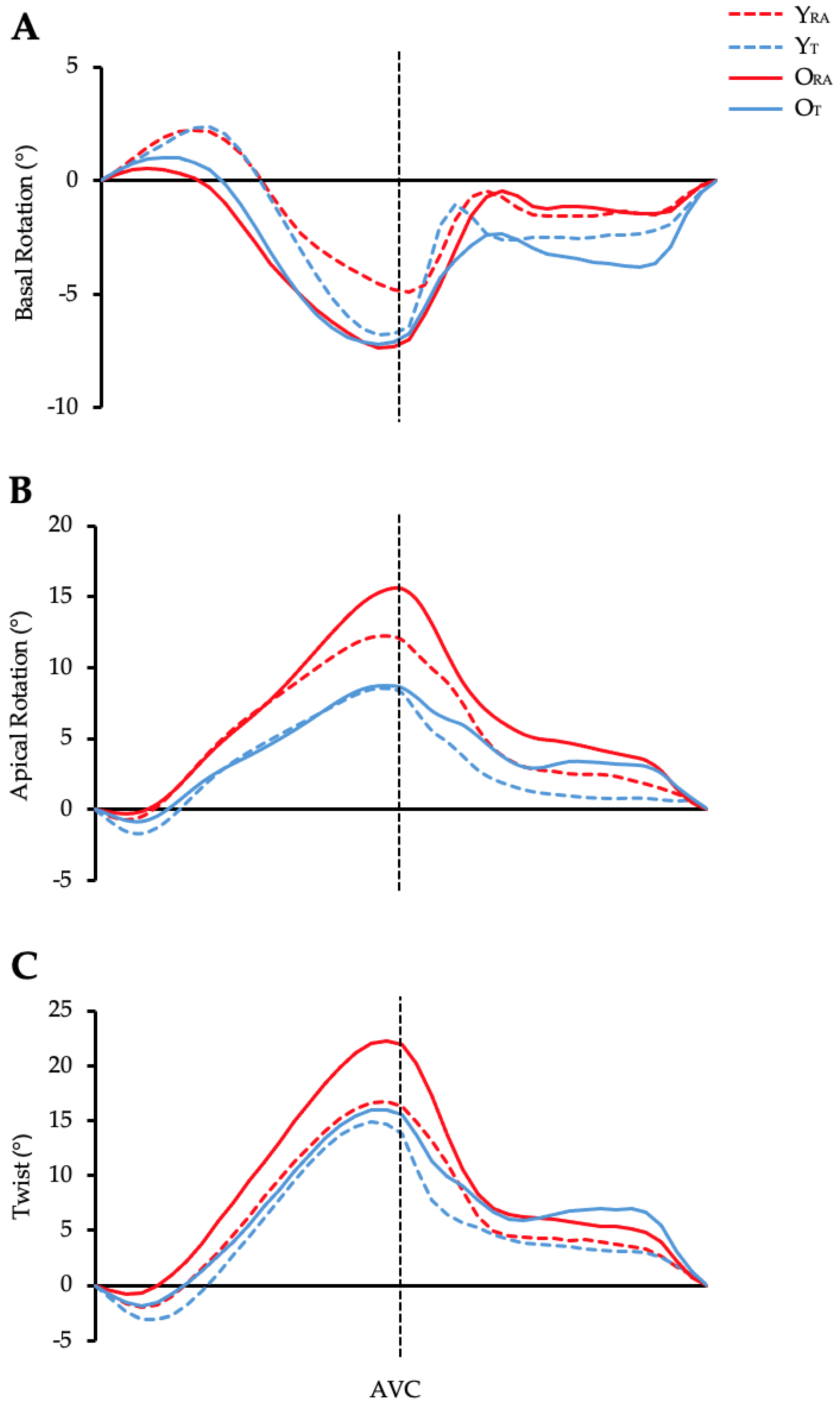
Data pertaining to LV structure, volumes, and function have been published previously [30]; see Supplementary Material Files S2 and S3 for the reader’s interest. Figure 2A–C illustrates average temporal LV rotations and twist between groups.

3.1. Left Ventricular Basal Rotation

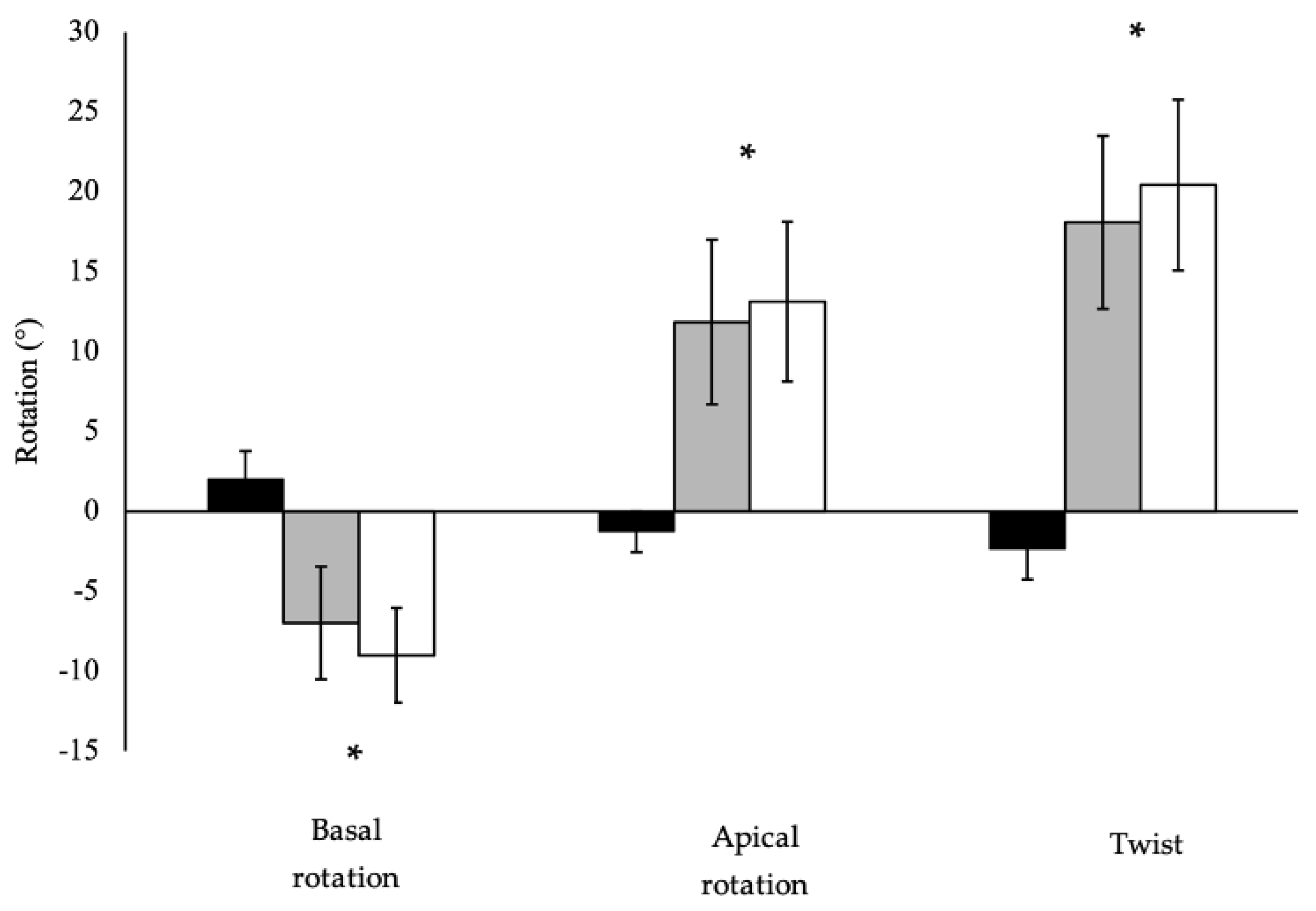
Pooled (n = 40) basalpeak was significantly lower than basaltotal (p < 0.001; Figure 3). Basal rotation and respective timings are presented in Table 1 and Table 2, respectively. Older cohorts demonstrated lower basalearly than younger cohorts (p = 0.025), while basalpeak (p = 0.164), basaltotal (p = 0.841) and normalised basal rotation (p = 0.094) demonstrated no age effect. No significant training status effects or interaction effects were observed for basal rotation for basalearly (p = 0.679; p = 0.742, respectively), basalpeak (p = 0.421; p = 0.482, respectively), basaltotal (p = 0.273; p = 0.568, respectively) or normalised basal rotation (p = 0.421; p = 0.482, respectively). Similarly, time-to-peak basalearly and basalpeak rotation did not differ based on age (p = 0.720; p = 0.251, respectively) or training status (p = 0.534; p = 0.937, respectively), with no interaction (p = 0.942; p = 0.731, respectively).

3.2. Left Ventricular Apical Rotation

Pooled (n = 40) apicalpeak was significantly lower than apicaltotal (p < 0.001; Figure 3). Apical rotation and respective timings are presented in Table 1 and Table 2, respectively. Apicalearly (p = 0.379), apicalpeak (p = 0.206), apicaltotal (p = 0.310), and normalised peak apical rotation (p = 0.105) did not differ between young and old. Similarly, apicalearly demonstrated no significant difference in trained than recreationally-active groups (p = 0.069), whereas trained groups demonstrated significantly lower apicalpeak (p < 0.001), apicaltotal (p = 0.004) and normalised peak apical rotation than recreationally-active groups (p < 0.001). No significant age x training interactions were observed for indices of apicalearly (p = 0.393), apicalpeak (p = 0.173), apicaltotal (p = 0.112) or normalised apical rotation (p = 0.201). Time-to-peak apicalearly and apicalpeak rotation did not differ based on age (p = 0.635; p = 0.204, respectively), or training status (p = 0.295; p = 0.545, respectively), with no interaction (p = 0.980; p = 0.563, respectively).
Time displacement did not differ based on age (p = 0.882) or training status (p = 0.648), with no interaction (p = 0.435, Table 2).

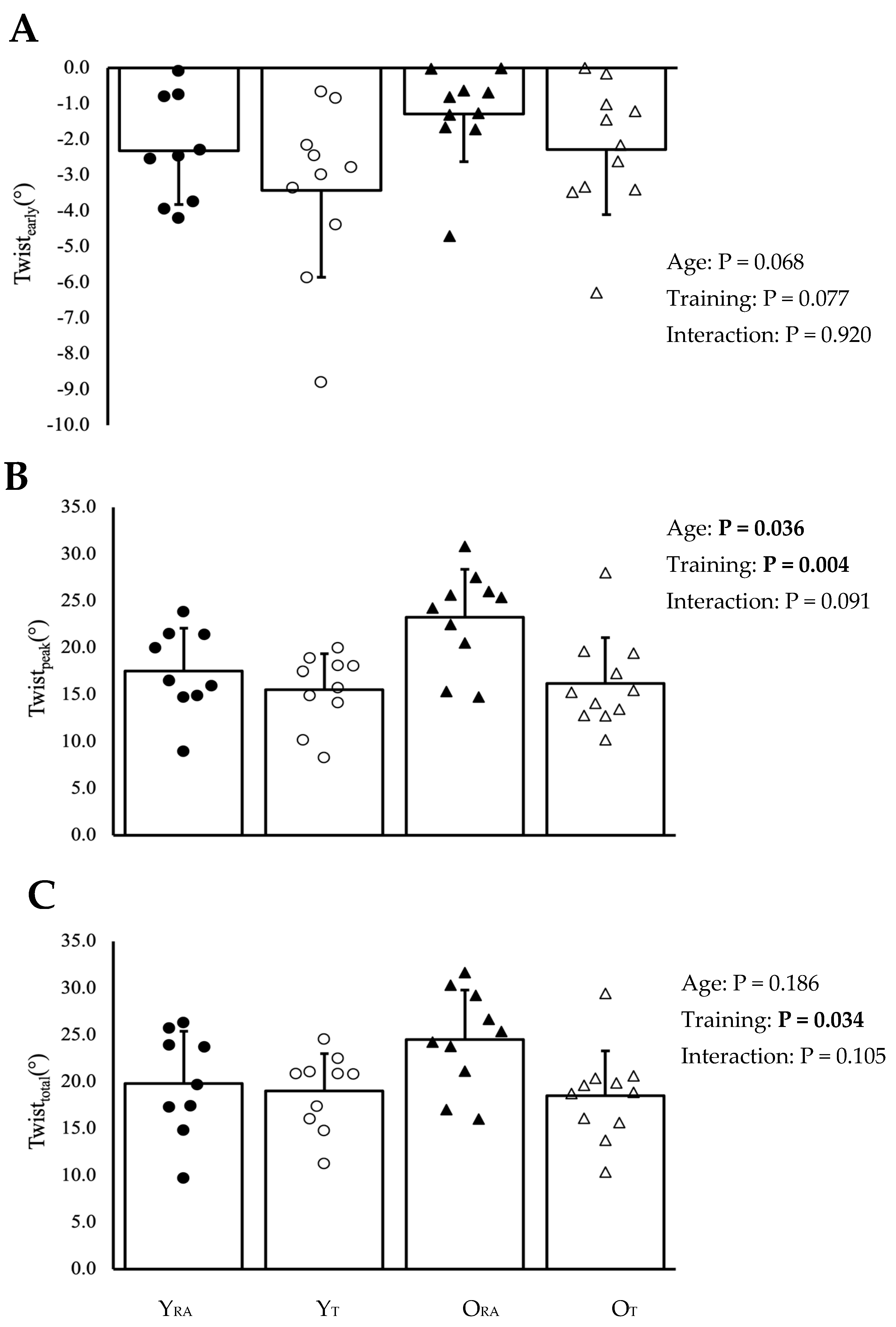
3.3. Left Ventricular Twist

Pooled (n = 40) twistpeak was significantly lower than twisttotal (p < 0.001; Figure 3). Twistearly, twistpeak and twisttotal are illustrated in Figure 4A–C, respectively. Time-to-peak twistearly and twistpeak are presented in Table 2. There were no statistically significant differences in twistearly between older and younger groups (p = 0.068) or between trained and recreationally-active groups (p = 0.077), with no significant interaction (p = 0.920). Twistpeak was higher in older than in younger cohorts (p = 0.036) and lower in trained than in recreationally-active groups (p = 0.004), with a non-significant interaction (p = 0.091). In contrast, twisttotal did not differ between young and old cohorts (p = 0.186) but remained significantly lower in trained than in recreationally-active groups (p = 0.034), with no significant interaction (p = 0.105). Torsion (LV twistpeak normalized to LV length) was significantly greater in older than younger groups (p = 0.009) and lower in trained than recreationally-active cohorts (p = 0.001), with a non-significant interaction (p = 0.119). Time-to-peak twistearly did not differ based on age (p = 0.865) or training status (p = 0.446), with no interaction (p = 0.890). Conversely, twistpeak was later in older than younger groups (p = 0.018), and there was no significant difference in time to twistpeak (p = 0.710) based on training status or an interaction (p = 0.402).

4. Discussion

The principal findings from this exploratory study were that LV twistpeak was higher in older than younger males, yet twistpeak was lower in trained than recreationally-active males. Moreover, the findings of this study suggest that twistearly may influence twistpeak during systole, such that there was less difference in LV twisting between young and older aged groups, as reflected by twisttotal. This is, to our knowledge, the first documentation that early mechanics prior to ejection may contribute to observed peak values near end-systole.

4.1. Age-Related Differences in LV Twist

Twistpeak and torsion were higher in older than younger groups, which aligns with the known ageing process [7,8,9,10,11]. Moreover, our present observations of lower basalearly with ageing, yet similar apicalearly, agree with others [26,27]. Additionally, we noted a smaller twistearly in older than younger males, which approached statistical significance.
An imbalance between epicardial and endocardial fibres from subendocardial fibrosis/dysfunction is a recurrent proposal for the age-associated increase in twistpeak [11,12]. This theory is plausible since less opposing rotation within the endocardium would permit greater dominance of epicardial rotation, leading to heightened overall twist [40]. Examination of mechanics prior to twistpeak has provided more insight into these age-related changes, potentially related to the endocardium. During IVC, endocardial shortening and epicardial stretching produce basalearly and apicalearly [22]. Thus, since endocardial mechanical activity is responsible for basalearly [24,27], it is possible that ageing may reduce endocardial shortening during IVC, explaining less basalearly that we and others have reported in older than in younger groups [27]. This result is further extended by our findings of lower twistearly, while not achieving statistical significance, in older groups than in younger. Altered basalearly may, therefore, represent a sensitive marker to endocardial dysfunction in this context associated with advancing age [28]. Still, it is unclear why apicalearly was not different between ages, which is also due to endocardial shortening [27].
It has also been proposed that temporal alignment of apical and basal rotation may contribute to an age-related increase in simultaneous twist [27]. Results from this study are not in agreement with those from van Dalen et al. [27] since relative timings of apical and basal rotations did not differ with age, nor did the absolute time displacement between respective peak timings. Possible reasons for the discrepancies are not clear, although the acquisition of displacement time may provide some insight. An inherent limitation of 2D speckle tracking means that apical and basal images are acquired separately in different cardiac cycles, and thus, absolute heart rates and loading conditions could influence the timing alignment between rotations captured at the base and apex.

4.2. LV Twist and Exercise Training Status

Twistpeak, twisttotal, apicalpeak and apicaltotal were lower in exercise trained groups in this study compared to recreationally-active participants. These observations, in consideration of the differences with healthy ageing, suggest exercise training status in older age mitigates an age-related difference in twist. Existing data concerning ageing, exercise, and twist are in their infancy, but findings to date are conflicting. Our observations disagree with others who have observed similar resting twist between middle-aged trained and untrained men [19,20], yet concur with reports of lower twist in middle-aged athletes than age-matched controls [10,18]. Although all studies have included participants from endurance-based and highly dynamic modalities [41], such as cycling, triathlon, speed skating, running and swimming, the influence of specific activities will warrant further consideration. Indeed, the static components of highly dynamic, endurance activities vary [41], which has shown to influence the training status related differences in LV twist mechanics, albeit in younger cohorts [42]. In the current study, the twist and torsion observations were likely due to lower apicalpeak in OT, since basalpeak was unaltered; whereas Maufrais et al. [10] attributed the lower twist to basal adaptations only. The reason for this regional disparity is unclear, however, reduced twist has been more commonly accompanied by lower apical rotation and not the base, at least in younger athletes [42,43]. The causes for altered LV mechanics with chronic exercise across all ages have not been fully elucidated. It would seem unlikely that preserved longitudinal subendocardial functioning in OT is responsible since layer-specific shortening was similar to ORA [30]. In addition to LV strain, elite cyclists showed lower apical rotation and twist in both endocardial and epicardial layers [44].
It is not clear at this stage whether the lower twist and apical rotation reflect a reduction in systolic function or represent a functional reserve for exercise capacity [43]. Without a cohort design, it is difficult to determine if the lower twist is a response to chronic and extended exercise training. Indeed, 39 months of rowing training produced a similar twist and apical rotation as a baseline, which was preceded by an initial increase due to acute exercise [45]. Therefore, in our study, the long-term training of older adults may have lowered twist further. From a geometry perspective, less LV twist was related to larger mean wall thickness [43], and a reduction in twist following a more extended period of exercise training may have been associated with cellular hypertrophy [45]. Greater indexed mean wall thickness and LVmass in trained than recreationally-active [30] would support this proposition, with the latter potentially negating the requirement for a bigger systolic contribution to LV ejection in accordance with a larger stroke volume and lower heart rate [30]. Still, lower apical rotation in those with superior aerobic capacity occurred without LV structural adaptation [46] and thus, hypertrophy may not be a prerequisite for altered twist mechanics. Furthermore, changes in fibre angle are known to influence twist [47], as is the sphericity index [48]. However, the sphericity index did not differ between groups in this study [30]; therefore, fibre rearrangement angulation may require further exploration in the absence of differences in the sphericity index. Altered autonomic function following exercise training characterised by heightened parasympathetic and reduced sympathetic activity may influence LV mechanics [49], corresponding to higher and lower LV twist following dobutamine and esmolol infusion, respectively [49]. Although the mechanisms of adaptation require clarification, these data do indicate that chronic exercise training in older adults produces LV adaptations that present a younger phenotype than their untrained counterparts.

4.3. Temporal LV Systolic Twist

We extend existing studies by documenting for the first time that twistearly may be an important consideration when interpreting twistpeak. We demonstrated that twistpeak is significantly underestimated compared with twisttotal, which considers twistearly. Therefore, when assessing the age-related differences in LV twist mechanics, twisttotal, produces a different statistical outcome between young and old adults than twistpeak. Thus, twisttotal may reflect a narrowing in the amount of LV twisting between young and old. Although, caution must be applied when interpreting our results as we do not suggest that the age effect on twist is abolished because of twistearly, nor do we propose that when assessing twisttotal, pump function is equal between young and old males. Instead, it is to our knowledge the first documentation of a reduction in the ageing-related difference in twist through important considerations of mechanics during early systole. These data would suggest that conventional approaches to quantifying twistpeak and the anticipated differences between young and old may be inconsistent and, in some cases, underestimated since the preceding counterclockwise rotation during early-systole, below ‘zero’ has not been accounted for. Therefore, individual variations in twistearly may, in turn, impact the magnitude of difference in twistpeak, in this case, between different age groups. If older and younger ages are characterised by smaller and larger twistearly, respectively, the onset of counterclockwise twist may begin at different points in relation to the ‘zero line’. The subsequent peaktwist taken from the ‘zero line’ may, therefore, be over- or under-estimated. In turn, this may have implications for the appropriate quantification of LV function, although the contribution of twisting occurring before ‘zero’ to LV ejection requires clarification. Simultaneous assessment of LV twisting and structure/volume, like others have reported recently for strain-volume loops [50], may provide further insight into the contribution that twisttotal may have on systolic function. Although systole and diastole are linked by LV twist mechanics [13], twisttotal could be less informative in the context of early diastolic function since the maximum amount of twist would have already occurred. The impact of exercise training status appeared to be less influenced by twisttotal, which remained significantly lower in trained than recreationally-active groups. Together, these data highlight the importance of considering the temporal sequence of LV twist, including all phases of systole to reflect the nadir point on the twist-time curve.

4.4. Limitations

Study limitations pertaining to the cohorts used in this study have been reported previously [30]. Although the study sample size is small, we employed stringent inclusion criteria to facilitate separate homogenous groups related to healthy ageing and exercise training. Post-hoc power analysis between ORA and OT for twistpeak identified a calculated effect size of 1.4 (Cohen’s D) and achieved a power of 87%, suggesting sufficient statistical power in the current study. However, given the small sample size used in this study, results should still be interrupted with caution and replication work is needed to further elucidate the age- and exercise-related differences in twist mechanics using a larger sample size. Our healthy cohort may explain why twist was obtained in all participants (100%) since reports that only 19% (n = 206) of older males (69 ± 6 years) with various medical histories had adequate LV short-axis images at both the base and apex to enable LV twist [51]. Only men were included in the present study based on sex-dependent LV mechanics with ageing [9,52,53], and as a consequence, findings should not be generalised to the female population. There exists a paucity of data on the female community and it should, therefore, be investigated in relation to ageing and exercise. Without an experimental study design, we cannot ascertain causality for training and age-related changes in LV twist mechanics, nor group-based differences which are attributed to factors beyond assessment in this study (e.g., genetics). However, since a longitudinal study in OT athletes across multiple decades (31 ± 11 years of training) is not feasible, we used a cross-sectional study with training-based inclusion criteria for the OT to reflect a cohort of chronically trained males that is heterogenous to their recreationally active counterparts (ORA). We did not normalise LV twist simultaneously with LV length/structure, which future work may wish to do so when determining the contribution of LV twist to ejection across phases of the cardiac cycle, similar to strain-volume loops reported recently [50].

5. Conclusions

Findings from this exploratory study showed that while LV twist is higher in older than younger aged groups, LV twist is lower in exercise-trained than in recreationally-active males. Novel observations demonstrated that when considering twistearly to reflect the nadir point on the twist-time curve, the difference in the amount of LV twisting between young and old males is reduced.

Supplementary Materials

The following supporting information can be downloaded at: https://www.mdpi.com/article/10.3390/jcdd11100321/s1, Supplementary Material Table S1: Baseline physical and exercise characteristics including training habits and maximal oxygen uptake in young recreationally active (YRA), young trained (YT), old recreationally active (ORA) and old trained (OT) participants; Table S2: Left ventricular structure, geometry and volumes in young recreationally active (YRA), young trained (YT), old recreationally active (ORA) and old trained (OT) participants; Table S3: Ultrasonic measures of conventional left ventricular (LV) systolic and diastolic function in young recreationally active (YRA), young trained (YT), old recreationally active (ORA) and old trained (OT) participants.

Author Contributions

A.J.B., N.F.S., F.G., V.B.U. and A.K.C. contributed to the conception and design of the experiment. A.J.B. and A.K.C. performed the experiments with assistance from F.G. and A.K. A.J.B. and N.F.S. conducted the analysis. A.J.B., N.F.S., D.O. and A.K.C. interpreted the data. A.J.B. drafted the manuscript. All authors have read and agreed to the published version of the manuscript.

Funding

This research received no external funding.

Institutional Review Board Statement

The study was granted ethical approval from the University of the West of Scotland Science and Sport School Ethics Committee (0958).

Informed Consent Statement

Informed consent was obtained from all subjects involved in the study.

Data Availability Statement

Data are not openly available due to restrictions upon ethical approval. Please contact the corresponding author for data requests.

Conflicts of Interest

The authors declare no conflicts of interest.

References

  1. Chiao, Y.A.; Rabinovitch, P.S. The Aging Heart. Cold Spring Harb. Perspect. Med. 2015, 5, a025148. [Google Scholar] [CrossRef] [PubMed]
  2. Paneni, F.; Cañestro, C.D.; Libby, P.; Lüscher, T.F.; Camici, G.G. The Aging Cardiovascular System: Understanding It at the Cellular and Clinical Levels. J. Am. Coll. Cardiol. 2017, 69, 1952–1967. [Google Scholar] [CrossRef] [PubMed]
  3. Lower, R. Tractatus de Corde; Oxford University Press: London, UK, 1669; Volume 9. [Google Scholar]
  4. Mor-Avi, V.; Lang, R.M.; Badano, L.P.; Belohlavek, M.; Cardim, N.M.; Derumeaux, G.; Galderisi, M.; Marwick, T.; Nagueh, S.F.; Sengupta, P.P.; et al. Current and Evolving Echocardiographic Techniques for the Quantitative Evaluation of Cardiac Mechanics: ASE/EAE Consensus Statement on Methodology and Indications Endorsed by the Japanese Society of Echocardiography. Eur. J. Echocardiogr. 2011, 12, 167–205. [Google Scholar] [CrossRef]
  5. Nakatani, S. Left Ventricular Rotation and Twist: Why Should We Learn? J. Cardiovasc. Ultrasound 2011, 19, 1–6. [Google Scholar] [CrossRef]
  6. Bansal, M.; Kasliwal, R.R. How Do I Do It? Speckle-Tracking Echocardiography. Indian Heart J. 2013, 65, 117–123. [Google Scholar] [CrossRef]
  7. Sun, J.P.; Lam, Y.-Y.; Wu, C.-Q.; Yang, X.S.; Guo, R.; Kwong, J.S.W.; Merlino, J.D.; Yu, C.-M. Effect of Age and Gender on Left Ventricular Rotation and Twist in a Large Group of Normal Adults—A Multicenter Study. Int. J. Cardiol. 2013, 167, 2215–2221. [Google Scholar] [CrossRef]
  8. Kaku, K.; Takeuchi, M.; Tsang, W.; Takigiku, K.; Yasukochi, S.; Patel, A.R.; Mor-Avi, V.; Lang, R.M.; Otsuji, Y. Age-Related Normal Range of Left Ventricular Strain and Torsion Using Three-Dimensional Speckle-Tracking Echocardiography. J. Am. Soc. Echocardiogr. Off. Publ. Am. Soc. Echocardiogr. 2014, 27, 55–64. [Google Scholar] [CrossRef]
  9. Kocabay, G.; Muraru, D.; Peluso, D.; Cucchini, U.; Mihaila, S.; Padayattil-Jose, S.; Gentian, D.; Iliceto, S.; Vinereanu, D.; Badano, L.P. Normal Left Ventricular Mechanics by Two-Dimensional Speckle-Tracking Echocardiography. Reference Values in Healthy Adults. Rev. Espanola Cardiol. Engl. Ed. 2014, 67, 651–658. [Google Scholar] [CrossRef]
  10. Maufrais, C.; Schuster, I.; Doucende, G.; Vitiello, D.; Rupp, T.; Dauzat, M.; Obert, P.; Nottin, S. Endurance Training Minimizes Age-Related Changes of Left Ventricular Twist-Untwist Mechanics. J. Am. Soc. Echocardiogr. Off. Publ. Am. Soc. Echocardiogr. 2014, 27, 1208–1215. [Google Scholar] [CrossRef]
  11. Zhang, Y.; Zhou, Q.; Pu, D.; Zou, L.; Tan, Y. Differences in Left Ventricular Twist Related to Age: Speckle Tracking Echocardiographic Data for Healthy Volunteers from Neonate to Age 70 Years. Echocardiogr. Mt. Kisco N 2010, 27, 1205–1210. [Google Scholar] [CrossRef]
  12. Tavakoli, V.; Sahba, N. Assessment of Age-Related Changes in Left Ventricular Twist by 3-Dimensional Speckle-Tracking Echocardiography. J. Ultrasound Med. 2013, 32, 1435–1441. [Google Scholar] [CrossRef] [PubMed]
  13. Stöhr, E.J.; Shave, R.E.; Baggish, A.L.; Weiner, R.B. Left Ventricular Twist Mechanics in the Context of Normal Physiology and Cardiovascular Disease: A Review of Studies Using Speckle Tracking Echocardiography. Am. J. Physiol. Heart Circ. Physiol. 2016, 311, H633–H644. [Google Scholar] [CrossRef] [PubMed]
  14. Gibson-Moore, H. UK Chief Medical Officers’ physical activity guidelines 2019: What’s new and how can we get people more active? Nutr. Bull. 2019, 44, 320–328. [Google Scholar] [CrossRef]
  15. Myers, J.; Prakash, M.; Froelicher, V.; Do, D.; Partington, S.; Atwood, J.E. Exercise Capacity and Mortality among Men Referred for Exercise Testing. N. Engl. J. Med. 2002, 346, 793–801. [Google Scholar] [CrossRef]
  16. Beaumont, A.J.; Grace, F.M.; Richards, J.C.; Campbell, A.K.; Sculthorpe, N.F. Aerobic Training Protects Cardiac Function During Advancing Age: A Meta-Analysis of Four Decades of Controlled Studies. Sports Med. 2018, 49, 199–219. [Google Scholar] [CrossRef] [PubMed]
  17. Howden, E.J.; Sarma, S.; Lawley, J.S.; Opondo, M.; Cornwell, W.; Stoller, D.; Urey, M.A.; Adams-Huet, B.; Levine, B.D. Reversing the Cardiac Effects of Sedentary Aging in Middle Age—A Randomized Controlled Trial: Implications For Heart Failure Prevention. Circulation 2018, 137, 1549–1560. [Google Scholar] [CrossRef]
  18. Santoro, A.; Alvino, F.; Antonelli, G.; Cassano, F.E.; De Vito, R.; Cameli, M.; Mondillo, S. Age Related Diastolic Function in Amateur Athletes. Int. J. Cardiovasc. Imaging 2015, 31, 567–573. [Google Scholar] [CrossRef]
  19. Maufrais, C.; Doucende, G.; Rupp, T.; Dauzat, M.; Obert, P.; Nottin, S.; Schuster, I. Left Ventricles of Aging Athletes: Better Untwisters but Not More Relaxed during Exercise. Clin. Res. Cardiol. 2017, 106, 884–892. [Google Scholar] [CrossRef] [PubMed]
  20. Lee, L.S.; Mariani, J.A.; Sasson, Z.; Goodman, J.M. Exercise with a Twist: Left Ventricular Twist and Recoil in Healthy Young and Middle-Aged Men, and Middle-Aged Endurance-Trained Men. J. Am. Soc. Echocardiogr. Off. Publ. Am. Soc. Echocardiogr. 2012, 25, 986–993. [Google Scholar] [CrossRef]
  21. Sengupta, P.P.; Khandheria, B.K.; Korinek, J.; Wang, J.; Jahangir, A.; Seward, J.B.; Belohlavek, M. Apex-to-Base Dispersion in Regional Timing of Left Ventricular Shortening and Lengthening. J. Am. Coll. Cardiol. 2006, 47, 163–172. [Google Scholar] [CrossRef]
  22. Sengupta, P.P.; Tajik, A.J.; Chandrasekaran, K.; Khandheria, B.K. Twist Mechanics of the Left Ventricle: Principles and Application. JACC Cardiovasc. Imaging 2008, 1, 366–376. [Google Scholar] [CrossRef] [PubMed]
  23. Ashikaga, H.; van der Spoel, T.I.G.; Coppola, B.A.; Omens, J.H. Transmural Myocardial Mechanics during Isovolumic Contraction. JACC Cardiovasc. Imaging 2009, 2, 202–211. [Google Scholar] [CrossRef] [PubMed]
  24. Sengupta, P.P.; Korinek, J.; Belohlavek, M.; Narula, J.; Vannan, M.A.; Jahangir, A.; Khandheria, B.K. Left Ventricular Structure and Function: Basic Science for Cardiac Imaging. J. Am. Coll. Cardiol. 2006, 48, 1988–2001. [Google Scholar] [CrossRef] [PubMed]
  25. Sabatino, J.; Castaldi, B.; Di Salvo, G. How to Measure Left Ventricular Twist by Two-Dimensional Speckle-Tracking Analysis. Eur. Heart J. Cardiovasc. Imaging 2021, 22, 961–963. [Google Scholar] [CrossRef]
  26. Kim, H.-K.; Sohn, D.-W.; Lee, S.-E.; Choi, S.-Y.; Park, J.-S.; Kim, Y.-J.; Oh, B.-H.; Park, Y.-B.; Choi, Y.-S. Assessment of Left Ventricular Rotation and Torsion with Two-Dimensional Speckle Tracking Echocardiography. J. Am. Soc. Echocardiogr. 2007, 20, 45–53. [Google Scholar] [CrossRef] [PubMed]
  27. van Dalen, B.M.; Soliman, O.I.I.; Vletter, W.B.; ten Cate, F.J.; Geleijnse, M.L. Age-Related Changes in the Biomechanics of Left Ventricular Twist Measured by Speckle Tracking Echocardiography. Am. J. Physiol.-Heart Circ. Physiol. 2008, 295, H1705–H1711. [Google Scholar] [CrossRef]
  28. Takeuchi, M.; Otsuji, Y.; Lang, R.M. Evaluation of Left Ventricular Function Using Left Ventricular Twist and Torsion Parameters. Curr. Cardiol. Rep. 2009, 11, 225–230. [Google Scholar] [CrossRef]
  29. Johnson, C.; Forsythe, L.; Somauroo, J.; Papadakis, M.; George, K.; Oxborough, D. Cardiac Structure and Function in Elite Native Hawaiian and Pacific Islander Rugby Football League Athletes: An Exploratory Study. Int. J. Cardiovasc. Imaging 2018, 34, 725–734. [Google Scholar] [CrossRef]
  30. Beaumont, A.; Campbell, A.; Unnithan, V.; Grace, F.; Knox, A.; Sculthorpe, N. Long-Term Athletic Training Does Not Alter Age-Associated Reductions of Left-Ventricular Mid-Diastolic Lengthening or Expansion at Rest. Eur. J. Appl. Physiol. 2020, 120, 2059–2073. [Google Scholar] [CrossRef]
  31. Aksakal, E.; Kurt, M.; Ozturk, M.E.; Tanboga, I.H.; Kaya, A.; Nacar, T.; Sevimli, S.; Gurlertop, Y. The Effect of Incremental Endurance Exercise Training on Left Ventricular Mechanics: A Prospective Observational Deformation Imaging Study. Anadolu Kardiyol. Derg. Anatol. J. Cardiol. 2013, 13, 432. [Google Scholar] [CrossRef]
  32. Donal, E.; Rozoy, T.; Kervio, G.; Schnell, F.; Mabo, P.; Carré, F. Comparison of the Heart Function Adaptation in Trained and Sedentary Men After 50 and Before 35 Years of Age. Am. J. Cardiol. 2011, 108, 1029–1037. [Google Scholar] [CrossRef] [PubMed]
  33. Matelot, D.; Schnell, F.; Kervio, G.; Ridard, C.; Thillaye du Boullay, N.; Wilson, M.; Carre, F. Cardiovascular Benefits of Endurance Training in Seniors: 40 Is Not Too Late to Start. Int. J. Sports Med. 2016, 37, 625–632. [Google Scholar] [CrossRef] [PubMed]
  34. Fujimoto, N.; Prasad, A.; Hastings, J.L.; Arbab-Zadeh, A.; Bhella, P.S.; Shibata, S.; Palmer, D.; Levine, B.D. Cardiovascular Effects of 1 Year of Progressive and Vigorous Exercise Training in Previously Sedentary Individuals Older than 65 Years of Age. Circulation 2010, 122, 1797–1805. [Google Scholar] [CrossRef] [PubMed]
  35. van Dalen, B.M.; Vletter, W.B.; Soliman, O.I.I.; ten Cate, F.J.; Geleijnse, M.L. Importance of Transducer Position in the Assessment of Apical Rotation by Speckle Tracking Echocardiography. J. Am. Soc. Echocardiogr. 2008, 21, 895–898. [Google Scholar] [CrossRef]
  36. van Dalen, B.M.; Soliman, O.I.I.; Kauer, F.; Vletter, W.B.; van der Zwaan, H.B.; Cate, F.J.T.; Geleijnse, M.L. Alterations in Left Ventricular Untwisting with Ageing. Circ. J. Off. J. Jpn. Circ. Soc. 2010, 74, 101–108. [Google Scholar] [CrossRef]
  37. Stöhr, E.J.; González-Alonso, J.; Pearson, J.; Low, D.A.; Ali, L.; Barker, H.; Shave, R. Effects of Graded Heat Stress on Global Left Ventricular Function and Twist Mechanics at Rest and during Exercise in Healthy Humans. Exp. Physiol. 2011, 96, 114–124. [Google Scholar] [CrossRef]
  38. Oxborough, D.; George, K.; Birch, K.M. Intraobserver Reliability of Two-Dimensional Ultrasound Derived Strain Imaging in the Assessment of the Left Ventricle, Right Ventricle, and Left Atrium of Healthy Human Hearts. Echocardiogr. Mt. Kisco N 2012, 29, 793–802. [Google Scholar] [CrossRef]
  39. The Jamovi Project Jamovi. Available online: https://www.jamovi.org (accessed on 1 August 2024).
  40. Lumens, J.; Delhaas, T.; Arts, T.; Cowan, B.R.; Young, A.A. Impaired Subendocardial Contractile Myofiber Function in Asymptomatic Aged Humans, as Detected Using MRI. Am. J. Physiol. Heart Circ. Physiol. 2006, 291, H1573–H1579. [Google Scholar] [CrossRef]
  41. Mitchell, J.H.; Haskell, W.; Snell, P.; Van Camp, S.P. Task Force 8: Classification of Sports. J. Am. Coll. Cardiol. 2005, 45, 1364–1367. [Google Scholar] [CrossRef]
  42. Beaumont, A.; Grace, F.; Richards, J.; Hough, J.; Oxborough, D.; Sculthorpe, N. Left Ventricular Speckle Tracking-Derived Cardiac Strain and Cardiac Twist Mechanics in Athletes: A Systematic Review and Meta-Analysis of Controlled Studies. Sports Med. 2016, 47, 1145–1170. [Google Scholar] [CrossRef]
  43. Forsythe, L.; MacIver, D.H.; Johnson, C.; George, K.; Somauroo, J.; Papadakis, M.; Brown, B.; Qasem, M.; Oxborough, D. The Relationship between Left Ventricular Structure and Function in the Elite Rugby Football League Athlete as Determined by Conventional Echocardiography and Myocardial Strain Imaging. Int. J. Cardiol. 2018, 261, 211–217. [Google Scholar] [CrossRef] [PubMed]
  44. Nottin, S.; Doucende, G.; Schuster-Beck, I.; Dauzat, M.; Obert, P. Alteration in Left Ventricular Normal and Shear Strains Evaluated by 2D-Strain Echocardiography in the Athlete’s Heart: Left Ventricular Regional Strains in Athletes. J. Physiol. 2008, 586, 4721–4733. [Google Scholar] [CrossRef] [PubMed]
  45. Weiner, R.B.; DeLuca, J.R.; Wang, F.; Lin, J.; Wasfy, M.M.; Berkstresser, B.; Stöhr, E.; Shave, R.; Lewis, G.D.; Hutter, A.M.; et al. Exercise-Induced Left Ventricular Remodeling Among Competitive Athletes A Phasic Phenomenon. Circ. Cardiovasc. Imaging 2015, 8, e003651. [Google Scholar] [CrossRef] [PubMed]
  46. Stöhr, E.J.; McDonnell, B.; Thompson, J.; Stone, K.; Bull, T.; Houston, R.; Cockcroft, J.; Shave, R. Left Ventricular Mechanics in Humans with High Aerobic Fitness: Adaptation Independent of Structural Remodelling, Arterial Haemodynamics and Heart Rate. J. Physiol. 2012, 590, 2107–2119. [Google Scholar] [CrossRef] [PubMed]
  47. Taber, L.A.; Yang, M.; Podszus, W.W. Mechanics of Ventricular Torsion. J. Biomech. 1996, 29, 745–752. [Google Scholar] [CrossRef]
  48. van Dalen, B.M.; Kauer, F.; Vletter, W.B.; Soliman, O.I.I.; van der Zwaan, H.B.; ten Cate, F.J.; Geleijnse, M.L. Influence of Cardiac Shape on Left Ventricular Twist. J. Appl. Physiol. 2009, 108, 146–151. [Google Scholar] [CrossRef]
  49. Notomi, Y.; Popovic, Z.B.; Yamada, H.; Wallick, D.W.; Martin, M.G.; Oryszak, S.J.; Shiota, T.; Greenberg, N.L.; Thomas, J.D. Ventricular Untwisting: A Temporal Link between Left Ventricular Relaxation and Suction. Am. J. Physiol. Heart Circ. Physiol. 2008, 294, H505–H513. [Google Scholar] [CrossRef]
  50. Oxborough, D.; Heemels, A.; Somauroo, J.; McClean, G.; Mistry, P.; Lord, R.; Utomi, V.; Jones, N.; Thijssen, D.; Sharma, S.; et al. Left and Right Ventricular Longitudinal Strain-Volume/Area Relationships in Elite Athletes. Int. J. Cardiovasc. Imaging 2016, 32, 1199–1211. [Google Scholar] [CrossRef]
  51. Park, C.M.; March, K.; Williams, S.; Kukadia, S.; Ghosh, A.K.; Jones, S.; Tillin, T.; Chaturvedi, N.; Hughes, A.D. Feasibility and Reproducibility of Left Ventricular Rotation by Speckle Tracking Echocardiography in Elderly Individuals and the Impact of Different Software. PLoS ONE 2013, 8, e75098. [Google Scholar] [CrossRef]
  52. Hung, C.-L.; Gonçalves, A.; Shah, A.M.; Cheng, S.; Kitzman, D.; Solomon, S.D. Age- and Sex-Related Influences on Left Ventricular Mechanics in Elderly Individuals Free of Prevalent Heart FailureCLINICAL PERSPECTIVE. Circ. Cardiovasc. Imaging 2017, 10, e004510. [Google Scholar] [CrossRef]
  53. Nio, A.Q.X.; Stöhr, E.J.; Shave, R.E. Age-Related Differences in Left Ventricular Structure and Function between Healthy Men and Women. Climacteric J. Int. Menopause Soc. 2017, 20, 476–483. [Google Scholar] [CrossRef] [PubMed]
Figure 1. Example of identification for left ventricle twist mechanics, including twistearly, twistpeak and twisttotal on a twist-time curve during systole.
Jcdd 11 00321 g001
Figure 2. (AC) Temporal LV rotation ((A)—basal rotation; (B)—basal rotation) and twist (C) across the cardiac cycle in 5% increments. AVC, aortic valve closure (end-systole [100%]). Data are presented as group mean with error bars omitted for clarity.
Jcdd 11 00321 g002
Figure 3. Left ventricular early (black bars), peak (grey bars) and total (white bars) rotations from apical and basal levels, and net twist for the pooled cohort (n = 40). Data are presented as mean ± standard deviation. * indicates statistically significant difference between peak and total rotations within the respective pair at p < 0.001. Left ventricular early rotation/twist are presented for illustrative purposes.
Jcdd 11 00321 g003
Figure 4. (AC) Left ventricular twist mechanics for groups based on age and training status. Data are presented as mean ± SD, and bold p-values indicate statistical significance at p < 0.05. YRA, young recreationally active; YT, young trained; ORA, old recreationally active; OT, old trained.
Jcdd 11 00321 g004
Table 1. Left ventricular twist mechanics among age and exercise training groups.
YoungOldp-Value
Recreationally Active (YRA)Trained
(YT)
Recreationally
Active (ORA)
Trained
(OT)
AgeTrainingInteraction
Basalearly rotation (°)2.6 ± 2.02.7 ± 2.11.2 ± 1.11.6 ± 1.60.0250.6790.742
Basalpeak rotation (°)−5.4 ± 2.7−7.0 ± 2.5−7.6 ± 3.0−7.7 ± 4.30.1640.4210.482
Basaltotal rotation (°)−8.1 ± 2.8−9.7 ± 1.9−8.8 ± 3.5−9.3 ± 3.50.8410.2730.568
Normalised basal rotation (°/cm)−0.6 ± 0.3−0.7 ± 0.3−0.9 ± 0.4−0.9 ± 0.50.0940.5640.560
Apicalearly rotation (°)−0.9 ± 0.7−2.0 ± 1.4−0.9 ± 1.5−1.3 ± 1.20.3790.0690.393
Apicalpeak rotation (°)12.6 ± 3.9 9.5 ± 3.016.3 ± 6.19.3 ± 4.00.206<0.0010.173
Apicaltotal rotation (°)13.5 ± 3.911.5 ± 2.917.2 ± 6.210.6 ± 4.00.3100.0040.112
Normalised apical rotation (°/cm)1.4 ± 0.41.0 ± 0.31.9 ± 0.71.0 ± 0.50.105<0.0010.201
Torsion (°/cm)1.9 ± 0.51.6 ± 0.42.6 ± 0.6 1.8 ± 0.50.0090.0010.119
Data are presented as mean ± SD. Bold values indicate statistical significance at p < 0.05.
Table 2. Left ventricular mechanical timings during early and peak systole between age and exercise training groups.
YoungOldp-Value
Recreationally Active (YRA)Trained
(YT)
Recreationally
Active (ORA)
Trained
(OT)
AgeTrainingInteraction
Time to basalearly rotation (ms)272 ± 77295 ± 103263 ± 99281 ± 1250.7200.5340.942
Time to basalpeak rotation (ms)348 ± 50343 ± 37362 ± 73369 ± 480.2510.9370.731
Time to apicalearly rotation (ms)215 ± 76247 ± 107230 ± 83260 ± 930.6350.2950.980
Time to apicalpeak rotation (ms)363 ± 36385 ± 71397 ± 48397 ± 600.2040.5450.563
Time displacement (ms)−16 ± 52−42 ± 60−35 ± 66−28 ± 770.8820.6480.435
Time to twistearly (ms)233 ± 72 260 ± 112242 ± 86261 ± 960.8650.4460.890
Time to twistpeak (ms)350 ± 41355 ± 34389 ± 42374 ± 310.0180.7100.402
Data are presented as mean ± SD. Bold values indicate statistical significance at p < 0.05.
Disclaimer/Publisher’s Note: The statements, opinions and data contained in all publications are solely those of the individual author(s) and contributor(s) and not of MDPI and/or the editor(s). MDPI and/or the editor(s) disclaim responsibility for any injury to people or property resulting from any ideas, methods, instructions or products referred to in the content.