
Personalized Evidence-Based Management of Patent Ductus Arteriosus in Preterm Infants


Abstract
:1. Introduction
2. Basis for Treatment of PDA
2.1. Natural History of PDA Spontaneous Closure
2.2. Different Approaches in PDA Management
2.3. Evidence to Support Conservative PDA Treatment Approach
2.4. Evidence to Support Active PDA Treatment Approach
2.5. Defining Hemodynamically Significant PDA
3. Medical Treatment
3.1. Pharmacological Treatment
3.2. Expectant Treatment Approach
- (1)
- Fluid restriction: Restricting fluid intake to 120–150 mL/kg/day was thought to decrease the size of PDA and pulmonary over-circulation [53]. However, fluid restriction carries a risk of dehydration, renal failure, and compromised nutrition intake [54]. Waal et al. recommended careful avoidance of fluid overload rather than overt fluid restriction [54]. Waal et al. summarized 11 studies on other supportive care measures [54].
- (2)
- Diuretics: The use of diuretics, such as furosemide, may help to reduce pulmonary edema associated with PDA or BPD. However, furosemide has been linked to increased risk of ductal patency via stimulating the prostaglandin pathway [55,56]. Chlorothiazide without the prostaglandin effect can be used as an alternative but has less efficacy than furosemide [57].
- (3)
- Optimizing ventilator settings: Optimizing the ventilator settings, such as a higher PEEP, reducing inspiratory times, and permissive hypercapnia have been suggested to temporarily stabilize respiratory status while waiting for PDA closure [54,57]. However, mechanical ventilation independently increases the risk of BPD associated with PDA, especially in infant with a PDA needing mechanical ventilation over 7–10 days [58].
- (4)
3.3. Timing and Treatment Approaches
- (a)
- Prophylactic treatment (first 6–24 h after birth);
- (b)
- Early symptomatic treatment with hsPDA (<7 days after birth);
- (c)
- Late symptomatic treatment with hsPDA (≥ 7 days after birth);
- (d)
- Expectant treatment with non-hsPDA;
- (e)
- Discharge home with small asymptomatic PDA.
- (a)
- Prophylactic treatment (within first 6–24 h after birth)
- (b)
- Early symptomatic treatment with hsPDA (<7 days after birth)
- (c)
- Late symptomatic treatment with hsPDA (≥ 7 days after birth)
- (d)
- Expectant treatment with non-hsPDA
- (e)
- Discharge home with small asymptomatic PDA
4. Future Direction and Conclusions
Author Contributions
Funding
Institutional Review Board Statement
Informed Consent Statement
Data Availability Statement
Conflicts of Interest
References
- Hamrick, S.E.; Sallmon, H.; Rose, A.T.; Porras, D.; Shelton, E.L.; Reese, J.; Hansmann, G. Patent ductus arteriosus of the Preterm infant. Pediatrics 2020, 146, e20201209. [Google Scholar] [CrossRef]
- Sung, S.I.; Chang, Y.S.; Kim, J.; Choi, J.H.; Ahn, S.Y.; Park, W.S. Natural evolution of ductus arteriosus with noninterventional conservative management in extremely preterm infants born at 23–28 weeks of gestational age. PLoS ONE 2019, 14, e0212258. [Google Scholar] [CrossRef]
- Benitz, W.E. Committee on Fetus and Newborn; American Academy of Pediatrics. Patent ductus arteriosus in preterm infants. Pediatrics 2016, 137, e20153730. [Google Scholar] [CrossRef]
- Dice, J.E.; Bhatia, J. Patent ductus arteriosus: An overview. J. Pediatr. Pharmacol. Ther. 2007, 12, 138–146. [Google Scholar] [CrossRef]
- El-Khuffash, A.; James, A.T.; Corcoran, J.D.; Dicker, P.; Franklin, O.; Elsayed, Y.N.; Ting, J.Y.; Sehgal, A.; Malikiwi, A.; Harabor, A.; et al. A patent ductus arteriosus severity score predicts chronic lung disease or death before discharge. J. Pediatr. 2015, 167, 1354–1361.e2. [Google Scholar] [CrossRef]
- Ballabh, P. Intraventricular hemorrhage in premature infants: Mechanism of disease. Pediatr. Res. 2010, 67, 1–8. [Google Scholar] [CrossRef]
- Schmidt, B.; Davis, P.; Moddemann, D.; Ohlsson, A.; Roberts, R.S.; Saigal, S.; Solimano, A.; Vincer, M.; Wright, L.L. Trial of Indomethacin Prophylaxis in Preterms Investigators. Long-term effects of indomethacin prophylaxis in extremely low-birth-weight infants. N. Engl. J. Med. 2001, 344, 1966–1972. [Google Scholar] [CrossRef]
- Kluckow, M.; Jeffery, M.; Gill, A.; Evans, N. A randomized placebo-controlled trial of early treatment of the patent ductus arteriosus. Arch. Dis. Child. Fetal Neonatal Ed. 2014, 992, F99–F104. [Google Scholar] [CrossRef]
- Noori, S.; McCoy, M.; Friedlich, P.; Bright, B.; Gottipati, V.; Seri, I.; Sekar, K. Failure of ductus arteriosus closure is associated with increased mortality in preterm infants. Pediatrics 2009, 123, e138–e144. [Google Scholar] [CrossRef]
- Cotton, R.B.; Stahlman, M.T.; Bender, H.W.; Graham, T.P.; Catterton, W.Z.; Kovar, I. Randomized trial of early closure of symptomatic patent ductus arteriosus in small preterm infants. J. Pediatr. 1978, 93, 647–651. [Google Scholar] [CrossRef]
- Sathanandam, S.; Whiting, S.; Cunningham, J.; Zurakowski, D.; Apalodimas, L.; Waller, B.R.; Philip, R.; Qureshi, A.M. Practice variation in the management of patient ductus arteriosus in extremely low birth weight infants in the United States: Survey results among cardiologists and neonatologists. Congenit. Heart Dis. 2019, 14, 6–14. [Google Scholar] [CrossRef]
- Slaughter, J.L.; Reagan, P.B.; Bapat, R.V.; Newman, T.B.; Klebanoff, M.A. Nonsteroidal anti-inflammatory administration and patent ductus arteriosus ligation, a survey of practice preferences at US children’s hospital. Eur. J. Pediatr. 2016, 175, 775–783. [Google Scholar] [CrossRef]
- Lee, H.C.; Durand, D.J.; Danielsen, B.; Dueñas, G.V.; Powers, R.J. Hospital variation in medical and surgical treatment of patent ductus arteriosus. Am. J. Perinatol. 2015, 32, 379–386. [Google Scholar]
- Brissaud, O.; Guichoux, J. Patent ductus arteriosus in preterm infant: A survey of clinical practices in French neonatal intensive care units. Pediat Cardiol. 2011, 32, 607–614. [Google Scholar] [CrossRef]
- Bixler, G.M.; Powers, G.C.; Clark, R.H.; Walker, M.W.; Tolia, V.N. Changes in the diagnosis and management of patent ductus arteriosus from 2006-2015 in United States neonatal intensive care units. J. Pediatr. 2017, 189, 105–112. [Google Scholar] [CrossRef]
- De Klerk, J.C.A.; Engbers, A.G.J.; van Beek, F.; Flint, R.B.; Reiss, I.K.M.; Völler, S.; Simons, S.H.P. Spontaneous Closure of the Ductus Arteriosus in Preterm Infants: A Systematic Review. Front. Pediatr. 2007, 8, 541. [Google Scholar] [CrossRef]
- Semberova, J.; Sirc, J.; Miletin, J.; Kucera, J.; Berka, I.; Sebkova, S.; O’Sullivan, S.; Franklin, O.; Stranak, Z. Spontaneous Closure of Patent Ductus Arteriosus in Infants ≤1500 g. Pediatrics 2017, 140, e20164258. [Google Scholar] [CrossRef]
- Nielsen, M.R.; Aldenryd, A.E.; Hagstrøm, S.; Pedersen, L.M.; Brix, N. The chance of spontaneous patent ductus arteriosus closure in preterm infants born before 32 weeks of gestation is high and continues to increase until 5 years of follow-up. Acta Paediatr. 2022, 111, 2322–2330. [Google Scholar] [CrossRef]
- Mitra, S.; de Boode, W.P.; Weisz, D.E.; Shah, P.S. Interventions for patent ductus arteriosus (PDA) in preterm infants: An overview of Cochrane Systematic Reviews. Cochrane Database Syst. Rev. 2023, 4, CD013588. [Google Scholar]
- Ohlsson, A.; Shah, P.S. Ibuprofen for the prevention of patent ductus arteriosus in preterm and/or low birth weight infants. Cochrane Database Syst. Rev. 2020, 1, CD004213. [Google Scholar] [CrossRef]
- Clyman, R.I.; Liebowitz, M.; Kaempf, J.; Erdeve, O.; Bulbul, A.; Håkansson, S.; Lindqvist, J.; Farooqi, A.; Katheria, A.; Sauberan, J.; et al. PDA-TOLERATE trial: An exploratory randomized controlled trial of treatment of moderate-to-large patent ductus arteriosus at 1 week of age. J. Pediatr. 2019, 205, 41–48.e6. [Google Scholar] [CrossRef]
- Hundscheid, T.; Onland, W.; van Overmeire, B.; Dijk, P.; van Kaam, A.H.L.C.; Dijkman, K.P.; Kooi, E.M.W.; Villamor, E.; Kroon, A.A.; Visser, R.; et al. Early treatment versus expectative management of patent ductus arteriosus in preterm infants: A multicenter, randomized, non-inferiority trial in Europe (BeNeDuctus trial). BMC Pediatr. 2018, 18, 262. [Google Scholar] [CrossRef]
- Sankar, M.N.; Bhombal, S.; Benitz, W.E. PDA: To treat or not to treat. Congenit. Heart Dis. 2019, 14, 46–51. [Google Scholar] [CrossRef]
- Potsiurko, S.; Dobryanskyy, D.; Sekretar, L.; Salabay, Z. Randomized Noninferiority Trial of Expectant Management versus Early Treatment of Patent Ductus Arteriosus in Preterm Infants. Am. J. Perinatol. 2022; online ahead of print. [Google Scholar]
- El-Khuffash, A.; Bussmann, N.; Breatnach, C.R.; Smith, A.; Tully, E.; Griffin, J.; McCallion, N.; Corcoran, J.D.; Fernandez, E.; Looi, C.; et al. A Pilot Randomized Controlled Trial of Early Targeted Patent Ductus Arteriosus Treatment Using a Risk Based Severity Score (The PDA RCT). J. Pediatr. 2021, 229, 127–133. [Google Scholar] [CrossRef]
- De Waal, K.; Phad, N.; Stubbs, M.; Chen, Y.; Kluckow, M. A Randomized Placebo-Controlled Pilot Trial of Early Targeted Nonsteroidal Anti-Inflammatory Drugs in Preterm Infants with a Patent Ductus Arteriosus. J. Pediatr. 2021, 228, 82–86.e2. [Google Scholar] [CrossRef]
- Hundscheid, T.; Onland, W.; Kooi, E.M.W.; Vijlbrief, D.C.; de Vries, W.B.; Dijkman, K.P.; van Kaam, A.H.; Villamor, E.; Kroon, A.A.; Visser, R.; et al. Expectant Management or Early Ibuprofen for Patent Ductus Arteriosus. N. Engl. J. Med. 2023, 388, 980–990. [Google Scholar] [CrossRef]
- Cambonie, G.; Clyman, R.I.; Rozé, J.C. Management of persistent ductus arteriosus in very premature neonates. Results of the French TRIOCAPI trial, perspectives for clinicians, and subsequent studies on this topic. Arch. Pediatr. 2021, 28, 501–503. [Google Scholar] [CrossRef]
- Gupta, S.; Juszczak, E.; Subhedar, N.; Hardy, P.; Wyllie, J.; Johnson, S.; Kelsall, W.; Roberts, T.; Hutchison, E.; Bowler, U.; et al. 545 Does selective early treatment of Patent Ductus Arteriosus (PDA) with ibuprofen reduce death or Bronchopulmonary Dysplasia (BPD) at 36 weeks in extreme preterm babies? A Randomised Controlled Trial (Baby-OSCAR Trial). Arch. Dis. Child. 2022, 107, A148–A149. [Google Scholar]
- Weisz, D.E.; Mirea, L.; Resende, M.H.F.; Ly, L.; Church, P.T.; Kelly, E.; Kim, S.J.; Jain, A.; McNamara, P.J.; Shah, P.S. Outcomes of Surgical Ligation after Unsuccessful Pharmacotherapy for Patent Ductus Arteriosus in Neonates Born Extremely Preterm. J. Pediatr. 2018, 2195, 292–296.e3. [Google Scholar] [CrossRef]
- Sathanandam, S.; Mcnamara, P.; Pedra, C.; Toyoshima, K.; Malekzadeh-Milani, S.; Patkai, J.; Barspinar, O.; Uslu, H.S.; Promphan, W.; Khorana, M.; et al. A Global Perspective on PDA Management in the Extremely Premature: Shifting Trend Toward Transcatheter Closure. J. Soc. Cardiovasc. Angiogr. Interv. 2023, 2, 100968. [Google Scholar] [CrossRef]
- Al-Turkait, A.; Szatkowski, L.; Choonara, I.; Ojha, S. Management of patent ductus arteriosus in very preterm infants in England and Wales: A retrospective cohort study. BMJ Paediatr. Open 2022, 6, e001424. [Google Scholar] [CrossRef]
- Lokku, A.; Mirea, L.; Lee, S.K.; Shah, P.S.; Canadian Neonatal Network. Trends and outcomes of patent ductus arteriosus treatment in very preterm infants in Canada. Am. J. Perinatol. 2017, 34, 441–450. [Google Scholar] [CrossRef]
- Isayama, T.; Kusuda, S.; Reichman, B.; Lee, S.K.; Lehtonen, L.; Norman, M.; Adams, M.; Bassler, D.; Helenius, K.; Hakansson, S.; et al. Neonatal Intensive Care Unit-Level Patent Ductus Arteriosus Treatment Rates and Outcomes in Infants Born Extremely Preterm. J. Pediatr. 2020, 220, 34–39.e5. [Google Scholar] [CrossRef]
- Altit, G.; Saeed, S.; Beltempo, M.; Claveau, M.; Lapointe, A.; Basso, O. Outcomes of extremely premature infants comparing patent ductus arteriosus management approaches. J. Pediatr. 2021, 235, 49–57.e2. [Google Scholar] [CrossRef]
- Mitra, S.; Florez, I.D.; Tamayo, M.E.; Mbuagbaw, L.; Vanniyasingam, T.; Veroniki, A.A.; Zea, A.M.; Zhang, Y.; Sadeghirad, B.; Thabane, L. Association of Placebo, Indomethacin, Ibuprofen, and Acetaminophen With Closure of Hemodynamically Significant Patent Ductus Arteriosus in Preterm Infants: A Systematic Review and Meta-analysis. JAMA 2018, 319, 1221–1238. [Google Scholar] [CrossRef]
- Ohlsson, A.; Shah, S. Paracetamol (acetaminophen) for patent ductus arteriosus in preterm or low birth weight infants. Cochrane Database Syst. Rev. 2020, 1, CD010061. [Google Scholar] [CrossRef]
- Ohlsson, A.; Walia, R.; Shah, S. Ibuprofen for the treatment of patent ductus arteriosus in preterm or low birth weight (or both) infants. Cochrane Database Syst. Rev. 2020, 2, CD003481. [Google Scholar] [CrossRef]
- Mitra, S.; McNamara, P.J. Patent Ductus Arteriosus—Time for a definitive trial. Clin. Perinatol. 2020, 47, 617–639. [Google Scholar] [CrossRef]
- Liebowitz, M.; Katheria, A.; Sauberan, J.; Singh, J.; Nelson, K.; Hassinger, D.C.; Aucott, S.W.; Kaempf, J.; Kimball, A.; Fernandez, E.; et al. Lack of Equipoise in the PDA-TOLERATE Trial: A Comparison of Eligible Infants Enrolled in the Trial and Those Treated Outside the Trial. J. Pediatr. 2019, 213, 222–226.e2. [Google Scholar] [CrossRef]
- Philip, R.; Waller, B.R.; Chilakala, S.; Graham, B.; Stecchi, N.; Apalodimas, L.; Cunningham, J.; Washington, K.; Sathanandam, S. Hemodynamic and clinical consequences of early versus delayed closure of patent ductus arteriosus in extremely low birth weight infants. J. Perinatol. 2021, 41, 100–108. [Google Scholar] [CrossRef]
- Sathanandam, S.; Balduf, K.; Chilakala, S.; Washington, K.; Allen, K.; Knott-Craig, C.; Rush Waller, B.; Philip, R. Role of Transcatheter patent ductus arteriosus closure in extremely low birth weight infants. Catheter. Cardiovasc. Interv. 2019, 93, 89–96. [Google Scholar] [CrossRef]
- Sathanandam, S.; Gutfinger, D.; Morray, B.; Berman, D.; Gillespie, M.; Forbes, T.; Johnson, J.N.; Garg, R.; Malekzadeh-Milani, S.; Fraisse, A.; et al. Consensus Guidelines for the Prevention and Management of Periprocedural Complications of Transcatheter Patent Ductus Arteriosus Closure with the Amplatzer Piccolo Occluder in Extremely Low Birth Weight Infants. Pediatr. Cardiol. 2021, 42, 1258–1274. [Google Scholar] [CrossRef]
- Lemmers, P.; Vijlbrief, D.; Benders, M.; Alderliesten, T.; Veldhuis, M.; Baerts, W.; Koopman-Esseboom, C.; Groenendaal, F.; van Bel, F. Delayed Surgical Closure of the Patent Ductus Arteriosus: Does the Brain Pay the Price? J. Pediatr. 2023, 254, 25–32. [Google Scholar] [CrossRef]
- Singh, Y.; Fraisse, A.; Erdeve, O.; Atasay, B. Echocardiographic Diagnosis and Hemodynamic Evaluation of Patent Ductus Arteriosus in Extremely Low Gestational Age Newborn (ELGAN) Infants. Front. Pediatr. 2020, 8, 573627. [Google Scholar] [CrossRef]
- Kindler, A.; Seipolt, B.; Heilmann, A.; Range, U.; Rüdiger, M.; Hofmann, S.R. Development of a Diagnostic Clinical Score for Hemodynamically Significant Patent Ductus Arteriosus. Front. Pediatr. 2017, 5, 280. [Google Scholar] [CrossRef]
- Umapathi, K.K.; Muller, B.; Sosnowski, C.; Thavamani, A.; Murphy, J.; Awad, S.; Bokowski, J.W. A Novel Patent Ductus Arteriosus Severity Score to Predict Clinical Outcomes in Premature Neonates. J. Cardiovasc. Dev. Dis. 2022, 9, 114. [Google Scholar] [CrossRef]
- Sehgal, A.; Paul, E.; Menahem, S. Functional echocardiography in staging for ductal disease severity: Role in predicting outcomes. Eur. J. Pediatr. 2013, 72, 79–184. [Google Scholar] [CrossRef]
- McNamara, P.J.; Sehgal, A. Towards rational management of the patent ductus arteriosus: The need for disease staging. Arch. Dis. Child. Fetal Neonatal Ed. 2007, 92, F424–F427. [Google Scholar] [CrossRef]
- Shah, S.D.; Makker, K.; Zhang, M.; Harnett, S.; Aziz, K.B.; Hudak, M.L. Dual medication therapy (acetaminophen and ibuprofen) for the management of patent ductus arteriosus in preterm infants: A systematic review and meta-analysis. J. Perinatol. 2022, 42, 1654–1661. [Google Scholar] [CrossRef]
- Sivanandan, S.; Agarwal, R. Pharmacological Closure of Patent Ductus Arteriosus: Selecting the Agent and Route of Administration. Paediatr. Drugs 2016, 18, 123–138. [Google Scholar] [CrossRef]
- Smit, C.; Engbers, A.G.J.; Samiee-Zafarghandy, S.; van Donge, T.; Simons, S.H.P.; Flint, R.B.; Pfister, M.; Knibbe, C.A.J.; van den Anker, J.N. (Oral Ibuprofen Is More Effective than Intravenous Ibuprofen for Closure of a Patent Ductus Arteriosus: Can Pharmacokinetic Modeling Help Us to Understand Why? Neonatology 2023, 120, 81–89. [Google Scholar] [CrossRef]
- Bell, E.F.; Acarregui, M.J. Restricted versus liberal water intake for preventing morbidity and mortality in preterm infants. Cochrane Database Syst. Rev. 2014, 12, CD000503. [Google Scholar] [CrossRef]
- De Waal, K.; Prasad, R.; Kluckow, M. Patent ductus arteriosus management and the drift towards therapeutic nihilism—What is the evidence? Semin. Fetal Neonatal Med. 2021, 26, 101219. [Google Scholar] [CrossRef]
- Dudley, S.; Sen, S.; Hanson, A.; El Khuffash, A.; Levy, P.T. The role of furosemide and fluid management for a hemodynamically significant patent ductus arteriosus in premature infants. J. Perinatol. 2022, 42, 1703–1707. [Google Scholar] [CrossRef]
- Green, T.P.; Thompson, T.R.; Johnson, D.E.; Lock, J.E. Furosemide promotes patent ductus arteriosus in premature infants with the respiratory distress syndrome. N. Engl. J. Med. 1983, 308, 743–748. [Google Scholar] [CrossRef]
- El-Khuffash, A.; Levy, P.T.; Gorenflo, M.; Frantz, I.D., 3rd. The definition of a hemodynamically significant ductus arteriosus. Pediatr. Res. 2019, 85, 740–741. [Google Scholar] [CrossRef]
- Clyman, R.I.; Hills, N.K. Patent ductus arteriosus (PDA) and pulmonary morbidity: Can early targeted pharmacologic PDA treatment decrease the risk of bronchopulmonary dysplasia? Semin. Perinatol. 2023, 47, 151718. [Google Scholar] [CrossRef]
- Franz, A.R.; Engel, C.; Bassler, D.; Rüdiger, M.; Thome, U.H.; Maier, R.F.; Krägeloh-Mann, I.; Kron, M.; Essers, J.; Bührer, C.; et al. Effects of Liberal vs. Restrictive Transfusion Thresholds on Survival and Neurocognitive Outcomes in Extremely Low-Birth-Weight Infants: The ETTNO Randomized Clinical Trial. JAMA 2020, 324, 560–570. [Google Scholar] [CrossRef]
- Ding, R.; Zhang, Q.; Duan, Y.; Wang, D.; Sun, Q.; Shan, R. The relationship between platelet indices and patent ductus arteriosus in preterm infants: A systematic review and meta-analysis. Eur. J. Pediatr. 2021, 180, 699–708. [Google Scholar] [CrossRef]
- Kumar, J.; Dutta, S.; Sundaram, V.; Saini, S.S.; Sharma, R.R.; Varma, N. Platelet Transfusion for PDA Closure in Preterm Infants: A Randomized Controlled Trial. Pediatrics 2019, 143, e20182565. [Google Scholar] [CrossRef]
- Mitra, S.; Weisz, D.; Jain, A.; Jong, G. Management of the patent ductus arteriosus in preterm infants. Paediatr. Child. Health 2022, 27, 63–64. [Google Scholar] [CrossRef]
- Rutledge, A.D.; Wahlquist, A.E.; Patel, E.U.; Hlavacek, A.M.; Ryan, R.M.; Steflik, H.J. Patent Ductus Arteriosus Response to Treatment by Course and Associations with Perinatal and Clinical Factors. Am. J. Perinatol. 2023; online ahead of print. [Google Scholar] [CrossRef]
- Engbers, A.G.J.; Völler, S.; Flint, R.B.; Goulooze, S.C.; de Klerk, J.; Krekels, E.H.J.; van Dijk, M.; Willemsen, S.P.; Reiss, I.K.M.; Knibbe, C.A.J.; et al. The Effect of Ibuprofen Exposure and Patient Characteristics on the Closure of the Patent Ductus Arteriosus in Preterm Infants. Clin. Pharmacol. Ther. 2022, 112, 307–315. [Google Scholar] [CrossRef]
- Patra, A.; Thakkar, P.S.; Makhoul, M.; Bada, H.S. Objective Assessment of Physiologic Alterations Associated With Hemodynamically Significant Patent Ductus Arteriosus in Extremely Premature Neonates. Front. Pediatr. 2021, 9, 648584. [Google Scholar] [CrossRef]


| GA (weeks) | <28 | <30 | <32 | <37 | |
|---|---|---|---|---|---|
| DOL 3 [16] | 34% | 47% | 48% | 55% | |
| DOL 7 [16] | 41% | 77% | 63% | 78% | |
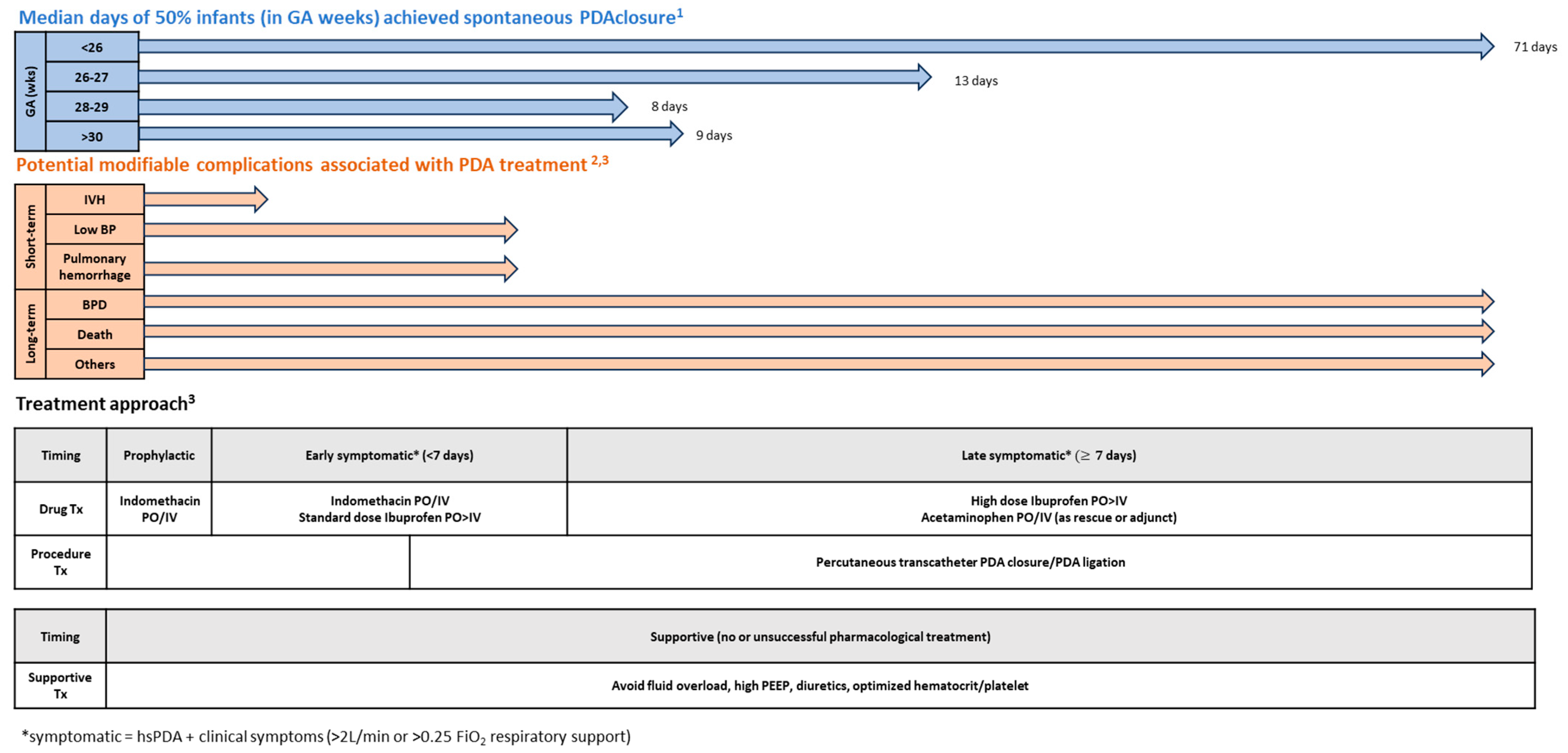
| GA (weeks) | <26 | <28 | <30 | >30 | |
| Median Day [17] | 71 | 13 | 8 | 9 | |
| Indomethacin | Ibuprofen | Acetaminophen (Paracetamol) | |
|---|---|---|---|
| Mechanism of action | Inhibits COX enzyme for prostaglandin synthesis | Same as indomethacin | Inhibits the peroxidation site of the COX enzyme for prostaglandin conversion |
| Standard regimen | 0.1 mg/kg/dose IV × 3 doses every 24 h (for 6–12 h after birth) | 10 mg/kg/dose PO or IV × 1, then 5 mg/kg/dose every 24 h × 2 doses | 15 mg/kg/dose PO or IV every 6 h × 12–28 doses |
| High dose regimen | 0.2 mg/kg/dose IV × 1 dose, then 0.1 mg/kg/dose IV every 24 h × 2 doses (for <48 h after birth) Or 0.2 mg/kg/dose IV every 24 h × 3 doses (for >2 days after birth) | 15 mg/kg/dose PO or IV × 1, then 7.5 mg/kg/dose every 24 h × 2 doses Or 20 mg/kg/dose PO or IV × 1, then 10 mg/kg/dose every 24 h × 2 doses (for >5 days after birth or >28 weeks GA) | |
| Repeat course | May repeat up to two course | May repeat up to two course | |
| Formulation | IV, no commercially available PO form for neonates | Both IV and PO forms. PO is more effective than IV | Both IV and PO forms |
| Half-life | 20 h; (12.5 h in >32-week GA; 17.5 h in <32-week GA) | 16 h for PO, 30.5 h for IV. Clearance is GA and chronological age dependent | |
| Elimination from body | 60% in urine, 40% in feces | Mostly in urine | Metabolized by liver |
| Adverse effects | Bleeding, negative renal effects with elevated creatinine or oliguria, bowel perforation in concurrent use of corticosteroids, further decrease mesenteric blood flow in the setting of hsPDA | Bleeding, negative renal effects (lower risk than indomethacin) | Hepatotoxicity |
| Efficacy and other considerations |
|
|
|
Disclaimer/Publisher’s Note: The statements, opinions and data contained in all publications are solely those of the individual author(s) and contributor(s) and not of MDPI and/or the editor(s). MDPI and/or the editor(s) disclaim responsibility for any injury to people or property resulting from any ideas, methods, instructions or products referred to in the content. |
© 2023 by the authors. Licensee MDPI, Basel, Switzerland. This article is an open access article distributed under the terms and conditions of the Creative Commons Attribution (CC BY) license (https://creativecommons.org/licenses/by/4.0/).
Share and Cite
Chan, B.; Singh, Y. Personalized Evidence-Based Management of Patent Ductus Arteriosus in Preterm Infants. J. Cardiovasc. Dev. Dis. 2024, 11, 7. https://doi.org/10.3390/jcdd11010007
Chan B, Singh Y. Personalized Evidence-Based Management of Patent Ductus Arteriosus in Preterm Infants. Journal of Cardiovascular Development and Disease. 2024; 11(1):7. https://doi.org/10.3390/jcdd11010007
Chicago/Turabian StyleChan, Belinda, and Yogen Singh. 2024. "Personalized Evidence-Based Management of Patent Ductus Arteriosus in Preterm Infants" Journal of Cardiovascular Development and Disease 11, no. 1: 7. https://doi.org/10.3390/jcdd11010007
APA StyleChan, B., & Singh, Y. (2024). Personalized Evidence-Based Management of Patent Ductus Arteriosus in Preterm Infants. Journal of Cardiovascular Development and Disease, 11(1), 7. https://doi.org/10.3390/jcdd11010007