
Inheritance of Monogenic Hereditary Skin Disease and Related Canine Breeds


Abstract
Simple Summary
Abstract
1. Introduction
2. Hereditary Epidermolysis Bullosa
3. Ichthyosis
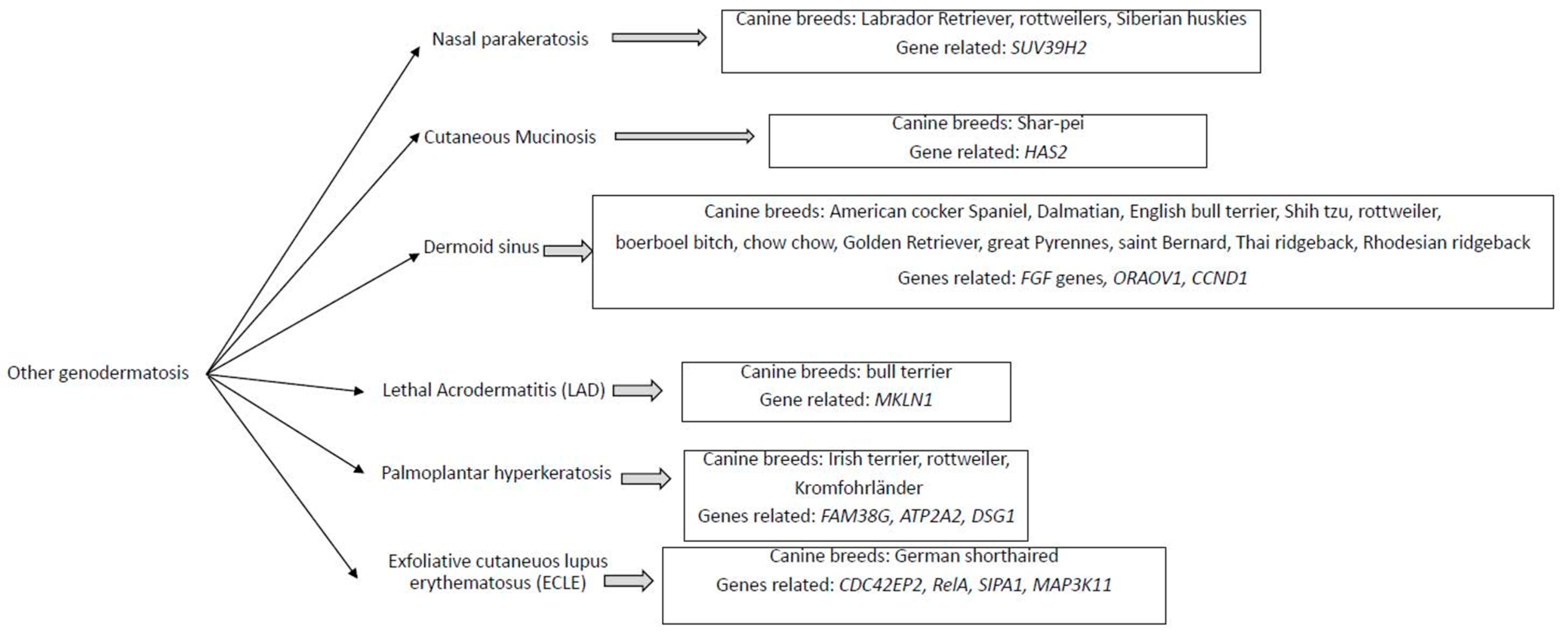
4. Other Genodermatosis
5. Conclusions
Author Contributions
Funding
Institutional Review Board Statement
Informed Consent Statement
Data Availability Statement
Acknowledgments
Conflicts of Interest
References
- Wade, C.M. Inbreeding and Genetic Diversity in Dogs: Results from DNA Analysis. Vet. J. 2011, 189, 183–188. [Google Scholar] [CrossRef] [PubMed]
- Leroy, G.; Baumung, R. Mating Practices and the Dissemination of Genetic Disorders in Domestic Animals, Based on the Example of Dog Breeding. Anim. Genet. 2011, 42, 66–74. [Google Scholar] [CrossRef] [PubMed]
- Cruz, F.; Vilà, C.; Webster, M.T. The Legacy of Domestication: Accumulation of Deleterious Mutations in the Dog Genome. Mol. Biol. Evol. 2008, 25, 2331–2336. [Google Scholar] [CrossRef]
- Mooney, J.A.; Yohannes, A.; Lohmueller, K.E. The Impact of Identity by Descent on Fitness and Disease in Dogs. Proc. Natl. Acad. Sci. USA 2021, 118, e2019116118. [Google Scholar] [CrossRef] [PubMed]
- Makino, T.; Rubin, C.-J.; Carneiro, M.; Axelsson, E.; Andersson, L.; Webster, M.T. Elevated Proportions of Deleterious Genetic Variation in Domestic Animals and Plants. Genome Biol. Evol. 2018, 10, 276–290. [Google Scholar] [CrossRef] [PubMed]
- Leeb, T.; Roosje, P.; Welle, M. Genetics of Inherited Skin Disorders in Dogs. Vet. J. 2021, 279, 105782. [Google Scholar] [CrossRef] [PubMed]
- Switonski, M.; Szczerbal, I.; Nowacka, J. The Dog Genome Map and Its Use in Mammalian Comparative Genomics. J. Appl. Genet. 2004, 45, 195–214. [Google Scholar]
- Wang, W.; Kirkness, E.F. Short Interspersed Elements (SINEs) Are a Major Source of Canine Genomic Diversity. Genome Res. 2005, 15, 1798–1808. [Google Scholar] [CrossRef]
- Lindblad-Toh, K.; Wade, C.M.; Mikkelsen, T.S.; Karlsson, E.K.; Jaffe, D.B.; Kamal, M.; Clamp, M.; Chang, J.L.; Kulbokas, E.J.; Zody, M.C.; et al. Genome Sequence, Comparative Analysis and Haplotype Structure of the Domestic Dog. Nature 2005, 438, 803–819. [Google Scholar] [CrossRef]
- Capt, A.; Spirito, F.; Guaguere, E.; Spadafora, A.; Ortonne, J.-P.; Meneguzzi, G. Inherited Junctional Epidermolysis Bullosa in the German Pointer: Establishment of a Large Animal Model. J. Investig. Dermatol. 2005, 124, 530–535. [Google Scholar] [CrossRef]
- Kiener, S.; Laprais, A.; Mauldin, E.A.; Jagannathan, V.; Olivry, T.; Leeb, T. LAMB3 Missense Variant in Australian Shepherd Dogs with Junctional Epidermolysis Bullosa. Genes 2020, 11, 1055. [Google Scholar] [CrossRef] [PubMed]
- Nagata, M.; Shimizu, H.; Masunaga, T.; Nishikawa, T.; Nanko, H.; Kariya, K.; Washizu, T.; Ishida, T. Dystrophic Form of Inherited Epidermolysis Bullosa in a Dog (Akita Inu). Br. J. Dermatol. 1995, 133, 1000–1003. [Google Scholar] [CrossRef]
- Magnol, J.-P.; Pin, D.; Palazzi, X.; Lacour, J.-P.; Gache, Y.; Meneguzzi, G. Characterization of a canine model of dystrophic bullous epidermolysis (DBE). Development of a gene therapy protocol. Bull. Acad. Natl. Med. 2005, 189, 107–119; discussion 119–121. [Google Scholar] [PubMed]
- Mauldin, E.A.; Wang, P.; Evans, E.; Cantner, C.A.; Ferracone, J.D.; Credille, K.M.; Casal, M.L. Autosomal Recessive Congenital Ichthyosis in American Bulldogs Is Associated with NIPAL4 (ICHTHYIN) Deficiency. Vet. Pathol. 2015, 52, 654–662. [Google Scholar] [CrossRef] [PubMed]
- Metzger, J.; Wöhlke, A.; Mischke, R.; Hoffmann, A.; Hewicker-Trautwein, M.; Küch, E.-M.; Naim, H.Y.; Distl, O. A Novel SLC27A4 Splice Acceptor Site Mutation in Great Danes with Ichthyosis. PLoS ONE 2015, 10, e0141514. [Google Scholar] [CrossRef]
- Credille, K.M.; Minor, J.S.; Barnhart, K.F.; Lee, E.; Cox, M.L.; Tucker, K.A.; Diegel, K.L.; Venta, P.J.; Hohl, D.; Huber, M.; et al. Transglutaminase 1-Deficient Recessive Lamellar Ichthyosis Associated with a LINE-1 Insertion in Jack Russell Terrier Dogs. Br. J. Dermatol. 2009, 161, 265–272. [Google Scholar] [CrossRef] [PubMed]
- Grall, A.; Guaguère, E.; Planchais, S.; Grond, S.; Bourrat, E.; Hausser, I.; Hitte, C.; Le Gallo, M.; Derbois, C.; Kim, G.-J.; et al. PNPLA1 Mutations Cause Autosomal Recessive Congenital Ichthyosis in Golden Retriever Dogs and Humans. Nat. Genet. 2012, 44, 140–147. [Google Scholar] [CrossRef] [PubMed]
- Graziano, L.; Vasconi, M.; Cornegliani, L. Prevalence of PNPLA1 Gene Mutation in 48 Breeding Golden Retriever Dogs. Vet. Sci. 2018, 5, 48. [Google Scholar] [CrossRef]
- Jagannathan, V.; Bannoehr, J.; Plattet, P.; Hauswirth, R.; Drögemüller, C.; Drögemüller, M.; Wiener, D.J.; Doherr, M.; Owczarek-Lipska, M.; Galichet, A.; et al. A Mutation in the SUV39H2 Gene in Labrador Retrievers with Hereditary Nasal Parakeratosis (HNPK) Provides Insights into the Epigenetics of Keratinocyte Differentiation. PLoS Genet. 2013, 9, e1003848. [Google Scholar] [CrossRef] [PubMed]
- von Bomhard, D.; Kraft, W. Idiopathic mucinosis cutis in Chinese Shar pei dogs: Epidemiology, clinical features, histopathologic findings and treatment. Tierarztl. Prax. Ausg. K Kleintiere Heimtiere 1998, 26, 189–196. [Google Scholar]
- Salmon Hillbertz, N.H.C.; Isaksson, M.; Karlsson, E.K.; Hellmén, E.; Pielberg, G.R.; Savolainen, P.; Wade, C.M.; von Euler, H.; Gustafson, U.; Hedhammar, A.; et al. Duplication of FGF3, FGF4, FGF19 and ORAOV1 Causes Hair Ridge and Predisposition to Dermoid Sinus in Ridgeback Dogs. Nat. Genet. 2007, 39, 1318–1320. [Google Scholar] [CrossRef]
- Fine, J.-D.; Bruckner-Tuderman, L.; Eady, R.A.J.; Bauer, E.A.; Bauer, J.W.; Has, C.; Heagerty, A.; Hintner, H.; Hovnanian, A.; Jonkman, M.F.; et al. Inherited Epidermolysis Bullosa: Updated Recommendations on Diagnosis and Classification. J. Am. Acad. Dermatol. 2014, 70, 1103–1126. [Google Scholar] [CrossRef] [PubMed]
- Medeiros, G.X.; Riet-Correa, F. Epidermolysis Bullosa in Animals: A Review. Vet. Dermatol. 2015, 26, 3–13, e1–e2. [Google Scholar] [CrossRef]
- Uitto, J.; Pulkkinen, L. Molecular Genetics of Heritable Blistering Disorders. Arch. Dermatol. 2001, 137, 1458–1461. [Google Scholar] [CrossRef] [PubMed]
- Mauldin, E.A.; Wang, P.; Olivry, T.; Henthorn, P.S.; Casal, M.L. Epidermolysis Bullosa Simplex in Sibling Eurasier Dogs Is Caused by a PLEC Non-Sense Variant. Vet. Dermatol. 2017, 28, 10-e3. [Google Scholar] [CrossRef]
- Herrmann, I.; Linder, K.E.; Meurs, K.M.; Friedenberg, S.G.; Cullen, J.; Olby, N.; Bizikova, P. Canine Junctional Epidermolysis Bullosa Due to a Novel Mutation in LAMA3 with Severe Upper Respiratory Involvement. Vet. Dermatol. 2021, 32, 379-e108. [Google Scholar] [CrossRef]
- Frattini, S.; Polli, M.; Cortellari, M.; Negro, A.; Bionda, A.; Riva, J.; Rizzi, R.; Marelli, S.; Crepaldi, P. Genetic Trend of the Junctional Epidermolysis Bullosa in the German Shorthaired Pointer in Italy. Vet. Rec. Open 2021, 8, e15. [Google Scholar] [CrossRef]
- Pigors, M.; Schwieger-Briel, A.; Leppert, J.; Kiritsi, D.; Kohlhase, J.; Bruckner-Tuderman, L.; Has, C. Molecular Heterogeneity of Epidermolysis Bullosa Simplex: Contribution of EXPH5 Mutations. J. Investig. Dermatol. 2014, 134, 842–845. [Google Scholar] [CrossRef]
- McGrath, J.A.; Stone, K.L.; Begum, R.; Simpson, M.A.; Dopping-Hepenstal, P.J.; Liu, L.; McMillan, J.R.; South, A.P.; Pourreyron, C.; McLean, W.H.I.; et al. Germline Mutation in EXPH5 Implicates the Rab27B Effector Protein Slac2-b in Inherited Skin Fragility. Am. J. Hum. Genet. 2012, 91, 1115–1121. [Google Scholar] [CrossRef]
- McGrath, J.A.; Mellerio, J.E. Ectodermal Dysplasia-Skin Fragility Syndrome. Dermatol. Clin. 2010, 28, 125–129. [Google Scholar] [CrossRef]
- Pigors, M.; Kiritsi, D.; Cobzaru, C.; Schwieger-Briel, A.; Suárez, J.; Faletra, F.; Aho, H.; Mäkelä, L.; Kern, J.S.; Bruckner-Tuderman, L.; et al. TGM5 Mutations Impact Epidermal Differentiation in Acral Peeling Skin Syndrome. J. Investig. Dermatol. 2012, 132, 2422–2429. [Google Scholar] [CrossRef] [PubMed]
- Pigors, M.; Kiritsi, D.; Krümpelmann, S.; Wagner, N.; He, Y.; Podda, M.; Kohlhase, J.; Hausser, I.; Bruckner-Tuderman, L.; Has, C. Lack of Plakoglobin Leads to Lethal Congenital Epidermolysis Bullosa: A Novel Clinico-Genetic Entity. Hum. Mol. Genet. 2011, 20, 1811–1819. [Google Scholar] [CrossRef] [PubMed]
- Bolling, M.C.; Veenstra, M.J.; Jonkman, M.F.; Diercks, G.F.H.; Curry, C.J.; Fisher, J.; Pas, H.H.; Bruckner, A.L. Lethal Acantholytic Epidermolysis Bullosa Due to a Novel Homozygous Deletion in DSP: Expanding the Phenotype and Implications for Desmoplakin Function in Skin and Heart. Br. J. Dermatol. 2010, 162, 1388–1394. [Google Scholar] [CrossRef]
- Hobbs, R.P.; Han, S.Y.; van der Zwaag, P.A.; Bolling, M.C.; Jongbloed, J.D.H.; Jonkman, M.F.; Getsios, S.; Paller, A.S.; Green, K.J. Insights from a Desmoplakin Mutation Identified in Lethal Acantholytic Epidermolysis Bullosa. J. Investig. Derm. 2010, 130, 2680–2683. [Google Scholar] [CrossRef] [PubMed]
- Jonkman, M.F.; Pasmooij, A.M.G.; Pasmans, S.G.M.A.; van den Berg, M.P.; Ter Horst, H.J.; Timmer, A.; Pas, H.H. Loss of Desmoplakin Tail Causes Lethal Acantholytic Epidermolysis Bullosa. Am. J. Hum. Genet. 2005, 77, 653–660. [Google Scholar] [CrossRef]
- Kiritsi, D.; Cosgarea, I.; Franzke, C.-W.; Schumann, H.; Oji, V.; Kohlhase, J.; Bruckner-Tuderman, L.; Has, C. Acral Peeling Skin Syndrome with TGM5 Gene Mutations May Resemble Epidermolysis Bullosa Simplex in Young Individuals. J. Investig. Dermatol. 2010, 130, 1741–1746. [Google Scholar] [CrossRef]
- Castañón, M.J.; Walko, G.; Winter, L.; Wiche, G. Plectin-Intermediate Filament Partnership in Skin, Skeletal Muscle, and Peripheral Nerve. Histochem. Cell Biol. 2013, 140, 33–53. [Google Scholar] [CrossRef]
- Wiche, G. Role of Plectin in Cytoskeleton Organization and Dynamics. J. Cell Sci. 1998, 111, 2477–2486. [Google Scholar] [CrossRef]
- Mauldin, E.A.; Credille, K.M.; Dunstan, R.W.; Casal, M.L. The Clinical and Morphologic Features of Nonepidermolytic Ichthyosis in the Golden Retriever. Vet. Pathol. 2008, 45, 174–180. [Google Scholar] [CrossRef]
- Olivry, T.; Linder, K.E.; Wang, P.; Bizikova, P.; Bernstein, J.A.; Dunston, S.M.; Paps, J.S.; Casal, M.L. Deficient Plakophilin-1 Expression Due to a Mutation in PKP1 Causes Ectodermal Dysplasia-Skin Fragility Syndrome in Chesapeake Bay Retriever Dogs. PLoS ONE 2012, 7, e32072. [Google Scholar] [CrossRef]
- South, A.P. Plakophilin 1: An Important Stabilizer of Desmosomes. Clin. Exp. Dermatol. 2004, 29, 161–167. [Google Scholar] [CrossRef] [PubMed]
- Has, C.; Bauer, J.W.; Bodemer, C.; Bolling, M.C.; Bruckner-Tuderman, L.; Diem, A.; Fine, J.-D.; Heagerty, A.; Hovnanian, A.; Marinkovich, M.P.; et al. Consensus Reclassification of Inherited Epidermolysis Bullosa and Other Disorders with Skin Fragility. Br. J. Dermatol. 2020, 183, 614–627. [Google Scholar] [CrossRef] [PubMed]
- Bardhan, A.; Bruckner-Tuderman, L.; Chapple, I.L.C.; Fine, J.-D.; Harper, N.; Has, C.; Magin, T.M.; Marinkovich, M.P.; Marshall, J.F.; McGrath, J.A.; et al. Epidermolysis Bullosa. Nat. Rev. Dis. Primers 2020, 6, 78. [Google Scholar] [CrossRef] [PubMed]
- Buchroithner, B.; Klausegger, A.; Ebschner, U.; Anton-Lamprecht, I.; Pohla-Gubo, G.; Lanschuetzer, C.M.; Laimer, M.; Hintner, H.; Bauer, J.W. Analysis of the LAMB3 Gene in a Junctional Epidermolysis Bullosa Patient Reveals Exonic Splicing and Allele-Specific Nonsense-Mediated MRNA Decay. Lab. Investig. 2004, 84, 1279–1288. [Google Scholar] [CrossRef]
- Liu, L.; Jung, S.-N.; Oh, C.; Lee, K.; Won, H.-R.; Chang, J.W.; Kim, J.M.; Koo, B.S. LAMB3 Is Associated with Disease Progression and Cisplatin Cytotoxic Sensitivity in Head and Neck Squamous Cell Carcinoma. Eur. J. Surg. Oncol. 2019, 45, 359–365. [Google Scholar] [CrossRef]
- Fine, J.-D.; Eady, R.A.J.; Bauer, E.A.; Bauer, J.W.; Bruckner-Tuderman, L.; Heagerty, A.; Hintner, H.; Hovnanian, A.; Jonkman, M.F.; Leigh, I.; et al. The Classification of Inherited Epidermolysis Bullosa (EB): Report of the Third International Consensus Meeting on Diagnosis and Classification of EB. J. Am. Acad. Dermatol. 2008, 58, 931–950. [Google Scholar] [CrossRef]
- Bruckner-Tuderman, L.; Nilssen, O.; Zimmermann, D.R.; Dours-Zimmermann, M.T.; Kalinke, D.U.; Gedde-Dahl, T.; Winberg, J.O. Immunohistochemical and Mutation Analyses Demonstrate That Procollagen VII Is Processed to Collagen VII through Removal of the NC-2 Domain. J. Cell Biol. 1995, 131, 551–559. [Google Scholar] [CrossRef]
- Dang, N.; Murrell, D.F. Mutation Analysis and Characterization of COL7A1 Mutations in Dystrophic Epidermolysis Bullosa. Exp. Dermatol. 2008, 17, 553–568. [Google Scholar] [CrossRef]
- Gache, Y.; Pin, D.; Gagnoux-Palacios, L.; Carozzo, C.; Meneguzzi, G. Correction of Dog Dystrophic Epidermolysis Bullosa by Transplantation of Genetically Modified Epidermal Autografts. J. Investig. Dermatol. 2011, 131, 2069–2078. [Google Scholar] [CrossRef]
- Baldeschi, C.; Gache, Y.; Rattenholl, A.; Bouillé, P.; Danos, O.; Ortonne, J.-P.; Bruckner-Tuderman, L.; Meneguzzi, G. Genetic Correction of Canine Dystrophic Epidermolysis Bullosa Mediated by Retroviral Vectors. Hum. Mol. Genet. 2003, 12, 1897–1905. [Google Scholar] [CrossRef]
- Gretzmeier, C.; Pin, D.; Kern, J.S.; Chen, M.; Woodley, D.T.; Bruckner-Tuderman, L.; de Souza, M.P.; Nyström, A. Systemic Collagen VII Replacement Therapy for Advanced Recessive Dystrophic Epidermolysis Bullosa. J. Investig. Dermatol. 2022, 142, 1094–1102.e3. [Google Scholar] [CrossRef] [PubMed]
- Mauldin, E.A. Canine Ichthyosis and Related Disorders of Cornification. Vet. Clin. N. Am. Small Anim. Pract. 2013, 43, 89–97. [Google Scholar] [CrossRef] [PubMed]
- Guaguère, É. A Practical. Guide to Canine Dermatology; Merial: Ingelheim am Rhein, German, 2008; ISBN 978-2-915758-11-5. [Google Scholar]
- Credille, K.M.; Barnhart, K.F.; Minor, J.S.; Dunstan, R.W. Mild Recessive Epidermolytic Hyperkeratosis Associated with a Novel Keratin 10 Donor Splice-Site Mutation in a Family of Norfolk Terrier Dogs. Br. J. Dermatol. 2005, 153, 51–58. [Google Scholar] [CrossRef] [PubMed][Green Version]
- Alperin, E.S.; Shapiro, L.J. Characterization of Point Mutations in Patients with X-Linked Ichthyosis. Effects on the Structure and Function of the Steroid Sulfatase Protein. J. Biol. Chem. 1997, 272, 20756–20763. [Google Scholar] [CrossRef]
- Bauer, A.; Waluk, D.P.; Galichet, A.; Timm, K.; Jagannathan, V.; Sayar, B.S.; Wiener, D.J.; Dietschi, E.; Müller, E.J.; Roosje, P.; et al. A de Novo Variant in the ASPRV1 Gene in a Dog with Ichthyosis. PLoS Genet. 2017, 13, e1006651. [Google Scholar] [CrossRef]
- Bauer, A.; De Lucia, M.; Jagannathan, V.; Mezzalira, G.; Casal, M.L.; Welle, M.M.; Leeb, T. A Large Deletion in the NSDHL Gene in Labrador Retrievers with a Congenital Cornification Disorder. G3 (Bethesda) 2017, 7, 3115–3121. [Google Scholar] [CrossRef][Green Version]
- Kiener, S.; Wiener, D.J.; Hopke, K.; Diesel, A.B.; Jagannathan, V.; Mauldin, E.A.; Casal, M.L.; Leeb, T. ABHD5 Frameshift Deletion in Golden Retrievers with Ichthyosis. G3 (Bethesda) 2021, 12, jkab397. [Google Scholar] [CrossRef]
- Casal, M.L.; Wang, P.; Mauldin, E.A.; Lin, G.; Henthorn, P.S. A Defect in NIPAL4 Is Associated with Autosomal Recessive Congenital Ichthyosis in American Bulldogs. PLoS ONE 2017, 12, e0170708. [Google Scholar] [CrossRef]
- Guaguere, E.; Bensignor, E.; Küry, S.; Degorce-Rubiales, F.; Muller, A.; Herbin, L.; Fontaine, J.; André, C. Clinical, Histopathological and Genetic Data of Ichthyosis in the Golden Retriever: A Prospective Study. J. Small Anim. Pract. 2009, 50, 227–235. [Google Scholar] [CrossRef]
- Owczarek-Lipska, M.; Thomas, A.; André, C.; Hölzer, S.; Leeb, T. Frequency of gene defects in selected European retriever populations. Schweiz. Arch. Tierheilkd. 2011, 153, 418–420. [Google Scholar] [CrossRef]
- Kienesberger, P.C.; Oberer, M.; Lass, A.; Zechner, R. Mammalian Patatin Domain Containing Proteins: A Family with Diverse Lipolytic Activities Involved in Multiple Biological Functions. J. Lipid Res. 2009, 50, S63–S68. [Google Scholar] [CrossRef] [PubMed]
- Grond, S.; Eichmann, T.O.; Dubrac, S.; Kolb, D.; Schmuth, M.; Fischer, J.; Crumrine, D.; Elias, P.M.; Haemmerle, G.; Zechner, R.; et al. PNPLA1 Deficiency in Mice and Humans Leads to a Defect in the Synthesis of Omega-O-Acylceramides. J. Investig. Dermatol. 2017, 137, 394–402. [Google Scholar] [CrossRef]
- Pichery, M.; Huchenq, A.; Sandhoff, R.; Severino-Freire, M.; Zaafouri, S.; Opálka, L.; Levade, T.; Soldan, V.; Bertrand-Michel, J.; Lhuillier, E.; et al. PNPLA1 Defects in Patients with Autosomal Recessive Congenital Ichthyosis and KO Mice Sustain PNPLA1 Irreplaceable Function in Epidermal Omega-O-Acylceramide Synthesis and Skin Permeability Barrier. Hum. Mol. Genet. 2017, 26, 1787–1800. [Google Scholar] [CrossRef]
- Puigdemont, A.; Furiani, N.; De Lucia, M.; Carrasco, I.; Ordeix, L.; Fondevila, D.; Ramió-Lluch, L.; Brazis, P. Topical Polyhydroxy Acid Treatment for Autosomal Recessive Congenital Ichthyosis in the Golden Retriever: A Prospective Pilot Study. Vet. Dermatol. 2018, 29, 323-e113. [Google Scholar] [CrossRef] [PubMed]
- Nakhaei, S.; Heidary, H.; Rahimian, A.; Vafadar, M.; Rohani, F.; Bahoosh, G.R.; Amirkashani, D. A New Case of Chanarin-Dorfman Syndrome with a Novel Deletion in ABHD5 Gene. Iran. Biomed. J. 2018, 22, 415–419. [Google Scholar] [CrossRef] [PubMed][Green Version]
- Eskiocak, A.H.; Missaglia, S.; Moro, L.; Durdu, M.; Tavian, D. A Novel Mutation of ABHD5 Gene in a Chanarin Dorfman Patient with Unusual Dermatological Findings. Lipids Health Dis. 2019, 18, 232. [Google Scholar] [CrossRef]
- Golda, M.; Mótyán, J.A.; Nagy, K.; Matúz, K.; Nagy, T.; Tőzsér, J. Biochemical Characterization of Human Retroviral-Like Aspartic Protease 1 (ASPRV1). Biomolecules 2020, 10, 1004. [Google Scholar] [CrossRef]
- Matsui, T.; Miyamoto, K.; Kubo, A.; Kawasaki, H.; Ebihara, T.; Hata, K.; Tanahashi, S.; Ichinose, S.; Imoto, I.; Inazawa, J.; et al. SASPase Regulates Stratum Corneum Hydration through Profilaggrin-to-Filaggrin Processing. EMBO Mol. Med. 2011, 3, 320–333. [Google Scholar] [CrossRef]
- Briand, A.; Cochet-Faivre, N.; Reyes-Gomez, E.; Jaraud-Darnault, A.; Tiret, L.; Chevallier, L. NIPAL4 Deletion Identified in an American Bully with Autosomal Recessive Congenital Ichthyosis and Response to Topical Therapy. Vet. Med. Sci. 2019, 5, 112–117. [Google Scholar] [CrossRef]
- Pigg, M.H.; Bygum, A.; Gånemo, A.; Virtanen, M.; Brandrup, F.; Zimmer, A.D.; Hotz, A.; Vahlquist, A.; Fischer, J. Spectrum of Autosomal Recessive Congenital Ichthyosis in Scandinavia: Clinical Characteristics and Novel and Recurrent Mutations in 132 Patients. Acta Derm Venereol. 2016, 96, 932–937. [Google Scholar] [CrossRef]
- Dahlqvist, J.; Westermark, G.T.; Vahlquist, A.; Dahl, N. Ichthyin/NIPAL4 Localizes to Keratins and Desmosomes in Epidermis and Ichthyin Mutations Affect Epidermal Lipid Metabolism. Arch. Dermatol. Res. 2012, 304, 377–386. [Google Scholar] [CrossRef]
- Mauldin, E.A.; Crumrine, D.; Casal, M.L.; Jeong, S.; Opálka, L.; Vavrova, K.; Uchida, Y.; Park, K.; Craiglow, B.; Choate, K.A.; et al. Cellular and Metabolic Basis for the Ichthyotic Phenotype in NIPAL4 (Ichthyin)-Deficient Canines. Am. J. Pathol. 2018, 188, 1419–1429. [Google Scholar] [CrossRef] [PubMed]
- Yen, M.-C.; Chou, S.-K.; Kan, J.-Y.; Kuo, P.-L.; Hou, M.-F.; Hsu, Y.-L. Solute Carrier Family 27 Member 4 (SLC27A4) Enhances Cell Growth, Migration, and Invasion in Breast Cancer Cells. Int. J. Mol. Sci. 2018, 19, 3434. [Google Scholar] [CrossRef] [PubMed]
- Schwenk, R.W.; Holloway, G.P.; Luiken, J.J.F.P.; Bonen, A.; Glatz, J.F.C. Fatty Acid Transport across the Cell Membrane: Regulation by Fatty Acid Transporters. Prostaglandins Leukot Essent Fat. Acids 2010, 82, 149–154. [Google Scholar] [CrossRef]
- Simpson, J.K.; Martinez-Queipo, M.; Onoufriadis, A.; Tso, S.; Glass, E.; Liu, L.; Higashino, T.; Scott, W.; Tierney, C.; Simpson, M.A.; et al. Genotype-Phenotype Correlation in a Large English Cohort of Patients with Autosomal Recessive Ichthyosis. Br. J. Dermatol. 2020, 182, 729–737. [Google Scholar] [CrossRef] [PubMed]
- Saldaña-García, N.; Espinosa-Fernández, M.G.; Serrano-Martín, M.D.M.; Vera Casaño, Á. A new SLC27A4 mutation associated with ichthyosis prematurity syndrome and compartment syndrome. An. Pediatr. 2020, 92, 308–310. [Google Scholar] [CrossRef] [PubMed]
- Li, S.; Green, J.F.; Jin, M. Impacts of Deletion and Ichthyosis Prematurity Syndrome-Associated Mutations in Fatty Acid Transport Protein 4 on the Function of RPE65. FEBS Lett. 2020, 594, 540–552. [Google Scholar] [CrossRef]
- Oji, V.; Traupe, H. Ichthyoses: Differential Diagnosis and Molecular Genetics. Eur. J. Dermatol. 2006, 16, 349–359. [Google Scholar]
- Radner, F.P.W.; Marrakchi, S.; Kirchmeier, P.; Kim, G.-J.; Ribierre, F.; Kamoun, B.; Abid, L.; Leipoldt, M.; Turki, H.; Schempp, W.; et al. Mutations in CERS3 Cause Autosomal Recessive Congenital Ichthyosis in Humans. PLoS Genet. 2013, 9, e1003536. [Google Scholar] [CrossRef]
- Caroppo, F.; Cama, E.; Salmaso, R.; Bertolin, C.; Salviati, L.; Belloni Fortina, A. A Novel KRT1 c.1433A>G p.(Glu478Gly) Mutation in a Newborn with Epidermolytic Ichthyosis. Clin. Case Rep. 2020, 8, 3079–3081. [Google Scholar] [CrossRef]
- Nellen, R.G.L.; Nagtzaam, I.F.; Hoogeboom, A.J.M.; Bladergroen, R.S.; Jonkman, M.F.; Steijlen, P.M.; van Steensel, M.A.M.; van Geel, M. Phenotypic Variation in Epidermolytic Ichthyosis: Clinical and Functional Evaluation of the Novel p.(Met339Lys) Mutation in the L12 Domain of KRT1. Exp. Dermatol. 2015, 24, 883–885. [Google Scholar] [CrossRef] [PubMed]
- Al Raddadi, A.A.; Habibullah, T.H.; Abdelaal, A.M.; Felimban, A.M.; Al Raddadi, H.A.; Satti, M.B. Epidermolytic Ichthyosis without Keratin 1 or 10 Mutations: A. Case Report. Saudi. J. Med. Med. Sci. 2018, 6, 36–39. [Google Scholar] [CrossRef] [PubMed]
- Bannoehr, J.; Balmer, P.; Stoffel, M.H.; Jagannathan, V.; Gaschen, V.; Kühni, K.; Sayar, B.; Drögemüller, M.; Howald, D.; Wiener, D.J.; et al. Abnormal Keratinocyte Differentiation in the Nasal Planum of Labrador Retrievers with Hereditary Nasal Parakeratosis (HNPK). PLoS ONE 2020, 15, e0225901. [Google Scholar] [CrossRef]
- Mecklenburg, L.; Hetzel, U.; Ueberschär, S. Epidermolytic Ichthyosis in a Dog: Clinical, Histopathological, Immunohistochemical and Ultrastructural Findings. J. Comp. Pathol. 2000, 122, 307–311. [Google Scholar] [CrossRef] [PubMed]
- Mauldin, E.A.; Elias, P.M. Ichthyosis and Hereditary Cornification Disorders in Dogs. Vet. Dermatol. 2021, 32, 567-e154. [Google Scholar] [CrossRef] [PubMed]
- Peters, J.; Scott, D.W.; Erb, H.N.; Miller, W.H. Hereditary Nasal Parakeratosis in Labrador Retrievers: 11 New Cases and a Retrospective Study on the Presence of Accumulations of Serum (‘serum Lakes’) in the Epidermis of Parakeratotic Dermatoses and Inflamed Nasal Plana of Dogs. Vet. Dermatol. 2003, 14, 197–203. [Google Scholar] [CrossRef]
- Senter, D.A.; Scott, D.W.; Miller, W.H.; Erb, H.N. Intracorneal Vacuoles in Skin Diseases with Parakeratotic Hyperkeratosis in the Dog: A Retrospective Light-Microscopy Study of 111 Cases (1973-2000). Vet. Dermatol. 2002, 13, 43–47. [Google Scholar] [CrossRef] [PubMed]
- Weirich, S.; Khella, M.S.; Jeltsch, A. Structure, Activity and Function of the Suv39h1 and Suv39h2 Protein Lysine Methyltransferases. Life 2021, 11, 703. [Google Scholar] [CrossRef] [PubMed]
- Wang, L.; Chakraborty, D.; Iqbal, K.; Soares, M.J. SUV39H2 Controls Trophoblast Stem Cell Fate. Biochim. Biophys. Acta Gen. Subj. 2021, 1865, 129867. [Google Scholar] [CrossRef] [PubMed]
- Dillberger, J.E.; Altman, N.H. Focal Mucinosis in Dogs: Seven Cases and Review of Cutaneous Mucinoses of Man and Animals. Vet. Pathol. 1986, 23, 132–139. [Google Scholar] [CrossRef] [PubMed]
- Welle, M.; Grimm, S.; Suter, M.; von Tscharner, C. Mast Cell Density and Subtypes in the Skin of Shar Pei Dogs with Cutaneous Mucinosis. Zentralbl. Vet. A 1999, 46, 309–316. [Google Scholar] [CrossRef] [PubMed]
- Whipple, K.M.; Kieran, E.A.; Dark, M.J.; Beatty, S.S. What Is Your Diagnosis? Cutaneous Mass from a Shar-Pei Dog. Vet. Clin. Pathol. 2020, 49, 365–366. [Google Scholar] [CrossRef] [PubMed]
- Zanna, G.; Fondevila, D.; Bardagí, M.; Docampo, M.J.; Bassols, A.; Ferrer, L. Cutaneous Mucinosis in Shar-Pei Dogs Is Due to Hyaluronic Acid Deposition and Is Associated with High Levels of Hyaluronic Acid in Serum. Vet. Dermatol. 2008, 19, 314–318. [Google Scholar] [CrossRef] [PubMed]
- Docampo, M.J.; Zanna, G.; Fondevila, D.; Cabrera, J.; López-Iglesias, C.; Carvalho, A.; Cerrato, S.; Ferrer, L.; Bassols, A. Increased HAS2-Driven Hyaluronic Acid Synthesis in Shar-Pei Dogs with Hereditary Cutaneous Hyaluronosis (Mucinosis). Vet. Dermatol. 2011, 22, 535–545. [Google Scholar] [CrossRef]
- Zanna, G.; Docampo, M.J.; Fondevila, D.; Bardagí, M.; Bassols, A.; Ferrer, L. Hereditary Cutaneous Mucinosis in Shar Pei Dogs Is Associated with Increased Hyaluronan Synthase-2 MRNA Transcription by Cultured Dermal Fibroblasts. Vet. Dermatol. 2009, 20, 377–382. [Google Scholar] [CrossRef] [PubMed]
- Heldin, P.; Basu, K.; Kozlova, I.; Porsch, H. HAS2 and CD44 in Breast Tumorigenesis. Adv. Cancer Res. 2014, 123, 211–229. [Google Scholar] [CrossRef]
- Suzuki, M.; Asplund, T.; Yamashita, H.; Heldin, C.H.; Heldin, P. Stimulation of Hyaluronan Biosynthesis by Platelet-Derived Growth Factor-BB and Transforming Growth Factor-Beta 1 Involves Activation of Protein Kinase C. Biochem. J. 1995, 307, 817–821. [Google Scholar] [CrossRef] [PubMed]
- Vigetti, D.; Clerici, M.; Deleonibus, S.; Karousou, E.; Viola, M.; Moretto, P.; Heldin, P.; Hascall, V.C.; De Luca, G.; Passi, A. Hyaluronan Synthesis Is Inhibited by Adenosine Monophosphate-Activated Protein Kinase through the Regulation of HAS2 Activity in Human Aortic Smooth Muscle Cells. J. Biol. Chem. 2011, 286, 7917–7924. [Google Scholar] [CrossRef] [PubMed]
- Yamane, T.; Kobayashi-Hattori, K.; Oishi, Y. Adiponectin Promotes Hyaluronan Synthesis along with Increases in Hyaluronan Synthase 2 Transcripts through an AMP-Activated Protein Kinase/Peroxisome Proliferator-Activated Receptor-α-Dependent Pathway in Human Dermal Fibroblasts. Biochem. Biophys. Res. Commun. 2011, 415, 235–238. [Google Scholar] [CrossRef]
- Weigel, P.H. Hyaluronan Synthase: The Mechanism of Initiation at the Reducing End and a Pendulum Model for Polysaccharide Translocation to the Cell Exterior. Int. J. Cell Biol. 2015, 2015, 367579. [Google Scholar] [CrossRef] [PubMed]
- Muller and Kirk’s Small Animal Dermatology-7th Edition. Available online: https://www.elsevier.com/books/muller-and-kirks-small-animal-dermatology/miller/978-1-4160-0028-0 (accessed on 6 February 2022).
- Rinna, C.; Reale, G.; Calafati, V.; Calvani, F.; Ungari, C. Dermoid Cyst: Unusual Localization. J. Craniofac. Surg. 2012, 23, e392–e394. [Google Scholar] [CrossRef] [PubMed]
- Hong, S.W. Deep Frontotemporal Dermoid Cyst Presenting as a Discharging Sinus: A Case Report and Review of Literature. Br. J. Plast. Surg. 1998, 51, 255–257. [Google Scholar] [CrossRef] [PubMed]
- Bailey, T.R.; Holmberg, D.L.; Yager, J.A. Nasal Dermoid Sinus in an American Cocker Spaniel. Can. Vet. J. 2001, 42, 213–215. [Google Scholar]
- van der Peijl, G.J.W.; Schaeffer, I.G.F. Nasal Dermoid Cyst Extending through the Frontal Bone with No Sinus Tract in a Dalmatian. J. Small Anim. Pract. 2011, 52, 117–120. [Google Scholar] [CrossRef]
- Burrow, R.D. A Nasal Dermoid Sinus in an English Bull Terrier. J. Small Anim. Pract. 2004, 45, 572–574. [Google Scholar] [CrossRef]
- Sturgeon, C. Nasal Dermoid Sinus Cyst in a Shih Tzu. Vet. Rec. 2008, 163, 219–220. [Google Scholar] [CrossRef]
- Bornard, N.; Pin, D.; Carozzo, C. Bilateral Parieto-Occipital Dermoid Sinuses in a Rottweiler. J. Small Anim. Pract. 2007, 48, 107–110. [Google Scholar] [CrossRef]
- Penrith, M.L.; van Schouwenburg, G. Dermoid Sinus in a Boerboel Bitch. J. S. Afr. Vet. Assoc. 1994, 65, 38–39. [Google Scholar]
- Booth, M.J. Atypical Dermoid Sinus in a Chow Chow Dog. J. S. Afr. Vet. Assoc. 1998, 69, 102–104. [Google Scholar] [CrossRef]
- Cornegliani, L.; Jommi, E.; Vercelli, A. Dermoid Sinus in a Golden Retriever. J. Small Anim. Pract. 2001, 42, 514–516. [Google Scholar] [CrossRef]
- Camacho, A.A.; Laus, J.L.; Valéri, V.; Valéri, F.V.; Nunes, N. Sinus Dermóide em Cão dos Pireneus. Braz. J. Vet. Res. Anim. Sci. 1995, 32, 170–172. [Google Scholar] [CrossRef][Green Version]
- Perazzi, A.; Berlanda, M.; Bucci, M.; Ferro, S.; Rasotto, R.; Busetto, R.; Iacopetti, I. Multiple Dermoid Sinuses of Type Vb and IIIb on the Head of a Saint Bernard Dog. Acta Vet. Scand. 2013, 55, 62. [Google Scholar] [CrossRef]
- Hathcock, J.T.; Clampett, E.G.; Broadstone, R.V. Dermoid Sinus in a Rhodesian Ridgeback. Vet. Med. Small Anim. Clin. 1979, 74, 53–56. [Google Scholar]
- Lambrechts, N. Dermoid Sinus in a Crossbred Rhodesian Ridgeback Dog Involving the Second Cervical Vertebra. J. S. Afr. Vet. Assoc. 1996, 67, 155–157. [Google Scholar]
- Antin, I.P. Dermoid Sinus in a Rhodesian Ridgeback Dog. J. Am. Vet. Med. Assoc. 1970, 157, 961–962. [Google Scholar]
- Gammie, J.S. Dermoid Sinus Removal in a Rhodesian Ridgeback Dog. Can. Vet. J. 1986, 27, 250–251. [Google Scholar]
- Hillbertz, N.H.C.S.; Andersson, G. Autosomal Dominant Mutation Causing the Dorsal Ridge Predisposes for Dermoid Sinus in Rhodesian Ridgeback Dogs. J. Small Anim. Pract. 2006, 47, 184–188. [Google Scholar] [CrossRef]
- Li, X. The FGF Metabolic Axis. Front. Med. 2019, 13, 511–530. [Google Scholar] [CrossRef]
- Luo, X.; Jiang, Y.; Chen, F.; Wei, Z.; Qiu, Y.; Xu, H.; Tian, G.; Gong, W.; Yuan, Y.; Feng, H.; et al. ORAOV1-B Promotes OSCC Metastasis via the NF-ΚB-TNFα Loop. J. Dent. Res. 2021, 100, 858–867. [Google Scholar] [CrossRef]
- Ha, S.Y.; Yeo, S.-Y.; Lee, K.-W.; Kim, S.-H. Validation of ORAOV1 as a New Treatment Target in Hepatocellular Carcinoma. J. Cancer Res. Clin. Oncol. 2021, 147, 423–433. [Google Scholar] [CrossRef]
- Jiang, L.; Yang, H.S.; Wang, Z.; Zhou, Y.; Zhou, M.; Zeng, X.; Chen, Q.M. ORAOV1-A Correlates with Poor Differentiation in Oral Cancer. J. Dent. Res. 2009, 88, 433–438. [Google Scholar] [CrossRef] [PubMed]
- Li, M.; Cui, X.; Shen, Y.; Dong, H.; Liang, W.; Chen, Y.; Hu, J.; Li, S.; Kong, J.; Li, H.; et al. ORAOV1 Overexpression in Esophageal Squamous Cell Carcinoma and Esophageal Dysplasia: A Possible Biomarker of Progression and Poor Prognosis in Esophageal Carcinoma. Hum. Pathol. 2015, 46, 707–715. [Google Scholar] [CrossRef]
- McEwan, N.A.; McNeil, P.E.; Thompson, H.; McCandlish, I.A. Diagnostic Features, Confirmation and Disease Progression in 28 Cases of Lethal Acrodermatitis of Bull Terriers. J. Small Anim. Pract. 2000, 41, 501–507. [Google Scholar] [CrossRef] [PubMed]
- Jiang, L.; Zeng, X.; Wang, Z.; Ji, N.; Zhou, Y.; Liu, X.; Chen, Q. Oral Cancer Overexpressed 1 (ORAOV1) Regulates Cell Cycle and Apoptosis in Cervical Cancer HeLa Cells. Mol. Cancer 2010, 9, 20. [Google Scholar] [CrossRef] [PubMed]
- McEwan, N.A. Malassezia and Candida Infections in Bull Terriers with Lethal Acrodermatitis. J. Small Anim. Pract. 2001, 42, 291–297. [Google Scholar] [CrossRef] [PubMed]
- Grider, A.; Mouat, M.F.; Mauldin, E.A.; Casal, M.L. Analysis of the Liver Soluble Proteome from Bull Terriers Affected with Inherited Lethal Acrodermatitis. Mol. Genet. Metab. 2007, 92, 249–257. [Google Scholar] [CrossRef] [PubMed][Green Version]
- Bauer, A.; Jagannathan, V.; Högler, S.; Richter, B.; McEwan, N.A.; Thomas, A.; Cadieu, E.; André, C.; Hytönen, M.K.; Lohi, H.; et al. MKLN1 Splicing Defect in Dogs with Lethal Acrodermatitis. PLoS Genet. 2018, 14, e1007264. [Google Scholar] [CrossRef] [PubMed]
- Binder, H.; Arnold, S.; Schelling, C.; Suter, M.; Wild, P. Palmoplantar Hyperkeratosis in Irish Terriers: Evidence of Autosomal Recessive Inheritance. J. Small Anim. Pract. 2000, 41, 52–55. [Google Scholar] [CrossRef]
- Plassais, J.; Guaguère, E.; Lagoutte, L.; Guillory, A.-S.; de Citres, C.D.; Degorce-Rubiales, F.; Delverdier, M.; Vaysse, A.; Quignon, P.; Bleuart, C.; et al. A Spontaneous KRT16 Mutation in a Dog Breed: A Model for Human Focal Non-Epidermolytic Palmoplantar Keratoderma (FNEPPK). J. Investig. Dermatol. 2015, 135, 1187–1190. [Google Scholar] [CrossRef] [PubMed]
- Schleifer, S.G.; Versteeg, S.A.; van Oost, B.; Willemse, T. Familial Footpad Hyperkeratosis and Inheritance of Keratin 2, Keratin 9, and Desmoglein 1 in Two Pedigrees of Irish Terriers. Am. J. Vet. Res. 2003, 64, 715–720. [Google Scholar] [CrossRef] [PubMed]
- Drögemüller, M.; Jagannathan, V.; Becker, D.; Drögemüller, C.; Schelling, C.; Plassais, J.; Kaerle, C.; Dufaure de Citres, C.; Thomas, A.; Müller, E.J.; et al. A Mutation in the FAM83G Gene in Dogs with Hereditary Footpad Hyperkeratosis (HFH). PLoS Genet. 2014, 10, e1004370. [Google Scholar] [CrossRef]
- Olivry, T.; Linder, K.E. Dermatoses Affecting Desmosomes in Animals: A Mechanistic Review of Acantholytic Blistering Skin Diseases. Vet. Dermatol. 2009, 20, 313–326. [Google Scholar] [CrossRef]
- Linek, M.; Doelle, M.; Leeb, T.; Bauer, A.; Leuthard, F.; Henkel, J.; Bannasch, D.; Jagannathan, V.; Welle, M.M. ATP2A2 SINE Insertion in an Irish Terrier with Darier Disease and Associated Infundibular Cyst Formation. Genes 2020, 11, 481. [Google Scholar] [CrossRef]
- Sayyab, S.; Viluma, A.; Bergvall, K.; Brunberg, E.; Jagannathan, V.; Leeb, T.; Andersson, G.; Bergström, T.F. Whole-Genome Sequencing of a Canine Family Trio Reveals a FAM83G Variant Associated with Hereditary Footpad Hyperkeratosis. G3 (Bethesda) 2016, 6, 521–527. [Google Scholar] [CrossRef]
- Backel, K.A.; Kiener, S.; Jagannathan, V.; Casal, M.L.; Leeb, T.; Mauldin, E.A. A DSG1 Frameshift Variant in a Rottweiler Dog with Footpad Hyperkeratosis. Genes 2020, 11, 469. [Google Scholar] [CrossRef]
- Rickman, L.; Simrak, D.; Stevens, H.P.; Hunt, D.M.; King, I.A.; Bryant, S.P.; Eady, R.A.; Leigh, I.M.; Arnemann, J.; Magee, A.I.; et al. N-Terminal Deletion in a Desmosomal Cadherin Causes the Autosomal Dominant Skin Disease Striate Palmoplantar Keratoderma. Hum. Mol. Genet. 1999, 8, 971–976. [Google Scholar] [CrossRef] [PubMed]
- Wang, P.; Zangerl, B.; Werner, P.; Mauldin, E.A.; Casal, M.L. Familial Cutaneous Lupus Erythematosus (CLE) in the German Shorthaired Pointer Maps to CFA18, a Canine Orthologue to Human CLE. Immunogenetics 2011, 63, 197–207. [Google Scholar] [CrossRef]
- Mauldin, E.A.; Morris, D.O.; Brown, D.C.; Casal, M.L. Exfoliative Cutaneous Lupus Erythematosus in German Shorthaired Pointer Dogs: Disease Development, Progression and Evaluation of Three Immunomodulatory Drugs (Ciclosporin, Hydroxychloroquine, and Adalimumab) in a Controlled Environment. Vet. Dermatol. 2010, 21, 373–382. [Google Scholar] [CrossRef]
- Ferrigno, A.; Hoover, K.; Blubaugh, A.; Rissi, D.; Banovic, F. Treatment of Exfoliative Cutaneous Lupus Erythematosus in a German Shorthaired Pointer Dog with Mycophenolate Mofetil. Vet. Dermatol. 2019, 30, 350-e102. [Google Scholar] [CrossRef] [PubMed]
- Hirsch, D.S.; Pirone, D.M.; Burbelo, P.D. A New Family of Cdc42 Effector Proteins, CEPs, Function in Fibroblast and Epithelial Cell Shape Changes. J. Biol. Chem. 2001, 276, 875–883. [Google Scholar] [CrossRef] [PubMed]
- Gugasyan, R.; Voss, A.; Varigos, G.; Thomas, T.; Grumont, R.J.; Kaur, P.; Grigoriadis, G.; Gerondakis, S. The Transcription Factors C-Rel and RelA Control Epidermal Development and Homeostasis in Embryonic and Adult Skin via Distinct Mechanisms. Mol. Cell Biol. 2004, 24, 5733–5745. [Google Scholar] [CrossRef] [PubMed]
- Ishida, D.; Su, L.; Tamura, A.; Katayama, Y.; Kawai, Y.; Wang, S.-F.; Taniwaki, M.; Hamazaki, Y.; Hattori, M.; Minato, N. Rap1 Signal Controls B Cell Receptor Repertoire and Generation of Self-Reactive B1a Cells. Immunity 2006, 24, 417–427. [Google Scholar] [CrossRef] [PubMed]
- Chadee, D.N.; Kyriakis, J.M. MLK3 Is Required for Mitogen Activation of B-Raf, ERK and Cell Proliferation. Nat. Cell Biol. 2004, 6, 770–776. [Google Scholar] [CrossRef] [PubMed]
- Leeb, T.; Leuthard, F.; Jagannathan, V.; Kiener, S.; Letko, A.; Roosje, P.; Welle, M.M.; Gailbreath, K.L.; Cannon, A.; Linek, M.; et al. A Missense Variant Affecting the C-Terminal Tail of UNC93B1 in Dogs with Exfoliative Cutaneous Lupus Erythematosus (ECLE). Genes 2020, 11, 159. [Google Scholar] [CrossRef]
- Correard, S.; Plassais, J.; Lagoutte, L.; Botherel, N.; Thibaud, J.-L.; Hédan, B.; Richard, L.; Lia, A.-S.; Delague, V.; Mège, C.; et al. Canine Neuropathies: Powerful Spontaneous Models for Human Hereditary Sensory Neuropathies. Hum. Genet. 2019, 138, 455–466. [Google Scholar] [CrossRef] [PubMed]
- Gutierrez-Quintana, R.; Mellersh, C.; Wessmann, A.; Ortega, M.; Penderis, J.; Sharpe, S.; Freeman, E.; Stevenson, L.; Burmeister, L. Hereditary Sensory and Autonomic Neuropathy in a Family of Mixed Breed Dogs Associated with a Novel RETREG1 Variant. J. Vet. Intern. Med. 2021, 35, 2306–2314. [Google Scholar] [CrossRef] [PubMed]
- Forman, O.P.; Hitti, R.J.; Pettitt, L.; Jenkins, C.A.; O’Brien, D.P.; Shelton, G.D.; De Risio, L.; Quintana, R.G.; Beltran, E.; Mellersh, C. An Inversion Disrupting FAM134B Is Associated with Sensory Neuropathy in the Border Collie Dog Breed. G3 (Bethesda) 2016, 6, 2687–2692. [Google Scholar] [CrossRef] [PubMed]
- Amengual-Batle, P.; Rusbridge, C.; José-López, R.; Golini, L.; Shelton, G.D.; Mellersh, C.S.; Gutierrez-Quintana, R. Two Mixed Breed Dogs with Sensory Neuropathy Are Homozygous for an Inversion Disrupting FAM134B Previously Identified in Border Collies. J. Vet. Intern. Med. 2018, 32, 2082–2087. [Google Scholar] [CrossRef] [PubMed]
- Jahns, H.; Vernau, K.M.; Nolan, C.M.; O’Neill, E.J.; Shiel, R.E.; Shelton, G.D. Polyneuropathy in Young Siberian Huskies Caused by Degenerative and Inflammatory Diseases. Vet. Pathol. 2020, 57, 666–674. [Google Scholar] [CrossRef] [PubMed]
- Plassais, J.; Lagoutte, L.; Correard, S.; Paradis, M.; Guaguère, E.; Hédan, B.; Pommier, A.; Botherel, N.; Cadiergues, M.-C.; Pilorge, P.; et al. A Point Mutation in a LincRNA Upstream of GDNF Is Associated to a Canine Insensitivity to Pain: A Spontaneous Model for Human Sensory Neuropathies. PLoS Genet. 2016, 12, e1006482. [Google Scholar] [CrossRef] [PubMed]


| Phenotype | Breed | Inheritance 1 | Reference |
|---|---|---|---|
| Junctional epidermolysis bullosa | German Pointer, Australian Shepherd | AR | [10,11] |
| Epidermolysis bullosa dystrophic | Golden Retriever, Akita Inu | AR | [12,13] |
| Ichthyosis | American Bulldog, Great Dane, Jack Russell Terrier, Golden Retriever | AR | [14,15,16,17,18] |
| Nasal parakeratosis | Labrador Retriever | AR | [19] |
| Mucinosis | Shar-Pei | ASD | [20] |
| Dermoid sinus | Rhodesian Ridgeback | AR | [21] |
| Classification Epidermolysis | Dog Breeds | Type of Ulceration | Gene Associated | Reference |
|---|---|---|---|---|
| Epidermolysis bullosa simplex (EBS) | Eurasier dog | Multifocal ulcers | PLEC | [25] |
| Epidermolysis bullosa junctional (EBJ) | German Pointer, Australian Shepherd | Skin and mucous membrane ulcers | LAMA3, LAMB3 | [10,11,26,27] |
| Dystrophic epidermolysis bullosa (DEB) | Golden Retriever, Akita Inu | Oral cavity ulcers | COL7A1 | [12,13] |
| Dog Breeds | Gene Associated | Inheritance 1 | Reference |
|---|---|---|---|
| Golden Retriever | ABHD5 | AR | [58] |
| PNPLA1 | AR | [17] | |
| German Shepherd | ASPRV1 | AD | [56] |
| American Bulldog | NIPAL4 | AR | [59] |
| Great Dane | SLC27A4 | AR | [15] |
| Jack Russell Terrier | TGM1 | AR | [16] |
Publisher’s Note: MDPI stays neutral with regard to jurisdictional claims in published maps and institutional affiliations. |
© 2022 by the authors. Licensee MDPI, Basel, Switzerland. This article is an open access article distributed under the terms and conditions of the Creative Commons Attribution (CC BY) license (https://creativecommons.org/licenses/by/4.0/).
Share and Cite
Marín-García, P.J.; Llobat, L. Inheritance of Monogenic Hereditary Skin Disease and Related Canine Breeds. Vet. Sci. 2022, 9, 433. https://doi.org/10.3390/vetsci9080433
Marín-García PJ, Llobat L. Inheritance of Monogenic Hereditary Skin Disease and Related Canine Breeds. Veterinary Sciences. 2022; 9(8):433. https://doi.org/10.3390/vetsci9080433
Chicago/Turabian StyleMarín-García, Pablo Jesús, and Lola Llobat. 2022. "Inheritance of Monogenic Hereditary Skin Disease and Related Canine Breeds" Veterinary Sciences 9, no. 8: 433. https://doi.org/10.3390/vetsci9080433
APA StyleMarín-García, P. J., & Llobat, L. (2022). Inheritance of Monogenic Hereditary Skin Disease and Related Canine Breeds. Veterinary Sciences, 9(8), 433. https://doi.org/10.3390/vetsci9080433