Communication

Immunohistochemical Expression of Platelet-Derived Growth Factor Receptor β (PDGFR-β) in Canine Cutaneous Peripheral Nerve Sheath Tumors: A Preliminary Study

1
Department of Veterinary Clinics, Institute of Biomedical Sciences Abel Salazar (ICBAS), University of Porto, R. Jorge Viterbo Ferreira 228, 4050-313 Porto, Portugal
2
Animal Science and Study Centre (CECA), Food and Agrarian Sciences and Technologies Institute (ICETA), P. Gomes Teixeira, Apartado 55142, 4051-401 Porto, Portugal
3
Department of Pathology and Molecular Immunology, Institute of Biomedical Sciences Abel Salazar (ICBAS), University of Porto, R. Jorge Viterbo Ferreira 228, 4050-313 Porto, Portugal
4
Institute of Molecular Pathology and Immunology (IPATIMUP), University of Porto, R. Júlio Amaral de Carvalho 45, 4200-135 Porto, Portugal
5
Institute for Research and Innovation in Health (I3S), University of Porto, R. Alfredo Allen 208, 4200-135 Porto, Portugal
*
Author to whom correspondence should be addressed.
Vet. Sci. 2022, 9(7), 345; https://doi.org/10.3390/vetsci9070345
Submission received: 31 May 2022 / Revised: 30 June 2022 / Accepted: 7 July 2022 / Published: 9 July 2022

Abstract

:

Simple Summary

The peripheral nerve sheath tumors are relatively common neoplasms, belong to the soft tissue sarcomas group, and are poorly investigated in veterinary medicine; the diagnosis is complex, and therapeutic options are limited. The platelet-derived growth factor receptors, namely the β subunit, are an important class of tyrosine kinase receptors that can be activated by genetic alterations and contribute to the process of carcinogenesis, so the inhibition of this receptor is an important therapeutic target. Using the immunohistochemical technique, this study aims to evaluate the expression of this receptor in 19 samples, 10 malignant and 9 benign tumors. The results showed that the majority of benign tumors, about 67% of cases, expressed the receptor in less than 25% of neoplastic cells and, in 80% cases of malignant tumors, the receptor was expressed in more than 25% of neoplastic cells. It was also found that, in the larger tumors, the expression of this receptor was significantly higher. With these findings it seems reasonable to speculate that the drugs able to inhibit this receptor, such as toceranib, may be considered in the therapeutic approach of these tumors.

Abstract

As in humans, the prevalence of tumors in companion animals is increasing dramatically and there is a strong need for research on new pharmacological agents particularly for the treatment of those tumors that are resistant to conventional chemotherapy agents such as soft tissue sarcomas (STS). Because malignant (MPNST) and benign peripheral nerve sheath tumors (BPNST) are relatively common STS in dogs, the aim of this retrospective study was to evaluate the immunohistochemical (IHC) expression of PDGFR-β, contributing to its characterization as a potential target for their treatment. A total of 19 samples were included, 9 histologically classified as benign and the other 10 as malignant. The results showed diffuse immunoexpression in the cytoplasm of neoplastic cells. Six (66.7%) BPNST expressed the receptor in less than 25% of neoplastic cells and only three (33.3%) exhibited labelling in more than 25% of neoplastic cells. In contrast, all MPNST expressed PDGFR-β, and in 8 (80%) of these samples, the receptor was expressed in more than 25% of neoplastic cells, and only 2 (20%) cases expressed the receptor in less than 25% of neoplastic cells. PDGFR-β expression was significantly higher in MPNST and larger tumors, suggesting that drugs able to inhibit the activity of this tyrosine kinase receptor, such as toceranib, may be considered in the approach of unresectable tumors and/or in the context of adjuvant or neoadjuvant therapies.

1. Introduction

Soft tissue sarcomas (STS) are tumors derived from tissues of mesenchymal origin, meaning they can arise from almost any anatomical site [1,2,3,4]. They are a heterogeneous population constituting 15% and 7% of all cutaneous and subcutaneous tumors in dogs and cats, respectively [5]. Most STS are single masses that appear in middle-aged to elderly dogs, with no racial or gender predisposition, but with a greater tendency to affect large breeds [5]. Clinical signs are directly related to the location and size of the tumor, but in most cases, animals are asymptomatic [5]. STS larger than 5 cm and located at the distal limbs, histological grade III and those excised with incomplete margins are considered to bear poor prognosis [5]. In such cases, the use of post-operative adjuvant therapies is recommended, including radiotherapy to control local disease and chemotherapy to control distant metastatic disease.
Malignant peripheral nerve sheath tumors (MPNST) are a subtype of STS arising from Schwann cells, perineural cells or endoneural and perineural fibroblasts and they account for 27% of all peripheral nervous system tumors in dogs [6,7]. They are microscopically non-encapsulated, consist of usually highly pleomorphic cells with numerous mitotic figures, have an infiltrative growth pattern and metastasize to distant organs [8,9,10]. PNST may also present a benign behavior and the most common benign PNST (BPNST) are schwannomas and neurofibromas, which are well circumscribed and located in the skin or subcutaneous tissue [11]. Both types of PNST are reported in different species [12].
Platelet-derived growth factor receptors (PDGFRs) α and β are tyrosine kinase type receptors (TKR) that participate in cellular physiological and pathological signaling pathways, mainly through paracrine mechanisms. In the adult physiological processes, they stimulate fibroblast and endothelial cell proliferation and are involved in tissue regeneration and fibrosis [13,14]. These receptors activate many signal transduction pathways, including PI3K, MAPK and phospholipase Cγ and AKT [15] that stimulate proliferation and cell growth and control the expression of anti-apoptotic genes [16,17,18].
However, PDGFRs can be aberrantly activated by several genetic alterations contributing to carcinogenesis [19,20]. Neoplasms of mesenchymal, glial and hematopoietic origins reveal dysfunctions of PDGFR [21]. The most frequent alterations are overexpression, constitutive activation of the tyrosine kinase domain, and post-transcriptional regulation by specific RNA sequences such as miRNA-34 [22].
In human medicine, PNST have been found to express specifically PDGFR-β so therapies targeting this receptor may have therapeutic potential [23,24]. In veterinary medicine, PDGFR-β expression has been investigated in canine osteosarcomas [25], nasal carcinomas [26], transitional cell carcinomas of the bladder [27], anal sac gland adenocarcinomas [28], oral melanomas [29], mammary carcinomas [30], liposarcomas [31], perivascular wall tumors [32], hemangiosarcomas and hemangiomas [33].
To the best of our knowledge, PDGFR-β has not been studied in canine PNST, and therefore, the goal of this preliminary study was to evaluate the PDGFR-β expression in these skin tumors through an immunohistochemical (IHC) assay, aiming to pave the way to investigate new potential therapeutic targets for effective management of patients affected by these tumors.

2. Materials and Methods

2.1. Samples and Histopathology

This study was conducted using archived samples from the Veterinary Pathology Laboratory of the University of Porto, after approval by the Ethical Committee for the Animal Wellbeing (ORBEA/P314/2019). Nineteen archived formalin-fixed paraffin-embedded (FFPE), Hematoxylin and Eosin (HE)-stained canine PNST tissue samples were selected, including 10 malignant cases (9 malignant schwannomas and one neurofibrosarcoma) and 9 benign schwannomas. All cases met the current diagnostic criteria [12,34,35,36], based on the evidence of typical histological features, such as the presence of specific growth patterns denominated Antoni A, characterized by well-defined cellular component, and Antoni B, characterized by hypocellular areas with loose fibrous to myxoid stroma and confirmed by immunohistochemical study with vimentin, S-100, NSE, GFAP and desmin; the immunostaining for all cases were positive for vimentin, S-100, NSE and GFAP, and negative for desmin. Clinical data regarding age, gender, breed, tumor size, location and margins were obtained from the histopathology reports. Breeds were grouped as miniature, small, medium, large and giant, according to the American Kennel Club standards. The neoplasm location was classified in two groups, limbs or trunk, and tumor size was categorized as <3 cm or ≥3 cm, using the longest tumor axis. Surgical margins were recorded and classified, accordingly to the pathologist as complete, close and/or incomplete. The pathologist considered complete margins whenever a distance between normal tissue and tumor cells were higher than 5 mm, close within 1–5 mm and if less than 1 mm it was considered infiltrated margins.

2.2. Immunohistochemistry (IHC)

Two µm thick tissue sections were subjected to IHC technique using a polymer-based system (Novocastra NovoLink-Max Polymer Detection SystemTM; Novocastra Laboratories, Newcastle, UK), according to the manufacturer’ instructions. Sections were dewaxed in xylene and rehydrated through graded alcohols. The antigen retrieval was performed using microwave radiation with Extran® solution (at 1:50 dilution) for approximately 10 min. Then, slides were cooled and washed in Tris-buffered saline (TBS, pH 7.6, 0.5 M). Endogenous peroxidase was blocked with the addition of Peroxidase Block solution (Novocastra™) for 12 min at room temperature. To inhibit the binding of non-specific proteins, the Protein Block solution (Novocastra ™) was added for 1 h, at room temperature. Slides were then incubated overnight at 4 °C with a rabbit monoclonal anti-PDGFR-β antibody (clone 28E1, Cell Signaling Technology, Danvers, MA, USA), diluted 1:50 in TBS with 5% bovine serum albumin (BSA). After washing the slides with TBS, the Post Primary solution (Novocastra™) was added for 30 min at room temperature, followed by the addition of the Polymer solution (Novolink™) for another 30 min. After washing with TBS, slides were incubated with DAB Substrate Buffer (Novolink™) and DAB Chromogen (Novocastra™). Finally, they were washed for 10 min and contrasted with hematoxylin. Positive controls consisted of sections from canine hemangiosarcoma and the immunostaining of macrophages, fibroblasts and vascular smooth muscle cells were used as internal positive controls [37]. Negative controls comprised sections in which the primary antibody was replaced by buffer solution. The cross reactivity of the primary antibody for canine tissues has been previously validated by Asa et al. (2012; 2013) [33,38].
Tumor cells were considered positive when there was distinct brown cytoplasmatic immunolabelling and tumors were grouped on the basis of the estimated percentage of immunoreactive cells (weakly positive from 0 to 25% and strongly positive if >25%) according to a previously described method [33,38]. The labelling intensity was classified into 2 groups: weak or intense.
The Ki-67 IHC method was performed as previously described by Teixeira et al. (2016) [39] using a monoclonal antibody (MIB-1, Dako, Glostrup, Denmark, 1:500). The proliferation index (PI) was defined as the percentage of immunopositive nuclei, determined by counting 1000 nuclei in the hot spot regions (×400). For statistical analysis, tumors were grouped according to the percentage of positive cells as <20% or ≥20%, a cut-off value previously defined by Watanabe et al. (2001) [40].
Slides were examined independently by three observers (C.A.C., I.A. and A.S.) and, in case of disagreement, a consensual result was reached through simultaneous evaluation of the slide using a multi-head microscope.

2.3. Statistical Analysis

The results were analyzed with the SPSS software (version 26.0; SPSS Inc., Statistical Products and Services, Chicago, IL, USA). For statistical analysis, tumors were grouped according to a cut-off value of 25% PDGFR-β positive cells. The relationship between PDGFR-β expression and clinical data, such as gender, tumor location (trunk or limbs), size (<3 cm and ≥3 cm) and breed (small versus medium + large + giant), as well as with histopathological factors, such as tumor type (BPNST vs. MPNST) and proliferation index (Ki-67 < 20% or ≥20%) were analyzed by Fisher’s exact test. ANOVA analysis was used to compare the mean Ki-67 index with the type of neoplasia and the expression of PDGFR-β. Values of p < 0.05 were considered statistically significant.

3. Results

Data regarding breed, age, gender, type of tumor, tumor location, dimensions, Ki-67 proliferation index and % of PDGFR-β expression are summarized in Table 1.
The mean age ± SD of the animals with benign neoplasms was 10.67 ± 2.35 years while those with malignant neoplasms were 9.00 ± 3.23 years old. In this population, the PNST occurrence was similar between males (10 cases—52.6%), and females (9 cases—47.4%), although the latter presented a higher number of malignant neoplasms (6/9) when compared to males (4/10).
Most of PNST was found in larger breed animals (11/14), and the majority of them were malignant (Table 2). All small breed dogs presented a benign PNST. Regarding surgical margins, 15 cases were incompletely removed (8 benign and 7 malignant), 9 in the limbs and 4 in the trunk (missing information in 2 cases).
Tumor behavior was independent of tumor location, but a significant association was found between MPNST and higher tumor sizes (Table 2); all MPNST had ≥3 cm. Although not statistically significant, MPNST had a slightly higher mean Ki-67 index (26.01 ± 20.09) than BPNST (15.66 ± 20.32) (Table 3). The relationship between the mean of Ki-67 index and PDGFR-β expression was also not statistically significant (Table 3).
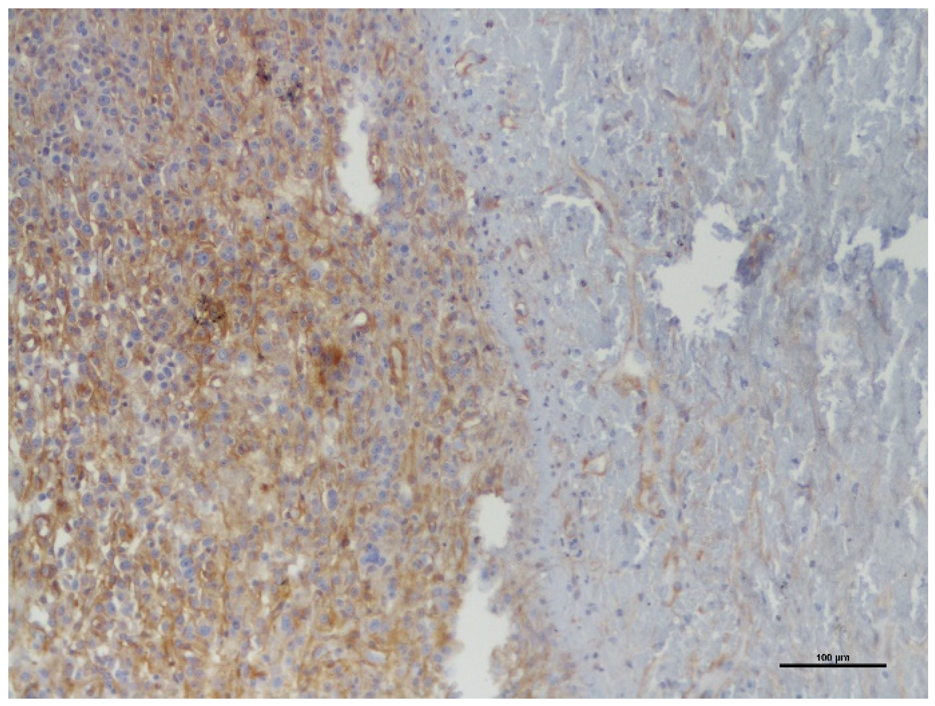
The PDGFR-β staining was diffuse cytoplasmatic in both benign and malignant PNST. The labelling intensity of BPNST was weak in 8 cases and intense in 1 case; in MPNST, it was weak in 6 cases and intense in 4 cases (Figure 1a,b). In addition, MPNST showed a more intense PDGFR-β expression at the tumor invasive front and areas of higher cytological atypia (Figure 2).
PDGFR-β was expressed in less than 25% of tumor cells in 6 of 9 BPNST samples (66.7%), including 1 case without immunoreactivity. All MPNST expressed PDGFR-β, with 80% of them (8/10) in >25% of cells. PDGFR-β expression was significantly higher in MPNST when compared to BPNST (Table 4). The tumor location, tumor size and Ki-67 index were not significantly related with the levels of PDGFR-β expression (Table 4).

4. Discussion

The activation of PDGFRs has an important role in the carcinogenesis of some human and animal neoplasms [19,20]. In dogs, the expression of PDGFR-β has been better characterized in hemangiosarcomas where it seems to contribute directly to a malignant behavior [33]. Several studies have shown that other canine neoplasms express this TKR suggesting that it may constitute an important therapeutic target [23,25,26,27,28,29,30,31,32,33,41]. To date, there are no published studies on the expression of this receptor in animal PNST, although it has already been studied in human PNST [24].
Despite the small sample in this preliminary study, it was possible to verify that the age of development of PNST does not appear to be related to their biological behavior, which is in accordance with the existing literature [5]. It has been described that STS appear in middle-aged to elderly dogs, and in large breeds, with no gender predisposition [2,3,5,42]. In this study, the prevalence of PNST was higher in large breed dogs, corroborating the previously described data [2,3,5]. The Ki-67 index and means were not significantly different between benign or malignant PNST and were also not related with the levels of PDGFR-β expression. The small number of cases and the great variability of Ki-67 values among them (which included two benign cases with very high Ki-67 index and two malignant with low Ki-67) may justify these results. Therefore, the value of Ki-67 as a proliferation marker to discriminate the clinical behavior (benign versus malignant) of theses tumors and the influence of PDGFR-β for tumor proliferation should be evaluated in larger series.
BPNST showed a significant lower expression of PDGFR-β compared to MPNST, suggesting that the receptor may have a role in the malignant transformation of these neoplasms, as also has been demonstrated in human PNST [23]. It has been reported that PDGFRs have a role in chemokine signaling to the tumor microenvironment and stimulate stromal cells contributing to mesenchymal carcinogenesis [43].
Larger tumors exhibited a higher level of expression of this TKR, suggesting that it may also contribute to tumor growth and perhaps a worse prognosis, but survival studies are needed to confirm this hypothesis. In addition, malignant PNST showed a more intense PDGFR-β expression at the tumor invasive front and areas of higher cytological atypia, suggesting that it may contribute to the tumor local invasive capacities [13,14,16,17,18].
Considering the overexpression of PDGFR-β in MPNST and in larger tumors, it seems reasonable to speculate that tyrosine kinase inhibitors (TKI) such as toceranib (Palladia®), may have a role in the treatment of the more aggressive PNST. In fact, the blockage of PDGFR-β expression with imatinib, a TKI used in human medicine, decreases the epithelial-mesenchymal transition (EMT) and the transcription of factor Slug, and it dramatically reduces the progression of human sarcomas [44].
This preliminary investigation had the strong limitation of the low number of cases, which limits the statistical power of the results, and therefore, further studies with a large number of PNST samples are warranted. Although PNST are not very common, they may have different clinical behaviors, and their proper diagnosis should be emphasized. The diagnostic process includes a panel of immunohistochemical markers that are expensive to perform and, because of that, many of these tumors are diagnosed only as cutaneous sarcomas and not distinguished from other soft tissue sarcomas, which may have consequences in terms of prognosis and therapeutic approach.

5. Conclusions

In summary, this study described, for the first time, the PDGFR-β expression in canine PNST, evidencing its presence in the cytoplasm of neoplastic cells. Despite the small number of samples, it was demonstrated that malignant and larger PNST have a significantly higher level of this TKR, suggesting that the receptor may have a role in the pathogenesis and progression of these neoplasms. These preliminary results support the need for conducting more investigations and survival studies to understand the value of PDGFR-β at both prognostic and therapeutic levels. Considering that some of these tumors may be difficult to totally excise, they may require adjuvant or neoadjuvant treatments, justifying the need for new therapeutic strategies, such as toceranib, a veterinary TKI able to target PDGFR-β.

Author Contributions

Conceptualization, C.A.-C., A.M. and A.S.; data curation, C.A.-C., I.A. and A.S.: methodology, C.A.-C., I.A., F.C., A.R. and A.S.; writing—original draft preparation, C.A.-C., I.A. and A.S.; supervision, writing—review and editing, C.A.-C., A.M., I.A., F.C., A.R. and A.S. All authors have read and agreed to the published version of the manuscript.

Funding

This research received no external funding.

Institutional Review Board Statement

This study was conducted using archived samples from the Veterinary Pathology Laboratory of the University of Porto, after approval by the Ethical Committee for the Animal Wellbeing (ORBEA/P314/2019).

Informed Consent Statement

Informed consent was obtained from all subjects involved in this study.

Data Availability Statement

The data that support the findings of this study are available from the corresponding author upon reasonable request.

Acknowledgments

The authors would like to thank the Department of Pathology and Molecular Immunology and the Laboratory of Veterinary Pathology of the Biomedical Sciences Institute of Abel Salazar (ICBAS)-University of Porto for their kind cooperation and contribution of this study.

Conflicts of Interest

The authors declare no conflict of interest.

References

  1. Bostock, D.E.; Dye, M.T. Prognosis after surgical excision of canine fibrous connective tissue sarcomas. Vet. Pathol. 1980, 17, 581–588. [Google Scholar] [CrossRef]
  2. Dernell, W.S.; Withrow, S.J.; Kuntz, C.A.; Powers, B.E. Principles of treatment for soft tissue sarcomas. Clin. Tech. Small Anim. Pract. 1998, 13, 59–64. [Google Scholar] [CrossRef]
  3. Ehrhart, N. Soft-tissue sarcomas in dogs: A review. J. Am. Anim. Hosp. Assoc. 2005, 41, 241–246. [Google Scholar] [CrossRef] [PubMed]
  4. Mayer, M.N.; Larue, S.M. Soft tissue sarcomas in dogs. Can. Vet. J. 2005, 46, 1048–1052. [Google Scholar] [PubMed]
  5. Liptak, J.M.; Christensen, N.I. Soft Tissue Sarcomas. In Withrow & MacEwen’s Small Animal Clinical Oncology, 6th ed.; Vail, D.M., Thamm, D.H., Liptak, J.M., Eds.; Elsevier Saunders: St. Louis, MO, USA, 2019; Chapter 22; pp. 404–431. [Google Scholar]
  6. Suzuki, S.; Uchida, K.; Nakayama, H. The effects of tumor location on diagnostic criteria for canine malignant nerve sheath tumors (MPNSTs) and the markers for distinction between canine MPNSTs and canine perivascular wall tumors. Vet. Pathol. 2014, 51, 722–736. [Google Scholar] [CrossRef] [Green Version]
  7. LeCouteur, R.A.; Withrow, S.J. Tumors of the nervous system. In Small Animal Clinical Oncology, 4th ed.; Withrow, S.J., MacEwen, E.G., Eds.; Elsevier Saunders: St. Louis, MO, USA, 2007; pp. 659–685. [Google Scholar]
  8. Koestner, A.; Armed Forces Institute of Pathology; WHO Collaborating Center for Worlwide Reference on Comparative Oncology. Histological Classification of Tumors of the Nervous System of Domestic Animals; Armed Forces Institute of Pathology in cooperation with the American Registry of Pathology and the World Health Organization Collaborating Center for Worldwide Reference on Comparative Oncology: Washington, DC, USA, 1999; pp. 37–38. [Google Scholar]
  9. Gross, T.L.; Ihrke, P.J.; Walder, E.J.; Affolter, V.K. Neural and Perineural Tumors. In Skin Diseases of the Dog and Cat, 2nd ed.; Blackwell Science Ltd.: Oxford, UK, 2005; pp. 786–796. [Google Scholar]
  10. Silva, E.O.; Goiozo, P.F.I.; Pereira, L.G.; Headley, S.A.; Bracarense, A.F.R.L. Concomitant malignant pulmonary peripheral nerve sheath tumour and benign cutaneous peripheral nerve sheath tumour in a dog. J. Comp. Path. 2017, 157, 46–50. [Google Scholar] [CrossRef]
  11. Chijiwa, K.; Uchida, K.; Tateyama, S. Immunohistochemical evaluation of canine peripheral nerve sheath tumors and other soft tissue sarcomas. Vet. Pathol. 2004, 41, 307–318. [Google Scholar] [CrossRef]
  12. Vucicevic, I.; Marinkovic, D.; Kukolj, V.; Nesic, S.; Anicic, M.; Durdevic, B.; Aleksic-Kovacecic, S. Immunohistochemical distinguish between canine peripheral nerve sheath tumors and perivascular wall tumors. Acta Vet. Beograd. 2019, 69, 290–299. [Google Scholar] [CrossRef] [Green Version]
  13. Alvarez, R.H.; Kantarjian, H.M.; Cortes, J.E. Biology of platelet-derived growth factor and its involvement in disease. Mayo Clin. Proc. 2006, 81, 1241–1257. [Google Scholar] [CrossRef]
  14. Demoulin, J.B.; Essaghir, A. PDGF receptor signaling networks in normal and cancer cells. Cytokine Growth Factor Rev. 2014, 25, 273–283. [Google Scholar] [CrossRef]
  15. Liu, K.W.; Hu, B.; Cheng, S.Y. Platelet-derived growth factor receptor alpha in glioma: A bad seed. Chin. J. Cancer 2011, 30, 590–602. [Google Scholar] [CrossRef] [PubMed]
  16. Seger, R.; Krebs, E.G. The MAPK signaling cascade. FASEB J. 1995, 9, 726–735. [Google Scholar] [CrossRef] [PubMed]
  17. Nicholson, K.M.; Anderson, N.G. The protein kinase B/Akt signalling pathway in human malignancy. Cell Signal. 2002, 14, 381–395. [Google Scholar] [CrossRef]
  18. Tokunaga, E.; Oki, E.; Egashira, A.; Sadanaga, N.; Morita, M.; Kakeji, Y.; Maehara, Y. Deregulation of the Akt pathway in human cancer. Curr. Cancer Drug Targets 2008, 8, 27–36. [Google Scholar] [CrossRef]
  19. Ostman, A.; Heldin, C.H. PDGF receptors as targets in tumor treatment. Adv. Cancer Res. 2007, 97, 247–274. [Google Scholar] [CrossRef]
  20. Toffalini, F.; Demoulin, J.B. New insights into the mechanisms of hematopoietic cell transformation by activated receptor tyrosine kinases. Blood 2010, 116, 2429–2437. [Google Scholar] [CrossRef] [Green Version]
  21. Paulsson, J.; Ehnman, M.; Ostman, A. PDGF receptors in tumor biology: Prognostic and predictive potential. Future Oncol. 2014, 10, 1695–1708. [Google Scholar] [CrossRef]
  22. Peng, Y.; Guo, J.J.; Liu, Y.M.; Wu, X.L. MicroRNA-34A inhibits the growth, invasion and metastasis of gastric cancer by targeting PDGFR and MET expression. Biosci. Rep. 2000, 34, 247–256. [Google Scholar] [CrossRef]
  23. Aoki, M.; Nabeshima, K.; Koga, K.; Hamasaki, M.; Suzumiya, J.; Tamura, K.; Iwasaki, H. Imatinib mesylate inhibits cell invasion of malignant peripheral nerve sheath tumor induced by platelet-derived growth factor-BB. Lab. Investig. 2007, 87, 767–779. [Google Scholar] [CrossRef]
  24. Ohishi, J.; Aoki, M.; Nabeshima, K.; Suzumiya, J.; Takeuchi, T.; Ogose, A.; Hazokazi, M.; Yamashita, Y.; Owasaki, H. Imatinib mesylate inhibits cell growth of malignant peripheral nerve sheath tumors in vitro through supression of PDGFR-β. BMC Cancer 2013, 13, 224. [Google Scholar] [CrossRef] [Green Version]
  25. Maniscalco, L.; Iussich, S.; Morello, E.; Martano, M.; Biolatti, B.; Riondato, F.; Salda, L.D.; Romanucci, M.; Malatesta, D.; Bongiovanni, L.; et al. PDGFs and PDGFRs in canine osteossarcoma: New targets for innovative therapeutic strategies in comparative oncology. Vet. J. 2013, 195, 41–47. [Google Scholar] [CrossRef] [PubMed]
  26. Gramer, I.; Killick, D.; Scase, T.; Chandry, D.; Marrington, M.; Blackwood, I. Expression of VEGFR and PDGFR-α/-β in 187 canine nasal carcinomas. Vet. Comp. Oncol. 2016, 15, 1041–1050. [Google Scholar] [CrossRef] [PubMed]
  27. Walters, L.; Martin, O.; Price, J.; Sulla, M.M. Expression of receptor tyrosine kinase targets PDGFR-β, VEGFR-2 and KIT in canine transitional cell carcinoma. Vet. Comp. Oncol. 2018, 16, 117–122. [Google Scholar] [CrossRef] [PubMed]
  28. Brown, R.J.; Newman, S.J.; Durtschi, D.C.; LeBlanc, A.K. Expression of PDGFR-β and Kit in canine anal sac apocrine gland adenocarcinoma using tissue immunohistochemistry. Vet. Comp. Oncol. 2011, 10, 74–79. [Google Scholar] [CrossRef]
  29. Iussich, S.; Maniscalco, L.; Di Sciuva, A.; Iotti, B.; Morello, E.; Martano, M.; Gattino, F.; Buracco, P.; De Maria, R. PDGFRs expression in dogs affected by malignant oral melanomas: Correlation with prognosis. Vet. Comp. Oncol. 2016, 15, 462–469. [Google Scholar] [CrossRef]
  30. Altamura, G.; Uberti, B.; Galiero, G.; Martano, M.; Pirro, A.; Russo, M.; Borzacchiello, G. Expression and activation of platelet-derived growth factor β receptor, mitogen-activated protein/extracelular signal-regulated kinase kinase (MEK) and extracelular signal-regulated kinase (ERK) in canine mammary tumours. Res. Vet. Sci. 2017, 110, 29–33. [Google Scholar] [CrossRef]
  31. Avallone, G.; Pellegrino, V.; Roccabianca, P.; Lepri, E.; Crippa, L.; Beha, G.; De Tolla, L.; Sarli, G. Tyrosine Kinase Receptor Expression in Canine Liposarcoma. Vet. Pathol. 2017, 54, 212–217. [Google Scholar] [CrossRef]
  32. Avallone, G.; Stefanello, D.; Boracchi, P.; Gelain, M.E.; Tresoldi, E.; Roccabianca, P.; Ferrari, R.; Turin, L. Growth Factors and COX2 Expression in Canine Perivascular Wall Tumors. Vet. Pathol. 2015, 52, 1034–1040. [Google Scholar] [CrossRef] [Green Version]
  33. Asa, S.A.; Murai, A.; Murakami, M.; Hoshino, H.; Mori, T.; Maruo, K.; Khater, A.; El-sawak, A.; el-Aziz, E.A.; Yanai, T.; et al. Expression of platelet-derived growth factor and its receptors in spontaneous canine hemangiosarcoma and cutaneous hemangioma. Histol. Histopathol. 2012, 27, 601–607. [Google Scholar] [CrossRef]
  34. Koestner, A.; Higgins, R.J.; Meuten, D.J. Primary Tumors of the peripheral nervous system. In Tumors in Domestic Animals, 4th ed.; Iowa State University Press: Ames, IA, USA, 2002; pp. 731–735. [Google Scholar]
  35. Schulman, F.Y.; Johnson, T.O.; Facemire, P.R.; Fanburg-Smith, J.C. Feline peripheral nerve sheath tumors: Histologic, immunohistochemical and clinicopathological correlation (59 Tumors in 53 cats). Vet. Pathol. 2009, 46, 1166–1180. [Google Scholar] [CrossRef] [Green Version]
  36. Joshi, R. Learning from eponyms: Jose Verocay and Verocay bodies, Antoni A and B areas, Nils Antoni and Schwannomas. Indian Derm. Online J. 2012, 3, 215–219. [Google Scholar] [CrossRef] [PubMed]
  37. Nakagawa, M.; Naruko, T.; Ikura, Y.; Komatsu, R.; Iwasa, Y.; Kitabayashi, C.; Inoue, T.; Itoh, A.; Yoshiyama, M.; Ueda, M. A decline in platelet activation and inflammatory cell infiltration is associated with the phenotypic redifferentiation of neointimal smooth muscle cells after bare-metal stent implantation in acute coronary syndrome. J. Atheroscler. Thromb. 2010, 17, 675–687. [Google Scholar] [CrossRef] [PubMed] [Green Version]
  38. Asa, S.A.; Mori, T.; Maruo, K.; Khater, A.; El-sawak, A.; el-Aziz, E.A.; Yanai, T.; Sakai, H. Analysis of genomic mutation and immunohistochemistry of platelet-derived growth factor receptors in canine vascular tumours. Vet. Comp. Oncol. 2013, 13, 237–245. [Google Scholar] [CrossRef] [PubMed]
  39. Teixeira, S.; Amorim, I.; Rema, A.; Faria, F.; Gartner, F. Molecular heterogeneity of canine cutaneous peripheral nerve sheath tumors: A drawback in the diagnosis refinement. In Vivo 2016, 30, 819–827. [Google Scholar] [CrossRef] [Green Version]
  40. Watanabe, T.; Oda, Y.; Tamiya, S.; Kinukawa, N.; Masuda, K.; Tsuneyoshi, M. Malignant peripheral nerve sheath tumours: High Ki67 labelling index is the significant prognostic indicator. Histopathology 2001, 39, 187–197. [Google Scholar] [CrossRef]
  41. Heaton, C.M.; Fernandes, A.F.A.; Jark, P.C.; Pan, X. Evaluation of toceranib for treatment of apocrine gland sac anal adenocarcinoma in dogs. J. Vet. Intern. Med. 2020, 34, 873–881. [Google Scholar] [CrossRef]
  42. Dennis, M.M.; McSporran, K.D.; Bacon, N.J.; Schulman, F.Y.; Foster, R.A.; Powers, B.E. Prognostic factors for cutaneous and subcutaneous soft tissues sarcomas in dogs. Vet. Pathol. 2011, 48, 73–84. [Google Scholar] [CrossRef] [Green Version]
  43. Pender, A.; Jones, R.L. Olaratumab: A platelet-derived growth factor receptor-α-blocking antibody for the treatment of soft tissue sarcoma. Clin. Pharmacol. 2017, 9, 159–164. [Google Scholar] [CrossRef] [Green Version]
  44. Chang, K.K.; Yoon, C.; Yi, B.C.; Tap, W.D.; Simon, M.C.; Yoon, S.S. Platelet-derived growth factor receptor-α and -β promote cancer stem cell phenotypes in sarcomas. Oncogenesis 2018, 7, 47. [Google Scholar] [CrossRef] [Green Version]
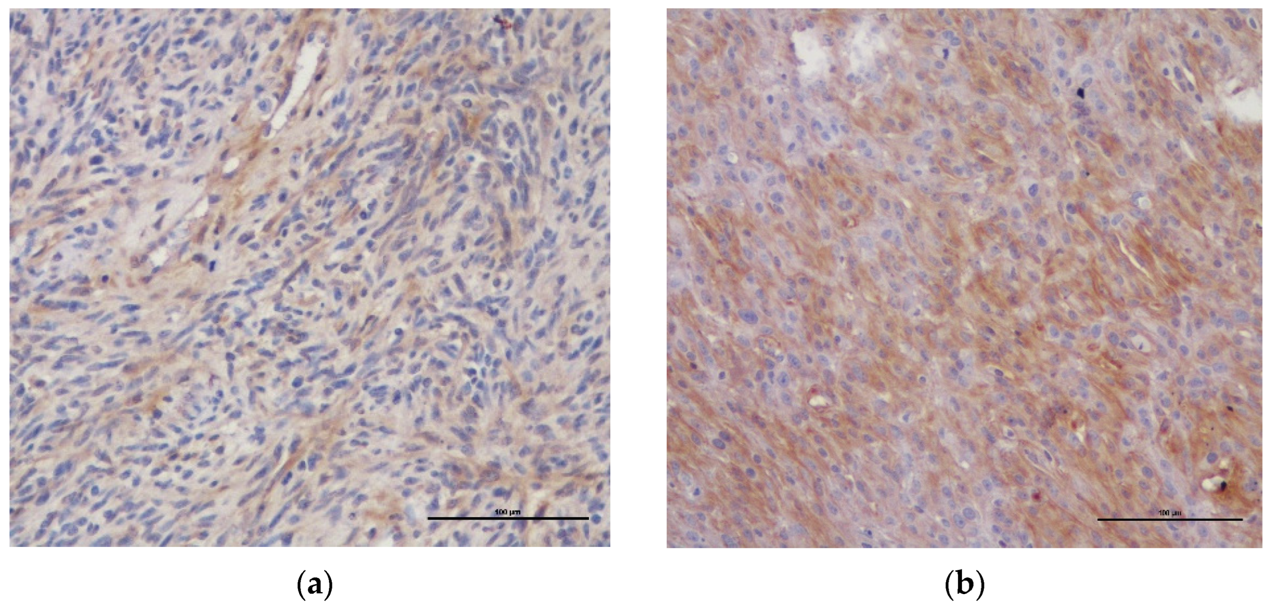
Figure 1. Immunohistochemical PDGFR-β staining in canine cutaneous PNST (×200): (a) MPNST with weak cytoplasmic staining in more than 25% of neoplastic cells; (b) MPNST with intense cytoplasmic staining in more than 25% of neoplastic cells.
Vetsci 09 00345 g001
Figure 2. Immunohistochemical staining of PDGFR-β in canine cutaneous MPNST (×100). Tumor invasive front showing high levels of PDGFR-β expression.
Vetsci 09 00345 g002
Table 1. Clinical data, type of tumor, Ki-67 index and percentage of PDGFR-β expression in dogs with cutaneous PNST.
Case
Nr.
BreedAge (Years)GenderType of
Tumor
Location of TumorDimensions of Tumor (cm)Ki-67
PI (%)
% PDGFR-β
Expression
1M. Schnauzer13MBT2.5 × 2.560.8≤25%
2Mixed breed12FBL4 × 3.515.8>25%
3Poodle9MBNR3 × 2.536.3>25%
4X Rottweiler8MBNRNR4.3≤25%
5Mixed breed8MBL4 × 2.81.8≤25%
6Mixed breed10FBL2 × 13.8≤25%
7G. Shepherd14FBT4 × 214.1≤25%
8Poodle13MBL1.2 × 1.21.2≤25%
9Boxer9MBT2 × 12.8>25%
10Port. Pointer9MMT8 × 838.9>25%
11St. Bernard10FML10 × 1062.6≤25%
12Siberian husky13FML4.5 × 414.1>25%
13Labr. Retriever2MMT3 × 323.5>25%
14Boxer7MML4.5 × 3.526.2>25%
15Boxer7FMT6.5 × 621.4>25%
16Mixed breed12MML3 × 254.0≤25%
17Mixed breed12FMT10.5 × 86.4>25%
18Dalmatian10FML4 × 2.510.2>25%
19Boxer8FML4 × 22.8>25%
M—Male; F—Female; B—BPNST; M—MPNST; L—Limbs; T—Trunk; PI—Proliferation index.
Table 2. Relationship between tumor type and clinicopathological variables in canine PNST.
VariableNr. of TumorsBPNST (Number; %)MPNST (Number; %)p Value
Gender
Female93 (33.3)6 (66.7)0.24
Male 106 (60.0)4 (40.0)
Breed *
Small33 (100.0)0 (0)0.05
Medium, Large, Giant113 (27.3)8 (72.7)
Tumor Location *
Trunk73 (42.9)4 (57.1)0.64
Limbs104 (40.0)6 (60.0)
Tumor size *
<3 cm44 (100.0)0 (0)0.02
≥3 cm144 (28.6)10 (71.4)
Fields with * have a lower number of cases due to unknown data. Group differences were assessed by Fisher’s exact test.
Table 3. Relationship between the mean Ki-67 expression, tumor type and PDGFR-β expression in canine PNST.
VariableNr. of TumorsMean Ki-67Standard Deviation (SD)p Value
(ANOVA)
Tumor Type
Benign915.6620.320.28
Malignant1026.0120.09
PDGFR-β expression
≤25%825.3328.380.45
>25%1118.0412.48
Table 4. Relationship between PDGFR-β expression and clinicopathological variables in 19 canine PNST.
VariableNr. of TumorsTumors with ≤25% PDGFR-β Expression (Number; %)Tumors with >25% PDGFR-β Expression
(Number; %)
p Value
Tumor type
Benign96 (66.7)3 (33.3)0.05
Malignant102 (20.0)8 (80.0)
Tumor Location *
Trunk 72 (28.6)5 (71.4)0.35
Limbs105 (50.0)5 (50.0)
Tumor size *
<3 cm43 (75.0)1 (25.0)0.13
≥3 cm144 (28.6)10 (71.4)
Ki-67 Index
<20%115 (45.5)6 (54.5)0.55
≥20%83 (37.5)5 (62.5)
Fields with * have a lower number of cases due to unknown data. Group differences were assessed by Fisher’s exact test.
Publisher’s Note: MDPI stays neutral with regard to jurisdictional claims in published maps and institutional affiliations.